NERVOUS SYSTEM.
Reading edition
I. THE CENTRAL NERVOUS SYSTEM.
1. The Spinal Cord. Medulla spinalis
(Figs. 133-136).—The spinal cord is that portion of the nervous system which occupies the vertebral canal; it is continuous craniad with the brain. It has the form of a somewhat flattened cylinder and extends from the foramen magnum into the caudal region. It diminishes in diameter after entering the sacral region.
The cord has a cervical and a lumbar enlargement, the former (Fig. 133) marking the origin of the nerves which pass to the fore limb, and the latter (Fig. 136) the origin of those which pass to the hind limb.
The cervical enlargement (Fig. 133) lies in that part of the vertebral canal bounded by the fourth to seventh cervical and first thoracic vertebræ. Caudad of the first thoracic vertebra the cord continues of nearly uniform diameter to the lumbar enlargement (Fig. 136) which stretches from the third to the seventh lumbar vertebræ (inclusive). Caudad of the seventh lumbar vertebra it diminishes uniformly in diameter and ends in a slender cord, the filum terminale (Fig. 136, C), which may be traced into the caudal region.
The surface of the cord is marked by a number of longitudinal grooves or sulci, and fissures. The most prominent of these is the anterior median fissure (Fig. 134, c), along the ventral median line; into this the pia mater dips. The posterior median sulcus (Fig. 133, e; Fig. 134, a) is a shallow furrow along the dorsal median line. The anterior fissure and the posterior sulcus thus divide the cord into lateral halves. Each half is subdivided by the anterior and posterior lateral sulci.
The posterior lateral sulcus (Fig. 133, f; Fig. 134, b) lies at the side of the posterior median sulcus. It is broad and shallow and has the posterior roots of the spinal nerves emerging from its bottom.
The anterior lateral sulcus exists only after the forcible pulling out of the anterior (ventral) roots of the spinal nerves. It then marks the line along which they originate from the cord.
In cross-section (Fig. 134) the substance of the cord appears to the naked eye as composed of a darker central “gray matter” (g) and of an outer “white matter” (f). The gray matter has in section the form of the letter H. The cord is really tubular, having a central canal (h) which appears in section in the middle of the cross-bar of the H, while the two ends of each vertical bar extend toward the anterior and posterior lateral fissures.
The white matter is divided into funiculi (or columns) by the fissures and sulci, so that there is in each half of the cord an anterior, a posterior, and a lateral white funiculus.
In the cervical region (Fig. 134) there appears between the posterior lateral and posterior median sulci an intermediate sulcus which divides the posterior funiculus in this region into two. The median slenderer of these is the fasciculus gracilis (d) or column of Goll. The lateral and thicker is the fasciculus cuneatus (e) or column of Burdach.
The membranes of the spinal cord:
The dura mater is a tough fibrous membrane directly continuous with the dura mater of the brain. It differs from the dura mater of the brain in two particulars:
1. At the foramen magnum it splits into two layers, one of which lines the bony vertebral canal and forms its periosteum, while the other covers the cord.
2. It is separated from the cord by a considerable space. The dura mater is continuous with the fibrous sheaths of the spinal nerves at their points of exit. Along the sides of the cord it is connected to the pia mater by a delicate strand of connective tissue probably equivalent to the “ligamentum denticulatum” of man.
The arachnoid is a delicate cellular membrane lying beneath the dura mater, between it and the pia mater. It forms a continuous investment for the cord, is not vascular, and is said not to dip into the fissures of the cord.
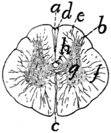
Fig. 134.—Cross-section of the Spinal Cord in the Cervical Region.
a, posterior median sulcus; b, posterior lateral sulcus; c, anterior median fissure; d, fasciculus gracilis; e, fasciculus cuneatus; f, white matter; g, gray matter; h, central canal.
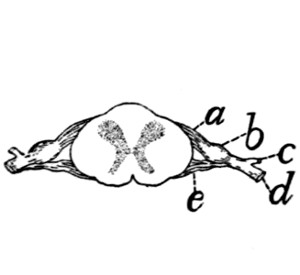
Fig. 135.—Section of Spinal Cord, showing the Origin of a Pair of Spinal Nerves.
a, dorsal root; b, spinal ganglion; c, dorsal ramus; d, ventral ramus; e, ventral root.
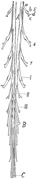
Fig. 136.—Caudal Portion of the Spinal Cord.
A, lumbar enlargement; B, cauda equina; C, filum terminale; 5-7, fifth to seventh lumbar nerves. I-III, the three sacral nerves. a, dorsal roots; b, spinal ganglia; c, dorsal rami; d, ventral rami.
The pia mater invests the cord closely and contains some blood-vessels. It is a delicate membrane which dips into the fissures and sulci of the cord and is connected to it by numerous strands of connective tissue that pass from it into the substance of the cord. The nerves pierce it.
Spinal Nerves.—From the spinal cord arise the spinal nerves. Of these there are about thirty-eight pairs in the cat. Eight are cervical, thirteen thoracic, seven lumbar, three sacral, and seven or eight caudal. Those leaving the cervical (Fig. 133, 5-8 and I) and lumbar (Fig. 136) enlargements are larger than the others. The first cervical nerve leaves the vertebral canal through the atlantal foramen, the second leaves between the arches of the atlas and axis, while all the others leave the vertebral canal by way of the intervertebral foramina. Each nerve arises from the cord by a dorsal and a ventral root. The dorsal root is chiefly sensory, the ventral motor in character. The dorsal root (radix posterior) (Figs. 133, 135, and 136, a) begins as a number (twelve or more) of separate nerve-bundles which emerge from the posterior lateral groove. These roots lie nearly in a single plane and pass laterad, converging to penetrate a ganglion, the spinal ganglion (b) (or ganglion of the posterior root). All the spinal ganglia except the first and second are situated in the intervertebral foramina or within the vertebral canal. The first and second are situated among the muscles surrounding the place of exit of the nerves.
The ventral root (radix anterior) (Fig. 135, e) arises as a larger number of small fibre-bundles which do not lie in a single plane, so that in a transverse section several rootlets may appear in a single section. The rootlets converge to form a single mass which joins the dorsal root just as it emerges from the spinal ganglion. The nerve formed by the junction of the ventral and dorsal roots is one of the spinal nerves.
The direction in which the nerves leave the cord varies. In the cervical region (Fig. 133) and cranial part of the lumbar region it is nearly laterad; at the cervical and lumbar enlargements it is laterocaudad. The nerves in the sacral and caudal regions pass almost directly caudad to reach the intervertebral foramina and form thus a brush which surrounds the filum terminale (Fig. 136, C) and is called the cauda equina (Fig. 136, B).
Each spinal nerve immediately after leaving the intervertebral foramen divides into two branches, a dorsal or posterior branch or ramus (c), and a ventral or anterior ramus (d). The dorsal ramus is in each case small (except in the first and second cervical nerves), and is distributed to the longitudinal muscles and integument of the back.
The ventral rami are larger and each is connected a short distance beyond its origin with the sympathetic system by a short ramus communicans or communicating branch. Each is then distributed to the integument and muscles of the ventral part of the body, including the limbs.
The ventral rami which pass to the limbs are much larger than the others. The ventral rami are further distinguished from the dorsal by the fact that they frequently unite with one another to form plexuses.
The peripheral distribution of the spinal nerves is described later.
2. The Brain. Encephalon.
—The brain is that portion of the central nervous system that is included within the cranial cavity. It is a direct continuation of the spinal cord, and presents many of the same essential characters as the latter, with great modifications in details.
The structure of the brain can best be understood if it be considered as a modified continuation of the spinal cord, and the relation of the parts to the essential parts of the cord noted. The spinal cord is a nearly straight tube, with a central cavity and thick walls. The brain is likewise tubular, with the cavities enlarged or subdivided in places, with the walls greatly thickened, and with a number of bends and constrictions in the tube. The relation of the structure of the brain to that of the spinal cord is most easily perceived by an examination of the brain of some lower vertebrate, as the frog or shark, and such an examination should be made before proceeding to the study of the more complicated brain of the cat.
In the following account of the brain of the cat all parts will be described as far as possible in relation with the tubular structure of the brain. The brain will be considered as a hollow structure, having central cavities, and the solid portions will be brought into relation as parts of the roof, sides, or floor of the cavities. The cavities of the brain, forming a direct continuation of the central canal of the spinal cord, are known as ventricles (ventriculi).
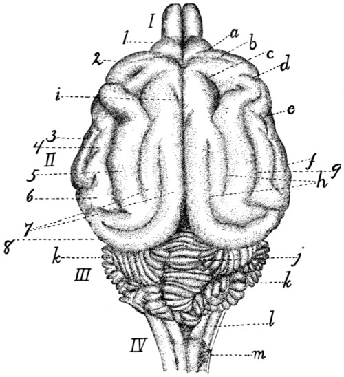
Fig. 137.—Dorsal Surface of the Brain.
I, olfactory bulbs; II, cerebral hemispheres; III, cerebellum; IV, medulla. a, præsylvian sulcus; b, cruciate sulcus; c, sulcus ansatus; d, coronal sulcus; e, anterior sulcus; f, posterior sulcus; g, suprasylvian sulcus; h, separate parts of the lateral sulcus; i, end of splenial sulcus; j, vermis of cerebellum; k, hemispheres of cerebellum; l, roof of fourth ventricle; m, first cervical nerve. 1, orbital lobe (or gyrus); 2, sigmoid gyrus; 3, anterior sylvian gyrus; 4, ectosylvian gyrus; 5, suprasylvian gyrus; 6, posterior sylvian gyrus; 7, parts of marginal gyrus; 8, gyrus compositus posterior.
In a general view of the cat’s brain from the dorsal side (Fig. 137) four subdivisions are discernible. At the caudal end is a small stalk-like portion (IV) which is clearly a slightly modified continuation of the spinal cord; this is the myelencephalon or medulla oblongata. Just craniad of this, rising high above it, is an irregularly lobulated rounded portion, the cerebellum (III). This is produced as a thickening of the wall of the original tube; it forms the dorsal part of the metencephalon. These two portions of the brain have a common cavity, lying within the medulla oblongata and ventrad of the cerebellum, and formed by a widening of the cavity of the spinal cord; it is known as the fourth ventricle. Craniad of the cerebellum, separated from it by a deep transverse fissure, are two very large portions, the hemispheres of the cerebrum (II), separated from each other by a deep longitudinal fissure; they constitute together the telencephalon. The hemispheres are produced by a sort of forking of the original central tube;—by outgrowths on the sides of the tube, the central cavity extending into the outgrowths. The two outgrowths extend dorsad, as well as craniad and caudad from the original place of origin, forming the larger part of the brain; the extensions of the central cavity which they contain are known as the lateral ventricles. Finally, in front of the two hemispheres are seen the two small olfactory bulbs (I) which are mere extensions of the two hemispheres and contains cavities which are extensions of the lateral ventricles.
In a ventral view of the entire brain (Fig. 138) certain parts are visible which in the dorsal view are hidden by the large cerebral hemispheres. At the caudal end is seen, as before, the medulla oblongata, or myelencephalon, and craniad of this, on the ventral side of the cerebellum, a broad transverse tract, the pons (i); this, like the cerebellum, forms part of the metencephalon. All this part of the brain formed by the myelencephalon and metencephalon lies caudad of (behind) the rest of the brain and is marked off from it by a great fissure; it therefore receives as a whole the name hindbrain or rhombencephalon.
Just craniad of the pons are seen two short diverging arm-like bands of fibres (g), enclosing a small triangular space between them. These two arms with the space between them are all that is visible of a division of the brain which in dorsal view is completely covered by the backward projection of the cerebral hemispheres. This is the midbrain or mesencephalon. In this portion of the brain the continuation of the central cavity is a narrow canal which receives the name cerebral aqueduct (aqueductus cerebri) or aqueduct of Sylvius.
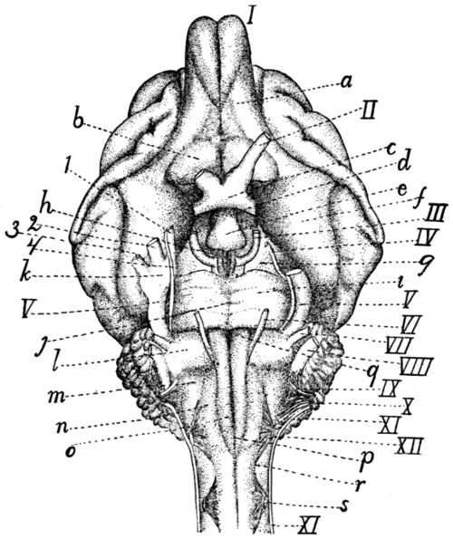
Fig. 138.—Ventral Surface of the Brain.
I-XII, the twelve cranial nerves in order; I, olfactory bulb; II, N. opticus; III, N. oculomotorius; IV, N. trochlearis; V, N. trigeminus (1, mandibular nerve; 2, maxillary nerve; 3, ophthalmic nerve; 4, sensory root); VI, N. abducens; VII, N. facialis; VIII, N. acusticus; IX, N. glossopharyngeus; X, N. vagus; XI, N. accessorius; XII, N. hypoglossus. a, tractus olfactorius; b, substantia perforata anterior; c, optic chiasma; d, infundibulum; e, hypophysis; f, lobus pyriformis, or tractus postrhinalis; g, pedunculi cerebri; h, substantia perforata posterior; i, pons; j, sulcus basilaris of pons; k, semilunar (or Gasserian) ganglion; l, trapezium; m, area ovalis; n, area elliptica; o, pyramidal tracts; p, anterior or ventral fissure; q, foramen cæcum; r, anterior lateral sulcus; s, first cervical nerve.
Just craniad of the midbrain is a small rather irregular area, bounded craniad by the band-like optic chiasma (c), from which arise the optic nerves (II), and showing caudad of this band a rounded irregular projection (e). This is a fourth division of the brain which is likewise covered dorsad by the hemispheres; it is the ’tween-brain or diencephalon. Its cavity is known as the third ventricle. Just craniad of the ’tween-brain, in the median line, is a deep fissure, showing that here in reality is the termination of the original tube, the further extension craniad being due to the pushing forward of the lateral outgrowths, or cerebral hemispheres, which extend in ventral view a considerable distance further craniad. The hemispheres lie also caudad, laterad, and dorsad of the midbrain and ’tween-brain, so that these parts are almost enveloped by the hemispheres. The hemispheres and ’tween-brain are sometimes taken together as forming a single portion, the forebrain or prosencephalon.
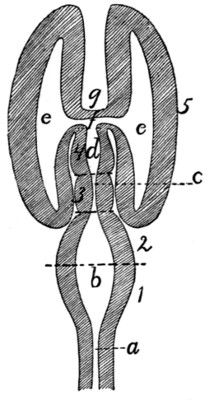
Fig. 139.—Diagram of Brain from Above.
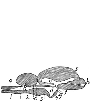
Fig. 140.—Diagram of Brain from the Side.
1, myelencephalon; 2, metencephalon; 3, mesencephalon; 4, diencephalon; 5, telencephalon. a, canal of the spinal cord; b, fourth ventricle; c, cerebral aqueduct (aqueduct of Sylvius); d, third ventricle; e, lateral ventricle; f, interventricular foramen (or foramen of Monroe); g, lamina terminalis; h, olfactory bulb.
There are thus altogether five principal subdivisions of the brain, each enclosing a portion of the central cavity. These are the myelencephalon, the metencephalon (these two enclosing the fourth ventricle), the mesencephalon (enclosing the cerebral aqueduct), the diencephalon (enclosing the third ventricle), and the two cerebral hemispheres, constituting together the telencephalon, and enclosing the two lateral ventricles. A plan of the brain, considered as a tube enclosing cavities, is given in Figs. 139 and 140. Fig. 139 shows the plan considered as viewed from the dorsal side, Fig. 140 in a lateral view. These figures are of course pure diagrams, showing only what may be called the plan of structure, and omitting all details. In the actual conditions many important modifications even of the main features of the plan are met with.
In the following description the parts of the brain will be taken up in this order:
A. Rhombencephalon (primitive hindbrain).
1. Myelencephalon.
2. Metencephalon.
B. Mesencephalon (primitive midbrain).
3. Mesencephalon.
C. Prosencephalon (primitive forebrain).
4. Diencephalon.
5. Telencephalon.
A. Rhombencephalon.—
1. Myelencephalon.
—The myelencephalon or medulla oblongata is transitional between the spinal cord and the brain. It has in general the structural peculiarities of the cord, but these undergo in it a gradual transformation into the arrangements characteristic of the brain.
It has the form of a flattened and truncated cone, which widens craniad and is limited at the cranial end by the pons (Fig. 138, i) ventrally and laterally; by the cerebellum (Fig. 137, III) dorsally. The cranial portion of its dorsal surface is overhung by the cerebellum. The points of origin of the roots of the first pair of cervical nerves (Fig. 138, s) may be taken as indicating the boundary between the spinal cord and the medulla oblongata; there is no other external marking to show the limits of the two.
The central canal of that part of the medulla which is overhung by the cerebellum is greatly widened (Fig. 141, h). The cavity thus formed is flattened and triangular, with the apex of the triangle caudad, and is the caudal part of the fourth ventricle (Fig. 141, h). The roof is very thin (Fig. 143, n) and is intimately connected with the pia mater, so that in removing the latter the thin roof of the fourth ventricle is removed with it and the cavity of the fourth ventricle is left exposed. The thin roof of the fourth ventricle is known as the velum medullare posterius (Fig. 143, n).
The ventral (anterior) fissure (Fig. 138, p) of the cord passes onto the medulla oblongata and ends at the pons (i), its end being slightly deeper and forming what is known as the foramen cæcum (q). The dorsal (posterior) sulcus (Fig. 141, a) is well marked; it ends at the caudal boundary of the fourth ventricle (h).
The anterior lateral sulcus (Fig. 138, r) may be traced craniad from the origin of the ventral roots of the first cervical nerves (s), along the lateral border of the area elliptica (n), then curving mediad to the lateral border of the pyramis (o), and finally reaching the pons (i). Its position is marked by the origin of the roots of the hypoglossal nerve (XII).
The posterior lateral sulcus (Fig. 141, b), marked on the spinal cord by the origin of the dorsal nerve-roots, curves laterad at the sides of the fourth ventricle (h) owing to the increasing width of the latter, and ends at an elevated area of oblique fibres, the area ovalis (f).
The columns or funiculi bounded by the longitudinal fissures present the following peculiarities:
The anterior white funiculus of the cord is replaced in the medulla by the pyramidal tracts (pyramides) (Fig. 138, o). The pyramidal tracts are formed by fibres which emerge from beneath the pons and pass caudad to disappear just craniad of the level of the first cervical nerve (s). The pyramidal tracts are bounded medially by the anterior median fissure (p), but laterally each is separated from the anterior lateral sulcus (r) over its caudal part by an elongated area elliptica (n), the human homologue of which is uncertain. It perhaps represents the oliva.
Laterad of the cranial portion of the pyramids is an irregular area known as the trapezium (l) which abuts caudad on the area elliptica (n) and the area ovalis (m). The area ovalis (Fig. 138, m; Fig. 141, f) (or zonula Arnoldi) is abroad band of oblique fibres which passes from the lateral side of the area elliptica craniodorsad to disappear under the cerebellum.
The posterior white funiculus was divided in the cervical region into two, the fasciculus gracilis (column of Goll) and the fasciculus cuneatus (column of Burdach). The fasciculus gracilis (Fig. 141, c) extends to the caudal end of the fourth ventricle (h) and ends there in an expansion, the clava (d), which forms the posterior boundary of the fourth ventricle. The fasciculus cuneatus (e) passes laterad on account of the width of the fourth ventricle and appears to end at the area ovalis (f), but it may be seen passing beneath the area ovalis, emerging at its cranial border and turning dorsad to enter the cerebellum. It forms the side walls of a part of the shallow fourth ventricle.
The lateral funiculi are divided by longitudinal furrows into three divisions. The dorsal one of these is the fasciculus cuneatus lateralis or column of Rolando (Fig. 141, g). It accompanies the fasciculus cuneatus into the cerebellum.
The entire mass formed by the fasciculus gracilis, the fasciculus cuneatus medialis, and fasciculus cuneatus lateralis is known as the corpus restiforme, and since its fibres pass into the cerebellum it is sometimes known as the pedunculus cerebelli (or crus cerebelli ad medullam).
The following cranial nerves arise from the medulla oblongata (Fig. 138).
The twelfth nerve (XII) (N. hypoglossus) arises by ten or fifteen rootlets from the ventral surface of the medulla oblongata in the anterior lateral fissure (r) opposite the caudal portion of the area elliptica (n).
The eleventh cranial nerve (XI) (N. accessorius) arises by numerous rootlets from the lateral surface of the medulla oblongata and of the spinal cord as far caudad as the sixth or seventh cervical nerve. These rootlets join to form a nerve which enters the cranium through the foramen magnum and is closely associated at its point of exit with the glossopharyngeal (IX) and vagus (X) nerves. The line of origin on the medulla passes between the dorsal and ventral roots of the cervical nerves and is along the middle of the lateral white funiculi.
The tenth cranial nerve (X) (N. vagus) arises by about eighteen very delicate rootlets from the surface of the area ovalis. The rootlets are divided into a dorsal and a ventral series. The dorsal series (about twelve) arise in the groove which separates the fasciculus cuneatus medialis from the fasciculus cuneatus lateralis. The ventral series arise somewhat ventrad of this groove. These rootlets are to be distinguished from those of the ninth nerve by their smaller size.
The ninth cranial nerve (IX) (N. glossopharyngeus) arises from the area ovalis from a line craniad of the dorsal line of origin of the vagus roots (X) and between these and those of the auditory (VIII). It arises by a number of rootlets which are larger than those of the vagus (X), with which this nerve is closely associated.
The eighth cranial nerve (VIII) (N. acusticus) appears at the lateral end of the trapezium (l). It arises from an elevation (Fig. 141, i) which is continued dorsomediad along the cranial border of the area ovalis.
The seventh cranial nerve (VII) (N. facialis) leaves the lateral border of the trapezium (l) near its cranial edge, between the fifth and eighth nerves. It is much smaller than the eighth nerve.
The sixth cranial nerve (VI) (N. abducens) arises by about six bundles from the groove between the pyramids and the trapezii and passes craniad.
2. Metencephalon.
—The metencephalon includes the pons and the cerebellum.
The pons (Fig. 138, i) is a mass of transverse fibres which forms the ventral and cranial part of the primitive hindbrain. It is a modification of the latter brought about by the development of the cerebellum, and the degree of its development is in direct ratio to that of the cerebellar and cerebral hemispheres. The pons forms a projecting mass of fibres which is marked by a median longitudinal groove, the sulcus basilaris (j), which indicates the course of the basilar artery (Fig. 121, c). Laterad the fibres of the pons converge somewhat and turning dorsad disappear in the cerebellum, forming the brachia pontis (Fig. 141, l).
The fifth cranial nerve (Fig. 138, V) (N. trigeminus) arises by two roots from the caudal border of the pons, near the lateral end. The ventral root (4) is small; the dorsal one is much larger and soon forms the large semilunar ganglion (k) from which three branches (1, 2, and 3) diverge. The ventral root (4) joins one of these branches (1).
The cerebellum (Fig. 137, III) is formed by an increase in size of the cranial portion of the primitive hindbrain. This increase has affected principally the surface of the roof, so that as the cerebellum has grown it has been thrown into many folds, the exact form of which varies in different specimens. The cerebellum has at the same time increased in size and has thus extended laterad as well as caudad and craniad. It thus touches the cerebrum in front (separated from it by the tentorium) and aids it in concealing the midbrain and ’tween-brain in dorsal view, while caudad in the same view it conceals the greater part of the medulla. The connections of the cerebellum with adjacent parts of the brain are also overhung and concealed.
The whole surface of the cerebellum is thrown up into numerous folds or gyri, separated from one another by deep fissures or sulci, which appear at first to render the surface wholly irregular. The entire mass is, however, divisible into a central portion, which from its resemblance to a segmented worm is called the vermis (j) (its cranial part is the superior vermis, and its caudal part the inferior vermis), and into lateral portions, the hemispheres (k). The vermis (j) occupies a median longitudinal position, and its gyri and sulci are in the main transverse. It is not directly connected with adjacent parts, and its ventral surface extends farther caudad and craniad than that of the hemispheres. The ventral part of the superior vermis is fitted against the posterior corpora quadrigemina.
The hemispheres (k) may again be subdivided into groups of gyri which have received special names. One of these, the so-called appendicular lobe, fits into the appendicular fossa of the petrous bone.
The cerebellum is connected to the adjacent parts by three tracts of fibres, sometimes known as crura cerebelli. The tract connecting it with the medulla oblongata is the corpus restiforme; that connecting it with the pons is the brachium pontis (Fig. 141, l); these have been described. A third tract passes craniad to the corpora quadrigemina (Fig. 141, p and q); this is the brachium conjunctivum (Fig. 141, k).
The cerebellum is composed of white and gray matter, the latter on the surface (Fig. 143, III). The folds of its surface present thus a contrivance for increasing the amount of gray matter. The white matter forms a central mass from which tracts extend into the folds. The whole mass of white matter has thus in section (more particularly in a longitudinal section of the vermis) the appearance of a tree, whence the name arbor vitæ (Fig. 143).
The fourth ventricle (Fig. 141, h; Fig. 143, m) is the cavity of the original hindbrain. It begins caudad at the clava (Fig. 141, d) as a widening and continuation of the central canal of the spinal cord and extends craniad, becoming wider and passing ventrad of the cerebellum. It becomes narrower craniad and ends at the posterior corpora quadrigemina (p), where it becomes continuous with the aqueductus cerebri (Fig. 141, o; Fig. 143, j). The cavity is shallow and is encroached upon dorsally by the vermis of the cerebellum (Fig. 143).
The floor of the cavity is known as the fossa rhomboidea (Fig. 141, h). It is formed by the continuation of the gray matter which surrounds the central canal of the cord. It is marked by a median longitudinal groove. At its widest part are seen two considerable tracts of white fibres (striæ medullares) which pass from near the median line laterad and extend into the auditory (eighth) nerves. The floor caudad and craniad of these striæ is marked by a number of elevations and depressions. Similarly situated elevations in the human brain differ from one another slightly in color, are made up of gray matter, and are the centres of origin of most of the cranial nerves. Their homologues in the cat appear not to have been determined.
The side walls of the fourth ventricle (Fig. 141) are formed by the following in order, beginning caudad: the clava (d), the corpus restiforme, brachium pontis (l), brachium conjunctivum (k), and caudal corpora quadrigemina (p) (colliculi inferiores).
The roof (Fig. 143) is formed caudad by a thin layer of non-nervous matter which is closely associated with the pia mater. This thin layer is known as the velum medullare posterius (n). It connects the dorsal surface of the medulla with the caudal border of the cerebellum. The pia mater covering this portion of the roof is vascular and is folded in toward the floor of the ventricle, forming the choroid plexus of the fourth ventricle. In the middle the roof of the fourth ventricle is the cerebellum, while craniad the roof is the velum medullare anterius (Fig. 143, l). This is a thin layer just craniad of the cerebellum, connecting it with the corpora quadrigemina, and attached laterally to the brachia conjunctiva. Here the fourth ventricle narrows craniad and becomes continuous with the slender aqueductus cerebri (Fig. 143, j) (aqueduct of Sylvius). The narrowed portion of the brain is frequently known as the isthmus rhombencephali.
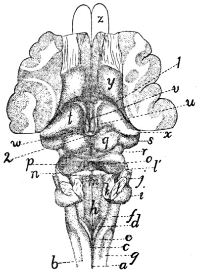
Fig. 141.—Dorsal Surface of Myelencephalon, Mesencephalon, and Diencephalon.
The cerebellum and the greater part of the cerebral hemispheres have been removed. a, posterior sulcus of cord; b, posterior lateral sulcus; c, fasciculus gracilis; d, clava; e, fasciculus cuneatus; f, area ovalis; g, fasciculus cuneatus lateralis; h, fossa rhomboidea or floor of fourth ventricle; i, projection formed by origin of auditory nerve; j, facial nerve; k, cut end of brachium conjunctivum; l, cut end of brachium pontis (l′, brachium pontis); m, velum medullare anterius; n, fourth nerve; o, depression marking caudal end of aqueductus cerebri (aqueduct of Sylvius); p, caudal corpora quadrigemina (colliculi posteriores); q, cranial corpora quadrigemina (colliculi anteriores); r, brachium quadrigeminum inferius; s, corpus geniculatum mediale; t, thalamus; u, striæ medullares; v, third ventricle; w, pulvinar; x, corpus geniculatum laterale; y, corpus striatum; z, outlines of olfactory bulbs. 1, boundary between hemispheres and ’tween-brain; 2, pineal body.
The fourth cranial nerve (Fig. 141, n; Fig. 138, IV) (N. trochlearis) arises from the brain at the craniolateral angle of the velum medullare anterius.
B. Mesencephalon.—
3. Mesencephalon.
—The mesencephalon or midbrain includes the corpora quadrigemina (Fig. 141) and the pedunculi cerebri (Fig. 142). In the primary midbrain there is a pronounced thickening of the walls accompanied by a reduction of the central canal. The midbrain does not thus become very large and is concealed in the dorsal view by the cerebellum and cerebrum, though its floor appears in the ventral view just craniad of the pons (Fig. 138, g). Its narrow canal is the aqueductus cerebri (aqueduct of Sylvius) (Fig. 143, j). Its roof forms the corpora quadrigemina, and its floor the pedunculi cerebri.
In a dorsal view (Fig. 141) the roof is seen to be marked by two pairs of elevations, the corpora quadrigemina (p and q). The cranial pair (q) (known as the colliculi superiores) are circular in outline, surrounded on all sides except the cranial one by a deep groove. From the cranial side a tract of fibres (brachium quadrigeminum superius, or arm of the cranial corpus) extends craniad and disappears beneath the thalamus (t). Between the anterior or cranial corpora quadrigemina lies the pineal body or epiphysis (corpus pineale) (Fig. 143, y; Fig. 141, 2), a portion of the roof of the ’tween-brain. The caudal corpora (Fig. 141, p) are larger than the cranial ones, and ovoid in shape with the long axis vertical. They are united in the median line, and the velum medullare anterius (m) stretches between their caudal borders. The brachium of the caudal corpus quadrigeminum (brachium quadrigeminum inferius) (r) extends craniad and disappears beneath a considerable elevation, the corpus geniculatum mediale (s).
Crossing this brachium is seen a small tract of fibres which extends ventrad, crosses the pedunculus cerebri, and reaches the medial border of the latter. It is the tractus transversus peduncularis (Fig. 142, b). Ventrad of the caudal corpus quadrigeminum is seen a triangular area of oblique fibres which corresponds in position to the human lemniscus.
The pedunculi cerebri (peduncles of the cerebrum) form the ventral part of the midbrain. They appear in a ventral view of the entire brain (Fig. 138) as two broad tracts of fibres (g) emerging from beneath the pons and diverging from one another as they pass craniad, finally disappearing beneath the cerebral hemispheres. Each is made up of many fibre-bundles, which are apparent in surface view (Fig. 142). The peduncles are separated by a small triangular space, which is marked by a median longitudinal sulcus. In this space, just caudad of the mammillary bodies, is a small area through which a number of blood-vessels pass into the brain. This is known as the posterior perforated area (or substance) (Fig. 142, j). The cerebral peduncles (a) are crossed by the tractus transversus peduncularis (b) (see above).
The third cranial nerve (Fig. 138, III) (N. oculomotorius) leaves the brain at the medial border of the cerebral peduncle (g), just caudad of the tractus transversus peduncularis.
The aqueductus cerebri (Fig. 143, j; Fig. 153, d) (or aqueduct of Sylvius) is the continuation craniad of the fourth ventricle. It is a narrow passage, one or two millimeters in diameter, lying dorsad of the pedunculi cerebri and ventrad of the corpora quadrigemina.
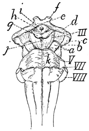
Fig. 142.—Ventral Surface of the Midbrain and ’Tween-brain, with the Pons.
a, pedunculi cerebri; b, tractus transversus peduncularis; c, corpus geniculatum mediale; d, optic tract; e, optic chiasma; f, optic nerve; g, mammillary bodies; h, tuber cinereum; i, opening for infundibulum (which has been removed); j, posterior perforated area; k, pons. III, V, VII, VIII, third, fifth, seventh, and eighth cranial nerves.
C. Prosencephalon.—The prosencephalon or primitive forebrain includes the diencephalon or ’tween-brain and the telencephalon or cerebral hemispheres.
4. Diencephalon.
—The diencephalon or ’tween-brain includes the thalami and the other parts bounding the third ventricle. The diencephalon is seen in entire brains only in ventral view (Fig. 138, c, d, e, etc.)
The diencephalon may be considered as forming almost or quite the most cranial portion of the median nervous tube,—the cranial wall of the third ventricle (the lamina terminalis) (Fig. 143, d), ending in the median line in the deep fissure between the hemispheres of the cerebrum. Parts of the brain which extend farther craniad than this are lateral portions, due to the forward growth of the lateral hemispheres. The two hemispheres of the cerebrum may be considered as lateral outgrowths of the central ’tween-brain; these outgrowths have extended dorsad, laterad, craniad, and caudad, so as to cover almost completely the ’tween-brain.
In early stages the cerebral hemispheres are projections from the cranial end of the ’tween-brain, so that the plane of junction was nearly transverse, the cranial end of the ’tween-brain joining the caudal end of the hemispheres. With the increasing size and backward growth of the latter, the attachment to the ’tween-brain has been shifted from a cranial to nearly a lateral position, and at the same time the originally lateral surface of the ’tween-brain has become nearly caudal. This is shown in Fig. 141, the deep fissure at 1 marking the line of attachment between the ’tween-brain and the hemispheres. The dorsoventral plane of junction of ’tween-brain and hemispheres is (as Fig. 141 shows) not wholly lateral, but oblique, passing from its cranial end near the middle line caudolaterad.
A second peculiarity of the ’tween-brain lies in the thinness of its roof. The roof is exceedingly thin and is so intimately connected with the pia mater that they cannot be removed separately. The ventral thick floor of the ’tween-brain is directly continuous with the similar floor of the cerebrum; but where the roof of the ’tween-brain joins the roof of the cerebrum along the oblique plane already indicated, the roof is very thin and is intimately connected to the pia mater, and is at the same time folded into the lateral ventricles to form, together with the pia mater, the choroid plexuses of the lateral ventricles (Fig. 148, e). When the pia mater is removed the thin roof of the brain along the line of junction of the ’tween-brain and the cerebrum is brought away and there appears to be a direct communication between lateral ventricles and the exterior.
We may now take up the parts of the diencephalon in detail.
a. Thalamus (Fig. 141, t).—The two thalami are large oblique rounded ridges, forming the sides of the ’tween-brain. They lie just craniad of the cranial corpora quadrigemina (q), but separated from them by a broad groove, and are completely covered by the caudally projecting part of the cerebral hemispheres. The medial larger end of each thalamus is near the middle line; thence the thalamus extends caudolaterad, and rises at its lateral extremity into a sharply rounded projection, the corpus geniculatum laterale (x). From this projection the thalamus is continued ventrad and then craniomediad as a large band of fibres, the optic tract (tractus opticus) (Fig. 142, d), which extends to the optic chiasma (e), where it passes into the optic nerves (f). On the caudal border of the thalamus, near the median line, is a very faint projection, the pulvinar (Fig. 141, w); this lies just craniad of the lateral border of the cranial corpus quadrigeminum (q). Just ventrad of the corpus geniculatum laterale (x) is the prominent rounded corpus geniculatum mediale (s); this is connected by a prominent ridge, the brachium quadrigeminum inferius (r), with the caudal corpus quadrigeminum (p). In a similar manner the brachium quadrigeminum superius passes from the cranial corpus quadrigeminum (q) into the thalamus itself.
Between the two thalami there exists a groove, the sulcus hypothalamicus. Over this groove lies the roof of the third ventricle, forming the choroid plexus of the third ventricle (Fig. 143, w). The medial surface of the two thalami are flat and extend directly ventrad, forming part of the lateral boundary of the third ventricle (Fig. 143, h). The two medial surfaces meet over a considerable area across the narrow cavity of the third ventricle, and unite, forming the massa intermedia (Fig. 143, f) or intermediate mass of the thalamus (“middle commissure”). This connection of the thalami of the two sides is thus not a primitive one, forming no part of the roof or floor of the central cavity, but is a secondary connection due to a growing together of a part of the two sides of the ventricle across its cavity. Along the dorsal edge of the medial border of each thalamus passes a distinct white strand, the stria medullaris (Fig. 141, u); the two striæ meet in an arch caudad, lying beneath the pineal body (2).
The thalamus is separated craniolaterad by a groove (1) from the corpus striatum (y), on the floor of the cerebral hemisphere.
b. The roof of the third ventricle is thin and united with the pia mater, as already stated. The pia mater bears many blood-vessels, and the two are folded into the groove between the optic thalami, forming the lamina chorioidea epithelialis, or choroid plexus of the third ventricle (Fig. 143, w). The roof is attached to the dorsomedial borders of the thalami and becomes continuous with the choroid plexus of the lateral ventricles (Fig. 148, e) at the craniolateral borders of the thalami.
The pineal body (corpus pineale) or epiphysis (Fig. 141, 2; Fig. 143, y) is a small conical body which is formed as an outgrowth of the caudal part of the roof of the third ventricle. It lies on the roof of the brain between the two cranial corpora quadrigemina (Fig. 141, q). It is hollow, containing an extension of the third ventricle. From its craniolateral angles two white strands, the habenulæ, extend into the striæ medullares (Fig. 141, u) of the thalami.
Just ventrad of the pineal body is a transverse band of white fibres, lying in the caudal part of the roof of the third ventricle. This interconnects the two thalami, and forms the posterior commissure (Fig. 143, z′) (commissura posterior). From this commissure a thin sheet of tissue extends to the pineal body.
c. The floor of the third ventricle appears in a ventral view of the brain (Fig. 138, Fig. 142) as a somewhat diamond-shaped space craniad of the pedunculi cerebri (Fig. 142, a) and bounded along its cranial margin by the optic tracts (Fig. 142, d). The optic tracts come from the thalami, as already described; they converge and unite to form the optic chiasma (Fig. 142, e), from which the optic nerves (f) diverge.
Immediately caudad of the optic chiasma lies a considerable rounded gray elevation, the tuber cinereum (Fig. 142, h). This bears on its ventral surface in the natural condition the infundibulum (Fig. 138, d) with the hypophysis (Fig. 138, e); in cases where the two latter structures have been removed (Fig. 142) the tuber cinereum (h) bears a small longitudinal opening (i) for attachment of the infundibulum. The infundibulum (Fig. 138, d) is a hollow extension of the floor of the third ventricle, and is attached to the middle of the ventral surface of the tuber cinereum. It bears at its ventral end the hypophysis (pituitary body) (Fig. 138, e), a vascular non-nervous body of unknown function. The hypophysis is lodged in the sella turcica of the sphenoid bone.
At its caudal border the tuber cinereum bears two white elevations, the mammillary bodies (corpora mammillaria) (Fig. 142, g).
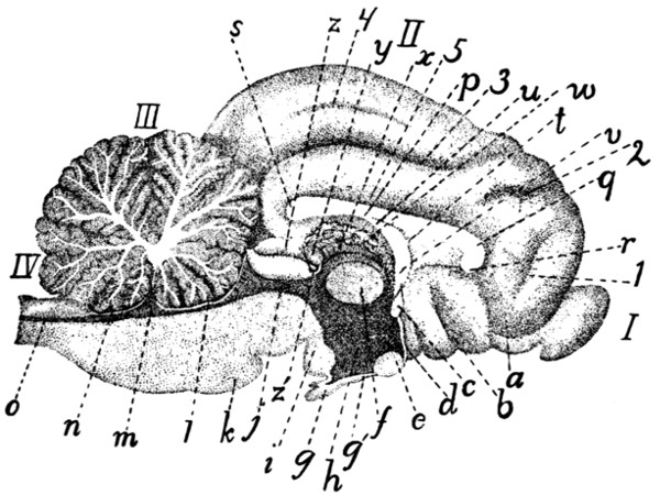
Fig. 143.—Longitudinal Median Section of Brain.
I, olfactory bulb; II, cerebrum; III, section of cerebellum, showing “arbor vitæ”; IV, medulla oblongata. a, tractus olfactorius; b, substantia perforata anterior; c, anterior commissure; d, lamina terminalis; e, optic chiasma; f, massa intermedia of thalamus; g′, infundibulum; g, hypophysis; h, third ventricle; i, mammillary body; j, aqueductus cerebri (or aqueduct of Sylvius); k, pons; l, velum medullare anterius; m, fourth ventricle; n, velum medullare posterius; o, continuation of canal of spinal cord; p, corpus callosum (body); q, genu of corpus callosum; r, rostrum; s, splenium; t, septum pellucidum; u, fornix; v, pillars of fornix; w, choroid plexus of third ventricle; x, stria medullaris; y, corpus pineale; z, cranial corpus quadrigeminum; z′, posterior commissure. 1, sulcus falcialis; 2, sulcus cruciatus; 3, sulcus splenialis; 4, sulcus marginalis; 5, sulcus supracallosalis (or callosalis).
The third ventricle (Fig. 143, h; Fig. 141, v) is a very narrow slit-like space, of considerable extent dorsoventrally, but less than a millimeter in width; it lies between the medial ends of the thalami (Fig. 141), and extends ventrad into the tuber cinereum, and thence into the infundibulum (Fig. 143, g′). Dorsad it extends into the corpus pineale (Fig. 143, y). The third ventricle communicates caudally near its dorsal border with the aqueductus cerebri (j); craniolaterally with the lateral ventricles, through the interventricular foramen (foramen of Monroe). Its cranial boundary forms in the middle line a thin plate, the lamina terminalis (d), which is, morphologically, the cranial termination of the cerebrospinal axis; it lies at the bottom of the deep fissure between the cerebral hemispheres. At the dorsal border of the lamina terminalis is a strong transverse band of fibres connecting the two sides of the brain; this is the anterior commissure (c). The cavity of the third ventricle is much encroached upon by the meeting and secondary union of the two thalami across the middle line, forming the massa intermedia (f).
The boundaries of the third ventricle, in order, are as follows, beginning dorsocraniad of the communication with the aqueductus cerebri: the posterior commissure (Fig. 143, z′), the pineal body (y), the choroid plexus of the third ventricle (w), the columns of the fornix (v), the anterior commissure (c), the lamina terminalis (d), the tuber cinereum with the infundibulum (g′), the substantia perforata posterior, and the midbrain. The lateral boundaries are formed by the thalami (Fig. 141, t).
5. Telencephalon.
—The telencephalon includes the two cerebral hemispheres. The name cerebrum is also applied to this portion of the brain; frequently, however, the name cerebrum is used as signifying the entire mass of the brain craniad of the rhombencephalon,—therefore including mesencephalon, diencephalon, and telencephalon. The term will here be used as synonymous with telencephalon.
The cerebral hemispheres arise as two lateral outgrowths from the cranial end of the primitive forebrain. They have undergone great increase in size in the course of evolution, so as to form the larger part of the brain; at the same time important modifications of structure have taken place. In the original condition the medial faces of the two hemispheres are not connected, the two being separate outgrowths of the ’tween-brain, and connected only through the latter. But secondary connections have been formed across the fissure between the two hemispheres, resulting in the production of the corpus callosum (Fig. 143, p; Fig. 147; Figs. 149-152, a), a broad transverse band of white fibres connecting the two hemispheres dorsad of the ’tween-brain. Ventrad of the corpus callosum another secondary union has resulted in the production of the fornix (Fig. 143, u).
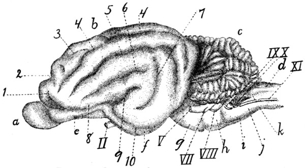
Fig. 144.—Lateral Surface of the Brain.
a, olfactory bulb; b, cerebral hemisphere; c, cerebellum; d, medulla oblongata; e, tractus olfactorius; f, lobus pyriformis or tractus postrhinalis; g, pons; h, trapezium; i, pyramid; j, area elliptica; k, area ovalis. II, N. opticus; V, N. trigeminus; VII, N. facialis; VIII, N. acusticus; IX, N. glossopharyngeus; X, N. vagus; XI, N. accessorius. 1. sulcus præsylvius; 2, sulcus cruciatus; 3, sulcus ansatus; 4, sulcus lateralis; 5, sulcus suprasylvius; 6, sulcus anterior; 7, sulcus posterior; 8, sulcus rhinalis; 9. fissura Sylvii; 10. sulcus rhinalis posterior.
External Features.—With increase in size the mass of the cerebrum shows externally a tendency to divide into three lobes, one craniad, the frontal (Fig. 145, A); one caudoventrad, the temporal (B); and one caudodorsad, the occipital (C). The two latter are not distinctly marked off from one another. The limit between the temporal and frontal lobes is marked by a short deep fissure, the lateral fissure (fissura cerebri lateralis), or fissure of Sylvius (Fig. 144, 9; Fig. 145, a). Each lobe is thrown up into elevations or gyri, which are separated by grooves or sulci; these are described below. The homology of the cerebral gyri and sulci of the cat with those of man is in most cases uncertain.
The sulci and gyri of the cerebral hemispheres may be described briefly in their main features as follows: On the lateral surface of the hemisphere (Fig. 145) the lateral fissure (fissura cerebri lateralis), or fissure of Sylvius, separating temporal and frontal lobes, forms the most convenient point of departure for an understanding of the fissures. The fissure of Sylvius is short but deep, and is formed early during development. In man it covers an area, the insula, at its bottom, which may be seen by separating the sides of the fissure. In the cat the insula is rudimentary and can be demonstrated only with difficulty.
The lateral surface of the hemisphere is marked by fissures which form three concentric arches (b-c, d, and e), irregular and incomplete, about the fissure of Sylvius. These arches all open ventrad. The first arch (b-c) (that next to the fissure of Sylvius) lacks the central part, the keystone, so that a complete arch is not formed, but only the two sides of the arch, as two separate sulci (b and c). The one of these craniad of the fissure of Sylvius is the sulcus anterior (b); the other is the sulcus posterior (c). In the dog this arch is usually complete and the sulcus forming it is frequently known as the ectosylvian sulcus (sulcus ectosylvius).
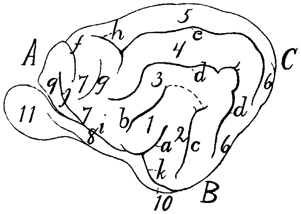
Fig. 145.—Diagram of the Sulci and Gyri on the Lateral Surface of the Hemisphere.
A, frontal lobe; B, temporal lobe; C, occipital lobe. a, lateral fissure or fissure of Sylvius; b, sulcus anterior; c, sulcus posterior (the broken line connecting the ends of a and b serves to show how the first arch is completed to form the ectosylvian sulcus, as in the dog); d, sulcus suprasylvius; e, sulcus lateralis; f, sulcus cruciatus; g, sulcus coronalis; h, sulcus ansatus; i, sulcus rhinalis; j, sulcus præsylvius; k, sulcus rhinalis posterior. 1, anterior Sylvian gyrus; 2, posterior Sylvian gyrus; 3, gyrus ectosylvius; 4, gyrus suprasylvius; 5, gyrus marginalis; 6, gyrus compositus posterior; 7, sigmoid gyrus; 8, olfactory tract; 9, orbital gyrus; 10, lobus pyriformis; 11, olfactory bulb.
The gyri included between the fissure of Sylvius on the one hand and the anterior and posterior sulci on the other are known as the anterior (1) and posterior (2) Sylvian gyri.
The second arch is formed by the suprasylvian sulcus (sulcus suprasylvius) (d). The gyrus between the anterior and posterior sulci ventrad and the suprasylvian sulcus dorsad is the ectosylvian gyrus (3) (because dorsad of the ectosylvian sulcus).
The third arched sulcus, next to the medial margin of the hemisphere, is variable: it forms the lateral sulcus (e). The gyrus lying between the suprasylvian and lateral sulci is the suprasylvian gyrus (4) (because dorsad of the suprasylvian sulcus). The lateral sulcus is sometimes broken into two (as in Fig. 137, h), the two ends overlapping so as to leave a small gyrus between the two ends.
The gyrus dorsomediad of the lateral sulcus, extending onto the medial surface of the hemisphere as far as the splenial sulcus, is the marginal gyrus (5).
The four sets of gyri included between these three sets of arched sulci and the fissure of Sylvius unite caudad and craniad to form single gyri. The caudal one is the gyrus compositus posterior (6). The cranial one (less regular than the caudal one) is the sigmoid gyrus (7). The latter curves about the cruciate sulcus (f) (described below).
In the cranial part of the lateral surface of the hemisphere are certain sulci and gyri which do not belong to the system above described. The cruciate sulcus (f) is a short transverse sulcus passing from the lateral surface of the hemisphere onto its medial surface, where it extends caudad about a centimeter. Curving around the lateral end of this, separated from it by part of the sigmoid gyrus, is the short coronal sulcus (g). Connected usually with the end of the lateral sulcus (e) and running nearly parallel with the cruciate sulcus is the short sulcus ansatus (h).
At its ventral end the fissure of Sylvius (a) joins a longitudinal groove passing craniad and caudad from the point of junction. That part which passes craniad is the sulcus rhinalis (i); it forms the dorsolateral boundary of the olfactory tract (8). Passing dorsocraniad from the sulcus rhinalis is a sulcus which separates a very small cranial lobe from the rest of the hemisphere; this is the supraorbital or præsylvian sulcus (j). The portion of the hemisphere craniad of this sulcus is the orbital gyrus (9).
The sulcus rhinalis (i) is continuous caudad with the sulcus rhinalis posterior (or sulcus postrhinalis) (k). This extends caudad onto that portion of the hemisphere which faces the cerebellum. It forms the lateral boundary of a large elongated oval lobe lying at the side of the ventral floor of the midbrain and ’tween-brain. This lobe has been called tractus postrhinalis or lobus pyriformis (Fig. 145, 10; Fig. 138, f).
In addition to the sulci and gyri above mentioned, any given specimen will usually show a number of small inconstant sulci and gyri in various regions; these inconstant structures will not be here described.
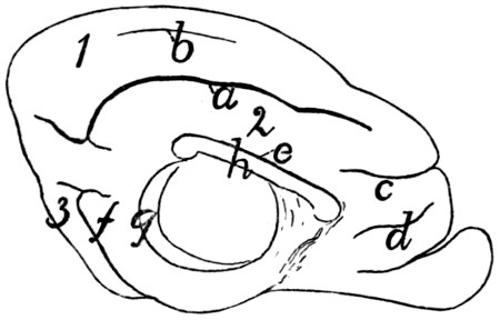
Fig. 146.—Diagram of the Sulci and Gyri on the Medial Surface of the Hemisphere.
a, sulcus splenialis; b, sulcus marginalis; c, sulcus cruciatus; d, sulcus falcialis; e, sulcus supracallosalis; f, sulcus rhinalis posterior; g, hippocampal sulcus; h, corpus callosum. 1, gyrus marginalis; 2, gyrus fornicatus; 3, gyrus compositus posterior.
On the medial surface of the hemisphere (Fig. 146), the following arrangement of sulci and gyri seems to be typical. Some distance from the dorsal margin a long sulcus runs parallel with the margin; this is the sulcus splenialis (a). The marginal gyrus (1) is dorsad of the splenial sulcus, passing onto the lateral surface of the hemisphere, where it is bounded by the lateral sulcus. The splenial sulcus extends onto the caudal surface of the hemisphere. A shallow sulcus marginalis (b) occurs frequently between the sulcus splenialis and the dorsal margin of the hemisphere. Cranioventrad or the cranial end of the splenial sulcus is that portion of the sulcus cruciatus (c) that lies on the medial surface of the hemisphere. Ventrad of this is a short shallow furrow which has been called the sulcus falcialis (d).
Immediately dorsad of the corpus callosum (h), separating its dorsal surface from a part of the hemisphere, is the supracallosal or callosal sulcus (e). Between the supracallosal (e) and splenial (a) sulci is the gyrus fornicatus (2).
On the caudal surface of the hemisphere (that facing the cerebellum) appear the ends of the sulcus splenialis (a) and the sulcus rhinalis posterior (f). Hidden in the natural condition by the thalami and optic tract is the broad hippocampal sulcus (g), marking externally the course of the hippocampus.
A further extension of the surface of the cerebrum has taken place in connection with the sense of smell. A mass of gray matter, the olfactory bulb (Fig. 144, a), reckoned as a part of the cerebrum, is separated from the latter and lies against the cribriform plate of the ethmoid bone. From it the olfactory fibres pass through the perforations of the plate to the olfactory mucous membrane. The olfactory bulb contains a cavity, a part of the lateral ventricle.
The bulb lies against the ventral surface of the frontal lobe and projects craniad of it. It is connected to the cerebrum by a tract of fibres, the olfactory tract (Fig. 138, a), which is divisible into two roots, medial and lateral. The medial root comes from the medial surface of the frontal lobe, where it is continuous with a tract extending to the cranial end of the corpus callosum. The lateral root is traceable from an elevated gyrus-like portion of the cerebrum which lies at the side of the infundibulum and is known as the lobus pyriformis or tractus postrhinalis (Fig. 138, f). The lateral root is divisible into a medial white strand and a lateral gray strand.
That part of the brain comprising the olfactory bulb and the parts intimately related to it are frequently included under the term rhinencephalon.
In the triangular area between the two olfactory tracts and craniad of the optic chiasma appears a mass of gray matter, subdivided by a longitudinal fissure. This possesses numerous openings through which blood-vessels pass to the brain substance, and is thence known as the anterior perforated substance (substantia perforata anterior) (Fig. 138, b).
Internal Structures of the Cerebrum.—The cavity of each of the cerebral hemispheres is known as a lateral ventricle. The two lateral ventricles constitute the first and second of the ventricles of the brain, whence the application of the names third and fourth ventricles to the cavities of the ’tween-brain and hindbrain. The lateral ventricles do not grow at the same rate as the walls of the hemispheres, so that they remain comparatively small. The cavity of each ventricle is further reduced in size by the development on its floor of a large ridge-like thickening, the corpus striatum (Fig. 148, f).
The dorsal wall or roof of the lateral ventricle joins the thin roof of the third ventricle on each side along an oblique curved line (Fig. 141, 1) which follows the cranial or lateral border of the thalamus (Fig. 141, t). Along this line the thin roof of the brain is folded in together with the pia mater to form the choroid plexus of the lateral ventricles (lamina chorioidea epithelialis) (Fig. 148, e). When this is pulled out there is left a fissure, the “great transverse fissure of the cerebrum” (Fig. 141, 1). Just dorsad of the groove between the thalami there runs a tract of white fibres known as the fornix (Fig. 148, a). The two halves of the fornix separate at the cranial ends of the thalami and pass ventrad, forming thus the pillars of the fornix (Fig. 148, b; Fig. 143, v). Dorsad of the cranial end of the fornix the corpus callosum (Fig. 143, p) passes from one hemisphere to the other. Caudad of the pillars of the fornix, the lateral ventricles communicate with the third ventricle by way of the interventricular foramen (foramen of Monroe).
The parts of the cerebrum may now be taken up in detail.
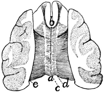
Fig. 147.—The Corpus Callosum.
The dorsal portion of the hemispheres has been dissected away, then sliced off, showing the plate of transverse fibres forming the corpus callosum. a, splenium; b, genu; c, line marking the medial edge of the hemispheres; d, line marking the lateral boundary of the supracallosal sulcus; laterad of this line the corpus callosum lies in the substance of the hemispheres, which have been dissected away; e, line marking medial limit of cut surface.
The corpus callosum (Fig. 147; Fig. 143, p; Figs. 149-152, a) is a broad transverse band of fibres forming a secondary connection between the medial walls of the two hemispheres, dorsad of the roof of the third ventricle. Its outer surface (Fig. 147) is exposed at the bottom of the fissure which separates the hemispheres. On each side it passes laterad, forming the roof of the lateral ventricle. Its cranial part lies dorsad of the corpus striatum, and its caudal part dorsad of the thalamus. Laterally its fibres radiate into the substance of the hemispheres. At its cranial end the corpus callosum bends ventrad and then caudad (Fig. 143). The part which turns to pass ventrad is the genu (Fig. 143, q) or knee, while the part which projects caudad is the rostrum (r). The caudal border of the corpus callosum is also thickened and turned ventrad and is called the splenium (Fig. 143, s); it lies dorsad of the cranial corpora quadrigemina (z). The caudal half of the ventral surface of the corpus callosum is united with the fornix (Fig. 143, u).
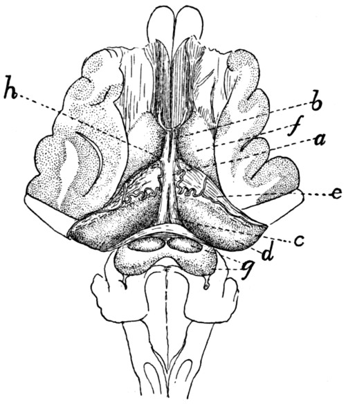
Fig. 148.—Fornix, Hippocampus, and Corpus Striatum.
The dorsal portion of the hemispheres has been dissected away and the corpus callosum removed. a, fornix; b, columns or pillars of the fornix; c, crura of the fornix; d, hippocampus; e, choroid plexus of the lateral ventricles overlying the fimbria (the choroid plexus shows an artery); f, corpus striatum; g, corpora quadrigemina; h, position of the interventricular foramen (foramen of Monroe).
The fornix (Fig. 148, a; Fig. 143, u, v; Figs. 150-152, b) consists of an arched tract of longitudinal fibres near the medial border of each hemisphere, ventrad of the corpus callosum. Each tract begins in the mammillary bodies, and passes dorsad, the two converging until they run side by side, forming a cylindrical dorsoventral bundle known as the columns or pillars of the fornix (Fig. 143, v; Fig. 150, e), which cross the anterior commissure (Fig. 143, c; Fig. 150, f) caudad of the latter. Caudad of the pillars of the fornix, between these and the thalamus, lies on each side the interventricular foramen or foramen of Monroe, a small opening which connects the lateral ventricles with the third ventricle. Dorsad of the foramen the fornix turns caudad, the two fibre-tracts of each hemisphere lying side by side and closely connected (Fig. 148, a), forming thus another secondary union between the medial surfaces of the two hemispheres. This portion of the fornix is the corpus or body (Fig. 148, a); it lies dorsad of the roof of the third ventricle and passes to the splenium (Fig. 143, s) of the corpus callosum, and its dorsal surface unites with the ventral surface of the latter (Fig. 143). Caudad the two halves of the fornix diverge, forming the crura of the fornix (Fig. 148, c); these and the body are continuous laterally with the hippocampus (Fig. 148, d) and the fimbria (Fig. 148, beneath e).
The anterior commissure (Fig. 143, c; Fig. 150, f) is a transverse band of white fibres which stretches from one hemisphere to the other about half way between the interventricular foramen or foramen of Monroe and the floor of the third ventricle, and just craniad of the pillars of the fornix. This tract of fibres is developed in the original wall of the third ventricle, so that it does not form a secondary connection between the halves of the cerebrum, as do the fornix and corpus callosum. It lies dorsad of the lamina terminalis (Fig. 143, d) and is continuous with it.
The septum pellucidum (Fig. 143, t) is a vertical partition which separates the lateral ventricles and fills the interval between the corpus callosum dorsad and the fornix ventrad. It is triangular and translucent. It is formed from the medial walls of the two hemispheres and therefore is made up of two laminæ which embrace between them a space which originally was a part of the fissure separating the hemispheres. This space has been called the fifth ventricle.
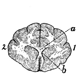
Fig. 149.—Caudal Surface of Transverse Section of Brain through the Genu of the Corpus Callosum.
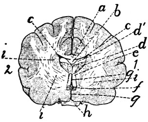
Fig. 150.—Caudal Surface of Transverse Section of Brain through the Optic Chiasma and the Region of the Interventricular Foramen (Foramen of Monroe).
Fig. 149.—Gray matter dotted; white matter with a few lines. 1, right hemisphere; 2, left hemisphere. a, corpus callosum, section through genu and rostrum; b, cavity of lateral ventricles.
Fig. 150.—1, right hemisphere; 2, left hemisphere. a, corpus callosum; b, fornix (cut); c, cavity of lateral ventricles; d, fimbria (d′, cut surface; d, caudal uncut surface of the part that curves ventrad); e, pillars of fornix; f, anterior commissure; g, parts of third ventricle; h, optic chiasma; i, parts of corpus striatum.
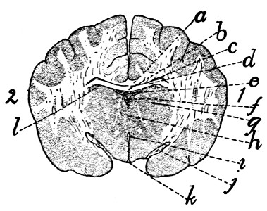
Fig. 151.—Caudal Surface of Transverse Section of the Brain through the Tuber cinereum and Infundibulum, Craniad of the Hypophysis.
a, corpus callosum; b, fornix (continuous at the sides with the hippocampus); c, cavity of lateral ventricles; d, hippocampus; e, choroid plexus of the third ventricle (roof of the diencephalon); f, dorsal part of cavity of the third ventricle; g, section of thalamus; h, massa intermedia of the thalami, dividing the third ventricle into a dorsal (f) and a ventral (i) portion; i, ventral part of third ventricle; j, part of optic tracts; k, thin wall of infundibulum, with part of cavity of third ventricle; l, line separating roof of thalamus (diencephalon) from floor of hemispheres (telencephalon).
The hippocampus (Fig. 148, d; Fig. 152, d) is an elongated rounded elevation of the floor of the lateral ventricle. It is continuous mediad with the fornix (Fig. 148, a) and extends thence along the inferior horn of the lateral ventricle to its end in the temporal lobe. It is somewhat narrower at its lateral end. It is thus curved into a semicircle in conformity with the inferior horn of the ventricle. Its dorsal surface is convex and looks into the lateral ventricle; its ventral surface is concave and rests upon the thalamus and the optic tract. Along the craniolateral edge of the hippocampus is a broad, clearly marked fibre-tract, the fimbria; this runs parallel to the choroid plexus of the lateral ventricle and beneath it, and is continuous at its medial end with the fornix.
The corpus striatum (Fig. 148, f; Fig. 141, y; Fig. 150, i) is a fusiform or ovoid elevation of the floor of the lateral ventricle. One of its narrow ends lies about opposite the middle of the thalamus (Fig. 141, t) and it extends thence obliquely mediad nearly parallel with the thalamus. In the groove between it and the thalamus is the choroid plexus of the lateral ventricles (Fig. 148, e). Internally the corpus striatum is made up of several layers of different texture (Fig. 150, i), whence its name.
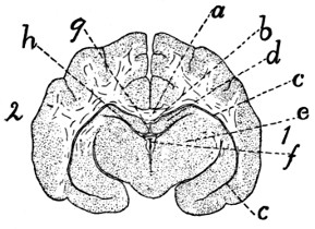
Fig. 152.—Caudal Surface of Transverse Section of the Brain through the Pineal Body and Thalami.
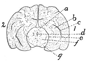
Fig. 153.—Caudal Surface of Transverse Section of the Brain through the Cranial Pair of Corpora Quadrigemina.
Fig. 152.—a, corpus callosum; b, fornix; c, inferior horn of lateral ventricles; d, hippocampus; e, thalamus; f, third ventricle; g, pineal body; h, space between the floor of the hemisphere (hippocampus) and roof of the ’tween-brain (thalamus).
Fig. 153.—a, cranial corpora quadrigemina; b, hippocampus; c, part of inferior horn of lateral ventricle; d, aqueductus cerebri (aqueduct of Sylvius); e, space between the outer surface of the midbrain and the lower surface of the hemisphere; f, cross-section of midbrain; g, pedunculi cerebri.
The choroid plexus of the lateral ventricles (lamina chorioidea epithelialis) (Fig. 148, e).—The line of junction of the roof of the third ventricle (i.e., the choroid plexus of the third ventricle) and the cerebral hemispheres is along the lateral (or cranial) border of the thalamus (Fig. 141, t). Along this line (Fig. 141, 1) the brain-wall remains very thin and becomes closely associated with the pia mater, so that on the removal of the pia there is left the “transverse fissure” which leads into the lateral ventricle. In an early stage, before the cerebrum has grown far caudad, the fissure is exposed in dorsal view, but after the hemispheres have covered the thalami the fissure appears to be in the lower face of the hemisphere. Through this fissure the pia mater extends upward into the lateral ventricles as a fold separated from the ventricle by the thin brain-wall. This fold is vascular and is known as the choroid plexus of the lateral ventricle (Fig. 148, e). It is a fringe-like fold lying on the fimbria and extending from the foramen of Monroe (at h) nearly to the end of the inferior horn of the lateral ventricle.
The lateral ventricles are the extensions into the hemispheres of the originally unpaired cavity of the forebrain. Each consists of a portion parallel to the basis cranii and lying at the side of the septum pellucidum between the corpus callosum dorsad and the corpus striatum, hippocampus, and fornix ventrad, and of two horns, an anterior and an inferior horn. The inferior horn (Fig. 152, c) is a narrow cleft, crescent-shaped in cross-section, which follows the dorsal surface of the hippocampus (Fig. 148, d; Fig. 152, d) along the temporal lobe to its end almost against the tuber cinereum. It thus passes first caudad and then ventrad and finally mediad. The choroid plexus and hippocampus project into it from its floor. The anterior horn extends ventrad and then slightly caudad in the frontal lobe, following the corpus striatum (Fig. 148, f), on which it lies.
The interventricular foramen (or foramen of Monroe).—If the choroid plexus of the lateral ventricle (Fig. 148, e) is followed to its medial end, it is found to pass through a foramen (at h) in which it becomes continuous with the opposite plexus or roof of the third ventricle. This foramen leads from one lateral ventricle to the other and is connected ventrally by a median opening with the third ventricle. It is thus Y-shaped or T-shaped and is the interventricular foramen (or foramen of Monroe). The foramen lies opposite the middle of the corpus striatum and caudad of the pillars of the fornix.
Membranes of the Brain.—The membranes of the brain are the dura mater, the arachnoid, and the pia mater.
The dura mater is a strong fibrous membrane lining the cranial cavity and covering the brain. It is strongly attached to the projections of the base of the skull and to the tentorium. It dips between the cerebral hemispheres and olfactory bulbs, forming the falx cerebri. It likewise dips between the cerebrum and the cerebellum, covering both surfaces of the bony tentorium. The dura mater does not pass into the fissures or sulci of the surface of the brain. It becomes continuous with the sheath of the cranial nerves as they leave the skull. It is continuous with the dura mater of the spinal cord.
The arachnoid and pia mater are essentially similar to the same structures on the spinal cord. The pia mater dips into the fissures and sulci of the brain.
II. THE PERIPHERAL NERVOUS SYSTEM.
1. Cranial Nerves. Nervi cerebrales.—
I. N. olfactorius.
—The fasciculi of the olfactory nerves arise from the olfactory bulb (Fig. 144, a) and pass through the foramina of the cribriform plate, upon which the bulb lies, to be distributed to the olfactory mucous membrane of the nose.
II. N. opticus.
—The second nerve or optic (Fig. 138, II) arises from the optic chiasma (Fig. 138, c), passes through the optic foramen, and extends in an S-shaped curve to the eyeball. Its course is craniodorsad. It pierces the sclerotic and choroid coats of the eye and spreads out into the retina.
III. N. oculomotorius.
—The third or oculomotor nerve arises (Fig. 138, III) from the pedunculus cerebri and passes into the orbit through the orbital fissure. It passes between the lateral and superior recti, sends a large branch to the superior rectus, supplies the medial rectus and the retractor oculi, passes laterad of the optic nerve and supplies the inferior rectus, and sends a long branch to the inferior oblique (Fig. 166, f). At the point where the branch is given off to the inferior oblique muscle is the triangular reddish ciliary (or ophthalmic) ganglion, about two millimeters in diameter. It is directly attached to the inferior oblique branch of the third nerve and receives a delicate sensory filament from the ophthalmic nerve. From the ciliary ganglion proceed two short ciliary nerves which pass along each side of the optic nerve to the eyeball. These nerves penetrate the sclerotic at the sides of the eyeball to be distributed to the ball.
IV. N. trochlearis.
—The fourth nerve, the trochlearis (or patheticus), arises from the lateral border of the velum medullare anterius, as already described (Fig. 141, n). It passes craniad, emerges through the orbital fissure in company with the oculomotor, abducens, and ophthalmic, passes dorsad of the superior rectus muscle, and reaches (Fig. 154, l) the caudal border of the belly of the superior oblique muscle near its middle. It supplies only the superior oblique.
V. N. trigeminus.
—The fifth nerve, the trigeminus (or trifacial) arises (Fig. 138, V) by two roots, a large sensory and a small motor root (4), as already described (p. 347). One of these, the motor (4), is smaller and more ventral; the other, the sensory root, is larger and dorsal. The dorsal root soon enlarges to form a large ganglion, the semilunar (or Gasserian) ganglion (Fig. 138, k), from which three branches diverge. One branch is joined by the ventral root (4), which passes over the ventral surface of the semilunar ganglion; and the nerve thus formed is the mandibular division (1) of the fifth nerve. Of the other two branches from the ganglion, the middle and longest is the maxillary nerve (2), and the smallest is the ophthalmic (3). The mandibular nerve is thus mixed, motor and sensory, while the others are sensory.
1. N. ophthalmicus.—The ophthalmic or first division of the fifth nerve arises from the semilunar (or Gasserian) ganglion. It passes out of the cranial cavity and into the orbit by way of the orbital fissure, in company with the third, fourth, and sixth nerves and with the extension of the carotid (arterial) plexus.
It passes between the superior and medial recti along with the third nerve, crosses dorsad of the optic nerve, and divides into infratrochlear and ethmoidal branches. In the orbital fissure it gives off the frontal nerve, and while crossing the optic it gives off the long ciliary nerve.
a. N. frontalis.—The frontal nerve passes along the lateral border of the superior oblique muscle and then laterad of the pulley to near the middle of the supraorbital crest of the frontal bone. Here it passes out of the orbit and is distributed to the integument of the upper eyelid and the adjacent region at the side of the nose.
b. N. infratrochlearis.—The infratrochlear nerve passes between the superior rectus and the superior oblique in the first part of its course. It then passes ventrad of the superior oblique and ventrad of the pulley to be distributed to the integument of the upper eyelid near the inner angle.
c. N. ethmoidalis.—The ethmoidal nerve passes along with the ethmoidal artery through the ethmoidal foramen (or foramina) in the orbital plate of the frontal bone. It is finally distributed to the mucosa of the nose and to the cartilage and integument of the snout.
d. N. ciliaris longus.—The long ciliary nerve arises from the ophthalmic and passes along the optic nerve to be distributed to the eyeball. It divides into several branches before penetrating the sclerotic.
One or two small communicating branches to the ciliary ganglion are given off at about the same point as the long ciliary nerve. (For a description of this ganglion see the account of the oculomotor nerve, page 369.)
2. N. maxillaris.—The maxillary nerve, the second division of N. trigeminus, rises from the semilunar (Gasserian) ganglion and leaves the skull by the foramen rotundum. It is the sensory nerve of the palate, upper teeth and upper lip, and of part of the forehead and cheek.
On leaving the foramen the maxillary nerve divides into three branches, the two infraorbital nerves (Fig. 154, f) and the sphenopalatine (Fig. 154, g). Two smaller branches are likewise given off either within or just outside of the foramen, the lachrymal nerve (Fig. 154, j) and the zygomatic (subcutaneus malæ) (Fig. 154, i).
a. N. lachrymalis (Fig. 154, j; Fig. 155, m).—The lachrymal nerve passes along the periorbita to the lachrymal gland (Fig. 154, 11), to which it gives branches. It then continues caudad of the zygomatic process of the temporal to the integument; here it turns caudad and is distributed to the integument in the region between the eye and the external ear (Fig. 155, m). It anastomoses with the zygomatic branch of the seventh nerve (Fig. 155, h).
b. N. zygomaticus (subcutaneus malæ) (Fig. 154, i).—This arises with the preceding and follows it for some distance. It passes through a foramen in the frontal process of the malar bone and is distributed to the lower eyelid and adjacent integument.
c. Nn. infraorbitales (Fig. 154, f; Fig. 155, l).—The infraorbital nerves are two of nearly equal size. They pass through the orbit ventrad of the eyeball to the infraorbital canal. On their course each divides once or twice and each divides again in the infraorbital foramen, so that about eight branches emerge from the infraorbital foramen and diverge to the integument and whiskers of the upper lip and to the side and wing of the nose (Fig. 155, l). In the infraorbital canal, and before reaching it, branches are given to the molar teeth, and a branch continues in the bone to the canine, incisor, and premolar teeth.
d. N. sphenopalatinus (Fig. 154, g).—The sphenopalatine nerve turns mediad from the infraorbitals, directing its course toward the sphenopalatine foramen. Before reaching this it gives off the greater palatine nerve (N. palatinus major), which enters the posterior palatine canal and passes to the hard palate. The sphenopalatine then usually divides into two branches which pass along side by side to enter the sphenopalatine ganglion. This is a large elongated triangular ganglion lying on the dorsal surface of the external pterygoid muscle, just laterad of the sphenopalatine foramen.
The following nerves are connected with the sphenopalatine ganglion:
a. N. palatinus minor.—This leaves the craniolateral angle of the ganglion and passes to the soft palate.
b. N. nasalis posterior.—The posterior nasal nerve enters the nasal cavity by the sphenopalatine foramen and is distributed to the mucosa of the ventral and middle parts of the nasal cavity.
c. N. canalis pterygoidii (Vidian Nerve).—This is a large nerve which leaves the caudal angle of the sphenopalatine ganglion and passes caudad. It enters the orbital fissure, lying in a groove on its ventral wall (the cranial end of this groove is sometimes converted into a canal). The groove ends caudally in a foramen which pierces the sphenoid bone between the wing and the body and lies just mediad of the foramen rotundum. The groove and foramen constitute the pterygoid canal (from which the nerve is named). After emerging from the pterygoid canal onto the ventral surface of the basisphenoid the nerve enters the tympanic bulla along with the Eustachian tube, lying on the medial side of the latter. Just after entering, on reaching the internal carotid artery, it divides into two. One of these, N. petrosus superficialis major (p. 375), passes into the hiatus facialis of the petrous bone and joins the facial nerve. The other, N. petrosus profundus, accompanies the internal carotid artery caudad, turning therefore out of the bulla and passing caudad along its medial side; it finally joins the superior cervical ganglion of the sympathetic system.
3. N. mandibularis.—The third division of the fifth, the mandibular nerve, takes origin by a strong root from the semilunar (or Gasserian) ganglion, and receives also after separating from the ganglion the smaller ventral root (portio minor) of the fifth nerve. The root from the ganglion is sensory; the smaller root is motor, so that the mandibular nerve is both sensory and motor. The nerve passes through the foramen ovale, and sends off at once the following branches:
a. N. auriculotemporalis (Figs. 154 and 155, n).—This passes dorsad between the cartilaginous auditory meatus and the zygomatic process of the temporal bone (Fig. 154, n), emerges at the caudal border of the masseter muscle (Fig. 155, n), and divides into two chief branches. One, the auricular branch, passes along the cranial side of the external ear and is distributed to its integument. The other, the temporal branch, passes along the zygomatic arch, gives branches to the temporal muscle and to the skin, follows the zygomatic arch almost to the angle of the mouth, and anastomoses with the superior buccal branch of the facial (Fig. 155, k).
b. N. temporalis profundus.—One or more large branches passing mediad of the zygomatic arch to the temporal muscle.
c. N. massetericus.—The masseteric nerve passes dorsocraniad to the masseter muscle.
d. N. pterygoideus.—One or more pterygoid branches pass to the pterygoid muscles. A small twig from the nerve to the internal pterygoid passes into the tympanic cavity and supplies the tensor tympani muscle.
e. N. buccinatorius (Fig. 154, e).—This passes craniad along the dorsolateral surface of the pterygoid muscles (Fig. 154, 8) to the angle of the mouth. Here it divides into branches to the masseter muscle, the mucosa of the mouth, and to the lips.
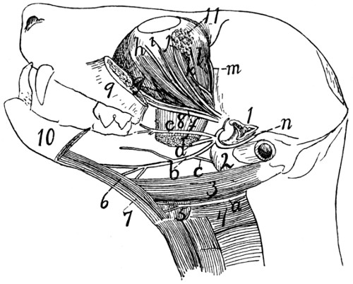
Fig. 154.—Dissection showing a Number of the Cranial Nerves.
The mandible and zygomatic arch have been cut and removed; the temporal and internal pterygoid muscles are also removed. a, N. hypoglossus; b, N. lingualis; c, N. chorda tympani; d, N. alveolaris inferior (cut); e, N. buccinatorius; f, Nn. infraorbitales; g, N. sphenopalatinus; h, branch of N. oculomotorius; i, N. zygomaticus; j, N. lachrymalis; k, N. abducens; l, N. trochlearis; m, N. frontalis; n, N. auriculotemporalis. 1, cut end of zygomatic process of temporal; 2, tympanic bulla; 3, M. digastricus; 4, M. constrictor pharyngis medius; 5, M. hyoglossus; 6, M. genioglossus; 7, M. geniohyoideus; 8, M. pterygoideus externus (cut); 9, cut cranial end of zygomatic arch; 10, mandible (cut); 11, lachrymal gland.
After giving off the above branches the mandibular nerve passes three or four millimeters laterad along the root of the zygomatic process of the temporal bone, and divides into two large branches, the inferior alveolar (or dental) nerve (d) and the lingual nerve (b).
f. N. alveolaris inferior (Fig. 154, d).—This passes toward the mandibular foramen, but before reaching it gives off a branch to the mylohyoid and digastric muscles. It then enters the foramen, passes through the mandibular canal, and gives numerous inferior dental branches to the teeth. At the mental foramen (or foramina) it passes out of the canal and divides into several mental nerves, to the chin, lower lip, and mucosa of the lower jaw.
g. N. lingualis (Fig. 154, b).—The lingual nerve passes between the internal and external pterygoid muscles, and two or three millimeters from its origin it receives the chorda tympani nerve (c), a communicating branch from the facial (see page 376). It passes onto the ventral surface of the external pterygoid, thence onto the side of the tongue, beneath the mylohyoid. On the side of the tongue it divides into branches which enter the tongue and are distributed to its mucous membrane. One branch passes to the mucosa of the pharynx. Just before entering the tongue a branch is given off to the sublingual and submaxillary glands.
VI. N. abducens.
—The sixth nerve, the abducens, arises from the medulla, as already described (Fig. 138, VI, and page 347). It passes into the orbit through the orbital fissure, then extends obliquely cranioventrad along the medial surface of the lateral rectus muscle (Fig. 154, k). At about the middle of the length of the muscle it divides into two or three branches which enter at once into the lateral rectus and supply it.
VII. N. facialis
(Fig. 155).—The seventh or facial nerve arises from the trapezium at the caudal margin of the pons, craniad of the origin of the eighth nerve, as described in the account of the brain (Fig. 138, VII). It passes into the internal auditory meatus, traverses the facial canal through the petrous bone, and emerges at the stylomastoid foramen.
Within the facial canal the nerve bears an enlargement, the ganglion geniculi. It gives off within the canal a branch to the stapedius muscle, and the superficial petrosal and chorda tympani nerves.
The superficial petrosal (N. petrosus superficialis major) nerve passes into the hiatus facialis, through the canal which forms its continuation, and joins the sphenopalatine ganglion (p. 373).
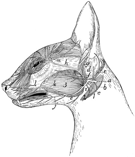
Fig. 155.—Superficial Nerves of the Face.
a, N. facialis; b, branch to digastric; c, N. auricularis posterior; d, branch to inside of ear; e, ventral ramus; f, dorsal ramus; g, temporal nerve; h, zygomatic nerve; i, nerve to stylohyoid muscle; j, inferior buccal nerve; k, superior buccal nerve; l, infraorbital branches of fifth nerve; m, lachrymal branches of fifth nerve; n, auriculotemporal branches of fifth nerve. 1, M. digastricus; 2, M. stylohyoideus; 3, M. masseter.
The chorda tympani is given off two or three millimeters before the emergence of the facial at the stylomastoid foramen. It passes into the tympanic cavity, extends across it between the malleus and incus, passing close against the tensor tympani muscle, and leaves it (by a small cleft, the canal of Huguier) between the bulla and the squamous portion of the temporal. It then passes craniad (Fig. 154, c), ventrad of the root of the zygomatic process of the temporal, and joins the lingual nerve (Fig. 154, b) (branch of the third division of the fifth nerve).
On emerging from the stylomastoid foramen the facial nerve (Fig. 155, a) gives off at once a small branch (b) to the digastric muscle (1), and a larger branch, N. auricularis posterior (c), which passes dorsocaudad about the base of the ear, supplying some of its muscles. Another small branch (d) pierces the cartilaginous external ear and is distributed to its inner surface. The facial nerve then curves craniad about the proximal part of the cartilaginous external ear, and divides five or six millimeters from its emergence into two main branches, which may be designated as the dorsal (f) and ventral (e) rami. A third small branch may also rise from the point of union of the two; it is usually, however, a twig from the dorsal ramus, and passes to M. submentalis.
The dorsal ramus (f) sends two or three small branches to the cheek (including the one just mentioned), and divides into the temporal (g) and the zygomatic (h) branches. The former (g) passes along the cranial margin of the external ear, supplying the superficial muscles, and extends into the temporal region, where it lies deeper than the terminal branches of the lachrymal nerve (m) (from the fifth cranial). The zygomatic branch (h) passes across the malar bone to the caudal angle of the eye, sends branches into both eyelids, anastomoses with twigs from the lachrymal branch (m) of the fifth nerve, and passes along the medial side of the eye to the lateral surface of the nose, where it ramifies.
The ventral ramus (e) gives off a minute branch (i) to the stylohyoid muscle (2), then proceeds toward the angle of the mouth and divides into superior (k) and inferior (j) buccal branches, the former to the muscles of the upper lip and the contiguous regions, the latter to those of the lower lip and chin.
The seventh nerve thus supplies most of the muscles of the head except those of mastication, and of these it supplies the digastric.
VIII. N. acusticus.
—The eighth or auditory nerve takes origin (Fig. 138, VIII) from the floor of the fourth ventricle, as before described (page 347). It passes into the internal auditory meatus and is distributed to the internal ear within the petrous bone.
IX. N. glossopharyngeus
(Fig. 156, a).—The ninth or glossopharyngeal nerve takes origin (Fig. 138, IX) from the side of the medulla, as described in the account of the brain. Close to its origin its trunk bears a small enlargement, the ganglion superius. It passes along with the tenth and eleventh nerves through the jugular foramen. At its exit it has a ganglionic enlargement, the ganglion petrosum; this is connected by fine fibres with the ganglion nodosum (Fig. 156, d) of the vagus. The glossopharyngeal (Fig. 156, a) then passes craniad over the surface of the tympanic bulla (15) and mediad of the digastric muscle. It continues mediad of the carotid artery, and as it approaches the cranial cornu of the hyoid divides into two portions, one of which passes to the muscles and mucosa of the pharynx, and the other to the tongue, where it is the special nerve of taste.
X. N. vagus.
—The vagus nerve arises from the side of the medulla in the manner described under the Brain (page 346 and Fig. 138, X). It passes through the jugular foramen along with the glossopharyngeal and accessory nerves.
Cervical Portion of the Vagus (Fig. 156, d, d′).—In the foramen, or just before entering it, it presents a ganglionic enlargement, the ganglion jugulare (or “ganglion of the root”), and a short distance beyond the foramen it forms a second ganglion, the ganglion nodosum (d) (or “ganglion of the trunk”), which lies dorsocaudad of the superior cervical sympathetic ganglion (e). The ganglia of the vagus and sympathetic are closely bound together by connective tissue, and that portion of the vagus craniad of the ganglion nodosum is interconnected by a network of nervous fibres with the sympathetic (e), hypoglossal (b), and accessory (c) nerves. From the ganglion nodosum the vagus (d′) passes caudad, closely bound up with the sympathetic; the two lie at the side of the common carotid artery. Just before entering the thorax the two separate, the vagus being the larger and lying ventrad of the sympathetic. (In rare cases the two are distinctly separated throughout their length.)
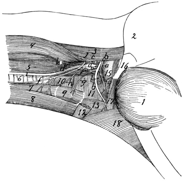
Fig. 156.—Glossopharyngeal, Hypoglossal, Vagus, Sympathetic, and First Cervical Nerves in the Neck.
a, N. glossopharyngeus; b, N. hypoglossus; c, N. accessorius; d, ganglion nodosum of vagus nerve; d′, N. vagus; e, ganglion cervicale superius of sympathetic; f, ramus descendens of N. hypoglossus (united with first cervical); g, branch to M. thyrohyoid from ramus descendens of N. hypoglossus; h, N. laryngeus superior of N. vagus; i, united vagus and sympathetic nerves; j, first cervical nerve; k, second cervical nerve; l, N. pharyngeus from vagus. 1, M. masseter; 2, outline of external ear; 3, M. splenius; 4, M. levator scapulæ ventralis; 5, M. longus capitis; 6, trachea; 7, M. sternothyreoideus; 8, M. sternohyoideus; 9, M. thyreohyoideus; 10, M. constrictor pharyngis inferior; 11, M. constrictor pharyngis medius; 12, cut end of M. stylohyoideus; 13, M. hyoglossus; 14, M. stylopharyngeus; 15, bulla tympani; 16, M. jugulohyoideus; 17, M. styloglossus; 18, M. mylohyoideus.
Branches of the vagus in the cervical region (Fig. 156).
a. N. auricularis.—This leaves the ganglion jugulare, passes into the petrous bone to the facial canal, leaves the skull with the facial nerve by the stylomastoid foramen, and is distributed to the external ear.
b. N. pharyngeus (l).—This leaves the vagus craniad of the ganglion nodosum, passes ventrad, sends a small communicating branch to N. laryngeus superior (h), and is distributed to the pharyngeal muscles and cranial part of the œsophagus.
c. N. laryngeus superior (h).—This arises from the ganglion nodosum, passes ventrad, crossing the medial surface of the carotid artery, and reaches the larynx at about the level of the caudal end of M. thyreohyoideus. It passes into the larynx and supplies its mucosa.
Thoracic Portion of the Vagus (Fig. 157).—As the vagus (b) and sympathetic (c) separate to enter the thorax, the vagus (b) lies more ventrad. The right vagus now lies along the lateral surface of the trachea, the left one (Fig. 157) along that of the œsophagus. At about the level of the first rib or further craniad there arise, apparently from the vagus (but really from the sympathetic), two slender nerves which pass caudad parallel and close to the vagus, lying slightly dorsad of it. These are the cardiac nerves (i); they arise from the sympathetic farther craniad in the neck region, accompany the vagus, and seem to branch from it. In cases where vagus and sympathetic are separate throughout their courses, these nerves arise clearly from the sympathetic. The cardiac nerves pass to the heart and aid in forming a network of nerves known as the cardiac plexus (k).
The vagus usually receives also, at about the level of the first rib, one or two communicating branches from the middle cervical ganglion (d) of the sympathetic. It then passes to the roots of the lungs, crossing the lateral surface of the aortic arch on the left side, and the medial surface of the azygos vein on the right side. As it crosses the aortic arch the left vagus gives off N. laryngeus inferior (j). This curves around the caudal side of the aortic arch to the lateral surface of the trachea (o), then extends on the lateral and ventral surface of the trachea craniad into the neck region. In the neck it passes to the dorsolateral side of the trachea and reaches the larynx. It passes into the larynx between the cricoid and thyroid cartilages, and supplies the muscles of the larynx. It anastomoses with the superior laryngeal.
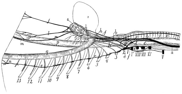
Fig. 157.—Sympathetic, Vagus, and Phrenic Nerves in the Neck and Thorax.
1-13, the thirteen ribs (cut); V-VIII, the fifth to eighth cervical nerves (cut); I′, first thoracic nerve (cut). a, combined trunk of vagus and sympathetic; b, vagus; c, sympathetic; d, middle cervical ganglion of sympathetic; e, inferior cervical ganglion; e′, communicating branch to sixth, seventh, and eighth cervical nerves; f, phrenic nerve; g, loop of sympathetic about subclavian artery, between the middle and inferior cervical ganglia; h, cardiac branches from the inferior cervical ganglion; i, cardiac branch passing from sympathetic along with vagus to heart; j, inferior laryngeal nerve; k, pulmonary and cardiac plexus; l, ventral œsophageal branch of vagus; l′, similar branch from the opposite side; m, dorsal œsophageal branch of vagus; n, great splanchnic nerve; o, trachea; p, œsophagus; q, aorta; r, heart; s, root of lung.
The right inferior laryngeal nerve is given off from the right vagus much further craniad than the left; it curves around the right subclavian artery just caudad of the origin of the internal mammary artery, then passes to the trachea, and has a course and distribution like that of the left side.
On reaching the root of the lungs the vagus divides into numerous branches which form the pulmonary plexus (k) over the roots of the lungs, and extends onto the basis of the heart as the cardiac plexus. From these plexuses numerous branches pass to the heart, lungs, pulmonary artery, pericardium, etc. The cardiac branches from the sympathetic, mentioned above, are also connected with the cardiac plexus.
Caudad of the pulmonary plexus the vagus is continued on each side as two trunks, a dorsal (m) and a ventral (l) which pass along the œsophagus (p). The ventral branches of right and left vagi (l and l′) unite a short distance caudad of the root of the lung, and the single trunk extends into the abdominal cavity on the ventral surface of the œsophagus. The dorsal branches of right and left vagi unite farther back, near the diaphragm, and the single trunk thus formed (m) enters the abdominal cavity on the dorsal surface of the œsophagus. Both divisions give fine nerves to the œsophagus.
Abdominal Portion of the Vagus (Fig. 164, page 407).—After passing through the diaphragm the ventral division (Fig. 164, l) reaches the lesser curvature of the stomach (1), over which it ramifies, some branches being traceable almost to the pylorus. The network of branches thus formed is the anterior gastric plexus. Some twigs from the ventral division pass transversely across the cranial end of the stomach and join the plexus formed by the dorsal division.
The dorsal division (m) of the vagus reaches the greater curvature of the stomach, where it ramifies, forming the posterior gastric plexus. Branches from this anastomose with the anterior gastric plexus and with the cœliac plexus of the sympathetic (e).
XI. N. accessorius.
—The accessory (or spinal accessory) nerve arises (Fig. 138, XI) by numerous rootlets from the lateral surface of the medulla and of the spinal cord as far caudad as the fifth to seventh cervical nerve. These spinal rootlets join to form a nerve which enters the cranium through the foramen magnum. After receiving the rootlets from the medulla, it leaves the cranial cavity along with the vagus and glossopharyngeal by the jugular foramen. Just outside the foramen (Fig. 156, c) it becomes involved in a plexus of fine branches which interconnect it with the vagus, sympathetic, and hypoglossal. It then turns laterad and caudad (Fig. 158, 1) and pierces the cleidomastoid muscle (d), to which it gives small branches. It then divides: one branch enters the sternomastoid muscle; the other passes caudad along the dorsal border of the levator scapulæ ventralis, sends branches to the clavotrapezius, and may be traced to the acromiotrapezius and spinotrapezius, which it supplies.
XII. N. hypoglossus
(Fig. 156, b).—The twelfth or hypoglossal nerve arises from the ventral side of the medulla (Fig. 138, XII), as previously described. It emerges from the skull through the hypoglossal canal. At first it passes ventrad, then gradually turns craniad, following thus a curved course and passing successively laterad of the vagus and sympathetic nerves (Fig. 156, d and e), the common carotid artery, and the cranial cornu of the hyoid bone. A short distance from its origin the nerve gives off a ramus descendens (Fig. 156, f), which receives a communicating branch from the first cervical nerve (j), and divides into two branches, one (g) to the thyrohyoid muscle (9), the other to the sternohyoid (8) and sternothyroid muscles (7). The main nerve passes beneath the mylohyoid muscle into the tongue, at first following the lingual artery, then laterad of it, then crossing it again. It sends branches to all the muscles of the tongue, and may be traced to its tip.
The hypoglossal nerve is involved in the plexus just outside of the jugular foramen, with which the vagus, sympathetic, and accessory nerves are also connected.
2. Spinal Nerves.
—The origin and general features of the spinal nerves have been described in connection with the account of the spinal cord (page 337). A description of their peripheral distribution will now be given.
A. Cervical Nerves.
—There are eight pairs of cervical nerves. The first leaves the vertebral canal through the atlantal foramen. The second passes out between the arches of the atlas and axis, not through a special intervertebral foramen, so that its ganglion lies among the muscles of the back of the neck. The others emerge through the intervertebral foramina, the eighth one from between the last cervical and first thoracic vertebræ.