THE SKELETON OF THE CAT. (continued 2)
Reading edition
The middle portion of the dorsal surface is formed by the frontals (5). Each frontal presents laterally a prominent zygomatic process (f), extending ventrolaterad toward a corresponding (frontal) process (h) of the malar bone. These two processes mark the boundary between the orbital fossa (craniad) and the temporal fossa (caudad). Craniad of the zygomatic process of the frontal a sharp margin separates the dorsal surface of the skull from the wall of the orbital fossa: this is the supraorbital arch or margin (i).
The cranial portion of the dorsal surface is formed by the maxillary (8), nasal (7), and premaxillary bones (9). Just craniad of the nasals, bounded ventrad and craniad by the premaxillaries, appears the large opening of the nares (j), leading into the nasal cavity.
The zygomatic arch (g) is formed by the zygomatic process of the temporal (m) and the malar or zygomatic bone (6). Each presents near its middle a prominent dorsocaudally directed process, the frontal process (h) of the malar bone. The zygomatic arch forms the lateral boundary of the temporal and orbital fossæ, which are separated by a line connecting the frontal process of the malar (h) and the zygomatic process of the frontal (f).
A portion of the floor of the orbit and the opening of the lachrymal canal (o) may also be seen in dorsal view; they are described in connection with the lateral surface.
The caudal surface of the skull is formed largely by the occipital bone (Fig. 17), surrounding the foramen magnum (Fig. 17, d). At the sides of the foramen magnum are the two prominent curved occipital condyles (e) for articulation with the atlas. Craniolaterad of the condyles, separated from them by a deep notch, are the jugular processes (f) of the occipital, closely applied to the caudal ends of the tympanic bullæ.
Dorsad of the foramen magnum are faint indications of a median ridge running dorsad, the external occipital crest (Fig. 17, i); this rises at its junction with the lambdoidal ridge to form the prominent external occipital tubercle (Fig. 39, b). The dorsal and dorsolateral boundaries of the posterior surface are formed by the lambdoidal ridge (Fig. 17, h; Fig. 39, a).
The lateral surface of the skull (Fig. 40) is much more complicated than the dorsal and posterior surfaces. Caudally the occipital condyles (a) and external occipital crest (b) are visible; dorsocaudad the sagittal crest (c).
Extending from the caudal end of the sagittal crest the lambdoidal ridge (d) is seen passing ventrocraniad to the tympanic bulla, thence craniad to the root of the zygomatic arch. In the ventral part of the caudal region the tympanic bulla (e) is visible with the jugular process (f) of the occipital pressed close against its caudal end. Just craniad of the jugular process the mastoid process (g) of the temporal rests against the side of the bulla. Beneath the cranial edge of this process is the opening of the stylomastoid foramen (h) for the seventh nerve, while just ventrad of the foramen is the small pit (i) in the tympanic bulla for the reception of the tympanohyal bone. Craniad of the stylomastoid foramen is the large opening of the external auditory meatus (j), leading into the middle ear.
Immediately dorsocraniad of the external auditory meatus the zygomatic arch begins as the zygomatic process (k) of the temporal bone. On the cranial surface of the base of this process is the deep mandibular fossa (l) for the condyle of the mandible. This fossa is bounded caudally by the prominent postmandibular process (m).
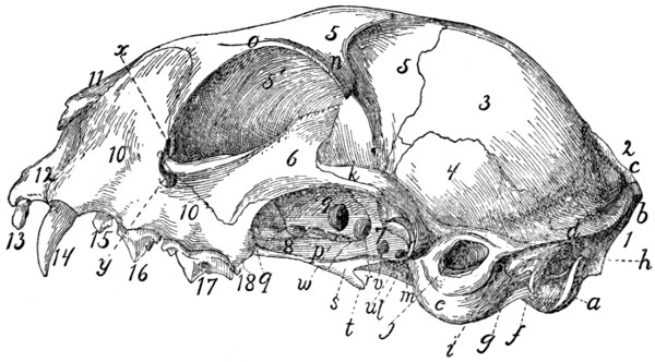
Fig. 40.—Skull, Side View.
1, occipital bone; 2, interparietal; 3, parietal; 4, temporal; 5, 5′, frontal; 6, malar; 7, sphenoid; 8, palatine; 9, presphenoid; 10, maxillary; 11, nasal; 12, premaxillary; 13, incisor teeth; 14, canine; 15, 16, 17, premolars; 18, molar. a, occipital condyle; b, external occipital crest; c, sagittal crest; d, lambdoidal ridge; e, tympanic bulla; f, jugular process; g, mastoid process; h, stylo-mastoid foramen; i, pit for tympanohyal bone; j, external auditory meatus; k, zygomatic process of temporal bone; l, mandibular fossa; m, postmandibular process; n, zygomatic process of the frontal; o, supraorbital margin; p, external pterygoid fossa; q, sphenopalatine foramen; r, orbital fissure; s, internal pterygoid fossa; t, hamulus; u, foramen ovale; v, foramen rotundum; w, optic foramen; x, opening of lachrymal canal; y, infraorbital foramen.
All that portion of the lateral surface of the skull which lies craniodorsad of the lambdoidal ridge may be divided (excluding the zygomatic arch) into three main parts, the temporal fossa, the orbital fossa, and the face. The boundaries of the temporal fossa have been given. The orbital fossa is bounded externally by a prominent semicircular ridge formed chiefly by the zygomatic arch, the zygomatic process of the frontal (n), and the supraorbital arch (o) of the frontal, which may be traced to the cranial root of the zygomatic arch. The orbital fossa may be considered to end caudally and ventrally at the level of the optic foramen (w); ventrad of it are certain smaller fossæ. Immediately ventrad is the long external pterygoid fossa (p), from which arises part of the external pterygoid muscle. This fossa begins at the sphenopalatine foramen (q) and extends caudad to the orbital fissure (r); it is separated by a ridge from the orbital fossa. Caudoventrad of the external pterygoid fossa and separated from it by a sharp ridge is the small narrow internal pterygoid fossa (s), which extends ventrad without interruption on to the surface of the hamulus (t) and caudad to within two or three millimeters of the tympanic bulla. From it the internal pterygoid muscle takes origin. The hamulus (t) projects caudoventrad in this region, forming a prominent feature in a lateral view.
Four foramina leading into the cranial cavity are visible in a lateral view of the skull, craniad of the tympanic bulla. The one nearest the bulla is the foramen ovale (u) for the third division of the fifth nerve; next craniad of this is the foramen rotundum (v) for the second division of the fifth nerve. These two foramina pierce the alisphenoid: just craniad of them, between the alisphenoid and the orbitosphenoid, is the large orbital fissure (r) (foramen lacerum anterius), which transmits the third, fourth, and sixth cranial nerves and the first division of the fifth. Dorsocraniad of the orbital fissure is the optic foramen (w), for the optic nerve.
Ventrad of the cranial portion of the orbit is the large sphenopalatine foramen (q), for the nerves and arteries of the same name. Just craniad of this is the small caudal opening of the posterior palatine canal, which passes through the substance of the palatine bone and opens on its ventral surface near its cranial margin. Just dorsad of the cranial root of the zygomatic arch is the opening of the lachrymal canal (x), while the root of the arch is pierced by the large infraorbital foramen (y), which transmits the infraorbital nerves and artery from the orbit.
The teeth (13-18), implanted along the alveolar border of the maxillary and premaxillary, form a prominent feature in a lateral view: they are described in the account of the alimentary canal.
The ventral surface of the skull (Fig. 41) is very complex. It is separated by the orbits into a caudal and a cranial portion, united by a narrow median trough-like part. Laterad of this trough-like part are visible parts of the orbit and the zygomatic arches, which do not properly belong to the ventral surface and have already been described.
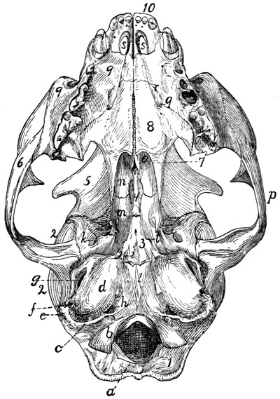
Fig. 41.—Skull, Ventral View.
1, occipital bone; 2, temporal; 3, sphenoid; 4, presphenoid; 5, frontal; 6, malar; 7, vomer; 8, palatine; 9, maxillary; 10, premaxillary. a, foramen magnum; b, occipital condyles; c, jugular process; d, tympanic bulla; e, mastoid process; f, stylomastoid foramen; g, external auditory meatus; h, jugular foramen; i, styliform process; j, groove for Eustachian tube; k, foramen ovale; l, foramen rotundum; m, pterygoid process of sphenoid; n, perpendicular plate of palatine; o, choanæ or posterior nares; p, zygomatic arch; q, cranial end of posterior palatine canal; r, palatine grooves; s, foramina incisiva or anterior palatine foramina; t, opening of pterygoid canal.
Caudally there appear in the ventral view the foramen magnum (a), occipital condyles (b), and jugular processes (c). In front of the jugular processes the two tympanic bullæ (d) form prominent features, with the mastoid process (e), the stylomastoid foramen (f), and the external auditory meatus (g) on their lateral surfaces. All these structures have been described. The tympanic bullæ (d) are placed with long axes directed craniomediad, so that they converge toward their cranial ends. At the caudomedial angle of each bulla is the large jugular foramen (h), for the ninth, tenth, and eleventh nerves. Opening into the mediocaudal margin of the jugular foramen is the smaller hypoglossal foramen, for the twelfth nerve.
The craniomedial end of the tympanic bulla projects craniad as the styliform process (i). Just laterad of this process is the opening (j) into the tympanic bulla by which the tuba auditiva or Eustachian tube passes into the middle ear. A faint groove for the tube passes craniomediad from this opening, on the surface of the sphenoid. Craniolaterad of the opening for the tuba auditiva is the foramen ovale (k); craniad of this the foramen rotundum (l) is faintly indicated. On the surface of the sphenoid just craniad of the styliform process of the bulla tympani is the minute opening of the pterygoid canal (t). The orbital fissure and optic foramen are not seen in the ventral view.
The middle region of the ventral surface is narrow: it is formed by a trough-like fossa which is bounded laterally by the pterygoid processes (m) of the sphenoid and the perpendicular plates of the palatines (n). Ventrad of this lies, in the natural condition, the soft palate, converting the fossa into the nasal portion of the pharynx or nasopharynx. Craniad this fossa is bounded by the free caudal edges of the palatines; beneath which the fossa communicates with the nasal cavity by the two choanæ (o). Laterad of this median fossa are visible in the ventral view parts of the temporal and orbital fossæ, bounded laterally by the zygomatic arches (p).
The cranial part of the ventral surface is a somewhat triangular plane area formed by the palatal portions of the palatines (8), maxillaries (9), and premaxillaries (10), which together constitute the hard palate (palatum durum). Laterad and craniad this area is bounded by the alveolar borders of the maxillaries and premaxillaries bearing the teeth. The hard palate is marked near the cranial border of the palatine bones with two or more foramina which form the cranial termination of the posterior palatine canal (q). Two faint grooves pass from these foramina a short distance craniad, gradually converging: these are known as the palatine grooves (r) (sulci palatini). Near the cranial end of the hard palate are two large openings close together near the middle line: these are the foramina incisiva (or anterior palatine foramina) (s).
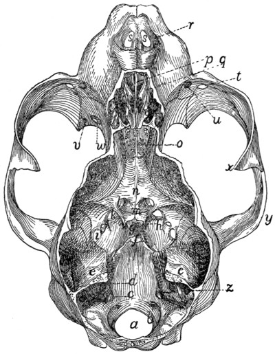
Fig. 42.—Skull, with Dorsal Surface Removed, showing the Cranial and Nasal Cavities.
a, foramen magnum; b, caudal end of hypoglossal canal; c, jugular foramen; d, internal auditory meatus; e, tentorium, forming the cranial boundary of the cerebellar fossa; f, dorsum sellæ; g, sella turcica; h, anterior clinoid processes; i, foramen ovale; j, foramen rotundum; k, orbital fissure; l, optic foramen; m, chiasmatic groove; n, presphenoid bone; o, cribriform plate; p, lamina perpendicularis of ethmoid; q, labyrinths of ethmoid; r, nares; s, foramina incisiva or anterior palatine foramina; t, infraorbital foramen; u, opening of the lachrymal canal; v, caudal opening of posterior palatine canal; w, sphenopalatine foramen; x, frontal process of the malar; y, zygomatic process of the temporal; z, appendicular fossa, in the petrous bone.
Cavities of the Skull
(Figs. 42 and 43).—The bones of the cranial portion of the skull enclose the cranial cavity for the brain; the facial bones enclose the nasal cavity, for the olfactory organ.
The cranial cavity is divisible into three principal fossæ: the cerebellar fossa (Fig. 43, I) caudad, for the cerebellum; the cerebral fossa (II) in the middle, for the cerebrum; the small olfactory fossa (III) at the cranial end for the olfactory bulb of the brain.
The cerebellar fossa (I) is bounded caudally by the occipital bone enclosing the foramen magnum (Fig. 42, a). Its ventral surface is formed by the basilar portion of the occipital and the petrous portions of the temporals; its lateral surface by the mastoid portions of the temporals and parts of the parietals and occipital. Its roof is formed by the parietals and interparietal. Craniad the cerebellar fossa is partly separated from the cerebral fossa by the tentorium (Fig. 42, e; Fig. 43, f) formed by the two parietals: this encloses a quadrangular opening by which the two fossæ communicate. The caudal, dorsal, and lateral walls of the cerebellar fossa are deeply marked by fossæ for the lobes of the cerebellum; the small appendicular fossa (Fig. 43, e), forming a deep indentation in the petrous bone near its dorsocaudal end, is particularly noticeable.
The following openings are found in the walls of the cerebellar fossa. Caudad is the large foramen magnum (Fig. 42, a) by which the brain-cavity communicates with the vertebral canal. Near the caudal margin of the foramen magnum, on its lateral side, just mediad of the dorsal end of the occipital condyle, is the caudal opening of the condyloid canal (Fig. 43, a) which passes craniad through the substance of the occipital bone to open just caudad of the petrous: it transmits a vein. The condyloid canal varies greatly in size in different specimens. A few millimeters craniad of the edge of the foramen magnum on the floor of the fossa is the small opening of the hypoglossal canal (Figs. 42 and 43, b), for the twelfth nerve. Just craniad of this, at the caudomedial border of the petrous, is the large jugular foramen (c). On the petrous itself, near the middle, is the internal, auditory meatus (d) divided into the dorsal facial canal for the seventh nerve, and a ventral passage for the eighth nerve. At the cranial end of the cerebellar fossa is the large opening bounded by the free edges of the tentorium.
The cerebral fossa forms much the largest part of the cranial cavity. It is bounded by the parietals (Fig. 43, 3′), squamous portions of the temporals (4), frontals (8), the sphenoid (5), and presphenoid (6). A slight rounded ridge on its lateral wall at about the position of the suture between the frontals and parietals separates a smaller cranial portion sometimes called the anterior fossa, from a larger caudal portion sometimes known as the middle fossa of the cranial cavity. The walls of the cerebral cavity are marked with numerous ridges and shallow furrows for the cerebral convolutions.
The floor of the cerebral cavity is bounded caudad by the prominent dorsum sellæ (Fig. 42, f; Fig. 43, g), just craniad of which is the rounded depression known as the sella turcica (Fig. 42, g; Fig. 43, h), for lodgment of the hypophysis. A number of foramina pierce the floor of the cavity in this region. Just ventrad of the cranial tip of the petrous portion of the temporal is the small foramen lacerum (medius). Craniad and laterad of this is a row of four foramina: the caudal one is the foramen ovale (Fig. 42, i); then come in order the foramen rotundum (j), the orbital fissure (k), and the optic foramen (Fig. 42, l; Fig. 43, k). The two optic foramina are connected by the shallow transverse chiasmatic groove (Fig. 42, m), for the optic chiasma. Another small foramen continues caudad from a groove on the floor of the orbital fissure; this opens on the ventral surface of the sphenoid, between the wing and the body of the bone. The groove and foramen constitute the pterygoid canal, which transmits a nerve,—the nerve of the pterygoid canal, or Vidian nerve.
The cranial cavity narrows at its cranial end to form the small olfactory fossa (Fig. 43, III) which lodges the olfactory bulbs. This is bounded by the frontals and the lamina cribrosa (Fig. 42, o) of the ethmoid; caudad it opens directly into the cerebral fossa. Numerous openings through the lamina cribrosa for the olfactory fibres connect the olfactory fossa with the nasal cavity. The roof of the fossa is marked by a prominent median crest from the united edges of the frontals.
The nasal cavity is almost completely filled by the ethmoid and vomer and the conchæ nasales. Its roof is formed by the nasal bones and portions of the frontals; its sides by the frontals, lachrymals, maxillaries, premaxillaries, and palatine bones; its floor by the horizontal plates of the palatines, maxillaries, and premaxillaries.
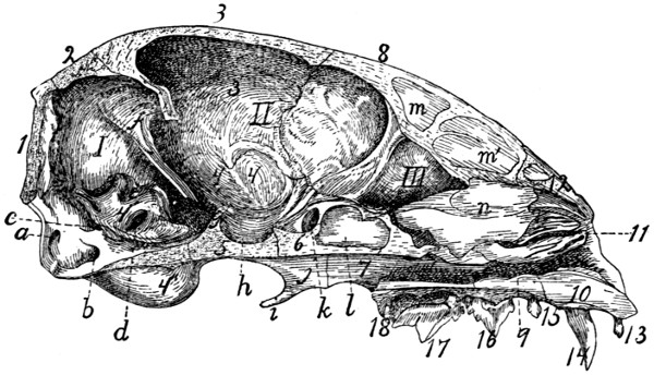
Fig. 43.—Skull, Median Longitudinal Section, showing the Cavities.
I, cerebellar fossa; II, cerebral fossa; III, olfactory fossa. 1, occipital bone; 2, interparietal; 3, 3′, parietal; 4, temporal (4, squamous portion; 4′, petrous portion; 4″, tympanic portion); 5, sphenoid; 6, presphenoid; 7, palatine; 8, frontal; 9, maxillary; 10, premaxillary; 11, ethmoid; 12, nasal; 13, incisor teeth; 14, canine; 15, 16, 17, premolars; 18, molar. a, condyloid canal; b, hypoglossal canal; c, jugular foramen; d, internal auditory meatus; e, appendicular fossa; f, tentorium; g, dorsum sellæ; h, sella turcica; i, hamular process; j, pterygoid process of sphenoid; k, optic foramen; l, presphenoid sinus; m, m′, frontal sinus; n, lamina perpendicularis of the ethmoid (broken at cranial edge).
The nasal cavity opens craniad by the large nares (Fig. 39, j; Fig. 42, r), which are bounded by the premaxillary and nasal bones. In the natural condition this opening is divided by a median cartilage which is continuous with the lamina perpendicularis (Fig. 43, n) of the ethmoid, thus forming a partition which divides the nasal cavity into two separate halves. From the floor of the cranial part of the cavity rises a ridge formed of the nasal crests of the maxillaries and premaxillaries, and the cranial portion of the vomer. Farther caudad the vomer spreads out in a horizontal plane and separates from the floor of the cavity, so that the nasal cavity is thereby divided by a horizontal partition into dorsal and ventral portions. The ventral portion is small, forming the inferior meatus of the nose; it ends caudally at the choanæ (posterior nares, Fig. 41, o) which lead into the nasopharynx. That portion of the nasal cavity lying dorsad of the vomer is almost completely filled by the ethmoid and the conchæ nasales, superior and inferior. It is bounded caudally by the lamina cribrosa of the ethmoid (Fig. 42, o). The nasal cavity communicates with the cranial cavity by the foramina for the olfactory fibres in the lamina cribrosa; with the nasopharynx by the choanæ; with the exterior of the body by the nares; with the mouth-cavity by the foramina incisiva or anterior palatine foramina (Fig. 42, s); with the orbit by the sphenopalatine foramen and the nasolachrymal canal. It communicates directly also with the frontal sinuses (Fig. 43, m, m′), the sphenoidal sinuses (Fig. 43, l), and with the cells of the labyrinths of the ethmoid.
JOINTS AND LIGAMENTS OF THE SKULL.
Sutures of the Skull.—The bones of the skull join each other by means of immovable articulations known as sutures. These sutures are designated by combining the names of the bones between which they are situated: as, sphenofrontal suture (sutura sphenofrontalis), between the sphenoid and frontal; nasomaxillary suture (sutura nasomaxillaris), between the nasal and maxillary bones. When a suture joins the two corresponding bones of opposite sides the prefix inter is used, as the intermaxillary suture (sutura intermaxillaris) between the maxillaries. The sutures bounding the parietals have, however, received special names not derived in this manner. The suture caudad of the parietals, separating them from the occipital and interparietal, is known as the lambdoidal suture; that between the two parietals is the sagittal suture; that separating the parietals and squamous portions of the temporals is the squamous suture; that between the parietals and frontals is the coronal suture. The suture separating the two frontals also is known as the frontal suture, in place of interfrontal.
Articulations of the Mandible.—In man the two halves of the mandible are united craniad, so as to form a single bone. In the cat the two halves are separate, but articulate closely at the symphysis menti by a thin interarticular cartilage.
The articulation of the mandible at the mandibular fossa of the temporal is covered with a close articular capsule. The mandibular fossa is lined with cartilage. A slender ligament passes from the angular process of the mandible caudad to the external auditory meatus, being attached to the latter about 8 millimeters from its medial end. This is the stylomandibular ligament.
V. BONES OF THE THORACIC EXTREMITIES.
Scapula
(Figs. 44 and 45).—The scapula may be described as a flat triangular bone with one angle rounded. It lies beneath the muscles on the lateral face of the thorax near its cranial end. From its lateral surface there projects a flat ridge (Fig. 44, g), the spine of the scapula. The ventral end of the ridge is free as a curved process, the acromion process (Fig. 44, j).
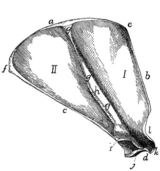
Fig. 44.—Scapula, Lateral Surface.
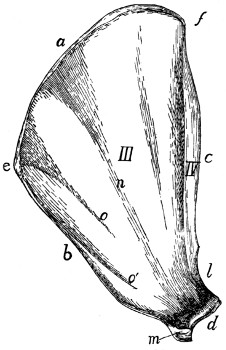
Fig. 45.—Scapula, Medial Surface.
I, fossa supraspinata; II, fossa infraspinata; III, fossa subscapularis; IV, fossa for teres major. a, vertebral border; b, coracoid border; c, glenoid border; d, glenoid angle and fossa; e, coracovertebral angle; f, glenovertebral angle; g, spine; h, tuberosity of the spine; i, metacromion; j, acromion; k, supraglenoidal tubercle; l, incisura scapulæ; m, coracoid process; n, groove indicating portion of spine; o, o′, ridges for attachment of muscle-fibres.
The ventral angle of the scapula (d), the glenoid angle (lateral angle of human anatomy), is much heavier than the others and bears a concave, pear-shaped articular facet, the glenoid fossa, for articulation with the humerus. The border with which this angle is more nearly continuous may be called the glenoid border (c) (axillary border of human anatomy).
Near the narrower cranial end of the glenoid fossa is a small curved projection of the bone, the coracoid process (Fig. 45, m). The border upon which it lies is the coracoid border (b) (superior border of human anatomy). The third border is turned toward the vertebral column and is the vertebral border (a).
The angle between the glenoid and vertebral borders is the glenovertebral angle (f) (inferior angle of human anatomy), and that between the coracoid and vertebral borders the coracovertebral angle (e) (medial angle of human anatomy).
The medial or costal surface (Fig. 45) is smooth and nearly flat. A shallow furrow (n) marks the position of the spine of the scapula. Between the furrow and the coracoid border are two oblique parallel ridges (o and o′) for the insertion of muscle-fibres. Near the glenoid border is a well-marked ridge separating the subscapular fossa (III), comprising the greater part of the medial surface of the scapula, from the fossa in which the teres major muscle has origin (IV). The surface presents several nutrient foramina usually directed toward the glenoid angle.
The lateral surface (dorsal surface of human anatomy) (Fig. 44) is divided by the spine (g) into two portions. The portion of the scapula craniad of the spine and the cranial surface of the spine bound the supraspinous fossa (fossa supraspinata) (I), while the surface caudad of the spine and the caudal portion of the spine bound the infraspinous fossa (fossa infraspinata) (II).
The spine (g) begins as a triangular elevated area in the middle of the vertebral margin and runs toward the glenoid angle. It rises gradually for about two-fifths of its length and then the margin becomes broader and the spine remains of the same height to its glenoid end. There is a rough thickening, the tuberosity (h) of the spine, situated on its free border about midway between the tip of the acromion and the vertebral end of the spine. The spine is inclined toward the glenoid margin so as to form an angle of about 60 degrees with the caudal half of the lateral surface.
At the base of the acromion process (j) the margin of the spine presents a flat triangular projection, the metacromion (i), directed toward the glenoid border.
The acromion (j) continues in the direction of the spine. It is thicker than the spine, smooth and rounded on both its surfaces and both its borders, and its apex is connected by fibrous tissue to the clavicle.
The coracoid border (b) presents a slight rounded notch, the incisura scapulæ or suprascapular notch (l), just dorsad of the glenoid angle, and at its ventral end bears the coracoid process (m) which is directed ventromediad.
The glenoid angle (d) is the only one requiring special mention. Between the root of the coracoid process and the glenoid cavity it presents a tubercle, the supraglenoidal or bicipital tubercle (k), for the tendon of origin of the biceps muscle. The glenoid angle is separated by a contracted neck from the rest of the bone. Between this angle and the inner margin of the acromion there is left a deep notch, the great scapular notch.

Fig. 46.—Clavicle.
a, medial end; b, lateral end.
Clavicle. Clavicula
(Fig. 46).—The clavicle in the cat is greatly reduced. It is a slender curved rod of bone imbedded in the muscles of the shoulder and connected by fibrous tissue to the apex of the acromion process. The lateral end (b) is slightly enlarged.
Humerus
(Figs. 47 and 48).—The humerus forms the support of the upper arm and articulates by its proximal end with the scapula at the glenoid cavity, and by its distal end with the radius and ulna, the bones of the lower arm. It is a nearly cylindrical bone with enlarged ends, and is so curved that its dorsal and ventral borders are hooked at the opposite ends so that it has the form of an Italic f.
The proximal end of the bone bears on its dorsomedial portion a thickening, the head of the humerus (a), which bears a smooth ovoid articular facet by which the bone articulates with the glenoid cavity of the scapula. The head is not separated from the body by a distinct anatomical neck as in the human humerus.
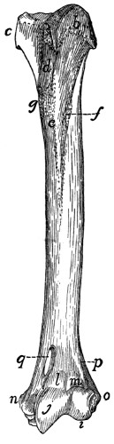
Fig. 47.—Humerus, Ventral Side.
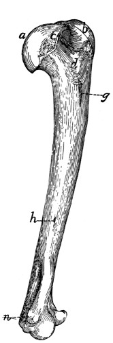
Fig. 48.—Humerus, Medial Side.
a, head; b, greater tuberosity; c, lesser tuberosity; d, bicipital groove; e, pectoral ridge; f, deltoid ridge; g, rough area for insertion of latissimus dorsi and teres major; h, nutrient foramen; i, capitulum; j, trochlea; l, coronoid fossa; m, radial fossa; n, medial epicondyle; o, lateral epicondyle; q, supracondyloid foramen.
Along the lateral border of the proximal end of the shaft is a high rough ridge semicircular in side view, the great tuberosity (b). It gives attachment to muscles and is marked on its dorsal border by a deep depression for the tendon of the infraspinatus muscle. On the medial margin of the proximal end closely associated with the head is a smaller elevation, the lesser tuberosity (c), also for muscular attachment.
Between the greater and lesser tuberosities on the ventral surface is seen a broad groove, the sulcus intertubercularis or bicipital groove (d), which passes distad onto the surface of the shaft. In the natural state it is converted into a canal by overlying tendons and lodges the tendon of the biceps muscle.
The shaft is nearly cylindrical at its middle, but its dorsoventral diameter is slightly greater than its mediolateral diameter. Its proximal end is flattened mediolaterad, while its distal end is flattened dorsoventrad.
From the ventral margin of the greater tuberosity a ridge, the pectoral ridge (e), is continued onto the surface of the shaft, and from the dorsal margin another ridge, the deltoid ridge (f), passes distad and ventrad so as to meet the pectoral ridge near the middle of the ventral surface of the bone. On the medial margin of the bone near the junction of the first and second fourths is a roughened area (g) for the attachment of the tendons of the latissimus dorsi and teres major muscles, and on the same surface near the junction of the second and last thirds is a nutrient foramen (h).
The distal end of the bone presents a smooth saddle-shaped articular surface, which, in well-marked bones, is divided, when seen from the ventral surface, by a slight nearly median ridge into two unequal portions, lateral and medial (i and j). The lateral half is rounded and is called the capitulum (i). It is broader ventrad than dorsad, and is not continued onto the dorsal surface of the bone. It is for articulation with the proximal end of the radius.
The medial half of the surface, the trochlea (j), is concave and passes directly into the capitular surface laterad, but is limited mediad by a sharp ridge. It is continued onto the dorsal surface of the bone, where it is limited also laterad by a ridge. It articulates with the semilunar notch of the ulna.
Proximad of the trochlea the dorsal surface presents a deep fossa, the olecranon fossa, which receives the olecranon of the ulna when the arm is straightened. On the ventral surface (Fig. 47) are two shallower fossæ separated by a longitudinal ridge. The one over the trochlea receives the coronoid process of the ulna when the arm is bent, and is called thence the coronoid fossa (l). The one over the capitulum, the radial fossa (m), receives a triangular facet on the proximal end of the radius at the same time. Between the radial and coronoid fossæ on one side and the olecranon fossa on the other is only a thin plate of bone. On the medial surface of the distal end is a considerable roughened elevation, the medial epicondyle (n) (epitrochlea). It gives origin to flexor muscles and to the ulnar collateral ligaments of the elbow-joint. Opposite the medial epicondyle over the capitulum is the lateral epicondyle (o) for the origin of extensor muscles of the forearm and of the radial collateral ligaments of the elbow-joint. From the lateral epicondyle a ridge, the lateral supracondyloid ridge (p), continues proximad, curving onto the dorsal surface of the bone and ending about opposite the junction of the deltoid and pectoral ridges.
Proximad of the medial epicondyle the bone is pierced near its medial margin by an oblique oval foramen, the supracondyloid foramen (q).
Radius
(I, Figs. 49 and 50).—In the usual position the radius lies with its proximal end on the lateral side of the arm, articulating with the capitulum of the humerus. The proximal end is thus laterad of the proximal end of the ulna. Its distal end, however, lies on the medial side of the distal end of the ulna, so that the radius in the natural position crosses ventrad of the ulna.
The radius is a curved bone slightly flattened dorsoventrally, with enlarged ends. It may be described as consisting of a shaft and of a proximal and a distal end. Its proximal end presents on the ventral surface a tuberosity, the bicipital tuberosity (c), for the insertion of the tendon of the biceps muscle. Proximad of this the bone is contracted to form a neck (b) which is surmounted by a head (a). The head has on its proximal surface a depressed oval facet by which it articulates with the capitulum, and on its ulnar border a long narrow facet, the articular circumference (d), for articulation with the radial notch of the ulna; also a triangular facet (e), which fits into the radial fossa of the humerus.
The shaft is convex dorsad and concave ventrad. The distal end is somewhat pyramidal. From its medial or radial side a wedge-shaped process, the styloid process (f), extends distad. The distal surface of the end together with the lateral surface of the styloid process form a concave articular cavity (g) which fits against the scapholunar bone.
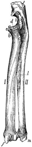
Fig. 49.—Radius and Ulna, Dorsolateral View.
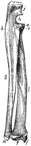
Fig. 50.—Radius and Ulna, Ventromedial View.
I, radius; II, ulna. a, head of radius; b, neck; c, bicipital tuberosity; d, articular circumference; e, facet for radial fossa of humerus; f, styloid process of radius; g, facet for articulation of scapholunar bone; h, semilunar (or great sigmoid) notch of ulna; i, coronoid process; j, olecranon; k, area for insertion of brachialis and clavobrachial muscles; l, rough area for attachment of interosseous membrane; m, styloid process of ulna.
The dorsal surface of the distal end is marked by longitudinal grooves for tendons, and its lateral or ulnar surface bears a concave facet for articulation with the ulna.
Ulna
(II, Figs. 49 and 50).—The ulna is a long slender bone, flattened mediolaterad. It is enlarged at its proximal end and becomes gradually smaller toward the distal end.
The proximal end is marked ventrally by a deep excavation, the semilunar notch, or great sigmoid cavity (h). By the saddle-shaped articular surface of the semilunar notch it articulates with the trochlea. This articular surface is divided into two parts by a transverse non-articular area. The distal boundary of the semilunar notch is a blunt process, the coronoid process (i), which bears on its lateral surface a concave facet, the radial notch, for the head of the radius.
The portion of the bone proximad of the semilunar notch is called the olecranon (j). It fits into the olecranon fossa of the humerus when the arm is straightened, and is rough at its end for the insertion of tendons.
The body of the ulna becomes triangular distad. The distal end is slightly larger than the shaft just proximad of it, and bears on its radioventral side a hemispherical head for articulation with the radius. Distad of the head the bone continues as the flattened styloid process (m), which projects distad from its dorsolateral side and is smooth on the medial side of its apex, for articulation with the cuneiform bone of the wrist.
Carpus
(Fig. 51).—The carpus (wrist) consists of seven bones arranged in two rows, three in the proximal row and four in the distal row. Beginning on the medial side of the hand (thumb or radial side), the first bone in the proximal row is the scapholunar (a) (equal to the scaphoid or navicular and lunar of the human hand). It articulates with the radius. The next is the cuneiform (b), articulating with the styloid process of the ulna, and the next, which is attached to the cuneiform and projects freely ventrad, is the pisiform (c).
In the distal row the bone on the radial side is the trapezium (d); the next is the trapezoid (e), the next the os magnum (f), and the last the unciform (g). The distal row articulates with the metacarpals or bones of the palm of the hand (1-5).
In the kitten the scapholunar is represented by three bones, the scaphoid or navicular, on the radial side, the lunare, between the scaphoid and the cuneiform, and a centrale, which lies distad of the other two.
Scapholunar Bone. Os scapholunaris (Fig. 51, a).—The scapholunar is a quadrangular bone with the ventroradial angle produced into a blunt process. Its proximal surface is smooth and articulates with the distal end of the radius. The distal end is marked by oblique ridges and articulates with the unciform, os magnum, trapezoid, and trapezium. The ulnar surface articulates with the cuneiform, and the dorsal surface of the ventroradial process with the radial sesamoid.
Cuneiform Bone. (Os triquetrum BNA) (Fig. 51, b).—The cuneiform bone has the form of a flattened pyramid. Its base articulates with the unciform, its proximoulnar surface with the pisiform except at its dorsal margin, where it articulates with the styloid process of the ulna. On its proximoradial surface is a smooth facet for articulation with the scapholunar.
Pisiform Bone. Os pisiforme (Fig. 51, c).—The pisiform bone is about twice as long as broad, with enlarged ends. Its dorsal end articulates with the cuneiform, and on its proximal surface, separated from the above by a smooth ridge, is a smooth facet for articulation with the styloid process of the ulna.
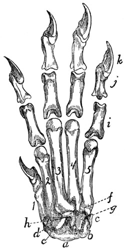
Fig. 51.—Carpus, Metacarpus, and Phalanges, Dorsal Surface.
a, scapholunar bone; b, cuneiform; c, pisiform; d, trapezium; e, trapezoid; f, os magnum; g, unciform; h, radial sesamoid; i, proximal phalanges; j, second phalanges; k, distal phalanges; 1, 2, 3, 4, 5, metacarpals in order from the radial side.
Unciform Bone. (Os hamatum BNA) (Fig. 51, g).—The unciform is a wedge-shaped bone with the apex of the wedge directed proximad, and smooth for articulation with the scapholunar. By a part of its ulnar surface it articulates with the cuneiform, and by its radial surface with the os magnum. Its distal end articulates with the fourth and fifth metacarpals.
Os magnum. (Os capitatum BNA) (Fig. 51, f).—The os magnum may be described as an oblong plate bearing on its proximal surface a semicircular ridge which crosses it diagonally. The proximal end of the bone articulates with the scapholunar. Its distal end articulates with the third metacarpal except near its ventroulnar angle, where it articulates with the fourth metacarpal. Its ulnar surface articulates with the unciform. Its radial border articulates with the trapezoid, the third metacarpal, and, by two facets, with the second metacarpal.
Trapezoid. (Os multangulum minus BNA.) (Fig. 51, e).—The trapezoid is somewhat wedge-shaped, with the apex of the wedge pointing ventrad. Its proximal side articulates with the scapholunar, its distal side with the second metacarpal, its ulnar side with the os magnum, and its radial side with the trapezium.
Trapezium. (Os multangulum majus BNA.) (Fig. 51, d).—The trapezium has the form of a triangular prism curved into a semicircle. The convex face looks proximad and articulates by its ventral half with the scapholunar. The ulnar surface articulates with the second metacarpal dorsally, and ventrally with the trapezoid. Its radial surface articulates with the first metacarpal.
Bones of the Hand or Manus
(Fig. 51, 1-5).—The Metacarpals. Metacarpus.—The metacarpals are the five bones of the palm of the hand; they are numbered from one to five, beginning with the thumb. They are cylindrical elongated bones with enlarged ends. The distal end is called the head, and the proximal end the base. Each head bears a hemispherical articular facet which is marked over its ventral half by a prominent smooth ridge. The surface dorsad of the ridge articulates with the proximal head of a phalanx. The ridge and the surface at its sides are for a pair of sesamoid bones.
The first metacarpal (1) is the shortest. Its head is oblique, and it articulates by the ulnar half of its proximal surface with the trapezium (d); by the radial half with the radial sesamoid (h).
The second metacarpal (2) is marked on the proximal part of its dorsal surface by an oblique groove passing from the radial side distad to the ulnar side. The base articulates with the trapezoid (e). The ulnar surface of the proximal end articulates with the os magnum (f) and third metacarpal, while the radial surface articulates with the trapezium (d).
The third metacarpal (3) is the longest, and its base is rhomboidal with a projecting dorsoradial angle separated by a groove from the rhomboid surface. The proximal end articulates with the os magnum (f) and second metacarpal; the radial surface of the proximal end with the second, and the ulnar surface with the fourth, metacarpal.
The fourth metacarpal (4) has a base similar in form to that of the third, and when placed in position with the fifth the two form a hemispherical facet which articulates with the unciform (g) and os magnum (f). The fourth metacarpal articulates by its radial side with the third, and by its ulnar side with the fifth.
The fifth metacarpal (5) articulates by its proximal end with the unciform (g), and by the radial side of its proximal end with the fourth metacarpal.
Digits (Fig. 51).—The first digit of the hand is called the pollex (thumb), the second the index, the third the medius, the fourth the annularis, the fifth the minimus.
The first digit has two phalanges, each of the others three phalanges.
Phalanges (Fig. 51, i, j, k).—The phalanges of the proximal row (i) are elongated, flattened dorsoventrally and curved so as to be longitudinally convex dorsad. All have thickened ends. The proximal end is notched, and its proximal surface looks dorsad and is concave for the head of the metacarpal. The distal end is pulley-shaped, and the pulley surface extends farther on to the ventral than on to the dorsal surface, and serves for articulation with the middle phalanx.
The phalanges of the middle row (j) are like those of the proximal row, but shorter. The proximal surface is triangular and marked by a median facetted ridge. The whole surface is smooth and adapted to the distal end of the phalanx of the first row. The distal end is transversely elongated, so as to be cylindrical, and projects more toward the ulnar than toward the radial side. The distal phalanx (k) articulates with this cylinder so that when it is fully extended it lies on the ulnar side of the middle phalanx.
The distal phalanx (k) has the form of a quadrangular prism. It is excavated on its proximal surface for articulation with the middle phalanx. Its distal surface presents dorsad a deep excavation from the bottom of which arises a compressed plate of bone having the form of a bird’s beak. The depression receives the base of a claw, and the beak-like projection supports the claw.
Sesamoid Bones of the Hand. Ossa sesamoidea.—The hand contains, in addition to those already described, eleven small bones that are developed in tendons.
One of these, the radial sesamoid (Fig. 51, h), is closely applied to the radial end of the scapholunar bone. It is developed in the tendon of the extensor brevis pollicis muscle.
The other ten occur in pairs as small flattened curved bones on the ventral side of the joint between each metacarpal and the phalanx with which it articulates.
JOINTS AND LIGAMENTS OF THE THORACIC LIMBS.
The shoulder-joint is an arthrodial or ball-and-socket joint. The bones entering into its formation are the scapula and the humerus.
The capsular ligament or articular capsule is very ample and allows for extended movement of the humerus. It is attached to the edge of the glenoid fossa of the scapula and passing distad covers the head of the humerus and is inserted at the line of junction of the shaft and the epiphysis which forms the head of the bone. On the lateral side of the ventral surface of the humerus the attachment continues distad about two centimeters along the lateral edge of the bicipital groove. On the medial side the insertion passes over the proximal end of the lesser tuberosity. A strong transverse band passes from the greater tuberosity to the lesser tuberosity and bridges the bicipital groove, converting it into a canal. The lateral and medial parts of the capsule are strengthened by thicker bands of fibres, the more prominent medial one of which passes from the coracoid process of the scapula to the lesser tuberosity. To the capsule are closely united parts of the supraspinatus, infraspinatus, coracobrachialis, and subscapularis muscles. A synovial membrane lines the capsule within and forms a sheath around the biceps tendon, so that the latter does not actually enter the synovial capsule.
The elbow-joint (Figs. 52 and 53) is a ginglymus or hinge-joint. The bones which enter into it are the humerus, radius, and ulna.
The capsule of the joint forms a sac, with the following attachments to the bones: (1) To the humerus it is attached at the proximal edge of the coronoid and radial fossæ; to the sides of the capitulum and trochlea distad of the two epicondyles, and to the distal edge of the olecranon fossa. (2) To the ulna it is attached at the edges of the radial and semilunar notches; (3) to the radius around the articular facet, two or three centimeters distad of the border. Many of the muscles of this region are closely attached to the capsule.
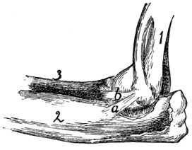
Fig. 52.—Ligaments of Elbow-joint, Medial Side.
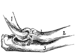
Fig. 53.—Elbow-joint, Lateral View.
Fig. 52.—1, humerus; 2, ulna; 3, radius. a and b, the two medial collateral ligaments.
Fig. 53.—1, humerus; 2, radius; 3, ulna. a, dorsal collateral ligament; b, ventral collateral ligament; c, annular ligament.
Closely connected with the capsule of the joint are the collateral ligaments. The two medial collateral ligaments (Fig. 52) arise from the medial epicondyle. One (b) passes distad and laterad to the interval between the radius and ulna; here it divides, one branch going to the head of the radius, while the other is attached to the lateral surface of the ulna at the edge of the semilunar notch. The second medial ligament (a) lies dorsad of the first; it passes to the medial surface of the ulna, at the distal edge of the semilunar notch.
The two collateral ligaments on the lateral side (Fig. 53) arise from the lateral epicondyle. The ventral one (b) passes almost directly distad and is inserted into the lateral surface of the proximal end of the radius about one centimeter from the articular surface; its inner surface is partly united to the annular ligament (c) of the radius. The dorsal one (a) is attached to the lateral border of the semilunar notch of the ulna.
Articulations of Radius and Ulna.—The proximal radio-ulnar articulation (Fig. 53) is by a pivot-joint or trochoid. The two bones are held in place by the annular ligament (Fig. 53, c). This is attached on the lateral side to the dorsal border of the radial notch of the ulna, passes around the head of the radius, receiving some ligamentous fibres which come from the lateral epicondyle, and is attached to the coronoid process of the ulna. The annular ligament is closely united with the capsule of the joint.
The radius and ulna are united for about their middle third by the thin interosseous membrane, which fills the interosseous space between their adjacent edges.
The Wrist.—At the wrist or carpus there are in reality three joints, the first between the radius and ulna proximad and the first row of carpals distad, the second between the two rows of carpal bones, the third between the distal row of carpals and the metacarpals. The first two are movable joints; the third is not. Each of these three joints has a capsule, and the bones entering into the joints are interconnected by numerous ligaments. These ligaments are named by combining the names of the two bones which they interconnect. Ligaments which interconnect bones of the same row in the carpus are sometimes distinguished as interosseous ligaments, as contrasted with intercarpal ligaments, which connect together bones of different rows. According to their position the ligaments may also be distinguished as dorsal, ventral, and lateral. Detailed descriptions and figures of all these ligaments are given by Strauss-Durckheim.
Metacarpals.—The joint between the carpals and metacarpals has been described. At the distal end of the metacarpals the articulations with the phalanges have each a capsule. The joint is further strengthened by a double lateral ligament on each side. The two sesamoid bones at each joint are interconnected by a strong transverse ligament, and each is connected with the head of the metacarpal and the base of the first phalanx by a lateral ligament.
Phalanges.—Between the phalanges the joints possess capsules, and each has a radial and an ulnar lateral ligament.
VI. BONES OF THE PELVIC EXTREMITIES.
Innominate Bones. Ossa innominata. (Os coxæ BNA)
(Figs. 54 and 55).—The two innominate bones articulate with the sacrum and extend thence caudoventrad and finally turn mediad and unite in the middle line, forming the symphysis pubis. They thus form an arch, the pelvic arch, pelvic girdle or pelvis, which is closed dorsad by the sacrum.
In the middle of the lateral surface of each bone is a hemispherical depression, the acetabulum (Fig. 55, d), which receives the head of the femur.
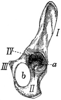
Fig. 54.—Innominate Bone of Kitten, Ventrolateral View.
I, ilium; II, ischium; III, pubis; IV, acetabular bone. a, acetabulum; b, obturator foramen.
In the kitten each innominate bone is composed of three principal parts united by sutures (Fig. 54). From the sacrum to the acetabulum is a single bar, the ilium (I). Caudad of the acetabulum are two bars. The dorsal one of these is the ischium (II), and the ventral one is the pubis (III). The ischium enters into the formation of the acetabulum (a), but the pubis does not. The two bones, however, are in contact at the ventral edge of the acetabulum. From this point they diverge, but unite with one another again near the middle line and thus enclose an oval foramen, the obturator foramen (b). Wedged between the ilium, ischium, and pubis at their point of junction and helping to form the acetabulum is a small irregular bone, the acetabular bone (IV). In the adult cat these four parts are united into a single bone which is nevertheless usually described, ignoring the acetabular piece, as made up of ilium (Fig. 55, I), ischium (II), and pubis (III).
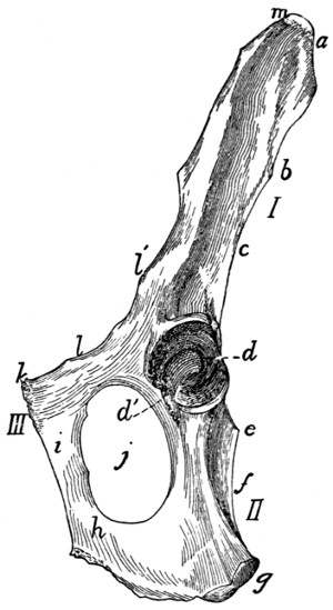
Fig. 55.—Innominate Bone of Adult Cat, Ventrolateral View.
I, ilium; II, ischium; III, pubis. a, crest of the ilium; b, posterior inferior spine; c, great sciatic notch; d, acetabulum; d′, incisura acetabuli; e, spine of the ischium; f, lesser sciatic notch; g, tuberosity of the ischium; h, ramus of the ischium; i, ramus of the pubis; j, obturator foramen; k, pubic tubercle; l, iliopectineal line; l′, ilio-pectineal eminence; m, anterior superior process.
The ilium (I) is somewhat contracted at the middle and broader at its ends. One end enters into the acetabulum (Fig. 55, d) and forms about one-fifth the articular surface. This end is also the thickest part of the bone. The lateral surface of the ilium is concave for the attachment of muscles. The medial surface is smooth over its acetabular half and rough over its sacral half. The rough portion is marked at its junction with the smooth portion by the ear-shaped auricular impression by which the bone articulates directly with the sacrum. The caudal half of that part of the medial surface craniad of the auricular impression gives attachment to the ilio-sacral ligaments which bind the ilium to the sacrum. The dorsal border is straight at its cranial end and concave and rounded at its caudal end. Between the two portions and at the dorsal edge of the auricular surface is a protuberance corresponding to the posterior inferior spine (Fig. 55, b) of the human ilium. The concavity of the dorsal border (c) corresponds to the great sciatic notch of the human ilium. At its caudal end is the short spine of the ischium (e), which is not a part of the ilium. The ventral border of the ilium is broad caudad, becoming narrower craniad. The lateral margin of the ventral border is continued to a tuberosity at the edge of the acetabulum; its medial margin is called the iliopectineal line (l) and extends on the pubis to the symphysis. An eminence, the iliopectineal eminence (l′), on the iliopectineal line, lies opposite to the acetabulum at about the junction of the ilium and pubis. The cranial end of the bone is thickened, forming the crest (a) of the ilium. At the junction of the crest with the ventral border is a projection, the anterior superior process (m) of human anatomy.
The pubis (III) (including the acetabular bone) enters into the formation of the acetabulum (d) constituting about one-sixth the circumference, but less than one-sixth its area. It may be described as a flat, curved bone, contracted at the middle and expanded at the ends. The dorsal end enters into the acetabulum; the ventral end unites with the opposite bone at the symphysis pubis and sends caudad a projection, the ramus (i) of the pubis, which unites with the ramus of the opposite bone to form about two-thirds of the entire symphysis. At the sides of the symphysis a slightly marked angle projects craniad from each of the pubic bones; these two together constitute the pubic tubercle (k), for the origin of the rectus abdominis muscle. The surfaces of the ramus are smooth. One of its borders is concave and enters into the formation of the obturator foramen (j). Another of its borders is the iliopectineal line (l). Its third border is rough for the symphysis.
The ischium (II) has the form of a triangular prism contracted at the middle. Its cranial end forms nearly two-thirds of the acetabulum. Its caudal end bears dorsad a rough thickening, the tuberosity of the ischium (g). From the caudal half of the ventral border of the bone a sickle-shaped process, the ramus (h) of the ischium, curves medioventrad and then craniad and joins the ramus of the pubis. Its medial border is rough and enters into the symphysis, forming the caudal one-third. The lateral angle of the bone is rounded. Its dorsal angle is marked near the cranial end by the spine (e) of the ischium. The concavity between this spine and the tuberosity corresponds to the lesser sciatic notch (f) of human anatomy.
The acetabulum (d) is cup-shaped. The ventral one-sixth of its border is deficient and a broad groove extends from the deficiency to the bottom of the cup. The deficiency, incisura acetabuli, or acetabular notch (d′), is closed naturally by a ligament, and the groove gives origin to the ligament (ligamentum teres) which attaches the head of the femur.
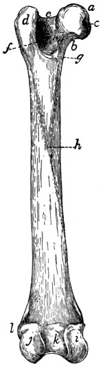
Fig. 56.—Left Femur, Ventral Side.
a, head; b, neck; c, depression for round ligament; d, great trochanter; e, trochanteric fossa; f, intertrochanteric line; g, lesser trochanter; h, linea aspera; i, medial condyle; j, lateral condyle; k, intercondyloid fossa; l, lateral epicondyle.
Femur
(Fig. 56).—The femur is the proximal bone of the posterior extremity. It consists of enlarged proximal and distal ends connected by a nearly cylindrical shaft. The proximal end presents on its medial side a hemispherical head (a) which fits into the acetabulum. It is supported by a neck (b) which is contracted near the head and expanded dorsoventrally where it joins the remainder of the bone. The medial surface of the head presents near its ventral border a depression (c) for the insertion of the round ligament of the femur. Ventrad the articular surface of the head extends as an acute projection onto the shaft, so that the whole articular surface appears somewhat pear-shaped. On the lateral side of the proximal end opposite the head is a projecting mass, the great trochanter (d), forming the end of the shaft. On the medial side of the great trochanter at its junction with the neck is a deep fossa, the trochanteric fossa or digital fossa (e) for the insertion of muscles. From the ventral surface of the great trochanter a ridge, the intertrochanteric line (f), is continued distad, ending in a pyramidal projection, the lesser trochanter (g), which serves for the insertion of muscles. A second ridge is continued to the lesser trochanter from the neck. A slight but well-marked ridge, the spiral ridge or line, runs round two sides of the neck parallel to the second ridge.
The shaft is nearly straight and cylindrical. A rough line is continued along its ventral surface from the lesser trochanter, and a similar line along its lateral surface from the greater trochanter; these unite ventrad to form the linea aspera (h). On its ventral surface is a nutrient foramen, directed proximad.
The shaft gradually widens distad and ends in two condyles (i and j) which are continuous dorsad but separated ventrad by a deep notch, the intercondyloid fossa (k). The distal surface of the shaft and condyles is articular. This articular surface is larger on the lateral condyle (j). The part of the articular surface on the end of the shaft (patellar surface) is for the patella; that part of it on the condyles and separated by the notch is for the tibia.
On the lateral surface of the lateral condyle is a slight prominence, the lateral epicondyle (l), and on the medial surface of the medial condyle is another prominence, the medial epicondyle; both are for the attachment of ligaments.
Patella
(Fig. 1, r).—The patella is a small flat bone with a pear-shaped outline, having its apex distad. It lies against the articular surface at the lower end of the shaft of the femur. It thus covers the knee-joint. The inner surface is smooth and convex from side to side, but concave in a proximodistal line. It fits against the lower end of the femur. Its outer surface is rough and concave. It is a sesamoid bone inserted in the tendon of the quadriceps femoris muscle.
Three other sesamoid bones are found in the region of the knee (see Fig. 61, p. 89). Two are in the tendons of the gastrocnemius muscle, proximad of the two condyles of the femur. The third is in the tendon of the popliteus muscle, just laterad of the lateral condyle of the femur.
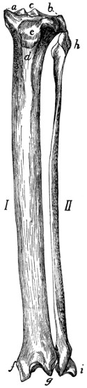
Fig. 57.—Tibia and Fibula of Left Leg, Dorsal View.
I, tibia; II, fibula. a, medial condyle of the tibia; b, lateral condyle; c, spine of the tibia; d, crest of the tibia; e, tubercle for attachment of the patellar ligament; f, medial malleolus; g, projection of dorsal surface of the tibia; h, head of fibula; i, lateral malleolus.
Tibia
(I, Fig. 57).—The tibia is the longer of the two bones of the leg between the knee and the ankle, and is the longest bone of the body. It has a triangular shaft and enlarged proximal and distal ends.
The proximal end is curved ventrad and projects into two prominences, the tuberosities, on either side. Each tuberosity bears on its proximal end an articular facet for the condyles of the femur; these are known respectively as the lateral and medial condyles (a and b) of the tibia. The condyles are oval, convex dorsoventrad, and concave from side to side. The two condyles are separated at the middle of their contiguous margins by a bicuspid projection, the spine of the tibia (c). They are continuous dorsad, but separated ventrad by a deep notch between the tuberosities, the popliteal notch. On the distal side of the lateral condyle is an elongated facet for the proximal end of the fibula.
The shaft is triangular, smallest at about its middle and enlarged at both ends. It presents dorsal, medial, and lateral borders, and medial, lateral, and ventral surfaces. The lateral surface is concave proximad. The medial surface is convex. The two are continuous at the distal end. At their proximal ends the border separating them is raised into a prominent ridge, the crest (d) of the tibia, the proximal end of which contributes to increase the proximal surface of the bone, and presents an oblong tubercle (e) for the insertion of the ligamentum patellæ (ligament of the quadriceps femoris muscle). The ventral surface is concave proximad where it abuts upon the tuberosities. Its proximal half is crossed by two rough parallel lines, the distal one of which crosses in a spiral course from the lateral to the medial border; near its beginning is a nutrient foramen.
The distal end extends farther distad on its medial side. The extension is the medial malleolus (f). The malleolus presents two grooves on its medial surface for the tendons of muscles. On the lateral side of the distal end is an oblique triangular facet for the distal end of the fibula. The ventral surface of the distal end presents an oblique border which passes from the apex of the malleolus proximolaterad. The dorsal surface extends into a V-shaped projection (g) between the malleolus and the fibular facet. The distal end presents an oblique ridge running from the apex of the V-shaped extension of the dorsal surface to near the base of the medial malleolus. The ridge and the concavities on either side of it, the medial one of which is deeper, fit against the proximal trochlear surface of the astragalus.
Fibula
(II, Fig. 57).—The fibula lies at the lateral side of the tibia in the shank. It is a slender triangular bone with enlarged proximal and distal ends.
The proximal end or head (h) is flattened. It bears a facet on its proximomedial surface for articulation with the tibia, and is longitudinally grooved on the outer surface.
The shaft has a very sharp medial border. This border is turned toward the tibia and gives attachment to the interosseous membrane, which runs between the tibia and fibula.
The distal end is expanded to form the lateral malleolus (i). This bears a facet on the proximal portion of its medial surface near its dorsal margin, for the tibia, and distad of this is a second facet for the astragalus. The ventral and lateral surfaces are grooved for tendons.
Tarsus
(Fig. 58).—The tarsus consists of seven bones. The longest of these, lying on the lateral side of the foot and forming the support of the heel, is the calcaneus or os calcis (a). It articulates distad with a bone, the cuboid (c), which bears the fourth and fifth metatarsals. Lying between the calcaneus and the tibia is the astragalus or talus (b), the distal end of which articulates with the boat-shaped navicular or scaphoid (d). The scaphoid bears on its distal surface the three cuneiform bones, lateral (e), medial (g), and intermediate, bearing the rudiment of the first metatarsal and the second and third metatarsals.
Astragalus. (Talus BNA) (Fig. 58, b).—The astragalus may be divided into body, neck, and head. The body is marked on its proximal surface by a deep pulley-like groove for the articulation with the distal end of the tibia, and on its lateral and medial surfaces by curved facets for articulation with the malleoli of the tibia and fibula. This entire surface for articulation with the bones of the leg is known as the trochlea. The lower surface is marked by two facets separated by a groove; these are for articulation with corresponding facets on the calcaneus. Distally the bone contracts to form the neck and enlarges at the end, forming the head, which is smooth on its distal surface for articulation with the navicular or scaphoid.
Calcaneus (Fig. 58, a, and Fig. 59).—The calcaneus (os calcis) is the largest bone of the foot and forms the heel. It is two or three times as long as broad and has six surfaces: dorsal, ventral, medial, lateral, proximal, and distal. The proximal one-half of the dorsal surface (Fig. 59) is smooth, while the distal half is broadened and bears two facets which are separated by a groove. These articulate with the corresponding facets on the astragalus. The medial facet is borne on a projection of the bone, the sustentaculum tali (c). Distad of the facets the surface is rough. The ventral surface is smooth. The proximal end (b) is grooved for the tendon of Achilles. The lateral surface is smooth and marked by a grooved tubercle, the peroneal tubercle (d), near the distal end. The medial surface is marked by part of the articular facet for the astragalus, and also by the grooved sustentaculum tali. The distal end (a) articulates with the cuboid.
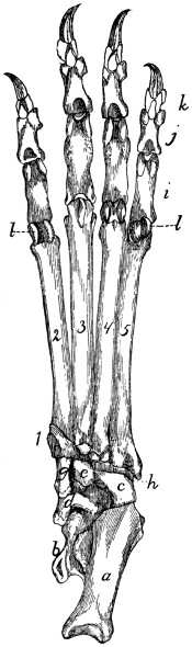
Fig. 58.—Tarsus, Metatarsus, and Phalanges of Left Foot, Ventral View.
a, calcaneus; b, astragalus; c, cuboid; d, scaphoid; e, lateral cuneiform; g, medial cuneiform; h, peroneal groove, for the tendon of the peroneus longus muscle; i, proximal phalanges; j, second row of phalanges; k, distal phalanges; l, sesamoid bones. 1, rudimentary first (medial) metatarsal; 2, 3, 4, 5, the other metatarsals.
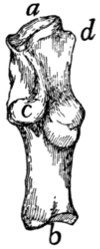
Fig. 59.—Calcaneus of Right Foot, Dorsal View.
a, distal facet for cuboid; b, proximal end with groove for tendon of Achilles; c, sustentaculum tali; d, peroneal tubercle.
Cuboid. Os cuboideum (Fig. 58, c).—The cuboid has somewhat the form of a cube and articulates by its proximal end with the calcaneus (a), and by its distal end with the fourth and fifth metatarsals. Its medial surface articulates with the scaphoid (d) and lateral cuneiform (e). The ventral surface is marked near its distal end by an oblique ridge, distad of which is a deep groove, the peroneal groove (h), for the tendon of the peroneus longus muscle.
Scaphoid. (Os naviculare pedis BNA) (Fig. 58, d).—The scaphoid is a boat-shaped bone. Its proximal surface is marked by a concave facet for the head of the astragalus (b), and its distal surface has three facets for the lateral (e), intermediate and medial (g) cuneiform bones. At the junction of the ventral with the medial surface is a prominent tubercle. The lateral surface bears two linear facets for articulation with the calcaneus (a) and cuboid (c).
Lateral Cuneiform. Ectocuneiform. Os cuneiforme tertium BNA (Fig. 58, e).—The lateral cuneiform is a wedge-shaped bone with a hooked process extending from the ventral sharp angle of the bone. It articulates by its proximal end with the scaphoid (d), and by its distal end with the third metatarsal. The medial surface bears near its distal end two facets for the second metatarsal, and on its proximal end a facet for the intermediate cuneiform. The caudal surface has a facet on its proximal end for the cuboid (c).
Intermediate Cuneiform. Mesocuneiform. Os cuneiforme secundum BNA.—The intermediate cuneiform is small and wedge-shaped, with the base of the wedge dorsad. It lies between the lateral cuneiform and the medial cuneiform, articulates by its proximal end with the middle facet of the scaphoid, and bears on its distal end the second metatarsal. It is not visible in ventral view.
Medial Cuneiform. Entocuneiform. Os cuneiforme primum BNA (Fig. 58, g).—The medial cuneiform lies on the medial side of the foot. It is a flat triangular bone about twice as long as broad, and broader at its proximal end than at the distal end. It bears on its distal end the rudimentary first metatarsal. The proximal end is oblique and bears a concave facet for the lateral distal facet of the scaphoid (d). The lateral surface has a concave facet at its proximal end for the intermediate cuneiform, while the distal portion is applied against the medial surface of the second metatarsal.
Bones of the Foot or Pes
(Fig. 58).—Metatarsals. Metatarsus (Fig. 58, 1-5).—The metatarsals are five in number. They bear a close resemblance to the metacarpals, but they may be distinguished by their bases.
The first metacarpal (1) is rudimentary and conical. Its base has a facet for the distal end of the medial cuneiform (g), while the outer surface fits into a depression on the inner surface of the base of the second metatarsal.
The second (2). The proximal surface of the base is triangular, corresponding to the distal end of the intermediate cuneiform. The medial surface is marked by two concavities, one along the proximal border for the distal end of the medial cuneiform, and one distad of this for the first metatarsal. The lateral surface bears on the proximal margin an oblique triangular facet dorsad and a similar facet ventrad, both for the lateral cuneiform (e). Distad of these facets is a rough ridge.
The third (3). The proximal end of its base is a triangular facet with the apex directed ventrad and the sides excavated. It is for the distal end of the lateral cuneiform (e). Its medial surface presents a depression which receives the ridge of the second metatarsal. On the lateral surface a short distance distad of the proximal border is a triangular concave facet, and near the proximal border ventrad a second concave facet. Both are for the fourth metatarsal.
The fourth (4). The proximal end is convex, notched medially and facetted for the cuboid (c). Its medial surface bears dorsad, a short distance from its proximal end, a smooth tubercle, and ventrad a small convex facet. Both articulate with facets on the lateral surface of the third metatarsal. The lateral surface has a sinuous facet along its dorsal border, and ventrad of this a depression. There is a second facet along the ventral border. Both facets are for the fifth metatarsal, and the depression is for ligaments.
The fifth (5) has its base flattened and expanded so as to be wedge-shaped, with the apex of the wedge directed proximad. Its dorsal end extends into a tubercle. It thus presents only lateral and medial surfaces. The medial surface shows two tubercles, one distad of the other. The distal tubercle and the distal half of the proximal tubercle are facetted and fit into the sinuous facet on the fourth metatarsal. A narrow facet on the ventral border of the surface articulates with the facet on the ventral border of the lateral surface of the fourth metatarsal. The proximal half of the distal tubercle is facetted for the cuboid (c). The lateral surface is smooth, non-articular, and obliquely grooved.
Phalanges (Fig. 58, i, j, k).—There are three phalanges in each of the four digits, and these are almost identical with those described for the manus.
Sesamoid Bones. Ossa sesamoidea (Fig. 58, l).—The sesamoid bones are found at the joints between the metatarsals and phalanges, and are in all respects like those of the manus.
JOINTS AND LIGAMENTS OF THE PELVIC LIMBS.
Ligaments of the Pelvis.—The ilium and sacrum are articulated at the auricular facet of the ilium and the corresponding rough surface of the sacrum. The joint is an amphiarthrosis, permitting very little movement. A capsular ligament surrounds the articular surface, being attached to the bones about its circumference; it is short and strong. Craniad of the capsule is a thick very short ligament, composed of very strong transverse fibres passing from the rough surface of the sacrum to the corresponding rough surface of the ilium. This forms the lateral iliosacral ligament, which is united at its caudal border to the capsule.
A strong, wide ligamentous band passes from the dorsal border of the ilium to the sides of the sacrum. This is indistinctly subdivided into several bands, which together represent the long and short posterior iliosacral ligaments of man.
Symphysis pelvis.—The medial borders of the pubis and ilium meet in the middle line ventrad of the pelvis and are here united by cartilage. The joint is strengthened by numerous small bands which pass across the line of junction from one side to the other; these occur on both surfaces.
The Hip-joint.—The hip-joint is an enarthrosis, or ball-and-socket joint in which more than half the spherical head of the femur is received into the acetabulum. The depth of the acetabulum is increased by a rim of fibrocartilage about its margin, forming the labrum glenoidale. This passes across the acetabular notch, forming the transverse ligament of the acetabulum; beneath it blood-vessels and nerves pass into the acetabular cavity.
The capsule of the joint is large and loose. It is attached about the margin of the acetabulum, and passes over the head of the femur, to be attached to the bone several millimeters distad of the head. It thus encloses both the head and the neck of the femur.
The ligamentum teres, or round ligament, is a very strong, short ligament which passes from the depression in the head of the femur to the bottom of the acetabulum.
The Knee-joint (Figs. 60 and 61).—The joint between the femur and the tibia is very complex. The surfaces of the condyles of the femur do not correspond to those of the condyles of the tibia. Between the ends of the two bones are placed two disks of cartilage, the menisci, or semilunar cartilages (Fig. 60, c and d; Fig. 61, a and b), of such a form that the congruity of articular surfaces is restored. Each meniscus has a proximal surface corresponding to the form of one of the condyles of the femur, and a distal surface corresponding to a condyle of the tibia. The menisci are held in position by ligaments. The knee-joint permits not only backward and forward movement, but also a small amount of rotary motion.
The joint has two capsules, one on the dorsal (convex) side, the other on the ventral side. The two communicate only by a small passageway lying within the joint between the ends of the bones. The dorsal one is attached to the femur several millimeters proximad of the patellar surface and some distance on each side of the latter. The patella is imbedded in its outer wall, and it is attached to the tibia on the edges of the articular surface of the latter, from the crest to the tuberosities. The capsule is also attached laterally and medially to the sides of the menisci, and is closely united to the patellar ligaments. Its cavity contains a mass of yellow fat.
The ventral capsule is attached to the borders of the articular surfaces of the femur and tibia on their ventral sides, to the menisci, and to the epicondyles of the femur and the tuberosities of the tibia. Its walls are stronger and its cavity smaller than those of the dorsal capsule.
The ligaments of the knee-joint (Figs. 60 and 61), aside from the capsules, may be classified into: (1) those which are connected with the patella; (2) collateral ligaments (Fig. 60, i and j), which pass from the epicondyles directly distad along the sides of the joint to the tibia or fibula; (3) crucial ligaments (Fig. 60, g and h; Fig. 61, c and d), which cross within the joint from one side of the femur to the opposite side of the tibia; (4) ligaments which hold the menisci in place (Fig. 60, e and f; Fig. 61, e and f).
(1) Ligaments of the Patella.—The patella is imbedded in the dorsal wall of the dorsal capsule of the joint. From its distal end a strong tendon or ligament passes distad to the crest of the tibia. This is known as the ligamentum patellæ: it may be considered a part of the tendon of M. quadriceps femoris. On the lateral side the capsule of the joint is strengthened by the transverse fibres of the tendon of M. plantaris, which aid in holding the patella in place.
(2) Collateral Ligaments.—Of these there are two. The ligamentum collaterale fibulare (Fig. 60, j) is attached to the lateral epicondyle of the femur and passes distad across the tendon of the plantaris muscle to the head of the fibula. Dorsad of the fibular ligament and parallel with it passes the tendon of origin of the extensor longus digitorum. The ligamentum collaterale tibiale (Fig. 60, i; Fig. 61, g) begins on the medial epicondyle of the femur and passes distad to the lateral tuberosity of the tibia; part of it passes one to one and a half centimeters distad of the tuberosity to be attached to a rough ridge on the side of the tibia.
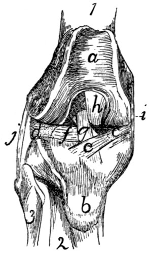
Fig. 60.—Knee-joint, from the Dorsal or Patellar Side.
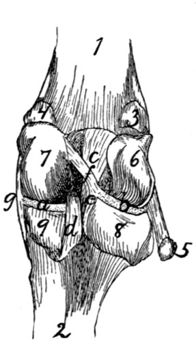
Fig. 61.—Knee-joint, from the Ventral or Flexor Side.
Fig. 60.—The patella has been removed. 1, femur; 2, tibia; 3, fibula. a, patellar surface of femur; b, tubercle for attachment of ligamentum patellæ; c, medial meniscus; d, lateral meniscus; e, f, ligaments of the menisci; g, ligamentum cruciatum anterius; h, ligamentum cruciatum posterius; i, ligamentum collaterale tibiale; j, ligamentum collaterale fibulare.
Fig. 61.—The capsule of the joint has been opened. 1, femur; 2, tibia (fibula not shown); 3, sesamoid bone in lateral head of M. gastrocnemius; 4, sesamoid in medial head of M. gastrocnemius; 5, tendon of M. popliteus, with sesamoid bone; 6, 7, lateral and medial condyles of the femur, with the intercondyloid notch between them; 8, 9, lateral and medial condyles of the tibia, with the popliteal notch between them; a, b, medial and lateral menisci; c, ligamentum cruciatum anterius; d, ligamentum cruciatum posterius; e, f, ligaments of the lateral meniscus; g, ligamentum collaterale tibiale.
(3) Crucial Ligaments.—There are two of these also. The ligamentum cruciatum anterius, or anterior crucial ligament (Fig. 60, g; Fig. 61, c), is a thick, strong ligament which begins on the dorsal part of the proximal end of the tibia nearer the medial side (Fig. 60, g), and passes ventrad and proximad, between the ends of the two bones forming the joint, into the intercondyloid fossa of the femur, and becomes attached to the medial surface of the lateral condyle of the femur (Fig. 61, c). It is composed of two partially separated bands, forming a slight angle with one another. It is crossed near its dorsal and ventral ends by two of the ligaments of the menisci. The ligamentum cruciatum posterius, or posterior crucial ligament (Fig. 60, h; Fig. 61, d), begins on the tibia at the edge of the popliteal notch (Fig. 61, d), nearer the medial side, and passes dorsad and proximad to be attached to the ventral edge of the patellar surface of the femur, in the intercondyloid fossa (Fig. 60, h).
(4) Ligaments of the Menisci.—There are five of these, connecting the menisci with the femur or tibia. One (Fig. 60, e) passes from the dorsal edge of the medial meniscus (c) transversely across the anterior crucial ligament (g) to the proximal end of the tibia nearer the lateral side. A second (Fig. 60, f) passes from the dorsal margin of the lateral meniscus (d) transversely beneath the anterior crucial ligament (g) to the proximal end of the tibia nearer the ventral side and medial border. A third (Fig. 61, f) passes from the ventral margin of the lateral meniscus obliquely across the anterior crucial ligament (c) to the lateral side of the medial condyle of the femur. The fourth (Fig. 61, e) is small, passing from the medial angle of the ventral border of the lateral meniscus distad to the popliteal notch. The fifth passes from the ventral border of the medial meniscus laterad beneath the posterior crucial ligament to the proximal end of the tibia, nearer the ventral and lateral sides.
Articulations between the Tibia and Fibula.—At the proximal end the fibula is as it were suspended from the distal side of the overhanging lateral tuberosity of the tibia by strong ligamentous tissue. The capsule of the joint is formed by an extension of the capsule of the knee-joint which passes between the tibia and fibula on the ventral side. Forming the dorsolateral wall of this extension is a strong, thick ligament which passes directly from the head of the fibula to the lateral surface of the lateral tuberosity of the tibia. A second more delicate ligament passes from the head of the fibula dorsoproximad to the tubercle laterad of the crest of the tibia, bridging a groove through which passes the tendon of M. extensor longus digitorum.
The tibia and fibula are connected throughout their length by an interosseus membrane. This is broad and very thin in its proximal part, narrower and thicker distad.
Distad the two malleoli forming the ends of the fibula and tibia are closely and immovably united. The capsule of the joint is here an extension proximad of the capsule of the articulation with the astragalus. On the dorsal side a short broad band of strong fibres passes from the surface of the tibia obliquely laterodistad to the border of the fibula; this forms the anterior ligament of the lateral malleolus. On the ventral side a very much weaker set of fibres forms the ventral wall of the articular capsule; it is called the posterior ligament of the lateral malleolus. Tibia and fibula are also connected on the dorsal side by the ligamentum transversum cruris, or transverse ligament of the lower leg (Fig. 91, 5), which spans the tendons of Mm. extensor longus digitorum and tibialis anterior. From the middle of the distal margin of this a slender supporting ligament passes distad and is inserted on the dorsal surface of the tarsus. The grooves in the two malleoli for the passage of the tendons are spanned by ligamentous fibres (retinacula) for holding the tendons in place.
Articulation between the Leg and the Foot.—At the distal end there is formed between the two malleoli a deep irregular fossa, into which is received the trochlea of the astragalus. The joint is covered by a large articular capsule, which passes also, as above noted, between the tibia and fibula. In addition to the capsule the following ligaments may be distinguished: (a) On the lateral side, (1) a short ligament from the fibula to the astragalus, directed toward the proximal end of the foot; (2) a ligament from the fibula to the calcaneus, attached to the latter proximad of the peroneal tubercle; (3) a stronger ligament from the fibula to the calcaneus, lying beneath the last-mentioned and directed toward the proximal end of the bone. (b) On the medial side may be distinguished (1) a strong ligament from the tibia (medial malleolus) to the sustentaculum tali, and passing thence onto the scaphoid; (2) a short ligament from the medial malleolus to the astragalus.
The Tarsus.—The articulations between the separate bones of the tarsus and between the tarsus and metatarsus have a considerable number of variously communicating articular capsules. The separate bones are connected by many ligaments. The ligaments of the ventral surface (ligamenta plantaria) are especially well developed. Here may be noticed particularly a very large calcaneocuboid ligament, and a large calcaneocuneiform ligament which passes from the sustentaculum tali to the medial cuneiform. Those on the dorsal surface (ligamenta dorsalia) are less strong and numerous. Many ligaments, longitudinal, transverse, and oblique, pass also between the separate bones (ligamenta interossea). Ligamenta lateralia, on the lateral and medial borders of the foot, are also distinguishable. Strauss-Durckheim enumerates ninety-four ligaments of the tarsus; an account of each of these does not form part of the plan of the present work.
The ligaments of the metatarsus and phalanges are of the same general character as in the forelimb. Of these Strauss-Durckheim enumerates thirty-six; they will not be described here.