Front matter (continued 9)
Reading edition
In the lower vertebrates the auditory organ is characterized by being surrounded with a cartilaginous capsule which springs from a special part of the axial cartilaginous skeleton on each side, known as the pair of parachordals. The latter, in Ammocœtes, form a pair of cartilaginous bars, which unite the trabecular bars with the branchial cartilaginous basket-work. They are recognized throughout the Vertebrata as distinct from the trabecular bars, thus forming a separate paired cartilaginous element between the trabeculæ and the branchial cartilaginous system, which of itself indicates a position for the auditory capsule between the prosomatic trabeculæ and the mesosomatic branchial cartilaginous system.
The auditory capsule and parachordals when formed are made of the same kind of cartilage as the trabeculæ, i.e. of hard cartilage, and are therefore formed from a gelatin-containing tissue, and not from muco-cartilage. Judging from the origin already ascribed to the trabeculæ, viz. their formation from the great prosomatic entochondrite or plastron, this would indicate that a second entochondrite existed in the ancestor of the vertebrate in the region of the junction of the prosoma and mesosoma, which was especially connected with the sense-organ to which the auditory organ owes its origin. This pair of entochondrites becoming cartilaginous would give origin to the parachordals, and subsequently to the auditory capsules, their position being such that the nerve to the operculum would be surrounded at its origin by the growth of cartilage.
On this line of argument it is very significant to find that the scorpions do possess a second pair of entochondrites, viz. the supra-pectinal entochondrites, situated between the nerve-cord and the pectens, so that if the ancestor of the Cephalaspid was sufficiently scorpion-like to have possessed a second pair of entochondrites and at the same time a pair of special sense-organs of the nature either of the pectens or flabella, then the origin of the auditory apparatus would present no difficulty.
It is also easy to see that the formation of the parachordals from entochondrites homologous with the supra-pectinal entochondrites, would give a reason why the VIIth or opercular nerve is involved with the VIIIth in the formation of the auditory capsule, especially if the special sense-organ which gave origin to the auditory organ was originally a pre-opercular sense-organ such as the flabellum, which subsequently took up a post-opercular position like that of the pecten.
The Evidence of Ammocœtes.
As to the auditory apparatus itself, we see that the elaborate organ for hearing—the cochlea—has been evolved in the vertebrate phylum itself. In the lowest vertebrates the auditory apparatus tends more and more to resolve itself into a simple epithelial sac, the walls of which in places bear auditory hairs projecting into the sac, and in part form otoliths. Such a simple sac forms the early stage of the auditory vesicle in Ammocœtes, according to Shipley; subsequently, by a series of foldings and growings together, the chambers of the ear of the adult Petromyzon, as figured and described by Retzius, are formed. Further, we see that throughout the Vertebrata this sac was originally open to the exterior, the auditory vesicle being first an open pit, which forms a vesicle by the approximating of its sides, the last part to close being known as the recessus labyrinthicus; in many cases, as in elasmobranchs, this part remains open, or communicates with the exterior by means of the ductus endolymphaticus.
Judging, therefore, from the embryological evidence, it would appear that the auditory organ originated as a special sense-organ, formed by modified epithelial cells of the surface, which epithelial surface becoming invaginated, came to line a closed auditory vesicle under the surface. This special sense-organ was innervated from a large ganglionic mass of nerve-cells, situated close against the peripheral sense-cells, the axis-cylinder processes of which formed the sensory roots of the nerve.
Yet another peculiarity of striking significance is seen in connection with the auditory organ of Ammocœtes. The opening of the cartilaginous capsule towards the brain is a large one (Fig. 154), and admits the passage not only of the auditory and facial nerves, but also of a portion of the peculiar tissue which surrounds the brain. The large cells of this tissue, with their feebly staining nuclei and the pigment between them, make them quite unmistakable; and, as I have already stated, nowhere else in the whole of Ammocœtes is such a tissue found. When I first noticed these cells within the auditory capsule, it seemed to me almost impossible that my interpretation of them as the remnant of the generative and hepatic tissue which surrounds the brain of animals such as Limulus could be true, for it seemed too unlikely that a part of the generative system could ever have become included in the auditory capsule. Still, they are undoubtedly there; and, as already argued with respect to the substance round the brain, they must represent some pre-existing tissue which was functional in the ancestor of Ammocœtes. If my interpretation is right, this tissue must be generative and hepatic tissue, and its presence in the auditory capsule immediately becomes a most important piece of evidence, for it proves that the auditory organ must have been originally so situated that a portion of the generative and hepatic mass surrounding the cephalic region of the nervous system followed the auditory nerve to the peripheral sense-organ.
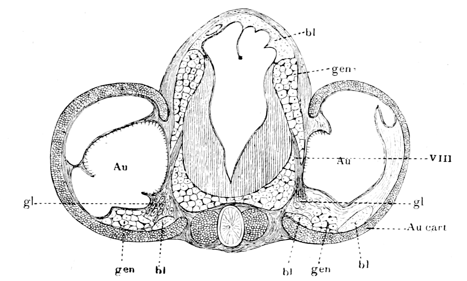
Fig. 154.—Transverse Section through Auditory Capsules and Brain of Ammocœtes.
Au., auditory organ; VIII, auditory nerve; gl., ganglion cells of VIIIth nerve; Au. cart., cartilaginous auditory capsule; gen., cells of old generative tissue round brain and in auditory capsule; bl., blood-vessels
Here there was a test of the truth of my theory ranking second only to the test of the median eyes; the strongest possible evidence of the truth of any theory is given when by its aid new and unexpected facts are brought to light. The theory said that in the group of animals from which the vertebrates arose, a special sense-organ of the nature of an auditory organ must have existed on the base of one of the appendages situated at the junction of the prosoma and mesosoma, and that into this basal part of the appendage a portion of the cephalic mass of generative and hepatic material must have made its way in close contiguity to the nerve of the special organ.
The only living example which nearly approaches the ancient extinct forms from which, according to the theory, the vertebrates arose, is Limulus, and, as has already been shown, in this animal, in the very position postulated by the theory, a large special sense-organ—the flabellum—exists, which, as already stated, may well have given rise to a sense-organ concerned with equilibration and audition. If, further, it be found that a diverticulum of the generative and hepatic material does accompany the nerve of the flabellum in the basal part of the appendage, then the evidence becomes very strong that the auditory organ of Ammocœtes, i.e. of the ancient Cephalaspids, was derived from an organ homologous with the flabellum; that, therefore, the material round the brain of Ammocœtes was originally generative and hepatic material; that, in fact, the whole theory is true, for all the parts of it hang together so closely that, if one portion is accepted, all the rest must follow. As pointed out in my address at Liverpool, and at the meeting of the Philosophical Society at Cambridge, it is a most striking fact that a mass of the generative and hepatic tissue does accompany the flabellar nerve into the basal part of this appendage. Into no other appendage of Limulus is there the slightest sign of any intrusion of the generative and hepatic masses; nowhere, except in the auditory capsule, is there any sign of the peculiar large-celled tissue which surrounds the brain and upper part of the spinal cord of Ammocœtes. The actual position of the flabellum on the basal part of the ectognath is shown in Fig. 155, A, and in Fig. 155, B, I have removed the chitin, to show the generative and hepatic tissue (gen.) lying beneath.
The reason why, to all appearance, the generative and hepatic mass penetrates into the basal part of this appendage only is apparent when we see (as Patten and Redenbaugh have pointed out) to what part of the appendage the flabellum in reality belongs.
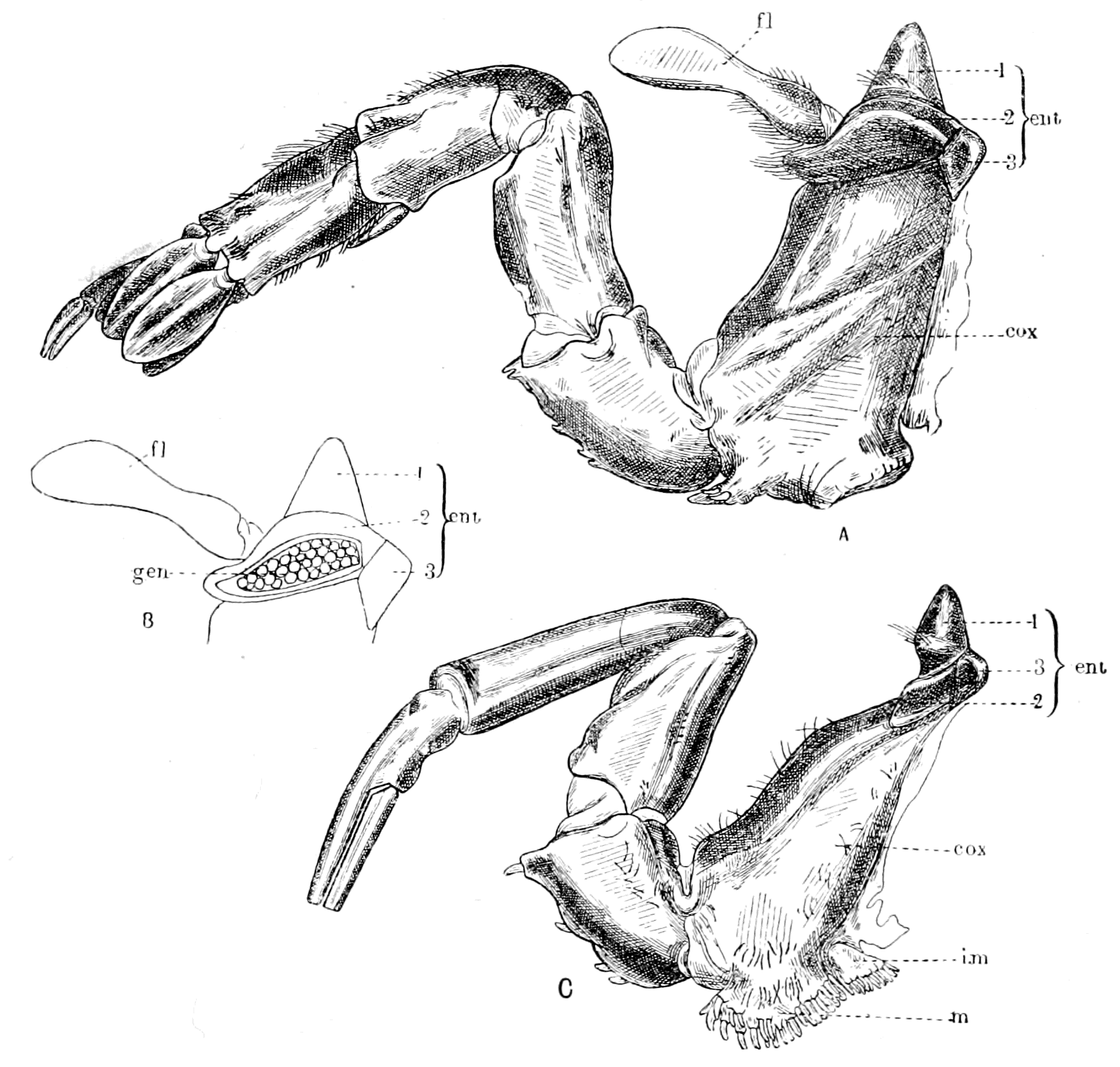
Fig. 155.—A, The Digging Appendage or Ectognath of Limulus; B, The Middle Protuberance (2) of the Entocoxite opened, to show the Generative and Hepatic Tissue (gen.) within it; C, One of the Prosomatic Locomotor Appendages or Endognaths of Limulus, for comparison with A.
fl., flabellum; cox., coxopodite; ent., entocoxite; m., mandible; i.m., inner mandible or epicoxite.
Patten and Redenbaugh, in their description of the prosomatic appendages of Limulus, describe the segments of the limbs as (1) the dactylopodite, (2) the propodite, (3) the mero- and carpo-podites, (4) the ischiopodite, (5) the basipodite, and (6) the coxopodite (cox. in Fig. 155). Still more basal than the coxopodite is situated the entocoxite (ent. in Fig. 155), which is composed of three sclerites or sensory knobs, to use Patten's description. The middle one of these three sclerites enlarges greatly in the digging appendage, and grows over the coxopodite to form the base from which the flabellum springs. Thus, as they have pointed out, the flabellum does not belong to the coxopodite of the appendage, but to the middle sensory knob of the entocoxite. Upon opening the prosomatic carapace, it is seen that the cephalic generative and hepatic masses press closely against the internal surface of the prosomatic carapace and also of the entocoxite, so that any enlargement of one of the sensory knobs of the entocoxite would necessarily be filled with a protrusion of the generative and hepatic masses. This is the reason why the generative and hepatic material apparently passes into the basal segment of the ectognath, and not into that of the endognaths; it does not really pass into the coxopodite of the appendage, but into an enlarged portion of the entocoxite, which can hardly be considered as truly belonging to the appendage. Kishinouye has stated that a knob arises in the embryo at the base of each of the prosomatic locomotor appendages, but that this knob develops only in the last or digging appendage (ectognath) forming the flabellum. Doubtless the median sclerites of the entocoxites of the endognaths represent Kishinouye's undeveloped knobs.
I conclude, therefore, that the flabellum, together with its basal part, is an adjunct to the appendage rather than a part of it, and might, therefore, easily remain as a separate and well-developed entity, even although the appendage itself dwindled down to a mere tentacle.
The evidence appears to me very strong that the flabellum of Limulus and the pecten of scorpions are the most likely organs to give a clue to the origin of the auditory apparatus of vertebrates. At present both the Eurypterids and Cephalaspids have left us in the lurch; in the former there is no sign of either flabellum or pecten; in the latter, no sign of any auditory capsule beyond Rohon's discovery of two small apertures situated dorsally on each side of the middle line in Tremataspis, which he considers to be the termination of the ductus endolymphaticus on each side. In both cases it is probable, one might almost say certain, that any such special sense-organ, if present, was not situated externally, but was sunk below the surface as in Ammocœtes.
The method by which such a sense-organ, situated externally on the surface of the animal, comes phylogenetically to form the lining wall of an internally situated membranous capsule is given by the ontogeny of this capsule, which shows step by step how the sense-organ sinks in and forms a capsule, and finally is entirely removed from the surface except as regards the ductus endolymphaticus.
Summary.
The special apparatus for hearing is of a very different character from that for vision or for smell, for its nerve belongs to the infra-infundibular group of nerves, and not to the supra-infundibular, as do those of the other two special senses. Of the five special senses the nerves for touch, taste, and hearing, all belong to the infra-infundibular segmental nerve-groups. The invertebrate origin, then, of the vertebrate auditory nerve must be sought for in the infra-œsophageal segmental group of nerves, and not in the supra-œsophageal.
The organs supplied by the auditory nerve are only partly for the purpose of hearing; there is always present also an apparatus—the semicircular canals—concerned with equilibration and co-ordination of movements. Such equilibration organs are not confined to the auditory nerve, but in the water-living vertebrates are arranged segmentally along the body, forming the organs of the lateral line in fishes; the auditory organ is but one of these lateral line organs, which has been specially developed.
These lateral line organs have been compared to similar segmental organs found in connection with the appendages in worms, especially the respiratory appendages. In accordance with this suggestion we see that they are all innervated from the region of the respiratory nerves—the vagus, glosso-pharyngeal, and facial—nerves which originally supplied the respiratory appendages of the palæostracan ancestor.
The logical conclusion is that the appendages of the Palæostraca possessed special sense-organs concerned with the perception of special vibrations, especially in the mesosomatic or respiratory region, and that somewhere at the junction of the prosoma and mesosoma, one of these sense-organs was specially developed to form the origin of the vertebrate auditory apparatus.
Impressed by this reasoning I made search for some specially striking sense-organ at the base of one of the appendages of Limulus, at the junction of the prosoma and mesosoma, and was immediately rewarded by the discovery of the extraordinary nature of the flabellum, which revealed itself as an elaborate sense-organ supplied with a nerve out of all proportion to its size. Up to this time no one had the slightest conception that this flabellum was a special sense-organ; the discovery of its nature was entirely due to the logical following out of the theory of the origin of vertebrates described in this book.
The structure of this large sense-organ is comparable with that of the sense-organs of the pectens of the scorpion, and of many other organs found on the appendages of various members of the scorpion group, of arachnids and other air-breathing arthropods. Many of these organs, such as the lyriform organs of arachnids, and the 'halteres' or balancers of the Diptera, are usually regarded as auditory and equilibration organs.
On all the mesosomatic appendages of Limulus very remarkable sense-organs are found, apparently for estimating pressures, which, when the appendages sank into the body to form with their basal parts the branchial diaphragms of Ammocœtes, could easily be conceived as remaining at the surface, and so giving rise to the lateral line organs.
Further confirmation of the view that an organ, such as the flabellum, must be looked upon as the originator of the vertebrate auditory organ, is afforded by the extraordinary coincidence that in Limulus a diverticulum of the generative and hepatic mass accompanies the flabellar nerve into the basal part of the digging appendage, while in Ammocœtes, accompanying the auditory nerve into the auditory capsule, there is seen a mass of cells belonging to that peculiar tissue which fills up the space between the brain and the cranial walls, and has already, on other grounds, been homologized with the generative and hepatic masses which fill up the encephalic region of Limulus.
For all these reasons special sense-organs, such as are found in the flabellum of Limulus and in the pectens of scorpions, may be looked upon as giving origin to the vertebrate auditory apparatus. In such case it is highly probable that the parachordals, with the auditory capsules attached, arose from a second entochondrite of the same nature as the plastron; a probability which is increased by the fact that the scorpion does possess a second entochondrite, which, owing to its special relations to the pecten, is known as the supra-pectinal entochondrite.
CHAPTER XII
THE REGION OF THE SPINAL CORD
Difference between cranial and spinal regions.—Absence of lateral root.—Meristic variation.—Segmentation of cœlom.—Segmental excretory organs.—Development of nephric organs; pronephric, mesonephric, metanephric.—Excretory organs of Amphioxus.—Solenocytes.—Excretory organs of Branchipus and of Peripatus, appendicular and somatic.—Comparison of cœlom of Peripatus and of vertebrate.—Pronephric organs compared to coxal glands.—Origin of vertebrate body-cavity (metacœle).—Segmental duct.—Summary of formation of excretory organs.—Origin of somatic trunk-musculature.—Atrial cavity of Amphioxus.—Pleural folds.—Ventral growth of pleural folds and somatic musculature.—Pleural folds of Cephalaspidæ and of Trilobita.—Significance of the ductless glands.—Alteration in structure of excretory organs which have lost their duct in vertebrates and in invertebrates.—Formation of lymphatic glands.—Segmental coxal glands of arthropods and of vertebrates.—Origin of adrenals, pituitary body, thymus, tonsils, thyroid, and other ductless glands.—Summary.
The consideration of the auditory nerve and the auditory apparatus terminates the comparison between the cranial nerves of the vertebrate and the prosomatic and mesosomatic nerves of the arthropod, and leaves us now free to pass on to the consideration of the vertebrate spinal nerves and the organs they supply. Before doing so, it is advisable to pass in review the conclusions already attained.
Starting with the working hypothesis that the central nervous system of the vertebrate has arisen from the central nervous system of the arthropod, but has involved and enclosed the alimentary canal of the latter in the process, so that there has been no reversal of surfaces in the derivation of the one form from the other, we have been enabled to compare closely all the organs of the head-region in the two groups of animals, and in no single case have we been compelled to make any startling or improbable assumptions. The simple following out of this clue has led in every case in the most natural manner to the interpretation of all the organs in the head-region of the vertebrate from the corresponding organs of the arthropod.
That it is possible to bring together all the striking resemblances between organs in the two classes of animals, such as I have done in preceding chapters, has been ascribed to a perverted ingenuity on my part—a suggestion which is flattering to my imaginative powers, but has no foundation of fact. There has been absolutely no ingenuity on my part; all I have done is to compare organs and their nerve-supply, as they actually exist in the two groups of animals, on the supposition that there has been no turning over on to the back, no reversal of dorsal and ventral surfaces. The comparison is there for all to read; it is all so simple, so self-evident that, given the one clue, the only ingenuity required is on the part of those who fail to see it.
The great distinction that has arisen between the two head-regions is the disappearance of appendages as such, never, however, of important organs on those appendages. If the olfactory organs of the one group were originally situated on antennules, the olfactory organs still remain, although the antennules as such have disappeared. The coxal excretory organs at the base of the endognaths remain and become the pituitary body. A special sense-organ, such as the flabellum of Limulus or the pecten of scorpion, remains and gives rise to the auditory organ. A special glandular organ, the uterus in the base of the operculum, remains, and gives rise to the thyroid gland. The branchiæ and sense-organs on the mesosomatic appendages remain, and even the very muscles to a large extent. As will be seen later, the excretory organs at the base of the metasomatic appendages remain. It is merely the appendage as such which vanishes either by dwindling away, or by so great an alteration as no longer to be recognizable as an appendage.
This dwindling process was already in full swing before the vertebrate stage; it is only a continuation of a previous tendency, as is seen in the dwindling of the prosomatic appendages in the Merostomata and the inclusion of the branchiæ within the body of the scorpion. Already among the Palæostraca, swimming had largely taken the place of crawling. The whole gradual transformation from the arthropod to the vertebrate is associated with a transformation from a crawling to a swimming animal—with the concomitant loss of locomotor appendages as such, and the alteration of the shape of the animal into the lithe fish-like form. The consideration of the manner in which this latter change was brought about, takes us out of the cranial into the spinal region.
If we take Limulus as the only living type of the Palæostraca, we are struck with the fact that the animal consists to all intents and purposes of prosomatic and mesosomatic regions only; the metasoma consisting of the segments posterior to the mesosoma is very insignificant, so that the large mass of the animal consists of what has become the head-region in the vertebrate; the spinal region, which has become in the higher vertebrates by far the largest region of the body, can hardly be said to exist in such an animal as Limulus. As to the Eurypterids and others, similar remarks may be made, though not to the same extent, for in them a distinct metasoma does exist.
In this book I have considered up to the present the cranial region as a system of segments, and shown how such segments are comparable, one by one, with the corresponding segments in the prosoma and mesosoma of the presumed arthropod ancestor.
In the spinal region such direct comparison is not possible, as is evident on the face of it; for even among vertebrates themselves the spinal segments are not comparable one by one, so great is the variation, so unsettled is the number of segments in this region. This meristic variation, as Bateson calls it, is the great distinctive character of the spinal region, which distinguishes it from the cranial region with its fixed number of nerves, and its substantive rather than meristic variation. At the borderland, between the two regions, we see how the one type merges into the other; how difficult it is to fix the segmental position of the spino-occipital nerves; how much more variable in number are the segments supplied by the vagus nerves than those anterior to them.
This meristic variation is a sign of instability, of want of fixedness in the type, and is evidence, as already pointed out, that the spinal region is newer than the cranial. This instability in the number of spinal segments does not necessarily imply a variability in the number of segments of the metasoma of the invertebrate ancestor; it may simply be an expression of adaptability in the vertebrate phylum itself, according to the requirements necessitated by the conversion of a crawling into a swimming animal, and the subsequent conversion of the swimming into a terrestrial or flying animal.
However many may have been the original number of segments belonging to the spinal region, one thing is certain—the segmental character of this region is remarkably clearly shown, not only by the presence of the segmental spinal nerves, but also by the marked segmentation of the mesoblastic structures. The question, therefore, that requires elucidation above all others is the origin of the spinal mesoblastic segments, i.e. of the cœlomic cavities of the trunk-region, and the structures derived from their walls.
Proceeding on the same lines as in the case of the cranial segments, it is necessary in the first instance to inquire of the vertebrate itself as to the scope of the problem in this region. In addition to the variability in the number of segments so characteristic of the spinal region, the complete absence in each spinal segment of a lateral root affords another marked difference between the two regions. Here, except, of course, at the junction of the spinal and cranial regions, each segmental nerve arises from two roots only, dorsal and ventral, and these roots are separately sensory and motor, and not mixed in function as was the lateral root of each cranial segment. Now, these lateral roots were originally the nerves supplying the prosomatic and mesosomatic appendages with motor as well as sensory fibres. The absence, therefore, of lateral roots in the spinal region implies that in the vertebrate none of the musculature belonging to the metasomatic appendages has remained. Consequently, as far as muscles are concerned, the clue to the origin of the spinal segments must be sought for in the segmentation of the body-muscles.
Here, in contradistinction to the cranial region, the segmentation is most marked, for the somatic spinal musculature of all vertebrates can be traced back to a simple sheet of longitudinal ventral and dorsal muscles, such as are seen in all fishes. This sheet is split into segments or myotomes by transverse connective tissue septa or myo-commata; each myotome corresponding to one spinal segment.
In addition to the evidence of segmentation afforded by the body-musculature in all the higher vertebrates, similar evidence is given by the segmental arrangement of parts of the supporting tissue to form vertebræ. Such segments have received the name of sclerotomes, and each sclerotome corresponds to one spinal segment.
Yet another marked peculiarity of this region is the segmental arrangement of the excretory organs. Just as our body-musculature has arisen from the uniformly segmented simple longitudinal musculature of the lowest fish, so, as we pass down the vertebrate phylum, we find more and more of a uniform segmental arrangement in the excretory organs.
The origin of all these three separate segmentations may, in accordance with the phraseology of the day, be included in the one term—the origin of the spinal mesoblastic segments—i.e. of the cœlomic cavities of the trunk-region and the structures derived from their walls.
The Origin of the Segmental Excretory Organs.
Of these three clues to the past history of the spinal region, the segmentation manifested by the presence of vertebræ is the least important, for in Ammocœtes there is no sign of vertebræ, and their indications only appear at transformation. Especially interesting is the segmentation due to the excretory organs, for the evidence distinctly shows that such excretory organs have steadily shifted more and more posteriorly during the evolution of the vertebrate.
In Limulus the excretory organs are in the prosomatic region—the coxal glands; these become in the vertebrate the pituitary body.
In Amphioxus the excretory organs are in the mesosomatic region, segmentally arranged with the gills.
In vertebrates the excretory organs are in the metasomatic region posterior to the gills, and are segmentally arranged in this region. Their investigation has demonstrated the existence of three distinct stages in these organs: 1. A series of segmental excretory organs in segments immediately following the branchial segments. This is the oldest of the three sets, and to these organs the name of the pronephros is given. 2. A second series which extends more posteriorly than the first, overlaps them to an extent which is not yet settled, and takes their place; to them is given the name of the mesonephros. 3. A third series continuous with the mesonephric is situated in segments still more posterior, supplants the mesonephros and forms the kidneys of all the higher vertebrates. This forms the metanephros.
These three sets of excretory organs are not exactly alike in their origin, in that the pronephric tubules are formed from a different portion of the cœlomic walls to that from which the meso- and metanephric tubules are formed, and the former alone gives origin to a duct, which forms the basis for the generative and urinary ducts, and is called the segmental duct. The mesonephric tubules, called also the Wolffian body, open into this duct.
In order to make the embryology of these excretory organs quite clear, I will make use of van Wijhe's phraseology and also of his illustrations. He terms the whole cœlomic cavity the procœlom, which is divisible into a ventral unsegmented part, the body-cavity or metacœlom, and a dorsal segmented part, the somite. This latter part again is divided into a dorsal part—the epimere—and a part connecting the dorsal part with the body-cavity, to which therefore he gives the name of mesomere.
The cavity of the epimere disappears, and its walls form the muscle and cutis plates of the body. The part which forms the muscles is known as the myotome, which separates off from the mesomere, leaving the latter as a blind sac—the mesocœlom—communicating by a narrow passage with the body cavity or metacœlom. At the same time, from the mesomere is formed the sclerotome, which gives rise to the skeletal tissues of the vertebræ, etc., so that van Wijhe's epimere and mesomere together correspond to the original term, protovertebra, or somite of Balfour; and when the myotome and sclerotome have separated off, there is still left the intermediate cell-mass of Balfour and Sedgwick, i.e. the sac-like mesocœle of van Wijhe, the walls of which give origin to the mesonephrotome or mesonephros. Further, according to van Wijhe, the dorsal part of the unsegmented metacœlom is itself segmented, but not, as in the case of the mesocœle, with respect to both splanchnopleuric and somatopleuric walls. The segmentation is manifest only on the somatopleuric side, and consists of a distinct series of hollow somatopleuric outgrowths, called by him hypomeres, which give rise to the pronephros and the segmental duct.
Van Wijhe considers that the whole metacœlom was originally segmented, because in the lower vertebrates the segmentation reaches further ventral-wards, so that in Selachia the body-cavity is almost truly segmental. Also in the gill-region of Amphioxus the cavities which are homologous with the body-cavity arise segmentally.
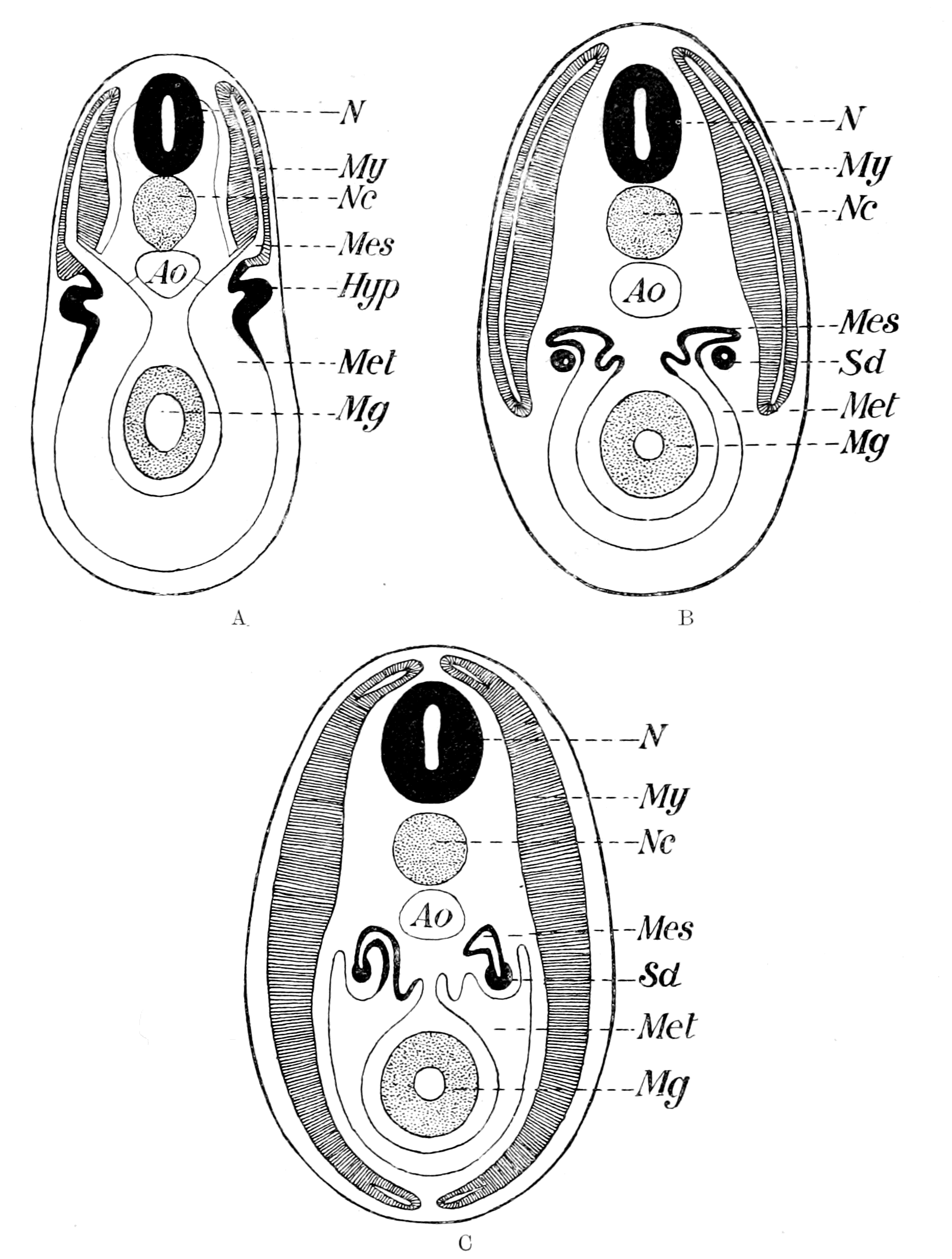
Fig. 156.—Diagrams to illustrate the Development of the Vertebrate Cœlom. (After van Wijhe.)
N., central nervous system; Nc., notochord; Ao., aorta; Mg., midgut. A, My., myocœle; Mes., mesocœle; Met., metacœle; Hyp., hypomere (pronephric). B and C, My., myotome; Mes., mesonephros; S.d., segmental duct (pronephric); Met., body cavity.
As is well known, Balfour and Semper were led, from their embryological researches, to compare the nephric organs of vertebrates with those of annelids, and, indeed, the nature of the vertebrate segmental excretory organs has always been the fact which has kept alive the belief in the origin of vertebrates from a segmented annelid. These segmental organs thus compared were the mesonephric tubules, and doubts arose, especially in the mind of Gegenbaur, as to the validity of such a comparison, because the mesonephric tubules did not open to the exterior, but into a duct—the segmental duct—which was an unsegmented structure opening into the cloaca; also because the segmental duct, which was the excretory duct of the pronephros, was formed first, and the mesonephric tubules only opened into it after it was fully formed. Further, the pronephros was said to arise from an outbulging of the somatopleuric mesoblast, which extended over a limited number of metameres, and was not segmental, but continuous. Gegenbaur and others therefore argued that the original prevertebrate excretory organ was the pronephros and its duct, not the mesonephros, from which they concluded that the vertebrate must have been derived from an unsegmented type of animal, and not from the segmented annelid type.
Such a view, however, has no further reason for acceptance, as it was based on wrong premises, for Rückert has shown that the pronephros does arise as a series of segmental nephric tubules, and is not unsegmented. He also has pointed out that in Torpedo the anterior part of the pronephric duct shows indications of being segmented, a statement fully borne out by the researches of Maas on Myxine, who gives the clearest evidence that in this animal the anterior part of the pronephric duct is formed by the fusion of a series of separate ducts, each of which in all probability once opened out separately to the exterior.
Rückert therefore concludes that Balfour and Semper were right in deriving the segmental organs of vertebrates from those of annelids, but that the annelid organs are represented in the vertebrate, not by the mesonephric tubules, but by the pronephric tubules and their ducts, which originally opened separately to the exterior. By the fusion of such tubules the anterior part of the segmental duct was formed, while its posterior part either arose by a later cœnogenetic lengthening, or is the only remnant of a series of pronephric tubules which originally extended the whole length of the body, as suggested also by Maas and Boveri. Rückert therefore supposed that the mesonephric tubules were a secondary set of nephric organs, which were not necessarily directly derived from the annelid nephric organs.
At present, then, Rückert's view is the one most generally accepted—the original annelid nephric organs are represented by the pronephric tubules and the pronephric duct, not by the mesonephric tubules, which are a later formation. This latter statement would hold good if the mesonephric tubules were found entirely in segments posterior to those containing the pronephric tubules; such, however, is said not to be the case, for the two sets of organs are said to overlap in some cases; even when they exist in the same segments, the former are said always to be formed from a more dorsal part of the cœlom than the pronephros, always to be a later formation, and never to give any indication of communicating with the exterior except by way of the pronephric duct.
The recent observations of Brauer on the excretory organs of the Gymnophiona throw great doubt on the existence of mesonephric and pronephric tubules in the same segment. He criticizes the observations on which such statements are based, and concludes that, as in Hypogeophis, the nephrotome which is cut off after the separation of the sclero-myotome gives origin to the pronephros in the more anterior regions, just as it gives origin to the mesonephros in the more posterior regions. In fact, the observations of van Wijhe and others do not in reality show that two excretory organs may be formed in one segment, the one mesonephric from the remains of the mesomere and the other pronephric from the hypomere, but rather that in such cases there is only one organ—the pronephros—part of which is formed from the mesomere and part from the hypomere. Brauer goes further than this, and doubts the validity of any distinction between pronephros and mesonephros, on the ground of the former arising from a more ventral part of the procœlom than the latter; for, as he says, it is only possible to speak of one part of the somite as being more ventral than another part when both parts are in the same segment; so that if pronephric and mesonephric organs are never in the same segment, we cannot say with certainty that the former arises more ventrally than the latter.
These observations of Brauer strongly confirm Sedgwick's original statement that the pronephric and mesonephric organs are homodynamous organs, in that they are both derived from the original serially situated nephric organs, the differences between them being of a subordinate nature and not sufficient to force us to believe that the mesonephros is an organ of quite different origin to the pronephros. So, also, Price, from his investigations of the excretory organs of Bdellostoma, considers that in this animal both pronephros and mesonephros are derived from a common embryonic kidney, to which he gives the name holonephros.
Brauer also is among those who conclude that the vertebrate excretory organs were derived from those of annelids; he thinks that the original ancestor possessed a series of similar organs over the whole pronephric and mesonephric regions, and that the anterior pronephric organs, which alone form the segmental duct, became modified for a larval existence—that their peculiarities were adaptive rather than ancestral. This last view seems to me very far-fetched, without any sufficient basis for its acceptance. According to the much more probable and reasonable view, the pronephros represents the oldest and original excretory organs, while the mesonephros is a later formation. Brauer's evidence seems to me to signify that the pronephros, mesonephros, and metanephros are all serially homologous, and that the pronephros bears much the same relation to the mesonephros that the mesonephros does to the metanephros. The great distinction of the pronephros is that it, and it alone, forms the segmental duct.
We may sum up the conclusions at which we have now arrived as follows:—
1. The pronephric tubules and the pronephric duct are the oldest part of the excretory system, and are distinctly in evidence for a few segments only in the most anterior part of the trunk-region immediately following the branchial region. They differ also from the mesonephric tubules by not being so clearly segmental with the myotomes.
2. The mesonephric tubules belong to segments posterior to those of the pronephros, are strictly segmental with the myotomes, and open into the pronephric duct.
3. All observers are agreed that the two sets of excretory organs resemble each other in very many respects, as though they arose from the same series of primitive organs, and, according to Sedgwick and Brauer, no distinction of any importance does exist between the two sets of organs. Other observers, however, consider that the pronephric organs, in part at all events, arise from a part of the nephrocœle more ventral than that which gives origin to the mesonephric organs, and that this difference in position of origin, combined with the formation of the segmental duct, does constitute a true morphological distinction between the two sets of organs.
4. All the recent observers are in agreement that the vertebrate excretory organs strongly indicate a derivation from the segmental organs of annelids.
The very strongest support has been given to this last conclusion by the recent discoveries of Boveri and Goodrich upon the excretory organs of Amphioxus. According to Boveri, the nephric tubules of Amphioxus open into the dorsal cœlom by one or more funnels. Around each funnel are situated groups of peculiar cells, called by him 'Fadenzellen,' each of which sends a long process across the opening of the funnel. Goodrich has examined these 'Fadenzellen,' and found that they are typical pipe-cells, or solenocytes, such as he has described in the nephridial organs of various members of the annelid group Polychæta. Also, just as in the Polychæta, the ciliated nephric tubule has no internal funnel-shaped opening into the cœlom, but terminates in these groups of solenocytes. "Each solenocyte consists of a cell-body and nucleus situated at the distal free extremity of a delicate tube; the proximal end of the tube pierces the wall of the nephridial canal and opens into its lumen. A single long flagellum arising from the cells works in the tube and projects into the canal."
The exceedingly close resemblance between the organs of Amphioxus and those of Phyllodoce, as given in his paper, is most striking, and, as he says, leads to the conclusion that the excretory organs of Amphioxus are essentially identical with the nephridia of certain polychæte worms.
It is to me most interesting to find that the very group of annelids, the Polychæta, which possess solenocytes so remarkably resembling those of the excretory organs of Amphioxus, are the highest and most developed of all the Annelida. I have argued throughout that the law of evolution consists in the origination of successive forms from the dominant group then alive, dominance signifying the highest type of brain-power achieved up to that time. The highest type among Annelida is found in the Chætopoda; from them, therefore, the original arthropod type must have sprung. This original group of Arthropoda gave rise to the two groups of Crustacea and Arachnida, in my opinion also to the Vertebrata, and, as already mentioned, it is convenient to give it a generalized name, the Protostraca, from which subsequently the Palæostraca arose.
The similarity between the excretory organs of Amphioxus and those of Phyllodoce suggests that the protostracan ancestor of the vertebrates arose from the highest group of the Chætopoda—the Polychæta. The evidence which I have already given points, however, strongly to the conclusion that the vertebrate did not arise from members of the Protostraca near to the polychæte stock, but rather from members in which the arthropod characters had already become well developed—members, therefore, which were nearer the Trilobita than the Polychæta. Such early arthropods would very probably have retained in part excretory organs of the same character as those found in the original polychæte stock, and thus account for the presence of solenocytes in the excretory organs of Amphioxus.
In connection with such a possibility, I should like to draw attention to the observations of Claus and Spangenberg on the excretory organs of Branchipus—that primitive phyllopod, which is recognized as the nearest approach to the trilobites at present living. According to Claus, an excretory apparatus exists in the neighbourhood of each nerve-ganglion, and Spangenberg finds a perfectly similar organ in the basal segment of each appendage—a system, therefore, of excretory organs as segmentally arranged as those of Peripatus. Claus considers that although these organs formed an excretory system, it is not possible to compare them with the annelid segmental organs, because he thought the cells in question arose from ectoderm. Now, the striking point in the description of the excretory cells in these organs, as described both by Claus and Spangenberg, is that they closely resemble the pipe-cells or solenocytes of Goodrich; each cell possesses a long tube-like projection, which opens on the surface. They appear distinctly to belong to the category of flame-cells, and resemble solenocytes more than anything else. According to Goodrich, the solenocyte is probably an ectodermal cell, so that even if it prove to be the case, as Claus thought, that these pipe-cells of Branchipus are ectodermal, they would still claim to be derived from the segmental organs of annelids, especially of the Polychæta, being, to use Goodrich's nomenclature, true nephridial organs, as opposed to cœlomostomes.
These observations of Claus and Spangenberg suggest not only that the primitive arthropod of the trilobite type possessed segmental organs in every segment directly derived from those of a polychæte ancestor, but also that such organs were partly somatic and partly appendicular in position. Such a suggestion is in strict accord with the observations of Sedgwick on the excretory organs of the most primitive arthropod known, viz. Peripatus, where also the excretory organs, which are true segmental organs, are partly somatic and partly appendicular. Further, the excretory organs of the Scorpion and Limulus group are again partly somatic and partly appendicular, receiving the name of coxal glands, because there is a ventral projection of the gland into the coxa of the corresponding appendage.
Judging from all the evidence available, it is probable that when the arthropod stock arose from the annelids, simultaneously with the formation of appendages, the segmental somatic nephric organs of the latter extended ventrally into the appendage, and thus formed a segmental set of excretory organs, which were partly somatic, partly appendicular in position, and might therefore be called coxal glands.
As already stated, all investigators of the origin of the vertebrate excretory organs are unanimous in considering them to be derived from segmental organs of the annelid type. I naturally agree with them, but, in accordance with my theory, would substitute the words "primitive arthropod" for the word "annelid," for all the evidence I have accumulated in the preceding chapters points directly to that conclusion. Further, the most primitive of the three sets of vertebrate segmental organs—the pronephros, mesonephros, and metanephros—is undoubtedly the pronephros; consequently the pronephric tubules are those which I consider to be more directly derived from the coxal glands of the primitive arthropod ancestor. Such a derivation appears to me to afford an explanation of the difficulties connected with the origin of the pronephros and mesonephros respectively, which is more satisfactory than that given by the direct derivation from the annelid.
The only living animal which we know of as at all approaching the most primitive arthropod type is, as pointed out by Korschelt and Heider, Peripatus; and Peripatus, as is well known, possesses a true cœlom and true cœlomic excretory organs in all the segments of the body. Sedgwick shows that at first a true cœlom, as typical as that of the annelids, is formed in each segment of the body, and that then this cœlom (which represents in the vertebrate van Wijhe's pro-cœlom) splits into a dorsal and a ventral part. In the anterior segments of the body the dorsal part disappears (presumably its walls give origin to the mesoblast from which the dorsal body-muscles arise), while the ventral part remains and forms a nephrocœle, giving origin to the excretory organs of the adult. According to von Kennel, the cavity becomes divided into three spaces, which for a time are in communication—a lateral (I.), a median (II.), and a dorso-median (III.). The dorso-median portion becomes partitioned off, and this, as well as the greater part of the lateral portion, which lies principally in the foot, is used up in providing elements for the formation of the body- and appendage-muscles respectively and the connective tissue.
In Fig. 157 I reproduce von Kennel's diagram of a section across a Peripatus embryo, in which I. represents the lateral appendicular part of the cœlom, II. the ventral somatic part, and III. the dorsal part which separates off from the ventral and lateral parts, and, as its walls give origin largely to the body-muscles, may be called the myocœle. The muscles of the appendages are formed from the ventral part of the original procœlom, just as I have argued is the case with the muscles of the splanchnic segmentation in vertebrates.
Sedgwick states that the ventral part of the cœlom extends into the base of each appendage, and there forms the end-sac of each nephric tubule, into which the nephric funnel opens, thus forming a coxal gland; this end-sac or vesicle in the appendage is called by him the internal vesicle (i.v.), because later another vesicle is formed from the ventral cœlom in the body itself, close against the nerve-cord on each side, which he calls the external vesicle (e.v.). (Cf. Fig. 158, taken from Sedgwick.) This second vesicle is, according to him, formed later in the development from the nephric tubule of the internal vesicle, so that it discharges its contents to the exterior by the same opening as the original tubule. Of course, as he points out, the whole system of internal and external vesicles and nephric tubules are all simply derivatives of the original ventral part of the cœlom or nephrocœle.
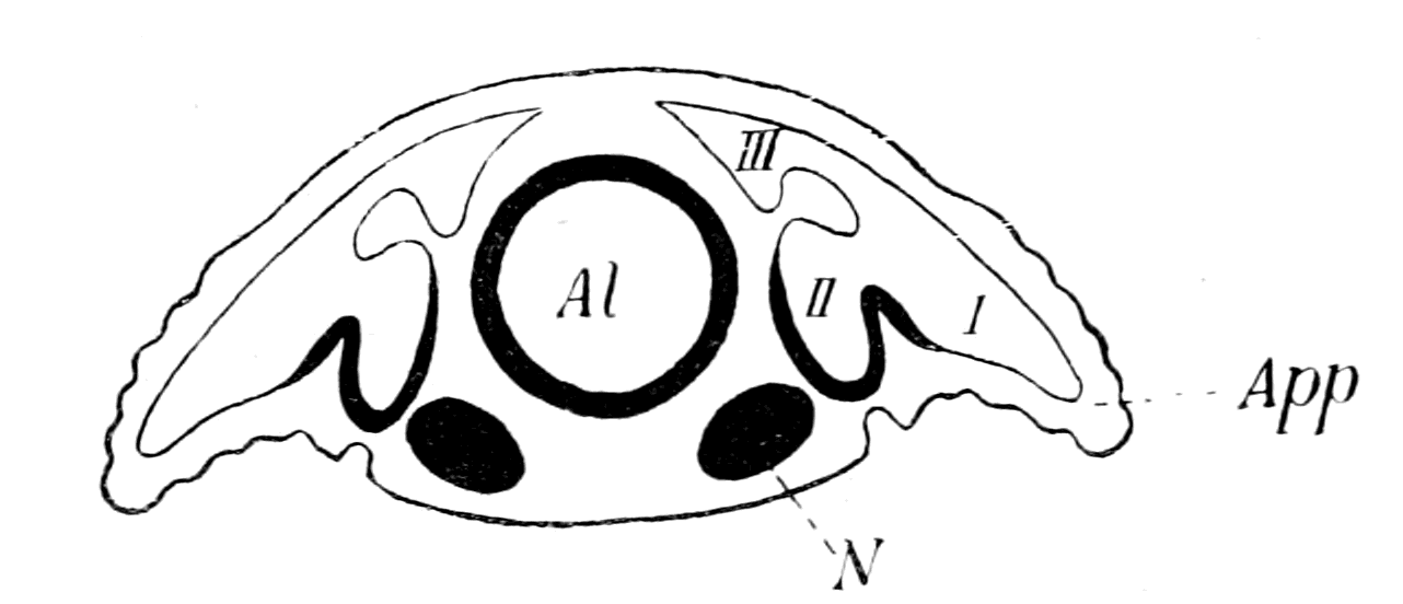
Fig. 157.—Transverse Section of Peripatus Embryo. (After von Kennel.)
Al., alimentary canal; N., nerve-cord; App., appendage; I, II, III, the three divisions (lateral, median, and dorso-median) of the cœlom.
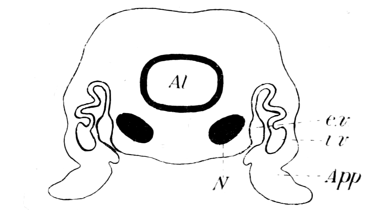
Fig. 158.—Section of Peripatus. (After Sedgwick.)
Al., alimentary canal; N., nerve-cord; App., appendage; i.v., internal, and e.v., external vesicles of the segmented excretory tubule (coxal gland).
Here, then, in Peripatus, and presumably, therefore, in members of the Protostraca, we see that the original segmental organs of the annelid have become a series of nephric organs, which extended into the base of the appendages, and may therefore be called coxal glands; also it is clear, from Sedgwick's description, that if the appendages disappeared, the nephric organs would still remain, not as coxal glands, but as purely somatic excretory glands. They would still be homologous with the annelid segmental organs, or with the coxal glands, but would arise in toto from a part of the ventral cœlom or nephrocœle, more dorsal than the former appendicular part, because the appendages and their enclosed cœlom are always situated ventrally to the body. Again, according to Sedgwick, the nephric tubules are connected with two cœlomic vesicles, the one in the appendage the internal vesicle, and the other, the so-called bladder, or the external vesicle, in the body itself, close against the nerve-cord. Sedgwick appears to consider that either of these vesicles may form the end-sac of a nephric tubule, for he discusses the question whether the single vesicle, which in each case gives origin to the nephridia of the first three legs, corresponds to the internal or external vesicle. He decides, it is true, in favour of the internal vesicle, and therefore considers the excretory organ to be appendicular, i.e. a coxal gland, in these segments as well as in those more posterior. Still, the very discussion shows that in his opinion, at all events, the external vesicle might represent the end-sac of the tubule, in the absence of the internal or appendicular vesicle.
Such an arrangement as Sedgwick describes in Peripatus is the very condition required to give rise to the pronephric and mesonephric tubules, as deduced by me from the consideration of the vertebrate, and harmonizes and clears up the controversy about the mesonephros and pronephros in the most satisfactory manner. Both pronephros and mesonephros are seen to be derivatives of the original annelid segmental organs, not directly from an annelid, but by way of an arthropodan ancestor; the difference between the two is simply that the pronephric organs were coxal glands, and indicate, therefore, the presence of the original metasomatic appendages, while the mesonephric organs were homologous organs, formed in segments of later origin which had lost their appendages. For this reason the pronephros is said to be formed, in part at least, from a portion of the cœlom situated more ventrally than the purely somatic part which gives rise to the mesonephros. For this reason Sedgwick, Brauer, etc., can say that the mesonephros is strictly homodynamous with the pronephros; while equally Rückert, Semon, and van Wijhe can say it is not homodynamous, in so far that the two organs are not derived strictly from absolutely homologous parts of the cœlom. For this reason Semon can speak of the mesonephros as a dorsal derivative of the pronephros, just as Sedgwick says that the external or somatic vesicle of Peripatus is a derivative of the appendicular nephric organ. For this reason the pronephros, or rather a part of it, is always derived from the somatopleuric layer, for, as is clear from Miss Sheldon's drawing, the part of the cœlom in Peripatus which dips into the appendage is derived from the somatopleuric layer alone.
Such a cœlom as that of Peripatus, Fig. 157, would represent the origin of the vertebrate cœlom, and would therefore represent the procœlom of van Wijhe. In strict accordance with this, we see that it separates into a dorsal part, the walls of which give origin to the somatic muscles, or at all events to the great longitudinal dorsal muscles of the animal, and a ventral part, which forms a nephrocœle, dips into the appendage, and gives origin to the muscles of the appendage. In the vertebrate, after the somatic dorsal part or myocœle has separated off, a ventral part is left, which forms a nephrocœle in the trunk-region, and gives origin to the splanchnic striated muscles in the cranial region, i.e. to the muscles which, according to my theory, were once appendicular muscles. This ventral nephrocœlic part is divisible in the trunk into a segmented part, which forms the excretory organs proper, and an unsegmented part, the metacœle or true body-cavity of the vertebrate.
This comparison of the procœlom of the vertebrate and arthropod signifies that the vertebrate metacœle was directly derived by ventral downgrowth from the arthropod nephrocœle, so that if, as I suppose, the vertebrate nervous system represents the conjoined nervous system and alimentary canal of the arthropod, then the vertebrate metacœle, or body-cavity, must have been originally confined to the region on each side of the central nervous system, and from this position have spread ventrally, to enclose ultimately the new-formed vertebrate gut. This means that the body-cavity (metacœle) of the vertebrate is not the same as the body-cavity of the annelid, but corresponds to a ventral extension of the nephrocœle, or ventral part of such body-cavity.
Such a phylogenetic history is most probable, because it explains most naturally and simply the facts of the development of the vertebrate body-cavity; for the mesoblast always originates in the neighbourhood of the notochord and central nervous system, and the lumen of the body-cavity always appears first in that region, and then extends laterally and ventrally on each side until it reaches the most ventral surface of the embryo, thus forming a ventral mesentery, which ultimately disappears, and the body-cavity surrounds the gut, except for the dorsal mesentery. Thus Shipley, in his description of the formation of the mesoblastic plates which line the body-cavity in Ammocœtes, describes them as commencing in two bands of mesoblast situated on each side, close against the commencing nervous system:—
"These two bands are separated dorsally by the juxtaposition of the dorsal wall of the mesenteron and the epiblast, and ventrally by the hypoblastic yolk-cells which are in contact with the epiblast over two-thirds of the embryo. Subsequently, but at a much later date, the mesoblast is completed ventrally by the downgrowth on each side of these mesoblastic plates. The subsequent downward growth is brought about by the cells proliferating along the free ventral edge of the mesoblast, these cells then growing ventralwards, pushing their way between the yoke-cells and epiblast."
The derivation of the vertebrate pronephric segmental organs from the metasomatic coxal glands of a primitive arthropod would mean, if the segmental organs of Peripatus be taken as the type, that such glands opened to the exterior on every segment, either at the base of the appendage or on the appendage itself. It is taken for granted by most observers that the pronephric segmental organs once opened to the exterior on each segment, and then, from some cause or other, ceased to do so, and the separate ducts, by a process of fusion, came to form a single segmental duct, which opened into the cloaca. Many observers have been led to the conclusion that the pronephric duct is epiblastic in origin, although from its position in the adult, it appears far removed from all epiblastic formations. However, at no time in the developmental history is there any clear evidence of actual fusion of any part of the pronephric organ with the epidermis, and the latest observer, Brauer, is strongly of opinion that there is never sufficiently close contact with the epidermis to warrant the statement that the epiblastic cells take part in the formation of the duct. All that can be said is, that the formation of the duct takes place at a time when the pronephric diverticulum is in close propinquity to the epidermis, before the ventral downgrowth of the myotome has taken place.
The formation of the anterior portion of the pronephric duct is, according to Maas in Myxine, and Wheeler in Petromyzon, undoubtedly brought about by the fusion of a number of pronephric tubules, which, according to Maas, are clearly seen in the youngest specimens as separate segmental tubes; each of these tubules is supplied by a capillary network from a segmental branch of the aorta, as in the tubules of Amphioxus according to Boveri, and does not possess a glomerulus.
The posterior part of the duct into which the mesonephric tubules enter possesses also a capillary network, which Maas considers to represent the original capillary network of a series of pronephric tubules, the only remnant of which is the duct into which the mesonephric tubules open. He therefore argues that the pronephric duct indicates a series of pronephric tubules, which originally extended along the whole length of the body, and were supplanted by the mesonephric tubules, which also belonged to the same segments.
I also think that the paired appendages which have left the pronephric tubules as signs of their past existence, existed originally, in the invertebrate stage, on every segment of the body. But I do not consider that such a statement is at all equivalent to saying that such pairs of tubules must have existed upon every one of the segments existing at the present day; for it seems to me that Rückert is much more likely to be right when he says that in Selachians the duct clearly does grow back, and is not formed throughout in situ; so that he gives a double explanation of the formation of the duct—a palingenetic anterior part formed by the fusion of the extremities of the original excretory tubules, to which a posterior cœnogenetic lengthening has been added.
It does not seem to me at all necessary that the immediate invertebrate ancestor of the vertebrate should have possessed excretory organs which opened out separately to the exterior on each segment; a fusion may already have taken place in the invertebrate stage, and so a single duct have been acquired for a number of organs. Such a suggestion has been made by Rückert, because of the fact discovered by Cunningham and E. Meyer, that the segmental organs of Lanice conchilega are on each side connected together by a single strong longitudinal canal. I would, however, go further than this and say, that even although the nephric organs of the polychæte ancestor opened out on every segment, and although the primitive arthropodan ancestor derived from such polychæte possessed coxal glands which opened out either on to or at the base of each appendage, similarly to those of Peripatus, yet the immediate arthropodan ancestor, with its palæostracan affinities, may already have possessed metasomatic coxal glands, all of which opened into a single duct, with a single opening to the exterior.
Judging from Limulus, such was very probably the case, for Patten and Hazen have shown (1) that the coxal glands of Limulus are segmental organs belonging to the prosomatic segments; (2) that the organs belonging to the cheliceral and ectognathal segments are not developed; (3) that the four glands belonging to the endognaths become connected together by a stolon, which communicates with a single nephric duct, opening to the exterior on the basal segment of the 5th prosomatic appendage (the last endognath). At no time is there any evidence of any separate openings or any fusion with the ectoderm, such as might indicate separate openings of these prosomatic coxal segmental organs. Thus we see that in Limulus, which is presumably much nearer the annelid condition than the vertebrate, all evidence of separate nephric ducts opening to the exterior on each prosomatic segment has entirely disappeared, just as is the case in the metasomatic coxal glands (i.e. the pronephros) of the vertebrate. What is seen in the prosomatic region of Limulus, and doubtless also of the Eurypterids, may very probably have occurred in the metasomatic region of the immediate invertebrate ancestors of the vertebrate, and so account for the single pronephric duct belonging to a number of pronephric organs.
The interpretation of these various embryological investigations may be summed up as follows:—
1. The ancestor of the vertebrates possessed a pair of appendages on each segment; into the base of each of these appendages the segmental excretory organ sent a diverticulum, thus forming a coxal gland.
2. Such coxal glands, even in the invertebrate stage, may have discharged into a common duct which opened to the exterior most posteriorly.
3. Then, from some cause, the appendages were rendered useless, and dwindled away, leaving only the pronephric organs to indicate their former presence. At the end of this stage the animal possessed vertebrate characteristics.
4. For the purpose of increasing mobility, of forming an efficient swimming instead of a crawling animal, the body-segments increased in number, always, as is invariably the case, by the formation of new ones between those already formed and the cloacal region, and so of necessity caused an elongation of the pronephric duct. Into this there now opened the ducts of the segmental organs formed by recapitulation, those, therefore, belonging to the body-segments—mesonephric—having nothing to do with appendages, for the latter had already ceased to exist functionally, and would not, therefore, be repeated with each meristic repetition.
This, so to speak, passive lengthening of the pronephric duct in consequence of the lengthening of the early vertebrate body by the addition of metameres, each of which contained only mesonephric and no pronephric tubules, is, to my mind, an example of a principle which has played an important part in the formation of the vertebrate, viz. that the meristic variation by which the spinal region of even the lowest of existing vertebrates has been formed, has largely taken place in the vertebrate phylum itself, and that such changes must be eliminated before we can picture to ourselves the pre-vertebrate condition. As an example, I may mention the remarkable repetition of similar segments pictured by Bashford Dean in Bdellostoma. Such repetition leads to passive lengthening of such parts as are already formed but are not meristically repeated: such are the notochord, the vertebrate intestine, the canal of the spinal cord, and possibly the lateral line nerve. The fuller discussion of this point means the discussion of the formation of the vertebrate alimentary canal; I will therefore leave it until I come to that part of my subject, and only say here that the evidence seems to me to point to the conclusion that at the time when the vertebrate was formed, the respiratory and cloacal regions were very close together, the whole of the metasoma being represented by the region of the pronephros alone.
Here, as always, the evidence of Ammocœtes tends to give definiteness to our conceptions, for Wheeler points out that up to a length of 7 mm. the pronephros only is formed; there is no sign of the more posteriorly formed mesonephros. Now we know, as pointed out in Chapter VI., p. 228, this is the time of Kupffer's larval stage of Ammocœtes. This is the period during which the invertebrate stage is indicated in the ontogeny, so that, in accordance with all that has gone before, this means that the metasoma of the invertebrate ancestor was confined to the region of the pronephros.
Again, take Shipley's account of the development of Petromyzon. He says—
"The alimentary canal behind the branchial region may be divided into three sections. Langerhans has termed these the stomach, midgut, and hindgut, but as the most anterior of these is the narrowest part of the whole intestine, it would, perhaps, be better to call it œsophagus. This part of the alimentary canal lies entirely in front of the yolk, and is, with the anterior region, which subsequently bears the gills, raised from the rest of the egg when the head is folded off. It is supported by a dorsal mesentery, on each side of which lies the head-kidney (pronephros)."
Further on he says—
"The hindgut is smaller than the midgut; its anterior limit is marked by the termination of the spiral valve, which does not extend into this region. The two segmental ducts open into it just where it turns ventrally to open to the exterior by a median ventral anus. Its lumen is from an early stage lined with cells which have lost their yolk, and it is in wide communication with the exterior from the first. This condition seems to be, as Scott suggests, connected with the openings of the ducts of the pronephros, for this gland is completed and seems capable of functioning long before any food could find its way through the midgut, or, indeed, before the stomodæum has opened."
Is there no significance in this statement of Shipley? Even if it be possible to find some special reason why the branchial and cloacal parts of the gut are freed from yolk and lined with serviceable epithelium a long time before the midgut, why should a bit of the midgut, which Shipley calls the œsophagus, which is connected with the region of the pronephros and not of the branchiæ, differ so markedly from the rest of the midgut? Surely the reason is that the branchial region of the gut, the pronephric region of the gut, and the cloacal region of the gut, belong to a different and earlier phase in the phylogenetic history of the Ammocœtes than does the midgut between the pronephric and cloacal regions. This observation of Shipley fits in with and emphasizes the view that the original animal from which the vertebrate arose consisted of a cephalic and branchial region, followed by a pronephric and cloacal region; the whole intermediate part of the gut, which forms the midgut, with its large lumen and spiral valve, and belongs to the mesonephric region, being a later formation brought about by the necessity of increasing the length of the body.
The Origin of the Somatic Trunk-Musculature and the Formation of an Atrial Cavity.
Next comes the question, why was the pronephros not repeated in the meristic repetition that took place during the early vertebrate stage? What, in fact, caused the disappearance of the metasomatic appendages, and the formation of the smooth body-surface of the fish?
The embryological evidence given by van Wijhe and others of the manner in which the original superficially situated pronephros is removed from the surface and caused to assume the deeper position, as seen in the later embryo, is perfectly clear and uniform in all the vertebrate groups. The diagrams at the end of van Wijhe's paper, which I reproduce here, illustrate the process which takes place. At first the myotome (Fig. 159, A) is confined to the dorsal region on each side of the spinal cord and notochord. Then (Fig. 159, B) it separates from the rest of the somite and commences to extend ventrally, thus covering over the pronephros and its duct, until finally (Fig. 159, C) it reaches the mid-ventral line on each side, and the foundations of the great somatic body-muscles are finally laid.
In order, therefore, to understand how the obliteration of the appendages took place, we must first find out what is the past history of the myotomes. Why are they confined at first to the dorsal region of the body, and extend afterwards to the ventral region, forcing by their growth an organ that was originally external in situation to become internal?
In the original discussion at Cambridge, I was accused of violating the important principle that in phylogeny we must look at the most elementary of the animals whose ancestors we seek, and was told that the lowest vertebrate was Amphioxus, not Ammocœtes; that therefore any argument as to the origin of vertebrates must proceed from the consideration of the former and not the latter animal. My reply was then, and is still, that I was considering the cranial region in the first place, and that therefore it was necessary to take the lowest vertebrate which possessed cranial nerves and sense-organs of a distinctly vertebrate character, a criterion evidently not possessed by Amphioxus. Such argument does not apply to the spinal region, so that, now that I have left the cranial region and am considering the spinal, I entirely agree with my critics that Amphioxus is likely to afford valuable help, and ought to be taken into consideration as well as Ammocœtes. The distinction between the value of the spinal (including respiratory) and cranial regions of Amphioxus for drawing phylogenetic conclusions is recognized by Boveri, who says that, in his opinion, "Amphioxus shows simplicity and undifferentiation rather than degeneration. If truly Amphioxus is somewhat degenerated, then it is so in its prehensile and masticatory apparatus, its sense organs, and perhaps its locomotor organs, owing to its method of living."
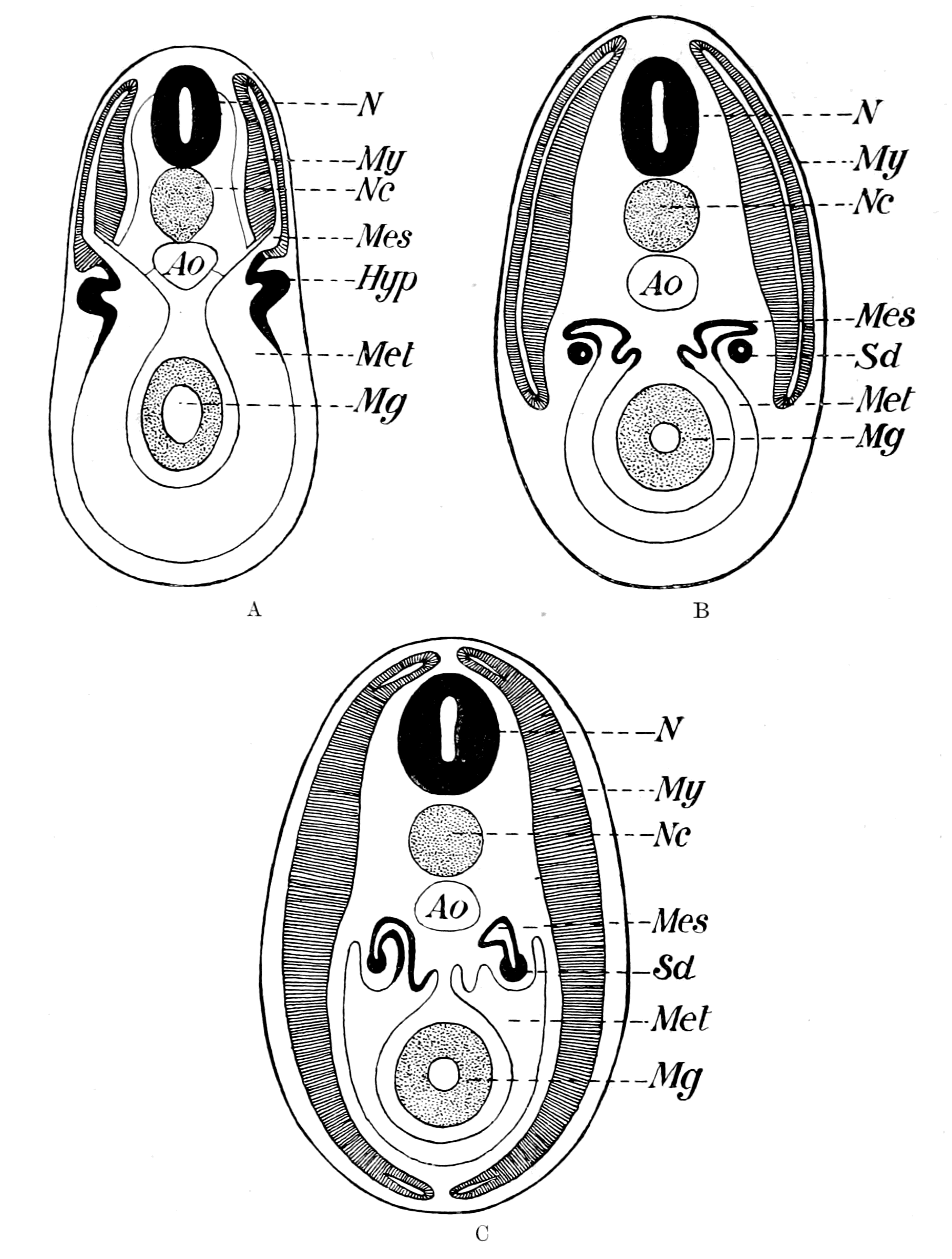
Fig. 159.—Diagrams to illustrate the Development of the Vertebrate Cœlom. (After van Wijhe.)
N., central nervous system; Nc., notochord; Ao., aorta; Mg., midgut. A, My., myocœle; Mes., mesocœle; Met., metacœle; Hyp., hypomere (pronephric). B and C, My., myotome; Mes., mesonephros; S.d., segmental duct (pronephric); Met., body-cavity.
Hatschek describes in Amphioxus how the cœlom splits into a dorsal segmented portion, the protovertebra, and a ventral unsegmented portion, the lateral plates. He describes in the dorsal part the formation of myotome and sclerotome, as in the Craniota. Also, he describes how the myotome is at first confined to the dorsal region in the neighbourhood of the spinal cord and notochord, and subsequently extends ventrally, until, just as in Ammocœtes, the body is enveloped in a sheet of somatic segmented muscles, the well-known myomeres.
The conclusion to be drawn from this is inevitable. Any explanation of the origin of the somatic muscles in Ammocœtes must also be an explanation of the somatic muscles in Amphioxus, and conversely; so that if in this respect Amphioxus is the more primitive and simpler, then the condition in Ammocœtes must be looked upon as derived from a more primitive condition, similar to that found in Amphioxus. Now, it is well known that a most important distinction exists between Amphioxus and Ammocœtes in the topographical relation of the ventral portion of this muscle-sheet, for in the former it is separated from the gut and the body-cavity by the atrial space, while in the latter there is no such space. Fürbringer therefore concludes, as I have already mentioned, that this space has become obliterated in the Craniota, but that it must be taken into consideration in any attempt at formulating the nature of the ancestors of the vertebrate.
Kowalewsky described this atrial space as formed by the ventral downgrowth of pleural folds on each side of the body, which met in the mid-ventral line and enclosed the branchial portion of the gut. According to this explanation, the whole ventral portion of the somatic musculature of the adult Amphioxus belongs to the extension of the pleural folds, the original body-musculature being confined to the dorsal region. This is expressed roughly on the external surface of Amphioxus by the direction of the connective tissue septa between the myotomes (cf. Fig. 162, B). These septa, as is well known, bend at an angle, the apex of which points towards the head. The part dorsal to the bend represents the part of the muscle belonging to the original body; the part ventral to the bend is the pleural part, and represents the extension into the pleural folds.
Lankester and Willey have attempted to give another explanation of the formation of the atrial cavity; they look upon it as originating from a ventral groove, which becomes a canal by the meeting of two outgrowths from the metapleure on each side. This canal then extends dorsalwards on each side, and so forms the atrial cavity; the metapleure still remains in the adult; the somatic muscles in the epipleure of the adult are the original body-muscles, and not extensions into an epipleuric fold, for there is no such fold.
This explanation is a possible conception for the post-branchial portion of the atrium, but is impossible for the branchial region; for, as Macbride points out, as must necessarily be the case, the point of origin of the atrial wall is, in all stages of development, situated at the end of the gill-slit. It shifts in position with the position of the gill-slit, but there can be no backwards extension of the cavity. Macbride therefore agrees with Kowalewsky that the atrial cavity is formed by the simultaneous ventral extension of pleural folds, and of the branchial part of the original pharynx. Thus, in his summing up, he states: "In the larva practically the whole sides and dorsal portion of the pharynx represent merely the hyper-pharyngeal groove and the adjacent epithelium of the pharynx of the adult, the whole of the branchial epithelium of the adult being represented by a very narrow strip of the ventral wall of the pharynx of the larva. The subsequent disproportionate growth of this part of the pharynx of the larva, and of the adjacent portion of the atrial cavity, has given the impression that the atrial cavity grew upwards and displaced other structures, which is not the case."
Further, van Wijhe states that the atrium extends beyond the atriopore right up to the anus, just as must have been the case if the pleural folds originally existed along the whole length of the body. His words are: "Allerdings hat sich das Atrium beim Amphioxus lanceolatus eigenthümlich ausgebildet, indem sich dasselbe durch den ganzen Rumpf bis an den Anus, d.h. bis an die Wurzel des Schwanzes ausdehnt."
We get, therefore, this conception of the origin of the somatic musculature of the vertebrate. The invertebrate ancestor possessed on each side, along the whole length of its body, a lateral fold or pleuron which was segmented with the body, and capable of movement with the body, because the dorsal longitudinal somatic muscles extended segmentally into each segment of the pleuron. By the ventral extension of these pleural folds, not only was the smooth body-surface of the vertebrate attained, but also the original appendages obliterated as such, leaving only as signs of their existence the branchiæ, the pronephric tubules, and the sense-organs of the lateral line system.
Such an explanation signifies that the somatic trunk-musculature of the vertebrate was derived from the dorsal longitudinal musculature of the body of the arthropod, and not from the ventral longitudinal musculature, and that therefore in the primitive arthropod stage the equivalent of the myotome of the vertebrate did not give origin to the ventral longitudinal muscles of the invertebrate ancestor. Now, as I have said, von Kennel states that in the procœlom of Peripatus a dorsal part (III. in Fig. 157) is cut off which gives origin to the dorsal body-musculature, while the ventral part which remains (I. and II. in Fig. 157) gives origin in its appendicular portion (I.) to the muscles of the appendage, and presumably in its ventral somatic portion (II.) to the ventral longitudinal muscles of the body. This dorsal cut-off part might be called the myotome, in the same sense as the corresponding part of the procœlom in the vertebrate is called the myotome. In both cases the muscles derived from it form only a part of the voluntary musculature of the animal, and in both cases the muscles in question are the dorsal longitudinal muscles of the body, to which must be added the dorso-ventral body-muscles. Now, the whole of my theory of the origin of vertebrates arose from the investigation of the structure of the cranial nerves, which led to the conception that their grouping is not, like the spinal, a dual grouping of motor and sensory elements, but a dual grouping to supply two sets of segments, characterized especially by the different embryological origin of their musculature. The one set I called the somatic segmentation, because the muscles belonging to it were the great longitudinal body-muscles; the other I called the splanchnic segmentation, because its muscles were those connected with the branchial and visceral arches. According to my theory, this latter segmentation was due to the segmentation of the appendages in the invertebrate ancestor; and in previous chapters, dealing as they do with the cranial region, attention was especially directed to the way in which the position of the striated splanchnic musculature could be explained by a transformation of the prosomatic and mesosomatic appendages. Now, I am dealing with the metasomatic region, in which it is true the appendages take a very subordinate place, but still something corresponding to the splanchnic segments of the cranial region might fairly be expected to exist, and I therefore desire to emphasize what appears to me to be the fact, that the musculature, which in the region of the trunk would correspond to that derived from the ventral segmentation of the mesoblast in the region of the head, may have arisen not only from the musculature of the appendages, but also from the ventral longitudinal musculature of the body of the invertebrate ancestor, for it seems probable that this latter musculature had nothing to do with the origin of the great longitudinal muscles of the vertebrate body, either dorsal or ventral.
The way in which I imagine the obliteration of the atrial cavity to have taken place is indicated in Fig. 160, B, which is a modification of a section across a trilobite-like animal as represented in Fig. 160, A. As is seen, the pleural folds on each side have nearly met the bulged-out ventral body-surface. A continuation of the same process would give Fig. 160, C, which is, to all intents and purposes, the same as Fig. 159, C, taken from van Wijhe, and shows how the segmental duct is left in the remains of the atrial cavity. The lining walls of the atrial cavity are represented very black, in order to indicate the presence of pigment, as indeed is seen in the corresponding position in Ammocœtes. In these diagrams I have represented the median ventral surface as a large bulged-out bag, without indicating any structures in it except the ventral extension of the procœlom to form the metacœlom. At present I will leave the space between the central nervous system and the ventral mesentery blank, as in the diagrams; in my next chapter I will discuss the possible method of formation within this blank space of the notochord and midgut. Boveri considers that the obliteration of the atrial cavity in the higher vertebrates is not complete, but that its presence is still visible in the shape of the pronephric duct. The evidence of Maas and others that the duct is formed by the fusion of the pronephric tubules is, it seems to me, conclusive against Boveri's view; but yet, as may be seen from my diagrammatic figures, the very place where one would expect to find the last remnant of the atrial cavity is exactly where the pronephric duct is situated. For my own part I should expect to find evidence of a former existence of an atrial cavity rather in the pigment round the pronephros and its duct than in the duct itself.
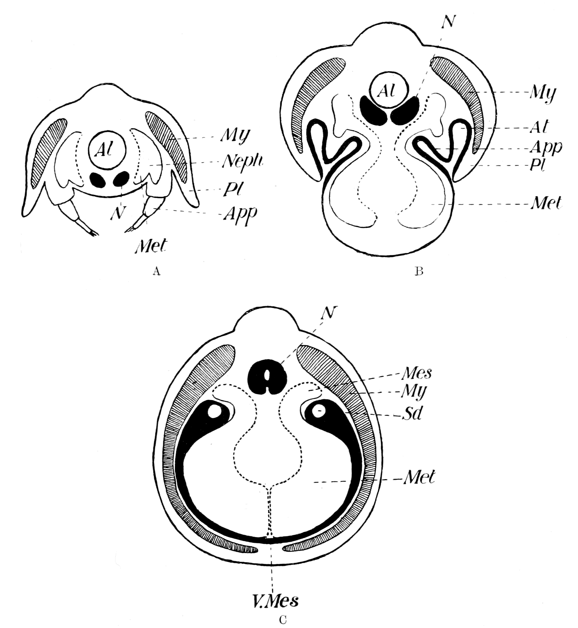
Fig. 160.—A, Diagram of Section through a Trilobite-like Animal; B, Diagram to illustrate Suggested Obliteration of Appendages and the Formation of an Atrial Cavity by the Ventral Extension of the Pleural Folds; C, Diagram to illustrate the Completion of the Vertebrate Type by the Meeting of the Pleural Folds in the Mid-ventral Line and the Obliteration of the Atrial Cavity.
Al., alimentary canal; N., nervous system; My., myotome; Pl., pleuron; App., appendage; Neph., nephrocœle; Met., metacœle; S.d., segmental duct; At., atrial chamber; V.Mes., ventral mesentery; Mes., mesonephros. The dotted line represents the splanchnopleuric mesoblast in all figures.
The conception that Amphioxus shows us how to account for the great envelope of somatic muscles which wraps round the vertebrate body, in that the ancestor of the vertebrate possessed on each side the body a segmented pleuron, is exactly in accordance with the theory of the origin of vertebrates deduced from the study of Ammocœtes, as already set forth in previous chapters. For we see that one of the striking characteristics of such forms as Bunodes, Hemiaspis, etc., is the presence of segmented pleural flaps on each side of the main part of the body; and if we pass further back to the great group of trilobites, we find in the most manifold form, and in various degrees of extent, the most markedly segmented pleural folds. In fact, the hypothetical figure (Fig. 160, A) which I have deduced from the embryological evidence, might very well represent a cross-section of a trilobite, provided only that each appendage of the trilobite possessed an excretory coxal gland.
The earliest fishes, then, ought to have possessed segmented pleural folds, which were moved by somatic muscles, and enveloped the body after the fashion of Ammocœtes and Amphioxus, and I cannot help thinking that Cephalaspis shows, in this respect also, its relation to Ammocœtes. It is well known that some of the fossil representatives of the Cephalaspids show exceedingly clearly that these animals possessed a very well-segmented body, and it is equally recognized that this skeleton is a calcareous, not a bony skeleton, and does not represent vertebræ, etc. It is generally called an aponeurotic skeleton, meaning thereby that what is preserved represents not dermal plates alone, or a vertebrate skeleton, but the calcified septa or aponeuroses between a number of muscle-segments or myomeres, precisely of the same kind as the septa between the myomeres in Ammocœtes. The termination of such septa on the surface would give rise to the appearance of dermal plates or scutes, or the septa may even have been attached to something of the nature of dermal plates. The same kind of picture would be represented if these connective tissue dissepiments of Ammocœtes were calcified, and the animal then fossilized. In agreement with this interpretation of the spinal skeleton of Cephalaspis, it may be noted that again and again, in parts of these dissepiments, I have found in old specimens of Ammocœtes nodules of cartilage formed, and at transformation it is in this very tissue that the spinal cartilages are formed.
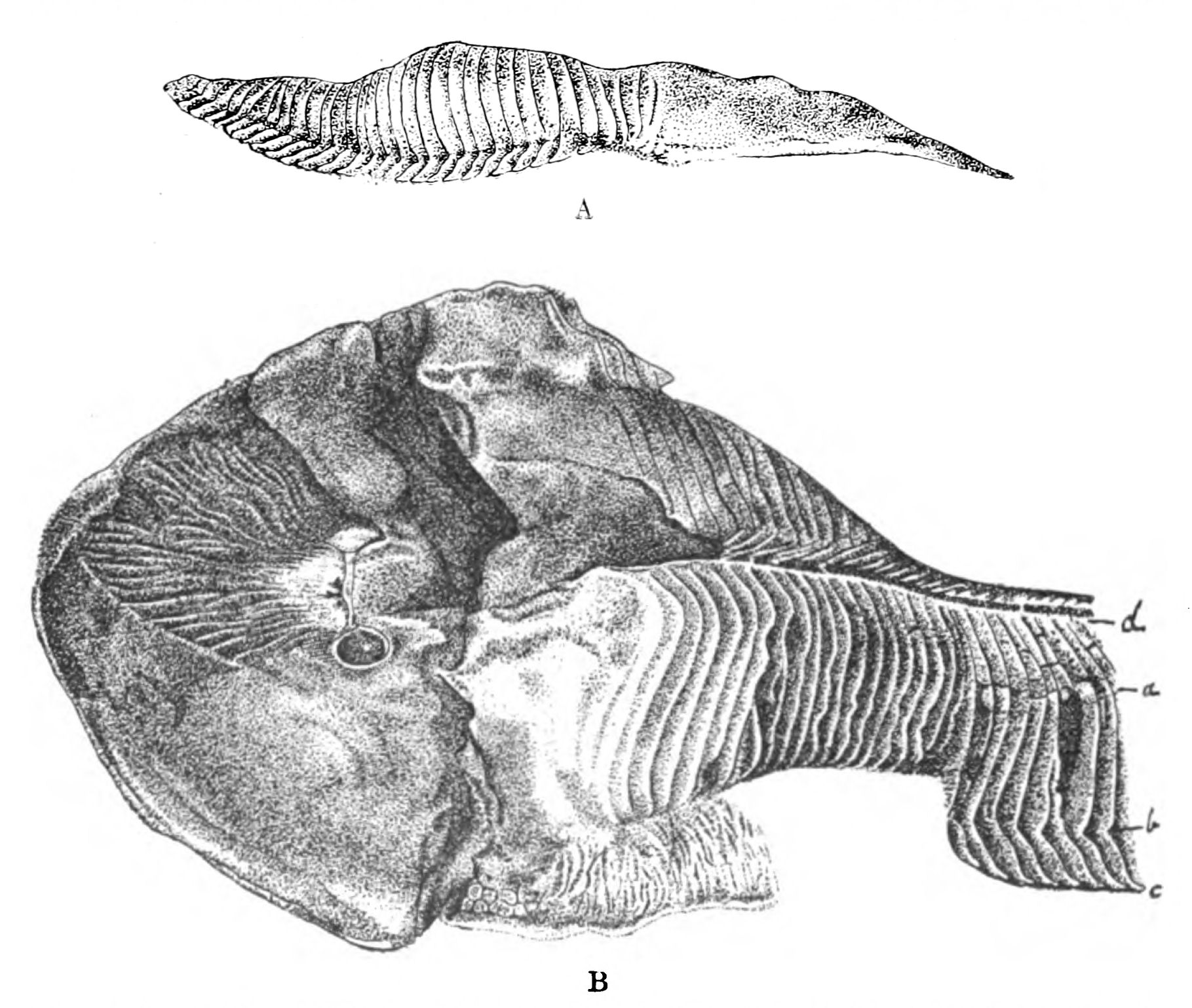
Fig. 161.—A, Facsimile of Woodward's Drawing of a Specimen of Cephalaspis Murchisoni, as seen from the side. The Cephalic Shield is on the Right and Caudal to it the Pleural Fringes are well shown; B, Another Specimen of Cephalaspis Murchisoni taken from the same block of Stone, showing the Dermoseptal Skeleton and in one place the Pleural Fringes, bc.
Now, the specimens of Cephalaspis all show, as seen in Fig. 161, that the skeletal septa cover the body regularly, and then along one line are bent away from the body to form, as it were, a fringe, or rather a free pleuron, which has been easily pushed at an angle to the body-skeleton in the process of fossilization. Patten thinks that this fringed appearance is evidence of a number of segmental appendages which were jointed to the corresponding body-segments, and in the best specimen at the South Kensington Natural History Museum he thinks such joints are clearly visible. He concludes, therefore, that the cephalaspids were arthropods, and not vertebrates. I have also carefully examined this specimen, and do not consider that what is seen resembles the joint of an arthropod appendage; the appearance is rather such as would be produced if the line of attachment of Patten's appendages to the body were the place where the pleural body folds became free from the body, and so with any pressure a bending or fracture of the calcified plates would take place along this line. There is, undoubtedly, an appearance of finish at the termination of these skeletal fringes, as though they terminated in a definitely shaped spear-like point, just as is seen in the trilobite pleuræ. This, again, to my mind, is rather evidence of pleural fringes than of true appendages.
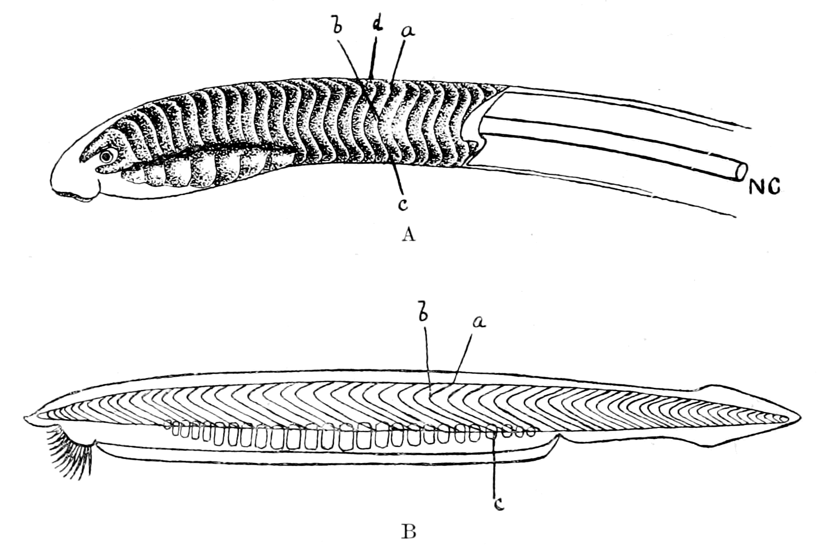
Fig. 162.—A, Arrangement of Septa in Ammocœtes (NC., position of notochord); B, Arrangement of Septa in Amphioxus.
As already argued, I look upon Ammocœtes as the only living fish at all resembling the cephalaspids; it is therefore instructive to compare the arrangement of this spinal dermo-septal skeleton of Cephalaspis with that of the septa between the myomeres in the trunk-region of Ammocœtes and Amphioxus. Such a skeleton in Ammocœtes would be represented by a series of plates overlapping each other, arranged as in Fig. 162, A, and in Amphioxus as in Fig. 162, B. I have lettered the corresponding parts of the two structures by similar letters, a, b, c. Ammocœtes differs in configuration from Amphioxus in that it possesses an extra dorsal (a, d) and an extra ventral bend. Ammocœtes is a much rounder animal than Amphioxus, and both the dorsal and ventral bends are on the extreme ventral and dorsal surfaces—surfaces which can hardly be said to exist in Amphioxus. The part, then, of such an aponeurotic skeleton in Ammocœtes which I imagine corresponds to b, c in Amphioxus, and therefore would represent the pleural fold, is the part ventral to the bend at b. In both the animals this bend corresponds to the position of the notochord NC.
The skeleton of Cephalaspis compares more directly with that of Ammocœtes than that of Amphioxus, for there is the same extra dorsal bend (Fig. 161, a, d) as in Ammocœtes; the lateral part of the skeleton again gives an angle a, b, c; the part from b to c would therefore represent the pleural fold. I picture to myself the sequence of events somewhat as follows:—
First, a protostracan ancestor, which, like Peripatus, possessed appendages on every segment into which cœlomic diverticula passed, forming a system of coxal glands; such glands, being derived from the segmental organs of the Chætopoda, discharged originally to the exterior by separate openings on each segment. It is, however, possible, and I think probable, that a fusion of these separate ducts had already taken place in the protostracan stage, so that there was only one external opening for the whole of these metasomatic coxal glands, just as there is only one external opening for the corresponding prosomatic coxal glands of Limulus. Then, by the ventral growth of pleural body-folds, such appendages became enclosed and useless, and the coxal glands of the post-branchial segments, with their segmental or pronephric duct, were all that remained as evidence of such appendages. This dwindling of the metasomatic appendages was accompanied by the getting-rid of free appendages generally, in the manner already set forth, with the result that a smooth fish-like body-surface was formed; then the necessity of increasing mobility brought about elongation by the addition of segments between those last formed and the cloacal region. Each of such new-formed segments was appendageless, so that its segmental organ was not a coxal gland, but entirely somatic in position, and formed, therefore, a mesonephric tubule, not a pronephric one. Such glands could no longer excrete to the exterior, owing to the enclosing shell of the pleural folds; but the pronephric duct was there, already formed, and so these nephric tubules opened into that, instead of, as in the case of the branchial slits, forcing their way through the pleural walls when the atrium became closed.
The Meaning of the Ductless Glands.
If it is a right conception that the excretory organs of the protostracan group, which gave origin to the vertebrates as well as to the crustaceans and arachnids, were of the nature of coxal glands, then it follows that such coxal glands must have existed originally on every segment, because they themselves were derived from the segmental organs of the annelids; it is therefore worth while making an attempt to trace the fate of such segmental organs in the vertebrate as well as in the crustacean and arachnid.
Such an attempt is possible, it seems to me, because there exists throughout the animal kingdom striking evidence that excretory organs which no longer excrete to the exterior do not disappear, but still perform excretory functions of a different character. Their cells still take up effete or injurious substances, and instead of excreting to the exterior, excrete into the blood, forming either ductless glands of special character, or glands of the nature of lymphatic glands.
The problem presented to us is as follows:—
The excretory organs of both arthropods and vertebrates arose from those of annelids, and were therefore originally present in every segment of the body. In most arthropods and vertebrates they are present only in certain regions; in the former case, as the coxal glands of the prosomatic or head-region; in the latter, as the nephric glands of the metasomatic or trunk-region, and, in the case of Amphioxus, of the mesosomatic or branchial region.
In the original arthropod, judging from Peripatus, they were present, as in the annelid, in all the segments of the body, and formed coxal glands. Therefore, in the ancestors of the living Crustacea and Arachnida, coxal glands must have existed in all the segments of the body, and we ought to be able to find the vestiges of them in the mesosomatic or branchial and metasomatic or abdominal regions of the body.
Similarly, in the vertebrates, derived, as has been shown, not from the annelids, but from an arthropod stock, evidence of the previous existence of coxal glands ought to be manifested in the prosomatic or trigeminal region, in the mesosomatic or branchial region, as well as in the metasomatic or post-branchial region.
How does an excretory organ change its character when it ceases to excrete to the exterior? What should we look for in our search after the lost coxal glands?
The answer to these questions is most plainly given in the case of the pronephros, especially in Myxine, where Maas has been able to follow out the whole process of the conversion of nephric tubules into a tissue resembling that of a lymph-gland.
He states, in the first place, that the pronephros possesses a capillary network, which extends over the pronephric duct, while the tubules of the mesonephros possess not only this capillary network, equivalent to the capillaries over the convoluted tubules in the higher vertebrates, but also a true glomerulus, in that the nephric segmental arteriole forms a coil (Knauel), and pushes in the wall of the mesonephric tubule. He describes the pronephros of large adult individuals as consisting of—
1. Tubules with funnels which open into the pericardial cœlom.
2. A large capillary network (the glomus) at the distal end.
3. A peculiar tissue (the 'strittige Gewebe' of the Semon-Spengel controversy), which Spengel considers to be composed of the altered epithelium of pronephric tubules, while Semon looks on it as an amalgamation of glomeruli.
Maas is entirely on the side of Spengel, and shows that this peculiar tissue is actually formed by modified pronephric tubules, which become more and more lymphatic in character.
He says: "The pronephros consists of a number of nephric tubules, placed separately one behind the other, which were originally segmental in character, each one of which is supplied by a capillary network from a segmental branch of the aorta. The tubules begin with many mouths (dorso-lateral and medial-ventral) in the pericardial cavity; on their other blind end they have lost their original external opening, and there, in the cranial portion of the head-kidney, before they have joined together to form a collecting duct, they, together with the vascular network, are transformed into a peculiar adrenal-like tissue. The most posterior of the segmental capillary nets retain their original character, and are concentrated into the separate capillary mass known as the glomus."
Later on he says: "Further, the separate head-kidney is more and more removed in structure from an excretory organ in the ordinary sense. One cannot, however, speak of it as an organ becoming rudimentary; this is proved not only by the progressive transformation of its internal tissue into a tissue of a very definite character, but also by the cilia in its canals, and the steady increase in the number of its funnels. It appears, therefore, to be the conversion of an excretory organ into an organ for the transference of fluid out of the cœlom into a special tissue, i.e. into its blood-sinus; in other words, into an organ which must be classed as belonging to the lymph-system."
In exact correspondence with this transformation of a nephric tubule into a ductless gland of the nature of a lymphatic gland, is the formation of the head-kidney in the Teleostea. Thus, Weldon points out that, though the observations of Balfour left it highly probable that the "lymphatic" tissue described by him was really a result of the transformation of part of the embryonic kidney, he did not investigate the details of its development. This was afterwards done by Emery, with the following results: "In those Teleostea which he has studied, Professor Emery finds that at an early stage the kidney consists entirely of a single pronephric funnel, opening into the pericardium, and connected with the segmental duct, which already opens to the exterior. Behind this funnel, the segmental duct is surrounded by a blastema, derived from the intermediate cell-mass, which afterwards arranges itself more or less completely into a series of solid cords, attaching themselves to the duct. These develop a lumen, and become normal segmental tubules, but it is, if I may be allowed the expression, a matter of chance how much of the blastema becomes so transformed into kidney tubules, and how much is left as the 'lymphatic' tissue of Balfour, this 'lymphatic' tissue remaining either in the pronephros only, or in both pro- and meso-nephros."
If we turn now to the invertebrates, we see also how close a connection exists between lymphatic and phagocytic organs and excretory organs. The chief merit for this discovery is due to Kowalewsky, who, taking a hint from Heidenhain's work on the kidney, in which he showed how easy it was to find out the nature of different parts of the mammalian excretory organ by the injection of different substances, such as a solution of ammoniated carmine, or of indigo-carmine, has injected into a large number of different invertebrates various colouring matters, or litmus, or bacilli, and thus shown the existence, not only of known excretory organs, but also of others, lymphatic or lymphoid in nature, not hitherto suspected.
In all cases he finds that a phagocytic action with respect to solid bodies is a property of the leucocytes, and that these leucocytes which are found in the cœlomic spaces of the Annelida, etc., are apparently derived from the epithelium of such spaces. Also by the proliferation of such epithelium in places, e.g. the septal glands of the terrestrial Oligochæta, segmental glandular masses of such tissue are formed which take up the colouring matter, or the bacilli. In the limicolous Oligochæta such septal glands are not found, but at the commencement of the nephridial organ, immediately following upon the funnel, a remarkable modification of the nephridial wall takes place to form a large cellular cavernous mass, the so-called filter, which in Euaxes is full of leucocytes; the cells are only definable by their nuclei, and look like and act in the same way as the free leucocytes outside this nephridial appendage. As G. Schneider points out, the whole arrangement is very like that described by Kowalewsky in the leeches Clepsine and Nephelis, where, also immediately succeeding the funnel of the nephridial organ, a large accessory organ is found, which is part of the nephridium, and is called the nephridial capsule. This is the organ par excellence which takes up the solid carmine-grains and bacilli, and apparently, from Kowalewsky's description, contains leucocytes in large numbers. We see, then, that in such invertebrates, just as in the vertebrate, modifications of the true excretory organ may give rise to phagocytic glands of the nature of lymphatic glands. Further, these researches of Kowalewsky suggest in the very strongest manner that whenever by such means new, hitherto unsuspected glands are discovered, such glands must belong to the excretory system, i.e. must be derived from cœlomic epithelium, even when all evidence of any cœlom has disappeared. Kowalewsky himself was evidently so impressed with the same feeling that he heads one of his papers "The Excretory Organs of the Pantopoda," although the organs in question had been discovered by him by this method, and appeared as ductless glands with no external opening.
To my mind these observations of Kowalewsky are of exceeding interest, for it is immediately clear that if the segmental organs of the annelids, which must have existed on all the segments of the forefathers of the Crustacea and Arachnida (the Protostraca), have left any sign of their existence in living crustaceans and arachnids, then such indication would most likely take the form of lymphatic glands in the places where the excretory organs ought to have been.
Now, as already pointed out in Peripatus, such segmental organs were formed by the ventral part of the cœlom, and dipped originally into each appendage. We know also that each segment of an arachnid embryo possesses a cœlomic cavity in its ventral part which extends into the appendage on each side; this cavity afterwards disappears, and is said to leave no trace in the adult of any excretory coxal gland derived from its walls. If, however, it is found that in the very position where such organ ought to have been formed a segmentally arranged ductless gland is situated, the existence of which is shown by its taking up carmine, etc., then it seems to me that in all probability such gland is the modification of the original coxal gland.