Front matter (continued 11)
Reading edition
Let us, then, apply this throughout, and see what are the logical results of tracing back the various organs and tissues from the adult to the embryo.
The adult body is built up of different kinds of tissues, which fall naturally, from the standpoint of physiology, into groups. Such groups are, in the first place—
1. All those tissues which are connected with the central nervous system, including in that group the nervous system itself.
2. All those tissues which have no connection with the nervous system.
In the second group the physiologist places all germinal cells, all blood- and lymph-corpuscles, all plasma-cells and connective tissue and its derivatives—in fact, all free-living cells, whether in a free state or in a quiescent, so to speak encysted, condition, such as is found in connective tissue. In the first group the physiologist recognizes that the central nervous system is connected with all muscular tissues, whether striped or unstriped, somatic or splanchnic, and that such connection is of an intimate character. Further, all epithelial cells, either of the outer or inner surfaces, whether forming special sense-organs and glands, such as the digestive and sweat-glands, or not, are connected with the nervous system. Besides these structures, there is another set of organs as to which we cannot speak definitely at present, which must be considered separately, viz. all the cells, together with their derived organs, which line the body-spaces. Whatever may be the ultimate decision as to this group of cells, it must fall into one or other of the two main groups.
The members of these two groups are so interwoven with one another that either, if taken alone, would still give the form of the body, so that, in a certain sense, we can speak of the body as formed of two syncytia, separate from each other, but interlaced, of which the one forms a continuous whole by means of cells connected together by a fluid medium or by solid threads formed in such fluid medium, while the other does not form a syncytium in the sense that any cell of one kind may be connected with any cell of another kind, but a syncytium of which all the different elements are connected together only through the medium of the nervous system.
If we choose to speak of the body as made up of two syncytia in this way, we must at the same time recognize the fundamental difference in character between them. In the one case the elements are connected together only by what may be called non-living material; there is no direct metabolic activity caused by the action of one cell over a more distant cell in consequence of such connection, it is not a true syncytium; in the second case there is a living connection, the metabolism of one part is directly influenced by the activity of another, and the whole utility of the system depends upon such functional connection.
The tissues composing this second syncytium may be spoken of as the master-tissues of the body, and we may express this conception of the building up of the body of the higher Metazoa by saying that it is composed of a syncytial host formed of the master-tissues, which contains within its meshes a system of free-living cells, none of which have any connection with the nervous system. This syncytial host is in the adult composed of a number of double elements, a nerve-cell element, and an epithelial element, such as muscle-cell, gland-cell, etc., connected together by nerves; and if such connection is always present as we pass from the adult to the embryo, if there is no period when, for example, the neural element exists alone free from the muscle-cell, no period when the two can be seen to come together and join, then it follows that when the single-layered blastula stage is reached, muscle-cell and nerve-cell must have fused together to form a neuro-muscular cell. Similarly with all the other neuro-epithelial organs; however far apart their two components may be in the adult, they must come together and fuse in the embryo to form a neuro-epithelial element.
The close connection between muscle and nerve which has always been recognized by physiologists, together with the origin of muscle from a myo-epithelial cell in Hydra and other Cœlenterata, led the older physiologists to accept thoroughly Hensen's views of the neuro-epithelial origin of all tissues connected with the central nervous system. Of late years this conception has been largely given up owing to the statement of His that the nervous system arises from a number of neuroblasts, which are entirely separate cells, and have at first no connection with muscle-cells or any peripheral epithelial cells, but subsequently, by the outgrowing of an axial fibre, find their way to the muscle, etc., and connect with it. I do not think that His' statement by itself would have induced any physiologist to give up the conception of the intimate connection of muscle and nerve, if the work of Golgi, Ramón y Cajal, and others had not brought into prominence the neurone theory, i.e. that each element of the central nervous system is an independent element, without real connection with any other element and capable of influencing other cells by contact only. These two statements, emanating as they did from embryological and anatomical studies respectively, have done much to put into the background Hensen's conceptions of the syncytial nature of the motor, neural, and sensory elements, which make up the master-tissues of the body, and have led to the view that all the elements of the body are alike, in so far as they are formed of separate cells each leading an independent existence, without any real intimate connection with each other.
The further progress of investigation is, it seems to me, bringing us back to the older conception, for not only has the neuroblast theory proved very difficult for physiologists to accept, but also Graham Kerr, in his latest papers on the development of Lepidosiren, has shown that there is continuity between the nerve-cell and the muscle-cell from the very first separation of the two sets of elements; in fact, Hensen is right and His wrong in their respective interpretation of the earliest stages of the connection between muscle and nerve. So also, it seems to me, the intimate connection between the metabolism of the gland-cell, as seen in the submaxillary gland, and the integrity of its nervous connection implies that the connection between nerve-cell and gland-cell is of the same order as that between nerve-cell and muscle-cell. Graham Kerr also states in his paper that from the very commencement there is, he believes, continuity between nerve-cell and epithelial cell, but so far he has not obtained sufficiently clear evidence to enable him to speak positively on this point.
Further, according to the researches of Anderson, the cells of the superior cervical ganglion in a new-born animal will continue to grow healthily as long as they remain connected with the periphery, even though entirely separated from the central nervous system by section of the cervical sympathetic nerve, and conversely, when separated from the periphery, will atrophy, even though still connected with the central nervous system. So, also, on the sensory side, Anderson has shown that the ganglion-cells of the posterior root-ganglion will grow and remain healthy after separation of the posterior roots in a new-born animal, but will atrophy if the peripheral nerve is cut, even though they are still in connection with the central nervous system. Further, although section of a posterior root in the new-born animal does not affect the development of the nerve-cells in the spinal ganglion, and of the nerve-fibres connecting the posterior root-ganglion with the periphery, it does hinder the development of that part of the posterior root connected with the spinal ganglion.
These experiments of Anderson are of enormous importance, and force us, it seems to me, to the same conclusion as that to which he has already arrived. His words are (p. 511): "I suggest, therefore, that the section of peripheral nerves checked the development of motor and sensory neurones, not because it blocked the passage of efferent impulses in the first case and the reception of stimuli from the periphery in the second, but for the same reason in both cases, viz. that the lesion disturbed the chemico-physical equilibrium of an anatomically continuous (neuro-muscular or neuro-epithelial) chain of cells, by separating the non-nervous from the nervous, and that the changes occurring in denervated muscle, which I shall describe later (and possibly those in denervated skin), are in part due to the reciprocal chemico-physical disturbance effected in these tissues by their separation from the nervous tissues; also that the section of the posterior roots checked the development of those portions of them still attached to the spinal ganglia, because the chemico-physical equilibrium in those processes is maintained not only by the spinal ganglion-cells, but also by the intra-spinal cells with which these processes are anatomically continuous."
What is seen so strikingly in the new-born animal can be seen also in the adult, and in Anderson's paper references are given to the papers of Lugaro and others which lead to the same conclusion.
These experiments seem to me distinctly to prove that the connection between the elements of the peripheral organ and the proximate neurone is more than one of contact.
We can, however, go further than this, for, apart from the observations of Apathy, there is direct physiological evidence that the vitality of other neurones besides the terminal neurones is dependent upon their connection with the peripheral organ, even though their only connection with the periphery is by way of the terminal neurone. Thus, as is seen from Anderson's experiments, section of the cervical sympathetic nerve in a very young animal causes atrophy of many of the cells in the corresponding intermedio-lateral tract, cells which I supposed gave origin to all the vaso-constrictor, pilomotor, and sweat-gland nerves. A still more striking experiment given by Anderson is the effect of the removal of the periphery upon the medullation of those efferent fibres which arise from these same spinal cells, for, as he has shown, section of the nerves from the superior cervical ganglion to the periphery in a very young animal delays the medullation in the fibres of the cervical sympathetic—that is, in preganglionic fibres which are not directly connected with the periphery but with the terminal neurones in the superior cervical ganglion. So also on the afferent side a sufficiently extensive removal of sensory field will cause atrophy of the cells of Clarke's column, so that, just as in the case of the primary neurones, the secondary neurones show by their degenerative changes the importance of their connection with the peripheral organs.
In this way I can conceive the formation of a series of both efferent and afferent relays in the nervous system by proliferation of the original neural moiety of the neuro-epithelial elements, every one of which is dependent upon its connection with the peripheral epithelial elements for its due vitality, the whole system being a scheme for co-ordination of a larger and larger number of peripheral elements. Thus the cells of the vasomotor centre are in connection with the whole system of segmental vaso-constrictor centres in the lateral horns of the thoracic region of the cord, so that to cause atrophy of these cells a very extensive removal of the vascular system would be required. Each of the segmental centres in the cord supplies a number of sympathetic segments, the connection with all of which would have to be cut in order to ensure complete removal of the connection of each of its cells with the periphery, and finally each of the cells in the sympathetic ganglia supplies a number of peripheral elements, all of which must be removed to ensure complete severance.
Thus, if we take any arbitrary number, such as 4, to represent the number of peripheral organ-elements with which each terminal neurone is connected, and suppose that each neurone has proliferated into sets of 4, then a cell of the third order, such as a cell of the vasomotor centre, would require the removal of 64 peripheral elements to cause its complete separation from the periphery, one of the second order (a cell of the thoracic lateral horn) 16 elements, one of the first order (a cell of a sympathetic ganglion) 4 elements.
Such intimate inter-relationship between the neurones, both afferent and efferent, and their corresponding peripheral organs does not imply that all nerve-cells are necessarily as closely dependent upon some connection with the periphery, for just as the proliferation of epithelial or muscle-cells forms an epithelial or muscular sheet, the elements of which are so loosely, if at all, connected together that their metabolism is in no way dependent upon such connection, so also a similar proliferation of the neural elements may form connections between nerve-cell and nerve-cell of a similarly loose nature.
It is this kind of proliferation which, in my opinion, would bind together the separate relays of efferent and afferent neurones, and so give origin to reflex actions at different levels. Such neurones would not be in the direct chain of either the afferent or efferent neurones, and so not directly connected with the periphery, and could therefore be removed without affecting the vitality of either the efferent or afferent chain of neurones. In other words, the vitality of the cells on the efferent side ought not to be dependent on the integrity of the reflex arc. With regard to the development of the anterior roots, Anderson has shown that this is the case, for section of all the posterior roots conveying afferent impulses from the lower limb in a new-born animal does not hinder the normal development of the anterior roots supplying that limb. Also Mott, who originally thought that section of all the posterior roots to a limb caused atrophy of the corresponding anterior roots, has now come to the same conclusion as other observers, and can find no degeneration on the efferent side due to removal of afferent impulses.
Again, the process of regeneration after section of a nerve is not in favour of the neuroblast theory. There is no evidence that the cut end of a nerve can grow down and attach itself to a muscular or epithelial element without the assistance of a nerve tube down which to grow. When the cut nerves connected with the periphery degenerate, that applies only to the axis-cylinder and the medullary sheath, not to the neurilemma; the connective tissue elements remain alive and form a tube into which the growing axon finds its way, and so is conducted to the end-plate or end-organ of the peripheral structure.
Possibly, as suggested by Mott and Halliburton, the products of degeneration of the axis-cylinder and medullary sheath stimulate these connective tissue sheath-cells into active proliferation, and so bring about the great multiplication of cells arranged as cell-chains, which are so often erroneously spoken of as forming the young nerves. These sheath-cells are then supposed to re-form and secrete a pabulum which is important for the process of regeneration of the down-growing axis-cylinder and medullary sheath. Without such pabulum regeneration does not take place, as is seen in the central nervous system, where the sheath of Schwann is absent.
Again, it is becoming more and more doubtful whether the peripheral terminations of nerves are ever really free. As far as efferent nerves are concerned the nervous element may entirely predominate over the muscular or glandular, as in the formation of the electric organs of the Torpedo and Malapterurus, but still the final effect is produced by the alteration of the muscle or gland-cell. On the afferent side especially free nerve-terminations are largely recognized, or, as in Barker's book, nerves are spoken of as arising in connective tissue. Thus the numerous kinds of special sense-organs, such as Pacinian bodies, tendon-organs, genital corpuscles, etc., are all referred to by Barker under the heading of "sensory nerve beginnings in mesoblastic tissues." Yet the type of these organs has been known for a long time in the shape of Grandry's corpuscles or the tactile corpuscles in the duck's bill, where it has been proved that the nerve terminates in special large tactile cells derived from the surface-epithelium.
So also with all the others, further investigation tends to put them all in the same category, all special sensory organs originating from a localized patch of surface-epithelium. Thus Anderson has shown me in his specimens how the young Pacinian body is composed of rows of epithelial cells, into each of which a twig from the nerve passes. He has also shown me how, in the case of the tendon-organ, each nerve-fibre passes towards the attachment of the tendon and then bends back to supply the tendon-organ, thus indicating, as he suggests, how the nest of epithelial cells has wandered inwards from the surface to form the tendon-organ. Again, Meissner's corpuscles and Herbst's corpuscles are evidently referable to the same class as those of Grandry and Pacini.
Yet another instance of the same kind is to be found in the chromatophores of the frog and other animals which are under the influence of the central nervous system and yet have been supposed by various observers to be pigmented connective tissue cells. The most recent work of Leo Loeb and others has conclusively shown that such cells are invariably derived from the surface-epithelium.
Finally, in fishes we find the special sense-organs of the lateral line and other accessory sensory organs, all of which are indisputably formed from modified surface epithelial cells.
The whole of this evidence seems to me directly against Barker's classification of sensory nerve-beginnings in mesoblastic tissues; in none of these cases are we really dealing with free nervous tissue alone, the starting point is always a neuro-epithelial couple.
We may then, I would suggest, look upon the adult as formed of a neural syncytium, which we may call the host, which carries with it in its meshes a number of free cells not connected with the nervous system. If, then, we confine our attention to the host and trace back this neural syncytium to its beginnings in the embryo, we see that, from the very nature of the neuro-epithelial couple, each epithelial moiety must approach nearer and nearer to its neural moiety, until at last it merges with it; the original neuro-epithelial cell results, and we must obtain, as far as the host is concerned, a single-layered blastula as the origin of all Metazoa. It follows, further, that there must always be continuity of growth in the formation of the host, i.e. in the formation of the neuro-epithelial syncytium; that therefore cells which have been previously free cannot settle down and take part in its formation, as, for instance, in the case of the formation of any part of the gut-epithelium or of muscle-cells from free-living cells.
Further, since the neural moiety is the one element common to all the different factors which constitute the host, it follows that the convergence of each epithelial moiety to the neural moiety, as we pass from the adult to the embryo, is a convergence of all outlying parts to the neural moiety, i.e. to the central nervous system, if there is a concentrated nervous system. Conversely, in the commencing embryo the place from which the spreading out of cells takes place, i.e. from which growth proceeds, must be the position of the central nervous system, if the nervous system is concentrated. If the nervous system is diffuse, and forms a general sub-epithelial layer, then the growth of the embryo would take place over the whole surface of the blastula.
Turning now to the consideration of the second group of tissues, those that are not connected with the central nervous system, we find that they include among them such special cells as the germinal cells, free cells of markedly phagocytic nature, and cells which were originally free and phagocytic, but have settled down to form a supporting framework of connective tissue, and are known as plasma-cells. In the embryo we find also in many cases free cells in the yolk, forming more or less of a layer, which function as phagocytes and prepare the pabulum for the fixed cells of the growing embryo; these cells are known by the name of vitellophags, and in meroblastic vertebrate eggs form somewhat of a layer known by the name of periblast. Such cells must be included in the second group, and, indeed, have been said again and again to give origin to the free-living blood-corpuscles of the adult. In other cases they are said to disintegrate after their work is done.
In the adult the free-living lymphocytes and hæmocytes reproduce themselves from already existing free-living cells, but as we pass back to the embryo there comes a time, comparatively late in the history of the embryo, when such free-living cells are not found in the fluids of the body, and they are said to arise from the proliferation and setting free of cells which form a lining epithelium. Such formation of leucocytes has been especially described in connection with the lining epithelium of the cœlomic cavities, as stated in Chapter XII., so that anatomists look upon the origin of these free cells as being largely from the cœlomic epithelium, or mesothelium, as Minot calls it.
Then, again, the free cells which form the germinal cells can be traced back to a germinal epithelium, which also is part of the cœlom. Thus the suggestion arises that in the embryo a cellular lining is formed to a cœlomic cavity (mesothelium) composed of cells which have no communication with the nervous system, and are capable of a separate existence as free individuals, either in the form of germinal cells or of lymphocytes, hæmocytes, and plasma-cells, so that these latter free cells may be considered as living an independent existence in the body, and ministering to it in the same sense as the germ-cells live an independent existence in the body. Again, the function of this mesothelium apart from the germ-cell is essentially excretory and phagocytic. It is the cells of the excretory organs as well as the lymphocytes which pick up carmine-grains when injected. It is the cells of the modified excretory organs, as mentioned in Chapter XII., which, according to Kowalewsky and others, give origin to the free leucocytes.
We see, then, that the conception of a syncytial neuro-epithelial host holding in its meshes a number of free cells leads directly to the questions: What is the cœlom? To which category does its lining membrane belong? and further, also, What is the origin of these free cells?
The Metazoa have been divided into two great groups—those which possess a cœlom (the Cœlomata; Lankester's Cœlomocœla) and those which do not (Cœlenterata; Lankester's Enterocœla). As an example of the latter we may take Hydra, because it is a very primitive form, and because its development has been carefully worked out recently by Brauer.
In Hydra we find a dermal layer of cells and an inner layer of cells separated by a gelatinous mass known as mesoglœa; in this mass between the dermal and inner layers scattered cells are found, the interstitial cells. Now, according to Brauer the position of the germ in Hydra is the interstitial cell-layer. One cell of the ovarium becomes the egg-cell, the others have their substance changed into yolk-grains, forming the so-called pseudo-cells, and as such afford pabulum to the growing egg-cell. Thus we see that in between the dermal and gastral layer of cells a third layer of cells is found, composed of free living germ-cells, some of which, by the formation of yolk-granules, become degraded into pabulum for their more favoured kinsfolk. These interstitial cells are said to arise from the dermal layer, or ectoderm, but clearly, as in other cases, germ-cells constitute a class by themselves and cannot be spoken of as originating from ectoderm-cells or from hypoderm-cells.
So also in Porifera, Minchin states: "In addition to the collared cells of the gastral layer, and the various cell-elements of the dermal layer, the body-wall contains numerous wandering cells or amœbocytes, which occur everywhere among the cells and tissues. Though lodged principally in the dermal layer, they are not to be regarded as belonging to it, but as constituting a distinct class of cells by themselves. They are concerned probably with the functions of nutrition and excretion, and from them arise the genital products." Further (p. 31): "At certain seasons some of these cells become germ-cells; hence the wandering cells and the reproductive cells may be included together under the general term archæocytes." Also (p. 51): "The mesoglœa is the first portion to appear as a structureless layer between the dermal and gastral epithelia, and is probably a secretion of the former."
He also points out that in these, the very lowest of the Metazoa, the separate origin of these archæocytes can be traced back to a very early period of embryonic life. Thus in Clathrina blanca the ovum undergoes a regular and total cleavage, resulting in the formation of a hollow ciliated blastula of oval form. At one point, the future posterior pole of the larva, are a pair of very large granular cells with vesicular nuclei, which represent undifferentiated blastomeres and are destined to give rise to the archæocytes, and, therefore, also to the sexual cells of the adult. Thus, as he says, from the very earliest period a distinction is made between the "tissue-forming" cells (my syncytial host) and the archæocytes.
We see, then, that the origin of all these free-living cells can be traced back to the very earliest of the Metazoa. Here between the dermal and gastral layers a gelatinous material, the mesoglœa is secreted by these layers. This material is non-living, non-cellular. In it live free cells which may either be germ-cells, amœbocytes, or 'collencytes' (connective tissue cells). If this mesoglœa were a fluid secretion, then we should have a tissue of the nature of blood or lymph; if it were solid, then we should have the foundation of connective tissue, cartilage, and bone.
From this primitive tissue it is easy to see how the special elements of the vascular, lymphatic, and skeletal tissues gradually arose, the matrix being provided by the cells of the syncytial host and the cellular elements by the archæocytes. In fact, we have no right to speak of these lowest members of the Metazoa as not being triploblastic, as possessing nothing corresponding to mesoblast, for in these free cells in the mesoglœa we have the origin of the mesenchyme of the higher groups. Thus Lankester, talking of mesenchyme, says: "I think we are bound to bring into consideration here the existence in many Cœlentera of a tissue resembling the mesenchyme of Cœlomocœla. In Scyphomedusæ, in Ctenophora, and in Anthozoa, branched fixed and wandering cells are found in the mesoglœa which seem to be the same thing as a good deal of what is distinguished as mesenchyme in Cœlomocœla. These appear to be derived from both the primitive layers; some produce spicules, others fibrous substance, others again seem to be amœbocytes with various functions. It appears to be probable that, though it may be necessary to distinguish other elements in it, the mesenchyme of Cœlomocœla is largely constituted by cells, which are the mother-cells of the skeletotrophic group of tissues, and are destined to form connective tissues, blood-vessels, and blood."
Thus we see that the earliest Metazoa were composed of a dermal and gastral epithelium, with a sub-epithelial nervous system connecting the parts together, which formed, as it were, a host, carrying around free living cells of varying function, all of which may be looked on as derived from archæocytes, i.e. germ-cells. From these the cœlomatous animals arose, and here also we find, according to present-day opinion, that the cœlom arose in the first place in the very closest connection with the germ-cells or gonads. Thus Lankester, in his review of the history of the cœlom, states:—
"The numerous embryological and anatomical researches of the past twenty years seem to me to definitely establish the conclusion that the cœlom is primarily the cavity, from the walls of which the gonad cells (ova or spermata) develop, or which forms around those cells. We may suppose the first cœlom to have originated by a closing or shutting off of that portion of the general archenteron of Enterocœla (Cœlentera), in which the gonads developed as in Aurelia or as in Ctenophora. Or we may suppose that groups of gonad mother cells, having proliferated from the endoderm, took up a position between it and the ectoderm, and there acquired a vesicular arrangement, the cells surrounding the cavity in which liquid accumulated.
"The cœlom is thus essentially and primarily (as first clearly formulated by Hatschek) the perigonadial cavity or gonocœl, and the lining cells of gonadial chambers are cœlomic epithelium. In some few groups of Cœlomocœla the cœloms have remained small and limited to the character of gonocœls. This seems to be the case in the Nemertina, the Planarians, and other Platyhelmia. In some Planarians they are limited in number, and of individually large size; in others they are numerous."
When Lankester says that "the lining cells of gonadial chambers are cœlomic epithelium," that is equivalent to saying that the lining cells of the cœlom form an epithelium which was originally gonadial, provided that, as seems to me most probable, his second suggestion, of the cœlom being formed from gonadial mother-cells which have taken up an intermediate position between endoderm and ectoderm and there acquired a vesicular arrangement, is the true one. It does not seem to me possible to conceive of the gonads arising from cells of the epiblast or of the hypoblast, in the sense that such cells are differentiated cells belonging to a layer with a definite meaning. When we consider that the gonad gives origin to the whole of a new individual, that in the protozoan ancestors of the Metazoa their ultimate aim and object was the formation of gonads, it seems a wrong conception to speak of the gonads as formed from cells belonging either to the gut-wall or to the external epithelium. The gonads must stand in a category by themselves; they represent a whole, while the other cells represent only a part; they cannot therefore be derived from the latter. They may, and indeed do, give rise to cells of a subordinate character, but they cannot rightly be spoken of as derived from such cells. The very fact mentioned by Lankester, that in the lowest cœlomatous Metazoa, the Platyhelminthes, the cœloms are limited to the character of simple gonocœls, strongly points to the conclusion that all the cœlomic cells were originally of the nature of gonadial cells, and therefore free-living and independent of the rest of the cells of the body. Whether the germ-cells appear, as in Hydra, to be derived from the ectoblast, or, as is usually stated, from the endoblast, in neither case ought they to be classed with the internal or external epithelium; they are germ-cells, and the epithelium which they form is neither epiblastic nor hypoblastic, but germinal, forming originally a simple gonocœle, afterwards, in the higher forms, the cœlom with its cells of various function. Thus, to quote again from Lankester, "The cœlomic fluid and the cœlomic epithelium, as well as the floating corpuscles derived from that epithelium, acquire special properties and importance over and above the original functions subservient to the maturation of the gonadial cells ... the most important developments of the cœlom are in connection with the establishment of an exit for the generative products through the body-wall to the outer world, and further in the specialization of parts of its lining epithelium for renal excretory functions."
Such exits led very early to the formation of cœlomoducts, which are true outgrowths of the cœlom itself (p. 14): "The cœlomoducts and the gonocœls of which they are a part, frequently acquire a renal excretory function, and may retain both the function of genital conduits and of renal organs, or may, where several pairs are present (metamerized or segmented animals), subserve the one function in some segments of the body, and the other function in other segments."
The origin of the cœlom and its derivatives from a germinal membrane, as suggested by Lankester, appears to me most probable, and, if true, it carries with it conclusions of far-reaching importance, for it necessitates that all the cells which line true cœlomic cavities, and their derivatives, belong to the category of free-living cells, and are not connected with the nervous system. The cells in question are essentially those which line serous cavities and those which form excretory glands such as the kidneys. In the latter organ we ought especially to be able to obtain a clear answer to this question, for is it not a gland which secretes into a duct and might therefore be expected to be innervated in the same way as other secretory glands? Although there is a strong primâ facie presumption in favour of the existence of renal secretory nerves, yet according to the universal opinion of physiologists no evidence in favour of such nerves has hitherto been found; all the phenomena of excretion of urine consequent on nerve stimulation are explicable by the action of nerves on the renal vessels, not on the renal cells. Not only is the physiological evidence negative up to the present time, but also, I think, the histological. On the one hand, Retzius has failed to find nerve-connections with kidney-cells; on the other, Berkley has obtained such evidence with the Golgi method, but failed entirely with methylene blue. I do not myself think that the evidence of the Golgi method alone is sufficient without corroboration by other methods, and, in any case, Berkley's evidence does not show the nerve-fibres terminating in the kidney-cells, in the same way as can be shown by modern methods to exist in the case of epithelial cells of the surface, etc. Quite recently another paper on this subject has appeared by Smirnow, who appears to have obtained better results than those given by Berkley.
Apart from these physiological and histological considerations, this question is also dependent upon the nature of the development of the excretory organs, for, according to Lankester, all excretory organs may be divided into the two classes of nephridial organs and cœlomostomes, of which the former are largely derived from epiblast. We should, therefore, expect to find secretory nerves to nephridial organs, though possibly not to cœlomostomes. The kidneys of the Mammalia are supposed to be true cœlomostomes, although, according to Goodrich's researches, the excretory organs in Amphioxus are solenocytes, i.e. true nephridia.
As to the lining epithelium of the peritoneal, pleural, and pericardial cavities—i.e. the mesothelium—there is no definite evidence that these cells are provided with nerves. Such surfaces are remarkably insensitive in the healthy condition, and the pain in such cavities is essentially a pressure phenomenon and referable to special sense-organs, such as Pacinian bodies, etc., rather than to the mesothelium itself.
These sense-organs are identical in structure with those in the skin, and, as Anderson has shown, the nerves of these organs medullate at the same time as those in the skin, and both obtain their medullary sheaths earlier than any other nerves, whether afferent or efferent. However difficult it may be to explain this fact, only one conclusion seems to me possible—these Pacinian bodies, like the skin Pacinians, originate from a nest of surface epithelial cells, a conclusion which is extremely probable on my theory of the origin of vertebrates, but not, as far as I can see, on any other.
At the present moment the weight of evidence is, to my mind, in favour of the lining endothelium of the cœlomic cavities being composed of free cells, unconnected with the nervous system rather than the reverse, but I must confess that the question is undecided. If it be true that the cœlomic lining is partly enterocœlic and partly gonocœlic, as Lankester teaches, then it would be natural that its cells should be in connection with the nervous system, to some extent at all events. This view is, however, based on very slender foundations. If the mesothelium is composed of cells capable of becoming free, it cannot give rise to the skeletal muscles, and it cannot therefore be right to speak of the skeletal muscles as derived from the lining cells of a part of the primary cœlom. The phylogenetic history of the musculature of the different animals points strongly to its intimate connection with and derivation from surface epithelial cells rather than from cœlomic mesothelial cells. Thus in the cœlenterates, as seen in Hydra, the muscular layer arises directly from a modification of the surface epithelial cells; and right up to the annelids, even to the highest form in the Polychæta, we still see it stated that the musculature, both circular and longitudinal, arises from the ectoderm. In the Oligochæta and Hirudinea, according to Bergh, there are five rows of teloblasts on each side, of which four are ectodermic and give rise to the nerve-ganglia and the circular muscles, while one is mesoblastic and forms the nephridial organs and the longitudinal muscles. (The latter statement is, according to Bergh, well known, and is not particularly shown by him. These longitudinal muscle-bands always lie close against the nervous system at their first formation, and may well have been derived in connection with it.)
It is apparently only in the Vertebrata that the lining cells of the cœlomic cavity are definitely stated to give origin to the body-musculature, and taking into account on the one hand the evidence of Graham Kerr as to the intimate connection between nerve-cell and muscle-cell from the very beginning, and on the other the manner in which all the skeletal muscles of the adult are lined with a lymphatic endothelium, I am strongly inclined to believe that at the closing up of the myocœle, when the myomere separates from the mesomere, the lining cells remain scattered in among the forming muscle-cells and form the ultimate lymphatic tissue of the muscles. If this is really so, then the evidence in favour of the mesothelium being composed of free cells not connected with the nervous system would be much strengthened, for, on the one hand, an intimate relation exists between the connective tissue cells and the endothelium of the roots of the lymphatic vessels, a relation which, according to Virchow, has rendered it impossible to draw any sharp line of distinction between the two; and, on the other, the lymphatic endothelium merges into the lining cells of the great serous cavities of the body.
It is impossible to conceive of an animal possessing a nervous system which is not in connection with sensory and muscular tissues; an isolated nerve-cell is a meaningless possession; but it is equally natural to conceive of a germ-cell being isolated, capable of living an independent existence. Such a difference between the two kinds of tissues must have existed from the very commencement of the Metazoa, so that we must, it seems to me, imagine that in the formation of the Metazoa from the Protozoa the whole of the body of the latter did not break up into a mass of separate gonads, each capable of becoming a free-living protozoan similar to its parent, but that a portion proliferated into a multinucleated syncytium while the remainder formed the free-living gonads. This multinucleated syncytium, or host, as it might be called, would still continue to exist for the purpose of carrying further afield the immortal gonads, which need no longer be all shed at one time.
In such an animal as Volvox globator we have an indication of the very kind of animal postulated as connecting the single-celled Protozoa and the multi-cellular Metazoa, for it consists of a many-celled case which forms a hollow sphere, each of the cells being provided with flagella for the purpose of locomotion of the sphere, except a certain number which are not flagellated; the latter leave the case to swim freely in the fluid contained within the sphere, and forming spermaries and ovaries, conjugate, maturate, and then are set free by the rupture of the encircling locomotor host.
This conception of the predecessors of the Metazoa being composed of a mortal host, holding within itself the immortal sexual products, leads naturally to the idea of the separate development of the host from that of the germ-cells ab initio, so that the study of the development of the Metazoa means the study of two separate constituents of the metazoan individual—on the one hand, the elaboration of the elements forming the syncytial host, on the other, of those derived from the free-living independent germ-cells. The elaboration of the host means the differentiation of the protoplasm into epithelial, muscular, and nervous elements, by means of which the gonads were carried further afield and their nourishment as well as that of the host ensured.
The rôle of the nervous system as the middleman between internal and external muscular and epithelial surfaces was, I imagine, initiated from the very earliest time. The further evolution of the host consisted in a greater and greater differentiation and elaboration of this neuro-epithelial syncytium, with the result of a steadily increasing concentration and departmental centralization of the main factor of the syncytium; in other words, it led to the origin and elaboration of a central nervous system. In the interstices of this syncytium the gonads were placed, and at first, doubtless, the life of the host ended when all the germ-cells had been set free. 'Reproduce and die' was, I imagine, the law of the Metazoa at its earliest origin, and throughout the ages, during all the changes of evolution, the reminiscence of such law still manifests itself even up to the highest forms as yet reached. With the differentiation of the syncytial host there came also differentiation of the free-living gonads, so that only some of them attained to the perfection of independent existence, capable of continuing the species; while others became subordinate to the first and provided them with pabulum, manufacturing within themselves yolk-spherules, and thus in the shape of yolk-cells ministered to the developing egg-cell. Thus arose a germinal epithelium of which only a few of the elements passed out of the host as perfect individuals, the remainder being utilized for the nutrition of these few. Such yolk-cells of the germinal epithelium would still, however, retain their character as free cells totally independent of the syncytial host, and, situated as they were between the internal and external epithelium, capable of amœboid movement, would naturally have their phagocytic action utilized either as yolk-cells for the providing of pabulum to the egg-cell, or as excretory cells for the removal and rendering harmless of deleterious products of all kinds. Thus the free cells of the body would become differentiated into the three classes of germ-cells, yolk-cells, and excretory cells.
Further, the mass of gonads, which originally occupied so large a space within the interior of the host, necessarily, as the tissues of the host differentiated more and more, took up less and less space in proportion to the whole bulk of the host and formed a germinal mass of cells between the outer and inner epithelial layers. This germinal mass formed an epithelium, some of the members of which acted as scavengers for the inner and outer layers of the host, with the result that fluid accumulated between the two parts of the germinal epithelium in connection respectively with the external and internal epithelial surfaces of the host, and thus led to the formation of a gonocœle, which, by obtaining an external opening, a cœlomostome, gave origin to the cœlom.
Again, with the longer life of the host, the setting free of the gonads no longer necessitating the destruction of the host, and also the gonads themselves requiring a longer and longer time to be fed up to maturity, the bulk and complexity of the whole organism increased and special supporting structures became a necessity. The host itself could and did provide these to a certain extent by secretions from its epithelial elements, but the intermediate supports were provided by the system of phagocytic cells utilizing the fluids of the body, at first in the shape of plasma-cells able to move from place to place, then settling down to form a connective tissue framework, and, later on, cartilage and bone.
So also were gradually evolved the whole of the endothelial structures; the lymph-cells, blood-cells, etc., all having their origin from the free cells of the body, which themselves originated in the extension of a germinal epithelium. Just as in a bee-hive the egg-cells may form the fully developed sexual animal, whether drone or queen bee, or the asexual host of workers, so in the body of the Metazoa the free cells may form either male or female germ-cells spermatozoa, or ova, or a host of workers, scavengers, repairers, food-providers, all useful to the community, all showing their common origin by their absolute independence of the nervous system.
Two points of great importance follow from this method of looking at the problem. First, the evolution of the animal kingdom means essentially the evolution of the host, for that is what forms the individual; secondly, as the host is composed of a syncytium, the common factor of whose elements is the neural moiety, it follows that the tissue of central importance for the evolution of the host must be, as indeed it is, the nervous system. Further, seeing that the growth of the individual means the orderly spreading out of the epithelial moiety away from the neural moiety, it follows that the germ-band or germ-area from which growth starts must be in the position of the nervous system. If then, the nervous system in the animal is a concentrated one, then the growth will emanate from the position of such nervous system. If, on the other hand, the nervous system is diffused, then the growth will also be diffused.
In this book I have throughout argued that the ancestors of vertebrates belonged to a great group of animals which gave origin also to Limulus and scorpion-like animals; it is therefore instructive to see what is the nature of the development of such animals. For this purpose I will take the development of the scorpion, as given by Brauer, for he has worked out its development with great thoroughness and care. His papers show that the segmentation is discoidal, and results in an oval blastodermic area lying on a large mass of yolk. Very early there separates out in this area genital cells and yolk-cells, which latter move freely into the yolk and prepare it into a fluid pabulum for the nutrition of the cells of the embryonic shield or germ-band. These free yolk-cells do not take part in the formation of the germinal layers, nor does the endoderm when formed give origin to free yolk-cells.
The cells of the germ-band form a small compact area, in which by continual mitosis the cells become more than one-layered, and soon it is found that those cells which lie close against the fluid pabulum form a continuous layer and absorb the nutritious material for themselves and the rest of the embryo. While this area is thus increasing in thickness by continuous development, the group of genital cells remains always apart, increasing in number, but being always in a state of isolation from the cells of the rest of the growing area. Thus from the very first Brauer's observations on the development of the scorpion point to the formation of a syncytial host containing separate genital cells. The continuous layer of cells against the fluid pabulum, which is already functioning as a gut, and may therefore be called hypoblast, spreads continuously over the yolk, as also does the surface epithelial layer, or epiblast. Such spreading is always a continuous one for both surfaces, so that the yolk is gradually enclosed by a continuous orderly growth from the germ-band, and not by the settling down of free cells in the yolk here and there to form the gut-lining. This steady orderly development proceeds owing to the nourishment afforded by the activity of the free cells or vitellophags and the absorbing power of the hypoblast, a steady growth round the yolk which results in the formation of the gut-tube, the outer covering and all the muscular and excretory organs. Where, then, is this starting-point, this germ-band from which the whole embryo grows? It forms the mid ventral area of the adult animal, it corresponds exactly to the position of the central nervous system. The whole phenomenon of embryonic growth in the scorpion is exactly what must take place on the argument deduced from the study of the adult that the animal arises as a neuro-epithelial syncytium, and we see that that layer of cells which is situated next to the food-material forms the alimentary tube. It is not a question whether such layer is ventral or dorsal to the neural cells, but whether it is contiguous to or removed from the food-material.
Take, again, a meroblastic vertebrate egg as of the bird. Again we find free cells passing into the yolk to act as vitellophags, the so-called periblast cells; again we see that the embryo starts from a germ-band or embryonic shield, and spreads from there continuously and steadily; again we see that the layer of cells which lies against the yolk absorbs the fluid pabulum for the growing cells; again we see that the area from which the whole process of growth starts is that of the central nervous system, and again we see that those cells which are contiguous to the food form the commencing gut, and are therefore called hypoblast, though in this case they are ventral not dorsal to the neural layer.
The comparison of these two processes shows that there is one common factor, one thing comparable in the two, one thing that is homologous and is the essential in the formation of that part of the animal which I have called the host, and that is the central nervous system. Whether the epithelial layer which lies ventrally to it or the one that is dorsal forms the gut depends upon the position of the food-mass. Where the food is, there will be the absorbing layer. Where the food is not, there will be no gut formation, whatever may have been the previous history of that layer. If, then, we suppose, as I do, that the vertebrate arose from a scorpion-like animal without any reversal of dorsal and ventral surfaces, and that the central nervous system remained the same in the two animals, then the comparison of the development of the two embryos shows that the one would be derived from the other if the yolk-mass shifted from the dorsal to the ventral side of the nervous system. This would leave the dorsal epithelial layer of the original syncytium free from pabulum; it would no longer form the definite gut, but it would still tend to form itself in the same manner as before, would still grow from a ventrally situated germ-band dorsalwards to form a tube, would recapitulate its past history, and show how the alimentary canal of the arthropod became the neural canal of the vertebrate. Although this alimentary canal is formed in the same way as before, it is no longer recognized as homologous with the scorpion's alimentary canal, but because it no longer absorbs pabulum, and does not therefore form the definite gut, it is called an epiblastic tube, and, in the words of Ray Lankester, has no developmental importance.
All the arthropods are built up on the same type, and in all the development may in its broad outlines be referred to the type just mentioned. So also with the vertebrate group; in both cases the position of the central nervous system determines the starting area of embryonic growth. In both cases the absorbing layer shows the position of the definite gut. A concentrated nervous system of this type is common to all the segmented animals from the annelids to the vertebrates, and in all cases the germ-band which indicates the first formation of the embryo is in the position of this nervous system.
As far as the embryo is concerned, there is no great difficulty in the conception that the yolk-mass may have shifted from one side to the other in passing from the arthropod to the vertebrate, for in the arthropod the embryo at first is surrounded by yolk and then passes to the periphery of the egg. If it is permissible to speak of a dorsal and ventral surface to an egg, and we may imagine the egg held with such dorsal surface uppermost, then the yolk would be situated ventrally to the embryo, as in the vertebrate, if the protoplasmic cells of the embryo rose from their central position to the surface through the yolk, while if they sank through the yolk, the yolk would be situated dorsally to the embryo, as in the arthropod.
In cases where there is no yolk, or very little, as in Lucifer and Amphioxus respectively, the embryo is compelled to feed itself at a very early age; such embryos form a free-swimming pelagic ciliated blastula, the invagination of which, for the purpose of collecting food material out of the open sea, is the simplest method of obtaining nutriment. Here, as in other cases, it is the physiological necessity which determines the method of formation of the gut, and such similarity of appearance as exists between the gastrula of Lucifer and that of Amphioxus, by no means implies that the gut of the adult Lucifer is homologous with the gut of Amphioxus.
I have compared two meroblastic eggs of the two classes respectively, because the scorpion's egg is meroblastic. I imagine that no real difficulty arises with respect to holoblastic eggs, for the experiments of O. Hertwig and Samassa show that by centrifugalizing, stimulating, and breaking down of large spheres the holoblastic amphibian egg may be converted into a meroblastic one, and then development will proceed regularly, i.e. in this case also the growth proceeds from the animal pole; the large cells of the vegetal pole, like the yolk-cells of the meroblastic egg, manufacture pabulum for the growing syncytial host.
Summary.
Any attempt to discover how vertebrates arose from invertebrates must be based upon the study of Comparative Anatomy, of Palæontology, and of Embryology. The arguments and evidence put forward in the preceding chapters show most clearly how the theory of the origin of vertebrates from palæostracans is supported by the geological evidence, by the anatomical evidence, and by the embryological evidence. Of the three the latter is the strongest and most conclusive, if it be taken to include the evidence given by the larval stage of the lamprey.
The stronghold of embryology for questions of this sort is the Law of Recapitulation, which asserts that the history of the race is recapitulated to a greater or less extent in the development of the individual. In the previous chapters such recapitulation has been shown for all the organs of the vertebrate body. In this respect, then, embryology has proved of the greatest value in confirming the evidence of relationship between the palæostracan and the vertebrate, given by anatomical and geological study.
There is, however, another side to embryology, which claims that the tissues of all the Metazoa are built up on the same plan; that in all cases in the very early stage of the embryo three layers are formed, the epiblast, mesoblast, and hypoblast; that in all animals above the Protozoa these three layers are homologous, the epiblast in all cases forming the external or skin-layer, the hypoblast the internal or gut-layer.
Such a theory, therefore, as is advocated in this book, which turns the gut of the arthropod into the neural canal of the vertebrate, and makes a new gut for the vertebrate from the external surface must be wrong, as it flatly contradicts the fundamental germ-layer theory.
Of recent years grave doubts have been thrown upon the validity of this theory, doubts which have increased in force year by year as more and more facts have been discovered which are not in agreement with the theory. So much is it now discredited that any criticism against my theory, which is based upon it, weighs nothing in the balance against the positive evidence of recapitulation already stated. If the germ-layer theory is no longer credited, upon what fundamental laws is embryology based?
In this chapter I have ventured to suggest a reply to this question, based on the uniformity of the laws of growth throughout the existence of the individual.
In the adult animal the body is composed of two kinds of tissues, those which are connected with or at all events are under the control of the nervous system, and those which are capable of leading a free life independent of the nervous system. These two kinds of tissues can be traced back from the adult to the embryo, and it is the task of embryology to find out how these two kinds of tissue originate.
The following out of this line of thought leads to the conception that, throughout the Metazoa, the body is composed of a host which consists of the master-tissues of the body, and takes the form of a neuro-epithelial syncytium, within the meshes of which free living independent organisms or cells live, so to speak, a symbiotic existence.
The evidence points to the origin of all these free cells from germ-cells, and thus leads to the conception that the blastula stage of every embryo represents two kinds of cells, the one which will form the mortal host being the locomotor neuro-epithelial cell, the other the independent immortal symbiotic germ-cell. Such conception leads directly to the conclusion that the blastula stage of every member of the Metazoa is the embryonic representation of a Protozoan ancestor of the Metazoa; an ancestor, whose nature may be illustrated by such a living form as Volvox globator, which, like a blastula, is composed of a layer of cells forming a hollow sphere. These cells partly bear cilia, and so form a locomotor host, partly are of a different character, and form male and female germ-cells. The latter leave the surface of the sphere, pass as free individuals into its fluid contents, form spermaries and ovaries, and then by the rupture of the mortal locomotor host pass out into the external medium, as free swimming young Volvox.
It is of interest to note that such members of the Protozoa are among the most highly developed of the members of this great group.
From such a beginning arose in orderly evolution, on the one hand, all the neuro-muscular and neuro-epithelial structures of the body—the so-called master-tissues; on the other, the germ-cells, the blood-corpuscles, lymph-corpuscles plasma and excretory cells, connective tissue cells, cartilage and bone-cells, etc., all of them independent of the central nervous system, all traceable to a modification of the original germ-cells.
Such a view of the processes of embryology brings embryology into harmony with comparative anatomy and phylogeny, for it makes the central nervous system and not the alimentary canal the most important factor in the development of the host.
The growth of the individual, whether arthropod or vertebrate, spreads from the position of the central nervous system, regardless of whether that position is a ventral or dorsal one with respect to the yolk-mass. Where the pabulum is, there is the definite gut, the lining walls of which are called in the embryo, hypoblast; but when the pabulum is no longer there, although a tube is formed in the same manner as the alimentary canal of the arthropod, it is now called an epiblastic tube, and is known as the neural tube of the vertebrate.
This is the great fallacy of the germ-layer theory, a fallacy which consists of an argument in a vicious circle: thus the alimentary canal is homologous in all of the Metazoa, because it is formed of hypoblast, but there is no definition of hypoblast, except that it is always that layer which forms the definitive alimentary canal.
When, after the process of segmentation has been completed, a free swimming blastula results, unprovided with any store of pabulum in the shape of yolk, then the same physiological necessity causes such a form to obtain its nutriment from the surrounding medium. The simplest way to do this is by a process of invagination, in consequence of which food particles are swept into the invaginated part and then absorbed. For this reason in such cases true gastrulæ are formed, as in the case of Amphioxus among the vertebrates, and Lucifer among the crustaceans; such a formation does not in the least imply that the gut of the arthropod is homologous with that of the vertebrate. The resemblance between the two is not a morphological one, but due to the same physiological necessity. They are analogous formations, not homologous.
The muscular tissues are found to be formed in close connection with the nervous tissues, and in very many cases are described as formed from epiblast, so that there are strong reasons for placing them in a special category of the so-called mesoblastic tissues. If they be separated out, then it seems to me, the rest of the mesoblast would consist of the free-living cells of the body, which are not connected with the central nervous system. In watching, then, the formation of mesoblast, defined in this way, we are watching the separation out from the master-tissues of the body of the independent skeletal and excretory cells.
CHAPTER XV
FINAL REMARKS
Problems requiring investigation—
Giant nerve-cells and giant-fibres; their comparison in fishes and in arthropods; blood- and lymph-corpuscles; nature of the skin; origin of system of unstriped muscles; origin of the sympathetic nervous system; biological test of relationship.
Criticism of Balanoglossus theory.—Theory of parallel development.—Importance of the theory advocated in this book for all problems of Evolution.
The discussion in the last chapter on the "Principles of Embryology" completes the evidence which I am able to offer up to the present time in favour of my theory of the "Origin of Vertebrates." There are various questions which I have left untouched, but still are well worth discussion, and may be mentioned here. The first of these is the significance of the giant nerve-cells and giant nerve-fibres so characteristic of the brain-region of the lower vertebrates. In most fishes two very large cells are most conspicuous objects in any transverse section of the medulla oblongata at the level of entrance of the auditory nerves. Each of these cells gives off a number of processes, some of which pass in the direction of the auditory nerves and one very large axis-cylinder process which forms a giant-fibre, known by the name of a Mauthnerian fibre. Each Mauthnerian fibre crosses the middle line soon after its origin from the giant-cell, and passes down the spinal cord on the opposite side right to the tail. Here, near the end of the spinal cord, it breaks up into smaller fibres, which are believed by Fritsch and others to pass out directly into the ventral roots to supply the muscles of the tail. Thus Bela Haller says: "The Mauthnerian fibres are known to give origin to certain fibres which supply the ventral roots of the last three spinal nerves, so that their terminal branches serve, in all probability, for the innervation of the muscles of the tail-fin." They do not occur in the eel, according to Haller, or in Silurus, according to Kölliker. Their absence in those fishes, in which a well-developed tail-fin is also absent, increases the probability of the truth of Fritsch's original conclusion that these giant-fibres are associated axis-cylinders for certain definite co-ordinated movements of the fish, especially for the lateral movement of the tail.
In Ammocœtes, instead of two Mauthnerian fibres, a number of giant-fibres are found. They are called Müllerian fibres, and arise from giant-cells which are divisible into two groups. The first group consists of three pairs situated headwards of the level of exit of the trigeminal nerves. Two of these lie in front of the level of exit of the oculomotor nerves, and one pair is situated at the same level as the origin of the oculomotor nerves. The second group consists of a number of cells on each side at the level of the entrance of the fibres of the auditory nerves.
The Müllerian fibres largely decussate, as described by Ahlborn, and then become the most anterior portion of the white matter of the spinal cord, forming a group of about eight fibres on each side (Fig. 73). A few fibres are also found laterally, and slightly dorsally, to the grey matter. These giant-fibres pass down the spinal cord right to the anal region; their ultimate destination is unknown. Mayer considers that in the first part of their course they correspond to those tracts of fibres known as the "posterior longitudinal bundles" in other vertebrates. I imagine, therefore, that the spinal part of their course represents the two antero-lateral descending tracts. The second group of giant-cells, which appears to have some connection with the auditory nerves, may represent "Deiter's nucleus." The whole system is probably the central nervous part of a co-ordination mechanism, which arises entirely in the pro-otic or prosomatic region of the brain—the great co-ordinating and equilibrating region par excellence.
If we turn now to the arthropod it is a striking coincidence that in the crayfish and in the lobster the work of Retzius, of Celesia, of Allen, and of many others demonstrates the existence of an equilibration-mechanism for the swimming movements of the tail-muscles, which is carried out by means of giant-fibres. These giant-fibres are the axis-cylinder processes of giant-cells, situated exclusively in the brain-region, and they run through the whole ventral ganglionic chain in order to supply the muscles of the tail. In the ventral nerve-cord of the crayfish, according to Retzius, two specially large giant-fibres exist, each of which breaks up, at the last abdominal ganglion, into smaller fibres, which pass directly out with the nerves to the tail-fin. Allen has shown that, in addition to these two specially large giant-fibres, there are a number of others, some of which, similarly to the Müllerian fibres of Ammocœtes, cross the middle line, while some do not. Each of these arises from a large nerve-cell and passes to one or other of the last pair of abdominal ganglia. The latter fibres, he says, send off collaterals, while the two specially large giant-fibres do not. The cells which give origin to all these large, long fibres are situated in or in front of the prosomatic region of the brain, similarly to the giant-cells, which give rise to the corresponding Müllerian fibres of Ammocœtes. I do not know how far this system is represented in Limulus or Scorpio.
It is, to my mind, improbable that the Mauthnerian fibres pass out directly as motor fibres to the muscles of the tail-fin; it is more likely that they are conducting paths between the equilibration-mechanism in connection with the VIIIth nerve and the spinal centres for the movements of the tail. Similarly, with respect to the arthropod, it is difficult to believe that the motor fibres for the tail-muscles arise in the brain-region. In either case, the striking coincidence remains that the movements of the tail-end of the body are regulated by means of giant-fibres which arise from giant-cells in the head-region of the body in both the Arthropoda and the lowest members of the Vertebrata.
The meaning of this system of giant-cells and giant-fibres in both classes of animals is well worthy of further investigation.
Another important piece of comparative work which ought to help in the elucidation of this problem is the comparison of the blood- and lymph-corpuscles of the vertebrate with those of the invertebrate groups. As yet, I have not myself made any observations in this direction, and feel that it is inadvisable to discuss the results of others until I know more about the facts from personal observation.
The large and important question of the manner of formation of the vertebrate skin has only been considered to a slight extent. A much more thorough investigation requires to be made into the nature of the skin of the oldest fishes in comparison with the skin of Ammocœtes on the one side, and of Limulus and the Palæostraca on the other.
The muscular system requires further investigation, not so much the different systems of the striated voluntary musculature—for these have been for the most part compared in the two groups of animals in previous chapters—as the involuntary unstriped musculature, about which no word has been said. The origin of the different systems of unstriped muscles in the vertebrate is bound up with the origin of the sympathetic system and its relation to the cranial and sacral visceral systems. The reason why I have not included in this book the consideration of the sympathetic nervous system is on account of the difficulty in finding any such system in Ammocœtes. Also, so far as I know, the distribution of unstriped muscle in Ammocœtes has not been worked out.
One clue has arisen quite recently which is of great importance, and must be worked out in the future, viz. the extraordinary connection which exists between the action of the sympathetic nervous system and the action of adrenalin. This substance, which is obtained from the medullary part of the adrenal or suprarenal glands, when injected into an animal produces the same effects as stimulation of the nerves, which belong to the lumbo-thoracic outflow of visceral nerves, i.e. the system known as the sympathetic nervous system, which is distinct from both the cranial and sacral outflows of visceral nerves. The similarity of its action to stimulation of nerves is entirely confined to the nerves of this sympathetic system, and never resembles that of either the cranial or sacral visceral nerves.
Another most striking fact which confirms the great importance of this connection between the adrenals and the sympathetic nervous system from the point of view of the evolution of the latter system is that the extract of the adrenals always produces the same effect as that of stimulation of the nerves of the sympathetic system, whatever may be the animal from which the extract is obtained. Thus adrenalin obtained from the elasmobranch fishes will produce in the highest mammal all the effects known to occur upon stimulation of the nerves of its sympathetic system.
Further, the cells, which are always associated with the presence of this peculiar substance—adrenalin—stain in a characteristic manner in the presence of chromic salts. In Ammocœtes patches of cells which stain in this manner have been described in connection with blood-vessels in certain parts, so that, although I know of no definite evidence of the existence of cell-groups in Ammocœtes corresponding to the ganglia of the sympathetic system in other vertebrates, it is possible that further investigation into the nature and connection of these "chromaffine" cells may afford a clue to the origin of the sympathetic nervous system. At present it is premature to discuss the question further.
Finally, another test as to the kinship of two animals of different species must be considered more fully than I have been able to do up to the present time. This test is of a totally different nature to any put forth in previous pages. It is known as the "biological test" of relationship, and is the outcome of pathological rather than of physiological or anatomical research. It is possible that this test may prove the most valuable of all. At present we do not know sufficiently its limitations and its sources of error, especially in the case of cold-blooded animals, to be able to look upon it as decisive in a problem of the kind considered in this book.
The nature of this test is as follows: It has been found that the serum of the blood of another animal, when injected in sufficient quantity into a rabbit, will cause such a change in the serum of that rabbit's blood that when it is added to the serum of the other animal a copious precipitate is formed, although the serum of normal rabbit's blood when mixed with that of another animal will cause no precipitate whatever. This extraordinary production of a precipitate in the one case and not in the other indicates the production of some new substance in the rabbit's serum in consequence of the introduction of the foreign serum into the rabbit, which brings about a precipitate when the rabbit's serum containing it is mixed with the serum originally injected. The barbarous name "antibody" has been used to express this supposed substance in accordance with the meaning of such a word as "antitoxin," which has been a long time in use in connection with preventive remedies against pathogenic bacteria. Now, it is found that the rabbit's serum containing a particular "antibody" will cause a precipitate only when added to the serum of the blood of the animal from which the "antibody" was produced or to the serum of the blood of a nearly related animal.
Further, if that animal is closely related a precipitate will be formed nearly as copious as with the original serum, if more distantly related a cloudiness will occur rather than a precipitate, and if the relationship is still more distant the mixture of the two sera will remain absolutely clear. Thus this test demonstrates the close relationship of man to the anthropoid apes and his more distant relationship to monkeys in general. By this method very evident blood-relationships have been demonstrated, especially between members of the Mammalia.
I therefore started upon an investigation into the possibility of proving relationship in this way between Limulus and Ammocœtes, with the kind assistance of Mr. Graham Smith. I must confess I was not sanguine of success, as I thought the distance between Limulus and Ammocœtes was too great. Dr. Lee, of New York, kindly provided me with most excellent serum of Limulus, and the first experiments showed that the anti-serum of Limulus gave a most powerful precipitate with its own serum. Graham Smith then tried this anti-serum of Limulus with the serum of Ammocœtes, and to his surprise, and mine, he obtained a distinct cloudiness, indicative of a relationship between the two animals. This, however, is not considered sufficient, the reverse experiment must also succeed. I therefore, with Graham Smith, obtained a considerable amount of blood from the adult lampreys at Brandon, and produced an anti-serum of Petromyzon, which gave some precipitate with its own serum, but not a very powerful one. This anti-serum tried with Limulus gave no result whatever, but at the same time it gave no result with serum from Ammocœtes, so that the experiment not only showed that Petromyzon was not related to Limulus, but also was not related to its own larval form, which is absurd.
Considerable difficulties were encountered in preparing the Petromyzon anti-serum owing to the extreme toxic character of the lamprey's serum to the rabbit; in this respect it resembled that of the eel. It is possible that the failure of the lamprey's anti-serum was due to the necessity of heating the serum sufficiently to do away with its toxicity before injecting it into the rabbit. At this point the experiments have been at present left. It will require a long and careful investigation before it is possible to speak decisively one way or the other. At present the experiment is positive to a certain extent, and also negative; but the latter proves too much, for it proves that the larva is not related to the adult.
Some day I hope this "biological test" will be of use for determining the relationships of the Tunicata, the Enteropneusta, Amphioxus, etc., as well as of Limulus and Ammocœtes.
The origin of Vertebrates from a Palæostracan stock, as put forward in this book, gives no indication of the systematic position of the Tunicata or Enteropneusta. Neither the Tunicata nor Amphioxus can by any possibility be on the direct line of ascent from the invertebrate to the vertebrate. They must both be looked upon as persistent failures, relics of the time when the great change to the vertebrate took place. The Enteropneusta are on a different footing; in their case any evidence of affinity with vertebrates is very much more doubtful.
The observer Spengel, who has made the most exhaustive study of these strange forms, rejects in toto any connection with vertebrates, and considers them rather as aberrant annelids. The so-called evidence of the tubular central nervous system is worth nothing. There is not the slightest sign of any tubular nervous system in the least resembling that of the vertebrate. It is simply that in one place of the collar-region the piece of skin containing the dorsal nerve of the animal, owing to the formation of the collar, is folded, and thus forms just at this region a short tube. My theory explains in a natural manner every portion of the elaborate and complicated tube of the vertebrate central nervous system. In the Balanoglossus theory the evolution of the vertebrate tube in all its details from this collar-fold is simple guesswork, without any reasonable standpoint. Similarly, the small closed diverticulum of the gut in Balanoglossus, which is dignified with the name of "notochord," has no right to the name. As I have already said, it may help to understand why the notochord has such a peculiar structure, but it gives no help to understanding the peculiar position of the notochord. The only really striking resemblance is between the gill-slits of Amphioxus and of the Enteropneusta. In this comparison there is a very great difficulty, very similar to that of the original attempts to derive vertebrates from annelids—the gill-slits open ventrally in the one animal and dorsally in the other. In both animals an atrial cavity exists which is formed by pleural folds, and in these pleural folds the gonads are situated so that the similarity of the two branchial chambers seems at first sight very complete. In the Enteropneusta, however, there are certain forms—Ptychodera—in which these pleural folds have not met in the mid-line in this branchial region, and in these it is plainly visible that these folds, with their gonads, spring from the ventral mid-line and arch over the dorsal region of the body. Equally clearly Amphioxus shows that its pleural folds, with the gonads, spring from the dorsal side of the animal, and grow ventralwards until they fuse in the ventral mid-line (cf. Fig. 168).
As far, then, as this one single striking similarity between Amphioxus and the Enteropneusta is concerned it necessitates the reversal of dorsal and ventral surfaces to bring the two branchial chambers into harmony.
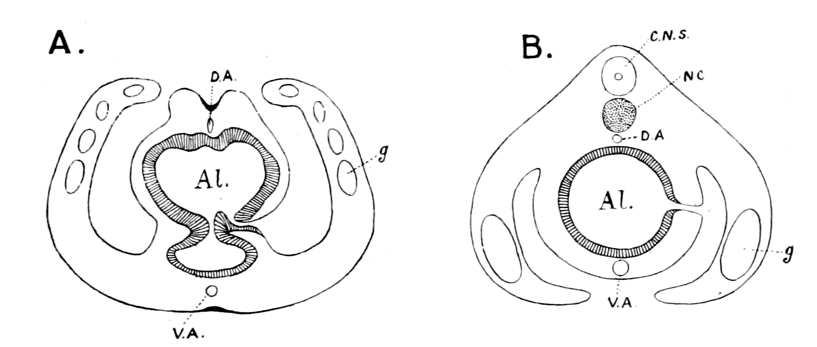
Fig. 168.—Diagram illustrating the Position of the Pleural Folds and Gonads in Ptychodera (A) and Amphioxus (B) respectively.
Al., alimentary canal; D.A., dorsal vessel; V.A., ventral vessel; g., gonads; NC., notochord; C.N.S., central nervous system.
In a mud-dwelling animal, like Balanoglossus, which possesses no appendages, no special sense-organs, it seems likely enough that ventral and dorsal may be terms of no particular meaning, and consequently what is called ventral in Balanoglossus may correspond to what is dorsal in Amphioxus; in this way the branchial regions of the two animals may be closely compared. Such comparison, however, immediately upsets the whole argument of the vertebrate nature of Balanoglossus based on the relative position of the central nervous system and gut, for now that part of its nervous system which is looked upon as the central nervous system in Balanoglossus is ventral to the gut, just as in a worm-like animal, and not dorsal to it as in a vertebrate.
There is absolutely no possibility whatever of making such a detailed comparison between Balanoglossus and any vertebrate, as I have done between a particular kind of arthropod and Ammocœtes. In the latter case not only the topographical anatomy of the organs in the two animals is the same, but the comparison is valid even to microscopical structure. In the former case the origin of almost all the vertebrate organs is absolutely hypothetical, no clue is given in Balanoglossus, not even to the segmented nature of the vertebrate. The same holds good with the evidence from Embryology and from Palæontology. I have pointed out how strongly the evidence in both cases confirms that of Comparative Anatomy. In neither case is the strength of the evidence for Balanoglossus in the slightest degree comparable. In Embryology an attempt has been made to compare the origin of the cœlom in Amphioxus and in Balanoglossus. In Palæontology there is nothing, only an assumption that in the Cambrian and Lower Silurian times a whole series of animals were evolved between Balanoglossus and the earliest armoured fishes, which have left no trace, although they were able to hold their own against the dominant Palæostracan race. The strangeness of this conception is that, when they do appear, they are fully armoured, as in Pteraspis and Cephalaspis, and it is extremely hard luck for the believers in the Balanoglossus theory that no intermediate less armoured forms have been found, especially in consideration of the fact that the theory of the origin from the Palæostracan does not require such intermediate forms, but finds that those already discovered exactly fulfil its requirements.
One difficulty in the way of accepting the theory which I have advocated is perhaps the existence of the Tunicata. I cannot see that they show any affinities to the Arthropoda, and yet they are looked upon as allied to the Vertebrata. I can only conclude that both they and Amphioxus arose late, after the vertebrate stock had become well established, so that in their degenerated condition they give indications of their vertebrate ancestry and not of their more remote arthropod ancestry.
In conclusion, the way in which vertebrates arose on the earth as suggested in this book carries with it many important far-reaching conclusions with respect to the whole problem of Evolution.
When the study of Embryology began, great hopes were entertained that by its means it would be possible to discover the pedigree of every group of animals, and for this end all the stages of development in all groups of animals were sought for and, as far as possible, studied. It was soon found, however, that the interpretation of what was seen was so difficult, as to give rise to all manner of views, depending upon the idiosyncrasy of the observer. At his will he decided whether any appearance was cœnogenetic or palingenetic, with the result that, in the minds of many, embryology has failed to afford the desired clue.
At the same time, the geological record was looked upon as too imperfect to afford any real help; it was said, and is said, that the Cambrian and pre-Cambrian periods were so immense, and the animals discovered in the lower Silurian so highly organized, as to compel us to ascribe the origination of all the present-day groups to this immense early period, the animals of which have left no trace of their existence as fossils.
In consequence of, or at all events following upon, the supposed failure of embryology and of geology to solve the problem of the sequence of evolution of animal life, a new theory has arisen, which goes very near to the denial of evolution altogether. This is the theory of parallel development. It discards the old picture of a genealogical tree with main branches arising at different heights, these again branching and branching into smaller and smaller twigs, and substitutes instead the picture of the ribs of a fan, every rib running independently of every other, each group represented by a rib reaching its highest development on the circumference of the fan and coming nearer and nearer to a common point at the handle of the fan. This point of convergence, where all the groups ultimately meet, is so far back as to reach to the lowest living organisms.
This, in my opinion, unscientific and inconceivable suggestion has arisen largely in consequence of a conception which has become firmly fixed in the minds of very many writers on this subject—the conception that in the evolution of every group, the higher members of the group are the most specialized in the peculiarities of that group, and it is impossible to obtain a new group with different peculiarities from such specialized members. If, then, a higher group is to arise from a lower, it must arise from the generalized members of that lower group, in other words, from the lowest members or those nearly akin to the next lower group.
Similarly, the highest members of this latter group are too specialized, and again we must go to the more generalized members of the group. In this way each separate specialized group is put on one side, and so the conception of parallel development comes into being.
The evidence given in this book dealing with the origin of vertebrates strikes at the foundations of this belief, for it presents an image of the sequence of evolution of animal forms in orderly upward progress, caused by the struggle for existence among the members of the race dominant at the time, which brought about the origin of the next higher group not from the lowest members of the dominant group, but from some one of the higher members of that group.
The great factor in evolution has been throughout the growth of the central nervous system; from that group of animals which possessed the highest nervous system evolved up to that time the next higher group must have arisen.
In this way we can trace without a break, always following out the same law, the evolution of man from the mammal, the mammal from the reptile, the reptile from the amphibian, the amphibian from the fish, the fish from the arthropod, the arthropod from the annelid, and we may be hopeful that the same law will enable us to arrange in orderly sequence all the groups in the animal kingdom.
This very same law of the paramount importance of the development of the central nervous system for all upward progress will, I firmly believe, lead to the establishment of a new and more fruitful embryology, the leading feature of which will be, as suggested in the last chapter, not the attempt to derive from the blastula three germ-layers common to all animals, but rather two sets of organs—those which are governed by the nervous system and those which are not—and thus by means of the development of the central nervous system obtain from embryology surer indications of relationship than are given at present.
The great law of recapitulation, which asserts that the past history of the race is indicated more or less in the development of each individual, a law which of late years has fallen somewhat into disrepute, owing especially to the difficulty of interpreting the embryological history of the vertebrate, is triumphantly vindicated by the theory put forward in this book. Each separate vertebrate organ, one after the other, as shown in the last chapter, indicates in its development the manner in which it arose from the corresponding organ of the arthropod. There is no failure in the evidence of embryology, the failure is in the interpretation thereof.
So, too, my theory vindicates the geological method. There is no failure here; on the contrary, the record of the rocks proclaims with startling clearness not only the sequence of evolution in the vertebrate kingdom itself, but the origin of the vertebrate from the most highly-developed invertebrate race.
The study of the comparative anatomy of organs down to the finest details has always been a most important aid in finding out relationship between animals or groups of animals. My theory endorses this view to the uttermost, and especially indicates the study of the central nervous system and its outgoing nerves as that comparative study which is most likely to afford valuable results.
As for the individual, so for the nation; as for the nation, so for the race; the law of evolution teaches that in all cases brain-power wins. Throughout, from the dawn of animal life up to the present day, the evidence given in this book suggests that the same law has always held. In all cases, upward progress is associated with a development of the central nervous system.
The law for the whole animal kingdom is the same as for the individual. "Success in this world depends upon brains."
BIBLIOGRAPHY AND INDEX OF AUTHORS
Author's name. Title of Paper. Pages of reference.
AHLBORN "Untersuchungen über das Gehirn der Petromyzonten" 210, 489
Zeitsch. f. wiss. Zool. Vol. 39. 1883
"Ueber die Segmentation des Wirbelthierkörpers" 260
Zeitsch. f. wiss. Zool. Vol. 40. 1884
AICHEL "Vergleichende Entwicklungsgeschichte und Stammesgeschichte der Nebennieren" 424, 428
Arch. f. Mikr. Anat. Vol. 56. 1900
ALCOCK 135, 287, 288, 289, 304, 307, 347, 445
"The Peripheral Distribution of the Cranial Nerves of Ammocœtes" 164, 171, 177, 188, 202, 297, 300, 310, 311, 316
Journ. of Anat. and Physiol. Vol. 33. 1898
"On Proteid Digestion in Ammocœtes" 58, 213, 442, 452
Journ. of Anat. and Physiol. Vol. 33. 1898
ALLEN "Studies on the Nervous System of Crustacea" 489
Q. J. Micr. Sci. Vol. 36. 1894
ANDERSON, H. K. 448, 470
"The Nature of the Lesions which hinder the Development of Nerve-cells and their Processes 466, 467, 469
Journ. of Physiol. Vol. 28. 1902
"On the Myelination of Nerve-fibres" 467, 477
Report of the Brit. Assn. 1898
APATHY "Das leitende Element des Nervensystems und seine topographischen Beziehung zu den Zellen" 467
Mitth. a. d. Zool. Stat. zu Neapel. Vol. 12. 1896
ASSHETON "On the Phenomenon of the Fusion of the Epiblastic Layers in the Rabbit and in the Frog" 42
Q. J. Micr. Sci. Vol. 37. 1894
"An Experimental Examination into the Growth of the Blastoderm of the Chick" 154
Proc. of Roy. Soc. Vol. 60. 1896
"On the Growth in Length of the Frog Embryo" 154
Q. J. Micr. Sci. Vol. 37. 1894
"A Re-investigation into the Early Stages of the Development of the Rabbit" 154
Q. J. Micr. Sci. Vol. 37. 1894
"The Primitive Streak of the Rabbit: the Causes which may determine its Shape, and the part of the Embryo formed by its Activity" 154
Q. J. Micr. Sci. Vol. 37. 1894
BALFOUR 'Comparative Embryology.' Vol. 2 73, 74, 94, 103, 104, 120, 181, 259, 424
London. 1881. Macmillan & Co.
"On the Origin and History of the Urino-genital Organs of Vertebrates" 390, 392
Journ. of Anat. and Physiol. Vol. 10. 1876
"On the Nature of the Organ in Adult Teleosteans and Ganoids, which is usually regarded as the Head-kidney or Pronephros" 420
Q. J. Micr. Sci. Vol. 22. 1882
BARKER 'The Nervous System' 470
London. 1901
BATESON "The Ancestry of the Chordata" 11
Q. J. Micr. Sci. Vol. 26. 1886
'Materials for the Study of Variation' 387
London. 1894
BEARD "The System of Branchial Sense Organs and their Associated Ganglia in Ichthyopsida" 262, 281, 283
Q. J. Micr. Sci. Vol. 26. 1885
"The Development of the Peripheral Nervous System in Vertebrates" 262, 281, 283
Q. J. Micr. Sci. Vol. 29. 1888
"The Old Mouth and the New" 318
Anat. Anzeiger. 1888
"The Source of Leucocytes and the True Function of the Thymus" 425, 426
Anat. Anzeiger. Vol. 18. 1900
"The Parietal Eye of the Cyclostome Fishes" 84
Q. J. Micr. Sci. Vol. 29. 1882
BECK AND LANKESTER "On the Muscular and Endo-skeletal Tissues of Scorpio" 171, 222, 224, 247, 268-277
Trans. Zool. Soc. Vol. 11. 1885
BEECHER "Natural Classification of the Trilobites" 283, 351, 436, 437
Amer. Journ. of Sci. Ser. 4. Vol. 3. 1897
BELL, C. 'The Nervous System of the Human Body' 155, 156, 183
London. 1830
BELLONCI "Système Nerveux et Organes des sens du Sphæroma serratum" 62, 90, 92, 101
Archiv. Ital. de Biol. Vol. 1. 1882
"Sur la structure et les rapports des lobes olfactives dans les Arthropods superieurs et les Vertébrés" 221,225
Archiv. Ital. de Biol. Vol. 3. 1883
BENHAM AND LANKESTER "On the Muscular and Endo-skeletal Systems of Limulus" 143, 171, 176, 177, 247
Trans. Zool. Soc. Vol. 11. 1885
BERGER "Untersuchungen über den Bau des Gehirns und der Retina der Arthropoden" 88-92, 97, 100, 101
Arbeit. a. d. Zool. Instit. Wien. Vol. 1. 1878
BERGH "Neue Beiträge zur Embryologie der Anneliden" 478
Zeitsch. f. wiss. Zool. Vol. 50. 1890
BERKLEY "The Intrinsic Nerves of the Kidney" 477
Bulletin of the Johns Hopkins Hospital. Vol. 4
BERNARD 'The Apodidæ: a Morphological Study' 284
Nature Series. 1892
BERTKAU "Beiträge zur Kenntniss der Sinnesorgane der Spinnen. 1. Die Augen der Spinnen" 369
Archiv. f. mikr. Anat. Vol. 27. 1886
BIEDERMANN 'Electro-physiology' 20
Translated by F. A. Welby. London. 1896
BLANCHARD Quoted by Huxley 225
'L'Organisation du Règne Animal. Arachnides' 109, 177, 190, 206, 313, 315
Paris. 1852
BLES "The Correlated Distribution of Abdominal Pores and Nephrostomes in Fishes" 431
Journ. of Anat. and Physiol. Vol. 32. 1898
BOBRETSKY 'Development of Astacus and Palæmon' 74
Kiew. 1873
BOURNE AND LANKESTER See Lankester and Bourne.
BOVERI "Die Nieren Canälchen des Amphioxus" 392, 395, 402, 407, 412, 426, 427
Zool. Jahrbuch. Vol. 5. 1892
BRAEM "Was ist ein Keimblatt" 460, 461, 462
Biol. Centralblatt. Vol. 15. 1895
BRAUER "Beiträge zur Kenntniss der Entwicklungsgeschichte des Skorpions" 62, 167, 222, 237, 281, 482
Zeit. f. wiss. Zool. Part I. Vol. 57. 1894 Part II. Vol. 59. 1895
"Beiträge zur Kenntniss der Entwicklung und Anatomie der Gymnophionen." III. 393, 394, 400, 402
"Die Entwicklung der Excretionsorgane" Zool. Jahrbuch. Vol. 16. 1902
"Ueber die Entwicklung von Hydra" 473
Zeit. f. wiss. Zool. Vol. 52. 1891
BÜTSCHLI "Notiz zur Morphologie des Auges der Muscheln" 114
Festschrift des Natur-hist-med. Vereins zu Heidelberg. 1886
BUJOR "Contribution a l'étude de la métamorphose de l'Ammocœtes branchialis en Petromyzon Planeri" 135, 304
Revue Biologique du Nord de la France. Vol. 3. 1891
CARLSON 177, 315, 316
CELESIA 'Differenziamento della proprietà inibitoria e dei funzioni coordinatrici nella catena gangliare dei crustacei decapodi' 489
Genoa. 1897
CLAUS "Untersuchungen über den Organismus und Entwicklung von Branchipus und Artemia" 90-92, 97, 100, 396
Arbeit a.d. Zool. Institut. Wien. Vol. 6. 1886
COPE "On the Phylogeny of the Vertebrata" 343
Proc. Amer. Philos. Soc. Vol. 30. 1892
CRONEBERG "Ueber die Mundtheile der Arachniden" 221-224, 241
Archiv. f. Naturgeschichte. 1880
CUÉNOT "Études sur le sang et les glandes lymphatiques dans la série animale; 2nd partie; invertébrés" 422
Arch. d. Zool. exper. gen. 2nd Ser. Vol. 9. 1891
CUNNINGHAM, J. T. "The Significance of Kupffer's Vesicle, with Remarks on other Questions of Vertebrate Morphology" 318
Q. J. Micr. Sci. Vol. 25. 1885
"The Nephridia of Lanice conchilega" 403
Nature. Vol. 36. 1887
DANA "On Cephalization" 53
Mag. of Nat. Hist. 1863
DEAN-BASHFORD 'Fishes, Living and Fossil' 344
New York. 1895
"On the Embryology of Bdellostoma Stouti" 405
Festschr. z. siebenzigsten Geburtstag. von C. v. Kupffer. Jena. 1899
DENDY "On the Parietal Sense-organs and Associated Structures in the New Zealand Lamprey (Geotria australis)" 80, 82
Q. J. Micr. Sci. Vol. 51. 1907
DIETL "Die Organisation des Arthropoden Gehirns" 101
Zeitsch. f. wiss. Zool. Vol. 27. 1876
DOHRN 'Der Ursprung der Wirbelthiere und das Princip des Functionswechsels' 14, 60, 185, 186, 317, 318
Leipzig. 1875
Studien zur Urgeschichte des Wirbelthiere Körpers. VIII. "Die Thyroidea bei Petromyzon, Amphioxus, und Tunicaten" 188, 195-198, 199, 212, 213
Mitth. Zool. Stat. z. Neapel. Vol. 6. 1886
"Neue Grundlagen zur Beurtheilung der Metamerie des Kopfes" 262, 263, 279