Front matter (continued 10)
Reading edition
This is what Kowalewsky has done. Thus he states that Metschnikoff had fed Mysis with carmine-grains, and found tubules at the base of the thoracic feet coloured red with carmine. He himself used an allied species, Parapodopsis cornutum, and found here also that the carmine was taken up by tubules situated in the basal segments of the feet. In Nebalia, feeding experiments with alizarin blue and carmine stained the antennal glands, and showed the existence of glands at the base of the eight thoracic feet. These glands resemble the foot-glands of Mysis, Parapodopsis, and Palæmon, and lie in the space through which the blood passes from the thoracic feet, i.e. from the gills, to the heart. In Squilla also, in addition to the shell-glands, special glands were discovered on the branchial feet on the path of the blood to the heart. These glands form continuous masses of cells which constitute large compact glands at the base of the branchial feet. Single cells of the same sort are found along the whole course of the branchial venous canal, right up to the pericardium.
These observations show that the Crustacea possess not only true excretory organs in the shape of coxal glands, i.e. antennary glands, shell-glands, etc., in the cephalic region, but also a series of segmental glands situated at the base of the appendages, especially of the respiratory appendages: a system, that is to say, of coxal glands which have lost their excretory function, through having lost their external opening, but have not in consequence disappeared, but still remain in situ, and still retain an important excretory function, having become lymphatic glands containing leucocytes. Such glands are especially found in the branchial appendages, and are called branchial glands by Cuénot, who describes them for all Decapoda.
Further, it is significant that the same method reveals the existence in Pantopoda of a double set of glands of similar character, one set in the basal segments of the appendage, and the other in the adjacent part of the body.
In scorpions also, Kowalewsky has shown that the remarkable lymphatic organ situated along the whole length of the nerve-cord in the abdominal region takes up carmine grains and bacilli; an organ which in Androctonus does not form one continuous gland, but a number of separate, apparently irregularly grouped, glandular bodies.
In addition to this median lymphatic gland, Kowalewsky has discovered in the scorpion a pair of lateral glands, to which he gives the name of lymphoid glands, which communicate with the thoracic body-cavity (i.e. the pseudocœle), are phagocytic, and, according to him, give origin to leucocytes by the proliferation of their lining cells, thus, as he remarks, reminding us of the nephridial capsules of Clepsine. These glands are so closely related in position to the coxal glands on each side that he has often thought that the lumen of the gland communicated with that of the coxal gland; he, however, has persuaded himself that there is no true communication between the two glands. Neither of these organs appears to be segmental, and until we know how they are developed it is not possible to say whether they represent fused segmental organs or not.
The evidence, then, is very strong that in the Crustacea and Arachnida the original segmental excretory organs do not disappear, but remain as ductless glands, of the nature of lymphatic glands, which supply leucocytes to the system.
Further, the evidence shows that the nephric organs, or parts of the cœlom in close connection with these organs, may be transformed into ductless glands, which do not necessarily contain free leucocytes as do lymph-glands, but yet are of such great importance as excretory organs that their removal profoundly modifies the condition of the animal. Such a gland is the so-called adrenal or suprarenal body, disease of which is a feature of Addison's disease; a gland which forms and presumably passes into the blood a substance of remarkable power in causing contraction of blood-vessels, a substance which has lately been prepared in crystalline form by Jokichi Takamine, and called by him "adrenalin"; a gland, therefore, of very distinctly peculiar properties, which cannot be regarded as rudimentary, but is of vital importance for the due maintenance of the healthy state.
In the Elasmobranchs two separate glandular organs have been called suprarenal; a segmental series of paired organs, each of which possesses a branch from the aorta and a sympathetic ganglion, and an unpaired series in close connection with the kidneys, to which Balfour gave the name of interrenal glands. Of these two sets of glands, Swale Vincent has shown that the extract of the interrenals has no marked physiological effect, in this respect resembling the extract of the cortical part of the mammalian gland, while the extract of the paired segmental organs of the Elasmobranch produces the same remarkable rise of blood-pressure as the extract of the medullary portion of the mammalian gland.
The development also of these two sets of glands is asserted to be different. Balfour considered that the suprarenals were derived from sympathetic ganglion-cells, but left the origin of the interrenals doubtful. Weldon showed that the cortical part of the suprarenals in the lizard was derived from the wall of the glomerulus of a number of mesonephric tubules. In Pristiurus, he stated that the mesoblastic rudiment described by Balfour as giving origin to the interrenals is derived from a diverticulum of each segmental tubule, close to the narrowing of its funnel-shaped opening into the body-cavity. With respect to the paired suprarenals he was unable to speak positively, but doubted whether they were derived entirely from sympathetic ganglia.
Weldon sums up the results of his observations by saying: "That all vertebrates except Amphioxus have a portion of the kidney modified for some unknown purpose not connected with excretion; that in Cyclostomes the pronephros alone is so modified, in Teleostei the pro- and part of the meso-nephros; while in the Elasmobranchs and the higher vertebrates the mesonephros alone gives rise to this organ, which has also in these forms acquired a secondary connection with certain of the sympathetic ganglia."
Since Weldon's paper, a large amount of literature on the origin of the adrenals has appeared, a summary of which, up to 1891, is given by Hans Rabl in his paper, and a further summary by Aichel in his paper published in 1900. The result of the investigations up to this latter paper may be summed up by saying that the adrenals, using this term to include all these organs of whatever kind, are in all cases, partly at all events, derived from some part of the walls of either the mesonephric or pronephric excretory organs, but that in addition a separate origin from the sympathetic nervous system must be ascribed to the medullary part of the organ and to the separate paired organs in the Elasmobranchs, which are equivalent to the medullary part in other cases.
The evidence, then, of the transformation of the known vertebrate excretory organs—the pronephros and the mesonephros—leads to the conclusion that in our search for the missing coxal glands of the meso- and pro-somatic regions, we must look for either lymphatic glands, or ductless glands of distinct importance to the body. I have already considered the question in the prosomatic region, and have given my reasons why the pituitary gland must be looked upon as the descendant of the arthropod coxal gland. In this case also the resulting ductless gland is still of functional importance, for disease of it is associated with acromegaly. If, as is possible, it is homologous with the Ascidian hypophysial gland, then it is confirmatory evidence that this latter is said by Julin to be an altered nephridial organ.
Finally, I come to the mesosomatic or branchial region; and here, strikingly enough, we find a perfectly segmental glandular organ of mysterious origin—the thymus gland—segmental with the branchiæ, not necessarily with the myotomes, belonging, therefore, to the appendicular system; and since the branchiæ represent, according to my theory, the basal part of the appendage, such segmental glands would be in the position of coxal glands. Here, then, in the thymus may be the missing mesosomatic coxal glands.
What, then, is the thymus?
The answer to this question has been given recently by Beard, who strongly confirms Kölliker's original view that the thymus is a gland for the manufacture of leucocytes, and that such leucocytes are directly derived from the epithelial cells of the thymus. Kölliker also further pointed out that the blood of the embryo is for a certain period destitute of leucocytes. Beard confirms this last statement, and says that up to a certain stage (varying from 10 to 16 mm. in length of the embryo) the embryos of Raja batis have no leucocytes in the blood or elsewhere. Up to this period the thymus-placode is well formed, and the first leucocytes can be seen to be formed in it from its epithelial cells; then such formation takes place with great rapidity, and soon an enormous discharge of leucocytes occurs from the thymus into the tissue-spaces and blood. He therefore concludes that all lymphoid tissues in the body arise originally from the thymus gland, i.e. from leucocytes discharged from the thymus.
The segmental branchial glands, known by the name of thymus, are, according to this view, the original lymphatic glands of the vertebrate; and it is to be noted that, in fishes and in Amphibia, lymphatic glands, such as we know them in the higher mammals, do not exist; they are characteristic of the higher stages of vertebrate evolution. In the lower vertebrates, the only glandular masses apart from the cell-lining of the body-cavity itself, which give rise to leucocyte-forming tissue, are these segmental branchial glands, or possibly also the modified post-branchial segmental glands, known as the head-kidney in Teleostea, etc.
The importance ascribed by Beard to the thymus in the formation of leucocytes in the lowest vertebrates would be considerably reduced in value if the branchial region of Ammocœtes possessed neither thymus glands nor anything equivalent to them. Such, however, is not the case. Schaffer has shown that in the young Ammocœtes masses of lymphatic glandular tissue are found segmentally arranged in the neighbourhood of each gill-slit—tissue which soon becomes converted into a swarming mass of leucocytes, and shows by its staining, etc., how different it is from a blood-space. The presence of this thymus leucocyte-forming tissue, as described by Schaffer, is confirmed by Beard, and I myself have seen the same thing in my youngest specimen of Ammocœtes.
Further, the very methods by which Kowalewsky has brought to light the segmental lymph-glands of the branchial region of the Crustacea, etc., are the same as those by which Weiss discovered the branchial nephric glands in Amphioxus—excretory organs which Boveri considers to represent the pronephros of the Craniota. In this supposition Boveri is right, in so far that both pronephros and the tubules in Amphioxus belong to the same system of excretory organs; but I entirely agree with van Wijhe that the region in Amphioxus is wrong. The tubules in Amphioxus ought to be represented in the branchial region of the Craniota, not in the post-branchial region; van Wijhe therefore suggests that further researches may homologize them with the thymus gland in the Craniota, not with the pronephros. This suggestion of van Wijhe appears to me a remarkably good one, especially in view of the position of the thymus glands in Ammocœtes and the nephric branchial glands in Amphioxus. If, as I have pointed out, the atrial cavity of Amphioxus has been closed in Ammocœtes by the apposition of the pleural fold with the branchial body-surface, then the remains of the position of the atrial chamber must exist in Ammocœtes as that extraordinary space between the somatic muscles and the branchial basket-work filled with blood-spaces and modified muco-cartilage. It is in this very space, close against the gill-slits, that the thymus glands of Ammocœtes are found, in the very place where the nephric tubules of Amphioxus would be found if its atrial cavity were closed completely. Instead, therefore, of considering with Boveri that the branchial nephric tubules of Amphioxus still exist in the Craniota as the pronephros, and that the atrial chamber has narrowed down to the pronephric duct, I would agree with van Wijhe that the pronephros is post-branchial, and suggest that by the complete closure of the atrial space in the branchial region the branchial nephric tubules have lost all external opening, and consequently, as in all other cases, have changed into lymphatic tissue and become the segmental thymus glands.
As van Wijhe himself remarks, the time is hardly ripe for making any positive statement about the relationship between the thymus gland and branchial excretory organs. There is at present not sufficient consensus of opinion to enable us to speak with any certainty on the subject, yet there is so much suggestiveness in the various statements of different authors as to make it worth while to consider the question briefly.
On the one hand, thymus, tonsils, parathyroids, epithelial cell-nests, and parathymus, are all stated to be derivatives of the epithelium lining the gill-slits, and Maurer would draw a distinction between the organs derived from the dorsal side of the gill-cleft and those derived from the ventral side—the former being thymus, the latter forming the epithelial cell-nests, i.e. parathyroids. The thymus in Ammocœtes, according to Schaffer, lies both ventral and dorsal to the gill-cleft; Maurer thinks that only the dorsal part corresponds to the thymus, the ventral part corresponding to the parathyroids, etc. Structurally, the thymus, parathyroids, and the epithelial cell-nests are remarkably similar, so that the evidence appears to point to the conclusion that, in the neighbourhood of the gill-slits, segmentally arranged organs of a lymphatic character are situated, which give origin to the thymus, parathyroids, tonsils, etc. Now, among these organs, i.e. among those ventrally situated, Maurer places the carotid gland, so that, if he is right, the origin of the carotid gland might be expected to help in the elucidation of the origin of the thymus.
The origin of the carotid gland has been investigated recently by Kohn, who finds that it is associated with the sympathetic nervous system in the same way as the suprarenals. He desires, in fact, to make a separate category for such nerve-glands, or paraganglia, as he calls them, and considers them all to be derivatives of the sympathetic nervous system, and to have nothing to do with excretory organs. The carotid gland is, according to him, the foremost of the suprarenal masses in the Elasmobranchs, viz. the so-called axillary heart.
In my opinion, nests of sympathetic ganglion-cells necessarily mean the supply of efferent fibres to some organ, for all such ganglia are efferent, and also, if they are found in the organ, would have been brought into it by way of the blood-vessels supplying the organ, so that Aichel's statement of the origin of the suprarenals in the Elasmobranchs seems to me much more probable than a derivation from nerve-cells. If, then, it prove that Aichel is right as to the origin of the suprarenals, and Kohn is right in classifying the carotid gland with the suprarenals, then Maurer's statements would bring the parathyroids, thymus, etc., into line with the adrenals, and suggest that they represent the segmented glandular excretory organs of the branchial region, into which, just as in the interrenals of Elasmobranchs, or the cortical part of the adrenals of the higher vertebrates, there has been no invasion of sympathetic ganglion-cells.
Wheeler makes a most suggestive remark in his paper on Petromyzon: he thinks he has obtained evidence of serial homologues of the pronephric tubules in the branchial region of Ammocœtes, but has not been able up to the present to follow them out. If what he thinks to be serial homologues of the pronephric tubules in the branchial region should prove to be the origin of the thymus glands of Schaffer, then van Wijhe's suggestion that the thymus represents the excretory organs of the branchial region would gain enormously in probability. Until some such further investigation has been undertaken, I can only say that it seems to me most likely that the thymus, etc., represent the lymphatic branchial glands of the Crustacea, and therefore represent the missing coxal glands of the branchial region.
This, however, is not all, for the appendages of the mesosomatic region, as I have shown, do not all bear branchiæ; the foremost or opercular appendage carries the thyroid gland. Again, the basal part of the appendage is all that is left; the thyroid gland is in position a coxal gland. It ought, therefore, to represent the coxal gland of this appendage, just as the thymus, tonsils, etc., represent the coxal glands of the rest of the mesosomatic appendages. In the thyroid gland we again see a ductless gland of immense importance to the economy, not a useless organ, but one, like the other modified coxal glands, whose removal involves far-reaching vital consequences. Such a gland, on my theory, was in the arthropod a part of the external genital ducts which opened on the basal joint of the operculum. What, then, is the opinion of morphologists as to the meaning of these external genital ducts?
In a note to Gulland's paper on the coxal glands of Limulus, Lankester states that the conversion of an externally-opening tubular gland (coxal gland) into a ductless gland is the same kind of thing as the history of the development of the suprarenal from a modified portion of mesonephros, as given by Weldon. Further, that in other arthropods with glands of a tubular character opening to the exterior at the base of the appendages, we also have coxal nephridia, such as the shell-glands of the Entomostraca, green glands of Crustacea (antennary coxal gland); and further on he writes: "When once the notion is admitted that ducts opening at the base of limbs in the Arthropoda are possibly and even probably modified nephridia, we immediately conceive the hypothesis that the genital ducts of the Arthropoda are modified nephridia."
So, also, Korschelt and Heider, in their general summing up on the Arthropoda, say: "In Peripatus, where the nephridia appear, as in the Annelida, in all the trunk-segments, a considerable portion of the primitive segments is directly utilized for the formation of the nephridia. In the other groups, the whole question of the rise of the organs known as nephridia is still undecided, but it may be mentioned as very probable that the salivary and anal glands of Peripatus, the antennal and shell-glands of the Crustacea, the coxal glands of Limulus and the Arachnida, as well as the efferent genital ducts, are derived from nephridia, and in any case are mesodermal in origin."
The necessary corollary to this exceedingly probable argument is that glandular structures such as the uterine glands of the scorpion already described, which are found in connection with these terminal genital ducts, may be classed as modified nephridial glands, and that therefore the thyroid gland of Ammocœtes, which, on the theory of this book, arose in connection with the opercular genital ducts of the palæostracan ancestor, represents the coxal glands of this fused pair of appendages. Such a gland, although its function in connection with the genital organs had long disappeared, still, in virtue of its original excretory function, persisted, and even in the higher vertebrates, after it had lost all semblance of its former structure and become a ductless gland of an apparently rudimentary nature, still, by its excretory function, demonstrates its vital importance even to the highest vertebrate.
By this simple explanation we see how these hitherto mysterious ductless glands, pituitary, thymus, tonsils, thyroid, are all accounted for, are all members of a common stock—coxal glands—which originally, as in Peripatus, excreted at the base of the prosomatic and mesosomatic appendages, and are still retained because of the importance of their excretory function, although ductless owing to the modification of their original appendages.
Finally, there is yet another organ in the vertebrate which follows the same law of the conversion of an excretory organ into a lymphatic organ when its connection with the exterior is obliterated, and that is the vertebrate body-cavity itself. According to the scheme here put forth, the body-cavity of the vertebrate arose by the fusion of a ventral prolongation of the original nephrocœle on each side; prolongations which accompanied the formation of the new ventral midgut, and by their fusion formed originally a pair of cavities along the whole length of the abdomen, being separated from each other by the ventral mesentery of the gut. Subsequently, by the ventral fusion of these two cavities, the body-cavity of the adult vertebrate was formed.
This is simply a statement of the known method of formation of the body-cavity in the embryo, and its phylogenetic explanation is that the body-cavity of the vertebrate must be looked upon as a ventral prolongation of the original ancestral body-cavity. Embryology clearly teaches that the original body-cavity or somite was confined to the region of the notochord and central nervous system, and there, just as in Peripatus, was divisible into a dorsal part, giving origin to the myocœle, and a ventral part, forming the nephrocœle. From this original nephrocœle are formed the pronephric excretory organs, the mesonephric excretory organs, and the body-cavity.
That the vertebrate body-cavity was originally a nephrocœle is generally accepted, and its excretory function is shown by the fact that it communicates with the exterior in all the lower vertebrates, either through abdominal pores or by way of nephridial funnels. Bles has shown how largely these two methods of communicating with the exterior mutually exclude each other. In the higher vertebrates both channels become closed, except in the case of the Fallopian tubes, and thus, so to speak, the body-cavity becomes a ductless gland, still, however, with an excretory function, but now, as in all other cases, forming a part of the lymphatic rather than of the true excretory system.
Summary.
The consideration of the formation of the vertebrate cranial region, as set forth in previous chapters, indicates that the ancestor of the vertebrates was not an arachnid purely or a crustacean purely, but possessed partly crustacean and partly arachnid characters. In order to express this conclusion, I have used the term Protostraca, invented by Korschelt and Heider, to indicate a primitive arthropod group, from which both arachnids and crustaceans may be supposed to have arisen, and have therefore stated that the vertebrate did not arise directly from the annelids, but from the Protostraca. Such an origin signifies that the origin of the excretory organs of the vertebrate must not be looked for in the segmental organs of the annelid, but rather in such modified annelid organs as would naturally exist in a primitive arthropod group. The nature of such organs may be inferred, owing to the fortunate circumstance that so primitive an arthropod as Peripatus still exists, and we may conclude that the protostracan ancestor possessed in every segment a pair of appendages and a pair of cœlomic cavities, which extended into the base of these appendages. The ventral portion of each of these cœlomic cavities separated off from the dorsal and formed a nephrocœle, giving origin to a segmental excretory organ, which, seeing that its end-vesicle was in the base of the appendage, and seeing also the nature of the known arachnid and crustacean excretory organs, may fitly be termed a coxal gland. This, then, is the working hypothesis to explain the difficulties connected with the origin of the pronephros and mesonephros—that the original segmental organs were coxal glands, and therefore indicated the presence of appendages. This hypothesis leads to the following conclusions:—
1. The coxal glands belonging to the post-branchial appendages of the invertebrate ancestor are represented by the pronephric tubules, and existed over the whole metasomatic region.
2. Such glands discharged into a common duct—the pronephric duct—which opened into the cloacal region, either in the protostracan stage, when the metasomatic appendages were still in existence, just as the coxal glands of the prosomatic region in Limulus discharge into a common duct, or else the pronephric duct was formed when the appendages were obliterated.
3. The metasomatic appendages disappeared owing to their enclosure by pleural folds, which, meeting in the mid-ventral line, not only caused the obliteration of the appendages, and gave a smooth fish-like body-surface to the animal, but also caused the formation of an atrial cavity.
4. Into these pleural folds the dorsal longitudinal muscles of the body extended, and ultimately reached to the ventral surface, thus forming the somatic muscles of the vertebrate body.
5. When the pleural folds had met in the mid-ventral line the animal had become a vertebrate, and was dependent for its locomotion on the movements of these somatic muscles, and not on the movements of appendages. Consequently, elongation of the trunk-region took place, for the purpose of increasing mobility, by the formation of new metameres.
6. Each of such metameres possessed its own segmental excretory organ, formed in the same way as the previous pronephric organs, but, as there were no appendages in these new-formed segments, the excretory organs took on the characters of a mesonephros, not a pronephros, and opened into the pronephric duct, because the direct way to the exterior was blocked by the enveloping pleural folds.
7. The group of annelids from which the protostracan ancestor of the vertebrates arose was the highest annelidan group, viz. the Polychæta, as shown by the nature of the excretory organs in Amphioxus.
8. The coxal glands of the protostracan ancestor existed on all the segments, and were, therefore, divisible into three groups, prosomatic, mesosomatic, and metasomatic; these three groups of coxal glands still exist in the vertebrate as ductless glands.
9. The prosomatic coxal glands form the pituitary body.
10. The mesosomatic coxal glands form the thymus, thyroid, parathyroids, tonsils, etc.
11. The metasomatic coxal glands form the adrenals.
12. The procœlom of the vertebrate is the procœlom of the protostracan ancestor, which splits into a dorsal part, the myocœle, and a ventral part, the nephrocœle. This latter part not only forms the pronephros and mesonephros, but also by a ventral extension gives origin to the walls of the vertebrate body-cavity or metacœle.
13. This ventral extension of the original nephrocœle at first excreted to the exterior, through abdominal pores, or through peritoneal funnels. When such paths to the exterior became closed, it also became a ductless gland, belonging to the lymphatic system.
CHAPTER XIII
THE NOTOCHORD AND ALIMENTARY CANAL
Relationship between notochord and gut.—Position of unsegmented tube of notochord.—Origin of notochord from a median groove.—Its function as an accessory digestive tube.—Formation of notochordal tissue in invertebrates from closed portions of the digestive tube.—Digestive power of the skin of Ammocœtes.—Formation of new gut in Ammocœtes at transformation.—Innervation of the vertebrate gut.—The three outflows of efferent nerves belonging to the organic system.—The original close contiguity of the respiratory chamber to the cloaca.—The elongation of the gut.—Conclusion.
In the previous chapters all the important organs of the arthropod have been found in the vertebrate in their appropriate place, of similar structure, and innervated from corresponding parts of the central nervous system. Such comparison is possible only as long as the ventral and dorsal surfaces of the vertebrate correspond with the respective surfaces of the arthropod, and no reversal is assumed. This method of comparative anatomy is the surest and most certain guide to the relationship between two animals, and when the facts obtained by the anatomical method are so strikingly confirmatory of the palæontological evidence, the combined evidence becomes so strong as to amount almost to a certainty that vertebrates did arise from arthropods in the manner mapped out in previous chapters, and not from a hypothetical group of animals, such as is postulated in the theory of their origin from forms like Balanoglossus.
The latter theory derives the alimentary canal of the vertebrate from that of the invertebrate, and finds in the latter the commencement of the notochord. In the comparison which I have made the alimentary canal of the invertebrate ancestor has become the tube of the central nervous system of the vertebrate, and there is no sign of a notochord whatever. All the organs of the arthropod have already been allocated; where the notochord is situated in the vertebrate there is nothing but a gap in the invertebrate, but the position of that gap can be settled with great accuracy from the previous comparison of organs in the two groups. So, also, the alimentary canal of the vertebrate is from the very nature of the case a new organ, yet, as has been shown in Chapter V., the comparison of the respiratory organs in the two groups gives a strong suggestion of the manner in which such a canal was formed.
The Origin of the Notochord.
The time has now come to endeavour to frame a plausible theory of the method of formation of the notochord and the new alimentary canal, and thus to complete the diagram on p. 413. The comparative method is no longer available, for these structures are both unrepresented as such in the arthropod; any suggested explanation, therefore, must be more tentative, and cannot give the same feeling of certainty as is the case with all the organs already considered. Our only chance of finding out the past history of the notochord lies in the embryological method, in the hope that, according to the 'law of recapitulation,' the ancestral history may be repeated in the ontogeny with sufficient clearness to enable some conclusion to be drawn.
At the outset, one point comes out clearly—the close relationship between the notochord and the vertebrate gut; they are both derived from the same layer, both parts of the same structure. On this point all embryologists are agreed; it is expressed in such statements as, "the notochord, as well as the alimentary canal, is formed from hypoblast"; "the notochord arises as a thickening in the dorsal wall of the alimentary canal." The two structures are so closely connected together that they must be considered together. If we can conjecture the origin of the one, we may be sure that we have the clue to the origin of the other. The two together form the one new organ which distinguishes the vertebrate from the arthropod, the only thing left which requires explanation for the completion of this strange history.
What, then, is the notochord? What are its characteristics? In the highest vertebrates it is conspicuous only in the embryo; with the development of the axial skeleton it is more and more squeezed out of existence, until in the adult it is no longer visible. By the 'law of recapitulation' this developmental history implies that, as we descend the vertebrate phylum, the notochord ought to be more and more conspicuous, more and more permanent during the life of the animal. Such is, indeed, found to be the case, until at last, in the lowest vertebrates, such as the lamprey, and in forms like Amphioxus, the notochord persists throughout the life of the animal as a large important axial supporting rod.
This rod has a number of striking characteristics which distinguish it from all other structures, and are the only means of guessing its probable origin. Its position in the body is always the same in all vertebrates and is very significant, for it lies just ventrally to the central nervous system, along nearly the whole length of the animal, not quite the whole length, for it invariably terminates close to the place where the infundibulum comes to the surface of the brain; it is, in fact, always confined to the infra-infundibular and spinal cord part of the central nervous system. Interpreting this into the language of the arthropod, it means that a rod was formed just ventrally to the nervous system, which extended the whole length of the infraœsophageal and ventral chain of ganglia, and terminated at the orifice of the mouth. Moreover, this rod was unsegmented, for the notochord is devoid of segmentation.
At the anterior end the rod tapers to a point, as in Fig. 166. In its middle part it is very large and conspicuous, cylindrical in shape; its interior is filled with a peculiar vacuolated tissue, different to any other known vertebrate tissue, which has therefore received the name of notochordal tissue. Outside this is a thick sheath formed of many layers, of which the external one gives the staining reactions of elastin, and is called the external elastic layer. Between this sheath and the notochordal tissue a thin layer of lining cells, of normal appearance, is conspicuous in Ammocœtes. These cells secrete the layers of the sheath, and have originally, by proliferation, given rise to the notochordal tissue. In the notochord of Ammocœtes there is no sign of either nerves, blood-vessels, or muscles.
The centre of the notochord presents the appearance of a slight slit, as though it had originated from a tube, and that is the opinion now generally held, for its mode of formation in the embryo is as that of a tube formed from an open groove, as will be explained immediately.
We may, then, conceive of the notochord as originally a tube lying in the mid-line just ventrally to the central nervous system, and extending from the original mouth to the end of the body. Translate this into the language of the arthropod and it denotes a tube on the mid-ventral surface of the body, which extended from mouth to anus. Such a tube might be formed from the mid-ventral surface as follows:—
In Fig. 163, A, the lining of the ventral surface between two appendages is represented flat, in B is shown how the formation of a solid rod may arise from the bulging of that ventral surface, and in C how a groove on that surface may lead to the formation of a tube between the two appendages. The difference between a notochordal rod formed as in B from that in C would be shown in the sheath, for in B the sheath would be formed from the cuticle of the lining cells, and in C from the basement membrane. The structure of the sheath is in accordance with the embryological evidence that the notochord is formed as a tube from a groove, as in C, and not as a solid rod as in B, for it possesses a well-marked elastin layer, and elastin has never yet been found as a constituent of any cuticular secretion, but invariably in connection with basement-membranes.
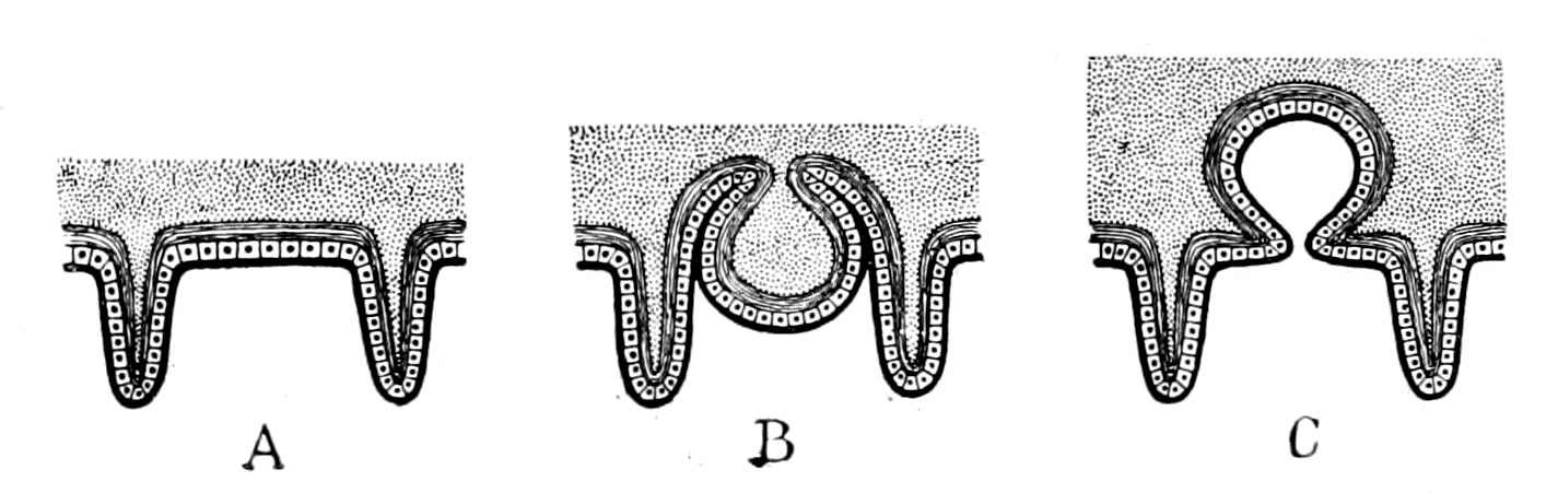
Fig. 163.—Diagram of two possible methods of the Formation of a Notochord.
The position, then, of the notochord and its method of formation suggests that the mid-ventral surface of the arthropod ancestor of the vertebrate formed a deep groove between the bases of all the prosomatic, mesosomatic, and metasomatic appendages, which was subsequently converted into a tube extending along the whole of the body between mouth and anus, and finally, by the proliferation of its lining cells and their conversion into notochordal tissue, became the notochordal rod of the vertebrate.
As already frequently stated, Apus and Branchipus are the two living arthropods which most nearly resemble the extinct trilobites. The beautiful specimens of Triarthrus (Fig. 165) found by Beecher give an idea of the under surface of the trilobite such as has never been obtained before, and demonstrate how closely the condition of things found in Apus (Fig. 164) was similar to that occurring in the trilobites. In both cases the mid-ventral surface of the animal formed a deep groove which extended the whole length of the animal; on each side of this groove in Apus are closely set the gnatho-bases of the appendages, in such a manner that the groove can be easily converted into a canal by the movements of these bases—a canal which, owing to the great number of the appendages and their closeness to each other, can be completely and efficiently closed.
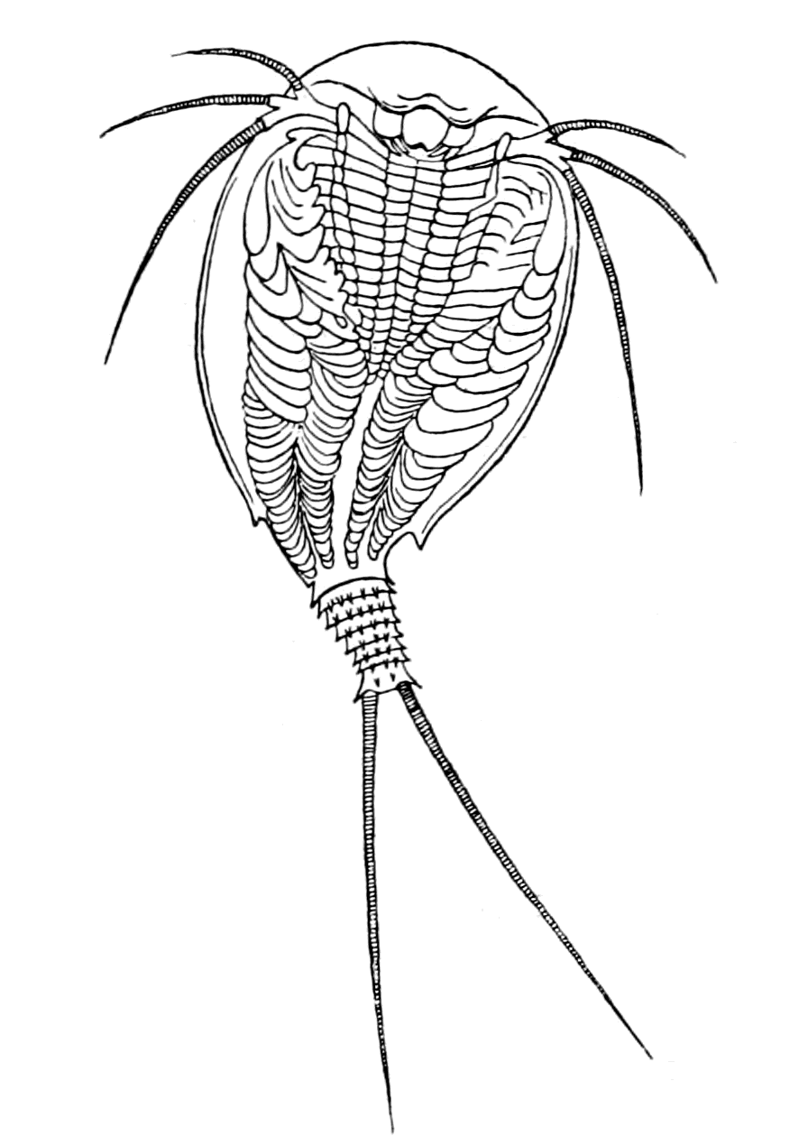
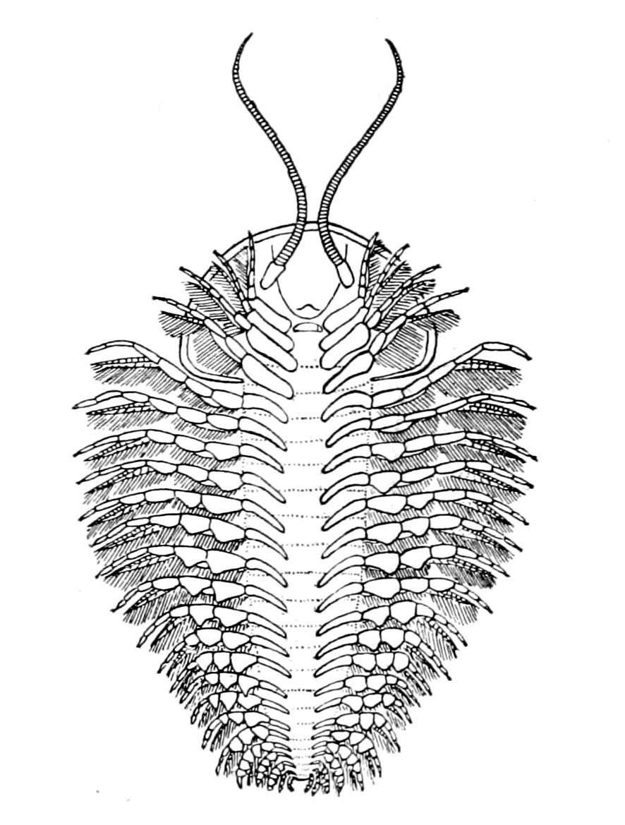
Fig. 164.—Under-Surface of Apus. (After Bronn.) Fig. 165.—Under-Surface of a Trilobite (Triarthrus). (From Beecher.)
All those who have seen Apus in the living state assert that this canal so formed is actually used by the animal for feeding purposes. By the movements of the gnatho-bases food is passed up from the hind end of the animal along the whole length of this ventral canal to the mouth, where it is taken in and swallowed. In this way Apus has been seen to swallow its own eggs.
In the trilobites there is a similar deep channel formed by the mid-ventral surface, similar gnatho-bases, and closely set appendages, and the membrane of this ventral groove was extremely thin.
Here, then, in the very group of animals which were the progenitors of the presumed palæostracan ancestor of the vertebrate—a group which is characterized by its extensive prevalence and its enormous variety of form during the great trilobite era—the formation of a mid-ventral canal out of this deep ventral groove is seen to be not only easy to imagine, but most probable, provided that a necessity arose for such a conversion.
For what purpose might such a tube have been formed? I would suggest that it might have acted as an accessory food-channel, which was of sufficient value at the time to give some advantage in the struggle for existence to those members of the group who were thus able to supplement their intake of food, but at the same time was so inefficient that it was quickly superseded by the new alimentary canal, and thus losing its temporary function, became solid, and was utilized to form an axial supporting rod.
There is a very considerable amount of evidence in favour of the view that the notochord was originally a digestive tube; in fact, as far as I know, this conclusion is universally accepted. The evidence is based essentially upon its development and upon its structure. It is formed in the vertebrate from the same layer as the alimentary canal, i.e. the hypoblast, and in Amphioxus it commences as a groove in the dorsal wall of the future alimentary canal; this groove then closes to form the tube of the notochord, and separates from the alimentary canal. Embryologically, then, the notochord is looked upon as a tube formed directly from the alimentary canal.
As regards its structure, its tissue is, as already stated, something sui generis. Notochordal tissue has no resemblance to bone or cartilage, or any of the usual supporting tissues. Such a tissue is not, however, entirely confined to the notochord of the vertebrates, but tissue closely resembling it has been found not only in Amphioxus and the Tunicata, but in certain other invertebrates, in the Enteropneusta (Balanoglossus, etc.), in Cephalodiscus, and in Actinotrocha. In all these latter cases, such a tissue is invariably found in disused portions of the alimentary canal; a diverticulum of the alimentary canal becomes closed, vacuolation of its lining cells takes place, and a tissue resembling notochordal tissue is formed.
Owing to the notochord being invariably so striking and mysterious a feature of the lowest vertebrates, the term vertebrate, which is inappropriate in the members of the group which do not yet possess vertebræ, has been largely superseded by the term chordate, with the result of attributing an undue preponderance to this tissue in any system of classification. Hence, wherever any animal has been found with a tissue resembling that of the notochord, enthusiasts have immediately jumped to the conclusion that a relationship must exist between it and the chordate animals; and, accordingly, they have classified such animals as follows: Amphioxus belongs to the group Cephalochorda because the notochord projects beyond the central nervous system; the Tunicata are called Urochorda because it is confined to the tail; the Enteropneusta, Hemichorda, because this tissue is confined to a small diverticulum of the gut, and, finally, Diplochorda has been suggested for Actinotrocha and Phoronis because two separate portions of the gut are transformed in this way.
This exaggerated importance given to any tissue resembling in structure that of the notochord is believed in by many of those who profess to be our teachers on this subject, the very men who can deliberately shut their eyes to the plain reading of the story of the pineal eyes, and say, "In our opinion this pineal organ was not an eye at all."
The only legitimate inference to be drawn from the similarity of structure between the notochord and these degenerated gut-diverticula, is that the structure of the notochord may have arisen in the same way, and that therefore the notochord may once have functioned as a gut. With cessation of its function its cells became vacuolated, as in these other cases, and its lumen became filled with notochordal tissue. This evidence strongly confirms the suggestion that the notochord was once a digestive tube, but by no means signifies that such tissue, wherever found, indicates the presence of a notochord.
In order to resemble a notochord, this tissue must possess not only a definite structure but a definite position, and this position is a remarkably striking and suggestive one. The notochordal tube is unsegmented, although the vertebrate is markedly segmented. But in all segmented animals the only unsegmented tube which extends the whole length of the body, from mouth to anus, is invariably the gut. In the vertebrate there are three such tubes: (1) the gut itself, (2) the central canal of the nervous system, and (3) the notochordal tube.
The first is the present gut, the second the gut of the invertebrate ancestor, and the third the tube in question.
These three unsegmented tubes, extending along the whole length of the segmented animal, constitute the great peculiarity of the vertebrate group; it is not the unsegmented notochord alone which requires explanation, but the presence of three such tubes in the same animal. Any one of them might be the unsegmented gut of the segmented animal. The most ventral tube is the actual gut of the present vertebrate; the most dorsal—the neural canal—was, according to my view, the original gut of the invertebrate ancestor; the middle one—the notochordal tube—was, in all probability, also once a gut, formed at the time when the exigencies of the situation made it difficult for food to pass along the original gut.
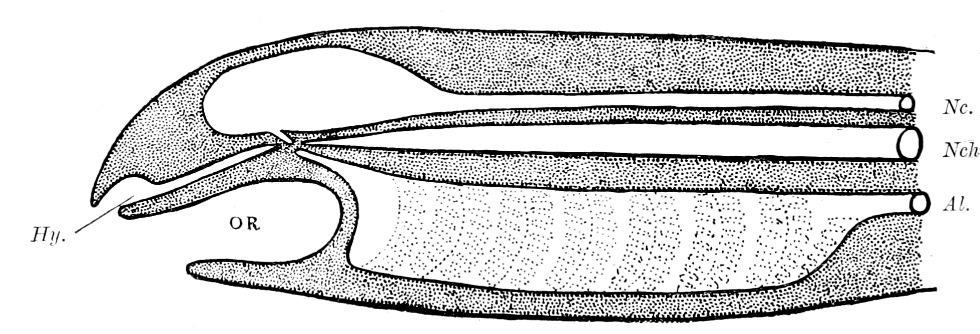
Fig. 166.—Diagram to show the Meeting of the Four Tubes in such a Vertebrate as the Lamprey.
Nc., neural canal with its infundibular termination; Nch., notochord; Al., alimentary canal with its anterior diverticulum; Hy., hypophysial or nasal tube; Or., oral chamber closed by septum.
Yet another circumstance in favour of this suggestion is the very striking position of the anterior termination of the notochord. It terminates at the point of convergence of three structures:—
(1) The tube of the hypophysis or nasal tube.
(2) The infundibulum or old mouth-termination.
(3) The notochordal tube.
To these may be added, according to Kupffer, in the embryonic stage, the anterior diverticulum of the gut (Fig. 166).
This is a very significant point. Here originally, in the invertebrate stage, the olfactory passage opened into the old mouth and œsophagus. Here, finally, in the completed vertebrate the same olfactory passage opens into the new pharynx. In the stage between the two it may well have opened into an intermediate gut, the notochordal tube, its separation from which would leave the end of the notochord blind, just as it had already left the end of the infundibulum blind.
The whole evidence points to the derivation of the notochord from a ventral groove on the surface of the animal, which closed to form a tube capable of acting as an accessory gut at the critical period before the new gut was fully formed. The essentials of a gut tube are absorption and digestion of food; is it likely that a tube formed as I have suggested would be efficient for such purposes?
As far as absorption is concerned, no difficulty would arise. The gut of the arthropod is lined with a thin layer of chitin, which is traversed, like all other chitinous surfaces, by fine canaliculi. Through these canaliculi, absorption of fluid material takes place, from the gut to the body. Similar canaliculi occur in the chitin covering the animal externally, so that, if such external surface formed a tube, and food in the right condition for absorption passed along it, absorption could easily take place through the chitinous surface. The evidence of Apus proves that food does pass along such a tube in the open condition, and in the trilobites the chitinous surface lining a similar groove was apparently very thin, a condition still more favourable to such an absorption process.
At first sight the second essential of a gut-tube—the power of digestion—appears to present an insuperable difficulty to this method of forming an accessory gut-tube, for it necessitates the formation of a secretion capable of digesting proteid material by the external cells of the body, whereas until recently it was supposed that such a function was confined to cells belonging to the so-called hypoblastic layer. Experiments were made now years ago of turning a Hydra inside out so that its internal layer should become external, and vice versâ, and they were said to have been successful. Such an animal could go on living and absorbing and digesting food, although its epiblastic surface was now its digestive internal surface. More recent observations have shown that these experiments were fallacious. At night-time, when the observer was not looking, the hydra reinverted itself, so that again its original digestive surface was inside and it lived and prospered as before.
Another piece of evidence of somewhat similar kind, which has not as yet been discredited, is seen in the Tunicata. In many of these, new individuals are formed from the parent by a process of budding, and it has been proved that frequently the gut of the new individual thus budded off arises not from the gut or hypoblastic layer of the parent, but from the surface or epiblastic layer. Such gut so formed possesses as efficient digestive powers as the gut of the parent.
The most remarkable evidence of all has been afforded by Miss Alcock's experiments. She examined the different tissues of Ammocœtes for the express purpose of finding out their power of digesting fibrin, with the result that the most active cells were those of the liver. Next in activity came the extract of the lining cells of the respiratory chamber and of the skin. The intestine itself when freed from the liver-secretion had very little digestive power; extracts of muscle, nervous system, and thyroid gland had no power whatever, but the extract of the skin-cells possessed a powerful digesting action.
Furthermore, it is not necessary to make an extract of the skin in order to obtain this digestive fluid, for under the influence of chloroform the skin of Ammocœtes secretes copiously, and this fluid thus secreted was found to possess strong digestive powers. So, also, Miss Alcock has demonstrated the power of digesting fibrin in a similar secretion of the epithelial cells lining the carapace of the crayfish. In both cases a very plausible reason for the presence of a digestive ferment in a skin-secretion is found in the necessity of preventing the growth of parasites, fungoid, or otherwise, especially in those parts where the animal cannot keep itself clean by 'preening.' Thus in a crayfish, in which the œsophageal commissures had been cut, fungus was found to grow on the ventral side, but not on the dorsal carapace. The animal was accustomed to keep its ventral surface clean by preening; owing to the paralysis it could not do so, and consequently the fungus grew there. In the lamprey I found that wherever there was a removal of the surface-epithelium, from whatever cause, that spot was immediately covered with a fungoid growth, although in the intact lamprey the skin was invariably smooth and clean.
I imagine, then, that this digestive power of the skin arose as a protective mechanism against parasitic attacks; it is self-evident how a tube formed of such material must ab initio act as a digestive tube.
In yet another respect this skin secretion of Ammocœtes is most instructive. The surface of Ammocœtes is absolutely smooth, no scales of any kind exist; this smoothness is due to the presence of a very well-defined cuticular layer secreted by the underlying epithelial cells. This cuticle is very much thicker than is usually found in vertebrates, and, strangely enough, has been thought to contain chitin. Whether it really contains chitin or not I am unable to say, but it certainly resembles a chitinous layer in one respect; it is perforated by innumerable very fine tubes or canaliculi, along which, by appropriate staining, it is easy to see the secretion of the underlying cell pass to the exterior (Fig. 140). This marked digestive power of the skin of Ammocœtes, together with the easy passage of the secretion through the thin cuticular layer, renders it almost certain that a tube formed from the deep ventral groove of the trilobite would, from the very first, act as a digestive as well as an absorbent tube; in other words, the notochord as soon as formed was able to act as an accessory digestive tube.
This suggested origin of the notochord from a groove along the mid-ventral surface of the body not only indicates a starting-point from a markedly segmented portion of the body, but also points to its formation at a stage previous to the formation of the operculum by the fusion of the two foremost mesosomatic appendages—indicates therefore its formation at a stage more nearly allied to the trilobite than to the sea-scorpion. The chance of ever finding any direct evidence of such a chordate trilobite stage appears to me exceedingly improbable, and I greatly fear that this conception of the mode of formation of the notochord can never be put to direct proof, but must always remain guesswork.
On the other hand, evidence of a kind in favour of its origin from a segmented part of the body does exist, and that evidence has this special value, that it is found only in that most primitive animal, Amphioxus.
This evidence is as follows:—
At fairly regular intervals, the sheath of the notochord is interrupted on each side of the mid-dorsal line by a series of holes, which penetrate the whole thickness of the sheath. This dorsal part is pressed closely against the spinal cord, and through these holes fibres appear to pass from the spinal cord to the interior of the notochord. So greatly do these fibres present the appearance of ventral roots to the notochord, that Miss Platt looks upon them as paired motor roots to the notochord, or at all events as once having been such motor roots. Lwoff and Rolph both describe a direct communication between the spinal cord and the notochord by means of fibres passing through these holes, without however looking upon this connection as a nervous one. Joseph alone asserts that no absolute connection exists, for the internal elastic layer of the notochord, according to him, is not interrupted at these holes, and forms, therefore, a barrier between the fibres from the spinal cord and those from the interior of the notochord. Still, whatever is the ultimate verdict as to these fibres, the suggestive fact remains of the spaces in the notochordal sheath and of the corresponding projecting root-like fibres from the spinal cord. The whole appearance gives the impression of some former connection, or rather series of connections, between the spinal cord and the notochord, such as would have occurred if nerves had once passed into the notochord. On the other hand, such nerves were not arranged segmentally with the myotomes, for, according to Joseph, in the middle of the animal ten to twelve such holes occur in one body-segment. In Apus the appendages are more numerous than the body-segments, so that it is not necessary for a segmental arrangement to coincide with that of the body-segments.
The Origin of the Alimentary Canal.
In close connection with the notochord is the alimentary canal. Any explanation of the one must be of assistance in explaining the other.
According to the prevalent embryological teaching, the body is formed of three layers, epiblast, hypoblast, and mesoblast, and the gastræa theory of the origin of all Metazoa implies of necessity that the formation of every individual commences with the formation of the gut. For this reason the alimentary canal must in every case be regarded as the earliest formed organ, however late in the development it may attain its finished appearance. Hence the notochord is spoken of as developed from the mid-dorsal wall of the alimentary canal. It is possible to look at the question the other way round, and suppose that the organ whose development is finished first is older than the one still in process of making. In this case it would be more right to say a ventral extension of the tissue, which gives rise to the notochord, takes place and forms the alimentary canal. It is, to my mind, perfectly possible, and indeed probable, that the formation of the vertebrate alimentary canal was a repetition of the same process which had already led to the formation of the notochordal tube. The formation of the anterior part of the alimentary canal in Ammocœtes at the time of transformation strongly suggests the marked similarity of the two processes.
Of all the startling surprises which occur at transformation, this formation of a new anterior gut is the most startling. From the oral chamber of Petromyzon two tubes start: the one leads into the gill-chambers, is known as the bronchus, and is entirely concerned with respiration; the other leads without a break from the mouth to the anus, has no connection with respiration, and is the alimentary canal of the animal. Any one looking at Petromyzon would say that its alimentary canal was absolutely non-respiratory in character. Before transformation, this kind of alimentary canal commences at the end of the respiratory chamber; from here to the anus it is of the same character as in Petromyzon, but in Ammocœtes the non-respiratory anterior part simply does not exist: the whole anterior chamber is both respiratory and affords passage to food. This part of the alimentary canal of the adult is formed anew. We see, then, here the formation of a part of the alimentary canal taking place, not in an embryo full of yolk, but in a free-living, independent, grown-up larval form in which all yolk has long since disappeared: a condition absolutely unique in the vertebrate kingdom, but one which more than any other may be expected to give a clue to the method of formation of a vertebrate gut.
The formation of this new gut can be easily followed at transformation, and was originally described by Schneider. His statement has been confirmed by Nestler, and its absolute truth has been demonstrated to me again and again by Miss Alcock, in her specimens illustrative of the transformation process. First, in the mid-dorsal line of the respiratory chamber a distinct groove is formed, the edges of which come together and form a solid rod. This solid rod blocks the opening of the respiratory chamber into the mid-gut, so that during this period of the transformation no food can pass out of the pharyngeal chamber. A lumen then begins to appear in this solid rod at the posterior end, which steadily advances mouthwards until it opens into the oral chamber and thus forms an open tube connecting the mouth with the gut.
Here, then, is the foundation of a new gut on very similar lines to that of the notochord, by the conversion of a groove into a tube. Still more suggestive is it to find that the tube so formed has no appearance whatever of segmentation; it is as unsegmented as the rest of the gut, although, as is seen in Fig. 62, the dorsal wall of the respiratory chamber from which it arose is as markedly segmented as any part of the animal. Here under our very eyes, in the course of a few days or weeks, an object-lesson in the process of the manufacture of an alimentary canal is carried out and completed, and the teaching of that lesson is that a gut-tube may be formed in the same way as the notochordal tube, by the conversion of a grooved surface into a canal, and that gut-tube so formed, like the notochord, loses all sign of segmentation, even although the original grooved surface was markedly segmented.
The suggestion then is, that the new gut may have been formed by a repetition of the same process which had already given origin to the notochord.
Such a method of formation is not, in my opinion, opposed to the evidence given by embryology, but in accordance with it; the discussion of this point will come best in the next chapter, which treats of the embryological evidence as a whole, and will therefore be left till then.
The Evidence given by the Innervation of the Vertebrate Alimentary Canal.
Throughout this investigation the one fixed landmark to which all other comparisons must be referred, is the central nervous system, and the innervation of every organ has given the clue to the meaning of that organ. So also it must be with the new alimentary canal; by its innervation we ought to obtain some insight into the manner of its origination. In any organ the nerves which are specially of value in determining its innervation, are of necessity the efferent or motor nerves, for the limits of their distribution in the organ are much more easily determined than those of the afferent or sensory nerves. The question therefore of primary importance in endeavouring to determine the nature of the origin of the alimentary canal from its innervation is the determination of the efferent supply to the musculature of its walls.
Already in previous chapters a commencement has been made in this direction; thus the musculature of the oral chamber has been derived directly from the musculature of the prosomatic appendages; the muscles which move the eyes from the prosomatic and mesosomatic dorso-ventral somatic muscles; the longitudinal body-muscles from the dorsal longitudinal somatic muscles of the arthropod; the muscles of respiration from the dorso-ventral muscles of the mesosomatic appendages.
In all these cases we have been dealing with striated musculature and consequently with only the motor nerves of the muscle; but the gut posterior to the pharyngeal or respiratory chamber contains unstriped instead of striped muscle, and is innervated by two sets of nerves, those which cause contraction and are motor, and those which cause relaxation and are inhibitory. It is by no means certain that these two sets of nerves possess equal value from a morphological point of view. The meaning of an inhibitory nerve is at present difficult to understand, and in this instance, is rendered still more doubtful owing to the presence of Auerbach's plexus along the whole length of the intestine—an elaborate system of nerve-cells and nerve-fibres situated between the layers of longitudinal and circular muscles surrounding the gut-walls, which has been shown by the recent experiments of Magnus, to constitute a special enteric nervous system.
One of the strangest facts known about the system of inhibitory nerves is their marked tendency to leave the central nervous system at a different level to the corresponding motor nerves, as is well known in the case of the heart, where the inhibitory nerve—the vagus—arises from the medulla oblongata, while the motor nerve—the augmentor or accelerator—leaves the spinal cord in the upper thoracic region. It is very difficult to obtain any idea of the origin of such a peculiarity; I know of only one suggestive fact, which concerns the innervation of the muscles which open and close the chela of the crayfish, lobster, etc. These muscles are antagonistic to each other, and both possess inhibitory as well as motor nerves. The central nervous system arrangements are of such a character that the contraction of the one muscle is accompanied by the inhibition of its opposer, and the nerves which inhibit the contraction of the one, leave the central nervous system with the nerves which cause the other to contract. Thus the inhibitory and motor nerves of either the abductor (opener) or adductor (closer) muscles of the crayfish claw do not leave the central nervous system together, but in separate nerves.
If now for some cause the one set of muscles either disappeared, or were so altered as no longer to present any appearance of antagonism, then there would be left a single set of muscles, the inhibitory and motor nerves of which would leave the central nervous system at different levels, and the older such systems might be, the greater would be the modification in the shape and arrangements of parts in the animal, so that the two sets of fibres might ultimately arise from very different levels.
As mentioned in the introductory chapter, the whole of this investigation into the origin of vertebrates arose from my work on the system of efferent nerves which innervate the vascular and visceral systems. One of the main points of that investigation was the proof that such nerves did not leave the central nervous system uniformly along the whole length of it, but in three great outflows, cranial, thoracico-lumbar, and sacral; there being two marked gaps separating the three outflows, caused by the interpolation of the plexuses for the innervation of the anterior and posterior limbs respectively. All these nerves are characterized by the presence of ganglion-cells in their course to the periphery, they are, therefore, distinguished from ordinary motor nerves to striated muscle in that their impulses pass through a ganglion-cell before they reach the muscle.
The ganglia of the large middle thoracico-lumbar outflow constitute the ganglia of the sympathetic system.
The functions of the nerves constituting these three outflows are very different, as I pointed out in my original papers. Since then a large amount of further information has been obtained by various observers, especially Langley and Anderson, which enable the following statements to be made:—
All the nerves which cause contraction of the unstriped muscles of the skin, whether pilomotor or not, all the nerves which cause secretion of sweat glands wherever situated, all the nerves which cause contraction or augmentation of the action of muscles belonging to the vascular system, all the nerves which are motor to the muscles belonging to all organs derived from the Wolffian and Müllerian ducts, e.g. the uterus, ureters, urethra, arise from the thoracico-lumbar outflow, never from the cranial or sacral outflows. It is essentially an efferent skin-system.
On the other hand, the latter two sets of nerves are concerned with the supply of motor nerves to the alimentary canal; they form essentially an efferent gut-system in contradistinction to the sympathetic or skin-system.
A marked distinction exists between these cranial and sacral nerves. The vagus never supplies the large intestine, the sacral nerves never supply the small intestine. Associated with the large intestine is the bladder, the whole system arising from the original cloacal region; the vagus never supplies the bladder, its motor nerves belong to the sacral outflow. The motor nerves to the ureters, to the urethra, and to the trigonal portion of the bladder between the ureters and the urethra, do not arise from the sacral outflow, but from the thoracico-lumbar. These muscles belong really to the muscles in connection with the Müllerian and Wolffian ducts and skin, not to the cloacal region.
The motor innervation then of the alimentary canal reveals this striking and suggestive state of affairs. The motor innervation of the whole of the small intestine arises from the cranial region, and is immediately followed by an innervation from the sacral region for the whole of the muscles of the cloaca. It thus indicates a head-region and a tail-region in close contiguity, the whole of the spinal cord region between these two extremes being apparently unrepresented. Not, however, quite unrepresented, for Elliott has shown recently that the ileo-colic valve at the junction of the small and large intestine is in reality an ileo-colic sphincter muscle, and that this muscle receives its motor nerves neither from the vagus nor from the sacral nerves, but from the thoracico-lumbar outflow or sympathetic system. This may mean one of two things, either that a band of fibres belonging to the skin-system has been added to the gut-musculature, for the purpose of forming a sphincter at this spot, or that the region between the vagus territory and the cloaca is represented by this small band of muscle. The second explanation seems to me the more probable of the two. Between the mesosomatic region represented by the vagus, and the cloacal region, there existed a small metasomatic region, represented by the pronephros, with its segmental duct, as already discussed in Chapter XII. That part of the new alimentary canal which belonged to this region is the short piece indicated by the ileo-colic sphincter, and innervated, therefore, from the same region as the organs derived from the segmental duct.
Such innervation seems to me to suggest that originally the vertebrate consisted, as far as its gut was concerned, of a prosomatic and mesosomatic (branchial) region, close behind which came the cloaca and anus. Between the two there was a short metasomatic region (possibly pronephric), so that the respiratory chamber did not open directly into the cloaca.
Such an interpretation is, I think, borne out by the study of the most ancient forms of fish. In Bothriolepis, according to Patten, and in Drepanaspis, according to Traquair, the cloacal region and anus follow immediately upon the posterior end of the head-shield, i.e. immediately after that region which presumably contained the branchiæ. Similarly, on the invertebrate side, all those forms which resembled Limulus must have possessed a very short region between the branchial and cloacal parts of the body. The original cloacal part of the vertebrate gut may well have been the original cloaca of the arthropod, into which its intestine emptied itself, especially when we see the tendency of the scorpion group of animals to form an accessory cloacal pouch known as the stercoral pouch or pocket.
Again, it is striking to see how, in certain of the scorpion group, e.g. Thelyphonus and Phrynus, there is a caudal massing of the central nerve-cells as well as a cephalic massing, so that their central nervous system is composed of a cephalic and caudal brain. These two brains are connected together by commissures extending the whole length of the body, in which I have been unable to find any sign of ganglion-cells. What this caudal brain innervates I do not know; it is, I think, a matter worth further investigation, especially as there are many indications in the vertebrate that the lumbo-sacral region of the cord possesses higher functions than the thoracic region.
The method of formation of the alimentary canal as indicated by its innervation is as follows:—
In front an oral chamber, formed, as already pointed out, by the modification of the prosomatic appendages, followed by a respiratory chamber, the muscles and branchiæ of which were the muscles and branchiæ of the mesosomatic appendages. This mesosomatic, or branchial, part was in close contiguity to the cloaca and anus, being separated from it only by a short tube formed in the metasomatic or pronephric region.
I imagine that this connection was originally in the form of an open groove, as already explained for both notochord and the anterior part of the gut itself in Ammocœtes; an open groove formed from the mid-ventral surface of the body, on each side of which were the remnants of the pronephric appendages. By the closure of this groove ventrally, and the growing round of the pleural folds, as already suggested, the remains of the pronephric appendages are indicated by the segmental duct and the form of the vertebrate body is attained.
Even in the branchial region the same kind of thing must, I think, have occurred. The grooved ventral surface became a tube, on each side of which were lying in regular order the in-sunk branchial appendages, the whole being subsequently covered by the pleural folds to form an atrial chamber. A tube thus formed from the grooved ventral surface would carry with it to the new ventral surface the longitudinal venous sinuses, and thus form, in the way already suggested, the heart and ventral aorta. Posterior to the heart in the pronephric region, the same process would give rise to the sub-intestinal vein.
The evidence of comparative anatomy bears out most conclusively the suggestion that in the original vertebrate the gut was mainly a respiratory chamber. In man and all mammals the oral chamber opens into a small pharynx, followed by the œsophagus, stomach and small intestine. Of this whole length, a very small part is taken up by the pharynx, in which, in the embryo, the branchial arches are found, showing that this represents the original respiratory part of the gut. In the ordinary fish this branchial part is much more conspicuous, occupies a large proportion of the gut, and in the lowest fishes, such as Ammocœtes and Amphioxus, the branchial region extends over a large portion of the animal, while the intestine proper is a straight tube, the length of which is insignificant in comparison with its length in the higher vertebrates.
Such a tube was able to act as a digestive tube, owing, as already pointed out, to the digestive powers of the skin-epithelium, and I imagine at first the respiratory chamber, seeing that it composed very nearly the whole of the gut, was at the same time the main digestive chamber; even in Ammocœtes its digestive power is superior to that of the intestine itself.
Just posterior to the branchial part a diverticulum of the gut was formed at an early stage, as seen in Amphioxus, and provided the commencement of the liver. This simple liver-diverticulum became the tubular liver of Ammocœtes, and formed, curiously enough, not a glandular organ of the same character as the liver of the higher vertebrates, but a hepato-pancreas, like the so-called liver of the arthropods, which also is a special diverticulum of the gut, or rather the main true gut of the animal. In both cases the liver is the chief agent in digestion, for in Ammocœtes the liver-extract is very much more powerful in the digestion of proteids than the extract of any other organ tried by Miss Alcock. Subsequently in the vertebrate the gastric and pancreatic glands arise and relieve the liver of the burden of proteid digestion.
It is, to my mind, somewhat significant that the liver on its first formation in the vertebrate should have arisen as a digestive organ of the same character as the so-called liver in the arthropods; whether it originally belonged to any separate segment is in our present state of knowledge difficult to say.
Conclusion.
In conclusion, I will endeavour to illustrate crudely the way in which, on my theory, the notochord and vertebrate gut may have been formed, the agencies at work being in the main two, viz. the dwindling of appendages as mere organs of locomotion, and the conversion of a ventral groove into a tube.
I imagine that, among the Protostraca, forms were found somewhat resembling trilobites with markedly polychætan affinities; which, like Apus, possessed a deep ventral groove from one end of the body to the other, and also pleural fringes, as in many trilobites. This might be called the Trilobite stage (Fig. 167, A).
This groove became converted into a tube and so gave rise to the notochord, while the appendages were still free and the pleuræ had not met to form a new ventral surface. This might be called the Chordate Trilobite stage (Fig. 167, B).
Then, passing from the protostracan to the palæostracan stage, the oral and respiratory chambers were formed, not communicating with each other, in the manner described in previous chapters, a ventral groove in the metasomatic region being the only connection between respiratory chamber and cloaca. This might be called the Chordate Palæostracan stage (Fig. 167, C).
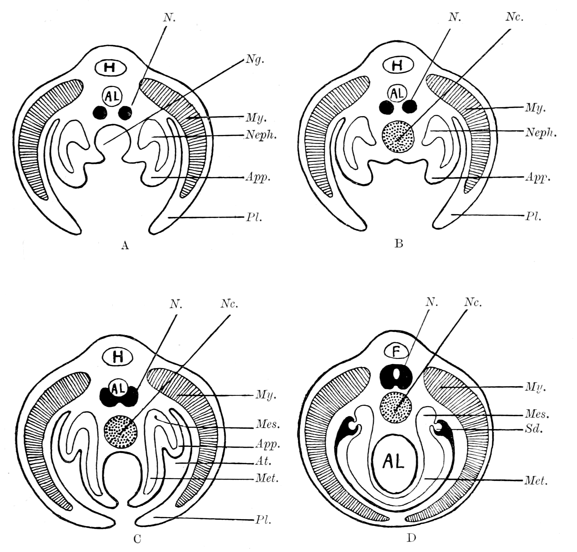
Fig. 167.—A, Diagram of Section through a Trilobite-like Animal; B, Diagram to illustrate the Suggested Formation of the Notochord from a Ventral Groove; C, Diagram to illustrate the Suggested Formation of the Post-Branchial Gut by the continuation of the same process of Ventral Groove-Formation, combined with Obliteration of Appendages and Growth of Pleural Folds; D, Diagram to illustrate the Completion of the Vertebrate Type by the Meeting of the Pleural Folds in the Mid-Ventral Line with the Obliteration of the Atrial Cavity and the Conversion of the Ventral Groove into the closed Alimentary Canal.
Al., alimentary canal; N., nervous system; My., myotome; Pl., pleuron; App., appendage; Neph., nephrocœle; Met., metacœle; Sd., segmental duct; Mes., mesonephros; At., atrial chamber; Nc., notochord; H., heart; F., fat body; Ng., notochordal groove. (These diagrams are intended to complete the diagrams on p. 413, which, as stated there, were purposely left incomplete.)
Finally, with the conversion of this groove into a tube, the opening of the oral into the respiratory chamber, and the formation of an atrium by the ventralwards growth of the pleural folds, the formation of a Vertebrate was completed (Fig. 167, D).
In my own mind I picture to myself an animal which possessed eurypterid and trilobite characters combined, in which a notochordal tube had been formed in the way suggested, and a respiratory chamber which communicated with the cloaca by means of a grooved channel along the mid-ventral line of the metasomatic portion of the body. On each side of this channel were the remains of the metasomatic appendages (pronephric). The whole was enveloped in the pleural folds, which probably at this time did not yet meet in the middle line to form a new ventral surface. This respiratory chamber, owing to the digestive power of the epidermis, assisted in the process of alimentation to such an extent as to supersede the temporary notochordal tube, with the effect of bringing about the conversion of the metasomatic groove into a closed canal, and so the formation of an alimentary tube continuous with the respiratory chamber. The amalgamation of the pleural folds ventrally completed the process, and so formed an animal resembling the Cephalaspidæ, Ammocœtes, or Amphioxus.
I have endeavoured in this chapter to make some suggestions upon the origin of the notochord and of the vertebrate gut in accordance with my theory of the origin of vertebrates. I feel, however, strongly that these suggestions are much more speculative than those put forward in the previous chapters, and of necessity cannot give the same feeling of soundness as those based directly upon comparative anatomy and histology. Still, the fact remains that the origin of the notochord is at present absolutely unknown, and that my speculation that it may have originated as an accessory digestive tube is at all events in accordance with the most widely spread opinion that it arises in close connection with an alimentary canal.
CHAPTER XIV
THE PRINCIPLES OF EMBRYOLOGY
The law of recapitulation.—Vindication of this law by the theory advanced in this book.—The germ-layer theory.—Its present position.—A physiological not a morphological conception.—New fundamental law required.—Composition of adult body.—Neuro-epithelial syncytium and free-living cells.—Meaning of the blastula.—Derivation of the Metazoa from the Protozoa. Importance of the central nervous system for Ontogeny as well as for Phylogeny.—Derivation of free-living cells from germ-cells.—Meaning of cœlom.—Formation of neural canal.—Gastrula of Amphioxus and of Lucifer.—Summary.
In a discussion upon this theory of mine, which took place at Cambridge on November 25 and December 2, 1895, it was said that such a theory was absolutely and definitely put out of court, because it contravened the principles of embryology, was opposed, therefore, to our surest guide in such matters; and the law was laid down with great assurance that no claim for genetic relationship between two groups of animals can be allowed which is based upon topographical and structural coincidences revealed by the study of the anatomy of two adult animals, however numerous and striking they may be, if there are fundamental differences in the embryology of the members of these two groups.
According to my theory the old gut of the arthropod still exists in the vertebrate as the tubular lining of the central nervous system, and the vertebrate has formed a new gut. According to the principles of embryology as held up to the present, in all animals above the Protozoa, the different structures of the body arise from three definite embryonic layers, the epiblast, mesoblast, and hypoblast, and in all cases the gut arises from the hypoblastic layer. In the vertebrate the gut also arises from the hypoblast, while the neural canal is epiblastic. My theory, then, makes the impossible assertion that what was hypoblast in the arthropod has become epiblast in the vertebrate, and what was epiblast in the arthropod has become hypoblast in the vertebrate. Such a conception is supposed to be so absolutely impossible that it only requires to be stated to be dismissed as an absurdity.
Against this opinion I claim boldly that my theory is not only not contrary to the principles of embryology, but is mainly based upon the teachings of embryology. I wish here not to be misunderstood. The great value of the study of embryology for questions of the sequence of the evolution of animals is to be found in what is known as the Law of Recapitulation, which asserts that every animal gives some indication in the stages of its individual development of its ancestral history. Naturally enough it cannot pass through all the stages of its past history with equal clearness, for what has taken millions of years to be evolved has to be compressed into an evolution lasting only a few months or weeks, or even less.
When in the highest vertebrate a vestigial organ, such as the pineal gland, can be traced back without leaving the vertebrate kingdom to a distinct median eye, such as is found in the lamprey, that rudimentary organ is evidence of an organ which was functional in the earliest vertebrates or their immediate ancestors. So it is generally with well defined vestigial organs found in the adult animal; they always indicate an organ which was functional in the near ancestor.
Passing from the adult to the embryo we still find the same law. Here, also, vestigial organs are met with, which may leave no trace in the adult, but indicate organs which were functional in the near ancestor. Thus, but for embryology, we should have no certainty that the air-breathing vertebrates had been derived from water-breathing fishes; the indication is not given by any close resemblance between the formation of the embryos in their earliest stages, but by the formation of vestigial gill-arches even in the embryos of the highest mammal.
For all questions of evolution the presence of vestigial organs in the embryo is the important consideration, for they give an indication of near ancestry; the early formation of the embryo concerns a much more remote ancestral period, all vestigial organs of which may well have been lost and obscured by cœnogenetic changes. Let us, then, consider the two things—the vestigial organs and the early formation of the embryo—separately, and see how far my opponents are justified in their statement that my theory contravenes the principles of embryology.
First, I will take the teachings of vestigial organs and the arrangement of organs found in the vertebrate embryo. Here it is impossible to say that my theory is contrary to the teaching of embryology, for as the previous chapters have shown again and again, the argument is based very largely upon the facts of embryology. In the first place, the comparison which I have chiefly made is a comparison between the larval form of a very low vertebrate and the arthropod group, a comparison which exists only for the larval form, and not for the adult. The whole theory, then, is based upon a developmental stage of the vertebrate, and not upon the anatomy of the adult.
Throughout the whole history it seems to me perfectly marvellous how completely the law of recapitulation is vindicated by my theory of the origin of the vertebrate. The theory asserts that the clue to the origin of vertebrates is to be found in the tubular nature of the central nervous system of the vertebrate; in that the vertebrate central nervous system is in reality formed of two things: (1) a central nervous system of the arthropod type, and (2) an epithelial tube in the position of the alimentary canal of the arthropod.
Is it possible for embryology to recapitulate such a phylogenetic history more clearly than is here the case? In order to avoid all possibility of our mistaking the clue, the nerve-tube in the embryo always opens into the anus at its posterior end, while in the larval Amphioxus it is actually still open to the exterior at the anterior end. The separateness of the tube from the nervous system at its first origin is shown especially well in the frog, where, as Assheton has pointed out, owing to the pigment in the cells of the external layer of epithelium, a pigmented tube is formed, on the outside of which the nervous tissue is lying, and step by step the gradual intermingling of the nerve-cells and the pigmented lining cells can be followed out.
Consider the shape of the nerve-tube when first formed in the vertebrate. At the cephalic end a simple bulged-out tube with two simple anterior diverticula, which passes into a narrow straight spinal tube; from this large cephalic bulging a narrow diverticulum, the infundibulum, passes to the ventral surface of the forming brain. This tube is the embryological expression of the simple dilated cephalic stomach, with its ventral œsophagus and two anterior diverticula, which opens into the straight intestine of the arthropod. Nay, more, by its very shape, and the invariable presence of two anterior diverticula, it points not only to an arthropod ancestry, but to a descent from a particular group of primitive arthropods. Then comes the formation of the cerebral vesicles, and the formation of the optic cup, telling us as plainly as can be how the invasion of nervous material over this simple cephalic stomach and its diverticula has altered the shape of the original tube, and more and more enclosed it with nervous elements.
So, too, in the spinal cord region. When the tube is first formed, it is a large tube, the latero-ventral part of which presents two marked bulgings; connecting these two bulgings is the anterior commissure. These two lateral bulgings, with their transverse commissure, represent, with marked fidelity, the ventral ganglion-masses of the arthropod with their transverse commissure, and occupy the same position with respect to the spinal tube, as the ganglion-masses do with respect to the intestine in the arthropod. Then the further development shows how, by the subsequent growth of the nervous material, the calibre of the tube is diminished in size, and the spinal cord is formed.
Again, I say, is it possible to conceive that embryology should indicate the nature of the origin of the vertebrate nervous system more clearly than it does?
It is the same with all the other organs. Take, for instance, the skeletal tissues. The study of the vertebrate embryo asserts that the cartilaginous skeleton arose as simple branchial bars and a simple cranio-facial skeleton, and also that the parenchymatous variety of cartilage represents the embryonic form. Word for word, the early embryonic stage of the vertebrate skeleton closely resembles the stage reached in the arthropod, as shown by Limulus, and again records, unmistakably, the past history of the vertebrate.
So, too, with the whole of the prosomatic region; the situation of the old mouth, the manner in which the nose of the cephalaspidian fishes arose from the palæostracan, are all shown with vivid clearness by Kupffer's investigations of the early stage of Ammocœtes, while at the same time the closure of the oral cavity by the septum shows how the oral chamber was originally bounded by the operculum. Nay, further, the very formation of this chamber embryologically was brought about by the forward growth of the lower lip, just as it must have been if the chilaria grew forward to form the metastoma.
So, too, the study of the embryo teaches that the branchiæ arise as ingrowths, that the heart arises as two longitudinal veins, just as the theory supposes from the facts provided by Limulus and the scorpions. No indication of the origin of the thyroid gland is given by the study of its structure in any adult vertebrate, but in the larval form of the lamprey there is still preserved for us a most graphic record of its past history.
The close comparisons which it is possible to make between the eye-muscles of the vertebrate and the recti muscles of the scorpion group on the one hand, and between the pituitary and coxal glands on the other, are based upon, or at all events are strikingly confirmed by, the study of the cœlomic cavities and the origin of these muscles in the two groups. In fact the embryological evidence of the double segmentation in the head and the whole nature of the cranial segments is one of the main foundation-stones on which the whole of my theory rests.
So it is throughout. Turn to the excretory organs—it is not the kidney of the adult animal which leads direct to the excretory organs of the primitive arthropod, but the early embryonic origin of that kidney.
So far from having put forward a theory which runs counter to the principles of embryology, I claim to have vindicated the great Law of Recapitulation which is the foundation-stone of embryological principles. My theory is largely based upon embryological facts, and its strength consists in the manner in which it links together into one harmonious whole, the facts of Embryology, Palæontology, Anatomy, and Physiology. Why, then, is it possible to assert that my theory disregards the principles of embryology, when, as we have seen, embryology is proclaiming as loudly as possible how the vertebrate arose? In my opinion, it is because the embryologists have to a large extent gone wrong in their fundamental principles, and have attached more weight to these faulty fundamental principles than to the obvious facts which, looked at thoughtfully, could not have failed to suggest a doubt as to the correctness of these 'principles.'
The current laws of embryology upon which such weight is laid are based on the homology of the germinal layers in all Metazoa, and state that in all cases after segmentation is finished a blastula is formed, from which there arises a gastrula, formed of an internal layer, the hypoblast, and an external layer, the epiblast; subsequently between these arises a third layer, the mesoblast. These layers are strictly morphological conceptions, and are stated to be homologous in all cases, so that the hypoblast of one animal must be homologous to the hypoblast of another. In order, therefore, to compare two adult animals for the purpose of finding kinship between them, it is necessary to find whether parts such as the gut, which in both cases have the same function, arise from the same germinal layer in the embryo. We can, in fact, have no certainty of kinship, even although the two animals are built up as far as the adult state is concerned on a remarkably similar plan, unless we can study their respective embryos and find out what parts arise from the hypoblast and what from the epiblast. The homology of the germinal layers constitutes in all cases of disputed relationship the court of final appeal. A new gut, therefore, in any animal can only be formed from hypoblast, and any theory, such as that advocated in this book, which deals with the formation of a new gut, and does not form that gut from pre-existing hypoblast, must of necessity be wrong and needs no further consideration.
Such is the result of current conceptions—conceptions which to be valid must be based upon an absolutely clear morphological definition of the formation of the germinal layers, a definition not based on their subsequent history and function, but determined solely by the uniformity of the manner of their origin.
What, then, is a germinal layer? How can we identify it when it first arises? What is the morphological criterion by which hypoblast can be distinguished from epiblast, or mesoblast from either?
This is the question put by Braem, in an admirable series of articles in the Biologisches Centralblatt, and is one that must be answered by every worker who bases his views of the process of evolution upon embryological investigation. As Braem points out, the germinal layers are definable either from a morphological or physiological standpoint. In the one case they must arise throughout on the same plan, and whatever be their fate in the adult, they must form at an early stage structures strictly homologous in all animals. In the other case the criterion is based on function, and the hypoblast, for instance, is that layer which is found afterwards to form the definitive alimentary canal. There is no longer any morphological homology; such layers are analogous; they may be, but are not necessarily, homologous. Braem gives a sketch of the history of the views held on the germinal layers, and shows how they were originally a purely physiological conception, and how gradually such conception changed into a morphological one, with the result that what had up to that time been looked upon as analogous structures became strictly homologous and of fundamental importance in deciding the position of any animal in the whole animal series.
This change of opinion was especially due to the lively imagination of Haeckel, who taught that the germinal layers of all Metazoa must be strictly homologous, because they were all derived from a common ancestral stock, represented by a hypothetical animal to which he gave the name Gastræa; an animal which was formed by the simple invagination of a part of the blastula, thus giving rise to the original hypoblast and epiblast, and he taught that throughout the animal kingdom the germinal layers were formed by such an invagination of a part of the blastula to form a simple gastrula. If further investigation had borne out Haeckel's idea, if therefore the hypoblast was in all cases formed as the invagination of a part of a single-layered blastula, then indeed the dogma of the homology of the germinal layers would be on so firm a foundation that no speculation which ran counter to it could be expected to receive acceptance; but that is just what has not taken place. The formation of the gastrula by simple invagination of the single-layered blastula is the exception, not the rule, and, as pointed out by Braem, is significantly absent in the earliest Metazoa; in those very places where, on the Gastræa theory, it ought to be most conspicuous.
Braem discusses the question most ably, and shows again and again that in every case the true criterion upon which it is decided whether certain cells are hypoblastic or not is not morphological but physiological. The decision does not rest upon the answer to the question, Are these cells in reality the invaginated cells of a single-celled blastula? but to the question, Do these cells ultimately form the definitive alimentary canal? The decision is always based on the function of the cells, not on their morphological position. Not only in Braem's paper, but elsewhere, we see that in recent years the physiological criterion is becoming more and more accepted by morphologists. Thus Graham Kerr, in his paper on the development of Lepidosiren, says: "It seems to me quite impossible to define a layer as hypoblastic except by asking one or other of the two questions: (1) Does it form the lining of an archenteric cavity? and (2) Does it become a certain part of the definitive epithelial lining of the gut?"
The appearance of Braem's paper was followed by a criticism from the pen of Samassa, who agrees largely with Braem, but thinks that he presses the physiological argument too far. He considers that morphological laws must exist for the individual development as well as for the phylogenetic, and finishes his article with the following sentence, a sentence in which it appears to me he expresses what is fast becoming the prevailing view: "Mit dem Satz, den man mitunter lesen kann: 'es muss doch auch für die Ontogenie allgemeine Gesetze geben' kann leicht Missbrauch getrieben werden; diese allgemeinen Gesetze giebt es wohl, aber sie liegen nicht auf flacher Hand und bis zu ihrer Erkenntnis hat es noch gute Wege; das eine kann man aber wohl heute schon sagen, die Keimblätterlehre gehört zu diesen allgemeinen Gesetzen nicht."
I conclude, then, that we ought to go back to a time previous to that of Haeckel and ask ourselves seriously the question, When we lay stress on the germinal layers and speak of this or that organ arising from this or that germinal layer, are we thereby adding anything to the knowledge that we already possess from the study of the anatomy and physiology of the adult body? If by hypoblast we only mean the internal surface or alimentary canal and its glands, etc., and by epiblast we mean the external surface or skin and its glands, etc., while mesoblast indicates the middle structures between the other two, then I fail to see what advantages we obtain by using Greek terms to express in the embryo what we express in English in the adult.
The evidence given by Braem, and it could be strengthened considerably, is conclusive against the morphological importance of the theory of the germinal layers, and transfers the fundamental importance of the early embryonic formation, from that of a three-layered embryo to that of a single-layered embryo—the blastula—from which, in various ways, the adult animal has arisen.
The derivation of both arthropod and vertebrate from such a single-layered animal is perfectly conceivable, even though the gut of the latter is not homologous with the gut of the former. We have seen that the teachings of embryology, as far as its later stages are concerned, afford one of the main supports upon which this theory rests. What, therefore, is required to complete the story is the way in which these later stages arise from the blastula stage; here, as in all cases, the ontogenetic laws must be in harmony with the phylogenetic; of the latter the most important is the steady development of the central nervous system for the upward progress of the animal race. The study of comparative anatomy indicates the central nervous system, not the gut, as the keystone of the edifice. So, also, it must be with ontogeny; here also the central factor in the formation of the adult from the blastula ought to be the formation of the central nervous system, not that of the gut.
Such, it appears to me, is the case, as may be seen from the following considerations.
The study of the development of any animal can be treated in two ways: either we can trace back from the adult to the very beginning in the ovum, or we can trace forward from the fertilized egg to the adult. Both methods ought to lead to the same result; the difference is, that in the first case we are passing from the more known to the less known, and are expressing the unknown in terms of the known. In the second case we are passing from the less known to the more known, and are expressing the known in speculative terms, invented to explain the unknown. What has just been said with respect to the germinal layers means that, however much we may study the embryo and try to express the adult in terms of it, we finally come back to the first way of looking at the question, and, starting with the adult, trace the continuity of function back to the first formation of cells having a separate function.