Front matter (continued 8)
Reading edition
This structure is therefore a calcareous infiltration of a tissue with cells in it. Where is there anything like it?
As soon as I saw Rohon's picture (Fig. 130), I was astounded at its startling resemblance to the structure of muco-cartilage as is seen in Fig. 131, taken from Ammocœtes. If such muco-cartilage were infiltrated with lime salts, then the muco-cartilaginous skeleton of Ammocœtes would be preserved in the fossil condition, and be comparable with that of Cephalaspis, etc.
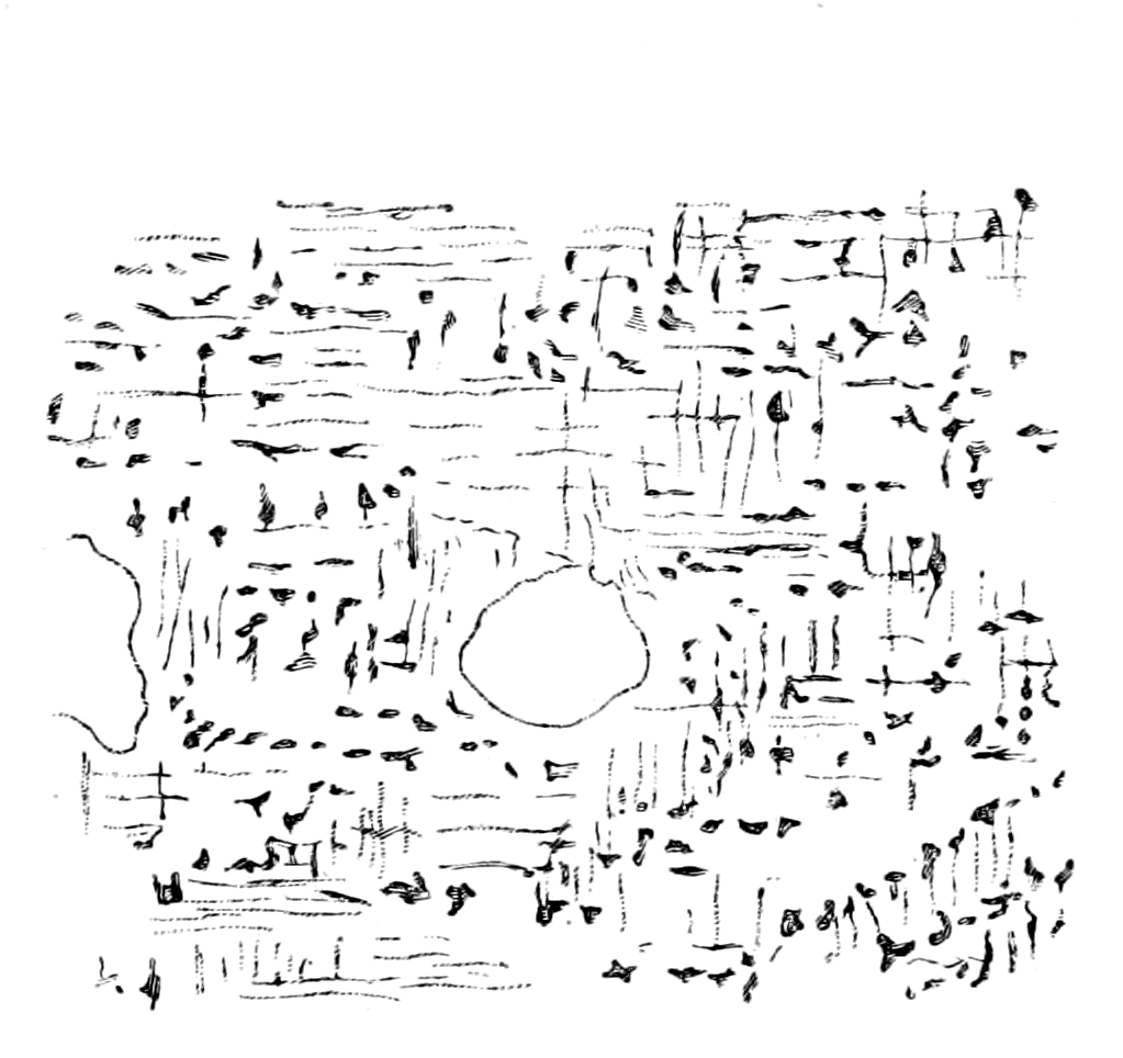
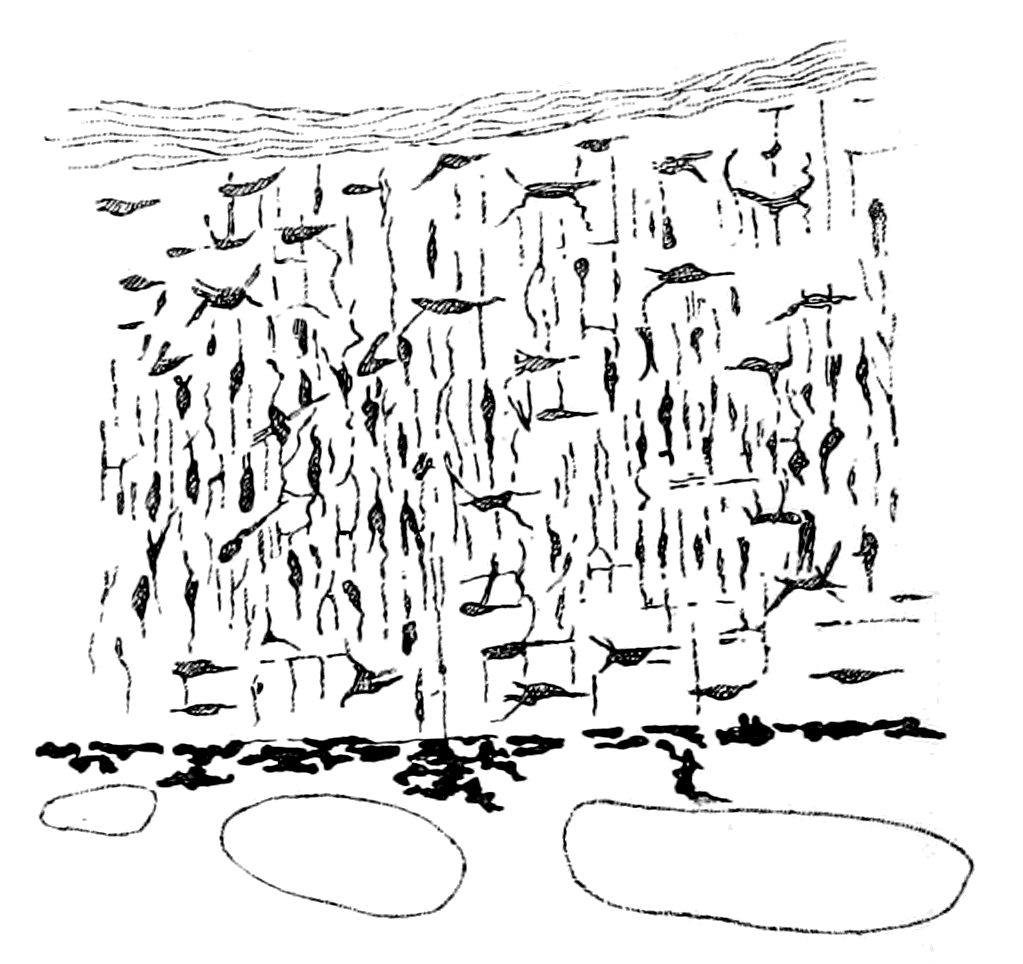
Fig. 130.—Section of a Head-Plate of a Cephalaspid. (From Rohon.) Fig. 131.—Section of Muco-Cartilage from Dorsal Head-Plate of Ammocœtes.
The whole structure is clearly remarkably like Rohon's picture of a section of the head-plate of a Cephalaspid (Fig. 130). In the latter case the matrix contains calcium salts, in the former it is composed of the peculiar homogeneous mucoid tissue which stains so characteristically with thionin. With respect to this calcification, it is instructive to recall the calcification in the interior of the branchial cartilages of Limulus, as described in Chapter III., for this example shows how easy it is to obtain a calcification in this chondro-mucoid material. With respect to the medullary spaces and smaller spaces in this tissue, as described by Rohon, I would venture to suggest that they need not all necessarily indicate blood-vessels, for similar spaces would appear in the head-shield of Ammocœtes if its muco-cartilage alone were preserved. Of these, some would indicate the position of blood-vessels, such, for instance, as of the external carotid which traverses this structure; but the largest and most internal spaces, resembling Rohon's medullary spaces, would represent muscles, being filled up with bundles of the upper lip-muscles.
The Muco-Cartilaginous Head-Shield of Ammocœtes.
The resemblance between the structure of the head-shield of Thyestes and the muco-cartilage of Ammocœtes, is most valuable, for muco-cartilage is unique, occurs in no other vertebrate, and every trace of it vanishes at transformation; it is essentially a characteristic of the larval form, and must, therefore, in accordance with all that has gone before, be the remnant of an ancestral skeletal tissue. The whole story deduced from the study of Ammocœtes would be incomplete without some idea of the meaning of this tissue. So also, as already mentioned, the skeleton of Ammocœtes is incomplete without taking this tissue into account. It is confined entirely to the head-region; no trace of it exists posteriorly to the branchial basket-work. It consists essentially of dorsal and ventral head-shields, connected together by the tentacular, metastomal, and thyroid bars, as already described. The ventral shield forms the muco-cartilaginous plate of the lower lip and the plate over the thyroid gland, so that the skeleton ventrally is represented by Fig. 118, B, which shows how the cartilaginous bars of the branchial basket-work are separated from each other by this thyroid plate. At transformation, with the disappearance of this muco-cartilaginous plate, the bars come together in the middle line, as in the more posterior portion of the branchial basket-work.
The dorsal head-shield of muco-cartilage covers over the upper lip, sends a median prolongation over the median pineal eyes and a lateral prolongation on each side as far as the auditory capsules, giving the shape of the head-shield of muco-cartilage, as in Fig. 118, C.
Not only then is the structure of the head-shield of a Cephalaspid remarkably like the muco-cartilage of Ammocœtes, but also its general distribution strangely resembles that of the Ammocœtes muco-cartilage.
Now, these head-shields in the Cephalaspidæ and Tremataspidæ vary very much in shape, as is seen by the comparison of Tremataspis and Auchenaspis with Cephalaspis and Eukeraspis, and yet, undoubtedly, all these forms belong to a single group, the Osteostraci.
The conception that Ammocœtes is the solitary living form allied to this group affords a clue to the meaning of this variation of shape, which appears to me to be possible, if not indeed probable. There is a certain amount of evidence given in the development of Ammocœtes which indicates that the branchial region of its ancestors was covered with plates of muco-cartilage as well as the prosomatic region.
The evidence is as follows:—
The somatic muscles of Ammocœtes form a continuous longitudinal sheet of muscles along the length of the body, which are divided up by connective tissue bands into a series of imperfect segments or myotomes. This simple muscular sheet can be dissected off along the whole of the head-region of the animal, with the exception of the most anterior part, without interfering with the attachments or arrangements of the splanchnic muscular system in the least. The reason why this separation can be so easily effected is to be found in the fact that the two sets of muscles are not attached to the same fascia. The sheet of fascia to which the somatic muscles are attached is separated from the fascia which encloses the branchial cavity by a space (cf. Figs. 63 and 64) filled with blood-spaces and cells containing fat, in which space is also situated the cartilaginous branchial basket-work. These branchial bars are closely connected with the branchial sheet of fascia, and have no connection with the somatic fascia, their perichondrium forming part of the former sheet. Upon examination, this space is seen to be mainly vascular, the blood-spaces being large and frequently marked with pigment; but it also possesses a tissue of its own, recognized as fat-tissue by all observers. The peculiarity of the cells of this tissue is their arrangement; they are elongated cells arranged at right angles to the plates of fascia, just as the fibres of the muco-cartilage are largely arranged at right angles to their limiting plates of perichondrium. These cells do not necessarily contain fat; and when they do, the fat is found in the centre of each cell, and does not push the protoplasm of the cell to the periphery, as in ordinary fat cells.
In Fig. 132, B, I give a specimen of this tissue stained by osmic acid; in Fig. 132, A, I give a drawing of ordinary muco-cartilage taken from the plate of the lower lip; and in Fig. 133, A, a modification of the muco-cartilage taken from the velum, which shows the formation of a tissue intermediate between ordinary muco-cartilage and this branchial fat-tissue.
Further, in fully-grown specimens of Ammocœtes, in the region of undoubted muco-cartilage, a fatty degeneration of the cells frequently appears, together with an increase in the blood spaces,—the precursor, in fact, of the great change which overtakes this tissue soon afterwards, at the time of transformation, when it is invaded by blood, and swept away, except in those places where new cartilage is formed. I conclude, then, that the tissue of this vascular space was originally muco-cartilage, which has degenerated during the life of the Ammocœtes. The fact that in most cases undoubted muco-cartilage is to be found here and there in this space, is strong confirmation of the truth of this conclusion.
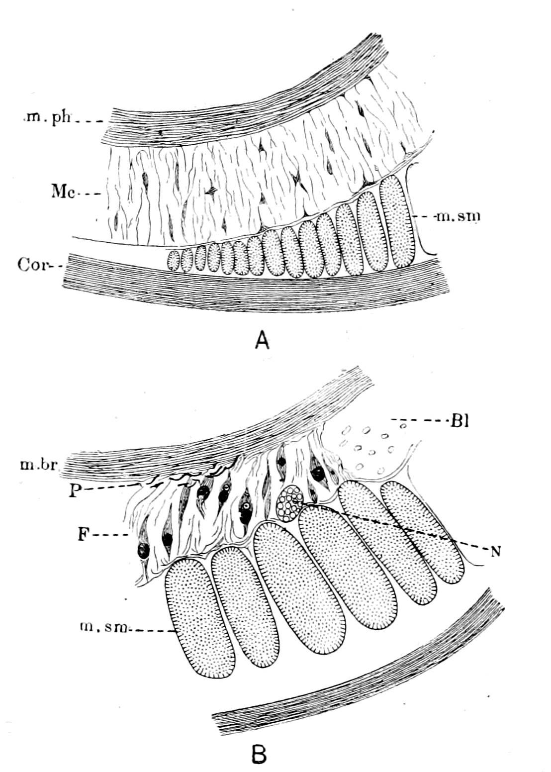
Fig. 132.—A, Muco-cartilage of Lower Lip (Mc.); m.ph., muscle of lower lip; m.sm., somatic muscle; Cor., laminated layer of skin. B, Degenerated Muco-cartilage of Branchial Region. F., fat layer; P., pigment; Bl., blood-space; N., somatic nerve; m.br., branchial muscle; m.sm., somatic muscle.
If this conclusion is correct, we may expect that it would be confirmed by the embryological history of the tissue, and we ought to find that in much younger stages a homogeneous tissue of the same nature as muco-cartilage fills up the spaces in the branchial region, where in the Ammocœtes only blood and fat-containing cells are present. For this purpose Shipley kindly allowed me to examine his series of sections through the embryo at various ages. These specimens are very instructive, especially those stained by osmic acid, which preserves the natural thickness of this space better than other staining methods. At an age when the branchial cartilages are seen to be formed, when no fat-cells are present, a distinctive tissue (Fig. 133, B) is plainly visible in the velum and at the base of the tentacles, in the very position where in the more advanced Ammocœtes muco-cartilage exists. Taking, then, this tissue as our guide, the specimens show that the space between the skin and the visceral muscles in which the cartilaginous basket-work lies is filled with a similar material. At this stage a sheet of embryonic tissue occupies the position where, later on, blood-spaces and fat-cells are found, and this tissue resembles that seen in the velum and other places where muco-cartilage is afterwards found.
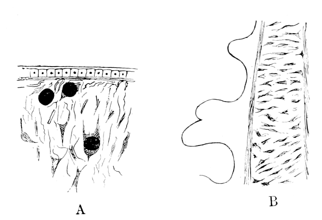
Fig. 133.—A, Muco-Cartilage of Velum; B, Embryonic Muco-Cartilage of Tentacular Bar.
I conclude, therefore, that originally the branchial or mesosomatic region was covered with a dorsal plate of muco-cartilage, which carried on its under surface the dorsal part of the branchial basket-work, and sprang from the central core of skeletogenous tissue around the notochord; this plate was separated from the plate which covered this region ventrally by the lateral grove in which the gill-slits are situated. The ventral plate carried on its under surface the ventral part of the branchial basket-work, and was originally continuous with the plate over the thyroid gland.
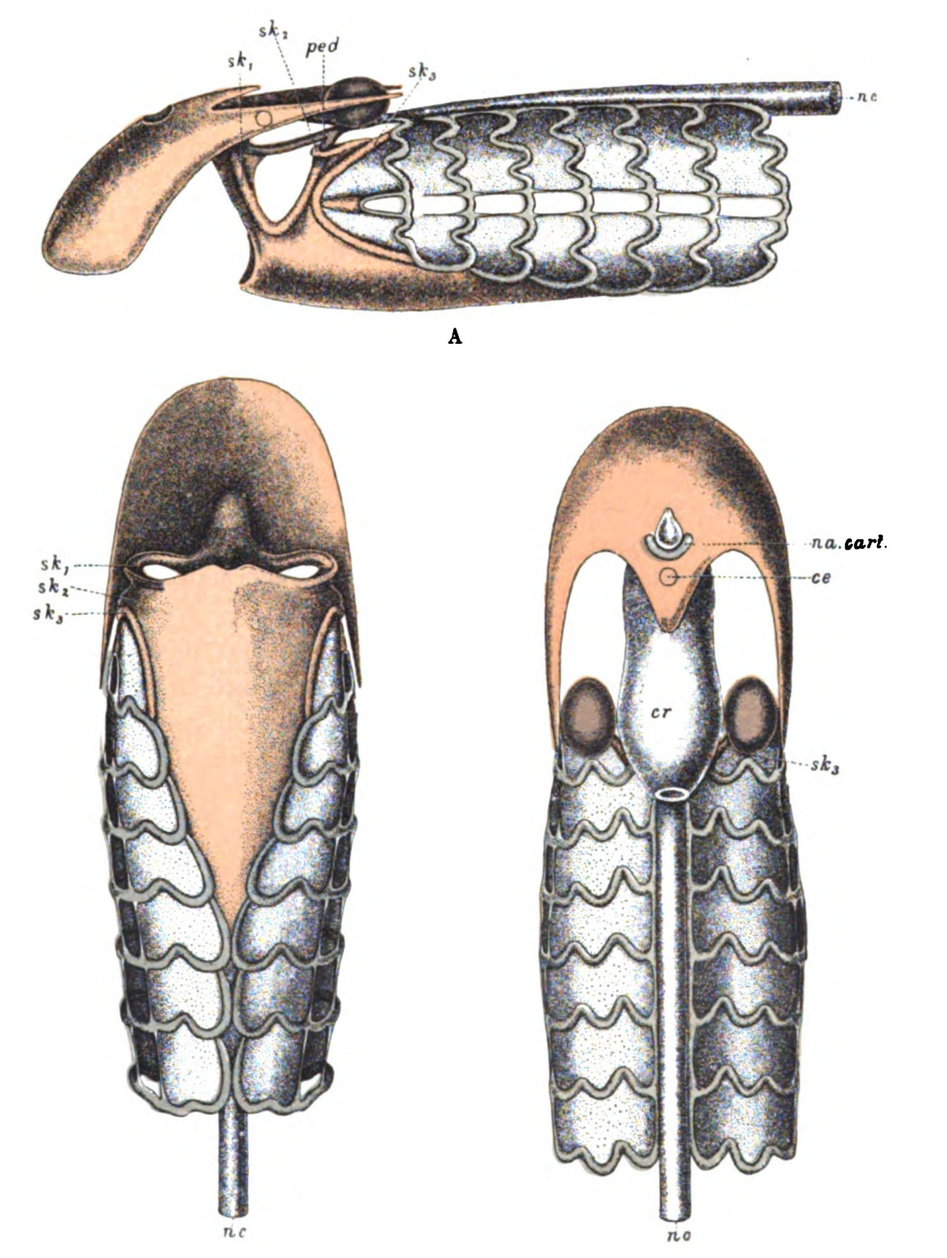
Fig. 134.—Skeleton of Head-Region of Ammocœtes. A, Lateral View; B, Ventral View; C, Dorsal View.
Muco-cartilage, red; soft cartilage, blue; hard cartilage, purple. sk1, sk2, sk3, skeletal bars; c.e., position of pineal eye; na. cart., nasal cartilage; ped., pedicle; cr., cranium; nc., notochord.
In Fig. 134, A, B, C, the cranial skeleton of Ammocœtes is represented from the dorsal, ventral, and lateral aspects. The muco-cartilage is coloured red, the branchial or soft cartilage blue, and the hard cartilage purple. The degenerated muco-cartilage of the branchial region is represented as an uncoloured plate, on which the branchial basket-work stands in relief. If it were restored to its original condition of muco-cartilage, it would represent a uniform plate, on the under surface of which the basket-work would be situated; and if it were calcified and made solid, the branchial basket-work would not show at all in these figures.
Is it possible to find the reason why this skeletal covering has degenerated so early before transformation, and why the thyroid plate remains intact until transformation? We see that all that part which has degenerated is covered over by the somatic muscles,—by, in fact, muscles which, being innervated by the foremost spinal nerves, belong naturally to the region immediately following the branchial. I suggest, therefore, that the original skeletal covering of muco-cartilage has remained intact only where it has not been invaded and covered over by somatic muscles, but has been invaded by blood and undergone the same kind of degenerative change as overtakes the great mass of this tissue at transformation wherever the somatic muscles have overgrown it.
The covering somatic muscles in the branchial region form a dorsal and ventral group, of which the latter is formed in the embryo much later than the former, the line of separation between the two groups being the lateral groove, with its row of branchial openings. This groove ends at the first branchial opening, but the ventral and dorsal somatic muscles continue further headwards. It is instructive to see that, although the lateral groove terminates, the separation between the two groups of muscles is still marked out by a ridge of muco-cartilage, represented in Fig. 134, A, which terminates anteriorly in the opercular bar.
Passing now to the prosomatic region, we find that here, too, the muco-cartilaginous external covering is divisible into a dorsal and a ventral head-plate, the ventral head-plate being the plate of the lower lip, and the dorsal head-plate the plate of muco-cartilage over the front part of the head. The staining reaction with thionin maps out this dorsal head-plate in a most beautiful manner, and shows that the whole of the upper lip-region in front of the nasal orifice is one large plate of muco-cartilage, obscured largely by the invasion of the crossing muscles of the upper lip, but left pure and uninvaded all around the nasal orifice, and where the upper and lower lips come together. In addition to this foremost plate, a median tongue of muco-cartilage covers over the pineal eye and fills up the median depression between the two median dorsal somatic muscles. Also, two lateral cornua pass caudalwards from the main frontal mass of muco-cartilage over the lateral eyes, forming the well-known wedge which separates the dorsal and lateral portions of the dorso-lateral somatic muscle. In fact, similarly to what we find in the branchial region, the muco-cartilaginous covering can be traced with greater or less completeness only in those parts which are not covered by somatic muscles.
In Fig. 134, A, B, C, this striking muco-cartilaginous head-shield, both dorsal and ventral, is shown. Seeing that the upper lip wraps round the lower one on each side, and that this most ventral edge of the upper lip contains muco-cartilage, as is seen in Fig. 117, the dorsal head-shield of muco-cartilage ought, strictly speaking, to extend more ventrally in the drawings. I have curtailed it in order not to interfere with the representation of the lower lip and tentacular muco-cartilages.
From what has been said, it follows that the past history of the skeletal covering of the whole head-region of Ammocœtes, both frontal and occipital, can be conjectured by means of the ontogenetic history of the foremost myomeres.
Dohrn and all other observers are agreed that during the development of this animal a striking forward growth of the foremost somatic myomeres takes place, so that, as Dohrn puts it, the body-musculature has extended forwards over the gill-region, and at the same time the gill-region has extended backwards. It is therefore probable that in the ancestral form the myotomes, innervated by the first spinal nerves, immediately succeeded the branchial region. Judging from Ammocœtes, the forward growth was at first confined to the dorsal region, and therefore invaded the dorsal head-plate, the ventral musculature being distinctly a later growth. With respect to this dorsal part of the myotomes, the first myotome is originally situated some distance behind the auditory capsule, and then grows forward towards the nasal opening; the lateral part, according to Hatschek, grows forward more quickly than the dorsal part, and splits itself above and below the eye into a dorso-lateral part, which extends up to the olfactory capsule, and a ventro-lateral part (m. lateralis capitis anterior, superior, and inferior), thus giving rise to the characteristic appearance of the muco-cartilaginous head-shield of Ammocœtes.
According, then, to the extent of the growth of these somatic muscles, the shape of the muco-cartilaginous head-shield will vary, and if it were calcified and then fossilized we should obtain fossil head-shields of widely differing configuration, although such fossils might be closely allied to each other. This is just what is found in this group. Let the muco-cartilage extend over the whole of the branchial region of Ammocœtes, the resulting head-shield would be as in Fig. 135, A; the branchial bars below the muco-cartilaginous shield might or might not be evident, and the line between the branchial and the trigeminal region might or might not be indicated. Such a head-shield would closely resemble those of Didymaspis and Tremataspis respectively. Now suppose the somatic musculature to encroach slightly on the branchial region and also laterally to the end of the anterior branchial region, then we should obtain a shape resembling that of Thyestes (Fig. 135, B). Continue the same process further, the lateral muscle always encroaching further than the median masses, until the whole or nearly the whole branchial region is invested, and we get the head-shield of Cephalaspis (Fig. 135, C); further still, that of Keraspis, and yet still further, that of Ammocœtes (Fig. 135, D).
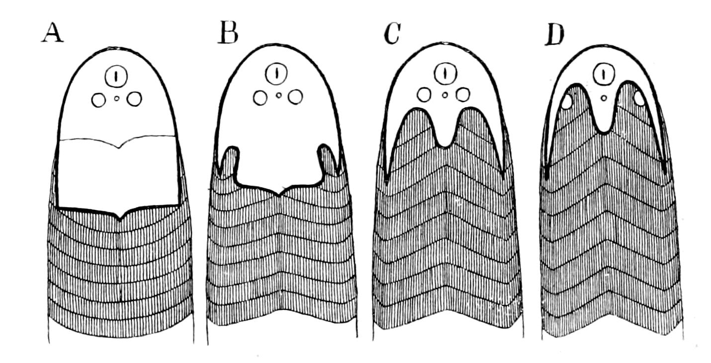
Fig. 135.—Diagrams to show the different shapes of Head-Shields due to the forward growth of the Somatic Musculature.
A, Didymaspis; B, Auchenaspis; C, Cephalaspis; D, Ammocœtes.
So close is this similarity, from the comparative point of view, between the dorsal head-shield of the Osteostraci and the dorsal cephalic region of Ammocœtes that it justifies us in taking Ammocœtes as the nearest living representative of such types; it is justifiable, therefore, to interpret by means of Ammocœtes the position of other organs in these forms. First and foremost is the hard plate known as the post-orbital plate, so invariably found. In Fig. 134, C, I have inserted (cr.) the position of the membranous cranium of Ammocœtes, and it is immediately evident that the primordial cranium of the Osteostraci must occupy the exact position indicated by this median hard plate. For this very reason this median plate would be harder than the rest in order to afford a better protection to the brain underneath. This plate, because of its position, may well receive the same name as the similar plate in the trilobite and various palæostracans and be called the glabellum.
Evidence of Segmentation in the Head-Shield—Formation of Cranium.
We may thus conceive the position of the nose, lateral eyes, median eyes, and cranium in these old fishes. In addition, other indications of a segmentation in this head-region have been found. The most striking of all the specimens hitherto discovered are some of Thyestes verrucosus, discovered by Rohon, in which the dorsal shield has been removed, and so we are able to see what that dorsal shield covered.
In Fig. 136, I reproduce his drawing of one of his specimens from the dorsal and lateral aspects. These drawings show that the frontal part of the shield covered a markedly segmented part of the animal; five distinct segments are visible apart from the median most anterior region. This segmented region is entirely confined to the prosomatic region, i.e. to the region innervated by the trigeminal nerve. An indication of similar markings is given in Lankester's figure of Eukeraspis pustuliferus (see Fig. 127, B), and, indeed, evidence of a segmentation under the antero-lateral border of the head-shield is recognized at the present time, not only in the Cephalaspidæ, but also in the Pteraspidæ, as was pointed out to me by Smith Woodward in the specimens at the British Museum. Also, in Cyathaspis, Jaekel has drawn attention to markings of a similar segmental nature (Fig. 137).
There seems, then, little doubt but that these primitive fishes possessed something in this region which was of a segmental character, and indicated at least five segments, probably more.
Rohon entitles his discovery 'the segmentation of the primordial cranium.' It would, I think, be better to call it the segmentation of the anterior region of the head, for that is in reality what his figures show, not the segmentation of the primordial cranium, which, to judge from Ammocœtes, was confined to the region of the glabellum.
What is the interpretation of this appearance?
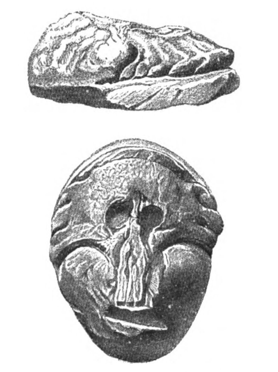
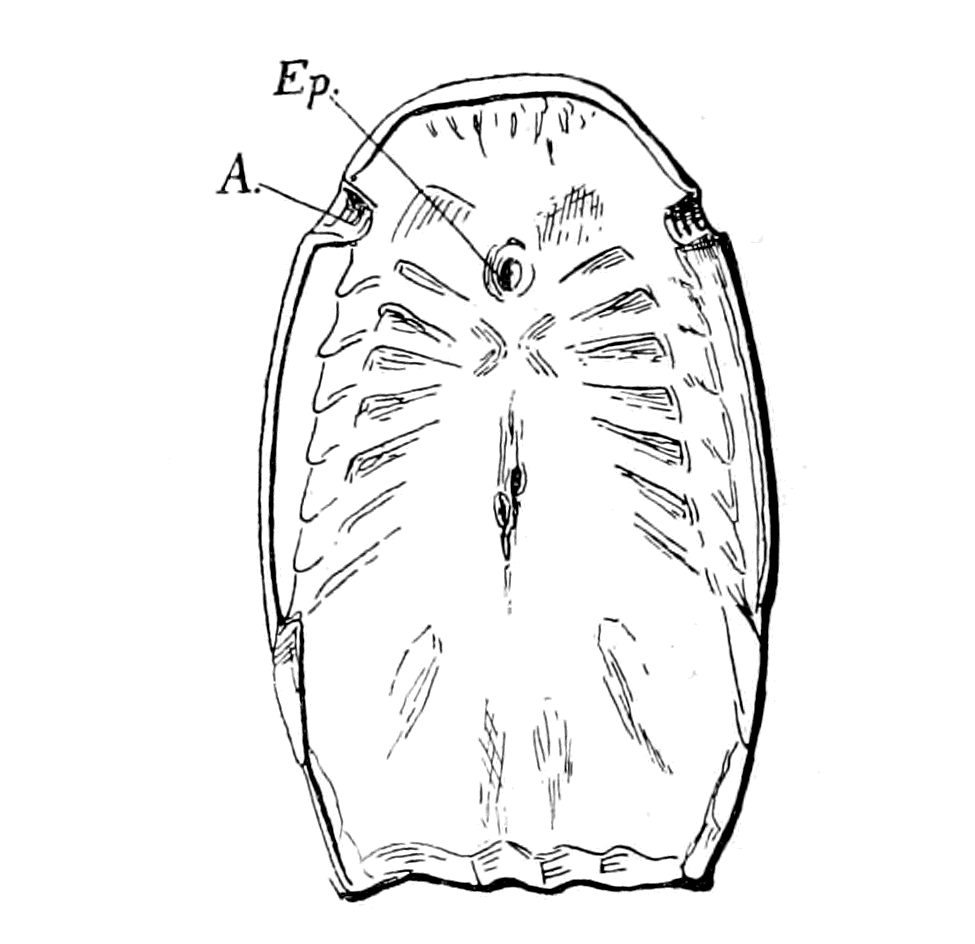
Fig. 136.—Lateral and Dorsal Views of the Frontal and Occipital Regions of the Head-Shield of Thyestes, after Removal of the Outer Surface. (From Rohon.) Fig. 137.—Under Surface of Head-Shield of Cyathaspis. (From Jaekel.) A., lateral eyes; Ep., median eyes.
Any segmentation in the head-region must be indicative of segments belonging to the trigeminal or prosomatic region, or of segments belonging to the vagus or mesosomatic region. Many palæontologists, looking upon segmentation as indicative of gills and gill-slits, have attempted to interpret such markings as branchial segments, regardless of their position. As the figures show, they extend in front of the eyes and reach round to the front middle line, a position which is simply impossible for gills, but points directly to a segmentation connected with the trigeminal nerve. Comparison with Ammocœtes makes it plain enough that the markings in question are prosomatic in position, and that the gill-region must be sought for in the place where Schmidt and Rohon located it in Thyestes, viz. the so-called occipital region.
This discovery of Rohon's is, in my opinion, of immense importance, for it indicates that, in these early fishes, the prosomatic segmentation, associated with the trigeminal nerve, was much more well-marked than in any fishes living in the present day. Why should it be more well-marked? Turning to the palæostracan, it is very suggestive to compare the markings on their prosomatic carapace with these markings. Again and again we find indications of segmentation in these fossils similar to those seen in the ancient fishes. Thus in Fig. 138 I have put side by side the palæostracan Bunodes and the fish Thyestes, both life size. In the latter I have indicated Rohon's segments; in the former the markings usually seen.
From the evidence of Phrynus, Mygale, etc., as already pointed out, such markings in the palæostracan fossils would indicate the position of the tergo-coxal muscles of the prosomatic appendages, even though such appendages have not yet been discovered, and it is significant that in all these cases there is a distinct indication of a median plate or glabellum in addition to the segmental markings. Especially instructive is the evidence of Phrynus, as is seen by a comparison of Figs. 107 and 108, which shows clearly that this median plate (glab.) covered the brain-region, a brain-region which is isolated and protected from the tergo-coxal muscles by the growth dorsalwards of the flanges of the plastron. In this way an incipient cranium of a membranous character is formed, which helps to give attachment to these tergo-coxal muscles. As such cranium is derived directly from the plastron, it is natural that it should ultimately become cartilaginous, just as occurs when Ammocœtes becomes Petromyzon and the cartilaginous cranium of the latter arises from the membranous cranium of the former. In Galeodes also the growth dorsalwards of the lateral flanges of the plastron to form an incipient cranium in which the brain lies is very apparent.
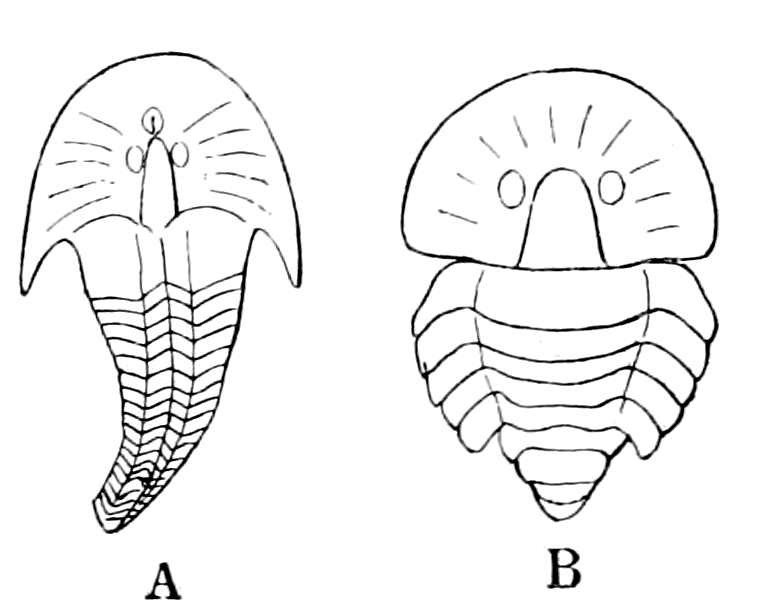
Fig. 138.—A, Outline of Thyestes Verrucosus with Rohon's Segments indicated; B, Outline of Bunodes Lunula with Lateral Eyes inserted.
Both figures natural size.
I venture, then, to suggest that in the Osteostraci the median hard plate or glabellum protected a brain which was enclosed in a membranous cranium, very probably not yet complete in the dorsal region—certainly not complete if the median pineal eyes so universally found in these ancient fishes were functional—a cranium derived from the basal trabeculæ, in precisely the same manner as we see it already in its commencement in Phrynus and other scorpions. With the completion of this cranium and its conversion into cartilage, and subsequently into bone, an efficient protection was afforded to the most vital part of the animal, and thus the hard head-shield of the Palæostraca and of the earliest fishes was gradually supplanted by the protecting bony cranium of the higher vertebrates.
Step by step it is easy to follow in the mind's eye the evolution of the vertebrate cranium, and because it was evolved direct from the plastron, the impossibility of resolving it into segments is at once manifest; for although the plastron was probably originally segmented, as Schimkéwitsch thinks, all sign of such segmentation had in all probability ceased, before ever the vertebrates first made their appearance on the earth.
It follows further, from the comparison here made, that those antero-lateral markings indicative of segments, found so frequently in these primitive fishes, must be interpreted as due not to gills but to aponeuroses, due to the presence of muscles which moved prosomatic appendages, muscles which arose from the dorsal region in very much the same position as do the muscles of the lower lip in Ammocœtes; the latter, as already argued, represent the tergo-coxal muscles of the last pair of prosomatic appendages—the chilaria or metastoma. Such an interpretation of these markings signifies that the first-formed fishes must have possessed prosomatic appendages of a more definite character than the tentacles of Ammocœtes, something intermediate between those of the palæostracan and Ammocœtes.
For my part I should not be in the least surprised were I to hear that something of the nature of appendages in this region had been found, especially in view of the well-known existence of the pair of appendages in the members of the Asterolepidæ—large, oar-like appendages which may well represent the ectognaths.
The Relationship of the Ostracoderms.
Of the three groups of fishes—the Heterostraci, the Osteostraci, and the Antiarcha—the last is Devonian, and therefore the latest in time of the three, while the earliest is the first group, as both Pteraspis and Cyathaspis have been found in lower levels of the Silurian age than any of the Osteostraci, and, indeed, Cyathaspis has been discovered in Sweden in the lower Silurian. This, the earliest of all groups of fishes, is confined to two forms only—Pteraspis and Cyathaspis,—for Scaphaspis is now recognized to be the ventral shield of Pteraspis.
Hitherto a strong tendency has existed in the minds both of the comparative anatomist and the palæontologist to look on the elasmobranchs as the earliest fishes, and to force, therefore, these strange forms of fish into the elasmobranch ranks. For this purpose the same device is often used as has been utilized in order to account for the existence of the Cyclostomata, viz. that of degeneration. The evidence I have put forward is very strongly in favour of a connection between the cyclostomes and the cephalaspids, and agrees therefore with all the rest of the evidence that the jawless fishes are more ancient than those which bore jaws—the Gnathostomata.
This is no new view. It was urged by Cope, who classified the Heterostraci, Osteostraci, and Antiarcha under one big group—the Agnatha—from which subsequently the Gnathostomata arose. Cope's arguments have not prevailed up to the present time, as is seen in the writings of Traquair, one of the chief authorities on the subject in Great Britain. He is still an advocate of the elasmobranch origin of all these earliest fishes, and claims that the latest discoveries of the Silurian deposits (Thelodus Pagei) and other members of the Cœlolepidæ confirm this view of the question.
This view may be summed up somewhat as follows:—
Cartilaginous jaws would not fossilize, and the Ostracoderms may have possessed them.
They may have degenerated from elasmobranchs just as the cyclostomes are supposed to have degenerated.
Seeing that bone succeeds cartilage, the presence of bony shields in Cephalaspis, etc., indicates that their precursors were cartilaginous, presumably elasmobranch fishes.
Of these arguments the strongest is based on the supposed bony covering of the Osteostraci, with the consequent supposition that their ancestors possessed a cartilaginous covering. This argument is entirely upset, if, as I have pointed out, the structure of the cephalaspid shield is that of muco-cartilage and not of bone. If these plates are a calcified muco-cartilage, then the whole argument for their ancestry from animals with a cartilaginous skeleton falls to the ground, for muco-cartilage is the precursor not only of bone, but also of cartilage itself.
The evidence, then, points strongly in favour of Cope's view that the most primitive fishes were Agnatha, after the fashion of cyclostomes, as is also believed by Smith Woodward, Bashford Dean, and Jaekel.
Among living animals, as I have shown, the Limulus is the sole survivor of the palæostracan type, and Ammocœtes alone gives a clue to the nature of the cephalaspid, i.e. the osteostracan fish. Older than the latter is the heterostracan, Pteraspis, and Cyathaspis. Is it possible from their structure to obtain any clue as to the actual passage from the palæostracan to the vertebrate?
Here again, as in the case of the Osteostraci, a relationship to the elasmobranch has been supposed, for the following reasons:—
The latest discoveries in the Silurian and Devonian deposits have brought to light strange forms such as Thelodus and Drepanaspis, of which the latter from the Devonian must, according to Traquair, be included in the Heterostraci. It possessed, as seen in Fig. 139, large plates, after the fashion of Pteraspis, and also many smaller ones.
The former, from the upper Silurian, belongs to the Cœlolepidæ, and was covered over with shagreen composed of small scutes, after the fashion of an elasmobranch. Traquair suggests that Thelodus arose from the original elasmobranch stock; that by the fusion of scutes such a form as Drepanaspis occurred, and, with still further fusion, Pteraspis.
There are always two ways of looking at a question, and it seems to me possible and more probable to turn the matter round and to argue that the original condition of the surface-covering was that of large plates, as in Pteraspis. By the subsequent splitting up of such plates, Drepanaspis was formed, and later on, by further splitting, the elasmobranch, Thelodus being a stage on the way to the formation of an elasmobranch, and not a backward stage from the elasmobranch towards Pteraspis.
This method of looking at the problem seems to me to be more in consonance with the facts than the reverse; for, as pointed out by Jaekel, the fishes with large plates are the oldest, and in Cyathaspis, the very oldest of all, the size of the plates is most conspicuous; he considers, therefore, this preconceived view that large plates are formed by the fusion of small ones must give way to the opposite belief.
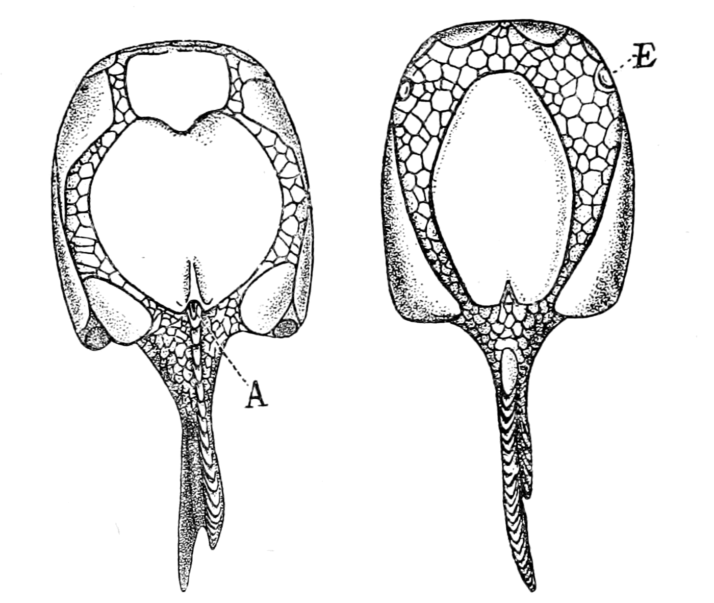
Fig. 139.—Drepanaspis. Ventral and Dorsal Aspects. (After Lankester.)
A., anus; E., lateral eyes.
So also Rohon, as quoted by Traquair, who, in his first paper accepted Lankester's view that the ridges of the pteraspidian shield were formed by the fusion of a linear arrangement of numbers of placoid scales, suggests in his second paper that these ridges may have been the most primitive condition of the dermal skeleton of the vertebrate, out of which, by differentiation, the dermal denticles (placoid scales) of the selachian, as well as their modifications in the ganoids, teleosteans, and amphibians, have arisen.
One thing is agreed upon on all sides; no sign of bone-corpuscles is to be found in this dermal covering of Pteraspis. In the deeper layers are large spaces, the so-called pulp-cavities leading into narrow canaliculi, the so-called dentine canals. The structure is looked upon as similar to that of the pulp and dentine canals of many fish-scales.
On the other hand, this dermal covering of Pteraspis has been compared by Patten with the arrangement of the chitinous structure of certain parts of the external covering of Limulus, a comparison which to my mind presents a great difficulty. The chitin-layers in Limulus are external to the epidermal cells, being formed by them; the layers in Pteraspis which look like chitin must have been internal to the epidermal layer; for each vascular canal which passes from a pulp-cavity on its way to be distributed into the dentine canals of the ridge gives off short side branches, which open directly into the groove between the ridges. If these canals were filled with blood they could not possibly open directly into the open grooves between the ridges; these openings must, therefore, have been covered over with an epithelial layer which covered over the surface of the animal, and consequently the chitin-like structure must have been internal to the epidermis, and not external, as on Patten's view. The comparison of this structure with the dentine of fish-scales signifies the same thing, for in the latter the epidermis is external to the dentine-plates, the hard skeletal structure is in the position of the cutis, not of the cuticle.
The position appears to me to be this: the dermal cranial skeleton of vertebrates, whether it takes the form of a bony skull or of the dorsal plates of a cephalaspid or a pteraspid is, in all cases, not cuticular, i.e. is not an external formation of the epidermal cells, but is formed in tissue of the nature of connective tissue underlying the epidermis. On the contrary, the hard part of the head-carapace of the palæostracan is an external formation of the epidermal cells.
If, then, this tissue of Pteraspis is not to be looked upon as chitin, how can we imagine its formation? It is certainly not bone, for there are no bone-corpuscles; it is a very regular laminated structure resembling in appearance chitin rather than anything else.
As in all cases of difficulty, turn to Ammocœtes and let us see what clue there is to be found there. The skin of Ammocœtes is peculiar among vertebrates in many respects. It consists of a number of epidermal cells, as in Fig. 140, the varying function of which need not be considered here, covered over with a cuticular layer which is extraordinarily thick for the cuticle of a vertebrate skin; this cuticular layer is perforated with fine canaliculi, through which the secretion of the underlying cells passes, as is seen in Fig. 140, A and B. This cuticle corresponds to the chitinous covering of the arthropod, and like it is perforated with canaliculi, and, according to Lwoff, possibly contains chitin. The epidermal cells rest on a thick layer of most striking appearance (Fig. 141), for it resembles, in an extraordinary degree, when examined superficially, a layer of chitin; it is called the laminated layer, and is characterized by the extreme regularity of the laminæ. This appearance is due, as the observations of Miss Alcock show, to alternate layers of connective tissue fibres arranged at right angles to each other, each fibre running a straight course and possessing its own nucleus. Although the fibres in each layer are packed close together, they are sufficiently apart to form with the fibres of the alternate layers a meshwork rather than a homogeneous structure, and thus the surface view of this layer shows a regular network of very fine spaces through which nerve-fibres and fluid pass. This layer is easily dissolved in a solution of hypochlorite of soda, a fluid which dissolves chitin. Any one looking at Ammocœtes would say that the only part of its skin which resembles chitin is this laminated layer, and therefore the only part of its skin which would afford an indication of the nature of the skeleton of Pteraspis is this laminated layer, which belongs to the cutis, and not to the cuticle. Yet another significant peculiarity of this layer is its entire disappearance at transformation. Miss Alcock, in a research not yet published, has shown that this layer is completely broken up and absorbed at transformation; the cutis of Petromyzon is formed entirely anew, and no longer presents any regular laminated character, but resembles rather the sub-epidermal connective tissue layer of the skin of higher vertebrates. This laminated layer, then, just like the muco-cartilage, shows, by its complete disappearance at transformation, its ancestral character.
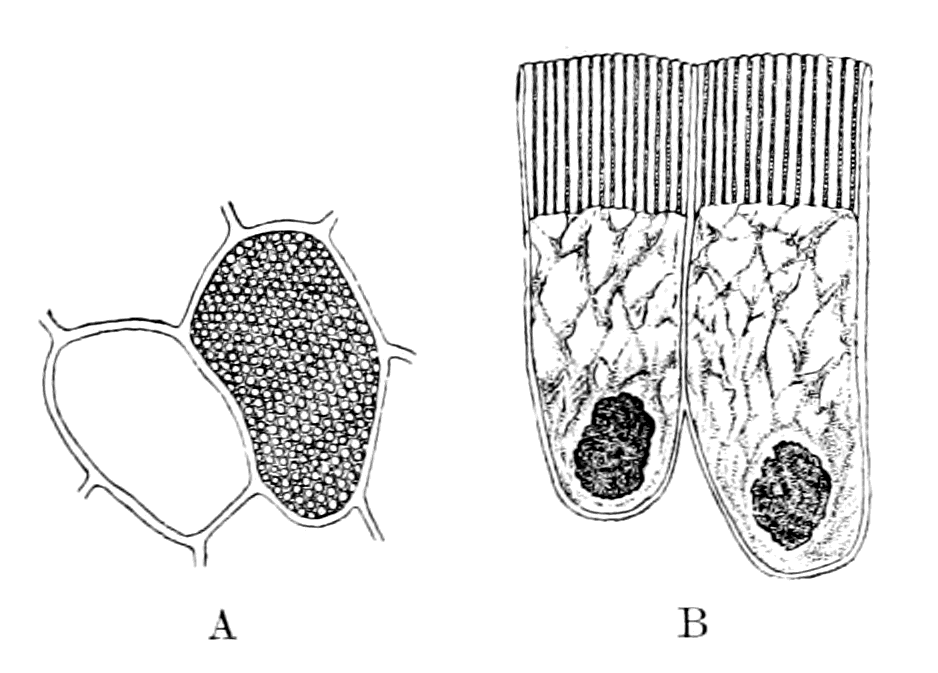
Fig. 140.—Epithelial Cells of Ammocœtes to show the Canaliculi in the Thick Cuticle (B). A, Transverse Section through the Cuticle.
Very suggestive is the arrangement of the different skeletal tissues in the head-region of Ammocœtes. Fig. 141 represents a section through the head near the pineal eye. Most internally is a, a section of the membranous cranium, then comes b, the muco-cartilaginous skeleton, then c, the laminated layer, and finally d, the external cuticle. If in Ammocœtes we possess an epitome of the history of the vertebrate, how would these layers be represented in the past ages, supposing they could be fossilized?
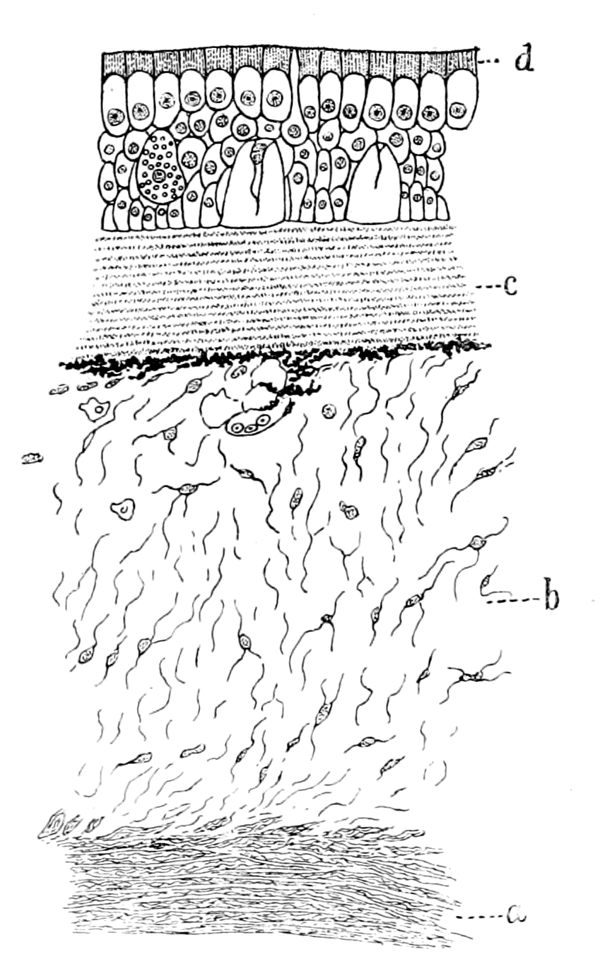
Fig. 141.—Section of Skin and Underlying Tissues in the Head-Region of Ammocœtes.
a, cranial wall; b, muco-cartilage; c, laminated layer; d, external cuticular layer.
The most internal layer a, by the formation of cartilage and then bone, represents the great mass of vertebrate fossils; the next layer b, by a process of calcification, as previously argued, represents the head-shield of the Osteostracan fishes; while the cuticular layer d, no longer thin, is the remnant of the Palæostracan head-carapace. Between these two layers, b and d, lies the laminated layer c. Intermediate to the Palæostracan and the Osteostracan comes the Heterostracan, with its peculiar head-shield—a head-shield whose origin is more easily conceivable as arising from something of the nature of the laminated layer than from any other structure represented in Ammocœtes.
My present suggestion, then, is this: the transition from the skeletal covering of the Palæostracan to that of the highest vertebrates was brought about by the calcification of successive layers from without inwards, all of which still remain in Ammocœtes and show how the external chitinous covering of the arthropod was gradually replaced by the deep-lying internal bony cranium of the higher vertebrates.
In Ammocœtes the layer which represents the covering of the Palæostracan has already almost disappeared. At transformation the layers representing the stage arrived at by the Heterostracan and the Osteostracan disappear; but the stage representing the higher vertebrates, far from disappearing, by the formation of cartilage reaches a higher stage and prepares the way for the ultimate stage of all—the formation of the bony cranium.
So much for the evidence as to the nature of the structure of the head-shield of the Pteraspidæ.
It suggests that these fishes were covered anteriorly with armoured plates derived from the cutis layer of the skin, a layer which was specially thickened and very vascular, apparently, to enable respiration to be very largely, if not entirely, effected by the surface of the body. It is difficult to understand how the sea-scorpions breathed, and it is easy to see how the formation of ventral and dorsal plates enclosing the mesosomatic appendages may at the outset have hindered the action of the branchiæ. The respiratory chamber, according to my view, had at first the double function of respiration and digestion. A new digestive apparatus was the pressing need at the time; it would, therefore, be of distinct advantage to remove, as much as possible, the burden of respiration from this incipient alimentary canal.
What can be said as to the shape of these ancient forms of fishes? Certain parts of them are absolutely known, other parts are guesswork. They are known to have possessed a dorsal shield, a ventral shield formerly looked upon as belonging to a separate species, called Scaphaspis, and a spine attached to the dorsal shield. The rest of their configuration, as given in Smith Woodward's restoration (Fig. 142) is guesswork; the fish-like body with its scales, the heterocercal tail, is based on the most insufficient evidence of something of the nature of scales having being found near the head-plates.
The dorsal shield is characterized by a pair of lateral eyes situated on the edge of the shield, not as in Cephalaspis near the middle line. In the middle line, where the rostrum meets the large dorsal plate, median eyes were situated. But the slightest sign of any median single nasal opening, such as is so characteristic of the head-shield of the Osteostraci and of Ammocœtes has never been discovered. The olfactory organ must have been situated on the ventral side as in the larval stage of Ammocœtes, or in the Palæostraca. Many of these head-shields are remarkably well preserved, and it is difficult to believe that an olfactory opening would not be seen if any such had existed, as it does in Thyestes.
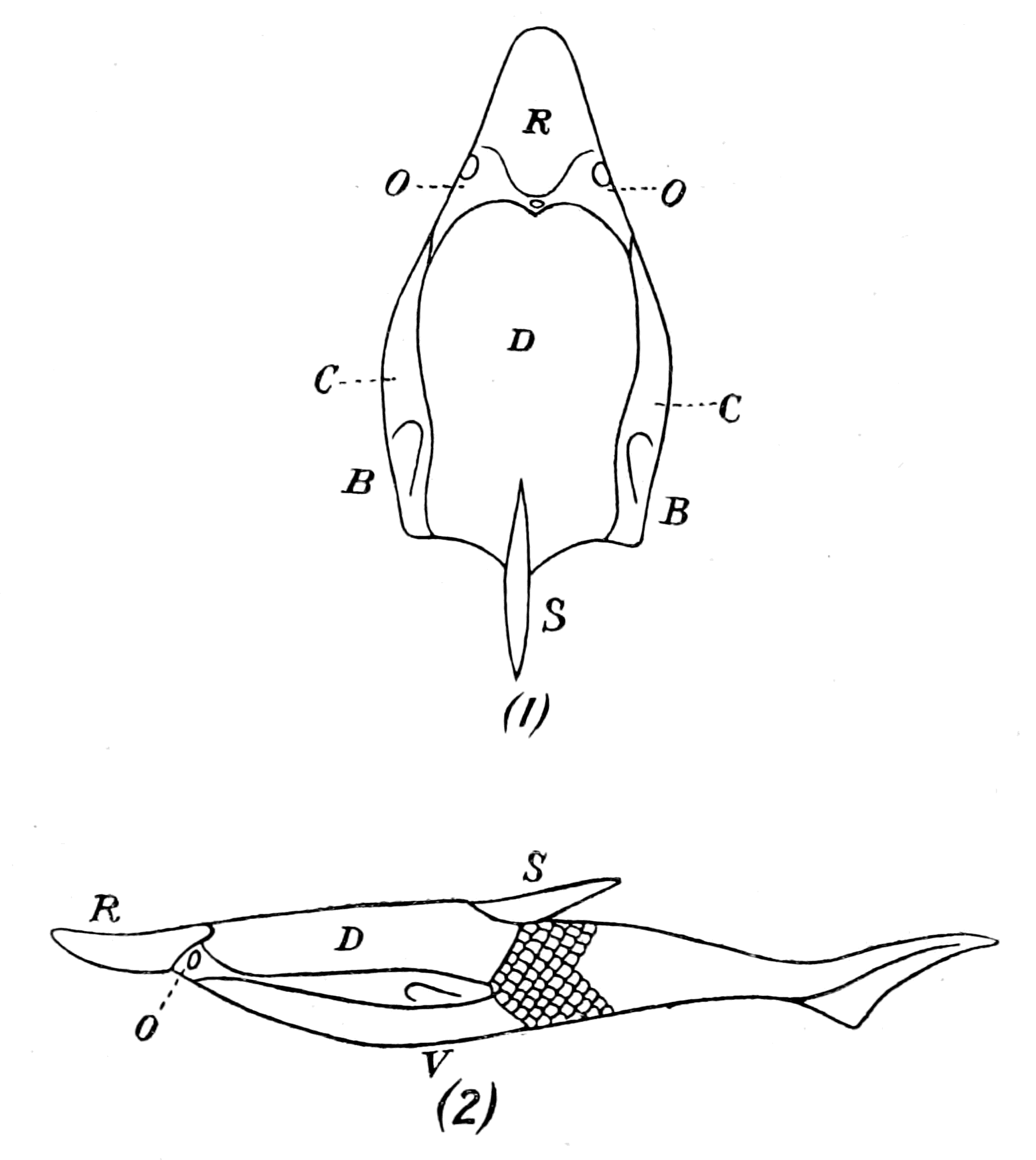
Fig. 142.—Restoration of Pteraspis. (After Smith Woodward.)
The difficulty of interpreting these types is the difficulty of understanding their method of locomotion; that is largely the reason why the spine has been placed as if projecting from the back, and a fish-like body with a heterocercal tail-fin added. If, on the contrary, the spine is a terminal tail-spine, then, as far as the fossilized remains indicate, the animal consisted of a dorsal shield, a ventral shield, and a tail-spine, to which must be added two apparently lateral pieces and a few scales. If the animal did not possess a flexible body with a tail-fin, but terminated in a rigid spike after the fashion of a Limulus-like animal, then it must have moved by means of appendages. At present we have not sufficient evidence to decide this question.
That the animal crawled about in the mud by means of free appendages is by no means an impossible view, seeing how difficult it is to find the remains of appendages in the fossils of this far-back time, even when we are sure that they existed. Thus, for many generations, the appendages of trilobites, which occur in such countless numbers, and in such great variety of form, were absolutely unknown, until at last, in consequence of a fortunate infiltration by pyrites, they were found by Beecher preserved down to the minutest detail. Even to this day no trace of appendages has been found in such forms as Hemiaspis, Bunodes, Belinurus, Prestwichia.
The whole question of the evidence of any prosomatic appendages in these ancient fishes is one of very great interest, and of late years has been investigated by Patten. It has long been known that forms such as Pterichthys and Bothriolepis possessed two large, jointed locomotor appendages, and Patten has lately obtained better specimens of Bothriolepis than have ever been found before, which show not only the general configuration of the fish, but also the presence of mandibles or gnathites in the mouth-region resembling those of an arthropod. These mandibles had been seen before (Smith Woodward), but Patten's specimens are more perfect than any previously described, and cause him to conclude that these ancient fish were of the nature of arthropods rather than of vertebrates.
Patten has also been able to obtain some excellent specimens of the under surface of the head of Tremataspis, which, as evident in Fig. 143, show the presence of a series of holes, ranging on each side from the mouth-opening, in a semicircular fashion towards the middle line. He considers that these openings indicate the attachments of appendages, in opposition to other observers, such as Jaekel, who look upon them as gill-slits. To my mind, they are not in the right position for gill-slits; they are certainly in a prosomatic rather than in a mesosomatic position, and I should not be at all surprised if further research justified Patten's position. So convinced is he of the presence of appendages in all these old forms, that he considers them to be arthropods rather than vertebrates, although, at the same time, he looks upon them as indicating the origin of vertebrates from arthropods. Here, perhaps, it is advisable to say a few words on Patten's attitude towards this question.
Two years after I had put forward my theory of the derivation of vertebrates from arthropods, Patten published, in the Quarterly Journal of Microscopical Science, simultaneously with my paper in that journal, a paper entitled "The Origin of Vertebrates from Arachnids." In this paper he made no reference to my former publications, but he made it clear that there was an absolutely fundamental difference between our treatment of the problem; for he took the old view that of necessity there must be a reversal of surfaces in order that the internal organs should be in the same relative positions in the vertebrate and in the invertebrate. He simply, therefore, substituted Arachnid for Annelid in the old theory. Because of this necessity for the reversal of surfaces he discarded the terms dorsal and ventral as indicative of the surfaces of an animal, and substituted hæmal and neural, thereby hopelessly confusing the issue and making it often very difficult to understand his meaning.
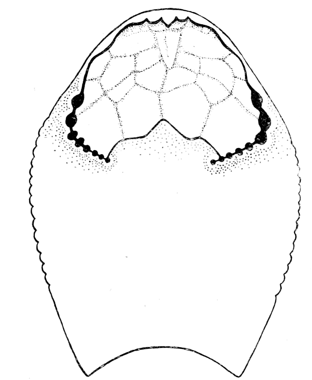
Fig. 143.—Under-Surface of Head-Region in Tremataspis. (After Patten.)
He still holds to his original opinion, and I am still waiting to find out when the reversal of surfaces took place, for his investigations lead him, as must naturally be the case, to compare the dorsal (or, as he would call it, the hæmal) surface of Bothriolepis, of the Cephalaspidæ, and of the Pteraspidæ with the dorsal surface of the Palæostraca.
All these ancient fishes are, according to him, still in the arthropod stage, have not yet turned over, though in a peculiarly unscientific manner he argues elaborately that they must have swum on their back rather than on their front, and so indicated the coming reversal. Because they were arthropods they cannot have had a frontal nose-organ; therefore, Patten looks upon the nose and the two lateral eyes of the Osteostraci as a complex median eye, regardless of the fact that the median eyes already existed.
Every atom of evidence Patten has brought forward, every new fact he has discovered, confirms my position and makes his still more hopelessly confused. Keep the animal the right side uppermost, and the evidence of the rocks confirms the transition from the Palæostracan to the Cyclostome; reverse the surfaces, and the attempt to derive the vertebrate from the palæostracan becomes so confused and hopelessly muddled as to throw discredit on any theory of the origin of vertebrates from arthropods. For my own part, I fully expect that appendages will be found not only in the Cephalaspidæ but also in the Pteraspidæ, and I hope Patten will continue his researches with increasing success. I feel sure, however, his task will be much simplified if he abandons his present position and views the question from my standpoint.
Summary.
The shifting of the nasal tube from a ventral to a dorsal position, as seen in Ammocœtes, is, perhaps, the most important of all clues in connection with the comparison of Ammocœtes to the Palæostracan on the one hand, and to the Cephalaspid on the other; for, whereas the exact counterpart of the opening of such a tube is always found on the dorsal head-shield in all members of the latter group, nothing of the kind is ever found on the dorsal carapace of the former group.
The reason for this difference is made immediately evident in the development of Ammocœtes itself, for the olfactory tube originates as a ventral tube—the tube of the hypophysis—in exactly the same position as the olfactory tube of the Palæostracan, and later on in its development takes up a dorsal position.
In fact, Ammocœtes in its development indicates how the Palæostracan head-shield became transformed into that of the Cephalaspid.
In another most important character Ammocœtes indicates its relationship to the Cephalaspidæ, for it possesses an external skeleton or head-shield composed of muco-cartilage, which is the exact counterpart of the so-called bony head-shield of the latter group; and still more strikingly the structure of the cephalaspidian head-shield is remarkably like that of muco-cartilage. In the one case, by the deposition of calcium salts, a hard external skeleton, capable of being preserved as a fossil, has been formed; in the other, by the absence of the calcium salts, a soft chondro-mucoid matrix, in which the characteristic cells and fibrils are embedded, distinguishes the tissue.
The recognition that the head-shields of these most primitive fishes were not composed of bone, but of muco-cartilage, the precursor of both cartilage and bone, immediately clears up in the most satisfactory manner the whole question of their derivation from elasmobranch fishes; for the main argument in favour of the latter derivation is the exceedingly strong one that bone succeeds cartilage—not vice versâ—therefore, these forms, since their head-shield is bony, must have arisen from some other fishes with a cartilaginous skeleton, most probably of an elasmobranch nature. Seeing, however, that the structure of their shields resembles muco-cartilage much more closely than bone, and that Ammocœtes forms a head-shield of muco-cartilage closely resembling theirs, there is no longer any necessity to derive the jawless fishes from the gnathostomatous; but, on the contrary, we may look with certainty upon the Agnatha as the most primitive group from which the others have been derived.
The history of the rocks shows that the group of fishes, Pteraspis and Cyathaspis, are older than the Cephalaspidæ—come, therefore, phylogenetically between the Palæostraca and the latter group. In this group the head-shields are of a very different character, without any sign of any structure comparable with that of bone, and although they possessed both lateral and median eyes, there is never in any case any trace of a dorsal nasal orifice. Their olfactory passage, like that of the Palæostraca, must have been ventral.
The remarkable comparison which exists between the head-shields of Ammocœtes and Cephalaspis, enables us to locate the position of the brain and cranium of the latter with considerable accuracy, and so to compare the segmental markings found in many of these fossils with the corresponding markings, found either in fossil Palæostraca or on the head-carapaces of living scorpions and spiders, such as Phrynus and Mygale. In all cases the cranial region was covered with a median plate, often especially hard, which corresponded to the glabellum of the trilobite; the growth of the cranium can be traced from its beginnings as the upturned lateral flanges of the plastron to the membranous cranium of Ammocœtes.
From such a comparison it follows that the segments, found in the antero-lateral region of the head-shield, were not segments of the cranium, but of parts beyond the region of the cranium, and from their position must have been segments supplied by the trigeminal nerve, and not by the vagus group; segments, therefore, which did not indicate gills and gill-slits, but muscles, innervated by the trigeminal nerve; muscles which, as indicated by the corresponding markings on the carapace of Phrynus, Mygale, etc., were the tergo-coxal muscles of the prosomatic appendages.
The discovery of the nature of these appendages in the Pteraspidæ and Cephalaspidæ, as well as in the Asterolepidæ (Pterichthys and Bothriolepis), is a problem of the future, though in the latter, not only have the well-known oar-like appendages been long since discovered, but Patten has recently found specimens of Bothriolepis which throw light on the anterior masticating gnathite-like appendages which these ancient forms possessed.
CHAPTER XI
THE EVIDENCE OF THE AUDITORY APPARATUS AND THE ORGANS OF THE LATERAL LINE
Lateral line organs.—Function of this group of organs.—Poriferous sense-organs on the appendages in Limulus.—Branchial sense-organs.—Prosomatic sense organs.—Flabellum.—Its structure and position.—Sense-organs of mandibles.—Auditory organs of insects and arachnids.—Poriferous chordotonal organs.—Balancers of Diptera.—Resemblance to organs of flabellum.—Racquet-organs of Galeodes.—Pectens of scorpions.—Large size of nerve to all these special sense-organs.—Origin of parachordals and auditory capsule.—Reason why VIIth nerve passes in and out of capsule.—Evidence of Ammocœtes.—Intrusion of glandular mass round brain into auditory capsule.—Intrusion of generative and hepatic mass round brain into base of flabellum.—Summary.
When speaking of the tripartite arrangement of the cranial nerves, an arrangement which gave the clue to the meaning of the cranial segments, I spoke of the trigeminal as supplying the sensory nerves to the skin in the head-region, and I compared this dorsal system of afferent nerves to the system of epimeral nerves in Limulus which supply the prosomatic and mesosomatic carapaces of Limulus with sensory fibres. I compared the ventral system of eye-muscle nerves with the system of nerves supplying the segmental dorso-ventral somatic muscles of the prosomatic region, and I compared the lateral system of mixed nerves with the nerves supplying the prosomatic and mesosomatic appendages of Limulus. I compared, also, the optic nerves and the olfactory nerves with the corresponding nerves in the same invertebrate group. My readers will see at once that one well-marked group of nerves—the auditory and lateral line system—has been entirely omitted up to the present, it has not even been mentioned in the scheme of the cranial segments; I have purposely reserved its consideration until now, because the organs these nerves supply, though situated in the skin, are of such a special character as to form a category by themselves. These nerves cannot be classed among the afferent nerves of the skin any more than the nerves of the optic and olfactory apparatus; they require separate consideration. A very extensive literature has grown up on the subject of this system of lateral line sense-organs and their innervation, the outcome of which is decisively in favour of this system being classed with the sense-organs supplied by the auditory nerve, so that in endeavouring to understand the position of the auditory nerve, we must always bear in mind that any theory as to its origin must apply to the system of lateral line nerves as well.
Now, although the auditory apparatus is common to all vertebrates, the lateral line system is not found in any land-dwelling animals; it belongs essentially to the fishes, and is, therefore, an old system so far as concerns the vertebrate group. Its sense-organs are arranged along the lateral line of the fish, and, in addition, on the head-region in three well-marked lines known as the supra-orbital, infra-orbital, and mandibular line systems. These sense-organs lie in the skin in a system of canals, and are innervated by a special nervous system different to that innervating adjacent skin-areas. The great peculiarity of their innervation consists in the fact that their nerves all belong to the branchial system of nerves; no fibres arise in connection with the trigeminal, but all of them in connection with the facial, glossopharyngeal and vagus nerves. In other words, although organs in the skin, their nerve-supply belongs to the lateral nervous system which supplies splanchnic and not somatic segments, a system which, according to the theory advanced in this book, originated in the nerves supplying appendages. The conclusion, therefore, is that in order to obtain some clue as to the origin of the sense-organs of this system in the assumed palæostracan ancestor, we must examine the mesosomatic appendages and see whether they possess any special sense-organs of similar function.
Further, considering that the auditory organ is to be regarded as a specially developed member of this system, we must especially look for an exceptionally developed organ in the region supplied by the auditory nerve.
The question of the origin of this system of lateral line sense-organs possesses a special interest for all those who attempt to obtain a solution of the origin of vertebrates, for the upholders of the view that the vertebrates have descended from annelids have always found its strongest support in the similarity of two sets of segmental organs found in annelids and vertebrates. On the one hand, great stress was laid upon the similarity of the segmental excretory organs in the two groups of animals, as will be discussed later; on the other, of the similarity of the segmentally arranged lateral sense-organs.
These lateral sense-organs of the annelids have been specially described by Eisig in the Capitellidæ, and, according to Lang, "there are many reasons for considering these lateral organs to be homologous with the dorsal cirri of the ventral parapodia of other Polychæta, and in the family of the Glyceridæ we can follow, almost step by step, the transformation of the cirri into lateral organs." Eisig describes them in the thoracic prebranchial region as slightly different from those in the abdominal branchial region; in the latter region, the ventral parapodia are gill-bearing, so that these lateral organs are in the branchial region closely connected with the branchiæ, just as is also the case in the vertebrates. It is but a small step from the gill-bearing ventral parapodia of the annelid to the gill-bearing appendages of the phyllopod-like protostracan; so that if we assume that this is the correct line along which to search for the origin of the vertebrate auditory apparatus, then, on my theory of the origin of the vertebrates from a group resembling the Protostraca, it follows that special sense-organs must have existed either on or in close connection with the branchial and prebranchial appendages of the protostracan ancestor of the vertebrates, which would form an intermediate link between the lateral organs of the annelids and the lateral and auditory organs of the vertebrates.
Further, these special sense-organs could not have been mere tactile hairs, but must have possessed some special function, and their structure must have been compatible with that function. Can we obtain any clear conception of the original function of this whole system of sense-organs?
A large amount of experimental work has been done to determine the function of the lateral line organs in fishes, and they have been thought at one time or another to be supplementary organs for equilibration, organs for estimating pressure, etc. The latest experimental work done by Parker points directly to their being organs for estimating slow vibrations in water in contradistinction to the quicker vibrations constituting sound. He concludes that surface wave-movements, whether produced by air moving on the water or solid bodies falling into the water, are accompanied by disturbances which are stimuli for the lateral line organs.
One of these segmental organs has become especially important and exists throughout the whole vertebrate group, whether the animal lives on land or in water—this is the auditory organ. Throughout, the auditory organ has a double function—the function of hearing and the function of equilibration. If, then, this is, as is generally supposed, a specialized member of the group, it follows that the less specialized members must possess the commencement of both these functions, just as the experimental evidence suggests.
In our search, then, for the origin of the auditory organ of vertebrates, we must look for special organs for the estimation of vibrations and for the maintenance of the equilibrium of the animal, situated on the appendages, especially the branchial or mesosomatic appendages; and, further, we must specially look for an exceptional development of such segmental organs at the junction of the prosomatic and mesosomatic regions.
Throughout this book the evidence which I have put forward has in all cases pointed to the same conclusion, viz. that the vertebrate arose by way of the Cephalaspidæ from some arthropod, either belonging to, or closely allied to, the group called Palæostraca, of which the only living representative is Limulus. If, then, my argument so far is sound, the appendages of Limulus, both prosomatic and mesosomatic, ought to possess special sense-organs which are concerned in equilibration or the appreciation of the depth of the water, or in some modification of such function, and among these we might expect to find that somewhere at the junction of the prosoma and mesosoma such sense-organs were specially developed to form the beginning of the auditory organ.
Now, it is a striking fact that the appendages of Limulus do possess special sense-organs of a remarkable character, which are clearly not simply tactile. Thus Gegenbaur, as already stated, has drawn attention to the remarkable branchial sense-organs of Limulus; and Patten has pointed out that special organs, which he considers to be gustatory in function, are present on the mandibles of the prosomatic appendages. I myself, as mentioned in my address to the British Association at Liverpool in 1896, searched for some special sense-organ at the junction of the prosoma and mesosoma, and was rewarded by finding that that extraordinary adjunct to the last locomotor appendage, known as the flabellum, was an elaborate sense-organ. I now propose to show that all these special sense-organs are constructed on a somewhat similar plan; that the structure of the branchial sense-organs suggests that they are organs for the estimation of water pressures; that among air-breathing arthropods sense-organs, built up on a somewhat similar plan, are universally found, and are considered to be of the nature of auditory and equilibration organs; and, what is especially of importance, in view of the fact that the most prominent members of the Palæostraca were the sea-scorpions, that the remarkable sense-organs of the scorpions known as the pectens belong apparently to the same group.
The Poriferous Sense-Organs of the Appendages in Limulus.
On all the branchial appendages in Limulus, special sense-organs are found of a most conspicuous character. They form in the living animal bluish convex circular patches, the situation of which on the appendages is shown in Fig. 58. These organs are not found on the non-branchial operculum. Gegenbaur, who was the first to describe them, has pointed out how the surface of the organ is closely set with chitinous goblets shaped as seen in Fig. 144, A, which do not necessarily project free on the surface, but are extruded on the slightest pressure. Each goblet fits into a socket in the chitinous covering, and is apparently easily protruded by variations of pressure from within. The whole surface of the organ on the appendage is slightly bulged in the living condition, and the chitin is markedly softer here than in the surrounding part of the limb. Each of these organs is surrounded by a thick protection of strongly branching spines. On the surface of the organ itself no spines are found, only these goblets, so that the surface-view presents an appearance as in Fig. 144, B. Each goblet possesses a central pore, which is the termination of a very fine, very tortuous, very brittle chitinous tubule (ch.t.), which passes from the goblet through the layers of the chitin into the subjacent tissue. The goblets vary considerably in size, a few very large ones being scattered here and there. The fine chitinous tubule is especially conspicuous in connection with these largest goblets. In the smaller ones there is the same appearance of a pore and a commencing tube, but I have not been able to trace the tube through the chitinous layers, as in the case of the larger goblets.
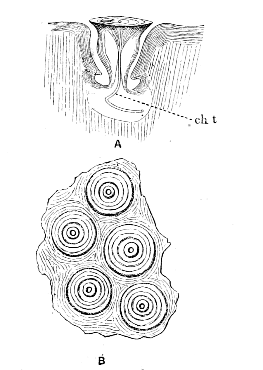
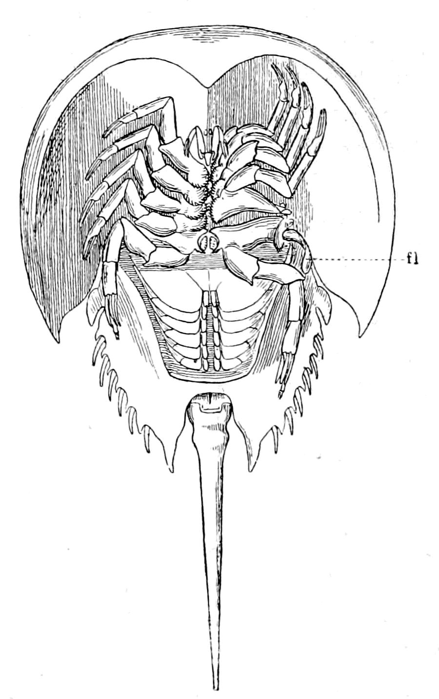
Fig. 144.—A, A Goblet from one of the Branchial Sense-organs of Limulus (ch.t., chitinous tubule); B, Surface View of a Portion of a Branchial Sense-Organ. Fig. 145.—The Endognaths of Limulus pushed out of the way on one side in order to show the position of the Flabellum (fl.) projecting towards the crack between the Prosomatic and Mesosomatic Carapaces.
Gegenbaur, in his picture, draws a straight tubule passing from every goblet among the fine canaliculi of the chitin. He says they are difficult to see, except in the case of the larger goblets. The tubule from the larger goblets is most conspicuous, and is in my sections always tortuous, never straight, as represented by Gegenbaur. A special branch of the appendage-nerve passes to these organs, and upon the fine branches of this nerve groups of ganglion-cells are seen, very similar in appearance to the groups described by Patten on the terminal branches of the nerves which supply the mandibular organs. At present I can see no mechanism by which the goblets are extruded or returned into place. In the case of the Capitellidæ, Eisig describes retractor muscles by means of which the lateral sense-organs are brought below the level of the surface, and he imagines that the protrusion is effected by hydraulic means, by the aid of the vascular system. In the branchial sense-organs of Limulus there are no retractor muscles, and it seems to me that both retraction and protrusion must be brought about by alterations of pressure in the vascular fluids. Certainly the cavity of the organ is very vascular. If this be so, it seems likely enough that such an organ should be a very delicate organ for estimating changes in the pressure of the external medium, for the position of the goblets would depend on the relation between the pressure of the fluid inside the organ and that on the surface of the appendage. Whether the chitinous tubule contains a nerve-terminal or not I am unable to decide from my specimens, but, judging from Patten's description of the similar chitinous tubules belonging to the mandibular organs, it is most highly probable that these tubules also contain a fine terminal nerve-fibre.
These organs, then, represent segmental branchial sense-organs, of which it can be said their structure suggests that they may be pressure-organs; but the experimental evidence is at present wanting.
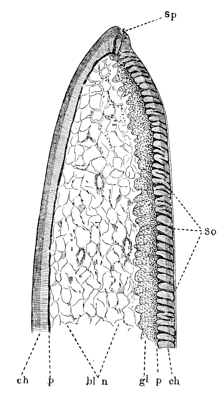
Passing now from the branchial to the prosomatic region, the first thing that struck me was the presence of that most conspicuous projection at the base of the last locomotor appendage, which is usually called the flabellum, and has been described by Lankester as an exopodite of this appendage. It is jointed on to the most basal portion of the limb (cf. Fig. 155), and projects dorsally from the limb into the open slit between the prosomatic and mesosomatic carapace, as is seen in Fig. 145 (fl.). Of its two surfaces, the undermost is very convex and the uppermost nearly flat from side to side, the whole organ being bent, so that when the animal is lying half buried in sand, entirely covered over by the prosomatic and mesosomatic carapaces except along this slit between the two, the upper flat or slightly convex surface of the flabellum is exposed to any movement of water through this slit, and owing to its possessing a joint, the direction of the whole organ can be altered to a limited extent. The whole of this flat upper surface is one large sense-organ of a striking character, thus forming a great contrast to the convex under surface, which is remarkably free from tactile spines or special sense-organs.
The nerve going to the flabellum is very large, almost as large as the nerve to the rest of the appendage, and the very large majority of the nerve-fibres turn towards the flat, uppermost side, where the sense-organ is situated. Between the nerve-fibres (n.) and the chitinous surface containing the special sense-tubes masses of cells (gl.) are seen, as in Fig. 146, apparently nerve-cells, which form a broad border between the nerve-fibres and the pigmented chitinogenous layer (p.). On the opposite side, nothing of the sort intervenes between the pigmented layer and the blood-spaces and nerve-fibres which constitute the central mass of the flabellum.
Fig. 146.—Section through Flabellum. ch., chitinous layers; s.o., sense-organs; sp., spike-organ; p., pigment layer; gl., ganglion cell layer; bl. and n., blood-spaces and nerves. Fig. 147.—Section parallel to the Surface of Flabellum, showing the Porous Terminations of the Sense-Organs and the Arrangement of the Canaliculi round them.
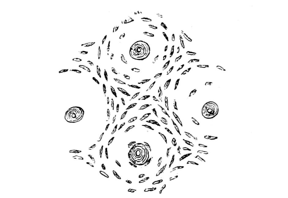
At present I am inclined to look upon this mass of cells as constituting a large ganglion, which extends over the whole length and breadth of the upper surface of the flabellum. At the same time, my preparations are not sufficiently clear to enable me to trace out the connections of these cells, especially their connections with the special sense-organs.
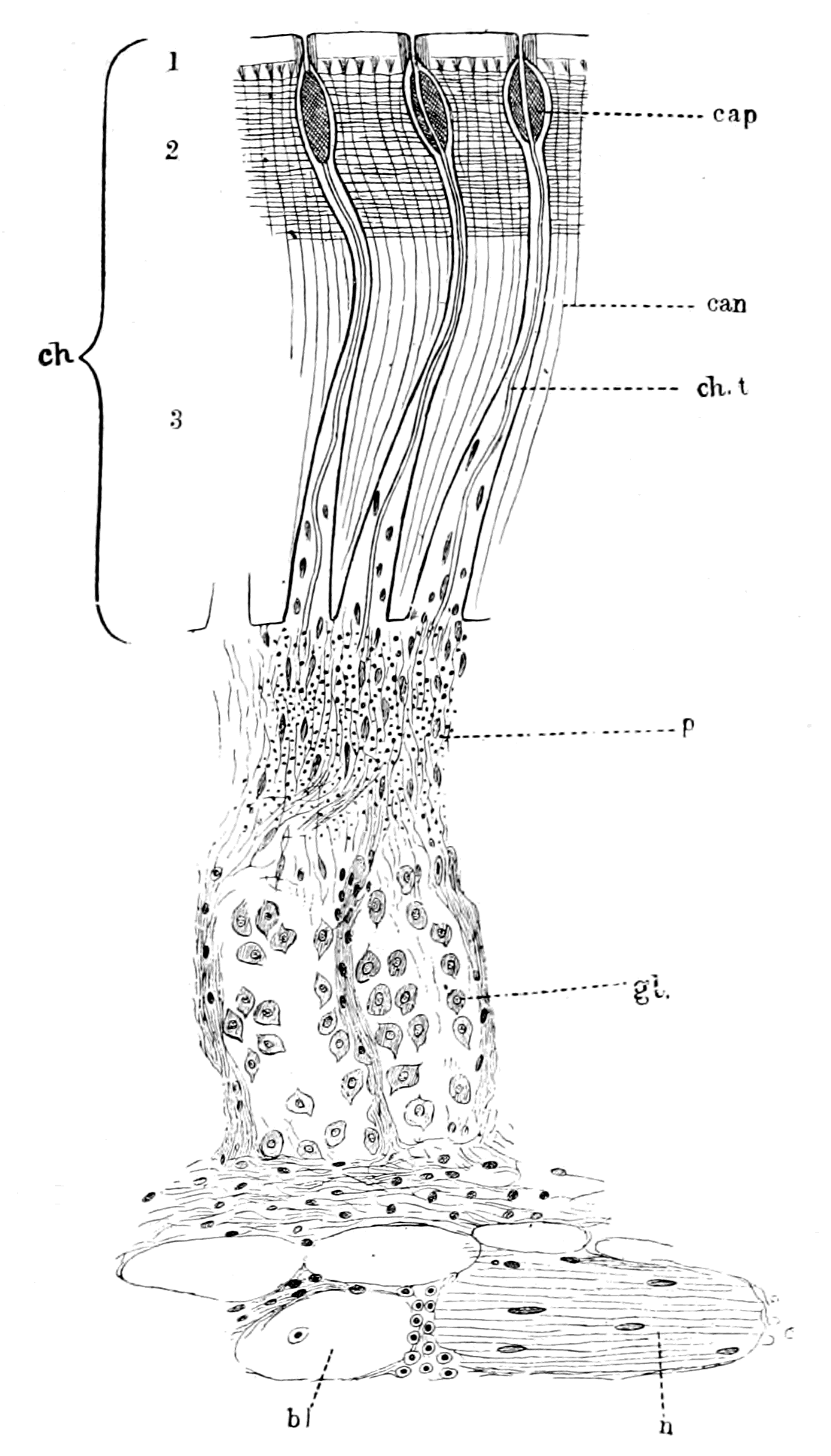
Fig. 148.—Section through the three Sense-Organs of Flabellum.
bl., blood-spaces; n., nerve; gl., layer of ganglion-cells; p., pigment layer; ch., 1, 2, 3, the three layers of chitin; ch.t., chitinous tubule in large tube of sense-organ; cap., capitellum or swollen extremity of large tube; can., very fine porous canals or canaliculi of chitin.
In Fig. 148 I give a magnified representation of a section through three of these flabellar sense-organs. As is seen, the section divides itself into four zones: (1) the chitinous layer (ch.); (2) the layer of pigment (p.) and hypodermal cells; (3) the layer of ganglion-cells (gl.); and (4) the layer of nerve-fibres (n.) and blood-spaces (bl.). The chitinous layer is composed of the usual three zones of the Limulus surface—externally (Fig. 148), a thin homogeneous layer, followed by a thick layer of chitin (3), in which the fine vertical tubules or canaliculi are well marked; the external portion (2) of this layer is differentiated from the rest by the presence of well-marked horizontal layers in addition to the canaliculi.
In this chitinous layer the special sense-organs are found. They consist of a large tube which passes through all the layers of the chitin except the thin homogeneous most external layer. This tube is conical in shape, its base, which rests on the pigmented layer, being so large and the organs so crowded together that a section of the chitin across the base of the tubes gives the appearance of a honeycomb, the septa of which is all that remains of the chitin. This large tube narrows down to a thin elongated neck as it passes through the chitin, and then, at its termination, bulges out again into an oval swelling (cap.) situated always beneath the homogeneous most external layer of chitin. Within this tube a fine chitinous tubule (ch. t.) is situated similar to that seen in the branchial sense-organs; it lies apparently free in the tube, not straight, but sinuous, and it passes right through all the chitinous layers to open at the surface as a pore; in the last part of its course, where it passes through the most external layer (1) of chitin, it lies always at right angles to the surface.
If the flabellum be stained with methylene blue and acid fuchsin, then all the canaliculi in the chitin show up as fine red lines, and present the appearance given in Fig. 148, and it is seen that each of the terminations of the tubules is surrounded in the homogeneous layer of chitin by a thick-set circular patch of canaliculi which pass to the very surface of the chitin, while the canaliculi in other parts terminate at the commencement of the homogeneous layer and do not reach the surface. Further, the contents of the oval swelling, and, indeed, of the tube as a whole, are stained blue, the chitinous tubule being either unstained or slightly pink in colour. We see, then, that the chitinous tubule alone reaches the surface, while the large tube, which contains the tubule, terminates in an oval swelling, which often presents a folded or wrinkled appearance, as in Fig. 149 (see also Patten's Fig. 1, Plate I.). This terminal bulging of the tube is reminiscent of the bulging in the chitinous tubes of the lyriform organs of the Arachnida, as described by Gaubert, and of the poriferous chordotonal organs in insects, as described by Graber (see Fig. 150). This terminal swelling is filled with a homogeneous refringent mass staining blue with methylene blue, in which I have seen no trace of a nucleus; through this the chitinous tubule makes its way without any sign of bulging on its part. Patten, in his description of the sense-organs on the mandibles of Limulus, which are evidently the same in structure as those on the flabellum, refers to this homogeneous mass as a coagulum. I doubt whether this is an adequate description; it appears to me to stain rather more readily than a blood-coagulum, yet in the sense of being structureless it resembles a coagulum.
The enormous number of these organs crowded together over the whole flat surface of the flabellum produces a very striking appearance when viewed on the surface. Such a view presents an appearance resembling that of the surface-view of the branchial sense-organs; in both cases the surface is covered with a great number of closely set circular plaques, in the centre of each of which is seen a well-marked pore. The circular plaques in the case of the flabellum are much smaller than those of the branchial sense-organs, and clearly are not protrusible as in the latter organs, the appearance as of a plaque being due to the ring of thickly-set canaliculi round the central tubule, as already described. When stained with methylene blue, the surface view of the flabellum under a low power presents an appearance of innumerable circular blue masses, from each of which springs a fine bent hair, terminating in a pore at the surface. The blue masses are the homogeneous substance (cap.) of the bulgings seen through the transparent external layer of chitin, and the hairs are the terminal part of the chitinous tubules. Patten has represented their appearance in the mandibles in his Fig. 2, Plate I.
The large tubes in the chitin alter in shape according to their position. Those in the middle of the sensory surface of the flabellum, in their course through the chitinous layers, are hardly bent at all; as they approach the two lateral edges of this surface, their long thin neck becomes bent more and more, the bending always being directed towards the middle of the surface (see Fig. 146); in this way the chitinous tubules increase more or less regularly in length from the centre of the organ to the periphery. The large basal part of the conical tube contains, besides the chitinous tubule, a number of nuclei which are confined to this part of the tube; some of these nuclei look like those belonging to nerve-fibres, others are apparently the nuclei of the chitinogenous membrane lining the tube. I have never seen any sign of nerve-cells in the tube itself.
The only other kind of sense-organ I have found in connection with these sense-organs are a few spike-like projections, the appearance of which is given in Fig. 149. I have always seen these in the position given in Fig. 146 (sp.), i.e. at the junction of the surface which contains the sense-organs and the surface which is free from them. They are, so far as I have seen, not very numerous; I have not, however, attempted to examine the whole sense-organ for the purpose of estimating their number and arrangement.
As is seen in Fig. 149, they possess a fine tubule of the same character as that of the neighbouring sense-organs, which apparently terminates at the apex of the projecting spike. They appear to belong to the same group as the other poriferous sense-organs, and are of special interest, because in their appearance they form a link between the latter and the poriferous sense-organs which characterize the pecten of the scorpion (cf. Fig. 152, C).
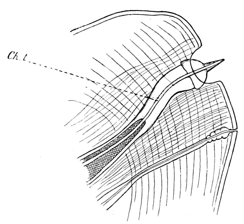
Fig. 149.—Spike-Organ of Flabellum.
ch.t., chitinous tubule.
Such, then, is the structure of this remarkable sense-organ of the flabellum, as far as I have been able to work it out with the materials at my disposal. It is evident that the flabellar organs, apart from the spike-organs, are of the same kind as those described by Patten on the mandibles and chelæ of Limulus, and therefore it is most probable that the nerve-terminals in the chitinous tubules, and the origin of the latter, are similar in the two sets of organs.
These organs, as Patten has described them, are situated in lines on the spines of the mandibles of the prosomatic locomotor appendages, and are grouped closely together to form a compact sense-organ on the surface of the inner mandible (Lankester's epicoxite) (i.m. in Fig. 155), so that a surface-view of the organ here gives the characteristic appearance of these poriferous sense-patches. Precisely similar organs are found on the chilaria, which are, in function at all events, simply isolated mandibles, to use Patten's terminology.
On the digging appendage (ectognath), as the comparison of Fig. 155, A and C, shows, the mandibular spines are almost non-existent, and the inner mandible or epicoxite is not present, so that the special sense-organ of this appendage is represented solely by the flabellum.
This sketch of the special sense-organs of Limulus shows that all the appendages of Limulus possess special sense-organs, with the exception of the operculum. All these sense-organs are formed on the same plan, in that they possess a fine chitinous tubule passing through the layers of chitin into the underlying hypodermal and nervous tissues, which terminates on the surface in a pore. The surface of the chitin where these pores are situated is perfectly smooth, although, in the case of the branchial sense-organs, the goblet-shaped masses of chitin, each of which contains a pore, are able to be pressed out beyond the level of the surface.
As to their functions, we unfortunately do not know much that is definite. Patten considers that he has evidence of a gustatory function in the case of the mandibular organs, and suggests also a temperature-sense in the case of some of these organs. The large organ of the flabellum and the branchial organs he has not taken into consideration. The situation of these organs puts the suggestion of any gustatory function, as far as they are concerned, out of the question; and I do not think it probable that such large specialized organs would exist only for the estimation of temperature, when one sees how, in the higher animals, the temperature-nerves and the nerves of common sensation are universally distributed over the body. As already stated, the structure of the branchial organs seems to me to point to organs for estimating varying pressures more than anything else, and I am strongly inclined to look upon the whole set of organs as the derivatives of the lateral sense-organs of annelids, such as are described by Eisig in the Capitellidæ. This is Patten's opinion with respect to the mandibular organs; and from what I have shown, these organs cannot be separated in type of structure from those of the flabellum and the branchial sense-organs.
In our search, then, for the origin of the vertebrate auditory organ in Limulus and its allies, we see so far the following indications:—
1. The auditory organ of the vertebrate is regarded as a special organ belonging to a segmentally arranged set of lateral sense-organs, whose original function was co-ordination and equilibration.
2. Such a set of segmentally arranged lateral sense-organs is found in annelids in connection with the dorsal cirri of the ventral parapodia.
3. If, as has been supposed, there is a genetic connection between (1) and (2) and if, as I suppose, the vertebrates did not arise from the annelids directly, but from a protostracan group, then it follows that the lateral sense-organs, one of which gave rise to the auditory organ, must have been situated on the protostracan appendages.
4. In Limulus, which is the sole surviving representative of the palæostracan group, such special sense-organs are found on both the prosomatic and mesosomatic appendages, and therefore may be expected to give a direct clue to the origin of the vertebrate auditory organ.
5. Both from its position, its size, and its specialization, the flabellum, i.e. an organ corresponding to the flabellum, must be looked upon as more likely to give a direct clue to the origin of the auditory organ than the sense-organs of the branchial appendages, or the so-called gustatory organs of the mandibles.
The Auditory Organs of Arachnids and Insects.
The difficulty of the investigating these organs consists in the fact that so little is known about them in those Arthropoda which live in the water; the only instance of any organ apparently of the nature of an auditory organ, is the pair of so-called auditory sacs at the base of the antennæ in various decapods. We are in a slightly better position when we turn to the land-living arthropods; here the presence of stridulating organs in so many instances carries with it the necessity of an organ for appreciating sound. It has now been shown that such stridulating organs are not confined to the Insecta, but are present also in the scorpion group, and I myself have added to their number by the discovery of a distinct stridulating apparatus in various members of the Phrynidæ. We may then take it for granted that arachnids as well as insects hear. Where is the auditory organ?
Many observers believe that certain surface-organs found universally among the spiders, to which Gaubert has given the name of lyriform organs, are auditory in function. His investigations show that they are universally present on the limbs and pro-meso-sternite of all spiders; that they are present singly, not in groups, on the limbs of Thelyphonus, and that a group of them exists on the second segment of each limb in the members of the Phrynus tribe. In the latter case this organ is the most elaborate of all described by him.
It is especially noticeable that they do not exist in Galeodes or in the scorpions, but in the former special sense-organs are found in the shape of the so-called 'racquet-organs,' on the basal segments of the most posterior pair of appendages, and also, according to Gaubert, on the extremity of the palps and the first pair of feet, while in the latter they occur in the shape of the pectens.
This observation of Gaubert suggests that the place of the lyriform organs in other arachnids is taken in Galeodes by the racquet-organs, and in the scorpions by the pectens. Bertkau, Schimkéwitsch, and Wagner, as quoted by Gaubert, all suggest that the lyriform organs of the arachnids belong to the same group of sense-organs as the porous chordotonal organs of the Insecta, sense-organs which have been found in every group of Insecta, and are generally regarded as auditory organs. Gaubert does not agree with this, and considers the lyriform organs to be concerned with the temperature-sense rather than with audition.
The chordotonal organs of insects have been specially studied by Graber. He divides them into two groups, the poriferous and the non-poriferous, the former being characterized by the presence of pores on the surface arranged in groups or lines. These poriferous chordotonal organs are remarkably constant in position, being found only at the base of the wings on the subcostal ridge, in marked contrast to the other group of chordotonal organs which are found chiefly on the appendages in various regions. The striking character of this fixity of position of these organs and the universality of their presence in the whole group, led Graber to the conclusion that in these poriferous chordotonal organs we are studying a form of auditory apparatus which characterized the ancestor of the insect-group. These organs are always well developed on the hind wings, and in the large group of Diptera the auditory apparatus has usurped the whole of the function of the wing; for the balancers or 'halteres,' as they are called, are the sole representatives of the hind wings, and they are usually considered to be of the nature of auditory organs. It is instructive to find that such an auditory organ serves not only for the purpose of audition, but also as an organ of equilibration; thus Lowne gives the evidence of various observers, and confirms it himself, that removal of the balancers destroys the power of orderly flight in the animal.
A striking peculiarity of these organs in the Insecta, as described by Graber, is the bulging of the porous canal near its termination (Fig. 150, C). This bulging is filled with a homogeneous, highly refractive material, from which, according to Lowne, a chordotonal thread passes, to be connected with a ganglion-cell and nerve. This sphere of refractive material he calls the 'capitellum' of the chordotonal thread. The presence of this material produces in a surface view an appearance as of a halo around the terminal plaque with its central pore; Graber has attempted to represent this by the white area round the central area (in Fig. 150, B). A very similar appearance is presented by the surface view of the flabellum in those parts where the tube runs straight to the surface, so that the refractive material which fills the oval bulging shines through the overlying chitin and appears to surround the terminal plaque with a translucent halo.
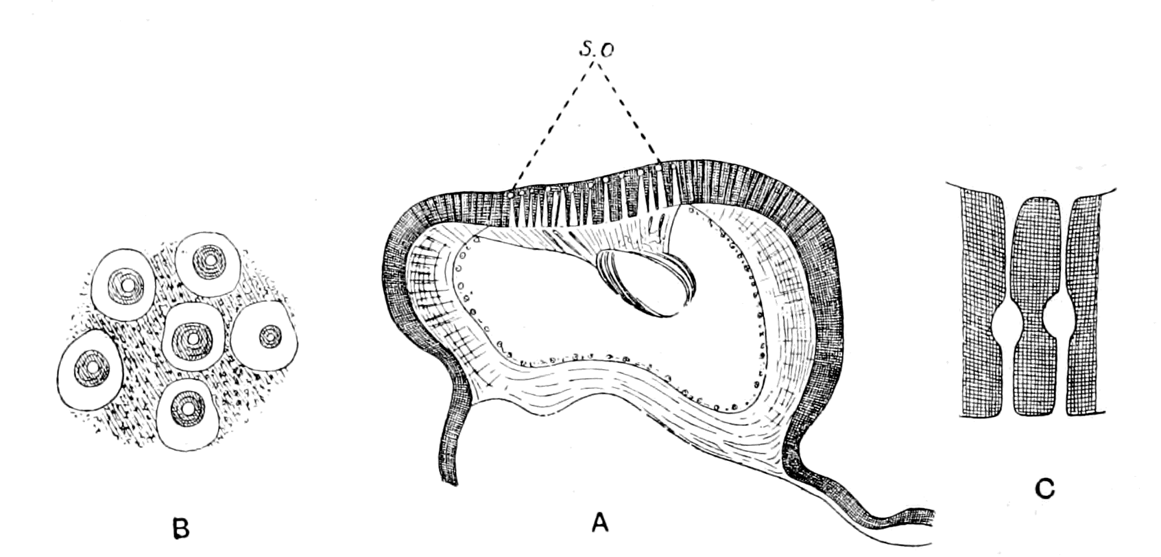
Fig. 150 (from Graber).—A, Section of Subcostal Nervure of Hind Wing of Dytiscus to show patch of Poriferous Organs (s.o.). B, Surface View of Poriferous Organs; the White Space round each Organ indicates the deeper lying Refringent Body which fills the bulging of the Canal seen in Transverse Section in C.
Such a peculiarity must have a very definite meaning, and suggests that the canals in the flabellum of Limulus and in the hind wings of insects belong to the same class of organ, the chitinous tubule with its nerve-terminal in the former corresponding to the chordotonal thread in the latter. One wonders whether this sphere of refractive material or 'capitellum' (to use Lowne's phraseology) is so universally present in order to act as a damper upon the vibrations of the chordotonal thread in the one case and of the chitinous tubule in the other, just as the membrana tectoria and the otoliths act in the case of the vertebrate ear.
Patten says that the only organs which seem to him to be comparable with the gustatory porous organs of Limulus are the sense-organs in the extremities of the palps and of the first pair of legs of Galeodes, as described by Gaubert. I imagine that he was thinking only of arachnids, for the comparison of his drawings with those of Graber show what a strong family resemblance exists between the poriferous sense-organs of Limulus and those of the insects. On the course of the terminal nerve-fibres, between the nerve-cell and their entrance into the porous chitinous canal, Graber describes the existence of rods or scolophores. On the course of the terminal fibres in the Limulus organ, between the nerve-cells and their entrance into the porous chitinous canal, Patten describes a spindle-shaped swelling, containing a number of rod-like thickenings among the fibrils in the spindle, which present an appearance reminiscent of the rods described by Graber.
It appears as though a type of sense-organ, characterized by the presence of pores on the surface and a fine chitinous canal which opens at these pores, was largely distributed among the Arthropoda. According to Graber, this kind of organ represents a primitive type of sense-organ, which was probably concerned with audition and equilibration, and he expresses surprise that similar organs have not been discovered among the Crustacea. It is, therefore, a matter of great interest to find that so ancient a type of animal as Limulus, closely allied to the primitive crustacean stock, does possess poriferous sense-organs upon its appendages which are directly comparable with these poriferous chordotonal organs of the Insecta.
The Pectens of Scorpions.
Among special sense-organs such as those with which I am now dealing, the pectens of scorpions and the 'racquet-organs' of Galeodes must, in all probability, be classed. I have given my reasons for this conclusion in my former paper.[2] At present such reasons are based entirely upon the structure of the organs; experimental evidence as to their function is entirely wanting. With respect to the pectens of the scorpion (Fig. 151), it has been suggested that they are of the nature of copulatory organs, a suggestion which may be dismissed without hesitation, for they are not constructed after the fashion of claspers, but are simply elaborate sense-organs, and, as such, are found equally in male or female. The only observer who has hitherto specially studied the structure of the sense-organs in the pecten is, as far as I know, Gaubert, and he describes their structure together with that of the sense-organs of the racquets of Galeodes, in connection with the lyriform organs of arachnids, as though he recognized a family resemblance between the three sets of organs.
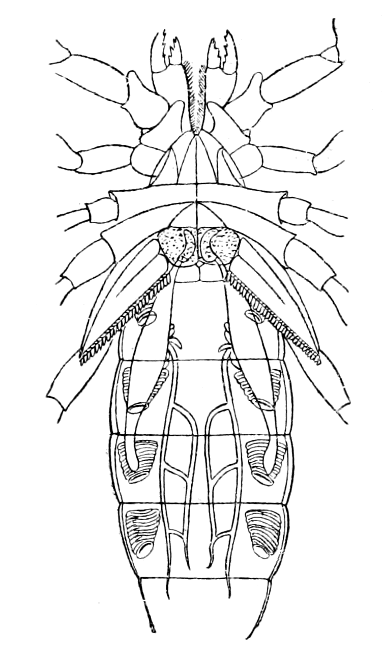
Fig. 151.—Under Surface of Scorpion (Androctonus).
The operculum is marked out with dots, and on each side of it is seen one of the pectens.
The pecten of the scorpions is an elaborate sense-organ, or rather group of sense-organs, the special organ being developed on each tooth of the comb; its surface, which is frequently flattened, being directed backwards and inwards, when the axis of the pecten is horizontal at right angles to the length of the body. The surface view of this part of the tooth resembles that of the branchial organs or of the flabellum in Limulus, in that it is thickly covered with circular patches, in the centre of which an ill-defined appearance as of a fine pore is seen. In Fig. 152, B, I give a sketch of the surface view of a part of the organ.
Transverse sections of a tooth of the comb of Scorpio Europæus present the appearance given in Fig. 152, A, and show that each of these circular patches is the surface-view of a goblet-shaped chitinous organ, Fig. 152, C, from the centre of which a short, somewhat cylindrical chitinous spike projects. Within this spike, and running through the goblet into the subjacent tissue, is a fine tubule. The series of goblets gives rise to the appearance of the circular plaques on the surface-view, while the spike with its tubule is the cause of the ill-defined appearance of the central pore, just as the terminal pore is much less conspicuous on surface-view in the spike-organs of the flabellum than in the purely poriferous organs, no part of which projects beyond the level of the chitinous surface.
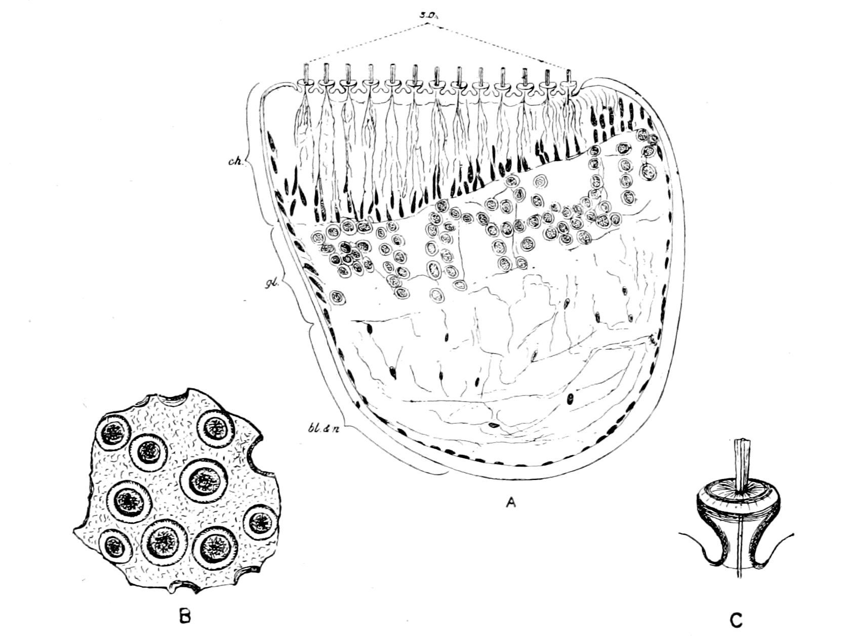
Fig. 152.—A, Section through Tooth of Pecten of Scorpion; B, Surface View of Sense-Organs; C, Goblet of Sense-Organ more highly magnified.
bl. and n., region of blood-spaces and nerves; gl., ganglion-cell layer; ch., modified chitinous layer; s.o., sense-organ.
The fine tubule is soon lost in the thickened but soft modification of the chitinous layer (ch.) which is characteristic of the sense-organ; at all events, I have not succeeded in tracing it through this layer with any more success than in the corresponding case of the tubules belonging to the smaller goblets of the branchial sense-organ of Limulus already described.
At the base of the modified chitinous layer a series of cells is seen, many, if not all, of which belong to the chitinogenous layer. Next to these is the marked layer of ganglion-cells (gl.), similar to those seen in the flabellum of Limulus. The rest of the space in the section of the tooth is filled up with nerves (n.) and blood-spaces (bl.) just as in the section, Fig. 146, of the flabellum of Limulus.
Gaubert does not appear to have seen the goblets at all clearly; he describes them simply as conical eminences, and states that they "recouvrent un pore analogue a celui des poils mais plus petit; il est rempli par le protoplasma de la couche hypodermique." From the ganglion, according to him, nervous prolongations pass, which traverse the chitinogenous layer and terminate at the base of the conical eminences. Each of these prolongations "présente sur son trajet, mais un peu plus près du ganglion que de sa terminaison périphérique, une cellule nerveuse fusiforme (g.) offrant, comme celles du ganglion, un gros noyau." He illustrates his description with the following, Fig. 153, taken from his paper.
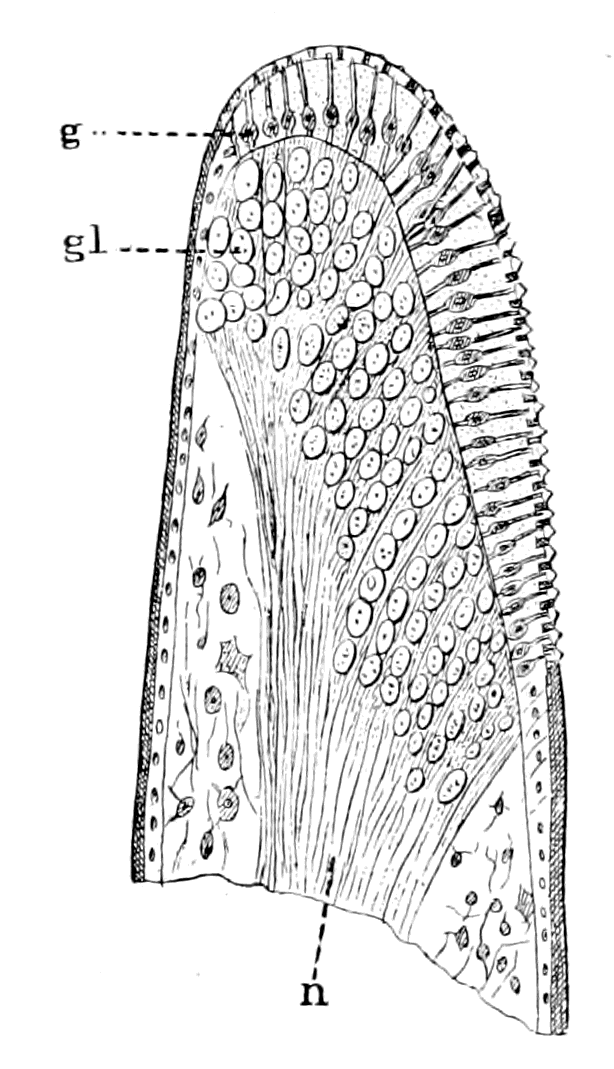
Fig. 153 (from Gaubert).—Section of a Tooth of Pecten of Scorpion.
n., nerve; gl., ganglion.
I have not been able to obtain any evidence of a fusiform nerve-cell on the course of the terminal nerve-fibres as depicted by him; fusiform cells there are in plenty, as depicted in my drawing, but none with a large nucleus resembling those of the main ganglion. In no case, either in the flabellum or in the branchial organs of Limulus, or in the pecten-organs, have I ever seen a ganglion-cell within the chitin-layer; all the nuclei seen there resemble those of the cells of the hypodermis or else the elongated nuclei characteristic of the presence of nerve-fibres. Gaubert's drawing is a striking one, and I have looked through my specimens to see whether there was anything similar, but have hitherto failed to obtain any definite evidence of anything of the kind.
I feel, myself, that an exhaustive examination of the structure and function of the pecten of scorpions ought to be undertaken. At present I can only draw the attention of my readers to the similarity of the arrangement of parts, and of the nature of the end-organs, in the sense-organs of the flabellum of Limulus and of the pecten of the scorpion. In both cases the special nerve-fibres terminate in a massive ganglion, situated just below the chitinogenous layer. In both cases the terminal fibres from these ganglion-cells pass through the modified chitinous layer to supply end-organs of a striking character; and although the end-organ of the pecten of the scorpion does not closely resemble the majority of the end-organs of the flabellum, yet it does resemble, on the one hand, the isolated poriferous spikes found on the flabellum (Fig. 149) and, on the other, the poriferous goblets found on the sense-patches of the branchial appendages of Limulus (Fig. 144, A), so that a combination of these two end-organs would give an appearance very closely resembling that of the pecten of the scorpion.
Finally, the special so-called 'racquet-organs' of Galeodes, which are found on the most basal segments of the last pair of prosomatic appendages, ought also to be considered here. Gaubert has described their structure, and shown how the nerve-trunk in the handle of the racquet splits up into a great number of separate bundles, which spread out fan-shaped to the free edge of the racquet; each of these separate bundles supplies a special sense-organ, which terminates as a conical eminence on the floor of a deep groove, running round the whole free edge of the racquet. This groove is almost converted into a canal, owing to the projection of its two sides. Gaubert imagines that the sense-organs are pushed forward out of the groove to the exterior by the turgescence of the whole organ; each of the nerve-fibres forming a bundle is, according to Gaubert, connected with a nerve-cell before it reaches its termination.
This sketch of the special sense-organs on the appendages of Limulus, of the scorpions, of Galeodes, and other arachnids, and their comparison with the porous chordotonal organs of insects, affords reason for the belief that we are dealing here with a common group of organs, which, although their nature is not definitely known, have largely been accredited with the functions of equilibration and audition, a group of organs among which the origin of the auditory organ of vertebrates must be sought for, upon any theory of the origin of vertebrates from arthropods.
Whenever in any animal these organs are concentrated together to form a special organ, it is invariably found that the nerve going to this organ is very large, out of all proportion to the size of the organ, and also that the nerve possesses, close to its termination in the organ, large masses of nerve-cells. Thus, although the whole hind wing in the blow-fly has been reduced to the insignificant balancers or 'halteres,' yet, as Lowne states, the nerves to them are the largest in the body.
The pectinal nerve in the scorpion is remarkable for its size, and so, also, is the nerve to the flabellum in Limulus, while the large size of the auditory nerve in the vertebrate, in distinction to the size of the auditory apparatus, has always aroused the attention of anatomists.
Throughout this book my attention has been especially directed to both Limulus and the scorpion group in endeavouring to picture to myself the ancestor of the earliest vertebrates, because the Eurypteridæ possessed such marked scorpion-like characteristics; so that in considering the origin of a special sense-organ, such as the vertebrate auditory organ near the junction of the prosoma and mesosoma, it seems to me that the presence of such marked special sense-organs as the flabellum on the one hand and the pecten on the other, must both be taken into account, even although the former is an adjunct to a prosomatic appendage, while the latter represents, according to present ideas, the whole of a mesosomatic appendage.
From the point of view that the VIIIth nerve represents a segment immediately posterior to that of the VIIth, it is evident that an organ in the situation of the pecten, immediately posterior to the operculum, i.e. according to my view, posterior to the segment originally represented by the VIIth nerve, is more correctly situated than an organ like the flabellum, which belongs to a segment anterior to the operculum.
On the other hand, from the point of view of the relationship between the scorpions and the king-crabs, it is a possibly debatable question whether the pecten really belongs to a segment posterior to the operculum. The position of any nerve in a series depends upon its position of origin in the central nervous system, rather than upon the position of its peripheral organ. Now, Patten gives two figures of the brain of the scorpion built up from serial sections. In both he shows that the main portion of the pectinal nerve arises from a swelling, to which he gives the name ganglion nodosum. This swelling arises on each side in close connection with the origin of the most posterior prosomatic appendage-nerve, according to his drawings, and posteriorly to such origin he figures a small nerve which he says supplies the distal parts of the sexual organs. This nerve is the only nerve which can be called the opercular nerve, and apparently arises posteriorly to the main part of the pectinal nerve. If this is so, it would indicate that the pectens arose from sense-organs which were originally, like the flabella, pre-opercular in position, but have shifted to a post-opercular position.
The Origin of the Parachordals and Auditory Cartilaginous Capsule.
In addition to what I have already said, there is another reason why a special sense-organ such as the pecten is suggestive of the origin of the vertebrate auditory organ, in that such a suggestion gives a clue to the possible origin of the parachordals and auditory cartilaginous capsules.