Front matter (continued 3)
Reading edition
3. The neurone of the third order, which terminates in the optic lobes of the brain by means of its neuraxons (the optic nerve).
We see, then, that the most recent researches agree with the older ones of Berger, Claus, and Bellonci, in picturing the retina of the primitive crustacean forms as formed of two ganglia only, and not of four, as in the specialized crustacean group the Malacostraca.
The comparison of the arthropod compound retina with that of the vertebrate shows, as one would expect upon the theory of the origin of vertebrates put forward in this book, that the latter retina is built up of two ganglia, as in the more primitive less specialized crustacean forms. The modern description of the vertebrate retina, based upon the Golgi method of staining, is exactly Parker's description of the simpler form of crustacean retina in which the 'neuropil' of the first ganglion is represented by the external molecular layer, and that of the second ganglion by the internal molecular layer; the three sets of neurones being, according to Parker's terminology:—
1. The neurones of the first order—viz. the visual cells—the nuclei of which form the external nuclear layer, and their long attenuated processes form synapses in the external molecular layer with
2. The neurones of the second order, the cells of which form the internal nuclear layer, and their processes form synapses in the internal molecular layer with
3. The neurones of the third order, the cells of which form the ganglionic layer and their neuraxons constitute the fibres of the optic nerve which end in the optic lobes of the brain.
Strictly speaking, of course, the visual cells with their elongated processes have no right to be called neurones: I only use Parker's phraseology in order to show how closely the two retinas agree even to the formation of synapses between the fine drawn-out processes of the visual cells and the neurones of the ganglion of the retina.
The Retina of the Lateral Eye of Ammocœtes.
As in the case of all other organs, it follows that if we are dealing here with a true genetic relationship, then the lower we go in the vertebrate kingdom the more nearly ought the structure of the retina to approach the arthropod type. It is therefore a matter of intense interest to determine the nature of the retina in Ammocœtes in order to see whether it differs from that of the higher vertebrates, and if so, whether such differences are explicable by reference to the structure of the arthropod eye.
Before describing the structure of this retina it is necessary to clear away a remarkable misconception, shared among others by Balfour, that this eye is an aborted eye, and that it cannot be considered as a primitive type. Thus Balfour says: "Considering the degraded character of the Ammocœte eye, evidence derived from its structure must be received with caution," and later on, "the most interesting cases of partial degeneration are those of Myxine and the Ammocœte. The development of such aborted eyes has as yet been studied only in the Ammocœte, in which it resembles in most important features that of other Vertebrata."
Again and again the aborted character of the eye is stated to be evidence of degeneration in the case of the lamprey. What such a statement means, why the eye is in any way to be considered as aborted, is to me a matter of absolute wonderment: it is true that in the larval form it lies under the skin, but it is equally true that at transformation it comes to the surface, and is most evidently as perfect an eye as could be desired. There is not the slightest sign of any degeneration or abortion, but simply of normal development, which takes a longer time than usual, lasting as it does throughout the life-time of the larval form.
Kohl, who has especially studied degenerated vertebrate eyes, discusses with considerable fulness the question of the Ammocœtes eye, and concludes that in aborted eyes a retarded development occurs, and this applies on the whole to Ammocœtes, "but with the important difference that in this case the period of retarded development is not followed by a stoppage, but on the contrary by a period of very highly intensified progressive development during the metamorphosis," with the result that "the adult eye of Petromyzon Planeri does not diverge from the ordinary type."
Referring in his summing up to this retarded development, he says: "Such reminiscences of embryonic conditions are after all present here and there in normally developed organs, and by no means entitle us to speak of abnormal development."
The evidence, then, is quite clear that the eye of Petromyzon, or, indeed, of the full-grown Ammocœtes, is in no sense an abnormal eye, but simply that its development is slow during the ammocœte stage. The retina of Petromyzon was figured and described by Langerhans in 1873. He describes it as composed of the following layers:—
(1) Membrana limitans interna. (2) Thick inner molecular layer. (3) Optic fibre layer. (4) Thick inner nuclear layer. (5) Peculiar double-layered ganglionic layer. (6) External molecular layer. (7) External nuclear layer. (8) Membrana limitans externa. (9) Layer of rods. (10) Pigment-epithelium.
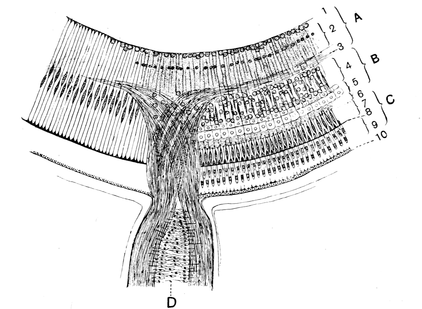
Fig. 41.—Retina and Optic Nerve of Petromyzon. (After Müller and Langerhans.)
On the left side the Müllerian fibres and pigment-epithelium are represented alone. The retina is divided into an epithelial part, C (the layer of visual rod-cells), and a neurodermal or cerebral part which is formed of, A, the ganglion of the optic nerve and, B, the ganglion of the retina. 1, int. limiting membrane; 2, int. molecular layer with its two layers of cells; 3, layer of optic nerve fibres; 4, int. nuclear layer; 5, double row of tangential fulcrum cells; 6, layer of terminal retinal fibres; 7, ext. nuclear layer; 8, ext. limiting membrane; 9, layer of rods; 10, layer of pigment-epithelium. D, axial cell layer (Axenstrang) in optic nerve. The layer 6 is drawn rather too thick.
He points out especially the peculiarity of layer (2) (2, Fig. 41), the inner molecular, in which two rows of nuclei are arranged with great regularity, the one row closely touching the membrana limitans interna, the other at the inner boundary of the middle third of the molecular layer. Of these two rows of nuclei, he describes the innermost as composed almost entirely of large nuclei belonging to ganglion cells, while the outermost is composed mainly of distinctly smaller nuclei, which in staining and appearance appear to belong not to nerve-cells but to the true reticular tissue of the molecular layer.
He also draws special attention to the remarkable layer (5) (5, Fig. 41), which is not found in the retina of the higher vertebrates, the cells of which, in his opinion, are of the nature of ganglion-cells.
W. Müller, in 1874, gave a most careful description of the eye of Ammocœtes and Petromyzon, and traced the development of the retina; the subsequent paper of Kohl does not add anything new, and his drawings are manifestly diagrams, and do not represent the appearances so accurately as Müller's illustrations. In the accompanying figure (Fig. 41) I reproduce on the right-hand side Müller's picture of the retina of Petromyzon, but have drawn it, as in Langerhans' picture, at the place of entry of the optic nerve.
From his comparison of this retina with a large number of other vertebrate retinas, he comes to the conclusion that the retina of all vertebrates is divisible into
A. An ectodermal (epithelial) part consisting of the layer of the visual cells, and
B. A neurodermal (cerebral) part which forms the rest of the retina.
Further, Müller points out that the neuroderm gives origin throughout the central nervous system to two totally different structures, on the one hand to the true nervous elements, on the other to a system of supporting cells and fibres which cannot be classed as connective tissue, for they do not arise from mesoblast, and are therefore called by him 'fulcrum-cells.' In the retina he recognizes two distinct groups of such supporting structures—(1) a system of radial fibres with well-marked elongated nuclei, which extend between the two limiting layers, and form at their outer ends a membrane-like expansion which was originally the outer limit of the retina, but becomes afterwards co-terminous with the membrana limitans externa, owing to the piercing through it of the external limbs of the rods. This system, which is known by the name of the radial Müllerian fibres (shown on the left-hand side of Fig. 41), has no connection with (2) the spongioblasts and neurospongium, which form a framework of neuroglia, in which the terminations of the optic ganglion and of the retinal ganglion ramify to form the molecular layers.
It is evident from Fig. 41 that the retina of Ammocœtes and Petromyzon differs in a striking manner from the typical vertebrate retina. The epithelial part (C) remains the same—viz. the visual rods, the external limiting membrane, and the external nuclear layer; but the cerebral part, the retinal ganglion (A and B), is remarkably different. It is true, it consists in the main of the small-celled mass known as the inner nuclear layer, and of the reticulated tissue or 'neuropil' known as the inner molecular layer, just as in all other compound retinal eyes; but neither the ganglion cell-layer nor the optic fibre-layer is clearly defined as separate from this molecular layer; on the contrary, it is matter of dispute as to what cells represent the ganglionic layer of higher vertebrates, and the optic fibres do not form a distinct innermost layer, but pass into the inner molecular layer at its junction with the inner nuclear layer. A comparison of this innermost part of the retina (A, Fig. 41), with the corresponding part in Berger's picture of Musca (n.l.o.g., Fig. 38), shows a most striking similarity between the two. In both cases the fibres of the optic nerve (O.n., Fig. 38) which cross at their entrance pass into the 'neuropil' of this part of the retinal ganglion, and are connected probably (though that is not proved in either case) with the cells of the ganglionic layer. In both cases we find two well-marked parallel rows of cells in this part of the retina, of which one, the innermost, is composed in Ammocœtes of large ganglion-cells, and the other mainly of smaller, deeper staining cells apparently supporting in function. Similarly, also, in Branchipus, as I conclude from my own observations as well as from those of Berger and Claus, the ganglionic layer is composed partly of true ganglion-cells and partly of supporting cells arranged in a distinct layer. This part, then, of the retina of Ammocœtes is remarkably like that of a typical arthropod retina, and forms that part of the retinal ganglion which may be called the ganglion of the optic nerve.
Next comes the ganglion of the retina (B, Fig. 41) (Parker's first optic ganglion), the cells of which form the small bipolar granule-cells of the inner nuclear layer; granule-cells arranged in rows just as they are shown in Claus' picture of the same layer in the retina of Branchipus (Fig. 40), just as they are found in the cortical layers of the optic ganglion of the pineal eye (ganglion habenulæ), in the optic lobes and other parts of the Ammocœtes brain, or in the cortical layers of the optic ganglia of all arthropods.
Between this small-celled nuclear layer (4, Fig. 41) and the layer of nuclei of the visual rod cells (7, Fig. 41) (the external nuclear layer), we find in the eye of Ammocœtes and Petromyzon two well-marked rows of cells of a most striking character—viz. the two remarkably regular rows of large epithelial-like cells with large conspicuous nuclei, which give the appearance of two opposing rows of limiting epithelium (5, Fig. 41), already mentioned in connection with the researches of Langerhans and W. Müller. Here, then, is a striking peculiarity of the retina of the lamprey, and according to Müller the obliteration of these two layers can be traced as we pass upwards in the vertebrate kingdom. Among fishes, they are especially well seen in the perch; in the higher vertebrates the whole layer is only a rudiment represented, he thinks, by the simple layer of round cells which lies close against the inner surface of the layer of terminal fibres (Nervenansätze), and is especially evident in birds and reptiles. In man and the higher mammals they are probably represented by the horizontal cells of the outer part of the inner nuclear layer.
Seeing, then, that they are most evident in Ammocœtes, and become less and less marked in the higher vertebrates, it is clear that their origin cannot be sought among the animals higher in the scale than Ammocœtes, but must, therefore, be searched for in the opposite direction.
Müller describes them as forming a very conspicuous landmark in the embryology of the retina, dividing it distinctly into two parts, an outer thinner, and an inner somewhat thicker part, the zone formed by them standing out conspicuously on account of the size and regularity of the cells and their lighter appearance when stained. Thus in his description of the retina of an Ammocœtes 95 mm. in length, he says, "The layer of pale tangentially elongated cells formed a double layer and produced the appearance of a pale, very characteristic zone between the outer and inner parts of the retina."
Let us now turn to the retina of the crustacean and see whether there is any evidence there that the retina is divisible into an outer and inner part, separated by a zone of characteristically pale staining cells with conspicuous nuclei. The most elaborate description of the development of the retina of Astacus is given by Reichenbach, according to whom the earliest sign of the formation of the retina is an ectodermic involution (Augen-einstülpung), which soon closes, so that the retinal area appears as a thickening. In close contiguity to this thickening, the thickening of the optic ganglion arises, so that that part of the optic ganglion which will form the retinal ganglion fuses with the thickened optic plate and forms a single mass of tissue. Later on a fold (Augen-falte) appears in this mass of tissue, in consequence of which it becomes divided into two parts. The lining walls of this fold form a double row of cells, the nuclei of which are most conspicuous because they are larger and lighter in colour than the surrounding nuclei, so that by this fold the retina is divided into an outer and an inner wall, the line of demarcation being conspicuous by reason of these two rows of large, lightly-staining nuclei.
Reichenbach is unable to say that this secondary fold is coincident with the primary involution, and that therefore the junction between the two rows of large pale nuclei is the line of junction between the retinal ganglion and the retina proper, because all sign of the primary involution is lost before the secondary fold appears.
Parker compares the appearances in the lobster with Reichenbach's description in the crayfish, and says that he finds only a thickening, no primary involution; at the same time he expressly states that in the very early stages his material was deficient, and that he had not grounds sufficient to warrant the statement that no involution occurs. He also finds that in the lobster the ganglionic tissue which arises by proliferation is divided into an outer and inner part; the separation is effected by a band of large, lightly-staining nuclei, which, in position and structure, resemble the band figured by Reichenbach. According to Parker, then, the line of separation indicated in the development by Reichenbach's outer and inner walls is not the line of junction between the retina and the retinal ganglion, as Reichenbach was inclined to think, but rather a separation of two rows of large ganglion-cells belonging to the retinal ganglion.
The similarity between these conspicuous layers of lightly-staining cells in Ammocœtes and in crustaceans is remarkably close, and in both cases observers have found the same difficulty in interpreting their meaning. In each case one group of observers looks upon them as ganglion-cells, the other as supporting structures. Thus in the lamprey, Müller considers them to belong to the supporting elements, while Langerhans and Kohl describe them as a double layer of ganglion-cells. In the crustacean, Berger in Squilla, Grenacher in Mysis, and Parker in Astacus, look upon them as supporting elements, while Viallanes in Palinurus considers them to be true ganglionic cells.
Whatever the final interpretation of these cells may prove to be, we may, it seems to me, represent an ideal compound retina of the crustacean type by combining the investigations of Berger, Claus, Reichenbach, and Parker in the following figure.
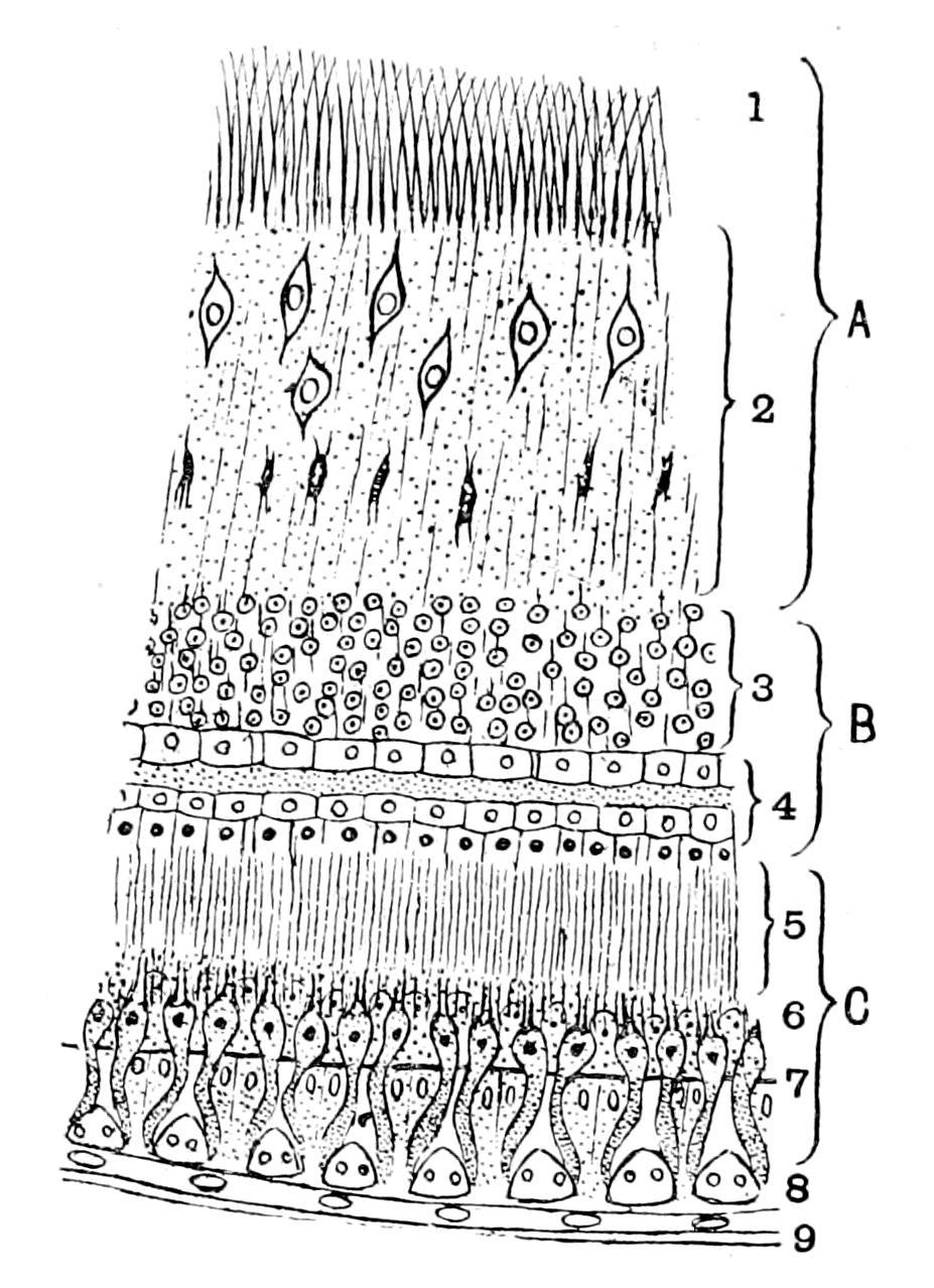
Fig. 42.—Ideal Diagram of the Layers in a Crustacean Eye.
The retina is divided into an epithelial part, C (the layer of retinular cells and rhabdomes), and a neurodermal or cerebral part, which is formed of, A, the ganglion of the optic nerve, and, B, the ganglion of the retina. 1, optic nerve fibres which cross at their entrance into the retina; 2, int. molecular layer with its two rows of cells; 3, int. nuclear layer; 4, Reichenbach's double row of large lightly-staining cells; 5, layer of terminal retinal fibres; 6, ext. nuclear layer; 7, ext. limiting membrane; 8, layer of crystalline cones; 9, cornea.
The comparison of this figure (Fig. 42) with that of the Petromyzon retina (Fig. 41) shows how great is the similarity of the latter with the arthropod type, and how the very points in which it deviates from the recognized vertebrate type are explainable by comparison with that of the arthropod. The most striking difference between the retinas in the two figures is that the layer of terminal nerve fibres (5, Fig. 42), which, after all, are only the elongated terminations of the retinal cells belonging to Parker's neurones of the first order, is very much longer than in Petromyzon or in any vertebrate, for the external molecular layer (6, Fig. 41) (Müller's layer of Nervenansätze) is very short and inconspicuous (in Fig. 41 it is drawn too thick).
Turning from the retina to the fibres of the optic nerve we again find a remarkable resemblance, for in Ammocœtes, as pointed out by Langerhans and carefully figured by Kohl, a crossing of the fibres of the optic nerve occurs as the nerve leaves the retina, just as is so universally the case in all compound retinas. To this crossing Kohl has given the name chiasma nervi optici, in distinction to the cerebral chiasma, which he calls chiasma nervorum opticorum. Further, we find that even this latter chiasma is well represented in the arthropod brain; thus Bellonci in Sphæroma, Berger, Dietl, and Krieger in Astacus, all describe a true optic chiasma, the only difference in opinion being, whether the crossing of the optic nerves is complete or not. Especially instructive are Bellonci's figures and description. He describes the brain of Sphæroma as composed of three segments—a superior segment, the cerebrum proper, a middle segment, and an inferior segment; the optic fibres, as is seen in Fig. 39, after crossing, pass direct into the middle segment, in the ganglia of which they terminate. From this segment also arises the nerve to the first antenna of that side—i.e. the olfactory nerve. The optic part, then, of this middle segment is clearly the brain portion of the optic ganglionic apparatus, and may be called the optic lobes, in contradistinction to the peripheral part, which is usually called the optic ganglion, and is composed of two ganglia, Op. g. I. and Op. g. II., as already mentioned. These optic lobes are therefore homologous with the optic lobes of the vertebrate brain.
The resemblance throughout is so striking as to force one to the conclusion that the retina of the vertebrate eye is a compound retina, composed of a retina and retinal ganglion of the type found in arthropods. From this it follows that the development of the vertebrate retina ought to show the formation of (1) an optic plate formed from the peripheral epidermis and not from the brain; (2) a part of the brain closely attached to this optic plate forming the retinal ganglion, which remains at the surface when the rest of the optic ganglion withdraws; (3) an optic nerve formed in consequence of this withdrawal, as the connection between the retinal and cerebral parts of the optic ganglion.
This appears to me exactly what the developmental process does show according to Götte's investigations. He asserts that the retina arises from an optic plate, being the optical portion of his 'Sinnes-platte.' At an early stage this is separated by a furrow (Furche) from the general mass of epidermal cells which ultimately form the brain. This separation then vanishes, and the retina and brain-mass become inextricably united into a mass of cells, which are still situated at the surface. By the closure of the cephalic plate and the withdrawal of the brain away from the surface, a retinal mass of cells is left at the surface connected with the tubular central nervous system by the hollow optic diverticulum or primary optic vesicle. If we regard only the retinal and nervous elements, and for the moment pay no attention to the existence of the tube, Götte's observation that the true retina has been formed from the optic plate (Sinnes-platte) to which the retinal portion of the brain (retinal ganglion) has become firmly fixed, and that then the optic nerve has been formed by the withdrawal of the rest of the brain (optic lobes), is word for word applicable to the description of the development of the compound retina of the arthropod eye, as has been already stated.
The Significance of the Optic Diverticula.
The origin of the retina from an optic epidermal plate in vertebrates, as in all other animals, brings the cephalic eyes of all animals into the same category, and leaves the vertebrate eye no longer in an isolated and unnatural position. In one point the retina of the vertebrate eye differs from that of a compound retina of an invertebrate; in the former, a striking supporting tissue exists, known as Müller's fibres, which is absent in the latter. This difference of structure is closely associated with another of the same character as in the central nervous system, viz. the apparent development of the nervous part from a tube. We see, in fact, that the retinal and nervous arrangements of the vertebrate eye are comparable with those of the arthropod eye, in precisely the same way and to the same extent as the nervous matter of the brain of the vertebrate is comparable with the brain of the arthropod. In both cases the nervous matter is, in structure, position, and function, absolutely homologous; in both cases there is found in the vertebrate something extra which is not found in the invertebrate—viz. a hollow tube, the walls of which, in the case of the brain, are utilized as supporting tissues for the nerve structures. The explanation of this difference in the case of the brain is the fundamental idea of my whole theory, namely, that the hollow tube is in reality the cephalic stomach of the invertebrate, around which the nervous brain-matter was originally grouped in precisely the same manner as in the invertebrate. What, then, are the optic diverticula?
"The formation of the eye," as taught by Balfour, "commences with the appearance of a pair of hollow outgrowths from the anterior cerebral vesicle. These outgrowths, known as the optic vesicles, at first open freely into the cavity of the anterior cerebral vesicle. From this they soon, however, become partially constricted, and form vesicles united to the base of the brain by comparatively narrow, hollow stalks, the rudiments of the optic nerves."
"After the establishment of the optic nerves, there takes place (1) the formation of the lens, and (2) the formation of the optic cup from the walls of the primary optic vesicle."
He then goes on to explain how the formation of the lens forms the optic cup with its double walls from the primary optic vesicle, and says—
"Of its double walls, the inner, or anterior, is formed from the front portion, the outer, or posterior, from the hind portion of the wall of the primary optic vesicle. The inner, or anterior, which very speedily becomes thicker than the other, is converted into the retina; in the outer, or posterior, which remains thin, pigment is eventually deposited, and it ultimately becomes the tesselated pigment-layer of the choroid."
The difficulties in connection with this view of the origin of the eye are exceedingly great, so great as to have caused Balfour to discuss seriously Lankester's suggestion that the eye must have been at one time within the brain, and that the ancestor of the vertebrate was therefore a transparent animal, so that light might get to the eye through the outer covering and the brain-mass; a suggestion, the unsatisfactory nature of which Balfour himself confessed. Is there really evidence of any part of either retina or optic nerve being formed from the epithelial lining of the tube?
This tube is formed as a direct continuation of the tube of the central nervous system, and we can therefore apply to it the same arguments as have been used in the discussion of the meaning of the latter tube. Now, the striking point in the latter case is the fact that the lining membrane of the central canal is in so many parts absolutely free from nervous matter, and so shows, as in the so-called choroid plexuses, its simple, non-nervous epithelial structure. This also we find in the optic diverticulum. Where there is no evidence of any invasion of the tube by nervous elements, there it retains its simple non-nervous character of a tube composed of a single layer of epithelial cells—viz. in that part of the tube which, as Balfour says, remains thin, in which pigment is eventually deposited, and which ultimately becomes the tesselated pigment-layer of the choroid. Nobody has ever suggested that this pigment-layer is nervous matter, or ever was, or ever will be, nervous matter; it is in precisely the same category as the membranous roof of the brain in Ammocœtes, which never was, and never will be, nervous matter. Yet, according to the old embryology both in the case of the eye and the brain, the pigment-layer and the so-called choroid plexuses are a part of the tubular nervous system.
Turning now to the optic nerve, Balfour describes it as derived from the hollow stalk of the optic vesicle. He says—
"At first the optic nerve is equally continuous with both walls of the optic cup, as must of necessity be the case, since the interval which primarily exists between the two walls is continuous with the cavity of the stalk. When the cavity within the optic nerve vanishes, and the fibres of the optic nerve appear, all connection is ruptured between the outer wall of the optic cup and the optic nerve, and the optic nerve simply perforates the outer wall, and becomes continuous with the inner one."
In this description Balfour, because he derived the optic nerve fibres from the epithelial wall of the optic stalk, of necessity supposed that such fibres originally supplied both the outer and inner walls of the optic cup and, therefore, seeing that when the fibres of the optic nerve appear they do not supply the outer wall, he supposes that their original connection with the outer wall is ruptured, because a discontinuity of the epithelial lining takes place coincidently with the appearance of the optic nerve-fibres, and, according to him, the optic nerve simply perforates the outer wall and becomes continuous with the inner one. This last statement is very difficult to understand. I presume he meant that some of the fibres of the optic nerve supplied from the beginning the inner wall of the optic cup, but that others which originally supplied the outer wall were first ruptured, then perforated the outer wall, and finally completed the supply to the inner wall or retina.
This statement of Balfour's is the necessary consequence of his belief, that the epithelial cells of the optic stalk gave rise to the fibres of the optic nerve. If, instead of this, we follow Kölliker and His, who state that the optic nerve-fibres are formed outside the epithelial walls of the optic stalk, and that the cells of the latter form supporting structures for the nerve-fibres, then the position of the optic nerve becomes perfectly simple and satisfactory without any rupturing of its connection with the outer wall and subsequent perforation, for the optic nerve-fibres from their very first appearance pass directly to supply the retina—i.e. the inner wall of the optic cup and nothing else.
They pass, as is well known, without any perforation by way of the choroidal slit to the inner surface of the inner wall (retina) of the optic cup; then, when the choroidal slit becomes closed by the expansion of the optic cup, the optic nerve naturally becomes situated in the centre of the base of the cup and spreads over its inner surface as that surface expands.
A section across the optic cup at an early stage at the junction of the optic stalk and optic cup would be represented by the upper diagram in Fig. 43; at a later stage, when the choroidal slit is closed, by the lower diagram.
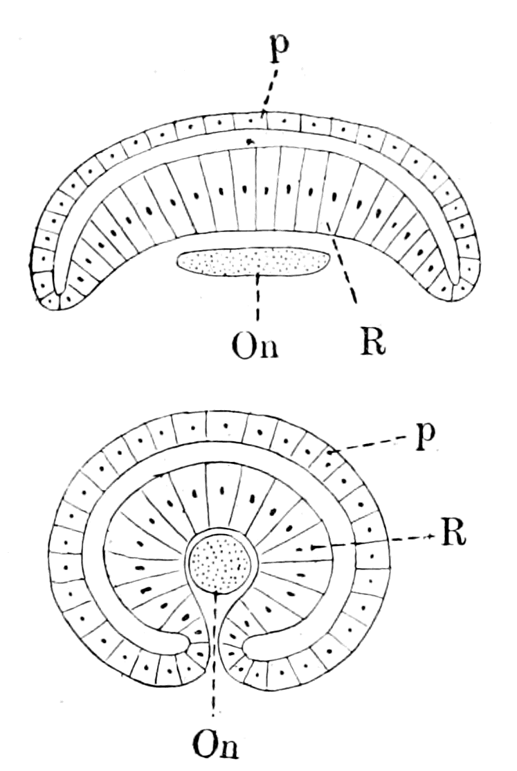
Fig. 43.—Diagram of the Relation of the Optic Nerve to the Optic Cup.
The upper diagram represents a stage before the formation of the choroidal slit, the lower one the stage of closure of the choroidal slit. R., retina; O.n., optic nerve; p., pigment epithelium.
The evident truth of this manner of looking at the origin of the optic nerve is demonstrated by the appearance of the optic nerve in Ammocœtes and Petromyzon. In the latter, although the development is complete, and the eye, and consequently also the optic nerve-fibres, are fully functional, there is still present in the axial core of the nerve a row of epithelial cells (Axenstrang) which are altered so as to form supporting structures, in the same way as a row of epithelial cells in the retina is altered to form the system of supporting cells known by the name of the Müllerian fibres.
The origin of this axial core of cells is perfectly clear, as has been pointed out by W. Müller. He says—
"The development of the optic nerve shows peculiarities in Petromyzon of such a character as to make this animal one of the most valuable objects for deciding the various controversial questions connected with the genesis of its elements. The lumen of the stalk of the primary optic vesicle is obliterated quite early by a proliferation of its lining epithelium. Also the original continuity of this epithelium with that of the pigment-layer is at an early period interrupted at the point of attachment of the optic stalk. This interruption occurs at the time when the fibres of the optic nerve first become visible."
Further on he says—
"The epithelium of the optic stalk develops entirely into supporting cells, which in Petromyzon fill up the original lumen and so form an axial core (Axenstrang) to the nerve-fibres which are formed entirely outside them; the projections of these supporting cells are directed towards the periphery, and so separate the bundles of the optic nerve-fibres. The mesodermal coat of the optic stalk takes no part in this separation; it simply forms the connective tissue sheath of the optic nerve. The development of the optic nerve in the higher vertebrates also obeys the same law, as I am bound to conclude from my own observations."
The evidence, then, of Ammocœtes is very conclusive. Originally a tube composed of a single layer of epithelial cells became expanded at the anterior end to form a bulb. On the outside of this tube or stalk the fibres of the optic nerve make their appearance, arising from the ganglion-cell layer of the retina, and, passing over the surface of the epithelial tube at the choroidal fissure, proceed to the brain by way of the optic chiasma. Owing to the large number of fibres, their crossing at the junction of the stalk with the bulb, and the narrowness at this neck, the obliteration of the lumen of the tube which takes place in the stalk is carried out to a still greater extent at this narrow part. The result of this is that all continuity of the cell-layers of the original tube of the optic stalk with those of both the inner and outer walls of the bulb is interrupted, and all that remains in this spot of the original continuous line of cells which connected the tube of the stalk with that of the bulb are possibly some of the groups of cells which are found scattered among the fibres of the optic nerve at their entrance into the retina. Such separation of the originally continuous elements of the epithelial wall of the optic stalk, which is apparent only at this neck of the nerve in Petromyzon, takes place along the whole of the optic nerve in the higher vertebrates, so that no continuous axial core of cells exist, but only scattered supporting cells.
If further proof in support of this view be wanted, it is given by the evidence of physiology, which shows that the fibres of the optic nerve are not different from other nerve-fibres of the central nervous system, but that they degenerate when separated from their nerve-cell, and that the nerve-cell of which the optic nerve-fibre is a process is the large ganglion-cell of the ganglionic layer of the retina. The origin of the ganglionic layer of the retina cannot therefore be separated from that of the optic nerve-fibres. If the one is outside the epithelial tube, so is the other, and what holds true of the ganglionic layer must hold good of the rest of the retinal ganglion and, from all that has been said, of the retina itself. We therefore come to the conclusion that the evidence is distinctly in favour of the view, that the retina and optic nerve in the true sense are structures which originally were outside a non-nervous tube, but, just like the central nervous system as a whole, have amalgamated so closely with the elements of this tube as to utilize them for supporting structures. One part of this non-nervous tube, its dorsal wall, like the corresponding part of the brain-tube, still retains its original character, and by the deposition of pigment has been pressed into the service of the eye to form the pigmented epithelial layer.
We can, however, go further than this, for we know definitely in the case of the retina what the fate of the epithelial cells lining this tube has been. They have become the system of supporting structures known as Müllerian fibres.
The epithelial layer of the primary optic vesicle can be traced into direct continuity with the lining epithelium of the brain cavity, as a single layer of epithelial cells in the core of the optic nerve, forming the optic stalk, which, in consequence of close contact, becomes the well-known axial layer of supporting cells. This epithelial layer of the optic stalk then expands to form the optic bulb, the outer or dorsal wall of which still remains as a single layer of epithelium and becomes the layer of pigment epithelium. This layer of epithelium becomes doubled on itself by the approximation of the inner or ventral wall of the optic cup to the outer or dorsal wall in consequence of the presence of the lens, and still remaining a single layer, forms the pars ciliaris retinæ; then suddenly, at the ora serrata, the single epithelial layer vanishes, and the layers of the retina take its place. It has long been known, however, that even throughout the retina this single epithelial layer still continues, being known as the fibres of Müller. This is how the fact is described in the last edition of Foster's "Text-book of Physiology," p. 1308—
"Stretching radially from the inner to the outer limiting membrane in all regions of the retina are certain peculiar-shaped bodies known as the radial fibres of Müller. Each fibre is the outcome of the changes undergone by what was at first a simple columnar epithelial cell. The changes are, in the main, that the columnar form is elongated into that of a more or less prismatic fibre, the edges of which become variously branched, and that while the nucleus is retained the cell substance becomes converted into neuro-keratin. And, indeed, at the ora serrata the fibres of Müller may be seen suddenly to lose their peculiar features and to pass into the ordinary columnar cells which form the pars ciliaris retinæ."
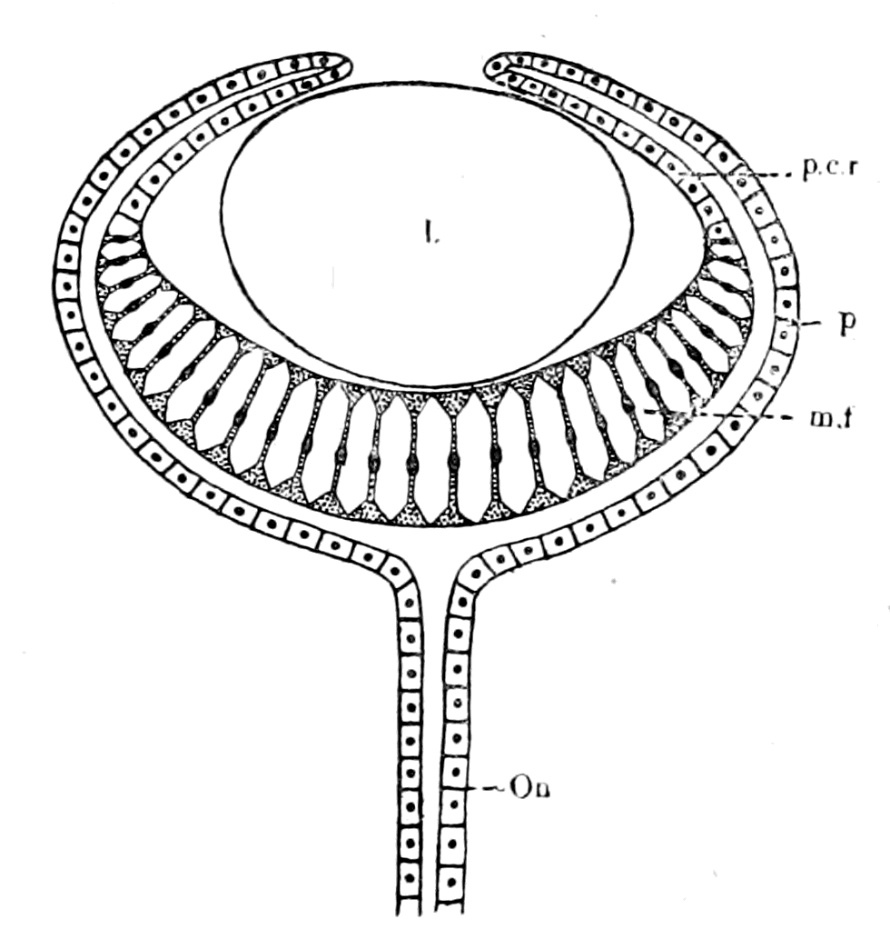
Fig. 44.—Diagram representing the Single-layered Epithelial Tube of the Vertebrate Eye after Removal of the Nervous and Retinal Elements.
O.n., axial core of cells in optic nerve; p., pigment epithelium; p.c.r., pars ciliaris retinæ; m.f., Müllerian fibres; l., lens.
It is then absolutely clear that the essential parts of the eye may be considered as composed of two parts—
1. A tube or diverticulum from the tube of the central nervous system, composed throughout of a single layer of epithelium, which forms the supporting axial cells in the optic nerve, the pigment epithelium and the Müllerian fibres of the retina. Such a tube would be represented by the accompanying Fig. 44, and the left side of Fig. 41.
2. The retina proper with the retinal ganglion and the optic nerve-fibres as already described. In this part supporting elements are found, just as in any other compound retina, of the nature of neuroglia, which are independent of the Müllerian fibres.
Of these two parts we have already seen that the second is to all intents and purposes a compound retina of a crustacean eye, and seeing that the single-layered epithelial tube is continuous with the single-layered epithelial tube of the central nervous system—i.e. with the cephalic part of the gut of the arthropod ancestor—it follows with certainty that the ancestor of the vertebrates must have possessed two anterior diverticula of the gut, with the wall of which, near the anterior extremity, the compound retina has amalgamated on either side, just as the infra-œsophageal ganglia have amalgamated with the ventral wall of the main gut-tube. In this way, and in this way alone, does the interpretation of the structure of the vertebrate lateral eye harmonize in the most perfect manner with the rest of the conclusions already arrived at.
The question therefore arises:—Have we any grounds for believing that the ancient forms of primitive crustaceans and primitive arachnids, which were so abundant in the time when the Cephalaspids appeared, possessed two anterior diverticula of the stomach, such as the consideration of the vertebrate eye strongly indicates must have been the case?
The beautiful pictures of Blanchard, and his description, show how, on the arachnid side, paired diverticula of the stomach are nearly universal in the group. Thus, although they are not present in the scorpions, still, in the Thelyphonidæ, Phrynidæ, Solpugidæ, Mygalidæ, the most marked characteristic of the stomach-region is the presence of four pairs of cœcal diverticula, which spread laterally over the prosomatic region. In the spiders the number of such diverticula increases, and the whole prosomatic region becomes filled up with these tubes. Blanchard considers that they form nutrient tubes for the direct nutrition of the organs in the prosoma, especially the important brain-region of the central nervous system. He points out that these animals are blood-suckers, and that, therefore, their food is already in a suitable form for purposes of nutrition when it is taken in by them, so that, as it were, the anterior part of the gut is transformed into a series of vessels or diverticula conveying blood directly to the important organs in the prosoma, by means of which they obtain nourishment in addition to their own blood-supply.
The universality of such diverticula among the arachnids makes it highly probable that their progenitors did possess an alimentary canal with one or more pairs of anterior diverticula. In the vertebrate, however, the paired diverticula are associated with a compound retina, a combination which does not occur among living arachnids; we must, therefore, examine the crustacean group for the desired combination, and naturally the most likely group to examine is the Phyllopoda, especially such primitive forms as Branchipus and Artemia, for it is universally acknowledged that these forms are the nearest living representatives of the trilobites. If, therefore, it be found that the retina and optic nerve in Artemia is in specially close connection with an anterior diverticulum of the gut on each side, then it is almost certain that such a combination existed also in the trilobites.
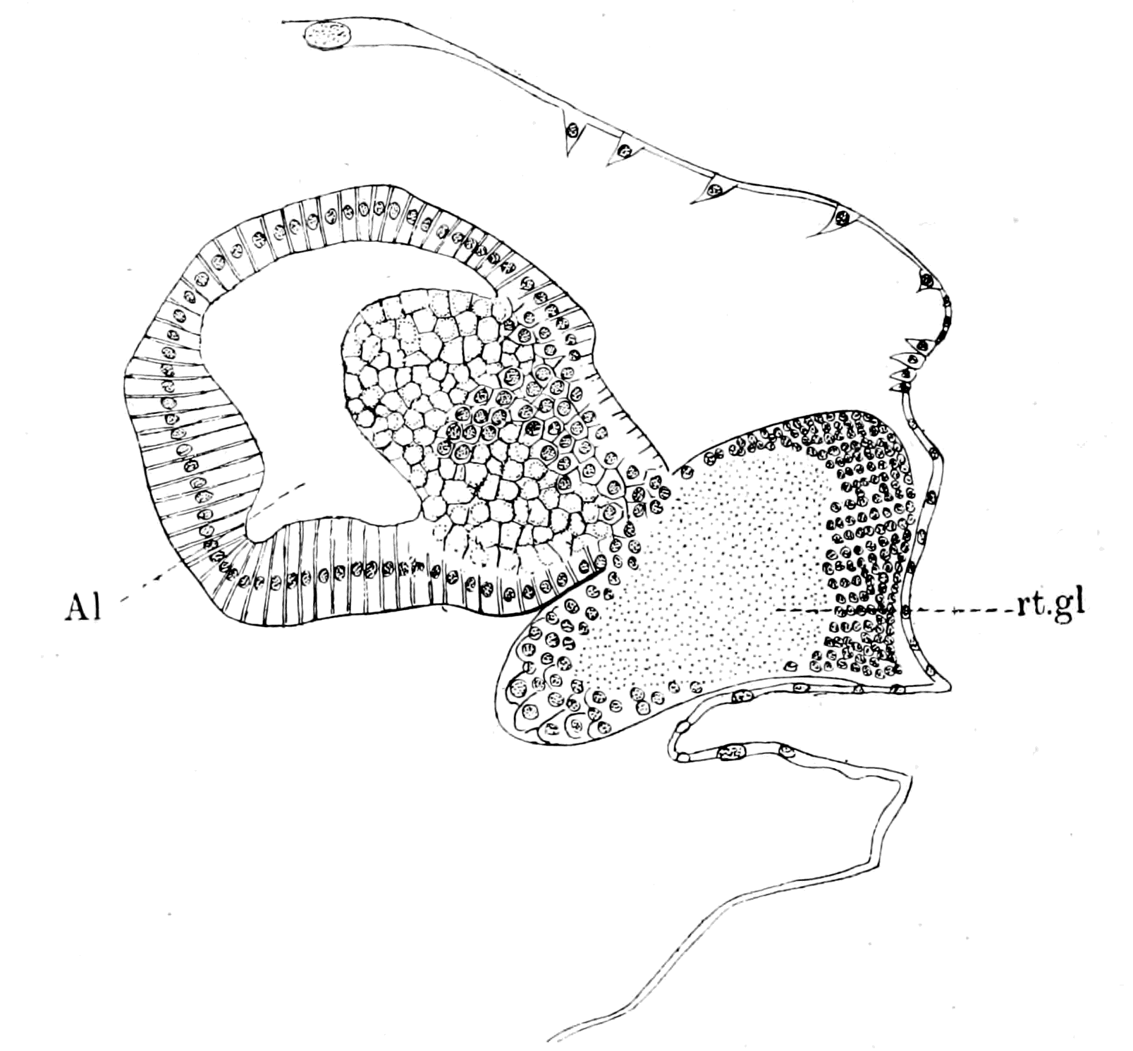
Fig. 45.—Section through one of the two Anterior Diverticula of the Gut in Artemia and the Retinal Ganglion.
The section is through the extreme anterior end of the diverticulum, thus cutting through many of the columnar cells at right angles to their axis. Al., gut diverticulum; rt. gl., retinal ganglion.
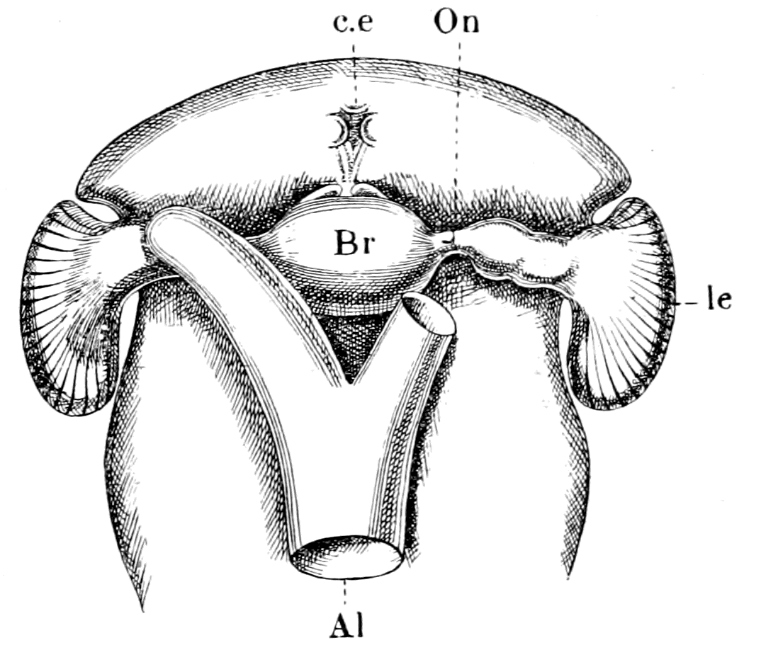
Fig. 46.—The Brain, Eyes, and Anterior Termination of the Alimentary Canal of Artemia, viewed from the Dorsal Aspect.
Br., brain; l.e., lateral eyes; c.e., median eyes; Al., alimentary canal.
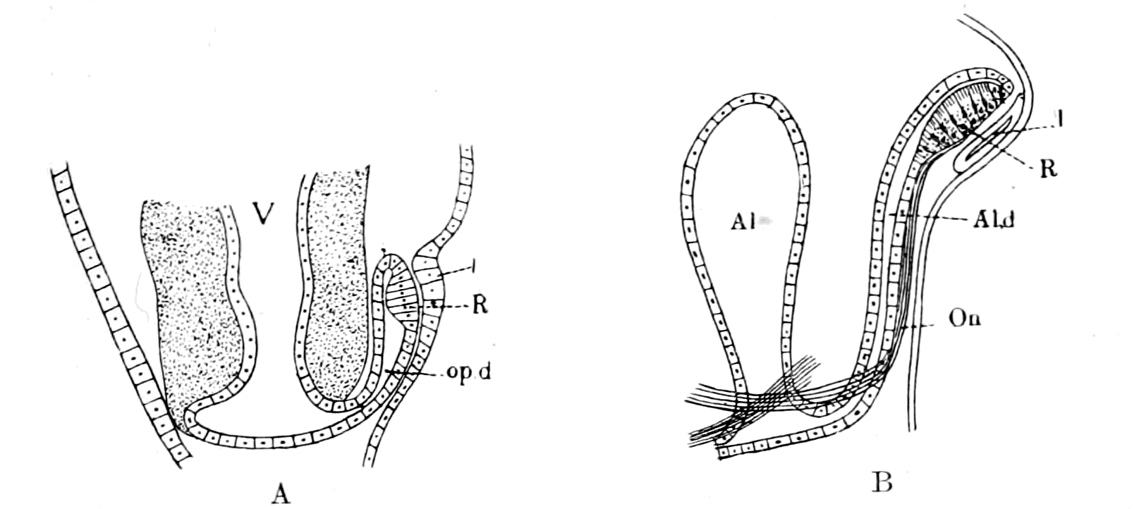
Fig. 47.—A, The Formation of the Retina of the Eye of Ammocœtes (after Scott); B, The Formation of the Retina of the Eye of Ammocœtes, on my theory.
R., retina; l., lens; O.n., optic nerve fibres; Al., cephalic end of invertebrate alimentary canal; V., cavity of ventricles of brain; Al.d., anterior diverticulum of alimentary canal; op.d., optic diverticulum.
My friend Mr. W. B. Hardy has especially investigated the nervous system of Artemia. In the course of his work he cut serial sections through the whole animal, and, as mentioned in my paper in the Journal of Anatomy and Physiology, he discovered that the retinal ganglion of each lateral eye is so closely attached to the end of the corresponding diverticulum of the gut that the lining cells of the ventral part of the diverticulum form a lining to the retinal ganglion (Fig. 45). In this animal there are only two gut-diverticula, which are situated most anteriorly. I have plotted out this series of sections by means of a camera lucida, with the result that the retina appears as a bulging attached ventro-laterally to the extremity of each gut-diverticulum, as is shown in Fig. 46. It is instructive to compare with this figure Scott's picture of the developing eye in Ammocœtes, where he figures the retina as a bulging attached ventrally to the extremity of the narrow tube of the optic diverticulum. In Fig. 47, A, I reproduce this figure of Scott, and by the side of it, Fig. 47, B, I have represented the origin of the vertebrate eye as I believe it to have occurred.
We see, then, this very striking fact, that in the most primitive of the Crustacea, not only are there two anterior diverticula of the gut, but also the retinal ganglion of the lateral eye is in specially close connection with the end of the diverticulum on each side. In fact, we find in the nearest living representative of the trilobites a retina and retinal ganglion and optic nerve, closely resembling that of the vertebrate, in close connection with an epithelial tube which has nothing to do with the organ of sight, but is one of a pair of anterior gut-diverticula. It is impossible to obtain more decisive evidence that the trilobites possessed a pair of gut-diverticula surrounded to a greater or less extent by the retina and optic nerve of each lateral eye.
Such anterior diverticula are commonly found in the lower Crustacea; they are usually known by the name of liver-diverticula, but as they take no part in digestion, and, on the contrary, represent that part of the gut which is most active in absorption, the term liver is not appropriate, and it is therefore better to call them simply the pair of anterior diverticula. Our knowledge of their function in Daphnia is given in a paper by Hardy and M‘Dougall, which does not appear to be widely known. Hardy succeeded in feeding Daphnia with yolk of egg in which carmine grains were mixed, and was able in the living animal to watch the whole process of deglutition, digestion, and absorption. The food, which is made into a bolus, is moved down to the middle region of the gut, and there digestion takes place. Then by an antiperistaltic movement the more fluid products of the digestion-process are sent right forward into the two anterior diverticula, where the single layer of columnar cells lining these diverticula absorbs these products, the cells becoming thickly studded with fat-drops after a feed of yolk of egg. The carmine particles, which were driven forward with the proteid- and fat-particles, are not absorbed, but are at intervals driven back by contractions of the anterior diverticula to the middle region of the gut.
These observations prove most clearly that the anterior diverticula have a special nutrient function, being the main channels by which new nutrient material is brought into the body, and, as pointed out by the authors, it is a remarkable exception in the animal kingdom that absorption should occur in that portion of the gut which is anterior to the part in which digestion occurs. In all these animals the two anterior diverticula extend forwards over the brain, and, as we have seen in Artemia, the anterior extremity of each one is so intimately related to a part of the brain—viz. the retinal ganglion—as to form a lining membrane to that mass of nerve-cells. It follows, therefore, that the nutrient fluid absorbed by the cells of this part of the gut-diverticulum must be primarily for the service of the retinal ganglion. In fact, the relations of this anterior portion of the gut to the brain as a whole suggest strongly that the marked absorptive function of this anterior portion of the gut exists in order to supply nutrient material in the first place to the most vital, most important organ in the animal—the brain and its sense-organs. This conclusion is borne out by the fact that in these lower crustaceans the circulation of blood is of a very inefficient character, so that the tissues are mainly dependent for their nutrition on the fluid immediately surrounding them. It stands to reason that the establishment of the anterior portion of the gut as a nutrient tube to the brain would necessitate a closer and closer application of the brain to that tube, so that the process of amalgamation of the brain with the single layer of columnar epithelial cells which constitutes the wall of the gut (which we see in its initial stage in the retinal ganglion of Artemia), would tend rapidly to increase as more and more demands were made upon the brain, until at last both the supra- and infra-œsophageal ganglia, as well as the retinal ganglia and optic nerves, were in such close intimate connection with the ventral wall of the anterior portion of the gut and its diverticula as to form a brain and retina closely resembling that of Ammocœtes.
Such an origin for the lateral eyes of the vertebrate explains in a simple and satisfactory manner why the vertebrate retina is a compound retina, and why both retina and optic nerve have an apparent tubular development.
At the same time one discrepancy still exists which requires consideration—viz. in no arthropod eye possessing a compound retina is the retina inverted. All the known cases of inversion among arthropods occur in eyes, the retina of which is simple, and are all natural consequences of the process of invagination by which the retina is formed. On the other hand, eyes with an inverted compound retina are not entirely unknown among invertebrates, for the eyes of Pecten and of Spondylus possess a retina which is inverted after the vertebrate fashion and still may be spoken of as compound rather than simple. It is clear that an invagination, the effect of which is an inversion of the retinal layer, would lead to the same result, whether the retinal optic nerves were short or long, whether, in fact, a retinal ganglion existed or not. Undoubtedly the presence of the retinal ganglion tends greatly to obscure any process of invagination, so that, as already mentioned, many observers, with Parker, consider the retina of the crustacean lateral eye to be formed by a thickening only, without any invagination, while Reichenbach says an obscure invagination does take place at a very early stage. So in the vertebrate eye most observers speak only of a thickening to form the retina, but Götte's observation points to an invagination of the optic plate at an early stage. So also in the eye of Pecten, Korschelt and Heider consider that the thickening, by which the retina is formed according to Patten, in reality hides an invagination process by means of which, as Bütschli suggests, an optic vesicle is formed in the usual manner. The retina is formed from the anterior wall of this vesicle, and is therefore inverted.
The origin of the inverted retina of the vertebrate eye does not seem to me to present any great difficulty, especially when one takes into consideration the fact that the retina is inverted in the arachnid group, only in the lateral eyes. The inversion is usually regarded as associated with the tubular formation of the vertebrate retina, and it is possible to suppose that the retina became inverted in consequence of the involvement of the eye with the gut-diverticulum. I do not myself think any such explanation is at all probable, because I cannot conceive such a process taking place without a temporary derangement—to say the least of it—of the power of vision, and as I do not believe that evolution was brought about by sudden, startling changes, but by gradual, orderly adaptations, and as I also believe in the paramount importance of the organs of vision for the evolution of all the higher types of the animal kingdom, I must believe that in the evolution from the Arthropod to the Cephalaspid, the lateral eyes remained throughout functional. I therefore, for my own part, would say that the inversion of the retina took place before the complete amalgamation with the gut-diverticulum, that, in fact, among the proto-crustacean, proto-arachnid forms there were some sufficiently arachnid to have an inverted retina, and at the same time sufficiently crustacean to possess a compound retina, and therefore a compound inverted retina after the vertebrate fashion existed in combination with the anterior gut-diverticula. Thus, when the eye and optic nerve sank into and amalgamated with the gut-diverticulum, neither the dioptric apparatus nor the nervous arrangements would suffer any alteration, and the animal throughout the whole process would possess organs of vision as good as before or after the period of transition.
Further, not only the retina but also the dioptric apparatus of the vertebrate eye point to its origin from a type that combined the peculiarities of the arachnids and the crustaceans. In the former it is difficult to speak of a true lens, the function of a lens being undertaken by the cuticular surface of the cells of the corneagen (Mark's 'lentigen'), while in the latter, in addition to the corneal covering, a true lens exists in the shape of the crystalline cones. Further, these crustacean lenses are true lenses in the vertebrate sense, in that they are formed by modified hypodermal cells, and not bulgings of the cuticle, as in the arachnid. We see, in fact, that in the compound crustacean eye an extra layer of hypodermal cells has become inserted between the cornea and the retina to form a lens. So also in the vertebrate eye the lens is formed by an extra layer of the epidermal cells between the cornea and the retina. The fact that the vertebrate eye possesses a single lens, though its retina is composed of a number of ommatidia, while the crustacean eye possesses a lens to each ommatidium, may well be a consequence of the inversion of the vertebrate retina. It is most probable, as Korschelt and Heider have pointed out, that the retina of the arachnid eyes is composed of a number of ommatidia, just as in the crustacean eyes and in the inverted eyes it is probable that the image is focussed on to the pigmented tapetal layer, and thence reflected on to the percipient visual rods. In such a method of vision a single lens is a necessity, and so it must also be if, as I suppose, eyes existed with an inverted compound retina. Owing to the crustacean affinities of such eyes, a lens would be formed and the retina would be compound: owing to the arachnid affinities, the retina would be inverted and the hypodermal cells which formed the lens would be massed together to form a single lens, instead of being collected in groups of four to form a series of crystalline cones.
To sum up: The study of the vertebrate eyes, both median and lateral, leads to most important conclusions as to the origin of the vertebrates, for it shows clearly that whereas, as pointed out in this and subsequent chapters, their ancestors possessed distinct arachnid characteristics, yet that they cannot have been specialized arachnids, such as our present-day forms, but rather they were of a primitive arachnid type, with distinct crustacean characteristics: animals that were both crustacean and arachnid, but not yet specialized in either direction: animals, in fact, of precisely the kind which swarmed in the seas at the time when the vertebrates first made their appearance. In the opinion of the present day, the ancestral forms of the Crustacea, which were directly derived from the Annelida, may be classed as an hypothetical group the Protostraca, the nearest approach to which is a primitive Phyllopod.
"Starting from the Protostraca," say Korschelt and Heider, "according to the present condition of our knowledge, we may, as has been already remarked, assume three great series of development of the Arthropodan stock, by the side of which a number of smaller independent branches have been retained. One of these series leads through the hypothetical primitive Phyllopod to the Crustacea; the second through the Palæostraca (Trilobita, Gigantostraca, Xiphosura) to the Arachnida; the third through forms resembling Peripatus to the Myriapoda and the Insecta. The Pantapoda and the Tardigrada must probably be regarded as smaller independent branches of the Arthropodan stock."
To these "three great series of development of the Arthropodan stock" the evidence of Ammocœtes shows that a fourth must be added, which, starting also from the Protostraca, and closely connected with the second, palæostracan branch, leads through the Cephalaspidæ to the great kingdom of the Vertebrata. Such a direct linking of the earliest vertebrates with the Annelida through the Protostraca is of the utmost importance, as will be shown later in the explanation of the origin of the vertebrate cœlom and urinary apparatus.
Summary.
The most important discovery of recent years which gives a direct clue to the ancestry of the vertebrates is undoubtedly the discovery that the pineal gland is all that remains of a pair of median eyes which must have been functional in the immediate ancestor of the vertebrate, seeing how perfect one of them still is in Ammocœtes. The vertebrate ancestor, then, possessed two pairs of eyes, one pair situated laterally, the other median. In striking confirmation of the origin of the vertebrate from Palæostracans it is universally admitted that all the Eurypterids and such-like forms resembled Limulus in the possession of a pair of median eyes, as well as of a pair of lateral eyes. Moreover, the ancient mailed fishes the Ostracodermata, which are the earliest fishes known, are all said to show the presence of a pair of median eyes as well as of a pair of lateral eyes. This evidence directly suggests that the structure of both the median and lateral vertebrate eyes ought to be very similar to that of the median and lateral arthropod eyes. Such is, indeed, found to be the case.
The retina of the simplest form of eye is formed from a group of the superficial epidermal cells, and the rods or rhabdites are formed from the cuticular covering of these cells; the optic nerve passes from these cells to the deeper-lying brain. This kind of retina may be called a simple retina, and characterizes the eyes, both median and lateral, of the scorpion group.
In other cases a portion of the optic ganglion remains at the surface, when the brain sinks inwards, in close contiguity to the epidermal sense-cells which form the retina; a tract of fibres connects this optic ganglion with the underlying brain, and is known as the optic nerve. Such a retina may be called a compound retina and characterizes the lateral eyes of both crustaceans and vertebrates. Also, owing to the method of formation of the retina by invagination, the cuticular surface of the retinal sense-cells, from which the rods are formed, may be directed towards the source of light or away from it. In the first case the retina may be called upright, in the second inverted.
Such inverted retinas are found in the vertebrate lateral eyes and in the lateral eyes of the arachnids, but not of the crustaceans.
The evidence shows that all the invertebrate median eyes possess a simple upright retina, and in structure are remarkably like the right median or pineal eye of Ammocœtes; while the lateral eyes possess, as in the crustaceans, an upright compound retina, or, as in many of the arachnids, a simple inverted retina. The lateral eyes of the vertebrates alone possess a compound inverted retina.
This retina, however, is extraordinarily similar in its structure to the compound crustacean retina, and these similarities are more accentuated in the retina of the lateral eye of Petromyzon than that of the higher vertebrates.
The evidence afforded by the lateral eye of the vertebrate points unmistakably to the conclusion that the ancestor of the vertebrate possessed both crustacean and arachnid characters—belonged, therefore, to a group of animals which gave rise to both the crustacean and arachnid groups. This is precisely the position of the Palæostracan group, which is regarded as the ancestor of both the crustaceans and arachnids. In two respects the retina of the lateral eyes of vertebrates differs from that of all arthropods, for it possesses a special supporting structure, the Müllerian fibres, which do not exist in the latter, and it is developed in connection with a tube, the optic diverticulum, which is connected on each side with the main tube of the central nervous system. These two differences are in reality one and the same, for the Müllerian fibres are the altered lining cells of the optic diverticulum, and this tube has the same significance as the rest of the tube of the nervous system; it is something which has nothing to do with the nervous portion of the retina but has become closely amalgamated with it. The explanation is, word for word, the same as for the tubular nervous system, and shows that the ancestor of the vertebrate possessed two anterior diverticula of its alimentary canal which were in close relationship to the optic ganglion and nerve of the lateral eye on each side. It is again a striking coincidence to find that Artemia, which with Branchipus represents a group of living crustaceans most nearly allied to the trilobites, does possess two anterior diverticula of the gut which are in extraordinarily close relationship with the optic ganglia of the retina of the lateral eyes on each side.
The evidence of the optic apparatus of the vertebrate points most remarkably to the derivation of the Vertebrata from the Palæostraca.
CHAPTER III
THE EVIDENCE OF THE SKELETON
The bony and cartilaginous skeleton considered, not the notochord.—Nature of the earliest cartilaginous skeleton.—The mesosomatic skeleton of Ammocœtes; its topographical arrangement, its structure, its origin in muco-cartilage.—The prosomatic skeleton of Ammocœtes; the trabeculæ and parachordals, their structure, their origin in white fibrous tissue.—The mesosomatic skeleton of Limulus compared with that of Ammocœtes; similarity of position, of structure, of origin in muco-cartilage.—The prosomatic skeleton of Limulus; the entosternite or plastron compared with the trabeculæ of Ammocœtes; similarity of position, of structure, of origin in fibrous tissue.—Summary.
The explanation of the two optic diverticula given in the last chapter accounts in the same harmonious manner for every other part of the tube around which the central nervous system of the vertebrate has been grouped. The tube conforms in all respects to the simple epithelial tube which formed the alimentary canal of the ancient type of marine arthropods such as were dominant in the seas when the vertebrates first appeared. The whole evidence so far is so uniform and points so strongly in the direction of the origin of vertebrates from these ancient arthropods, as to make it an imperative duty to proceed further and to compare one by one the other parts of the central nervous system, together with their outgoing nerves in the two groups of animals.
Before proceeding to do this, it is advisable first to consider the question of the origin of the vertebrate skeletal tissues, for this is the second of the great difficulties in the way of deriving vertebrates from arthropods, the one skeleton being an endo-skeleton composed of cartilage and bone, and the other an exo-skeleton composed of chitin. Here is a problem of a totally different kind to that we have just been considering, but of so fundamental a character that it must, if possible, be solved before passing on to the consideration of the cranial nerves and the organs they supply.
Is there any evidence which makes it possible to conceive the method by which the vertebrate skeleton may have arisen from the skeletal tissues of an arthropod? By the vertebrate skeleton I mean the bony and cartilaginous structures which form the backbone, the cranio-facial skeleton, the pectoral and pelvic girdles, and the bones of the limbs. I do not include the notochord in these skeletal tissues, because there is not the slightest evidence that the notochord played any part in the formation of these structures; the notochordal tissue is something sui generis, and never gives rise to cartilage or bone. The notochord happens to lie in the middle line of the body and is very conspicuous in the lowest vertebrate; with the development of the backbone the notochord becomes obliterated more and more, until at last it is visible in the higher vertebrates only in the embryo; but that obliteration is the result of the encroachment of the growing bone-masses, not the cause of their growth. Although, then, the notochord may in a sense be spoken of as the original supporting axial rod of the vertebrate, it is so different to the rest of the endo-skeleton, has so little to do with it, that the consideration of its origin is a thing apart, and must be treated by itself without reference to the origin of the cartilaginous and bony skeleton.
The Commencement of the Bony Skeleton in the Vertebrate.
What is the teaching of the vertebrate? What evidence is there as to the origin of the bony skeleton in the vertebrate phylum itself?
The axial bony skeleton of the higher Mammalia consists of two parts, (1) the vertebral column with its attached bony parts, and (2) the cranio-facial skeleton. Of these two parts, the bony tissue of the first arises in the embryo from cartilage, of the second partly from cartilage, partly from membrane.
In strict accordance with their embryonic origin is their phylogenetic origin: as we pass from the higher vertebrates to the lower these structures can be traced back to a cartilaginous and membranous condition, so that, as Parker has shown, the cranio-facial bony skeleton of the higher vertebrates can be derived directly from a non-bony cartilaginous skeleton, such as is seen in Petromyzon and the cartilaginous fishes.
Balfour, in his "Comparative Embryology," states that the primitive cartilaginous cranium is always composed of the following parts:—
1. A pair of cartilaginous plates on each side of the cephalic section of the notochord known as the parachordals (pa.ch., Fig. 49; iv., Fig. 48). These plates, together with the notochord (ch.) enclosed between them, form a floor for the hind and mid-brain.
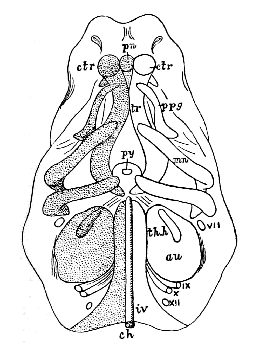
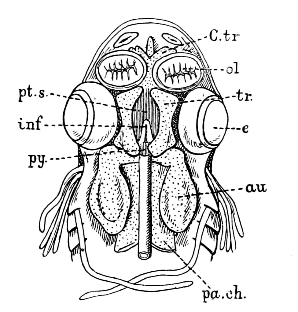
Fig. 48.—Embryo Pig, two-thirds of an inch long (from Parker), Elements of Skull seen from below. ch., notochord; iv., parachordals; au., auditory capsule; py., pituitary body; tr., trabecula; ctr., trabecular cornu; pn., pre-nasal cartilage; ppg., palato-pterygoid tract; mn., mandibular arch; th.h., first branchial arch; VII.-XII., cranial nerves. Fig. 49.—Head of Embryo Dog-fish (from Parker), Basal View of Cranium from above. ol., olfactory sacs; au., auditory capsule; py., pituitary body; pa.ch., parachordal cartilage; tr., trabecula; inf., infundibulum; pt.s., pituitary space; e., eye.
2. A pair of bars forming the floor for the fore-brain, known as the trabeculæ (tr). These bars are continued forward from the parachordals. They meet posteriorly and embrace the front end of the notochord, and after separating for some distance bend in again in such a way as to enclose a space—the pituitary space (pt.s.). In front of this space they remain in contact, and generally unite. They extend forward into the nasal region (pn.).
3. The cartilaginous capsules of the sense organs. Of these the auditory (au.) and the olfactory capsules (ol.) unite more or less intimately with the cranial walls; while the optic capsules, forming the usually cartilaginous sclerotics, remain distinct.
The parachordals and notochord form together the basilar plate, which forms the floor for that section of the brain belonging to the primitive postoral part of the head, and its extent corresponds roughly to that of the basioccipital of the adult skull.
The trabeculæ, so far as their mere anatomical relations are concerned, play the same part in forming the floor for the front cerebral vesicle as do the parachordals for the mid- and hind-brain. They differ, however, from the parachordals in one important feature, viz. that except at their hinder end they do not embrace the notochord. The notochord always terminates at the infundibulum, and the trabeculæ always enclose a pituitary space, in which lies the infundibulum (inf.) and the pituitary body (py.).
In the majority of the lower forms the trabeculæ arise quite independently of the parachordals, though the two sets of elements soon unite.
The trabeculæ are usually somewhat lyre-shaped, meeting in front and behind, and leaving a large pituitary space between their middle parts. Into this space the whole base of the fore-brain primitively projects, but the space itself gradually becomes narrowed until it usually contains only the pituitary body.
The trabecular floor of the brain does not long remain simple. Its sides grow vertically upwards, forming a lateral wall for the brain, in which in the higher types, two regions may be distinguished, viz. an alisphenoidal region behind, growing out from what is known as the basisphenoidal region of the primitive trabeculæ, and an orbito-sphenoidal region in front, growing out from the presphenoidal region of the trabeculæ. These plates form at first a continuous lateral wall of the cranium. The cartilaginous walls which grow up from the trabecular floor of the cranium generally extend upwards so as to form a roof, though almost always an imperfect roof, for the cranial cavity.
The basi-cranial cartilaginous skeleton reduces itself always into trabeculæ and parachordals with olfactory and auditory cartilaginous capsules.
In addition, a branchial skeleton exists, which consists of a series of bars known as the branchial bars, so situated as to afford support to the successive branchial pouches. An anterior arch known as the mandibular arch (Fig. 50, Mn.), placed in front of the hyo-mandibular cleft, and a second arch, known as the hyoid arch (Hy.), placed in front of the hyo-branchial cleft, are developed in all types; the succeeding arches are known as the true branchial arches (Br.), and are only fully developed in the Ichthyopsida. In all cases of jaw-bearing (gnathostomatous) vertebrates the first arch has become a supporting skeleton for the mouth (Fig. 51), and in the higher vertebrates in combination with the second or hyoid arch takes part in the formation of the ear-bones.
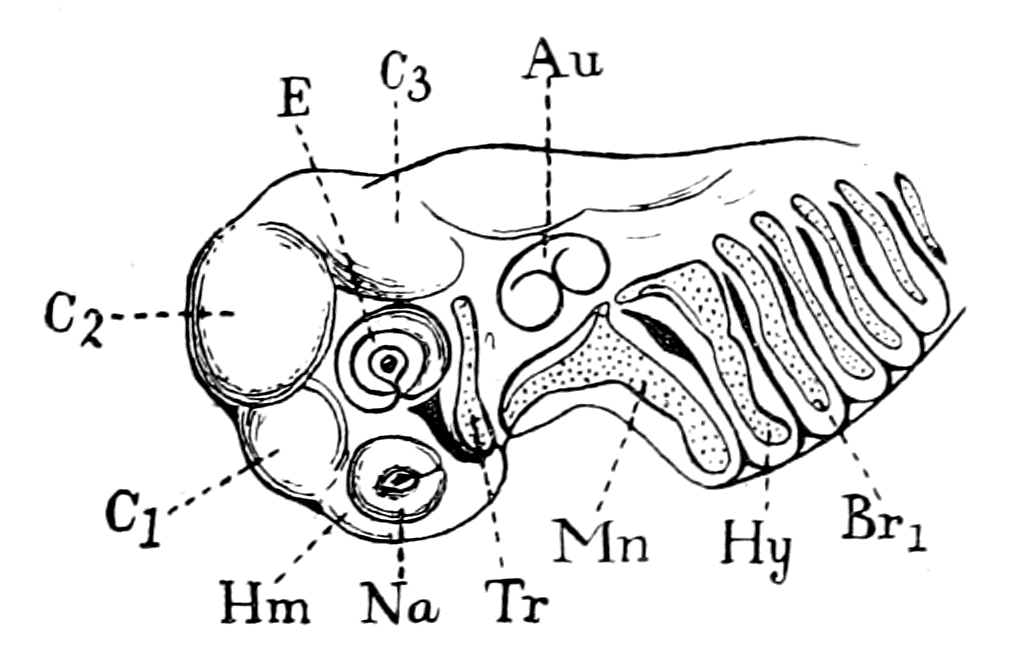
Fig. 50.—Head of Embryo Dog-fish, eleven lines long. (From Parker.)
Tr., trabecula; Mn., mandibular cartilage; Hy., hyoid arch; Br1., first branchial arch; Na., olfactory sac; E., eye; Au., auditory capsule; Hm., hemisphere; C1, C2, C3, cerebral vesicles.
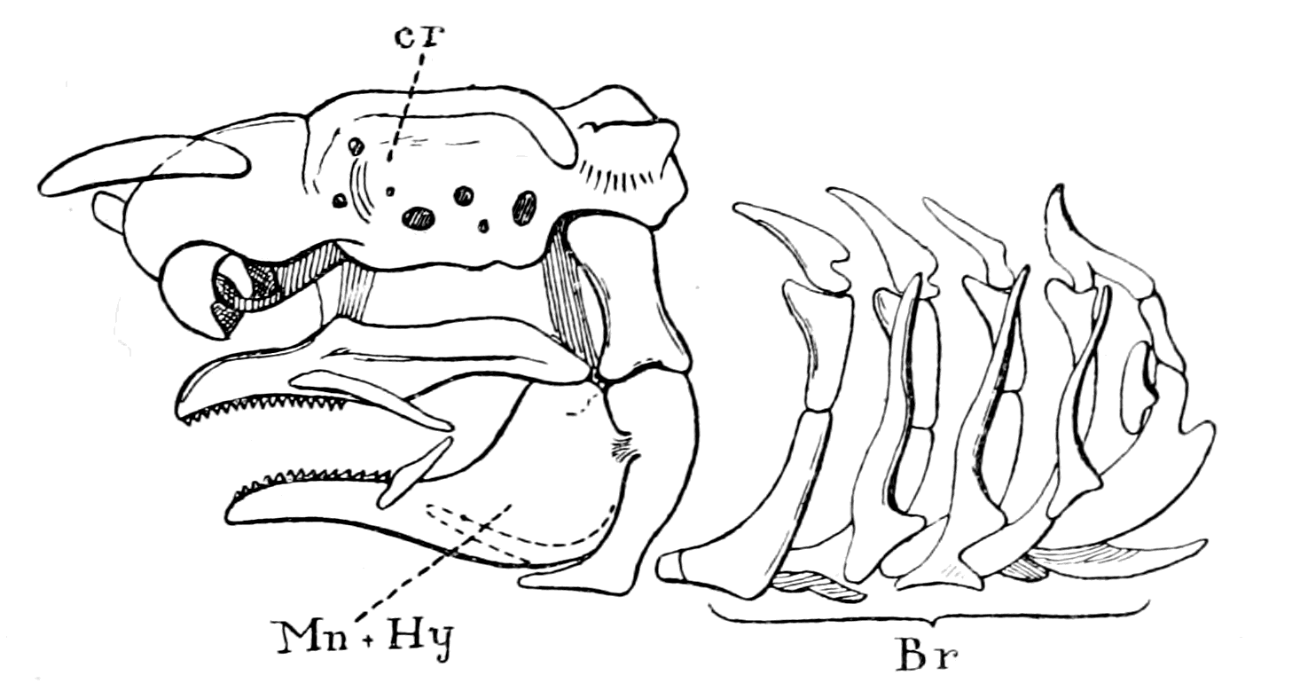
Fig. 51.—Skull of Adult Dog-fish, Side View. (From Parker.)
cr., cranium; Br., branchial arches; Mn. + Hy., mandibular and hyoid arches.
The true branchial arches persist, to a certain extent, in the Amphibia, and become still more degenerated in the Amniota (reptiles, birds, and mammals) in correlation with the total disappearance of a branchial respiration at all periods of their life. Their remnants become more or less important parts of the hyoid bone, and are employed solely in support of the tongue.
In no single animal is there any evidence that the foremost arch, the mandibular, is a true branchial arch. As low down as the Elasmobranchs it becomes divided into two elements which form respectively the upper and lower jaws; the hyoid arch, on the other hand, although it has altered its form and acquired the secondary function of supporting the mandibular arch, still retains its respiratory function.
The evidence afforded by the mode of formation of the skeletal tissues of vertebrates down to the Elasmobranchs indicates that the primitive cranial skeleton arose from two paired basal cartilages, the parachordals and trabeculæ, to which were attached respectively cartilaginous cases enclosing the organs of hearing and smell. In addition, the branchial portion of the cranial region was provided with cartilaginous bars arranged serially for the support of the branchiæ, with the exception of the foremost, the mandibular bar, which formed supporting tissues for the mouth—the upper and lower jaws.
Just as in past times the spinal nerves and the segments they supplied were supposed to represent the type on which the original vertebrate was built, so also the spinal vertebræ afforded the type of the segmented skeleton, and the anatomists of those days strove hard to resolve the cranio-facial skeleton into a series of modified vertebræ. Owing especially to the labours of Huxley, who showed that the segmentation in the head-region was essentially a segmentation due to the presence of branchial bars, this conception was finally laid to rest and nowadays it is admitted to be hopeless to resolve the cranium into vertebral segments. Still, however, the vertebrate is a segmented animal and its segmented nature is visible in the cranial region, so far as the skeletal tissues are concerned, in the shape of the series of branchial and visceral bars.
To this segmentation the name of 'branchiomeric' has been given, while that due to the presence of vertebræ is called 'mesomeric.'
As we have seen, the internal bony skeleton of the vertebrate commences as a cartilaginous and membranous skeleton. For this reason the preservation of such skeletons is impossible in the fossil form, unless the cartilage has become impregnated with lime salts, so that there is but little hope of ever obtaining traces of such structures in the fossils of the Silurian age either among the vertebrate or invertebrate remains. Fortunately for this investigation there are still living on the earth two representatives of that age; on the invertebrate side Limulus, and on the vertebrate side Ammocœtes.
The Elasmobranchs represent the most primitive of the gnathostomatous vertebrates. Below them come the Agnatha, known as the cyclostomatous fishes or Marsipobranchii, the lampreys (Petromyzon) and the hag-fishes (Myxine).
The skeleton of Petromyzon (Fig. 52) consists of a cranio-facial skeleton composed of a cartilaginous unsegmented cranium, with the basal trabeculæ and parachordals and a series of branchial and visceral cartilaginous bars forming the so-called branchial basket-work; to these must be added auditory and nasal capsules. In contradistinction to this elaborate cranio-facial skeleton, the spinal vertebral skeleton is represented only by segmentally arranged small pieces of cartilage formed in the connective tissue dissepiments between segmented sheets of body-muscles (myotomes).
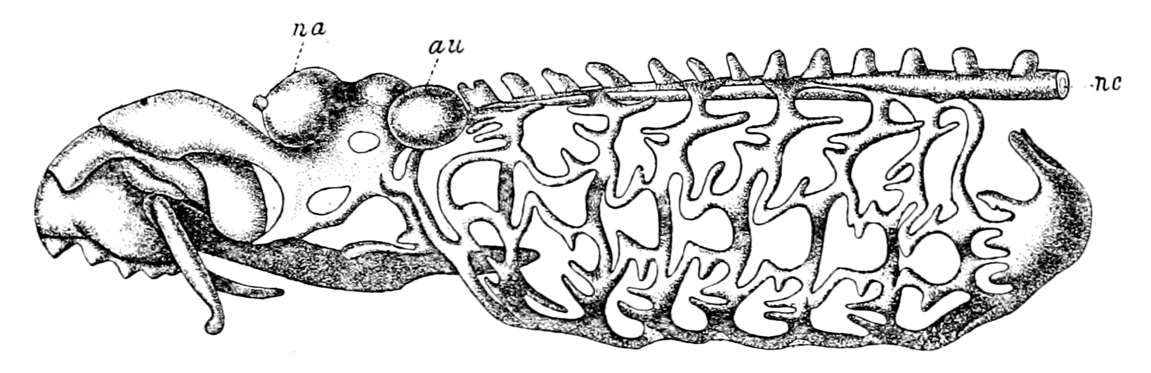
Fig. 52.—Skeleton of Petromyzon. (From Parker.)
na., nasal capsule; au., auditory capsule; nc., notochord.
But Petromyzon is derived from Ammocœtes by a remarkable process of transformation, and a most important part of that transformation is the formation of new cartilaginous structures. Thus we see that in Ammocœtes there is no sign of a cartilaginous vertebral column; at transformation the rudimentary vertebræ of Petromyzon are formed. In Ammocœtes the brain-case is a simple fibrous membranous covering; at transformation this becomes cartilaginous. In Ammocœtes there are no cartilaginous structures corresponding to the sub-ocular arches; these are all formed at transformation. It follows, that we can trace back the bony skeleton of the vertebrate head to the skeleton of Ammocœtes, and we may therefore conclude that the primitive cartilaginous skeleton of the vertebrate consisted of the following structures (Fig. 53, B), viz. the branchial bars forming a basket-work, the trabeculæ and parachordals, the auditory and nasal capsules—a clear proof that the cranial skeleton is older than the spinal. Of these structures the branchial bars are the only evidently segmented parts.
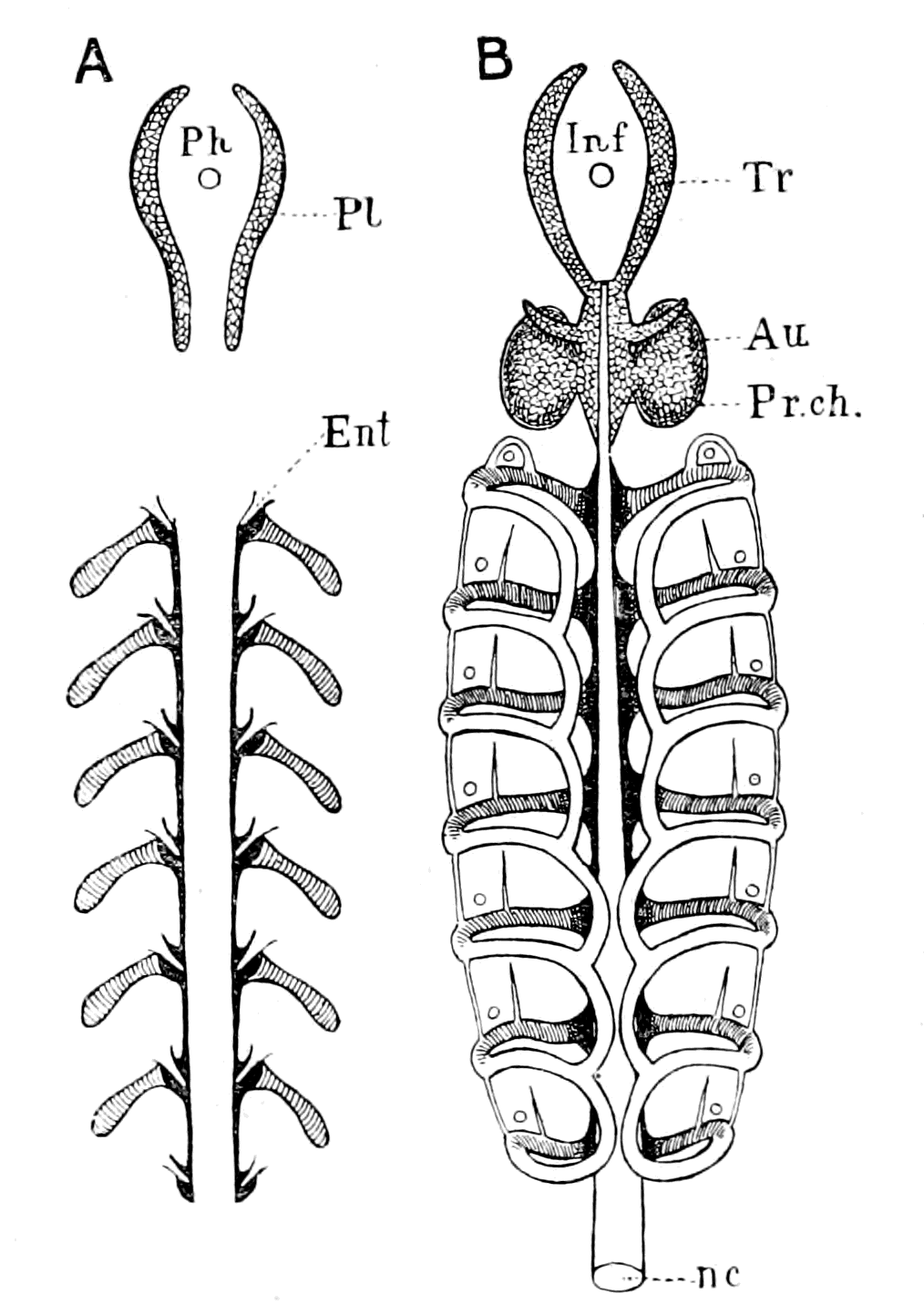
Fig. 53.—Comparison of Cartilaginous Skeleton of Limulus and Ammocœtes.
A, Diagram of cartilaginous skeleton of Limulus. Soft cartilage, entapophysial ligaments, deep black; branchial bars simply hatched; hard cartilage, lateral trabeculæ of entosternite, netted; Ph., position of pharynx.
B, Diagram of cartilaginous skeleton of Ammocœtes. Soft cartilage, sub-chordal cartilaginous bands, deep black; branchial basket-work (first formed part), simply hatched; hard cartilage, cranio-facial skeleton, trabeculæ, parachordals and auditory capsules, netted; Inf., position of tube of infundibulum (old œsophagus).
The Soft Cartilage of the Branchial Skeleton of Ammocœtes.
The study of Ammocœtes gives yet another clue to the nature of the earliest skeleton, for these two marked groups of cartilage—the branchial and basi-cranial—are characterized by a difference in structure as well as a difference in topographical position. J. Müller was the first to point out that these two sets of cartilages differ in appearance and constitution, and he gave to them the name of yellow and grey cartilage. Parker has described them fully under the terms soft and hard cartilage, terms which Schaffer has also used, and I shall also make use of them here. The whole of the branchial cartilaginous skeleton is composed of soft cartilage, while the basi-cranial skeleton, consisting of trabeculæ, parachordals, and auditory capsule, is composed of hard cartilage, the only soft cartilage in this region being that which forms the nasal capsule, not represented in Fig. 53, B.
These two groups of cartilage arise independently, so that at first the basi-cranial system is quite separate from the branchial, and only late in the history of the animal is a junction effected between the branchial system and the trabeculæ and parachordals, an initial separation which is especially striking when we consider that in this animal all the cartilaginous structures of any one system are continuous: there is no sign of anything in the nature of joints.
Of these two main groups, the branchial cartilages are formed first in the embryo, a fact which suggests that they are the most primitive of the vertebrate cartilages, and that, therefore, the first true formation of cartilage in the invertebrate ancestor may be looked for in the shape of bars supporting the branchial mechanism. The evidence of the origin of the cartilaginous structures in Ammocœtes is given by Shipley in the following words:—
"The branchial bases are the first part of the skeleton to appear. They arise about the 24th day as straight bars of cartilage, lying external and slightly posterior to the branchial vessel.
"The first traces of the basi-cranial skeleton appear on the 30th day as two rods of cartilage—the trabeculæ."
Our attention must, in the first place, be directed to this branchial basket-work of Ammocœtes.
Underlying the skin of Ammocœtes in the branchial region is situated the sheet of longitudinal body-muscles, divided into a series of segments or myotomes, which forms the somatic muscles so characteristic of all fishes. This muscular sheet is depicted on the left-hand side of Fig. 54. It does not extend over the lower lip or over that part in the middle line where the thyroid gland is situated. In these parts a sheet of peculiar tissue known by the name of muco-cartilage lies immediately under the skin, covering over the thyroid gland and lower lip. The somatic muscular sheet with the superjacent skin can be stripped off very easily owing to the vascularity and looseness of the tissue immediately underlying it. When this is done the branchial basket-work comes beautifully into view as is seen on the right-hand side of Fig. 54. It forms a cage within which the branchiæ and their muscles lie entirely concealed.
This is the great characteristic of this most primitive form of the branchial cartilaginous bars and distinguishes it from the branchial bars of other higher fishes, in that it forms a system of cartilages which lie external to the branchiæ—an extra-branchial system.
This branchial basket-work is simpler in Ammocœtes than in Petromyzon, and its actual starting-point consists of a main transverse bar corresponding to each branchial segment; from this transverse bar the system of longitudinal bars by which the basket-work is formed has sprung. These transverse bars arise from a cartilaginous longitudinal rod, situated close against the notochord on each side. These rods may be called the subchordal cartilaginous bands (Fig. 53), and, according to the observations of Schneider and others, each subchordal band does not form at first a continuous cartilaginous rod, but the cartilage is conspicuous only at the places where the transverse bars arise. In the youngest Ammocœtes examined by Schaffer, he could find no absolute discontinuity of the cartilage except between the first two transverse bars, but he says that the thinning between the transverse bars was so marked as to make it highly probable that at an earlier stage there was discontinuity. The whole system of branchial bars and subchordal rods is at first absolutely disconnected from the cranial system of trabeculæ and parachordals, and only later do the two systems join.
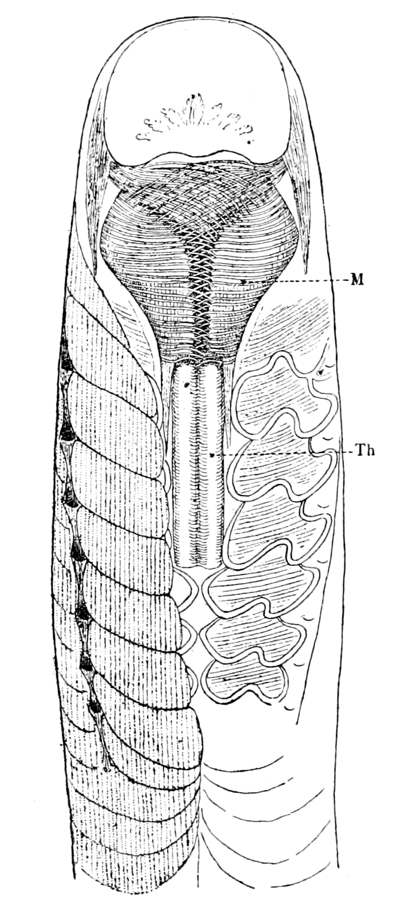
Fig. 54.—Ventral View of Head Region of Ammocœtes.
Th., thyroid gland; M., lower lip, with its muscles.
These observations on Ammocœtes lead most definitely to the conclusion that the starting-point of the whole cartilaginous skeleton of the vertebrate consisted of a series of transverse cartilaginous bars, for the purpose of supporting branchial segments; these were connected with two axial longitudinal cartilaginous rods, which at first contained cartilage only near the places of junction of the branchial bars. This system may be called the mesosomatic skeleton, as it is entirely confined to the branchial or mesosomatic region.
In addition to this primitive cartilaginous framework, which was formed for the support of the mesosomatic or respiratory segments, but at a slightly later period in the phylogenetic history, a separate cartilaginous system was formed for the support of the prosomatic segments, viz. the trabeculæ and parachordals with the auditory capsules: a system which was at first entirely separated from the mesosomatic, and, as we shall see, is more advanced in structure than the branchial system. Later still, the story is completed at the time of transformation to Petromyzon by the formation of the simple cartilaginous skull and the rudimentary vertebræ, the structure of which is also of a more advanced type.
The Structure of the Soft Branchial Cartilage.
Having considered the topographical position of the primitive branchial cartilaginous skeleton, we may now inquire, What was its structure and how was it formed?
In the higher vertebrates various forms of cartilage are described, viz. hyaline, fibro-cartilage, elastic cartilage, and parenchymatous cartilage. Of these, the parenchymatous cartilage is looked upon as the most primitive form, because it preserves without modification the characters of embryonic cartilage.
Embryology, then, would lead to the belief that the earliest form of cartilage in the vertebrate kingdom ought to be of this type, viz. large cells, each of which is enclosed in a simple capsule, so that the capsules of the cells form the whole of the matrix, and thus form a simple homogeneous honeycomb-structure, in the alveoli of which the cartilage-cells lie singly. If, then, the branchial cartilages of Ammocœtes are, as has just been argued, the representatives of the cartilaginous skeleton of the primitive vertebrate, it is reasonable to suppose that they should resemble in structure this embryonic cartilage. Such is undoubtedly the case: all observers who have described the branchial basket-work of Ammocœtes or Petromyzon have been struck with the extremely primitive character of the cartilage, and the last observer (Schaffer) describes it as composed of thin walls of homogeneous material, in which there are no lines of separation, which form a simple honeycomb-structure, in the alveoli of which the separate cells lie singly. These branchial cartilages are each surrounded by a layer of perichondrium, and in Fig. 55, A, I give a picture of a section of a portion of one of the bars.
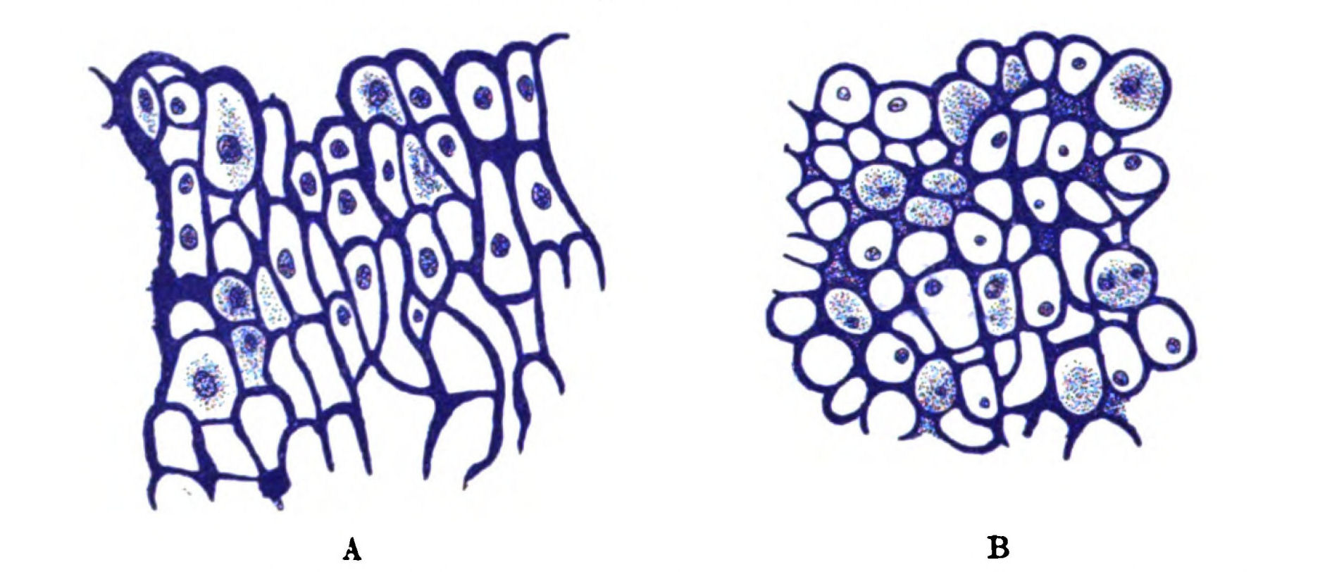
Fig. 55.—A, Branchial Cartilage of Ammocœtes, stained with Thionin. B, Branchial Cartilage of Limulus, stained with Thionin.
Hence we see that structurally as well as topographically the branchial bars of Ammocœtes justify their claim to be considered as the origin of the vertebrate cartilaginous framework.
On the Structure of the Muco-cartilage in Ammocœtes.
We can, however, go further than this, and ask how this cartilage itself is formed in Ammocœtes? The answer is most definite, most instructive and suggestive, for in all cases this particular kind of cartilage is formed from, or at all events in, a peculiar fibrous tissue, which was called by Schneider "Schleim-Knorpel," or muco-cartilage, a tissue which is distinguishable from other connective tissues, not only by its structural peculiarities, but also by its strong affinity for all dyes which differentiate mucoid or chondro-mucoid substances.
This muco-cartilage is thus described by Schneider:—The perichondrium in Ammocœtes is not confined to the true cartilaginous structures, but extends itself in the form of thin plates in definite directions. Between these plates of perichondrium a peculiar tissue (Fig. 56)—the muco-cartilage—exists, consisting of fibrillæ, whose direction is mainly at right angles to the planes of the perichondrial plates, with star-shaped cells in among them, and with the spaces between the fibrillæ filled up with a semi-fluid mass.
From this tissue all the primitive cartilages which resemble the branchial bars are formed, either by the invasion of chondroblasts from the surrounding perichondrium, or by the proliferation and encapsulation of the cells of the muco-cartilage itself.
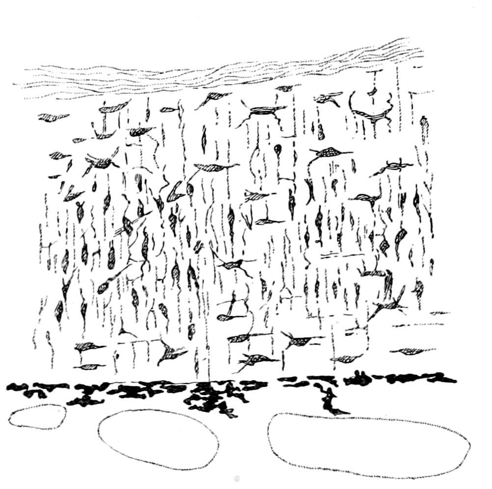
Fig. 56.—Section of Muco-cartilage from Dorsal Head-plate of Ammocœtes.
This very distinctive tissue—the muco-cartilage—is of very great importance in all questions of the origin of the skeletal tissues. In all descriptions of the skeletal tissues it has been practically disregarded until recent years when, besides my own observations, its distribution has been mapped out by Schaffer. Thus Parker, in his well-known description of the skeleton of the marsipobranch fishes, does not even mention its existence. Its importance is shown by its absolute disappearance at transformation and its non-occurrence in any of the higher vertebrates. It is entirely confined to the head-region, and its distribution there is most suggestive, for, as will be described fully later on, it forms a skeleton which both in structure and position resembles very closely the head-shields of cephalaspidian fishes. At the present part of my argument its more immediate interest lies in the method of tracing this tissue. For this purpose I made use of the micro-chemical reaction of thionin, a dye which, as shown by Hoyer, stains all mucin-containing substances a bright purple. Schaffer made use of a corresponding basophil stain, hæmalum. When stained with thionin, the matrix, or ground-substance of the branchial cartilages as well as the matrix or semi-fluid substance in which the fibrils of the muco-cartilaginous cells are embedded take on a deep purple colour, while the fibrous material of the cranial walls and other connective tissue strands, such as the perichondrium, are coloured light blue. Muco-cartilage, then, may be described as a peculiar form of connective tissue which differs from other connective tissue not only in its appearance but in its chemical composition, for unlike white fibrous tissue it contains a large amount of mucin, and this tissue is the forerunner of the earliest cartilaginous vertebrate skeleton, the branchial bars of Ammocœtes.
The conclusions to which we are led by the study of the structure, position, and mode of origin of these primitive cartilages of Ammocœtes may be thus summed up:—
1. The immediate ancestor of the vertebrate must have possessed a peculiar fibrous tissue—the ground-substance of which stained deep purple with thionin—in which cartilage arose.
2. The cartilage so formed was not like hyaline cartilage, but resembled in a striking manner parenchymatous cartilage.
3. This cartilage was situated partly in two axial longitudinal bands, partly as transverse bars, which supported the branchial apparatus.
The Prosomatic or Basi-cranial Skeleton of Ammocœtes.
Before searching for any evidence of a similar tissue in any invertebrate group, it is advisable to consider the other portion of the cartilaginous skeleton of Ammocœtes, which consists of the trabeculæ, parachordals and auditory capsules—the basi-cranial skeleton—and is composed of hard, not soft cartilage.
This basi-cranial skeleton represented in Fig. 53, B, is confined to the region of the notochord, the cranial walls being composed entirely of a white fibrous membrane. It is separated at first entirely from the sub-chordal portion of the branchial basket-work, and is composed of a foremost part, the trabeculæ (Tr.), and of a hindermost part, the parachordals (Pr.ch.), which are characterized by the attachment on each side of the large auditory capsule (Au.). In Ammocœtes the trabecular bars are continuous with the parachordals, the junction being marked by a small lateral projection on each side, which at transformation is seen to play an important part in the formation of the sub-ocular arch. The trabecular bar lies close against the notochord on each side up to its termination; it then bends away from the middle line and curves round until it meets its fellow on the opposite side, thus forming, as it were, the head of a racquet of which the notochord forms the splice in the handle. The strings of the racquet are represented by a thin membrane, in the centre of which the position of the infundibulum (Inf.) of the brain can be clearly seen. In an earlier stage of Ammocœtes the two trabecular horns do not meet, but are separated by connective tissue, which afterwards becomes cartilaginous.
As far, then, as the topography of this basi-cranial skeleton is concerned, the striking points are—the shape of the trabecular portion, diverging as it does around the infundibulum, and the presence on the parachordal portion of the two large auditory capsules.
These two points indicate, on the hypothesis that infundibulum and œsophagus are convertible terms, that two supporting structures of a cartilaginous nature must have existed in the ancestor of the vertebrate, the first of which surrounded the œsophagus, and the second was in connection with its auditory apparatus.
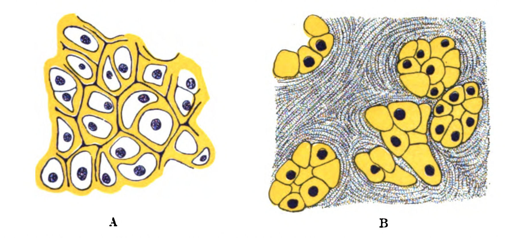
Fig. 57.—A, Cartilage of Trabeculæ of Ammocœtes, stained with Hæmatoxylin and Picric Acid. B, Nests of Cartilage Cells in Entosternite of Hypoctonus, stained with Hæmatoxylin and Picric Acid.
Structure of the Hard Cartilages.
The structure of this hard cartilage of the trabeculæ and auditory capsules resembles that of the soft, in so far that it consists of large cells with a comparatively small amount of intercellular substance. Schaffer, who has described it lately, considers that it is a nearer approach to hyaline cartilage than the soft, but yet cannot be called hyaline cartilage in the usual sense of the term. Its peculiarities and its differences from the soft are especially well seen by its staining reactions. I have myself been particularly struck with the effect of picrocarmine or combined hæmatoxylin and picric acid staining (Fig. 57). In the case of the soft cartilage the capsular substance stains respectively a brilliant red or blue, while that of the hard cartilage is coloured a deep yellow, so that the junction between the parachordals and the branchial cartilages is beautifully marked out. Then, again, with thionin, which gives so marked a reaction in the case of the soft cartilage, the hard cartilage of the auditory capsule is not stained at all, and in the trabeculæ the deep purple colour is confined to the mucoid cement-substance between the capsules, just as Schaffer has stated. The same kinds of reactions have been described by Schaffer: thus by double staining with hæmalum-eosin the hard cartilage stains red, the soft blue; and he points out that even with over-staining by hæmalum the auditory capsule remains colourless, just as I have noticed with thionin. He infers, precisely as I have done from the thionin reaction, that chondro-mucoid, which is so marked a constituent of the soft cartilage and of the muco-cartilage, is absent or present in but slight quantities in the hard cartilage. Similarly, he points out that double staining with tropœolin-methyl-violet stains the hard cartilage a bright orange colour, and the soft cartilage a violet.
The evidence, then, shows clearly that a marked chemical difference exists between these two cartilages, which may be expressed by saying that the one contains very largely a basophil substance, which we may speak of as belonging to the class of chondro-mucoid substances, while the other contains mainly an oxyphil substance, probably a chondro-gelatine substance.
We may perhaps go further and attribute this difference of composition to a difference of origin; for whereas the soft cartilage is invariably formed in a special tissue, the muco-cartilage, which shows by its reaction how largely it is composed of a mucoid substance, the hard cartilage is certainly, in the case of the cartilage of the cranium where its origin has been clearly made out, formed in the membranous tissue of the cranium of Ammocœtes—i.e. in a tissue which stains light blue with thionin, and contains a gelatinous rather than a mucoid substratum.
The best opportunity of finding out the mode of origin of the hard cartilage is afforded at the time of transformation, when so much of this kind of cartilage is formed anew. Unfortunately, it is very difficult to obtain the early transformation stages, consequently we cannot be said to possess any really exhaustive and definite account of how the new cartilages are formed. Bujor, Kaensche, and Schaffer all profess to give a more or less definite account of their formation, and the one striking impression left on the mind of the reader is how their descriptions vary. In one point only are they agreed, and in that I also agree with them, viz. the manner in which the new cranial walls are formed. Schaffer describes the process as the invasion of chondroblasts into the homogeneous fibrous tissue of the cranial walls. Such chondroblasts not only form the cartilaginous framework, but also assimilate the fibrous tissue which they invade, so that finally all that remains of the original fibrous matrix in which the cartilage was formed are these lines of cement-substance between the groups of cartilage cells, which, containing some basophil material, are marked out, as already mentioned (Fig. 57).
We may therefore conclude, from the investigation of Ammocœtes, that the front part of the basi-cranial skeleton arose as two trabecular bars, to which muscles were attached, situated bilaterally with respect to the central nervous system. These bars were composed of tendinous material with a gelatinous rather than a mucoid substratum, in which nests of cartilage-cells were formed, the cartilaginous material formed by these cells being of the hard variety, not staining with thionin, and staining yellow with picro-carmine, etc. By the increase of such nests and the assimilation of the intermediate fibrous material, the original fibro-cartilage was converted into the close-set semi-hyaline cartilage of the trabeculæ and auditory capsules, in which the fibrous material still marks out by its staining-reaction the limits of the cell-clusters.
Such I gather to be Schaffer's conclusions, and they are certainly borne out by my own and Miss Alcock's observations. As far as we have had an opportunity of observing at present, the first process at transformation appears to consist of the invasion of the fibrous tissue of the cranial wall by groups of cells which form nests of cells between the fibrous strands. These nests of cells form round themselves capsular material, and thus form cell-territories of cartilage, which squeeze out and assimilate the surrounding fibrous tissue, until at last all that remains of the original fibrous matrix is the lines of cement-substance which mark out the limits of the various cell-groups.
At present I am inclined to think that both soft and hard cartilage originate in a very similar manner, viz. by the formation of capsular material around the invading chondroblasts, and that the difference in the resulting cartilage is mainly due to the difference in chemical composition of the matrix of the connective tissue which is invaded. Thus the difference may be formulated as follows:—
The hard cartilage is formed by the invasion of chondroblasts into a fibrous tissue, which contains a gelatinous rather than a mucoid substratum, in contradistinction to the soft cartilage which is formed, probably also by the invasion of chondroblasts, in a tissue—the muco-cartilage—which contains a specially mucoid substratum.
Such, then, is the very clearly defined starting-point of the vertebrate skeleton—two distinct formations of different histological and chemical structure,—the one forming a segmented branchial skeleton, the other a non-segmented basi-cranial skeleton.
The Cartilaginous Skeleton of Limulus.
Among the whole of the invertebrates at present living on the earth, is there any sign of an internal cartilaginous skeleton that will give a direct clue to the origin of the primitive vertebrate skeleton? The answer to this question is most significant: only one animal among all those at present known possesses a cartilaginous skeleton, which is directly comparable with that of Ammocœtes, and here the comparison is very close—only one animal among the thousands of living invertebrate forms, and that animal is the only representative still surviving of the palæostracan group, which was the dominant race when the vertebrate first made its appearance. The Limulus, or king-crab, possesses a segmented branchial internal cartilaginous skeleton (Fig. 53, A), made up of the same kind of cartilage as the branchial skeleton of Ammocœtes, confined to the mesosomatic or branchial region, just as in Ammocœtes, forming, as in Ammocœtes, cartilaginous bars supporting the branchiæ, and these bars are situated externally to the branchiæ, as in Ammocœtes. In addition this animal possesses a basi-cranial internal semi-cartilaginous unsegmented plate known as the entosternite or plastron situated, with respect to the œsophagus, similarly to the position of the trabeculæ with respect to the infundibulum in Ammocœtes. Moreover, the cartilaginous cells in this tissue differ from those in the branchial region, in precisely the same manner as the hard cartilage differs from the soft in Ammocœtes.
This plastron, it is true, is found in other animals, all of which are members of the scorpion tribe, except in one instance, and this, strikingly enough, is the crustacean Apus—a strange primitive form, which is acknowledged to be the nearest representative of the Trilobita still living on the earth. None of these forms, however, possess any sign of an internal cartilaginous branchial skeleton, such as is possessed by Limulus. Scorpions, Apus, Limulus, are all surviving types of the stage of organization which had been reached in the animal world when the vertebrate first appeared.
The Mesosomatic or Respiratory Skeleton of Limulus, composed of Soft Cartilage.
Searching through the literature of the histology of the cartilaginous tissues in invertebrate animals, to see whether any cartilage had been described similar to that seen in the branchial cartilages of Ammocœtes, and whether such cartilage, if found, arose in a fibrous tissue resembling muco-cartilage, I was speedily rewarded by finding, in Ray Lankester's article on the tropho-skeletal tissues of Limulus, a picture of the cartilage of Limulus, which would have passed muster for a drawing of the branchial cartilage of Ammocœtes. This clue I followed out in the manner described in my former paper in the Journal of Anatomy and Physiology, and mapped out the topography of this remarkable tissue.