Front matter (continued 6)
Reading edition
The number of such segments, according to present opinion, is seven, viz.:—
(1) The foremost segment, which bears the cheliceræ.
(2, 3, 4, 5, 6) The next five segments, which carry the paired locomotor appendages; and
(7) The last segment, to which belongs a small abortive pair of appendages, known by the name of the chilaria, situated between the last pair of locomotor appendages and the operculum or first pair of mesosomatic appendages. These appendages are numbered from 1-7 in the accompanying drawing (Fig. 103).
Of these seven pairs of appendages, the significance of the first and the last has been matter of dispute. With respect to the first pair, or the cheliceræ, the question has arisen whether their nerves belong to the infra-œsophageal group, or are in reality supra-œsophageal.
It is instructive to observe the nature and the anterior position of this pair of appendages in the allied sea-scorpions, especially in Pterygotus, where the only chelate organs are found in these long, antennæ-like cheliceræ. In Slimonia and in Stylonurus they are supposed by Woodward to be represented by the small non-chelate antennæ seen in Fig. 8, B and C (p. 27), taken from Woodward. If such is the case, then these figures show that a pair of appendages is missing in each of these forms, for they possess only five free prosomatic appendages instead of six, as in Limulus and in Pterygotus. Similarly, Woodward only allowed five appendages for Pterygotus, so that his restorations were throughout consistent. Schmidt, in Pterygotus osiliensis has shown that the true number was six, not five, as seen in his restoration given in Fig. 8, A (p. 27).
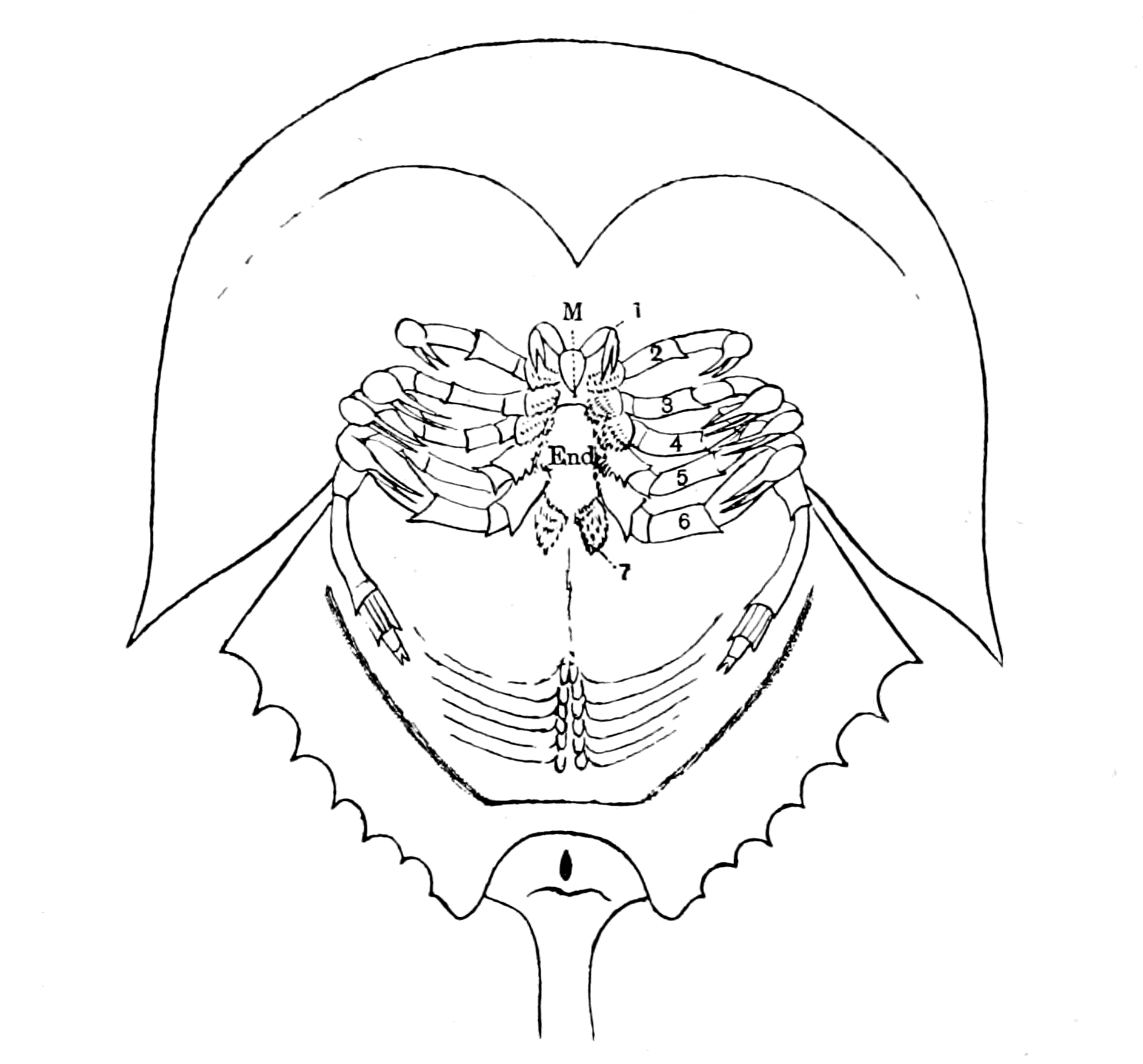
Fig. 103.—Ventral Surface of Limulus. (Taken from Kishinouye.)
The gnathic bases of the appendages have been separated from those of the other side to show the promesosternite or endostoma (End.).
With respect to Eurypterus, Schmidt figures an exceedingly minute pair of antennæ between the coxal joints of the first pair of appendages, thus making six pairs of appendages. Gerhard Holm, however, in his recent beautiful preparations from Schmidt's specimens and others collected at Rootziküll, has proved most conclusively that the cheliceræ of Eurypterus were of the same kind as those of Limulus. I reproduce his figure (Fig. 104) showing the small chelate cheliceræ (1) overhanging the mouth orifice, just as in Limulus or in Scorpio.
So, also, since Woodward's monograph, Laurie has discovered in Slimonia acuminata a small median pair of chelate appendages exactly corresponding to the cheliceræ of Limulus, or of Eurypterus, or of Scorpio. We may, therefore, take it for granted that such was also the case in Stylonurus, and that the foremost pair of prosomatic appendages in all these extinct sea-scorpions were in the same position and of the same character as the cheliceræ of the scorpions.
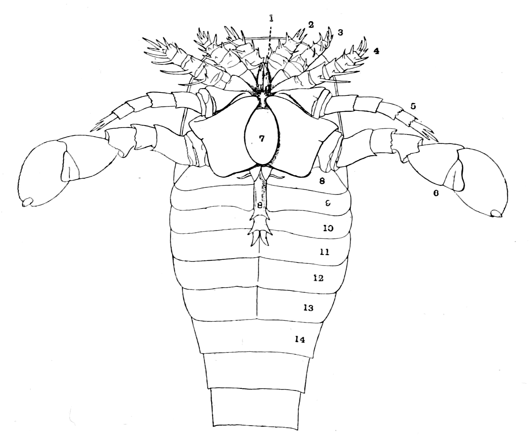
Fig. 104.—Eurypterus Fischeri. (From Holm.)
In the living scorpion and in Limulus the nerves to this pair of appendages undoubtedly arise from the foremost prosomatic ganglia, and the reason why they appear to belong to the supra-œsophageal brain-mass has been made clear by Brauer's investigations on the embryology of Scorpio; for he has shown that the cheliceral ganglia shift from the ventral to the dorsal side of the œsophagus during development, thus becoming pseudo-supra-œsophageal, though in reality belonging to the infra-œsophageal ganglia. This cheliceral pair of appendages is, in all probability, homologous with the second pair of antennæ in the crustacea.
I conclude, then, that the cheliceræ must truly be included in the prosomatic group, but that they stand in a somewhat different category to the rest of the prosomatic appendages, inasmuch as they take up a very median anterior and somewhat dorsal position, and their ganglia of origin are also exceptional in position.
Next for consideration come the chilaria (7 in Fig. 103), which Lankester did not consider to belong to appendages at all, but to be a peculiar pair of sternites. Yet their very appearance, with their spinous hairs corresponding to those of the other gnathites and their separate nerve-supply, all point distinctly to their being a modified pair of appendages, and, indeed, the matter has been placed beyond doubt by the observations of Kishinouye, who has found embryologically that they arise in the same way as the rest of the prosomatic appendages, and belong to a distinct prosomatic segment, viz. the seventh segment. In accordance with this, Brauer has found that in the scorpion there is in the embryo a segment, whose appendages degenerate, which is situated between the segment bearing the last pair of thoracic appendages and the genital operculum—a segment, therefore, comparable in position to the chilarial segment of Limulus.
Coming now to the five locomotor appendages, we find that they resemble each other to a considerable extent in most cases, with, however, certain striking differences. Thus in Limulus they are chelate, with their basal joints formed as gnathites, except in the case of the fifth appendage, in which the extremity is modified for the purpose of digging in the sand. In Pterygotus, Slimonia, Eurypterus, the first four of these appendages are very similar, and are called by Huxley and Woodward endognaths; in all cases they possess a basal part or sterno-coxal process, which acts as a gnathite or foot-jaw, and a non-chelate tactile part, which possesses no prehensile power, and in most cases could have had no appreciable share in locomotion, called by Huxley and Woodward the palpus. These small palps were probably retractile, and capable of being withdrawn entirely under the hood. The fifth appendage is usually different, being a large swimming organ in Pterygotus, Eurypterus, and Slimonia (Figs. 8 and 104), and is known as the ectognath.
Finally, in Drepanopterus Bembycoides, as stated by Laurie, all five locomotor appendages are built up after the same fashion, the last one not being formed as a paddle-shaped organ or elongated as in Stylonurus, but all five possess no special locomotor or prehensile power. According to Laurie this is a specially primitive form of the group.
It is significant to notice from this sketch that with the absence of special prehensile terminations such as chelæ, or the absence of special locomotor functions such as walking or swimming, these appendages tend to dwindle and become insignificant, taking up the position of mere feelers round the mouth, and at the same time are concentrated and pressed closely together, so that their appendage-nerves must also be close together.
This sketch therefore shows us that—
Of the six foremost prosomatic appendages, the cheliceræ and the four endognaths were, at the time when the vertebrates first appeared, in very many cases dwindling away; the latter especially no longer functioned as locomotor appendages, but were becoming more and more mere palps or tentacles situated round the mouth, which could by no possibility afford any help to locomotion.
On the contrary, the sixth pair of appendages—the ectognaths—remained powerful, being modified in many cases into large oar-like limbs by which the animal propelled itself through the water.
It is a striking coincidence that those ancient fishes, Pterichthys and Bothriolepis, should have possessed a pair of large oar-like appendages.
At this time, then, in strong contrast to the endognaths, the ectognaths, or sixth pair of appendages, remained strong and vigorous. What about the seventh pair, the chilaria of Limulus?
Of all the prosomatic appendages these are the most interesting from the point of view of my theory, for whereas in the scorpion of the present day they have dwindled away and left no trace except in the embryo, in the sea-scorpions of old, far from dwindling, they had developed and become a much more important organ than the chilaria of Limulus.
In all these animals a peculiarly striking and unique structure is found in this region known by the name of the metastoma, or lip-plate (Figs. 8 and 104 (7)); it is universally considered to be formed by the fusion of the two chilarial appendages.
All observers are agreed that this lip-plate was freely movable. Nieskowski considers that the movement of the metastoma was entirely in a vertical direction, whereby the cleft which is seen between the basal joints of all the pairs of locomotor appendages could be closed from behind. Woodward says it no doubt represents the labium, and served more effectually to enclose the posterior part of the buccal orifice, being found exteriorly to the toothed edges of the ectognaths or maxillipedes. Schmidt agrees with Nieskowski, and looks on the mestasoma as forming a lower lip within which the bases of the ectognaths worked.
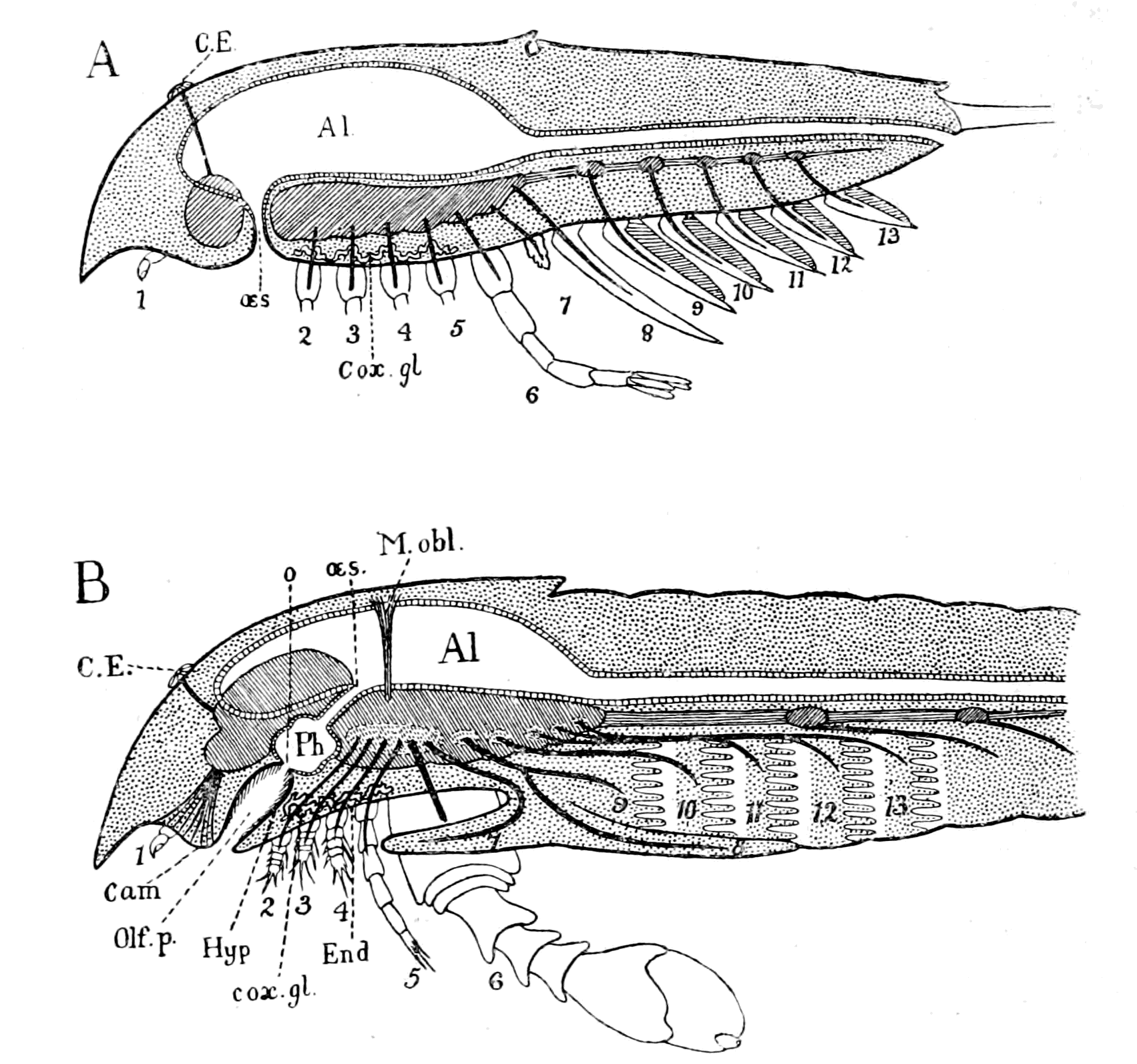
Fig. 105.—Diagram of Sagittal Median Section through A, Limulus, B, Eurypterus.
Quite recently Gerhard Holm has worked over again the very numerous specimens of Eurypterus Fischeri, which are obtainable at Rootziküll, and has thrown new light on the relation of the metastoma to the mouth-parts. His preparations show clearly that the true lower lip of Eurypterus was not the metastoma, for when the metastoma is removed another plate (End., Fig. 105, B) situated internally to it is disclosed, which, in his view, corresponds to the sternite between the bases of the pro-somatic appendages in Limulus, i.e. to the sternite called by Lankester, the pro-mesosternite (End., Fig. 103). This inner plate formed with the metastoma ((7) Fig. 105) and the ectognaths (6) a chamber closed posteriorly, within which the bases of the ectognaths worked. In other words, the removal of the metastoma discloses in Eurypterus the true anterior ventral surface of the animal which corresponds to that of Limulus, or of the scorpion group, with its pro-mesosternite and laterally attached gnathites or sterno-coxal processes. To this inner plate or pro-mesosternite Holm gives the name of endostoma.
To the anterior edge of the endostoma a thinner membrane is attached which passes inwards in the direction of the throat, and forms, therefore, the lower lip (Hyp., Fig. 105, B) of the passage of the mouth (olf. p.). This membrane bears upon its surface a tuft of hairs, which he thought were probably olfactory in function. Consequently, in his preliminary communication, he describes this lower lip as forming, in all probability, an olfactory organ; in his full communication he repudiates this suggestion, because he thinks it unlikely that such an organ would be situated within the mouth. I feel sure that if Holm had referred to Croneberg's paper, and seen how the true mouth in all the scorpion group is situated at the base of an olfactory passage, he would have recognized that his first suggestion is in striking accordance with the nature of the entrance to the mouth in other scorpions.
That Eurypterus also possessed a camerostome (cam.) seems to follow of necessity from its evident affinities both with Limulus and the scorpions. We see, in fact, that the mouth of these old sea-scorpions was formed after the fashion of Limulus, surrounded by masticatory organs in the shape of foot-jaws, and yet foreshadowed that of the scorpion, so that an ideal sagittal section of one of these old palæostracan forms would be obtained by the combination of actual sagittal sections through Limulus and a member of the scorpion group, with, at the same time, a due recognition of Holm's researches. Such a section is represented in Fig. 105, B, in which I have drawn the central nervous system and its nerves, the median eyes (C.E.), the olfactory organs (Cam.), the pharynx (Ph.), œsophagus (œs.), and alimentary canal (Al.), but have not tried to indicate the lateral eyes. I have represented the prosomatic appendages by numbers (1-7), and the foremost mesosomatic segments by numbers (8-13). I have placed the four endognaths and the nerves going to them close together, and made them small, mere tentacles, in recognition of the character of these appendages in Eurypterus, and have indicated the position and size of the large ectognath, with its separate nerve, by (6). If among the ancient Eurypterus-like forms, which were living at the time when vertebrates first appeared, there were some in which the ectognaths also had dwindled to a pair of tentacles, then such animals would possess a prosomatic chamber formed by a metastoma or accessory lip, within which were situated five pairs of short tactile appendages or tentacles. If the vertebrate were derived from such an animal, then the trigeminal nerve, as the representative of these prosomatic appendage-nerves, ought to be found to supply the muscles of this accessory lip and of these five pairs of tentacles in the lowest vertebrate.
This prosomatic or oral chamber, as it might be called, was limited posteriorly by the fused metastoma (7) and operculum (8), so that if in the same imaginary animal one imagines that the gill-chambers, instead of being separate, are united to form one large respiratory chamber, then, in such an animal, a prosomatic oral chamber, in which the prosomatic appendages worked, would be separated from a mesosomatic respiratory chamber by a septum composed of the conjoined basal portions of the mesosomatic operculum and the prosomatic metastoma, as indicated in the diagram. In this septum the nerves to the last prosomatic appendage (equivalent to the last part of the trigeminal in the vertebrate) and to the first mesosomatic (equivalent to the thyroid part of the facial) would run, as shown in the figure, close together in the first part of their course, and would separate when the ventral surface was reached, to pass headwards and tailwards respectively.
The Coxal Glands.
One more characteristic of these appendages requires mention, and that is the excretory glands situated at the base of the four endognaths known as the coxal glands. These glands are the main excretory organs in Limulus and the scorpions, and extend into the basal segments or coxæ of the four endognaths, not into those of the ectognaths or the chilaria (or metastoma). Hence their name, coxal glands; and, seeing the importance of the excretory function, it is likely enough that they would remain, even when the appendages themselves had dwindled away. With the concentration and dwindling of the endognaths these coxal glands would also be concentrated, so that in the diagram (Fig. 105) they would rightly be grouped together in the position indicated (cox. gl.).
Such a diagram indicates the position of all the important organs of the head-region except the special organs for taste and hearing. These, for the sake of convenience, I propose to take separately, in order at this stage of my argument not to overburden the simplicity of the comparison I desire to make with too much unavoidable detail.
The Prosomatic Region of Ammocœtes.
Let us now compare this diagram with that of the corresponding region in Ammocœtes and see whether or no any points of similarity exist.
With respect to this region, as in so many other instances already mentioned, Ammocœtes occupies an almost unique position among vertebrates, for the region supplied by the trigeminal nerve—the prosomatic region—consists of a large oral chamber which was separated from the respiratory chamber in the very young stage by a septum which is subsequently broken through, and so the two chambers communicate.
This chamber is bounded by the lower lip ventrally, the upper lip and trabecular region dorsally, and the remains of the septum or velum laterally and posteriorly. It contains a number of tentacles arranged in pairs within the chamber so as to form a sieve-like fringe inside the circular mouth; of these, the ventral pair are large, fused together, and attached to the lower lip.
All the muscles belonging to this oral chamber are of the visceral type, and are innervated by the trigeminal nerve. In accordance with the evidence obtained up to this point this means that such an oral chamber was formed by the prosomatic appendages of the invertebrate ancestor, similarly to the oral chamber just figured for Eurypterus.
This chamber in the full-grown Ammocœtes is not only open to the respiratory chamber, but is bounded by the large upper lip (U.L., Fig. 106, D). On the dorsal surface of this region, in front of the pineal eye (C.E.), is the most conspicuous opening of the olfactory tube (Na.), which olfactory tube passes from the dorsal region to the ventral side to terminate blindly at the very spot where the infundibulum comes to the surface of the brain. Here, also, is situated that extraordinary glandular organ known as the pituitary body (Pit.). A sagittal section, then, in diagram form, of the position of parts in the full-grown Ammocœtes, would be represented as in Fig. 106, D.
But, as argued out in the last chapter, the diagram of the adult Ammocœtes must be compared with that of a cephalaspidian fish; the diagram of the palæostracan must be compared with the larval condition of Ammocœtes. In other words, Fig. 106, B, must be compared with Fig. 106, C, which represents a section through the larval Ammocœtes as it would appear if it reached the adult condition without any forward growth of the upper lip or any breaking through of the septum between the oral and respiratory chambers. The striking similarity between this diagram and that of Eurypterus becomes immediately manifest even to the smallest details. The only difference between the two, except, of course, the notochord, consists in the closure of the mouth opening (o), in Fig. 106, B, by which the olfactory passage (olf. p.) of the scorpion becomes converted into the hypophysial tube (Hy.), Fig. 106, C, and later into the nasal tube (Na.), Fig. 106, D, of the full-grown Ammocœtes. That single closure of the old mouth is absolutely all that is required to convert the Eurypterus diagram into the Ammocœtes diagram.
Such a comparison immediately explains in the simplest manner a number of anatomical peculiarities which have hitherto been among the great mysteries of the vertebrate organization. For not only do the median eyes (C.E.) correspond in position in the two diagrams, and the infundibular tube (Inf.) and the ventricles of the brain (C.C.) correspond to the œsophagus (œs.) and the cephalic stomach (Al.), as already fully discussed; but even in the very place where the narrow œsophagus opened into the wider chamber of the pharynx (Ph.), there, in all the lower vertebrates, the narrow infundibular tube opens into the wider chamber of the membranous saccus vasculosus (sac. vasc.). This is the last portion of the membranous part of the tube of the central nervous system which has not received explanation in the previous chapters, and now it is seen how simple its explanation is, how natural its presence—it represents the old pharyngeal chamber of the palæostracan ancestor.
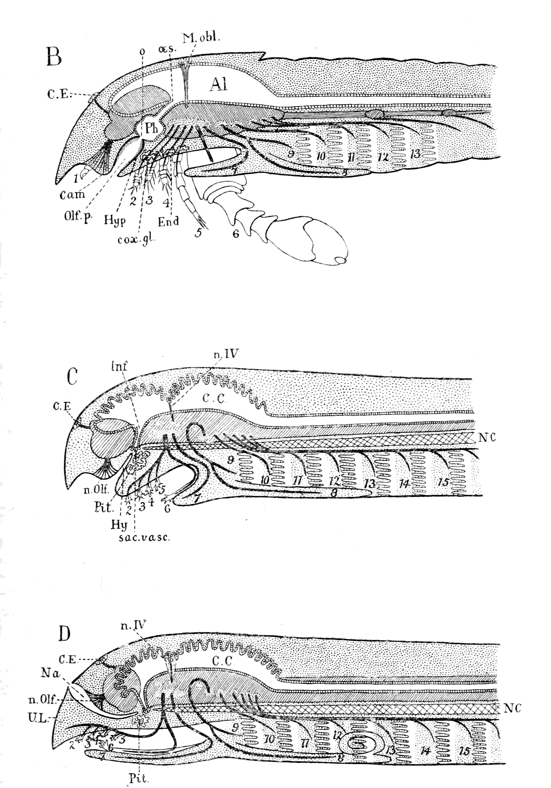
Fig. 106.—Diagram of Sagittal Median Section through B, Eurypterus; C, Larval Ammocœtes; D, Full-grown Ammocœtes.
Next among the mysteries requiring explanation is the pituitary body, that strange glandular organ always found so closely attached to the brain in the infundibular region that when it is detached in taking out the brain it leaves the infundibular canal patent right into the IIIrd ventricle. A comparison of the two diagrams indicates that such a glandular organ (Pit.), Fig. 106, C, was there because the coxal excretory glands (cox. gl.), Fig. 106, B, were in a similar position in the palæostracan ancestor—that, indeed, the pituitary body is the descendant of the coxal glands.
Finally, the diagrams not only indicate how the mesosomatic appendage-nerves supplying in the one case the operculum and the respiratory appendages correspond to the respiratory group of nerves, VII., IX., X., supplying in the other case the thyroid, hyoid, and branchial segments, but also that a similar correspondence exists between the prosomatic appendage-nerves in the one case and the trigeminal nerve in the other; a correspondence which supplies the reason why in the vertebrate a septum originally existed between an oral and respiratory chamber.
Such a comparison, then, leads directly to the suggestion that the trigeminal nerve originally supplied the prosomatic appendages, such appendages being: 1. The metastoma, which has become in Ammocœtes the lower lip supplied by the velar or mandibular branch of the trigeminal nerve (7); 2. The ectognath, which has become the large median ventral tentacle, called by Rathke the tongue, supplied by the tongue nerve (6); 3. The endognaths, which have been reduced to tentacles and are supplied by the tentacular branch of the trigeminal nerve (2, 3, 4, 5).
I have purposely put these two diagrams of the larval Ammocœtes and of Eurypterus before the minds of my readers at this early stage of my argument, so as to make what follows more understandable. I propose now to consider fully each one of these suggestive comparisons, and to see whether or no they are in accordance with the results of modern research.
In the first instance, the diagrams suggest that the trigeminal nerve originally supplied the prosomatic appendages of the palæostracan ancestor, while the eye-muscle nerves supplied the body-muscles of the prosoma.
As these appendages did not carry any vital organs such as branchiæ, but were mainly locomotor and masticatory in function, it follows that their disappearance as such would be much more complete than that of the mesosomatic branchial appendages. Most probably, then, in the higher vertebrates no trace of such appendages might be left; consequently the segmentation due to their presence would be very obscure, so that in this region the very reverse of what is found in the region of the vagus nerve would be the rule. There branchiomeric segmentation is especially evident, owing to the persistence of the branchial part of the branchial appendages; here, owing to the disappearance of the appendages, the segmentation is no longer branchiomeric, but essentially mesomeric in consequence of the persistence of the somatic eye-muscles.
In addition to the evidence of the appendages themselves, the number of prosomatic segments is well marked out in all the members of the scorpion group by the divisions of the central nervous system into well-defined neuromeres in accordance with the appendages, a segmentation the reminiscence of which may still persist after the appendages themselves have dwindled or disappeared. In accordance with this possibility we see that one of the most recent discoveries in favour of a number of segments in the head-region of the vertebrate is the discovery in the early embryo of a number of partial divisions in the brain-mass, forming a system of cephalic neuromeres which may well be the rudiments of the well-defined cephalic neuromeres of animals such as the scorpion.
The Evidence of the Prosomatic Musculature.
Even if the appendages as such become obscure, yet their muscles might remain and show evidence of their presence. The most persistent of all the appendage-muscles are the basal muscles which pass from coxa to carapace and are known by the name of tergo-coxal muscles. They are large, well marked, segmentally arranged muscles, dorso-ventral in direction, and, owing to their connecting the limb with the carapace, are likely to be retained even if the appendage dwindles away.
The muscular system of Limulus and Scorpio has been investigated by Benham and Miss Beck under Lankester's direction, and the conclusions to which Lankester comes are these—
The simple musculature of the primitive animal from which both Limulus and the scorpions arose consisted of—
1. A series of paired longitudinal dorsal muscles passing from tergite to tergite of each successive segment.
2. A similar series of paired longitudinal ventral muscles.
3. A pair of dorso-ventral muscles passing from tergite to sternite in each segment.
4. A set of dorso-ventral muscles moving the coxa of each limb in its socket.
5. A pair of veno-pericardial muscles in each segment.
Of these groups of muscles, any one of which would indicate the number of segments, Groups 1 and 2 do not extend into the prosomatic region, and Group 5 extends only as far as the heart extends in the case of both Limulus and the Scorpion group; so that we may safely conclude that in the Palæostraca the evidence of somatic segmentation in the prosomatic region would be given, as far as the musculature is concerned, by the dorso-ventral somatic muscles (Group 3), and of segmentation due to the appendages by the dorso-ventral appendage musculature (Group 4).
Therefore, if, as the evidence so far indicates, the vertebrate has arisen from a palæostracan stock, we should expect to find that the musculature of the somatic segments in the region of the trigeminal nerve did not resemble the segmental muscles of the spinal region, was not, therefore, the continuation of the longitudinal musculature of the body, but was dorso-ventral in position, and that the musculature of the splanchic segments resembled that of the vagus region, where, as pointed out in Chapter IV., the respiratory muscles arose from the dorso-ventral muscles of the mesosomatic appendages. This is, of course, exactly what is found for the muscles which move the lateral eyes of the vertebrate; these muscles, innervated by the IIIrd, IVth, and VIth nerves, afford one of the main evidences of segmentation in this region, are always grouped in line with the somatic muscles of spinal segments, and yet cannot be classed as longitudinal muscles. They are dorso-ventral in direction, and yet belong to the somatic system; they are exactly what one ought to find if they represent Group 3—the dorso-ventral body-muscles of the prosomatic segments of the invertebrate ancestor.
The interpretation of these muscles will be given immediately; at present I want to pass in review all the different kinds of evidence of segmentation in this region afforded by the examination of the invertebrate, whether living or fossil, so as to see what clues are left if the evidence of appendages fails us. I will take in the first instance the evidence of segmentation afforded by the presence of the musculature of Group 4, even when, as in the case of many fossils, no appendages have yet been found. In such animals as Mygale and Phrynus the prosomatic carapace is seen to be marked out into a series of elevations and depressions, and upon removing the carapace we see that these elevations correspond with and are due to the large tergo-coxal muscles of the appendages; so that if such carapace alone were found fossilized we could say with certainty: this animal possessed prosomatic appendages the number of which can be guessed with more or less certainty by these indications of segments on the carapace.
In those forms, then, which are only known to us in the fossil condition, in which no prosomatic appendages have been found, but which possess, more or less clearly, radial markings on the prosomatic carapace resembling those of Phrynus or Mygale, such radial markings may be interpreted as due to the presence of prosomatic appendages, which are either entirely concealed by the prosomatic carapace or dorsal head-plate, or were of such a nature as not to have been capable of fossilization.
The group of animals in question forms the great group of animals, chiefly extinct, classified by H. Woodward under the order of Merostomata. They are divided by him into the sub-order of Eurypteridæ, which includes—(1) Pterygotus, (2) Slimonia, (3) Stylonurus, (4) Eurypterus, (5) Adelophthalmus, (6) Bunodes, (7) Arthropleura, (8) Hemiaspis, (9) Exapinurus, (10) Pseudoniscus; and the sub-order Xiphosura, which includes—(1) Belinurus, (2) Prestwichia, (3) Limulus.
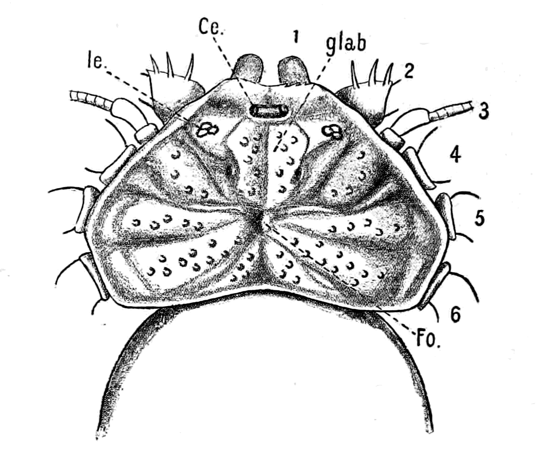
Fig. 107.—Phrynus Margine-Maculata.
Ce., median eyes; le., lateral eyes; glab., median plate over brain; Fo., fovea.
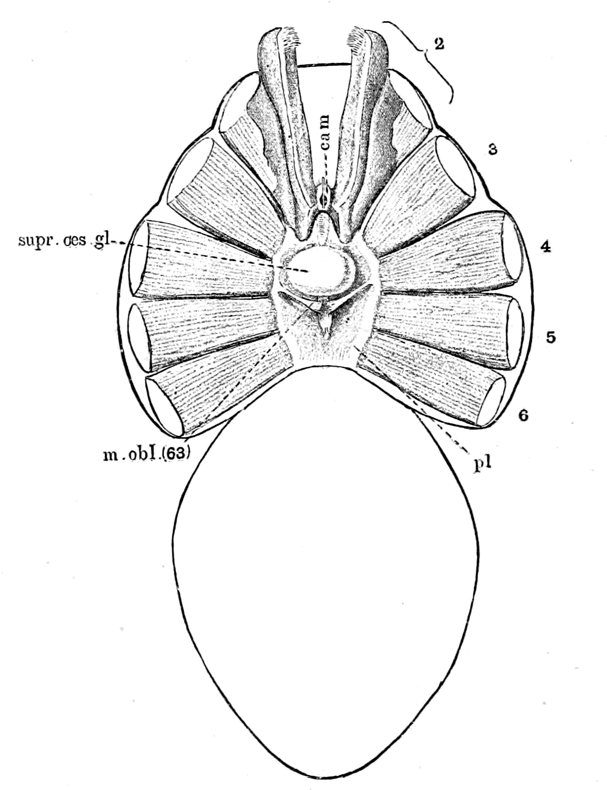
Fig. 108.—Phrynus sp. (?). Carapace removed.
cam., camerostome; pl., plastron.
The evidence of the Xiphosura and of the Hemiaspidæ conclusively shows, in Woodward's opinion, that the Merostomata are closely related to the Trilobita, and the Hemiaspidæ especially are supposed to be intermediate between the trilobites and the king-crabs. They are characterized, as also Belinurus and Prestwichia, by the absence of any prosomatic appendages, so that in these cases, as is seen in Fig. 12 (p. 30), representing Bunodes lunula, found in the Eurypterus layer at Rootziküll, we have an animal somewhat resembling Limulus in which the prosomatic appendages have either dwindled away and are completely hidden by the prosomatic carapace, or became so soft as not to be preserved in the fossilized condition. The appearance of the prosomatic carapace is, to my mind, suggestive of the presence of such appendages, for it is marked out radially, as is seen in the figure, in a manner resembling somewhat the markings on the prosomatic carapace of Mygale or Phrynus; the latter markings, as already mentioned, are due to the aponeuroses between the tergo-coxal muscles of the prosomatic appendages which lie underneath and are attached to the carapace.
A very similar radial marking is shown by Woodward in his picture of Hemiaspis limuloides, reproduced in Fig. 109, found in the Lower Ludlow beds at Leintwardine. This species has yielded the most perfect specimens of the genus Hemiaspis, which is recognized as differing from Bunodes by the possession of a telson.
It is striking to find that similar indications of segments have been found on the dorsal surface of the head-region in many of the most ancient extinct fishes, as will be fully discussed later on.
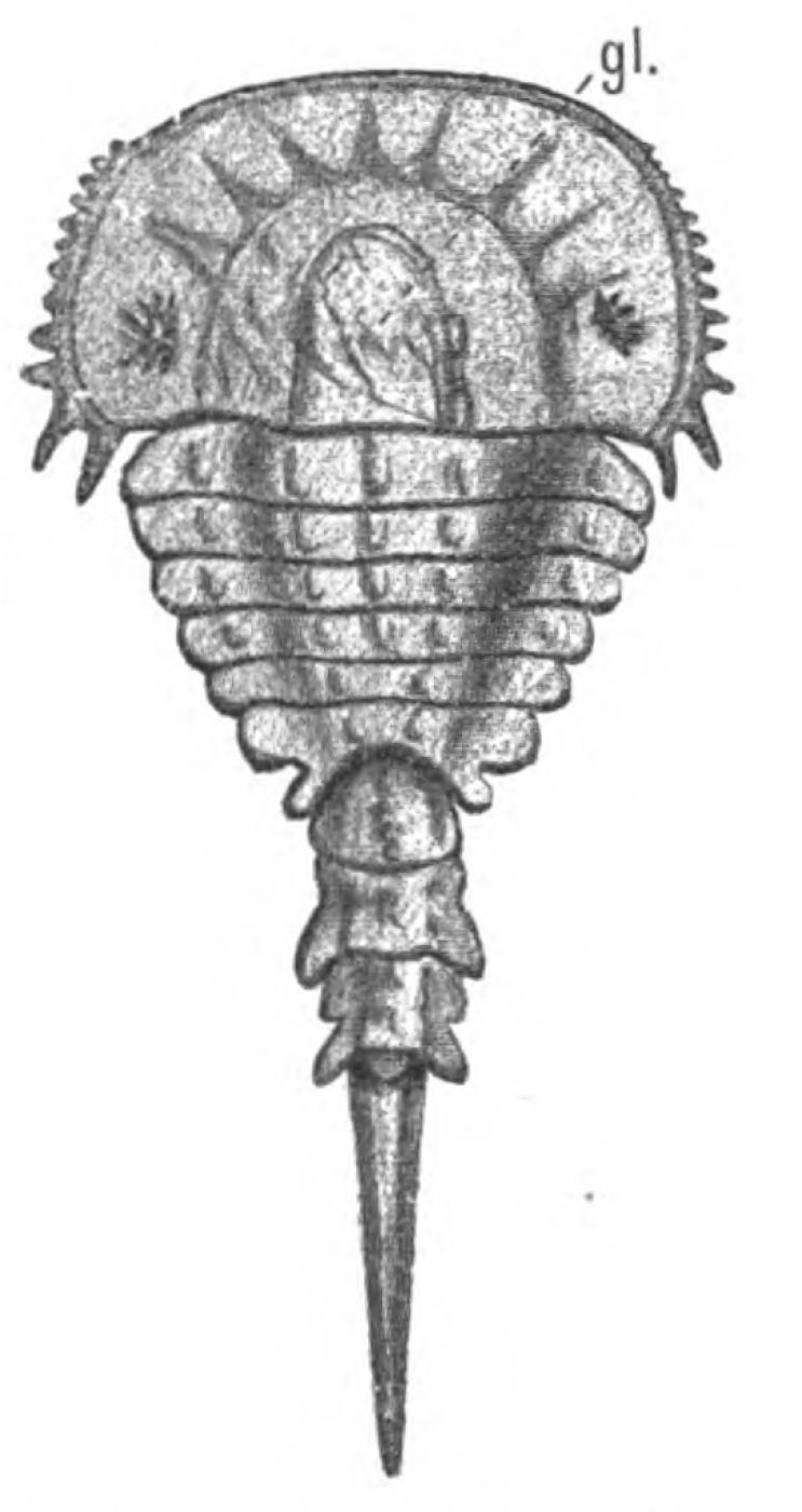
Fig.109.—Hemiaspis limuloides. (From Woodward.)
gl., glabellum.
The Evidence of Cœlomic Cavities.
In the head-region of the vertebrate, morphologists depend largely upon the embryonic divisions of the mesoderm for the estimation of the number of segments, and, therefore, upon the number of cœlomic cavities in this region, the walls of which give origin to the striated muscles of the head, so that the question of the number of segments depends very largely upon the origin of the muscles from the walls of these head-cavities. It is therefore interesting to examine whether a similar criterion of segmentation holds good in such a segmented animal as Limulus, or in the members of the scorpion group, in which the number of segments are known definitely by the presence of the appendages. In Limulus we know, from the observations of Kishinouye, that a series of cœlomic cavities are formed embryologically in the various segments of the mesosoma and prosoma, in a manner exceedingly similar to their mode of formation in the head-region of the vertebrate, and he has shown that in the mesosoma a separate cœlomic cavity exists for each segment, so that just as the dorso-ventral somatic muscles are regularly segmentally arranged in this region, so are the cœlomic cavities, and we should be right in our estimation of the number of segments in this region by the consideration of the numerical correspondence of these cavities with the mesomatic appendages. Similarly, in the vertebrate, we find every reason to believe that a single, separate head-cavity corresponds to each of the branchial segments in the opisthotic region, and therefore we should estimate rightly the number of segments by the division of the mesoderm in this region.
In the prosomatic region of Limulus, the dorso-ventral muscles are not arranged with such absolute segmental regularity as in the mesosomatic region, and Kishinouye's observations show that the cœlomic cavities in this region do not correspond absolutely with the number of prosomatic appendages. His words are:—
A pair of cœlomic cavities appears in every segment except the segments of the 2nd, 3rd, and 4th appendages, in which cœlomic cavities do not appear at all. At least eleven pairs of these cavities are produced. The eleventh pair belongs to the seventh abdominal segment.
The first pair of cœlomic cavities is common to the cephalic lobe and the segment of the first appendage (i.e. the cheliceræ).
The second cœlomic cavity belongs to the segment of the fifth appendage. It is well developed.
The ventral portion of the second cœlomic cavity remains as the coxal gland.
Consequently, if we were to estimate the number of segments in this region by the number of cœlomic cavities we should not judge rightly, for we should find only four cavities and seven appendages, as is seen in the following table:—
LIMULUS. VERTEBRATE.
Segments. Appendages. Eurypterid appendages. Cœlomic cavities. Cœlomic cavities.
1 Cheliceræ or 1st locomotor. Cheliceræ 1 Anterior

2 2nd locomotor Endognaths 2 Premandibular

3 3rd loco"
4 4th loco"
5 5th loco"
6 6th loco" Ectognath 3 Mandibular

7 Chilaria Metastoma 4
8 Operculum Operculum Genital 5 Hyoid


9 1st branchial 1st branchial 6
100 2nd brat" 2nd branchial 7 1st branchial
110 3rd bran" 3rd bran" 8 2nd brat"
120 4th bran" 4th bran" 9 3rd bran"
130 5th bran" 5th bran" 100 4th bran"
140 6th bran" 110
LIMULUS. VERTEBRATE.
Segments. Appendages. Eurypterid appendages. C.c. Cœlomic cavities.
1 Cheliceræ or 1st locomotor. Cheliceræ 1 Anterior
2 2nd locomotor Endognaths 2 Premandibular

3 3rd loco"
4 4th loco"
5 5th loco"
6 6th loco" Ectognath 3 Mandibular
7 Chilaria Metastoma 4
8 Operculum Opc. Genital 1st branchial 5 Hyoid
9 1st branchial 6
10 2nd brat" 2nd branchial 7 1st branchial
11 3rd bran" 3rd bran" 8 2nd brat"
12 4th bran" 4th bran" 9 3rd bran"
13 5th bran" 5th bran" 10 4th bran"
14 6th bran" 11
The second cavity would in reality represent four segments belonging to the 2nd, 3rd, 4th, 5th locomotor appendages, i.e. the very four segments which in the Eurypteridæ are concentrated together to form the endognaths, and we should be justified in putting this interpretation on it, because, according to Kishinouye, its ventral portion forms the coxal gland, and, according to Lankester, the coxal gland sends prolongations into the coxa of the 2nd, 3rd, 4th, 5th locomotor appendages. Similarly in the vertebrate, we find three head-cavities in the region which corresponds, on my theory, to the prosomatic region of Limulus, (1) the anterior cavity discovered by Miss Platt, (2) the premandibular cavity, and (3) the mandibular cavity, which, if they corresponded with the prosomatic cœlomic cavities of Limulus, would represent not three segments but seven segments, as follows:—the anterior cavity would correspond to the first cœlomic cavity, i.e. the cavity of the cheliceral segments in both Limulus and the Eurypteridæ; the premandibular, to the second cœlomic cavity, representing, therefore, the 2nd, 3rd, 4th, 5th prosomatic segments in Limulus and the endognathal segments in the Eurypteridæ; and the mandibular to the 3rd and 4th cœlomic cavities, representing the last locomotor and chilarial segments in Limulus, i.e. the ectognathal and metastomal segments in the Eurypteridæ.
It is worthy of note that, in respect to their cœlomic cavities, as in the position and origin of their nerves in the central nervous system, the first pair of appendages, the cheliceræ, retain a unique position, differing from the rest of the prosomatic appendages.
In the table I have shown how the vertebrate cœlomic cavities may be compared with those of Limulus. The next question to consider is the evidence obtained by morphologists and anatomists as to the number of segments supplied by the trigeminal nerve-group; this question will be considered in the next chapter.
Summary.
In Chapters IV. and V. I have dealt with the opisthotic segments of the vertebrate, including therein the segments supplied by the facial nerve, and shown that they correspond to the mesosomatic segments of the palæostracan; consequently the facial (VII.), glossopharyngeal (IX.), and vagus (X.) nerves originally supplied the branchial and opercular appendages.
In this chapter the consideration of the pro-otic segments is commenced, that is, the segments supplied by the trigeminal (V.) and the eye-muscle nerves (III., IV., VI.). I have considered the VIth nerve with the rest of the eye-muscle nerves for convenience' sake, though in reality it belongs to the same segment as the facial. Of these, that part of the trigeminal which innervates the muscles of mastication corresponds to the splanchnic segments, while the eye-muscle nerves belong to the corresponding somatic segments; but the pro-otic segments of the vertebrate ought to correspond to the prosomatic segments of the invertebrate, just as the opisthotic correspond to the mesosomatic. Therefore the motor part of the trigeminal ought to supply muscles which originally moved the prosomatic appendages, while the eye-muscles ought to have belonged to the somatic part of the same segments.
The first question considered is the number of segments which ought to be found in this region. In Limulus, the Eurypteridæ, and the scorpions there are seven prosomatic segments which carry (1) the cheliceræ, (2, 3, 4, 5) the four first locomotor appendages—the endognaths, (6) the large special appendage—the ectognath—and (7) the appendages, which in Limulus are known as the chilaria, and are small and insignificant, but in Eurypterus and other forms grow forwards, fuse together, and form a single median lip to an accessory oral chamber, which lip is known as the metastoma. Of these appendages the cheliceræ and endognaths tend to dwindle away and become mere tentacles, while the large swimming ectognath and metastoma remain strong and vigorous.
In this, the prosomatic region, the somatic segmentation is not characterized by the presence of the longitudinal muscle segments, for they do not extend into this head-region, but only by the presence of the segmental somatic ventro-dorsal muscles. Among the muscles of the appendages the system of large tergo-coxal muscles is especially apparent.
From these considerations it follows that the number of segments in this region in the vertebrate ought to be seven; that the musculature supplied by the trigeminal nerve ought to represent seven ventral or splanchnic segments, of which only the last two are likely to be conspicuous; and that the musculature supplied by the eye-muscle nerves ought to be dorso-ventral in direction, which it is, and represent seven dorsal or somatic segments.
A further peculiarity of this region, both in Limulus and the scorpions, is found in the excretory organs which are known by the name of coxal glands, because they extend into the basal joint, or coxa, of certain of the prosomatic limbs. The appendages so characterized are always the four endognaths, and it follows that if these four endognaths lose their locomotor power, become reduced in size, and concentrated together to form mere tentacles, then of necessity the coxal glands will be concentrated together, and tend to form a glandular mass in the region of the mouth; in fact, take up a position corresponding to that of the pituitary body in vertebrates.
Taking all these facts into consideration, it is possible to construct a drawing of a sagittal section through the head-region of Eurypterus, which will represent, with considerable probability, the arrangement of parts in that animal. This can be compared with the corresponding section through the head of Ammocœtes.
Now, as pointed out in the last chapter, the early stage of Ammocœtes is remarkably different from the more advanced stage; at that time the septum between the oral and respiratory chambers has not yet broken through, and the olfactory or nasal tube, known at this stage as the tube of the hypophysis, is directed ventrally, not dorsally.
The comparison of the diagram of Eurypterus with that of the early stage of Ammocœtes is remarkably close, and immediately suggests not only that the single nose of the former is derived from the corresponding organ in the palæostracan, but that the pituitary body is derived from the concentrated coxal glands, and the lower lip from the metastoma. The further working out of these homologies will be discussed in the next chapter.
In addition to the evidence of segmentation afforded by the appendages, there are in this region, in Limulus and the scorpion group, three other criteria of segmentation available to us, if from any cause the evidence of appendages fails us. These are—
1. The number of neuromeres are marked out in this region of the brain more or less plainly, especially in the young animal, just as they are also in the embryo of the vertebrate.
2. The segmentation is represented here, just as in the mesosomatic region, by two sets of muscle-segments; the one somatic, consisting of the segmentally arranged dorso-ventral muscles, the continuation of the group already discussed in connection with the mesosomatic segmentation, and the other appendicular characterized by the tergo-coxal muscles. These latter segmental muscles are especially valuable, for in such forms as Mygale, Phrynus, etc., their presence is indicated externally by markings on the prosomatic carapace, and thus corresponding markings found on fossil carapaces or on dorsal head-shields can be interpreted. These two sets of muscle-segments correspond in the vertebrate to the somatic and splanchnic segmentations.
3. In the vertebrate the segmentation in this region is indicated by the cœlomic or head-cavities, which are cavities formed in the mesoderm of the embryo, the walls of which give origin to the striated muscles of the head. In Limulus corresponding cœlomic cavities are found, which are directly comparable with those found in the vertebrate.
CHAPTER VIII
THE SEGMENTS BELONGING TO THE TRIGEMINAL NERVE-GROUP
The prosomatic segments of the vertebrate.—Number of segments belonging to the trigeminal nerve-group.—History of cranial segments.—Eye-muscles and their nerves.—Comparison with the dorso-ventral somatic muscles of the scorpion.—Explanation of the oculomotor nerve and its group of muscles.—Explanation of the trochlearis nerve and its dorsal crossing.—Explanation of the abducens nerve.—Number of segments supplied by the trigeminal nerves.—Evidence of their motor nuclei.—Evidence of their sensory ganglia.—Summary.
From the evidence given in the last chapter, combined with that given in Chapter IV., the probability of the theory that the trigeminal group of nerves of the vertebrate have been derived from the prosomatic group of nerves of the invertebrate can be put to the test by the answers to the following morphological and anatomical questions:—
1. Do we find in the vertebrate two segmentations in this region corresponding to the two segmentations in the branchial region, i.e. a somatic or dorsal series of segments, and a splanchnic or ventral series of segments? The latter would not be branchial, but rather of the nature of free tactile appendages; so that it is useless to look for or talk about gill-slits, although such appendages, being serially homologous with the branchial mesosomatic appendages, would readily give rise to the conception of branchial segments.
2. Is there morphological evidence that the trigeminal nerve is not the nerve belonging to a single segment, or even to two segments, but is really a concentration of at least six, probably seven, segmental nerves?
3. Is there morphological evidence that the oculomotor and trochlear nerves, which on all sides are regarded as belonging to the trigeminal segments, are not single nerves corresponding each to a single segment, but are the somatic motor roots belonging to the same segments as those to which the trigeminal supplies the splanchnic roots?
4. Do the mesoderm segments, which give origin to the eye-muscles, and therefore do the head-cavities of this region, correspond with the trigeminal segments? Considering the concentration of parts in this region and the difficulty already presented by the want of numerical agreement between the prosomatic appendages and the prosomatic cœlomic cavities in Limulus, it may very probably be difficult to determine the actual number of the mesoderm segments.
5. Is there anatomical evidence that the ganglion of origin of the motor part of the trigeminal nerve is not a single ganglion, but a representative of many, probably seven?
6. Is there anatomical evidence that the ganglia of origin of the oculomotor and trochlear nerves represent many ganglia?
7. Is there any evidence that the organs originally supplied by the motor part of the trigeminal nerve are directly comparable with prosomatic appendages?
It is agreed on all sides that in this region of the head there is distinct evidence of double segmentation, the dorsal mesoderm segments giving origin to the eye-muscles, and the ventral segments to the musculature innervated by the trigeminal nerve. Originally, according to the scheme of van Wijhe, two segments only were recognized, the dorsal parts of which were innervated by the IIIrd and IVth nerves respectively. Since his paper, the tendency has been to increase the number of segments in this region, as is seen in the following sketch, taken from Rabl, of the history of cranial segmentation.
History of Cranial Segmentation.
The first attempt to deal with this question was made by Goethe and Oken. They considered that the cranial skeleton was composed of a series of vertebræ, but as early as 1842 Vogt pointed out that only the occipital segments could be reduced to vertebræ. In 1869, Huxley showed that vertebræ were insufficient to explain the cranial segmentation, and that the nerves must be specially considered. The olfactory and optic nerves he regarded as parts of the brain, not true segmental nerves; the rest of the cranial nerves were segmental, with special reference to branchial arches and clefts, the facial, glossopharyngeal, and separate vagus branches supplying the walls of the various branchial pouches. In a similar manner, the supra- and infra-maxillary branches of the trigeminal were arranged on each side of the mouth, and the inner and outer twigs of the first (ophthalmic) branch of the trigeminal on each side of the orbito-nasal cleft, the trabecular and the supra-maxillary arches being those on each side of this cleft. Thus Huxley considered that there was evidence of a series of pairs of ventral arches belonging to the skull, viz. the trabecular and maxillary in front of the mouth, the mandibular, hyoid, and branchial arches behind, and that the Vth, VIIth, IXth, and Xth nerves were segmental in relation to these arches and clefts. Gegenbaur, in 1871 and 1872, considered that the branchial arches represented the lower arches of cranial vertebræ, and therefore corresponded to lower arches in the spinal region, i.e. the skull was composed of as many vertebræ as there are branchial arches. These vertebræ were confined to the notochordal part of the skull, the prechordal part having arisen secondarily from the vertebral part, while the number of vertebræ are at least nine, possibly more. The nerves which could be homologized with spinal nerves were, he thought, divisible into two great groups—(1) the trigeminal group, which included the eye-muscle nerves, the facial, and its dorsal branch, the auditory; (2) the vagus group, which included the glossopharyngeal and vagus.
Such was the outcome of the purely comparative anatomical work of Huxley and Gegenbaur—work that has profoundly influenced all the views of segmentation up to the present day.
Now came the investigations of the embryologists, of whom I will take, in the first instance, Balfour, whose observations on the embryology of the Selachians led him to the conclusion that besides the evidence of segmentation to be found in the cranial nerves and in the branchial clefts, further evidence was afforded by the existence of head-cavities, the walls of which formed muscles just as they do in the spinal region. He came to the conclusion that the first head-cavity belonged to one or more pre-oral segments, of which the nerves were the oculomotor, trochlearis, and possibly abducens; while there were seven post-oral segments, each with its head-cavity and its visceral arch, of which the trigeminal, facial, glossopharyngeal, and the four parts of the vagus were the respective nerves.
Marshall, in 1882, considered that the cranial segments were all originally respiratory, and that all the segmental nerves are arranged uniformly with respect to a series of gill-clefts which have become modified anteriorly and have been lost, to a certain extent, posteriorly. He included the olfactory nerves among the segmental nerves, and looked upon the olfactory pit, the orbito-nasal lacrymal duct, the mouth, and the spiracle as all modified gill-slits, so that he reckoned three pre-oral and oral segments belonging to the Ist, IIIrd, IVth, and Vth nerves, and eight post-oral segments belonging respectively to the VIIth and VIth nerves, and to the IXth nerve, and six segments belonging to the Xth nerve. He pointed out that muscles supplied by the oculomotor nerve develop from the outer wall of the first head-cavity; not, however, the obliquus superior and rectus externus, the latter originating probably from the walls of the third cavity.
In the same year, 1882, came van Wijhe's well-known paper, in which he showed that the mesoderm of the head in the selachian divided into two sets of segments, dorsal and ventral; that the dorsal segments were continuous with the body-somites, and that the ventral segments formed the lateral plates of mesoblast between each of the visceral and branchial pouches. He concluded that the dorsal somites were originally nine in number, that each was supplied with a ventral nerve-root, in the same way as the somites in the trunk, and that to each one a visceral pouch corresponded, whose walls were supplied by the corresponding dorsal nerve-root; of these nine segments, the ventral nerve-roots of the first three segments were respectively the oculomotor, trochlearis, and abducens nerves. The next three segments possessed no definable ventral root or muscles, and the seventh, eighth, and ninth segments possessed as ventral roots the hypoglossal nerve, with its muscular supply. The corresponding dorsal nerve-roots were the trigeminal, facial, auditory, glossopharyngeal and vagus nerves, the difference between cranial and spinal dorsal roots being that the former contain motor fibres.
Ahlborn, in 1884, drew a sharp distinction between the segments of the mesoderm and those of the endoderm. The former segmentation he called mesomeric, the latter branchiomeric. He considered the two segmentations to be independent, and concluded that the branchiomeric was secondary to the mesomeric, and therefore not of segmental value. As to the segments of the mesoderm in the head, the three hindmost or occipital in Petromyzontidæ remain permanently, and correspond to the three last segments in the selachian head. Of the anterior mesoderm segments, he considered that there were originally six, and that there are six typical eye-muscles in all Craniota, which have been compressed into three segments, as in Selachia.
Froriep (1885) showed in sheep-embryos and in chicks that the hypoglossal nerve belongs to three proto-vertebræ posterior to the vagus region, which were true spinal segments. He therefore modified Gegenbaur's conceptions to this extent: that portion of the skull designated by Gegenbaur as vertebral must be divided into two parts—a hind or occipital region, which is clearly composed of modified vertebræ and is the region of the hypoglossal nerves, and a front region, extending from the oculomotor to the accessorius nerves, which is characterized segmentally by the formation of branchial arches, but in which there is no evidence that proto-vertebræ were ever formed. He therefore divides the head-skeleton into three parts—
1. Gegenbaur's evertebral part—the region of the olfactory and optic nerves—which cannot be referred to any metameric segmentation.
2. The pseudo-vertebral, pre-spinal, or branchial part, clearly shown to be segmented from the consideration of the nerves and branchial arches, but not referable to proto-vertebræ—the region of the trigeminal and vagus nerves.
3. The vertebral spinal part—the region of the hypoglossal nerves.
He further showed that the ganglia of the specially branchial nerves, the facial, glossopharyngeal, and vagus, are at one stage in connection with the epidermis, so that these parts of the epidermis represent sense-organs which do not develop; these organs probably belonged to the lateral line system. As the connection takes place at the dorsal edge of the gill-slits, they may also be called rudimentary branchial sense-organs.
Since this paper of Froriep's, it has been generally recognized, and Gegenbaur has accepted Froriep's view, that the three hindmost metameres, which distinctly show the characteristics of vertebræ, belong to the spinal and not to the cranial region, so that the metameric segmentation of the cranial region proper has become more and more associated with the branchial segmentation. Froriep's discovery of the rudimentary branchial sense-organs as a factor in the segmentation question has led Beard to the conclusion that the olfactory and auditory organs represent in a permanent form two of these rudimentary branchial sense-organs. He therefore includes both the olfactory and auditory nerves in his list of cranial segmental nerves, and makes eleven cranial branchial segments in front of the spinal segments represented by the hypoglossal.
A still larger number of cranial segments is supposed to exist, according to the researches of Dohrn and Killian, in the embryos of Torpedo ocellata. The former, holding to the view that vertebrates arose from annelids, considered that the head was formed of a series of metameres, to each one of which a mesoderm-segment, a gill-arch, a gill-cleft, a segmental nerve and vessel belonged. He found in the front head-region of a Torpedo embryo, corresponding to van Wijhe's first four somites, no less than twelve to fifteen mesoderm segments, and concluded, therefore, that the eye-muscle nerves, especially the oculomotor, represented many segmental nerves, and were not the nerves of single segments; so, also, that the inferior maxillary part of the trigeminal and the hyoid nerve of the facial are probably not single nerves, but a fusion of several. Killian comes to much the same conclusion as Dohrn, for he finds seventeen to eighteen separate mesoderm segments in the head, of which twelve belong to the trigeminal and facial region.
Since Rabl's paper, a number of papers have appeared, especially from America, dealing with yet another criterion of the original segmentation of the head, viz. a series of divisions of the central nervous system itself, which are seen at a very early stage of development, and are called neuromeres; the divisions in the cranial region being known as encephalomeres, and those of the spinal region as myomeres. Locy's paper has especially brought these divisions into prominence as a factor in the question of segmentation. They are essentially segments of the epiblast and not of the mesoblast; they are conspicuous in very early stages, and appear to be in relation with the cranial nerves, according to Locy. He recognizes in Squalus acanthias, in front of the spino-occipital region, fourteen pairs of such encephalomeres and a median unsegmented termination, which may represent one more pair fused in the middle line, making at least fifteen. He distributes these fifteen segments as follows: fore-brain three and unsegmented termination, mid-brain two, and hind-brain nine.
Again, Kupffer, in his recent papers on the embryology of Ammocœtes, asserts that especial information as to the number of primitive segments is afforded by the appearance in the early stages of a series of epibranchial ganglia in connection with the cranial nerves, which remain permanently in the case of the vagus nerves, but disappear in the case of pro-otic nerves. He considers that the evidence points to the number of segments in the mid- and hind-brain region as being primitively fifteen, viz. six segments belonging to the trigeminal and abducens group, three segments belonging respectively to the facial, auditory, and glossopharyngeal, and six to the vagus.
From this sketch we see that the modern tendency is to make six segments at least out of the region of the trigeminal nerves rather than two. In this region, as already mentioned, the evidence of segmentation is based more clearly on the somatic than on the splanchnic segments. We ought, therefore, in the first place, to consider the teaching of the eye-muscles and their nerves and the cœlomic cavities in connection with them, and see whether the hypothesis that such muscles represent the original dorso-ventral somatic muscles of the palæostracan ancestor is in harmony with and explains the facts of modern research.
Eye-Muscles and their Nerves.
The only universally recognized somatic nerves belonging to these segments which exist in the adult are the nerves to the eye-muscles, of which, according to van Wijhe, the oculomotor is the nerve of the 1st segment, the trochlearis of the 2nd, and the abducens of the 3rd; while the nerves and muscles belonging to the 4th and 5th segments, i.e. the 2nd facial and glossopharyngeal segments respectively, show only the merest rudiments, and do not exist in the adult. One significant fact appears in this statement of van Wijhe, and is accepted by all those who follow him, viz. that the oculomotor nerve has equal segmental value with the trochlearis and the abducens, although it supplies a number of muscles, each of which, on the face of it, has the same anatomical value as the superior oblique or external rectus. Dohrn alone, as far as I know, as already pointed out, insists upon the multiple character of the oculomotor nerve.
As far as the anatomist is concerned, the evidence is becoming clearer and clearer that the nucleus of the IIIrd nerve is a composite ganglion composed of a number of nuclei, each similar to that of the trochlearis, so that if the trochlearis nucleus is a segmental motor nucleus, then the oculomotor nucleus is a combined nucleus belonging to at least four segmental nerves, each of which has the same value as that of the trochlearis.
The investigations of a number of anatomists, among whom may be mentioned Gudden, Obersteiner, Edinger, Kölliker, Gehuchten, all lead directly to the conclusion that this oculomotor nucleus is composed of a number of separate nuclei, of which the most anterior as also the Edinger-Westphal nucleus contains small cells, while the others contain large cells. Thus Edinger divides the origin of the oculomotor nerve into a small-celled anterior part and a larger posterior part, of which the cells are larger and distinctly arranged in three groups—(1) dorsal, (2) ventral, and (3) median. Between the anterior and posterior groups lies the Edinger-Westphal nucleus, which is small-celled; naturally, the large-celled group is that which gives origin to the motor nerves of the eye-muscles, the small-celled being possibly concerned with the motor nerves of the pupillary and ciliary muscles. I may mention that Kölliker considers that the anterior lateral nucleus has nothing to do with the oculomotor nerve, but is a group of cells in which the fibres of the posterior longitudinal bundle and of the deep part of the posterior commissure terminate.
These conclusions of Edinger are the outcome of work done in his laboratory by Perlia, who says that in new-born animals the nucleus of origin of the oculomotor nerve is made up of a number of groups quite distinct from each other, each group being of the same character as that of the trochlearis. He finds the same arrangement in various mammals and birds. Further, he finds that some of the fibres arise from the nucleus of the opposite side, thus crossing, as in the trochlearis; these crossing fibres belong to the most posterior of the dorsal group of nuclei, i.e. to the nerve to the inferior oblique muscle.
The evidence, therefore, points to the conclusion that the oculomotor nucleus is a multiple nucleus, each part of which gives origin to one of the nerves of one of the eye-muscles.
Edinger says that such an array of clinical observations exists, and of facts derived from post-mortem dissections, that one may venture to designate the portion of the nucleus from which the innervation of each individual ocular muscle comes. He gives Starr's table, the latest of these numerous attempts, begun by Pick. According to Starr, the nuclei of the nerves to the individual muscles are arranged from before backward, thus—
m. sphincter iridis. m. ciliaris.
m. levator palpebræ. m. rectus internus.
m. rectus superior. m. rectus inferior.
m. obliquus inferior.
Further, the evidence of the well-known physiological experiments of Hensen and Völckers that the terminal branches of the oculomotor nerve arise from a series of segments of the nucleus, arranged more or less one behind the other in a longitudinal row, leads them to the conclusion that the nuclei of origin are arranged as follows, proceeding from head to tail:—
Nearest brain. 1. m. ciliaris.
2. m. sphincter iridis.
3. m. rectus internus.
4. m. rectus superior.
5. m. levator palpebræ.
6. m. rectus inferior.
Most posterior. 7. m. obliquus inferior.
It is instructive to compare this arrangement of Hensen and Völckers with the arrangement of the origin of these muscles from the premandibular cavity as given by Miss Platt.
Thus she states that the most posterior part of the premandibular cavity is cut off so as to form a separate cavity, resembling, except in position, the anterior cavity; this separate, most posterior part gives origin to the inferior oblique muscle. She then goes on to describe how the dorsal wall of the remainder of the premandibular cavity becomes thickened, to form posteriorly the rudiment of the inferior rectus and anteriorly the rudiments of the superior and internal recti, a slight depression in the wall of the cavity separating these rudiments. The internal rectus is the more median of the two anterior muscles. In other words, her evidence points not only to a fusion of somites to form the premandibular cavity, but also to the arrangement of these somites as follows, from head to tail: (1) internal rectus, (2) superior rectus, (3) inferior rectus, (4) inferior oblique—an order precisely the same as that of Hensen and Völckers, and of Starr.
I conclude, from the agreement between the anatomical, physiological, and morphological evidence, that the IIIrd and IVth nerves contain the motor somatic nerves belonging to the same segments as the motor trigeminal, in other words, to the prosomatic segments, so that the eye-muscles, innervated by III. and IV., represent segmental muscles belonging to the prosoma. Further, I conclude that originally there were seven prosomatic segments, the first of which is represented by the anterior cavity described by Miss Platt, and does not form any permanent muscles; that the next four belong to the premandibular cavity, and the muscles formed are the superior rectus, internal rectus, inferior rectus, and inferior oblique; and that the last two belong to the mandibular cavity, the muscles formed being Miss Platt's mandibular muscle and the superior oblique. It is, to say the least of it, a striking coincidence that such an arrangement of the cœlomic cavities as here given should be so closely mimicked by the arrangement in the prosomatic region of Limulus as already mentioned; it suggests inevitably that the head-cavities of the vertebrate are nothing more than the prosomatic and mesosomatic segmental cœlomic cavities, as found in animals such as Limulus. In the table on p. 253, I have inserted the segments in the vertebrate for comparison with those of Limulus.
Before we can come to any conclusion as to the original position of these eye-muscles, it is necessary to consider the VIth nerve and the external rectus muscle. This nerve and this muscle belong to van Wijhe's 4th segment. The muscle is, therefore, the somatic segmental muscle belonging to the same segment as the facial and is, in fact, a segmental muscle belonging not to the prosoma, but to the mesosoma. Neal comes to the conclusion that the existing abducens is the only root which remains permanent among a whole series of corresponding ventral roots belonging to the opisthotic segments, and further points out that the external rectus was originally an opisthotic muscle which has taken up a pro-otic position, or, translating this statement into the language of Limulus, etc., it is a mesosomatic muscle which has taken up a prosomatic position.
There is, however, another muscle—the Retractor oculi—belonging to the same group which is innervated by the VIth nerve. Quite recently Edgeworth has shown that in birds and reptiles this muscle belongs to the hyoid segment; so that in this respect also the hyoid segment proclaims its double nature.
With respect to the external rectus muscle, Miss Platt has shown that the mandibular muscle is formed close alongside the external rectus, so that the two are in close relationship as long as the former exists.
Further, as already mentioned, the eye-muscles in Ammocœtes must be considered by themselves; they do not belong in structure or position to the longitudinal somatic muscles innervated by the spinal nerves; their structure is not the same as that of the tubular constrictor or branchial muscles, but resembles that structure somewhat; their position is dorso-ventral rather than longitudinal; they may be looked upon as a primitive type of somatic muscles segmentally arranged, the direction of which was dorso-ventral.
Anderson also has shown that the time of medullation of the nerves supplying these muscles is much earlier than that of the nerves belonging to the somatic trunk-muscles, their medullation taking place at the same time as that of the motor nerves supplying the striated visceral muscles; and Sherrington has observed that these muscles do not possess muscle-spindles, while all somatic trunk-muscles do. Both these observations are strong confirmation of the view that the eye-muscles must be classified in a different category to the ordinary somatic trunk muscle group.
What, then, is the interpretation of these various embryological and anatomical facts?
Remembering the tripartite division of each segmental nerve-group in Limulus into (1) dorsal or sensory somatic nerve, (2) appendage-nerve, and (3) ventral somatic nerve, I venture to suggest that the three nerves—the oculomotorius, the trochlearis, and the abducens—represent the ventral somatic nerves of the prosoma, and partly also of the mesosoma; that they are nerves, therefore, which may have originally contained sensory fibres, and which still contain the sensory fibres of the eye-muscles themselves, as stated by Sherrington. According to this suggestion, the eye-muscles are the sole survivors of the segmental dorso-ventral somatic muscles, so characteristic of the group from which I imagine the vertebrates to have sprung. In the mesosomatic region the dorso-ventral muscles which were retained were those of the appendages and not of the mesosoma itself, because the presumed ancestor breathed after the fashion of the water-breathing Limulus, by means of the dorso-ventral muscles of its branchial appendages, and not after the fashion of the air-breathing scorpion, by means of the dorso-ventral muscles of the mesosoma. The only mesosomatic dorso-ventral muscles which were retained were those of the foremost mesosomatic segments, i.e. those supplied by the VIth nerve, which were preserved owing to their having taken on a prosomatic position and become utilized to assist in the movements of the lateral eyes.
Let us turn now to the consideration of the corresponding musculature in Limulus and in the scorpion group. These muscles constitute the markedly segmental muscles to which I have given the name 'dorso-ventral somatic muscles.' They are most markedly segmental in the mesosomatic region, both in Limulus and in Scorpio, each mesosomatic segment possessing a single pair of these vertical mesosomatic muscles, as Benham calls them (cf. Fig. 58 (Dv.)). In the prosomatic region the corresponding muscles are not so clearly defined in Limulus; they are apparently attached to the plastron forming the group of plastro-tergal muscles. From Benham's description it is sufficiently evident that they formed originally a single pair to each prosomatic segment.
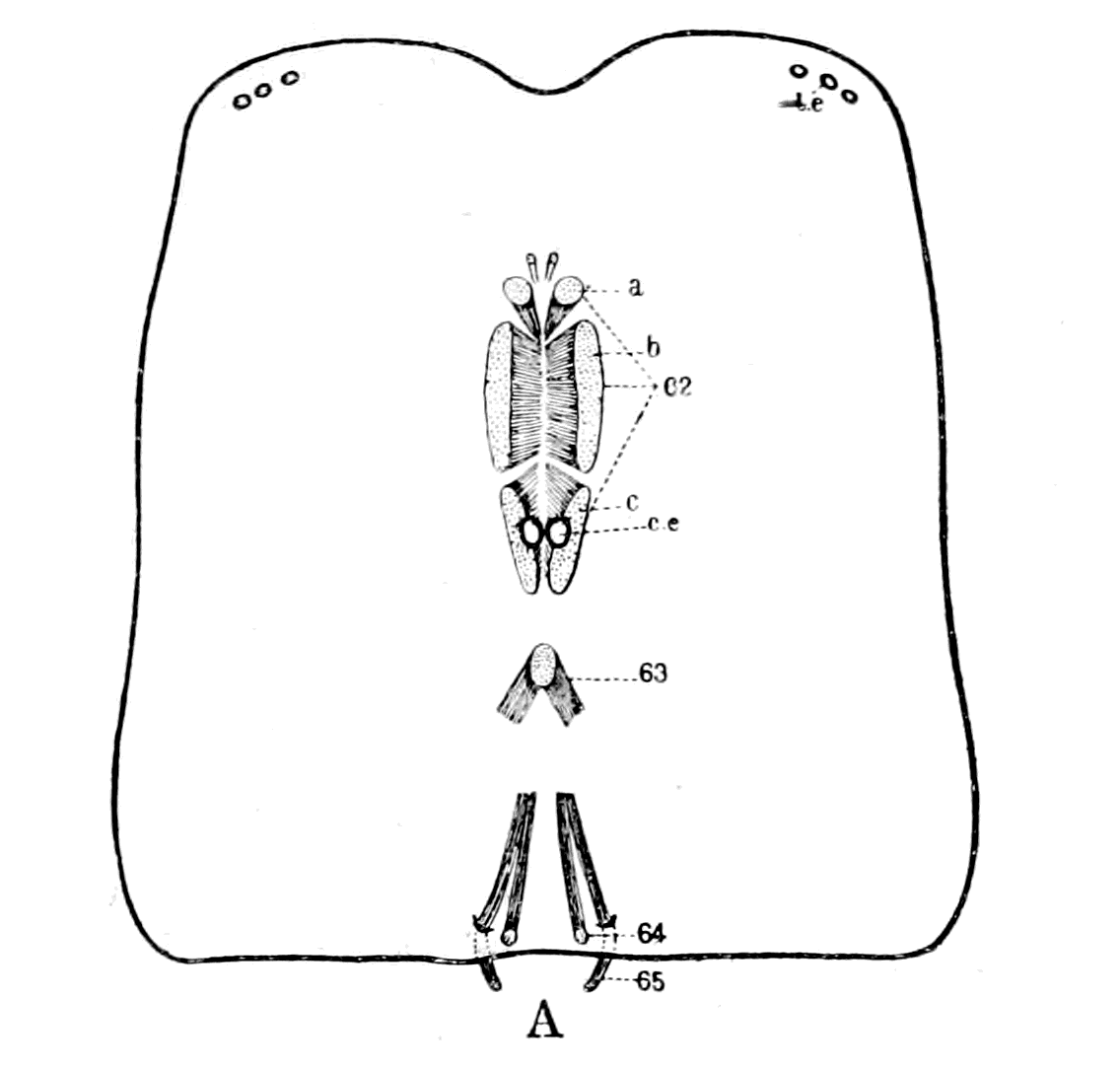
In Scorpio, according to Miss Beck, the dorso-ventral prosomatic muscles are situated near the middle line on each side and form the following well-marked series of pairs of muscles, shown in Fig. 110, A, taken from her paper, and thus described by her:—
1. The dorso-cheliceral-sternal muscle (61) is the most anterior of the dorso-ventral muscles. It is very small, and is attached to the carapace near the median line anteriorly to the central eyes.
2. The median dorso-preoral-entosclerite muscle (62) is a large muscle, between which and its fellow of the opposite side the eyes are situated. It is attached dorsally to the carapace and ventrally to the pre-oral entosclerite.
3. The anterior dorso-plastron muscle (63) is attached dorsally to the carapace in the middle line, being joined to its fellow of the opposite side. They separate, and are attached ventrally to the plastron. Through the arch thus formed the alimentary canal and the dorsal vessel pass.
4. The median dorso-plastron muscle (64) is attached dorsally to the posterior part of the carapace. It runs forward on the anterior surface of the posterior flap of the plastron to the body of the plastron, to which it is attached.
A. Dorso-ventral Muscles on Carapace of Scorpion. (From Miss Beck.)
B. Similar Muscles on Carapace of Eurypterus.
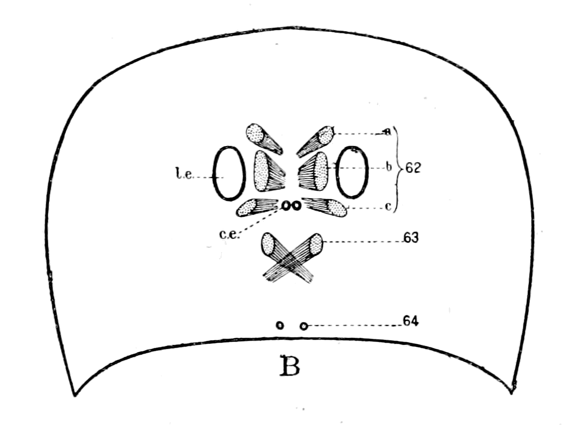
C. Similar Muscles on Head-Shield of a Cephalaspid. l.e., lateral eyes; c.e., central eyes; Fro., narial opening. 62-65 refer to Miss Beck's catalogue of the scorpion muscles.
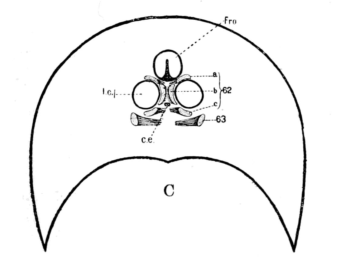
Fig. 110.
To these may be added, owing to its attachment to the plastron,
5. The posterior dorso-plastron muscle (65). This is the first of the dorso-ventral muscles attached to the mesosomatic tergites, being attached to the tergite of the first segment of the mesosoma.
This muscle is of interest, in connection with the prosomatic dorso-ventral muscles, because it is attached to the plastron, and runs a course in close contact with the muscle (64), the two muscles being attached dorsally close together, on each side of the middle line, the one at the very posterior edge of the prosomatic carapace, and the other at the very anterior edge of the mesosomatic carapace.
Taking these muscles separately into consideration, it may be remarked with respect to (61) that the cheliceral segment in its paired dorso-ventral muscles, as in its tergo-coxal muscles, takes up a separate position isolated from the rest of the prosomatic segments.
Next comes (62) the median dorso-preoral-entosclerite muscle, which is strikingly different from all the other dorso-ventral muscles in its large size and the extent of its attachment to the dorsal carapace, according to Miss Beck's figures. The reason of its large size is clearly seen upon dissection of the muscles in Buthus, for I find that, strictly speaking, it is not a single muscle, but is composed of a series of muscle-bundles, separated from each other by connective tissue. There are certainly three separate muscles included in this large muscle, which are attached in a distinct series along the pre-oral entosclerite, and present the appearance given in Fig. 110, A, at their attachment to the prosomatic carapace. Of this muscle-group the most anterior and the most posterior bundle are distinctly separate muscles; I am not, however, clear whether the middle bundle represents one or two muscles.
This division of Miss Beck's muscle (62) into three or four muscles brings the prosomatic region of the scorpion into line with the mesosomatic, and enables us to feel sure that a single pair of dorso-ventral somatic muscles belongs to each prosomatic segment just as to each mesosomatic, and, conversely, that each such single pair of muscles possesses segmental value in this region as much as in the mesosomatic.
It is very striking to see how in all the Scorpionidæ, in which the two median eyes are the principal eyes, this muscle group (62) on the two sides closely surrounds these two eyes, so that with a fixed pre-oral entosclerite, a slight movement of the eyes, laterally or anteriorly, owing to the flexibility of the carapace, might result as the consequence of their contraction. But this cannot be the main object of these muscles. The pre-oral entosclerite is firmly fixed to the camerostome, as is seen in Fig. 94, pr. ent., so that the main object of these muscles is, as Huxley has pointed out, the movement of this organ.
In order to avoid repetition of the long name given to this muscle group (62) by Miss Beck, because of their position, and for other reasons which will appear in the sequel, I will call this group of muscles the group of recti muscles. These recti muscles belong clearly to the segments posterior to the first prosomatic or cheliceral segment, and represent certainly three, probably four, of these segments, i.e. belong to the segments corresponding to the second, third, fourth, and fifth prosomatic locomotor appendages—the endognaths of the old Eurypterids.
The next pair of muscles is the pair of anterior dorso-plastron muscles (63). This muscle-pair evidently belongs to a segment posterior to the segments represented by the group already discussed, and belongs, therefore, in all probability to the same segment as the sixth pair of prosomatic appendages—the ectognaths of the old Eurypterids. This can be settled by considering either the nerve-supply or the embryological development. In the Eurypteridæ it seems most highly probable that the dorso-ventral muscles of each half of the segments belonging to the endognaths should be compressed together and separate from the dorso-ventral muscle belonging to the ectognathal segment, on account of the evident concentration and small size of the endognathal segments in contradistinction to the separateness and large size of the ectognathal segment.
The striking peculiarity of this muscle-pair, which distinguishes it from all other muscles in the scorpion, is the common attachment of the muscles of the two sides in the mid-dorsal line, so that the pair of muscles forms an arch through which the alimentary canal and dorsal blood-vessel pass.
The same dorso-ventral muscles are present in Phrynus, and in this animal the fibres of this pair of muscles (63) actually interlace before the attachment to the prosomatic carapace, so that the attachment of the muscle on each side overpasses the mid-dorsal line, and a true crossing occurs. In Fig. 108 the position of this pair of muscles is shown just posteriorly to the brain-mass. This muscle I will call the oblique muscle.
Finally we come to the muscles (64) and (65), the median and posterior dorso-plastron muscles, which run close together. Both muscles are attached to the plastron, and, therefore, to that extent belong to the prosomatic region; they are attached dorsally close to the junction of the prosoma and mesosoma. This position of the first mesosomatic dorso-ventral muscle belonging to the opercular segment may be compared with the position of the first mesosomatic dorso-ventral muscle in Limulus which has become attached to the prosomatic carapace; in both cases we see an indication that the foremost pair of mesosomatic dorso-ventral somatic muscles tend to take up a prosomatic position.
As to the pair of small muscles (64), I believe that they represent the dorso-ventral muscles of the seventh prosomatic segment (if the pair of muscles (63) belongs to the segment of the sixth locomotor prosomatic appendages), i.e. they belong to the chilarial segment or metastoma.
I desire to draw especial attention to the fact that the dorso-ventral muscle (64), which represents the seventh segment, always runs close alongside the dorso-ventral muscle (65), which represents the first mesosomatic or opercular segment.
The comparison, then, of these two sets of facts leads to the following conclusions:—
The foremost prosomatic or trigeminal segment stood separate and apart, being situated most anteriorly; the musculature of this segment does not develop, so that the only evidence of its presence is given by the anterior cœlomic cavity. This corresponds, according to my scheme, with the first or anterior cœlomic cavity of Limulus, and therefore represents, as far as the prosomatic appendages are concerned, the first prosomatic appendage-pair, or the cheliceræ; the appendage-muscles being the muscles of the cheliceræ, and the dorso-ventral somatic muscles the pair of dorso-cheliceral sternal muscles (61) in the scorpion. Both these sets of muscles, therefore, dwindle and disappear in the vertebrate.
Then came four segments fused together to form the premandibular segment, the characteristic of which is the apparent non-formation of any permanent musculature from the ventral mesoderm-segments, and the formation of the eye-muscles innervated by the oculomotor nerve from the dorsal mesoderm segments. These four segments have been so fused together that van Wijhe looked upon them as a single segment, and the premandibular cavity as the cavity of a single segment. They represent, according to my scheme, the segments belonging to the endognaths, i.e. the second, third, fourth, fifth pairs of prosomatic appendages; the premandibular cavity, therefore, represents the second cœlomic cavity in Limulus, which, according to Kishinouye, is the sole representative of the cœlomic cavities of the second, third, fourth, fifth prosomatic segments. The muscles derived from the ventral mesoderm-segments represent the muscles of these appendages, which therefore dwindle and disappear in the vertebrate, with the possible exception of the muscles innervated by the descending root of the trigeminal. The muscles derived from the dorsal mesoderm-segments, i.e. the eye-muscles supplied by the oculomotor nerve, represent the dorso-ventral somatic muscles of these four segments, muscles which are represented in the scorpion by the recti group of muscles, i.e. the median dorso-preoral-entosclerite muscles (62).
Then came two segments, the mandibular, in which muscles are formed both from the ventral and from the dorsal mesoderm-segments. From the former arose the main mass of muscles innervated by the motor root of the trigeminal, from the latter the superior oblique muscle and the mandibular muscle of Miss Platt, of which the former alone survives in the adult condition. These two segments are looked upon as a single segment by van Wijhe, of which the mandibular cavity is the cœlomic cavity. They represent, according to my scheme, the segments belonging to the sixth pair of prosomatic appendages or ectognaths, and the seventh pair, i.e. the chilaria or metastoma.
The first part, then, of the mandibular cavity represents the third cœlomic cavity in Limulus and the muscles derived from the ventral mesoderm, in all probability the muscles of the tongue in the lamprey (cf. Chap. IX.), which represents the ectognaths or sixth pair of prosomatic appendages, while the muscles derived from the dorsal mesoderm, i.e. the superior oblique muscles, represent the dorso-ventral somatic muscles of this segment, muscles which are represented in the scorpion group by the pair of anterior dorso-plastron or oblique muscles (63).
The second part of the mandibular cavity represents the 4th cœlomic cavity in Limulus and the muscles derived from the ventral mesoderm, in all probability the muscles of the lower lip in the lamprey (cf. Chap. IX.), which represents the metastoma; while the muscles derived from the dorsal mesoderm, i.e. Miss Platt's pair of mandibular muscles, represent the dorso-ventral somatic muscles of this segment, muscles which are represented in the scorpion group by the pair of median dorso-plastron muscles (64).
In connection with this last pair of muscles we find that the external rectus in the vertebrate represents the first dorso-ventral mesosomatic muscle in the scorpion, i.e. the posterior dorso-plastron muscle (65), and, as already mentioned (p. 267), that it always lies closely alongside the mandibular muscle, just as in the scorpion group muscle (65) always lies alongside muscle (64).
In the invertebrate as well as in the vertebrate this muscle is a mesosomatic muscle which has taken up a prosomatic position.
The question naturally arises, what explanation can be given of the fact that these dorso-ventral muscles attached on each side of the mid-dorsal line to the prosomatic carapace became converted into the muscles moving the eyeballs of the two lateral eyes? An explanation which must take into account not only the isolated position of the abducens nerve, but also the extraordinary course of the trochlearis. The natural and straightforward answer to this question appears to me quite satisfactory, and I therefore venture to commend it to my readers.
I have argued the case out to myself as follows: The lateral eyes must have been originally situated externally to the group of muscles innervated by the oculomotor nerve, for a sheet of muscle representing the superior internal and inferior rectus muscles could only wrap round the internal surface of each lateral eye; i.e. the arrangement of the muscle-sheet, as in the scorpion, about two median eyes, is in the wrong position, for if those two eyes, which are the main eyes in the scorpion, were to move outwards to become two lateral eyes, then such a muscle-group would form a superior external and inferior rectus group. The evidence, however, of Eurypterus and similar forms is to the effect that the lateral eyes became big and the median eyes insignificant and degenerate. If, then, with the degeneration of the one and the increasing importance of the other, these lateral eyes came near the middle line, then the muscular group (62), which I have called the recti group, would naturally be pressed into their service, and would form an internal and not an external group of eye-muscles.
In Fig. 110, A, taken from Miss Beck's paper, I have shown the relative position of the eyes and the segmental dorso-ventral prosomatic muscles on the carapace of the scorpion. In Fig. 110, B, I have drawn the prosomatic carapace of Eurypterus Scouleri, taken from Woodward's paper, with the eyes as represented there; in this I have inserted the segmental dorso-ventral muscles as met with in the scorpion, thereby demonstrating how, with the degeneration of the median eyes and the large size of the lateral eyes, the recti muscles of the scorpion would approach the position of an internal recti group to the lateral eyes, and so give origin to the group of muscles innervated by the oculomotor nerve. In the Eurypterus these large eyes are large single eyes, not separate ocelli, as in the scorpion.
All, then, that is required is that in the first formed fishes, which still possessed the dorso-ventral muscles of their Eurypterid ancestors, the lateral eyes should be the important organs of sight, large and near the mid-dorsal line. Such, indeed, is found to be the case. In amongst the masses of Eurypterids found in the upper Silurian deposits at Oesel, as described by Rohon, numbers of the most ancient forms of fish are found belonging to the genera Thyestes and Tremataspis. The nature of the dorsal head-shields of these fishes is shown in Fig. 14, which represents the dorsal head-shield of Thyestes verrucosus, and Fig. 111 that of Tremataspis Mickwitzi. They show how the two lateral eyes were situated close on each side of the mid-dorsal line in these Eurypterus-like fishes, in the very position where they must have been if the eye-muscles were derived from the dorso-ventral somatic muscles of a Eurypterid ancestor.
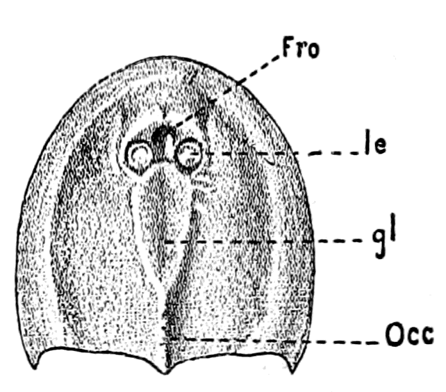
Fig. 111.—Dorsal Head-Shield of Tremataspis Mickwitzi. (From Rohon.)
Fro., narial opening; l.e., lateral eyes; gl., glabellum plate over brain; Occ., occipital spine.
In Lankester's words, one of the characteristics of the Osteostraci (Cephalaspis, Auchenaspis, etc.), as distinguished from the Heterostraci (Pteraspis), are the large orbits placed near the centre of the shield. The apparent exception of Thyestes mentioned by him is no exception, for orbits of the same character have since been discovered, as is seen in Rohon's figure (Fig. 14). In Fig. 110, C, I give an outline of the frontal part of the head-shield of a Cephalaspid, in which I have drawn the eye-muscles as in the other two figures.
Although all the members of the Osteostraci possess large lateral eyes towards the centre of the head-shield, the other group of ancient fishes, the Heterostraci, are characterized by the presence of lateral eyes far apart, situated on the margin of the head-shield on each side (cf. Fig. 142, o, p. 350).
So, also, on the invertebrate side, the lateral eyes of Pterygotus and Slimonia are situated on the margin of the prosomatic carapace, while those of Eurypterus and Stylonurus are situated much nearer the middle line of the prosomatic carapace.
Next comes the question of the superior oblique muscle and the trochlearis nerve. Why does this nerve (n.IV. in Fig. 106, C and D) alone of all the nerves in the body take the peculiar position it always does take? The only suggestion that I know of which sounds reasonable and worth consideration is that put forward by Fürbringer, which is an elaboration of the original suggestion of Hoffmann. Hoffmann suggested in 1889 that the trochlearis nerve represented originally a nerve for a protecting organ of the pineal eye, which became secondarily a motor nerve for the lateral eye as the pineal eye degenerated. Fürbringer differs from Hoffmann in that he considers that the nerve was originally a motor nerve, and was not transformed from sensory to motor, yet thinks Hoffmann's suggestion is in the right direction.
He points out that the crossing of the trochlearis is not a crossing of fibres between two centres in the central nervous system, but may be explained by the shifting of the peripheral organ, i.e. the muscle, from one side to the other, and the nerve following this shift. Consequently, says Fürbringer, the course of the nerve indicates the original position of the muscle, and therefore he imagines that the ancestor of the superior oblique muscle was a muscle the fibres of which were attached in the mid-dorsal line, and interlaced with those of the other side, the two muscles thus forming an arch through which the nervous system with its central canal passed. Then, for the sake of getting a more efficient pull, the crossing muscle-fibres became more definitely attached to the opposite side of the middle line, and finally obtained a new attachment on the opposite side, with the obliteration of the muscular arch; the nerve on each side, following the shifts of the muscle, naturally took up the position of the original muscular arch, and so formed the trochlear nerve, with its dorsal crossing. This explanation of Fürbringer's was associated by him with movements of the median pineal eyes, the length of their nerve, according to him, even yet indicating their previous mobility. This assumption is not, it seems to me, necessary. The length of the nerve is certainly no indication of mobility, for in Limulus and the scorpion group the nerve to each median eye is remarkably long, yet these eyes are immovably fixed in the carapace. All that is required is a pair of dorso-ventral muscles belonging to the segment immediately following the group of segments represented by the oculomotor nerves, the fibres of which should cross the mid-dorsal line at their attachment; for, seeing that the lateral eyes were originally so near this position, it follows that such muscles might form part of the muscular group belonging to the lateral eye without having previously moved the pineal eyes. In fact, Fürbringer's explanation requires as starting-point that the pair of muscles which ultimately become the superior oblique should have the exact position of the pair of dorso-ventral muscles in the scorpion, called by Miss Beck the anterior dorso-plastron muscles (63), which I have named the oblique muscles. Here, and here only, do we find an interlacement, across the mid-dorsal line, of the fibres of attachment of the muscles on the two sides, in consequence of which this pair of muscles is described by her as forming an arch encircling the alimentary canal and dorsal vessel. If, then, as I have previously argued, the primitive plastron formed a pair of trabeculæ, and the nervous system grew round the alimentary canal, such an arch would encircle the tubular central nervous system of the vertebrate.
Still more striking is this pair of muscles (63) in Phrynus (Fig. 108), where we see how the arch formed by them almost touches the posterior extremity of the supra-œsophageal brain-mass, crossing, therefore, over the beginning of the stomach region of the animal. The angle formed by the arch is much more obtuse than that formed in Scorpio, so that an actual crossing of the muscle-fibres has taken place at the point of attachment to the carapace. Also, only the part nearest the carapace is muscular, the rest forming a long tendinous prolongation of the plastron wall (the primordial cranium), as seen in the figure.
This muscle-pair is, as it should be, the pair of dorso-ventral muscles belonging to the segment immediately following on the group of segments represented by the recti muscles, i.e. according to previous argument, the segment belonging to the sixth pair of locomotor appendages or ectognaths; a muscle, therefore, which would arise in the vertebrate from the mandibular, and not from the premandibular cavity. A similar muscle probably existed in Eurypterus (M.obl. in Fig. 106, B), and, as in the case of the formation of the oculomotor group, derived from the recti group of the scorpion, would form the commencement of the superior oblique muscle in Thyestes and Tremataspis.
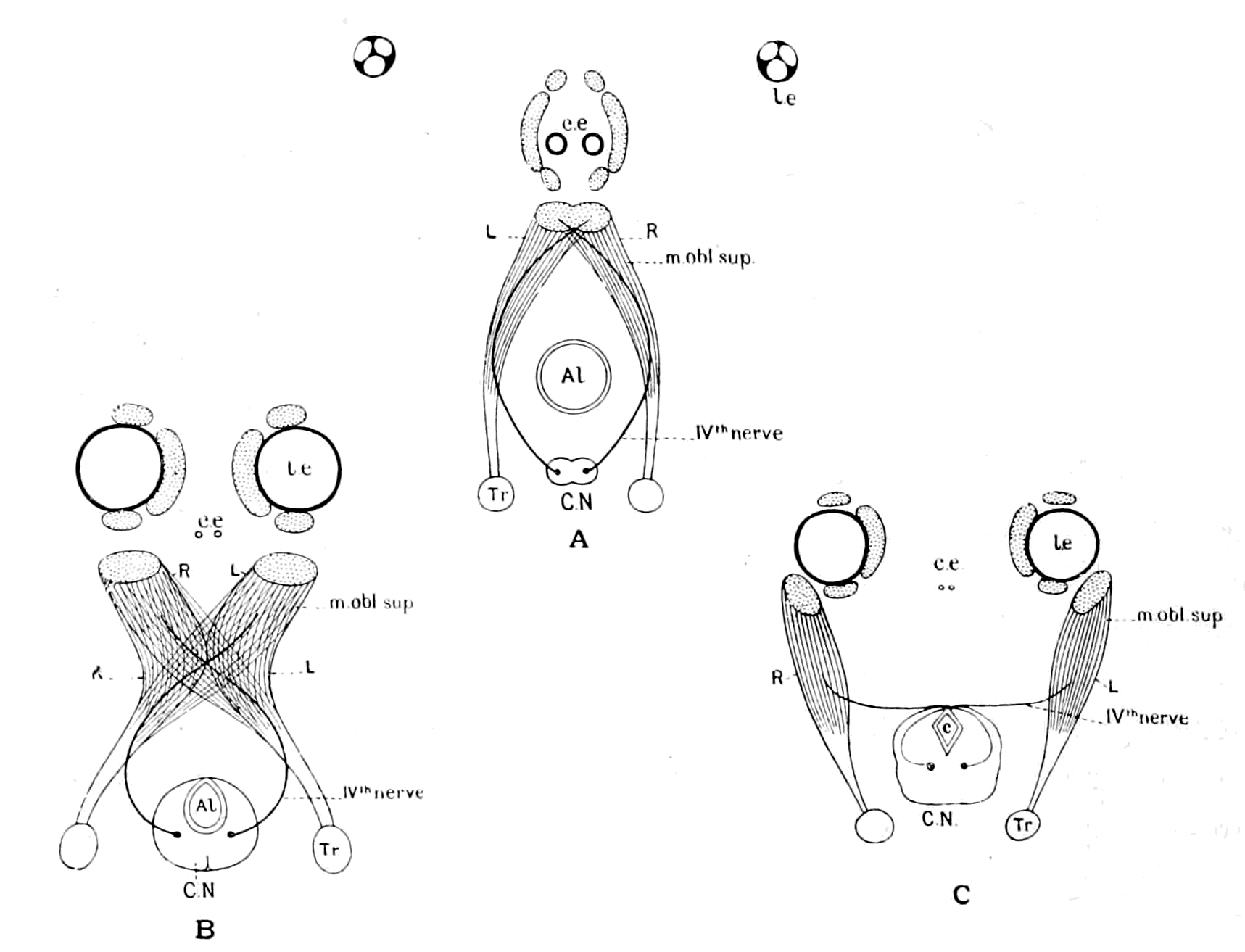
Fig. 112.—A, Diagram of Position of Oblique Muscle in Scorpion; B, Diagram of Transition Stage; C, Diagram of Superior Oblique Muscle in Vertebrate.
l.e., lateral eyes; c.e., central eyes; C.N., central nervous system; Al., alimentary canal; c., aqueductus Sylvii.
It is instructive to notice that the original position of attachment of this muscle is naturally posterior to that of the oculomotor group of muscles, and that Fürbringer, in his description of the eye-muscles of Petromyzon, asserts that this muscle in this primitive vertebrate form is not attached as in other vertebrates, but is posterior to the other muscles, so that he calls it the posterior rather than the superior oblique. The nature of the change by which the muscle known in the scorpion as the anterior dorso-plastron muscle (63) was probably converted into the superior oblique muscle of the vertebrate, is represented in the drawings Fig. 112, in which also are indicated the dwindling of the median eyes, and the progressive superiority of the lateral eyes, as well as the transformation of the recti muscle-group of the scorpion into the muscles supplied by the oculomotor nerve of the vertebrate.
With respect to the external rectus muscle, it follows naturally that if the muscles (64) and (65) are to follow suit with the rest of the group and become attached to the lateral eyes, they must take up an external position. These two muscles, which always run together, as seen in Fig. 110, A, the one belonging to the prosoma and the other to the mesosoma, are represented by the mandibular muscle of Miss Platt and the external rectus, the former derived from the walls of the last pro-otic head-cavity, the latter from the foremost of the opisthotic head-cavities.
Such, then, is the simple explanation of the origin of the eye-muscles which follows from my theory, and we see that the successive alterations of the position of the orbit, and, therefore, of the globe of the eye with its muscles, as we pass from Thyestes to man, is the natural consequence of the growth of the frontal bone, i.e. of the brain.
The Trigeminal Nerves and the Muscles supplied by them.
Turning now to the evidence as to the number of ventral segments, i.e. the motor and sensory supply to the prosomatic appendages afforded by the trigeminal nerve, we must, I think, come to the same conclusion as Dohrn, viz. that if there were originally seven dorsal or somatic segments in this region represented by: 1, Anterior cavity, muscle lost; 2, 3, 4, 5, muscles of the premandibular cavity, sup. rectus, inf. rectus, int. rectus, inf. oblique, supplied by IIIrd nerve; 6, 7, muscles of the mandibular cavity, sup. oblique, supplied by IVth nerve and muscle lost, there must have been also seven corresponding ventral or splanchnic segments supplied by the trigeminal. At present the evidence for such segments is nothing like so strong as for the corresponding somatic ones; there are, however, certain suggestive facts which point distinctly in this direction in connection with both the motor and sensory parts of the trigeminal. The origin of the trigeminal motor fibres in the central nervous system is most striking. We may take it for granted that a nucleus of cells giving origin to one or more segmental motor nerves will possess a greater or less longitudinal extension in the central nervous system, according to the number of fused separate segmental centres it represents. Thus a nucleus such as that of the IVth nerve or of the facial is small and compact in comparison to the extensive conjoint nucleus of the vagus and cranial accessory.
Upon examination of the motor nucleus of the trigeminal, we find a compact or well-defined nucleus, the nucl. masticatorius, the nerves of which supply the masseter, temporal, and other muscles, so that the anatomical evidence at first sight appears to bear out van Wijhe's conclusion that the motor trigeminal supplies at most two segments. Further examination, however, shows that this is not all, for the extraordinary so-called descending root of the Vth must be taken into consideration in any question of the origin of the motor elements, just as the equally striking ascending root enters into the consideration of the meaning of the sensory elements of the Vth.
It is not necessary here to discuss the controversy as to whether this descending root is motor or sensory. It is universally considered at present to be motor, and is believed to supply, as Kölliker suggested, among other muscles, the m. tensor tympani and the m. tensor veli palati. It is thus described by Obersteiner—
"From the region of the mid-brain the motor root receives an important addition of thick fibres, which form the cerebral or descending root. The large, round vesicular cells from which the fibres of the descending root arise form no single compact group, but are partly single, partly arranged like little bunches of grapes, as far as the region of the anterior corpora quadrigemina. The further we go brainwards, the smaller is the number of fibres. In the region of the anterior corpora quadrigemina, the few cells of origin are found more and more median; so that the uppermost trigeminal fibres descend in curves almost from the mid-line, as is shown by the exceptional occurrence of one or more of the characteristic cells above the aqueduct. At the height of the posterior commissure one finds the last of these trigeminal cells."
The anatomy of the Vth nerve reveals, then, three most striking facts:—
1. The motor nucleus of the Vth extends from the very commencement of the infra-infundibular region to nearly the commencement of the nucleus of the VIIth; in other words, the motor nucleus of the Vth extends through the whole prosomatic region, just as it must have done originally if its motor nerves supplied the muscles of the prosomatic appendages. Such an extended range of origin is indicative of the remains of an equally extended series of segmental centres or ganglia.
2. Of these centres the caudalmost have alone remained large and vigorous, constituting the nucleus masticatorius, which in the fish is divided into an anterior and posterior group, thus indicating a double rather than a single nucleus; while the foremost ones have dwindled away until they are represented only by the cells of the descending root, the muscles of these segments being still represented by possibly the tensor veli palati and the other muscles innervated from these cells.
3. The headmost of these cells takes up actually a position dorso-lateral to the central canal, so that the groups on each side nearly come together in the mid-dorsal line; a very unique and extraordinary position for a motor cell-group, but not improbable when we recall to mind Brauer's assertion as to the shifting of the foremost prosomatic ganglion-cells of the scorpion from the ventral to the dorsal side of the alimentary canal.
On the sensory side the evidence is also suggestive, the question here being not so much the distribution of the sensory nerves as the number of ganglia belonging to each of the cranial nerves.
With respect to this question, morphologists have come to the conclusion that there is a marked difference between spinal and cranial nerves, in that whereas the posterior root-ganglia of the spinal nerves arise from the central nervous system itself, i.e. from the neural crest, the ganglia of the cranial nerves arise partly from the neural crest, partly from the proliferation of cells on the surface of the animal; and because of the situation of these proliferating epidermal patches over the gill-clefts in the case of the vagus and glossopharyngeal nerves, they have been called by Froriep and Beard branchial sense-organs. Beard divides the cranial ganglia into two sets, one connected with the neural ridges, called the neural ganglia, and the other connected with the surface-cells, which he calls the lateral ganglia. This second set corresponds to Kupffer's epibranchial ganglia. Now it is clear that in the case of the vagus nerve, where, as is well shown in Ammocœtes, the nerve is not a single segmental nerve, but is in reality made up of a number of nerves going to separate branchial segments, the indication of such segments is not given by the main vagus ganglion or neural ganglion, but by the series of lateral ganglia. So also it is argued in the case of the trigeminal, that if in addition to the ganglion-cells arising from the neural crest separate ganglion-masses are found in the course of development, in connection with proliferating patches of the surface (plakodes, Kupffer calls them), then such isolated lateral ganglia are indications of separate segments, just as in the case of the vagus, even though the separate segments do not show themselves in the adult. So far the argument appears to me just, but the further conclusion that the presence of such plakodes shows the previous existence of branchial sense-organs, and, therefore, that such ganglia are epibranchial ganglia, indicating the position of a lost gill-slit, is not justified by the premises. If, as I suppose, the trigeminal nerve supplied a series of non-branchial appendages serially homologous with the branchial appendages supplied by the vagus, then it is highly probable that the trigeminal should behave with respect to its sensory ganglia similarly to the vagus nerve, without having anything to do with branchiæ.