Front matter (continued 2)
Reading edition
The difficulty lies rather in the position of the cerebellum and corpora quadrigemina, and the solution is as simple as it is conclusive.
Let us again turn to embryology and see what help it gives. In all vertebrates the dilated anterior portion of the nerve-tube does not, as it grows, increase in size uniformly, but a constriction appears on its dorsal surface at one particular place, so as to divide it into an anterior and posterior vesicle; then the latter becomes divided into two portions by a second constriction. In this way three cerebral vesicles are formed; these three primary cerebral vesicles indicate the region of the fore-brain, mid-brain, and hind-brain respectively. Subsequently the first cerebral vesicle becomes divided into two to form the prosencephalon and thalamencephalon, while the third cerebral vesicle is also divided into two to form the region of the cerebellum and medulla oblongata.
These constrictions are in the position of commissural bands of nervous matter; of these the limiting nervous strands between the thalamencephalon and mesencephalon and between the mesencephalon and the hind-brain are of primary importance. The first of these commissural bands is in the position of the posterior commissure connecting the two optic thalami. In close connection with this are found, on the mid-dorsal region, the two pineal eyes with their optic ganglia, the so-called ganglia habenulæ. From these ganglia a peculiar tract of fibre, known as Meynert's bundle, passes on each side to the ventral infra-infundibular portion of the brain. In other words, the first constriction of the dilated tube is due to the presence and growth of nervous material in connection with the median pineal eyes. Here in precisely the same spot, as will be fully explained in the next chapter, there existed in the arthropod ancestor a pair of median eyes situated dorsally to the cephalic stomach, the pre-existence of which explains the reason for the first constriction.
The second primary constriction separating the mid-brain from the hind-brain is still more interesting, for it is coincident with the position of the trochlear or fourth cranial nerve. In all vertebrates without exception this nerve takes an extraordinary course; all other nerves, whether cranial or spinal, pass ventralwards to reach their destination. This nerve passes dorsalwards, crosses its fellow mid-dorsally in the valve of Vieussens, where the roof of the brain is thin, and then passes out to supply the superior oblique muscle of the eye of the opposite side. The two nerves form an arch constricting the dilated tube at this place. In the lowest vertebrate (Ammocœtes) the constriction formed by this nerve-pair is evident not only in the embryonic condition as in other vertebrates, but during the whole larval stage. As Fig. 20, A and B, shows, the whole of the dorsal region of the brain up to the region of the pineal eye and ganglion habenulæ is one large membranous bag, except for the single constriction where the fourth nerve on each side crosses over. The explanation of this peculiarity is given in Chapter VII., and follows simply from the facts of the arrangement of that musculature in the scorpion-group which gave rise to the eye-muscles of the vertebrate.
In Ammocœtes both cerebellum and posterior corpora quadrigemina can hardly be said to exist, but upon transformation a growth of nervous material takes place in this region, and it is seen that this commencing cerebellum and the corpora quadrigemina arise from tissue that is present in Ammocœtes along the course of the fourth nerve.
Here, then, again Embryology does its best to tell us how the vertebrate arose. The formation of the two primary constrictions in the dilated anterior vesicle whereby the brain is divided into fore-brain, mid-brain, and hind-brain is simply the representation ontogenetically of the two nerve-tracts which crossed over the cephalic stomach in the prevertebrate stage, in consequence of the mid-dorsal position of the pineal eyes and of the insertion of the original superior oblique muscles.
The subsequent constriction by which the prosencephalon is separated from the thalamencephalon is in the position of the anterior commissure, that commissure which connects the two supra-infundibular nerve-masses, and is one of the first-formed commissures in every vertebrate. This naturally is simply the commissure between the two supra-œsophageal ganglia; anterior to it, in the middle line, equally naturally, the anterior end of the old stomach wall still exists as the lamina terminalis.
The other division in the hind-brain region, which separates the region of the cerebellum from the medulla oblongata, is due to the growth of the cerebellum, and indicates its posterior limit. In such an animal as the lamprey, where the cerebellum is only commencing, this constriction does not occur in the embryo.
From such simple beginnings as are seen in Ammocœtes, the higher forms of brain have been evolved, to culminate in that of man, in which the massive cerebrum and cerebellum conceals all sign of the dorsal membranous roof, those parts of the simple epithelial tube which still remain being tucked away into the cavities to form the various choroid plexuses.
In the whole evolution from the brain of Ammocœtes to that of man, the same process is plainly visible, viz. growth and extension of nervous material over the epithelial tube; extension dorsally and posteriorly of the supra-infundibular nervous masses (as seen in Fig. 19), combined with a dorsal growth of parts of the infra-infundibular nervous masses to form the cerebellum and posterior corpora quadrigemina.
Especially instructive is the formation of the cerebellum. It consists at first of a small mass of nervous tissue accompanying the fourth nerve, then by the growth of that mass surrounding and constricting a fold of the membranous roof, the worm of the cerebellum is formed, as in the dog-fish. This very constriction causes the membrane to be thrown into a lateral fold on each side, as seen in Fig. 24, and in the dog-fish the nervous material on each side, known as the fimbriæ, is already commencing to grow from the ventral mass of the medulla oblongata to surround these lateral membranous folds. These fimbriæ develop more and more in higher forms, and thus form the cerebellar hemispheres.
Not only does comparative anatomy confirm the teachings of embryology, but also pathology gives its quota in the same direction.
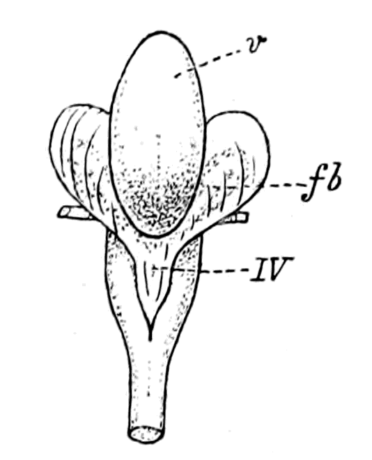
Fig. 24.—Cerebellum of Dog-fish.
v, worm of cerebellum; IV., membranous roof of fourth ventricle continuous with the membranous folds on each side. Through these the fimbriæ (fb.) can be dimly seen.
One of the striking facts about malformations and disease of the central nervous system is the frequency of cystic formations; spina bifida is a well-known instance. These cysts are merely epithelial non-nervous cysts formed from the epithelium of the central canal, difficult to understand if the whole nerve tube is one and entirely nervous, either actually or potentially, but natural and easy if we are really dealing with a simple epithelial tube on the outside of which the nervous material was originally grouped. The cystic formation belongs naturally enough to this tube, not to the nervous system.
Again, where animals such as lizards have grown a new tail, owing to the breaking off of the original one, it is found that the central canal extends into this new tail for some distance, but not the nervous material surrounding it; all the nerves supplying the new tail arise from the uninjured spinal cord above, the central canal with its lining layer of epithelial cells alone grows into the new-formed appendage.
To all intents and purposes the same thing is seen in the termination of the spinal cord in a bird-embryo; more and more, as the end of the tail is approached, does the nervous matter of the spinal cord grow less and less, until at last a naked central canal with its lining epithelium is alone left to represent the so-called nerve-tube.
All these different methods of investigation lead irresistibly to the one conclusion that the tubular nature of the central nervous system has been caused by the central nervous system enclosing to a greater or less extent a pre-existing, non-nervous, epithelial tube.
This must always be borne strictly in mind. The problem, therefore, which presents itself is the comparison of these two factors separately, in order to find out the relationship of the vertebrate to the invertebrate. The nervous system without the tube must be compared to other nervous systems, and the tube must be considered apart from the nervous system.
The Principle of Concentration and Cephalization.
The central nervous system of the vertebrate resembles that of all the Appendiculata in the fact that it is composed of segments joined together which give origin to segmental nerves. There is, however, a great difference between the two systems: the division into separate segments is not obvious to the eye in the vertebrate nervous system, while in the invertebrate we can see that it is composed of a series of separate pairs of ganglia joined together longitudinally by nervous strands known as connectives and transversely by the nerve-commissures. Such a simple segmented system is found in the segmented worms, and in the lower arthropods, such as Branchipus, no great advance has been made on that of the annelid. In the higher forms, however, a greater and greater tendency to fusion of separate ganglia exists, especially in the head-region, so that the infra-œsophageal ganglia, which, in the lower forms are as separate as those of the ventral chain, in the higher forms are fused together to form a single nervous mass.
This is the great characteristic of the advancement of the central nervous system among the Invertebrata, its concentration in the region of the head. It may be called the principle of cephalization, and is characteristic not only of higher organization in a group, but also of the adult as distinguished from the larval form. Thus in the imago greater concentration is found than in the caterpillar.
The segmented annelid type of nervous system consists of a supra-œsophageal ganglion, composed of the fused ganglia belonging to the pre-oral segments, and an infra-œsophageal chain of separate ganglia. With the concentration and modification around the mouth of the most anterior locomotor appendages to form organs for prehension and mastication of food, a corresponding concentration and fusion of the ganglia belonging to these segments takes place, so that finally, in the higher annelids, and in most of the great arthropod group, a fusion of a number of the most anterior ganglia has taken place to form the infra-œsophageal ganglion-mass.
The infra-œsophageal ganglia which are the first to fuse are those which supply the most anterior portion of the animal with nerves, and include always those anterior appendages which are modified for mastication purposes. To this part the name prosoma has been given; in many cases it forms a well-defined, distinct portion of the animal.
Succeeding this prosoma or masticatory region, there occurs in all gill-bearing arthropods a respiratory region, in many cases more or less distinctly defined, which has received the name of mesosoma. The rest of the body is called the metasoma.
In accordance with this nomenclature the central nervous system of many of the Arthropoda may be divided as follows:—
1. Pre-oral, or supra-œsophageal ganglia.
2. Infra-oral, or infra-œsophageal ganglia and ventral chain, which consist of three groups: prosomatic, mesosomatic, and metasomatic ganglia.
The infra-œsophageal ganglion-mass, then, in most of the Arthropoda may be spoken of as formed by the fusion of the prosomatic or mouth-ganglia, the mesosomatic and metasomatic remaining separate and distinct. The number of ganglia which have fused may be observed by examination of the embryo, in which it is easy to see indications of the individual ganglia or neuromeres, although all such indication has disappeared in the adult; thus the infra-œsophageal ganglia of the cray-fish have been shown to be constituted of six prosomatic ganglia.
In Fig. 25 I give figures of the central nervous system (with the exception of the abdominal or metasomatic ganglia) of Branchipus, Astacus, Limulus, Scorpio, Androctonus, Thelyphonus, and Ammocœtes. In all the figures the supra-œsophageal ganglia are lined horizontally, and their nerves shown, viz. optic (lateral eyes (II) and median eyes (II′)), olfactory (I) (first antennæ, camerostome, nose); then come the prosomatic ganglia (dotted), with their nerves (A) supplying the mouth parts, and the second antennæ or cheliceræ; then the mesosomatic (lined horizontally), with their nerves (B) supplying respiratory appendages. These figures show that the concentrated brain mass around the œsophagus of an arthropod which has arrived at the stage of Astacus, is represented by the supra-œsophageal ganglia and the fused prosomatic ganglia.
The next stage in the evolution of the brain is seen in the gradual inclusion of the mesosomatic ganglia, one after the other, into the infra-œsophageal mass of the already fused prosomatic ganglia. With this fusion is associated the loss of locomotion in these mesosomatic appendages, and their entire subservience to the function of respiration. Dana urges that cephalization is a consequence of functional alteration in the appendages, from organs of locomotion to those of mastication and respiration. Whether this be true or not, it is certainly a fact that in Limulus, the ganglion supplying the first mesosomatic appendage has fused with the prosomatic, infra-œsophageal mass. It is also a fact that the prosomatic appendages are the organs of mastication, their basal parts being arranged round the mouth so as to act as foot-jaws, while the mesosomatic appendages, though still free to move, have been reduced to such an extent as to consist mainly of their basal parts, which are all respiratory in function, except in the case of the first pair, where they carry the terminal ducts of the genital organs. In the next stage, that, of the scorpion, in which the mesosomatic appendages have lost all power of free locomotion, and have become internal branchiæ, another mesosomatic ganglion has fused with the brain mass, while in Androctonus two of the branchial mesosomatic ganglia have fused; and finally, in Thelyphonus and Phrynus, all the mesosomatic ganglia have coalesced with the fused prosomatic ganglia, while the metasomatic ganglia have themselves fused together in the caudal region to form what is known as the caudal brain.
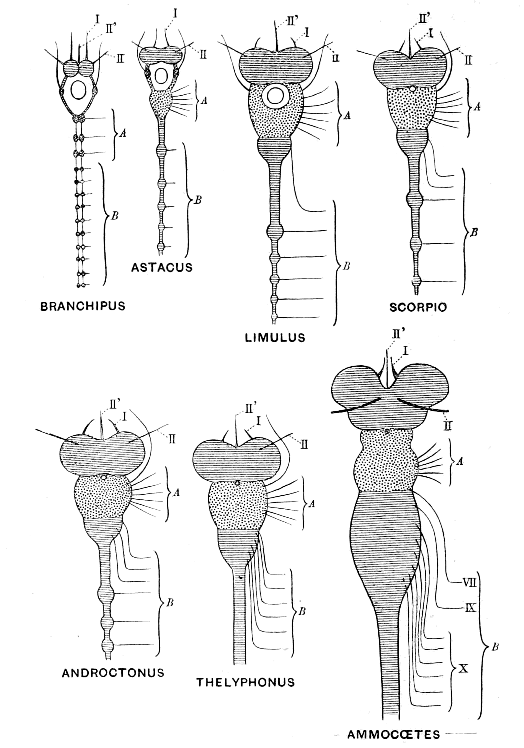
Fig. 25.—Comparison of Invertebrate Brains from Branchipus to Ammocœtes.
The brain in these animals may be spoken of as composed of three parts—(1) the fused supra-œsophageal ganglia, (2) the fused prosomatic ganglia, and (3) the fused mesosomatic ganglia. Such a brain is strictly homologous with the vertebrate brain, which also is built up of three parts—(1) the part in front of the notochord, the prechordal or supra-infundibular brain, which consists of the cerebral hemispheres, together with the basal and optic ganglia and corresponds, therefore, to the supra-œsophageal mass, with its olfactory and optic divisions lying in front of the œsophagus; (2 and 3) the epichordal brain, composed of (2) a trigeminal and (3) a vagus division, of which the first corresponds strictly to the fused prosomatic ganglia, and the second to the fused mesosomatic ganglia. Further, just as in the embryo of an arthropod it is possible, with more or less accuracy, to see the number of neuromeres or original ganglia which have fused to form the supra- and infra-œsophageal portions of its brain, so also in the embryo of a vertebrate we are able at an early stage to gain an indication, more or less accurate, of the number of neuromeres which have built up the vertebrate brain. The further consideration of these neuromeres, and the evidence they afford as to the number of the prosomatic and mesosomatic ganglia which have formed the epichordal part of the vertebrate brain, must be left to the chapter on the segmentation of the cranial nerves.
The further continuation of this process of concentration of separate segments, together with the fusion of the nervous system with the tube of the alimentary canal, leads in the simplest manner to the formation of the spinal cord of the vertebrate from the metasomatic ganglia of the ventral chain of the arthropod.
The Antagonism between Cephalization and Alimentation.
This concentration of the nervous system in the head-region, together with an actual increase in the bulk of the cephalic nervous masses, constitutes the great principle upon which the law of upward progress or evolution in the animal kingdom is based, and it illustrates in a striking manner the blind way in which natural selection works; for, as already explained, the central nervous system arose as a ring round the mouth, in consequence of which, with the progressive evolution of the animal kingdom, the œsophagus necessarily pierced the central nervous system at the cephalic end. At the same time, the very fact that the evolution was progressive necessitated the concentration and increase of the nervous masses in this very same œsophageal region.
Progress on these lines must result in a crisis, owing to the inevitable squeezing out of the food-channel by the increasing nerve-mass; and, indeed, the fact that such a crisis had in all probability arisen at the time when vertebrates first appeared is apparent when we examine the conditions at the present time.
Those invertebrates whose central nervous system is most concentrated at the cephalic end belong to the arachnid group, among which are included the various living scorpion-like animals, such as Thelyphonus, Androctonus, etc.
As already mentioned, the giants of the Palæostracan age were Pterygotus, Slimonia, etc., all animals of the scorpion-type—in fact, sea-scorpions. Now, all these animals, spiders and scorpions, without exception, are blood-suckers, and in all of them the concentrated cephalic mass of nervous material surrounds an œsophagus the calibre of which is so small that nothing but a fluid pabulum can be taken into the alimentary canal; and even for that purpose a special suctorial apparatus has in some species been formed on the gastric side of the œsophagus for the purpose of drawing blood through this exceedingly narrow tube.
In Fig. 25 this increasing antagonism between brain-power and alimentation, as we pass from such a form as Branchipus to the scorpion, is illustrated, and in Fig. 26 the relative sizes of the œsophagus and the brain-mass surrounding it is shown. The section shows that the food channel is surrounded by the white and grey matter of the brain as completely as the central canal of the spinal cord of the vertebrate is surrounded by the white and grey nervous material.
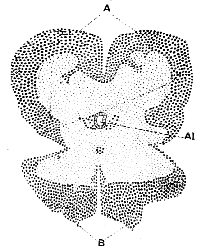
Fig. 26.—Transverse Section through the Brain of a Young Thelyphonus.
A, supra-œsophageal ganglia; B, infra-œsophageal ganglia; Al, œsophagus.
Truly, at the time when vertebrates first appeared, the direction and progress of variation in the Arthropoda was leading, owing to the manner in which the brain was pierced by the œsophagus, to a terrible dilemma—either the capacity for taking in food without sufficient intelligence to capture it, or intelligence sufficient to capture food and no power to consume it.
Something had to be done—some way had to be found out of this difficulty. The atrophy of the brain meant degeneration and the reduction to a lower stage of organization, as is seen in the Tunicata. The further development of the brain necessitated the establishment of a new method of alimentation and the closure of the old œsophagus, its vestiges still remaining as the infundibular canal of the vertebrate, meant the enormous upward stride of the formation of the vertebrate.
At first sight it might appear too great an assumption even to imagine the possibility of the formation of a new gut in an animal so highly organized as an arthropod, but a little consideration will, I think, show that such is not the case.
In the higher animals we are accustomed to speak of certain organs as vital and necessary for the further existence of the animal; these are essentially the central nervous system, the respiratory system, the circulatory system, and the digestive system. Of these four vital systems the first cannot be touched without the chance of degeneration; but that is not the case with the second. The passage from the fish to the amphibian, from the water-breathing to the air-breathing animal, has actually taken place, and was effected by the modification of the swim-bladder to form new respiratory organs—the lung; the old respiratory organs—the gills—becoming functionless, but still persisting in the embryo as vestiges. The necessity arose in consequence of the passage of the animal from water to land, and with this necessity nature found a means of overcoming the difficulty; air-breathing vertebrates arose, and from the very fact of their being able to extend over the land-surfaces, increased in numbers and developed in complexity in the manner already sketched out.
For a respiratory system all that is required is an arrangement by means of which blood should be brought to the surface, so as to interchange its gases with those of the external medium; and it is significant to find that of all vertebrates the Amphibia alone are capable of an effective respiration by means of the skin.
As to the circulatory system, it is exceedingly easily modified. An animal such as Amphioxus has no heart; in some the heart is systemic, in others branchial; in some there are more than one heart; in others there are contractile veins in addition to a heart. There is no difficulty here in altering and modifying the system according to the needs of the individual.
For a digestive system all that is required is an arrangement for the digestion and absorption of food, a mechanism which can arise easily if some of the cells of the skin possess digestive power. Now Miss Alcock has shown that some of the surface-cells of crustaceans secrete a fluid which possesses digestive powers, and she has also shown that certain of the cells in the skin of Ammocœtes possess digestive power.
The difficulty, then, of forming a new digestive system in the passage from the arthropod to the vertebrate is very much the same as the difficulty in forming a new respiratory system in the passage from the water-breathing fish to the air-breathing amphibian—a change which does not strike us as inconceivable, because we know it has taken place.
The whole argument so far leads to the conclusion that vertebrates arose from ancient forms of arthropods by the formation of a new alimentary canal, and the enclosure of the old canal by the growing central nervous system. If this conclusion is true, then it follows that we possess a well-defined starting-point from which to compare the separate organs of the arthropod with those of the vertebrate, and if, in consequence of such working hypothesis, each organ of the arthropod is found in the vertebrate in a corresponding position and of similar structure, then the truth of the starting-point is proved as fully as can possibly be expected by deductive methods. It is, in fact, this method of comparative anatomy which has proved the descent of man from the ape, the frog from the fish, etc.
Let us, then, compare all the organs of such a low vertebrate as Ammocœtes with those of an arthropod of the ancient type.
Life History of the Lamprey—not a Degenerate Animal.
The striking peculiarity of the lamprey is its life-history. It lives in fresh water, spending a large portion of its life in the mud during the period of its larval existence: then comes a somewhat sudden transformation-stage, characterized, as in the lepidopterous larva, by a process of histolysis, by which many of the larval tissues are destroyed and new ones formed, with the result that the larval lamprey, or Ammocœtes, is transformed into the adult lamprey, or Petromyzon. This transformation takes place in August, at all events in the neighbourhood of Cambridge, and later in the year the transformed lamprey migrates to the sea, grows in size and maturity, and returns to the river the following spring up to its spawning beds, where it spawns and forthwith dies. How long it lives in the Ammocœtes stage is unknown; I myself have kept some without transformation for four years, and probably they live in the rivers longer than that before they change from their larval state. It is absolutely certain that very much the longest part of the animal's life is spent in the larval stage, and that with the maturity of the sexual organs and the production of the fertilized ova the life of the individual ends.
Now, the striking point of this transformation is that it produces an animal more nearly comparable with higher vertebrates than is the larval form; in other words, the transformation from larva to adult is in the direction of upward progress, not of degeneration. It is, therefore, inaccurate to speak of the adult lamprey as degenerate from a higher race of fishes represented by its larval form—Ammocœtes. Its transformation does not resemble that of the tunicates, but rather that of the frog, so that, just as in the case of the tadpole, the peculiarities of its larval form may be expected to afford valuable indications of its immediate ancestry. The very peculiarities to which attention must especially be paid are those discarded at transformation, and, as will be seen, these are essentially characteristic of the invertebrate and are not found in the higher vertebrates. In fact, the transformation of the lamprey from the Ammocœtes to the Petromyzon stage may be described as the casting off of many of its ancestral invertebrate characters and the putting on of the characteristics of the vertebrate type. It is this double individuality of the lamprey, together with its long-continued existence in the larval form, which makes Ammocœtes more valuable than any other living vertebrate for the study of the stock from which vertebrates sprang.
Many authorities hold the view that the lamprey, like Amphioxus, must be looked upon as degenerate, and therefore as no more suitable for the investigation of the problem of vertebrate ancestry than is Amphioxus itself. This charge of degeneracy is based on the statement that the lamprey is a parasite, and that the eyes in Ammocœtes are under the skin. The whole supposition of the degeneracy of the Cyclostomata arose because of the prevailing belief of the time that the earliest fishes were elasmobranchs, and therefore gnathostomatous. From such gnathostomatous fishes the cyclostomes were supposed to have descended, having lost their jaws and become suctorial in habit in consequence of their parasitism.
The charge of parasitism is brought against the lamprey because it is said to suck on to fishes and so obtain nutriment. It is, however, undoubtedly a free-swimming fish; and when we see it coming up the rivers in thousands to reach the spawning-beds, and sucking on to the stones on the way in order to anchor itself against the current, or holding on tightly during the actual process of spawning, it does not seem justifiable to base a charge of degeneration upon a parasitic habit, when such so-called habit simply consists in holding on to its prey until its desires are satisfied. If, of course, its suctorial mouth had arisen from an ancestral gnathostomatous mouth, then the argument would have more force.
Dohrn, however, gives absolutely no evidence of a former gnathostomatous condition either in Petromyzon or, in its larval state, Ammocœtes. He simply assumes that the Cyclostomata are degenerated fishes and then proceeds to point out the rudiments of skeleton, etc., which they still possess. Every point that Dohrn makes can be turned round; and, with more probability, it can be argued that the various structures are the commencement of the skeletal and other structures in the higher fishes, and not their degenerated remnants. Compare the life-history of the lamprey and of the tunicate. In the latter case we look upon the animal as a degenerate vertebrate, because the larval stage alone shows vertebrate characteristics; when transformation has taken place, and the adult form is reached, the vertebrate characteristics have vanished, and the animal, instead of reaching a higher grade, has sunk lower in the scale, the central nervous system especially having lost all resemblance to that of the vertebrate. In the former case a transformation also takes place, a marvellous transformation, characterized by two most striking facts. On the one hand, the resulting animal is more like a higher vertebrate, for, by the formation of new cartilages, its cranial skeleton is now comparable with that of the higher forms, and the beginnings of the spinal vertebræ appear; by the increased formation of nervous material, its brain increases in size and complexity, so as to compare more closely with higher vertebrate brains; its eyes become functional, and its branchiæ are so modified, simultaneously with the formation of the new alimentary canal in the cranial region, that they now surround branchial pouches which are directly comparable to those of higher vertebrates. On the other hand, the transformation process is equally characterized by the throwing off of tissues and organs, one and all of which are comparable in structure and function with corresponding structures in the Arthropoda—the thyroid of the Ammocœtes, the tentacles, the muco-cartilage, the tubular muscles, all these structures, so striking in the Ammocœtes stage, are got rid of at transformation. Here is the true clue. Here, in the throwing off of invertebrate characters, and the taking on of a higher vertebrate form, especially a higher brain, not a lower one, Petromyzon proclaims as clearly as is possible that it is not a degenerate elasmobranch, but that it has arisen from Ammocœtes-like ancestors, even though Myxine, Amphioxus, and the tunicates be all stages on the downward grade from those same Ammocœtes-like ancestors.
As to the eyes, they are functional in the adult form and as serviceable as in any fish. There is no sign of degeneracy; it is only possible to speak of a retarded development which lasts through the larval stage.
Comparison of Brain of Ammocœtes with that of an Arthropod.
Seeing that the steady progress of the development of the central nervous system is the most important factor in the evolution of animals, it follows that of all organs of the body, the central nervous system must be most easily comparable with that of the supposed ancestor. I will, therefore, start by comparing the brain of Ammocœtes with that of arthropods, especially of Limulus and of the scorpion-group.
The supra-infundibular portion of the brain in vertebrates corresponds clearly to the supra-œsophageal portion of the invertebrate brain in so far that in both cases here is the seat of the will. Voluntary action is as impossible to the arthropod deprived of its supra-œsophageal ganglia as to the vertebrate deprived of its cerebrum. It corresponds, also, in that from it arise the nerves of sight and smell and no other nerves; this is also the case with the supra-œsophageal ganglia, for from a portion of these ganglia arise the nerves to the eyes and the nerves to the first antennæ, of which the latter are olfactory in function. Thus, in the accompanying figure, taken from Bellonci, it is seen that the supra-œsophageal ganglia consist of a superior segment corresponding to the cerebrum, a middle segment from which arise the nerves to the lateral eyes and to the olfactory antennæ, corresponding to the basal ganglia of the brain and the optic lobes, and, according to Bellonci, of an inferior segment from which arise the nerves to the second pair of antennæ. This last segment is not supra-œsophageal in position, but is situated on the œsophageal commissures. It has been shown by Lankester and Brauer in Limulus and the scorpion to be in reality the first ganglion of the infra-œsophageal series, and not to belong to the supra-œsophageal group.
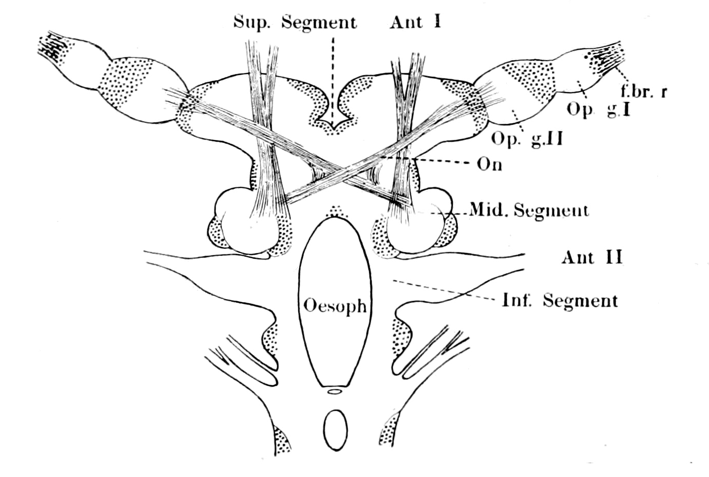
Fig. 27.—The Brain of Sphæroma serratum. (After Bellonci.)
Ant. I. and Ant. II., nerves to 1st and 2nd antennæ. f.br.r., terminal fibre layer of retina; Op. g. I., first optic ganglion; Op. g. II., second optic ganglion; O.n., optic nerve-fibres forming an optic chiasma.
Further, in Limulus, in the scorpion-group, and in all the extinct Eurypteridæ—in fact, in the Palæostraca generally—there are two median eyes in addition to the lateral eyes, which were innervated from these ganglia.
In Ammocœtes, then, if the supra-infundibular portion of the brain really corresponds to the supra-œsophageal of the palæostracan group, we ought to find, as indeed is the case, an optic apparatus consisting of two lateral eyes and two median eyes, innervated from the supra-infundibular brain-mass, and an olfactory apparatus built up on the same lines as in the scorpion-group, also innervated from this region. If, in addition, it be found that those two median eyes are degenerate eyes of the same type as the median eyes of Limulus and the scorpion-group, then the evidence is so strong as to amount to a proof of the correctness of the theory. This evidence is precisely what has been obtained in recent years, for the vertebrate did possess two median eyes in addition to the two lateral ones, and these two median eyes are degenerate eyes of the type found in the median eyes of arthropods and are not of the vertebrate type. Moreover, as ought also to be the case, they are most evident, and one of the pair is most nearly functional in the lowest perfect vertebrate, Ammocœtes.
Of all the discoveries made in recent years, the discovery that the pineal gland of the vertebrate brain was originally a pair of median eyes is by far the most important clue to the ancestry of the vertebrate, for not only do they correspond exactly in position with the median eyes of the invertebrates, but, being already degenerate and functionless in the lowest vertebrate, they must have been functional in a pre-vertebrate stage, thus giving the most direct clue possible to the nature of the pre-vertebrate stage. It is especially significant that in Limulus they are already partially degenerated. What, then, ought to be the structure and relation to the brain of the median and lateral eyes of the vertebrate if they originated from the corresponding organs of some one or other member of the palæostracan group?
This question will form the subject of the next chapter.
Summary.
The object of this book is to attempt to find out from what group of invertebrates the vertebrate arose; no attempt is made to speculate upon the causes of variation by means of which evolution takes place.
A review of the animal kingdom as a whole leads to the conclusion that the upward development of animals from an original cœlenterate stock, in which the central nervous system consists of a ring of nervous material surrounding the mouth, has led, in consequence of the elaboration of the central nervous system, to a general plan among the higher groups of invertebrates in the topographical arrangement of the important organs. The mouth is situated ventrally, and leads by means of the œsophagus into an alimentary canal which is situated dorsally to the central nervous system. Thus the œsophagus pierces the central nervous system and divides it into two parts, the supra-œsophageal ganglia and the infra-œsophageal ganglia. This is an almost universal plan among invertebrates, but apparently does not hold for vertebrates, for in them the central nervous system is always situated dorsally and the alimentary canal ventrally, and there is no piercing of the central nervous system by an œsophagus.
Yet a remarkable resemblance exists between the central nervous system of the vertebrate and that of the higher invertebrates, of so striking a character as to compel one school of anatomists to attempt the derivation of vertebrates from annelids. Now, the central nervous system of vertebrates forms a hollow tube, and a diverticulum of this hollow tube, known as the infundibulum, passes to the ventral surface of the brain in the very position where the œsophagus would have been if that brain had belonged to an annelid or an arthropod. This school of anatomists therefore concluded that this infundibular tube represented the original invertebrate œsophagus which had become closed and no longer opened into the alimentary canal owing to the formation of a new mouth in the vertebrate. As, however, the alimentary canal of the vertebrate is ventral to the central nervous system, and not dorsal, as in the invertebrate, it follows that the remains of the original invertebrate mouth into which the œsophagus (in the vertebrate the infundibular tube) must have opened must be searched for on the dorsal side of the vertebrate; and so the theory was put forward that the vertebrate had arisen from the annelid by the reversal of surfaces, the back of the one animal becoming the front of the other.
The difficulties in the way of accepting such reversal of surfaces have proved insuperable, and another school has arisen which suggests that evolution has throughout proceeded on two lines, the one forming groups of animals in which the central nervous system is pierced by the food-channel and the gut therefore lies dorsally to it, the other in which the central nervous system always lies dorsally to the alimentary canal and is not pierced by it. In both cases the highest products of the evolution have become markedly segmented animals, in the former, annelids and arthropods; in the latter, vertebrates. The only evidence on which such theory is based is the existence of low forms of animals, known as the Enteropneusta, the best known example of which is called Balanoglossus; they are looked upon as aberrant annelid forms by many observers.
This theory does not attempt to explain the peculiarities of the tube of the vertebrate central nervous system, or to account for the extraordinary resemblance between the structure and arrangement of the central nervous systems of vertebrates and of the highest invertebrate group.
Neither of these theories is satisfactory or has secured universal acceptance. The problem must be considered entirely anew. What are the guiding principles in this investigation?
The evolution of animal life on this earth can clearly, on the whole, be described as a process of upward progress culminating in the highest form—man; but it must always be remembered that whole groups of animals such as the Tunicata have been able to survive owing to a reverse process of degeneration.
If there is one organ more than another which increases in complexity as evolution proceeds, which is the most essential organ for upward progress, surely it is the central nervous system, especially that portion of it called the brain. This consideration points directly to the origin of vertebrates from the most highly organized invertebrate group—the Arthropoda—for among all the groups of animals living on the earth in the present day they alone possess a central nervous system closely comparable with that of vertebrates. Not only has an upward progress taken place in animals as a whole, but also in the tissues themselves a similar evolution is apparent, and the evidence shows that the vertebrate tissues resemble more closely those of the arthropod than of any other invertebrate group.
The evidence of geology points to the same conclusion, for the evidence of the rocks shows that before the highest mammal—man—appeared, the dominant race was the mammalian quadruped, from whom the highest mammal of all—man—sprung; then comes, in Mesozoic times, the age of reptiles which were dominant when the mammal arose from them. Preceding this era we find in Carboniferous times that the amphibian was dominant, and from them the next higher group—the reptiles—arose. Below the Carboniferous come the Devonian strata with their evidence of the dominance of the fish, from whom the amphibian was directly evolved. The evidence is so clear that each succeeding higher form of vertebrate arose from the highest stage reached at the time, as to compel one to the conclusion that the fishes arose from the race which was dominant at the time when the fishes first appeared. This brings us to the Silurian age, in which the evidence of the rocks points unmistakably to the sea-scorpions, king-crabs, and trilobites as being the dominant race. It was preceded by the great trilobite age, and the whole period, from the first appearance of the trilobite to the time of dwindling away of the sea-scorpions, may be designated the Palæostracan age, using the term Palæostraca to include both trilobites and the higher scorpion and king-crab forms evolved from them. The evidence of geology then points directly and strongly to the origin of vertebrates from the Palæostraca—arthropod forms which were not crustacean and not arachnid, but gave origin both to the modern-day crustaceans and arachnids. The history of the rocks further shows that these ancient fishes, when they first appeared, resembled in a remarkable manner members of the palæostracan group, so that again and again palæontologists have found great difficulty in determining whether a fossil is a fish or an arthropod. Fortunately, there is still alive on the earth one member of this remarkable group—the Limulus, or King-Crab. On the vertebrate side the lowest non-degenerate vertebrate is the lamprey, or Petromyzon, which spends a large portion of its existence in a larval stage, known as the Ammocœtes stage of the lamprey, because it was formerly considered to be a separate species and received the name of Ammocœtes. The larval stages of any animal are most valuable for the study of ancestral problems, so that it is most fortunate for the solution of the ancestry of vertebrates that Limulus on the one side and Ammocœtes on the other are available for thorough investigation and comparison. There are no trilobites still alive, but in Branchipus and Apus we possess the nearest approach to the trilobite organization among living crustaceans.
So strongly do all these different lines of argument point to the origin of vertebrates from arthropods as to make it imperative to reconsider the position of that school of anatomists who derived vertebrates from annelids by reversing the back and front of the animal. Let us not turn the animal over, but re-consider the position, the infundibular tube of the vertebrate still representing the œsophagus of the invertebrate, the cerebral hemispheres and basal ganglia the supra-œsophageal ganglia, the crura cerebri the œsophageal commissures, and the infra-infundibular part of the brain the infra-œsophageal ganglia. It is immediately apparent that just as the invertebrate œsophagus leads into the large cephalic stomach, so the infundibular tube leads into the large cavity of the brain known as the third ventricle, which, together with the other ventricles, forms in the embryo a large anterior dilated part of the neural tube. In the arthropod this cephalic stomach leads into the straight narrow intestine; in the vertebrate the fourth ventricle leads into the straight narrow canal of the spinal cord. In the arthropod the intestine terminates in the anus; in the vertebrate embryo the canal of the spinal cord terminates in the anus by way of the neurenteric canal. Keep the animal unreversed, and immediately the whole mystery of the tubular nature of the central nervous system is revealed, for it is seen that the nervous matter, which corresponds bit by bit with that of the arthropod, has surrounded to a greater or less extent and amalgamated with the tube of the arthropod alimentary canal, and thus formed the so-called central nervous system of the vertebrate.
The manner in which the nervous material has invaded the walls of the tube is clearly shown both by the study of the comparative anatomy of the central nervous system in the vertebrate and also by its development in the embryo.
This theory implies that the vertebrate alimentary canal is a new formation necessitated by the urgency of the case, and, indeed, there was cause for urgency, for the general plan of the evolution of the invertebrate from the cœlenterate involved the piercing of the anterior portion of the central nervous system by the œsophagus, while, at the same time, upward progress meant brain-development; brain-development meant concentration of nervous matter at the anterior end of the animal, with the result that in the highest scorpion and spider-like animals the brain-mass has so grown round and compressed the food-tube that nothing but fluid pabulum can pass through into the stomach; the whole group have become blood-suckers. These kinds of animals—the sea-scorpions—were the dominant race when the vertebrates first appeared: here in the natural competition among members of the dominant race the difficulty must have become acute. Further upward evolution demanded a larger and larger brain with the ensuing consequence of a greater and greater difficulty of food-supply. Nature's mistake was rectified and further evolution secured, not by degeneration in the brain-region, for that means degradation not upward progress, but by the formation of a new food-channel, in consequence of which the brain was free to develop to its fullest extent. Thus the great and mighty kingdom of the Vertebrata was evolved with its culminating organism—man—whose massive brain with all its possibilities could never have been evolved if he had still been compelled to pass the whole of his food through the narrow œsophageal tube, still existent in him as the infundibular tube. This, then, is the working hypothesis upon which this book is written. If this view is right, that the Vertebrate was formed from the Palæostracan without any reversal of surfaces, but by the amalgamation of the central nervous system and alimentary canal, then it follows that we have various fixed points of comparison in the central nervous systems of the two groups of animals from which to search for further clues. It further follows that from such starting-point every organ of importance in the body of the arthropod ought to be visible in the corresponding position in the vertebrate, either as a functional or rudimentary organ. The subsequent chapters will deal with this detailed comparison of organs in the arthropod and vertebrate respectively.
CHAPTER II
THE EVIDENCE OF THE ORGANS OF VISION
Different kinds of eye.—Simple and compound retinas.—Upright and inverted retinas.—Median eyes.—Median or pineal eyes of Ammocœtes and their optic ganglia.—Comparison with other median eyes.—Lateral eyes of vertebrates compared with lateral eyes of crustaceans.—Peculiarities of the lateral eye of the lamprey.—Meaning of the optic diverticula.—Evolution of vertebrate eyes.—Summary.
The Different Kinds of Eye.
In all animals the eyes are composed of two parts. 1. A set of special sensory cells called the retina. 2. A dioptric apparatus for the purpose of forming an image on the sensory cells. The simplest eye is formed from a modified patch of the surface-epithelium; certain of the hypodermal cells, as they are called, elongate, and their cuticular surface becomes bulged to form a simple lens. These elongated cells form the retinal cells, and are connected with the central nervous system by nerve-fibres which constitute an optic nerve; the cells themselves may contain pigment.
The more complicated eyes are modifications of this type for the purpose of making both the retina and the dioptric apparatus more perfect. According to a very prevalent view, these modifications have been brought about by invaginations of the surface-epithelium. Thus if ABCD (Fig. 28) represents a portion of the surface-epithelium, the chitinous cuticle being represented by the dark line, with the hypodermal cells beneath, and if the part C is modified to form an optic sense-plate, then an invagination occurring between A and B will throw the retinal sense-cells with the optic nerve further from the surface, and the layers B and A between the retina and the source of light will be available for the formation of the dioptric apparatus. The lens is now formed from the cuticular surface of A, and the hypodermal cells of A elongate to form the layer known by the name of corneagen, or vitreogen, the cells of B remaining small and forming the pre-retinal layer of cells. The large optic nerve end-cells of the retinal layer, C, take up the position shown in the figure, and their cuticular surface becomes modified to form rods of varying shape called rhabdites, which are attached to the retinal cells. Frequently the rhabdites of neighbouring cells form definite groups, each group being called a rhabdome. Whatever shape they take it is invariably found that these little rods (bacilli), or rhabdites, are modifications of the cuticular surface of the cells which form the retinal layer. Also, as must necessarily be the case from the method of formation, the optic nerve arises from the nuclear end of the retinal cells, never from the bacillary end. As in the case first mentioned, so in this case, the light strikes direct upon the bacillary end of the retinal cells; such eyes, therefore, are eyes with an upright retina.
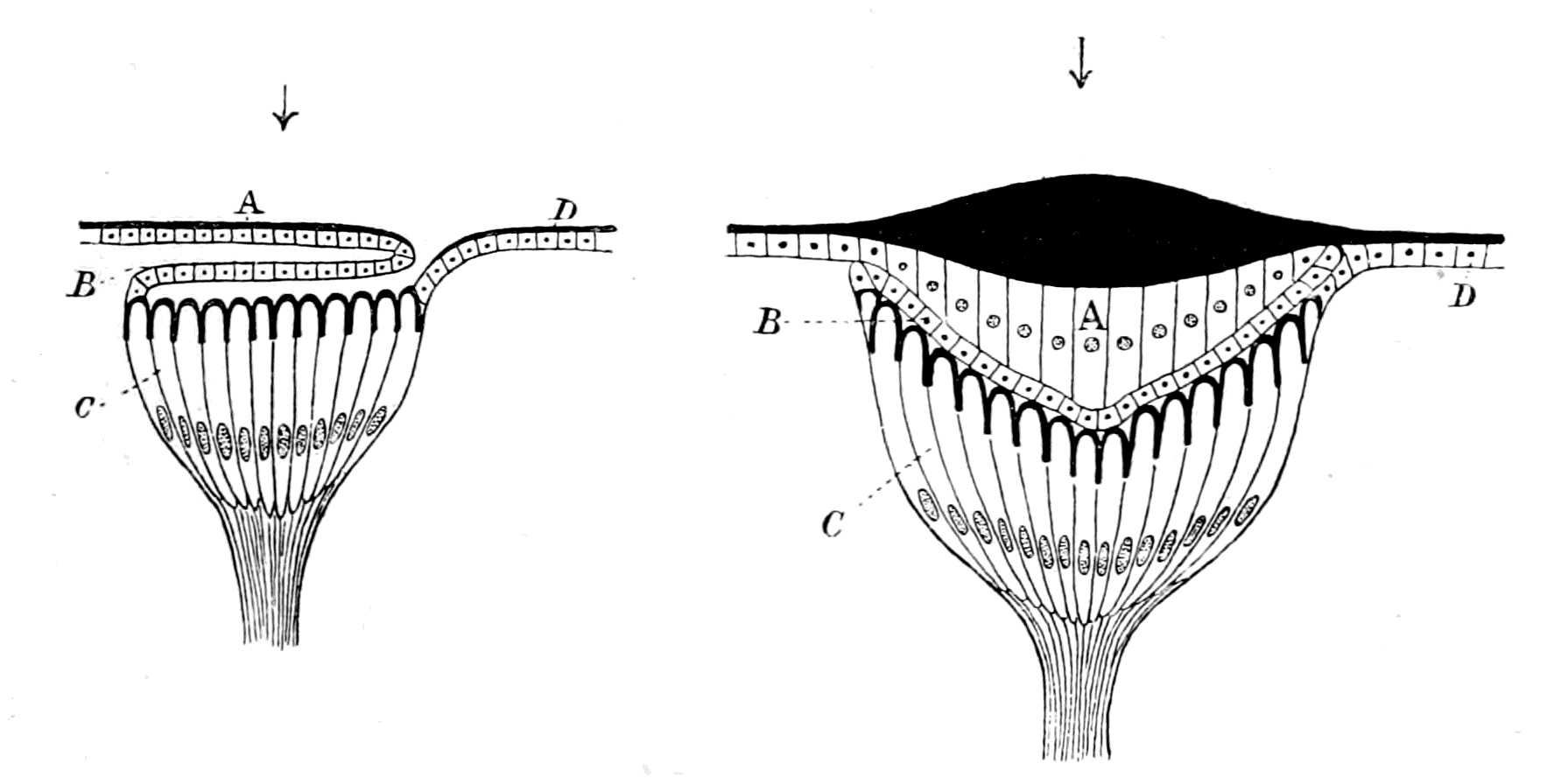
Fig. 28.—Diagram of Formation of an Upright Simple Retina.
It may happen that the part invaginated is the optic sense-plate itself, as would be the case if in the former figure, instead of C, the part B was modified to form a sense-plate. This will give rise to an eye of a character different from the former (Fig. 29). The optic nerve-fibres now lie between the source of light and the retinal end-cells, the layer A as before forms the cuticular lens, and its hypodermal cells elongate to form the corneagen; there is no pre-retinal layer, but, on the contrary, a post-retinal layer, C, called the tapetum, and, as is seen, the light passes through the retinal layer to the tapetum. The cuticular surface of the retinal cells forming the rods or bacilli is directed towards the tapetal layer away from the source of light, and the nuclei of the retinal cells are pre-bacillary in position, in contradistinction to the upright eye, where they are post-bacillary. The retinal end-cells are devoid of pigment, the pigment being in the tapetal layer.
Such an eye, in contradistinction to the former type, is an eye with an inverted retina; but still the same law holds as in the former case—the optic nerve-fibres enter at the nuclear ends of the cells, and the rods are formed from the cuticular surface.
In these eyes the pigmented tapetal layer is believed to act as a looking-glass; the dioptric apparatus throws the image on to its shiny surface, from whence it is reflected directly on to the rods, which are in close contact with the tapetum. A similar process has been suggested in the case of the mammalian lateral eye, with its inverted retina. Johnson describes the post-retinal pigmented layer as being frequently coloured and shiny, and imagines that it reflects the image directly back on to the rods.
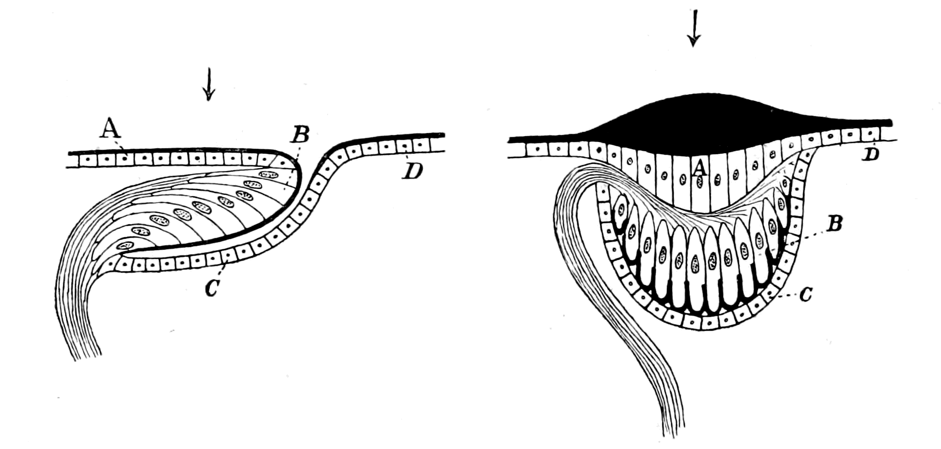
Fig. 29.—Diagram of Formation of an Inverted Simple Retina.
The arrow shows the direction of the source of light in this as in the preceding figure. In both figures the cuticular rhabdites are represented by thick black lines.
Thus we see that eyes can be placed in different categories, e.g. those with an upright retina and those with an inverted retina; also, according to the presence or absence of a tapetum, eyes have been grouped as tapetal or non-tapetal. All the eyes considered so far are called simple eyes, or ocelli; and a number of ocelli may be contiguous though separate, as in the lateral eyes of the scorpion. They may, however, come into close contact and form one single, large, compound eye. Such ocelli, in a very large number of cases, retain each its own dioptric apparatus, and therefore the external appearance of the compound eye represents not a single lens, but a large number of facets, as is seen in the eyes of insects. Owing to these differences, eyes have been divided into simple and compound, and into facetted and non-facetted.
Yet another complication occurs in the formation of eyes, which is, perhaps, the most important of all: the retinal portion of the eye, instead of consisting of simple retinal cells, with their accompanying rhabdites, may include within itself a portion of the central nervous system.
The rationale of such a formation is as follows: The external covering of the body is formed by a layer of external epithelial cells—the ectodermal cell-layer—and an underlying neural layer, of which the latter gives origin to the central nervous system. As development proceeds, this central nervous system sinks inwards, leaving as its connection with the ectoderm the sensory nerves of the skin. That part of the neural layer which underlies the optic plate forms the optic ganglion, and when the central nervous system leaves the surface to take up its deeper position, the strand of nerve-fibres known as the optic nerve, is left connecting it with the retinal cells as seen in Figs. 28, 29. It may, however, happen that part of the optic ganglion remains at the surface, in close connection with the retinal end-cells, when the rest of the central nervous system sinks inwards. The retina of such an eye is composed of the combined optic ganglion and retinal end-cells; the strand of nerve-fibres which is left as the connection between it and the rest of the brain, which is also called the optic nerve, is not a true peripheral nerve, as in the first case, but rather a tract of fibres connecting two parts of the brain, of which one has remained at the periphery. Such a retina, in contradistinction to the first kind, may be called a compound retina.
The optic ganglion, as seen in eyes with a simple retina, consists of a cortical layer of small, round nerve-cells, and an internal medulla of fine nerve-fibres, which form a thick network known as 'Punctsubstanz,' or in modern terminology, 'Neuropil.' Fibres which pass into this 'neuropil' from other parts of the brain connect them with the optic ganglion.
At the present time, owing to the researches of Golgi, Ramón y Cajal, and others, the nervous system is considered to be composed of a number of separate nerve-units, called neurones, each neurone consisting of a nerve-cell with its various processes; one of these—the neuraxon—constitutes the nerve-fibre belonging to that nerve-cell, the other processes—the dendrites—establish communication with other neurones. The place where these processes come together is called a synapse, and the tangle of fine fibres formed at a number of synapses forms the 'neuropil.'
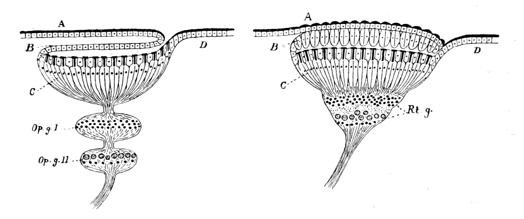
Fig. 30.—Diagram of Formation of an Upright Compound Retina.
ABCD, as in Fig. 28. Op. g. I. and Op. g. II., two optic ganglia which combine to form the retinal ganglion, Rt. g.
When, therefore, a compound retina is formed by the amalgamation of the ectodermal part—the retinal cells proper—with the neurodermic part—to which the name 'retinal ganglion' may be given,—such a retina consists of neuropil substance and nerve-cells, as well as the retinal end-cells. In all such compound retinas, the retinal ganglion is not single, but two optic ganglia at least are included in it, so that there are two sets of nerve-cells and two synapses are always formed; one between the retinal end-cells and the neurones of the first optic ganglion, which may be called the ganglion of the retina, the other between the first and second ganglia, which, seeing that the neuraxons of its cells form the optic nerve, may be called the ganglion of the optic nerve. The 'neuropil' formed by these synapses forms the molecular layers of the compound retina, and the cells themselves form the nuclear layers. Thus an upright compound retina, formed in the same way as the upright simple retina, would be illustrated by Fig. 30.
Further, in precisely the same way as in the case of the simple retina, such a compound retina may be upright or inverted. Thus, in the lateral eyes of crustaceans and insects, a compound retina of this kind is formed, which is upright; while in the vertebrates the compound retina of the lateral eyes is inverted.
The compound retina of vertebrates is usually described as composed of a series of layers, which may be analyzed into their several components as follows:—
Layer of rods and cones External nuclear layer External molecular layer Internal nuclear layer Internal molecular layer Optic nerve-cell layer Layer of optic nerve fibres retina proper Ectodermic part


ganglion of retina retinal ganglion neurodermic part

ganglion of optic nerve
Layer of rods and cones retina proper Ectodermic part
External nuclear layer
External molecular layer
ganglion of retina retinal ganglion neurodermic part
Internal nuclear layer
Internal molecular layer
ganglion of optic nerve
Optic nerve-cell layer
Layer of optic nerve fibres
The difference between the development of these two types of eye—those with a simple retina and those with a compound retina—has led, in the most natural manner, to the conception that the retina is developed, in the higher animals, sometimes from the cells of the peripheral epidermis, sometimes from the tissue of the brain—two modes of development termed by Balfour 'peripheral' and 'cerebral.' An historical survey of the question shows most conclusively that all investigators are agreed in ascribing the origin of the simple retina to the peripheral method of development, the retina being formed from the hypodermal cells by a process of invagination, while the cerebral type of development has been described only in the development of the compound retina. The natural conclusion from this fact is that, in watching the development of the compound retina, it is more difficult to differentiate the layers formed from the epidermal retinal cells and those formed from the epidermal optic ganglion-cells, than in the case of the simple retina, where the latter cells withdraw entirely from the surface. This is the conclusion to which Patten has come, and, indeed, judging from the text-book of Korschelt and Heider, it is the generally received opinion of the day that, as far as the Appendiculata are concerned, the retina, in the true sense—the retinal end-cells, with their cuticular rods,—is formed, in all cases, from the peripheral cells of the hypodermal layer, the cuticular rods being modifications of the general cuticular surface of the body. The apparent cerebral development of the crustacean retina, as quoted from Bobretsky by Balfour, is therefore in reality the development of the retinal ganglion, and not of the retina proper.
There is, I imagine, a universal belief that the natural mode of origin of a sense-organ, such as the eye, must always have been from the cells forming the external surface of the animal, and that direct origin from the central nervous system is a priori most improbable. It is, therefore, a matter of satisfaction to find that the evidence for the latter origin has universally broken down, with the single exception of the eyes of vertebrates and their degenerated allies; a fact which points strongly to the probability that a reconsideration of the evidence upon which the present teaching of the origin of the vertebrate eye is based will show that here, too, a confusion has arisen between that part formed from the epidermal surface and that from the optic ganglion.
The Median or Pineal Eyes.
Undoubtedly, in recent times, the most important clue to the ancestry of vertebrates has been given by the discovery that the so-called pineal gland in the vertebrate brain is all that remains of a pair of median or pineal eyes, the existence of which is manifest in the earliest vertebrates; so that the vertebrate, when it first arose, possessed a pair of median eyes as well as a pair of lateral eyes. The ancestor of the vertebrate, therefore, must also have possessed a pair of median eyes as well as a pair of lateral eyes.
Very instructive, indeed, is the evidence with regard to these median eyes, for one of the great characteristics of the ancient palæostracan forms is the invariable presence of a pair of median eyes as well as a pair of lateral eyes. In the living representative of such forms—Limulus—the pair of median eyes (Fig. 5) is well shown, and it is significant that here, according to Lankester and Bourne, these eyes are already in a condition of degeneration; so also in many of the Palæostraca (Fig. 7) the lateral eyes are the large, well-developed eyes, while the median eyes resemble those of Limulus in their insignificance.
We see, then, that in the dominant arthropod race at the time when the fishes first appeared, the type of eyes consisted of a pair of well-developed lateral eyes and a pair of insignificant, partially degenerated, median eyes. Further, according to all palæontologists, in the best-preserved head-shields of the most ancient fishes, especially well seen in the Osteostraci, in Cephalaspis, Tremataspis, Auchenaspis, Keraspis, a pair of large, prominent lateral eyes existed, between which, in the mid-line, are seen a pair of small, insignificant median eyes.
The evidence of the rocks, therefore, proves that the pair of median eyes which were originally the principal eyes (Hauptaugen), had already, in the dominant arthropod group been supplanted by a pair of lateral eyes, and had, in consequence, become small and insignificant, at the time when vertebrates first appeared. This dwindling process thus initiated in the arthropod itself has steadily continued ever since through the whole development of the vertebrates, with the result that, in the highest vertebrates, these median or pineal eyes have become converted into the pineal gland with its 'brain-sand.'
In the earliest vertebrate these median eyes may have been functional; they certainly were more conspicuous than in later forms. Alone among living vertebrates the right median eye of Ammocœtes is so perfect and the skin covering it so transparent that I have always felt doubtful whether it may not be of use to the animal, especially when one takes into consideration the undeveloped state of the lateral eyes in this animal, hidden as they are under the skin. Thus the one living vertebrate which is comparable with these extinct fishes is the one in which one of the pineal eyes is most well defined, most nearly functional.
Before passing to the consideration of the structure of the median eyes of Ammocœtes, it is advisable to see whether these median eyes in other animals, such as arachnids and crustaceans, belong to any particular type of eyes, for then assuredly the median eyes of Ammocœtes ought to belong to the same type if they are derived from them.
In the specialized crustacean, as in the specialized vertebrate, the median eyes have disappeared, at all events in the adult, but still exist in the primitive forms, such as Branchipus, which resemble the trilobites in some respects. On the other hand, the median eyes have persisted, and are well developed in the arachnids, both scorpions and spiders possessing a well-developed pair. The characteristics of the median eyes must then be especially sought for in the arachnid group.
Both scorpions and spiders possess many eyes, of which two are always separate and median in position, while the others form lateral groups; all these eyes possess a simple retina and a simple corneal lens. Grenacher was the first to point out that in the spiders two very distinct types of eye are found. In the one the retina is upright; in the other the retina is inverted, and the eye possesses a tapetal layer. The distribution of these two types is most suggestive, for the inverted retina is always found in the lateral eyes, never in the two median eyes; these always possess a simple upright retina.
In the crustaceans, the lateral eyes differ also from the median eyes, but not in the same way as in the arachnids; for here both types of eye possess an upright retina, but the retina of the lateral eyes is compound, while that of the median eyes is simple. In other words, the median eyes are in all cases eyes with a simple upright retina and a simple cuticular lens, while the retina of the lateral eyes is compound or may be inverted, according as the animal in question possesses crustacean or arachnid affinities. The lateral eye of the vertebrate, possessing, as it does, an inverted compound retina, indicates that the vertebrate arose from a stock which was neither arachnid nor crustacean, but gave rise to both groups—in fact, was a member of the great palæostracan group. What, then, is the nature of the median eyes in the vertebrate?
The Median Eyes of Ammocœtes.
The evidence of Ammocœtes is so conclusive that I, for one, cannot conceive how it is possible for any zoologist to doubt whether the parietal organ, as they insist on calling it, had ever been an eye, or rather a pair of eyes.
Anyone who examines the head of the larval lamprey will see on the dorsal side, in the median line, first, a somewhat circular orifice—the unpaired nasal opening; and then, tailwards to this, a well-marked circular spot, where the skin is distinctly more transparent than elsewhere. This spot coincides in position with the underlying dorsal pineal eye, which shines out conspicuously owing to the glistening whiteness of its pigment. Upon opening the brain-case the appearance as in Fig. 20 is seen, and the mass of the right ganglion habenulæ (G.H.R.), as it has been called, stands out conspicuously as well as the right or dorsal pineal eye (Pn.). Both eye and ganglion appear at first sight to be one-sided, but further examination shows that a left ganglion habenulæ is present, though much smaller than on the right side. In connection with this is another eye-like organ—the left or ventral pineal eye,—much more aborted, much less like an eye than the dorsal one; so also there are two bundles of peculiar fibres called Meynert's bundles, which connect this region with the infra-infundibular region of the brain; of these, the right Meynert's bundle is much larger than the left.
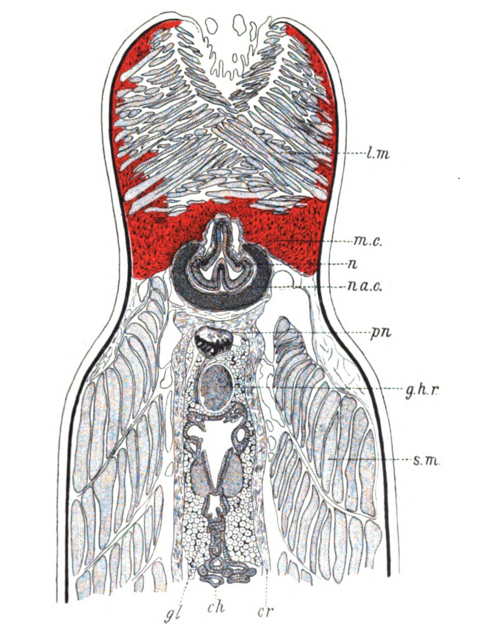
Fig. 31.—One of a Series of Horizontal Sections through the Head of Ammocœtes.
l.m., upper lip muscles; m.c., muco-cartilage; n., nose; na.c., nasal cartilage; pn., right pineal eye and nerve; g.h.r., right ganglion habenulæ; s.m., somatic muscles; cr., membranous wall of cranium; ch., choroid plexus; gl., glandular substance and pigment filling up brain-case.
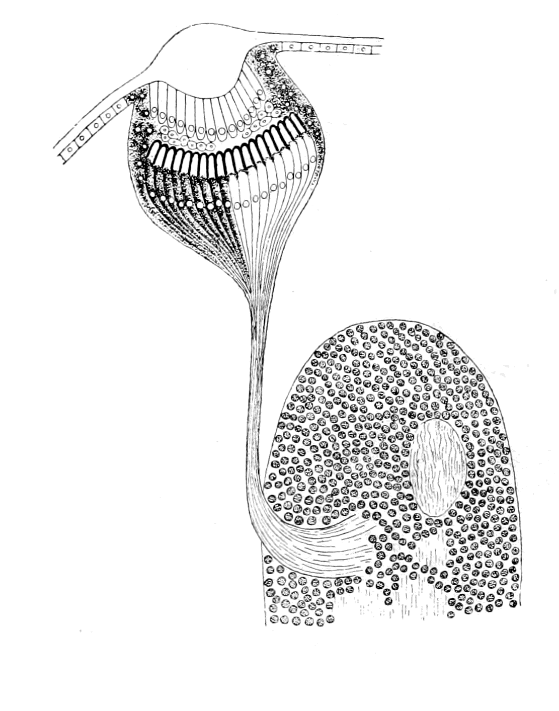
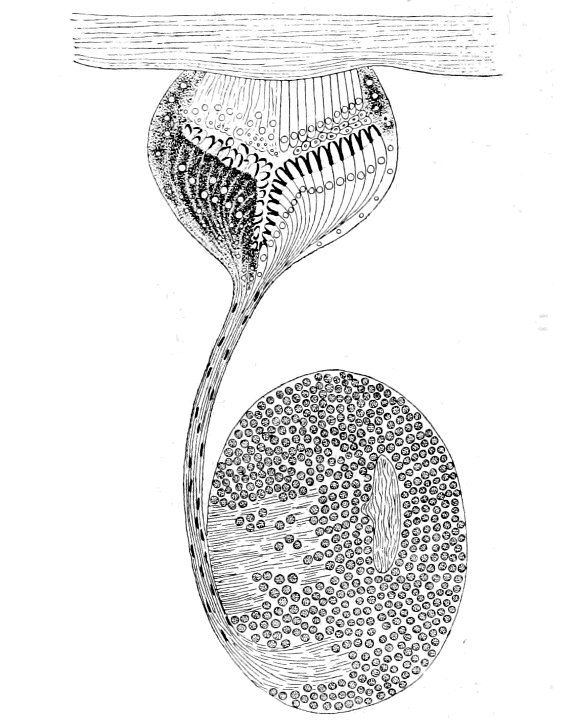
Fig. 32.—Eye of Acilius Larva, with its Optic Ganglion. On the right side the nerve end-cells have been drawn free from pigment. Fig. 33.—Pineal Eye of Ammocœtes, with its Ganglion Habenulæ. On the left side the eye is drawn as it appeared in the section. On the right side I have removed the pigment from the nerve end-cells, and drawn the eye as, in my opinion, it would appear if it were functional.
This difference between right and left indicates a greater degeneration on the left side, and points distinctly to a close relationship between the nerve-masses known as ganglia habenulæ and the median eyes. In my opinion this ganglion is, in part, at all events, the optic ganglion of the median eye on each side. It is built up on the same type as the optic ganglia of invertebrate simple eyes, with a cortex of small round cells and a medulla of fine nerve-fibres. Into this ganglion, on the right side, there passes a very well-defined nerve—the nerve of the dorsal eye. The eye itself with its nerve, pn., and its optic ganglion, g.h.r., is beautifully shown by means of a horizontal section through the head of Ammocœtes (Fig. 31). Originally, as described by Scott, the eye stood vertically above its optic ganglion, and presented an appearance remarkably like Fig. 32, which represents one of the simple eyes and optic ganglia of a larva of Acilius as described by Patten; then, with the forward growth of the upper lip, the right pineal eye was dragged forward and its nerve pulled horizontally over the ganglion habenulæ. For this reason the eye, nerve, and ganglion are better shown in a nearly horizontal than in a transverse section.
The optic nerve belonging to this eye is most evident and clearly shown in Fig. 31, and in the series of consecutive sections which follow upon this section; no doubt can arise as to the structure in question having been the nerve of the eye, even though, as is possible, it does not contain any functional nerve-fibres.
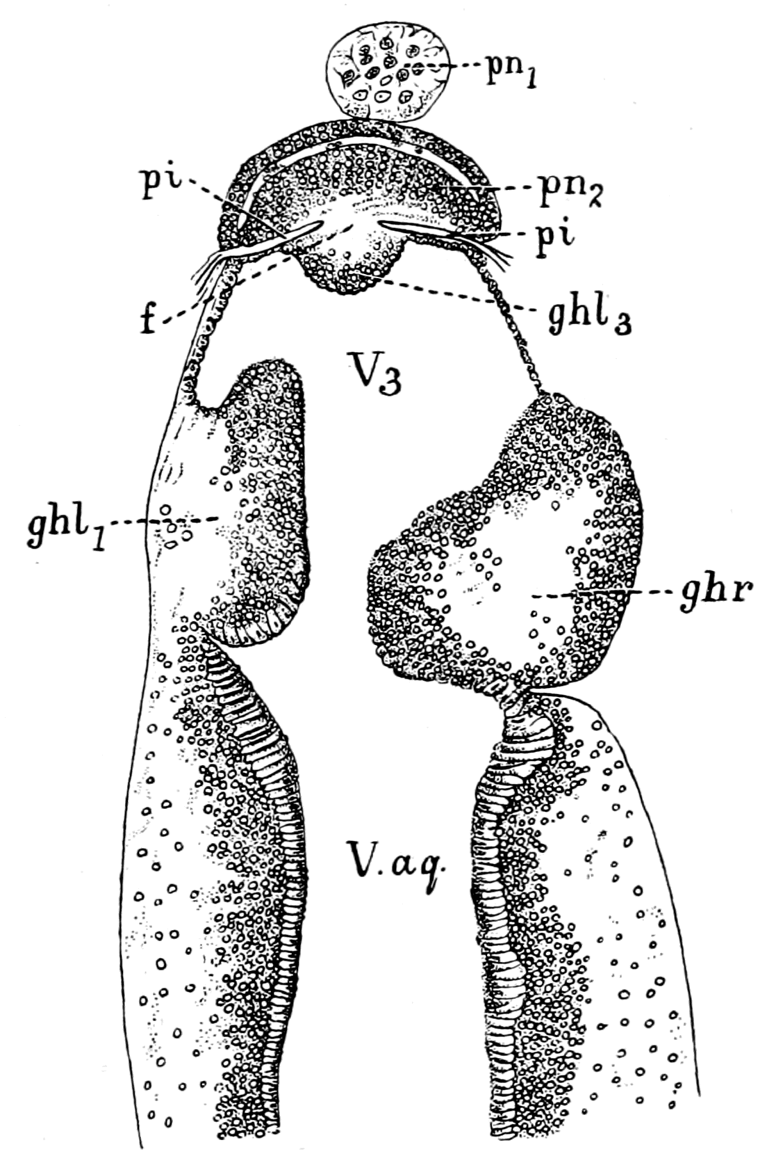
Fig. 34.—Horizontal Section through Brain of Ammocœtes, to show the Left, or Ventral Pineal Eye.
pn.2, left or ventral pineal eye; pn.1, last remnant of right, or dorsal pineal eye; g.h.r., right ganglion habenulæ; g.h.l.1, g.h.l.3, parts of left ganglion habenulæ; pi., fold of pia mater which separates the left ganglion habenulæ from the left pineal eye; f., strands of nerve-fibres connecting the left eye with its ganglion, g.h.l.3; V3, third ventricle; V.aq., ventricle of aquæduct.
The second, ventral or left, eye, belonging to the left ganglion habenulæ is very different in appearance, being much less evidently an eye. Fig. 34 is one of the same series of horizontal sections as Fig. 31, pn.1 being the last remnant of the right, or dorsal, eye, while pn.2 shows the left, or ventral, eye with its connection with the left ganglion habenulæ.
In a series of sections I have followed the nerve of the right pineal eye to its destination, as described in my paper in the Quarterly Journal of Microscopical Science, and have found that it enters into the ganglion habenulæ just as the nerve to any simple eye enters into its optic ganglion. This nerve, as I have shown, is a very distinct, well-defined nerve, with no admixture of ganglion-cells or of connective tissue, very different indeed to the connection between the left pineal eye and its optic ganglion. Here there is no defined nerve at all; but the cells of the ganglion habenulæ stretch right up to the remains of the eye itself. Seeing, then, that both the eye and ganglion on this side have reached a much further grade of degeneration than on the right side, it may be fairly concluded that the original condition of these two median eyes is more nearly represented by the right eye, with its well-defined nerve and optic ganglion, than by the left eye, or by the eyes in lizards and other animals which do not show so well-defined a nerve as is possessed by Ammocœtes. Quite recently Dendy has examined the two median eyes in the New Zealand lamprey Geotria australis. In this species the second eye is much better defined than in the European lamprey, and its connection with the ganglion habenulæ is more nerve-like. In neither eye, however, is the nerve so clean cut and isolated as is the nerve of the dorsal, or right, eye in the Ammocœtes stage of Petromyzon Planeri; in both, cells resembling those of the cortex of the ganglion habenulæ and connective tissues are mixed up with the nerve-fibres which pass from each eye to its respective optic ganglion.
The Right Pineal Eye of Ammocœtes.
The optic fibres of the right median eye of Ammocœtes are connected with a well-defined retina, the limits of which are defined by the white pigment so characteristic of this eye. This pigment is apparently calcium phosphate, which still remains as the 'brain-sand' of the human pineal gland. The cells, which are hidden by this pigment, were described by me in 1890 as the retinal end-cells with large nuclei. In 1893, Studniçka examined them more closely, and concluded that the retinal cells are of two kinds: the one, nerve end-cells, the sensory cells proper; the other, pigmented epithelial cells, which surround the sense-cells. The sense-cells may contain some of the white pigment, but not so much as the other cells. Similarly, in the median eyes of Limulus, Lankester and Bourne find it difficult to determine how far the retinal end-cells contain pigment and how far that pigment really is in the cells surrounding these nerve end-cells.
The interior of the eye presents the appearance of a cavity in shape like a cornucopia, the stalk of which terminates at the place where the nerve enters. This cavity is not empty, but the posterior part of it is filled with the termination of the nerve end-cells of the retina, as pointed out by me and confirmed by Studniçka. These terminations are free from pigment, and contain strikingly translucent bodies, which I have described in my paper in the Quarterly Journal, and called rhabdites, for they present the same appearance and are situated in the same position as are many of the rhabdites on the terminations of the retinal end-cells of arthropod eyes. Studniçka has also seen these appearances, and figures them in his second paper on the nerve end-cells of the pineal eye of Ammocœtes.
Up to this point the following conclusions may be drawn:—
1. Ammocœtes possesses a pair of median eyes, just as was the case with the most ancient fishes, and with the members of the contemporary palæostracan group.
2. The retina of one of these eyes is well-defined and upright, not inverted, and therefore in this respect agrees with that of all median eyes.
3. The presence of nerve end-cells, with pigment either in them or in cells around them, to the unpigmented ends of which translucent bodies resembling rhabdites are attached, is another proof that this retina agrees with that of the median eyes of arthropods.
4. The simple nature of the nerve with its termination in an optic ganglion closely resembling in structure an arthropod optic ganglion, together with Studniçka's statement that the nerve end-cells pass directly into the nerve, points directly to the conclusion that this retina is a simple, not a compound, retina, and that it therefore in this respect also agrees with the retina of all median eyes.
With respect to this last conclusion, neither I myself nor Studniçka have been able to see any definite groups of cells between the nerve end-cells and the optic nerve such as a compound retina necessitates.
On the other hand, Dendy describes in the New Zealand lamprey, Geotria australis, a cavity where the nerve enters into the eye, which he calls the atrium. This cavity is distinct from the main cavity of the eye, and is separated from it by a mass of cells similar in appearance to those of the cortex of the ganglion habenulæ. In these two eyes then, groups of cells, resembling in appearance those belonging to an optic ganglion, exist in the eyes themselves. This atrium is evidently that part of the central cavity which I have called the handle of the cornucopia in the European lamprey, and the very fact that it is separated from the rest of the central cavity is evidence that we are dealing here with a later stage in the history of the pineal eyes than in the case of the Ammocœtes of Petromyzon Planeri. Taking also into consideration the continuity of the mass of small ganglion-cells which surround this atrium with the cells of the ganglion habenulæ by means of the similar cells scattered along the course of the nerve, and also bearing in mind the fact already stated that in the more degenerate left eye of Ammocœtes the cells of the ganglion habenulæ extend right up to the eye itself, it seems more likely than not that these cells do not represent the original optic ganglion of a compound retina, but rather the subsequent invasion, by way of the pineal nerve, of ganglion-cells belonging to a portion of the brain. In the last chapter it has been suggested that the presence of the trochlear or fourth cranial nerve has given rise to the formation of the cerebellum by a similar spreading.
There is certainly no appearance in the least resembling a compound retina such as is seen in the vertebrate or crustacean lateral eye. In the median eyes of scorpions and of Limulus, cells are found within the capsule of the eye among the nerve-fibres and the nerve end-cells. These are especially numerous in the median eyes of Limulus, as described by Lankester and Bourne, and are called by them intrusive connective tissue cells. The meaning of these cells is not, to my mind, yet settled. It is sufficient for my purpose to point out that the presence of cells interneural in position among the nerve end-cells of the retina of the median eyes of Ammocœtes is more probable than not, on the assumption that the retina of these eyes is built up on the same plan as that of the median eyes in Limulus and the scorpions.
It is further to be borne in mind that these specimens of Geotria worked at by Dendy were in the 'Velasia' stage of the New Zealand lamprey, and correspond, therefore, more nearly to the Petromyzon than to the Ammocœtes stage of the European lamprey.
The Dioptric Apparatus.
Besides the retina, all eyes possess a dioptric apparatus. What is the evidence as to its nature in these vertebrate median eyes? Lankester and Bourne have divided the eyes of scorpions and Limulus into two kinds, monostichous and diplostichous. In the first the retinal cells are supposed to give rise to not only rhabdites but also the cuticular chitinous lens, so that the eye is one-layered; in the second the lens is formed by a well-marked hypodermal layer, in front of the retina, composed of elongated cells, so that these eyes are two-layered or diplostichous. The lateral eyes, according to them, are all monostichous, but the median eyes are diplostichous.
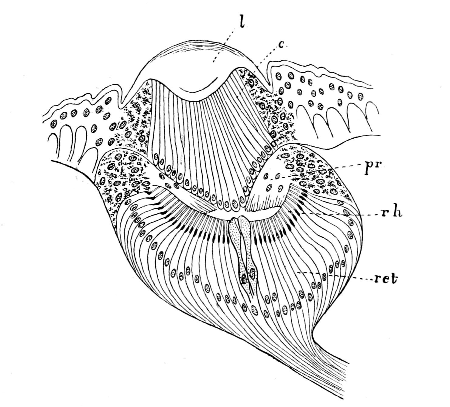
Fig. 35.—Eye of Acilius Larvæ. (After Patten.)
l., chitinous lens; c., corneagen; pr., pre-retinal layer; rh., rhabdites; ret., retinal end-cells.
This distinction is not considered valid by other observers. Thus, as already indicated, Patten looks on all these eyes as three-layered, and states that in all cases a corneagen or vitreogen layer exists, which gives origin to the lens. For my own part I agree with Patten, but we are not concerned here with the lateral eyes. It is sufficient to note that all observers are agreed that the median eyes are characterized by this well-marked cell-layer, the so-called vitreous or corneagen layer of cells.
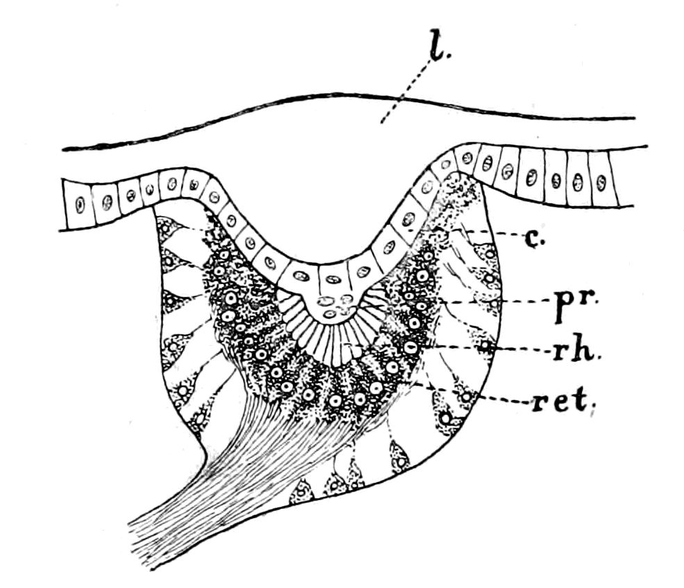
Fig. 36.—Eye of Hydrophilus Larva, with the Pigment over the Retinal End-cells.
l., chitinous lens; c., corneagen; pr., pre-retinal layer; rh., rhabdites; ret., retinal end-cells.
This layer (c., Fig. 35) is composed of much-elongated cells of the hypodermal layer, in each of which the large nucleus is always situated towards the base of the cell. The space between it and the retina contains, according to Patten the cells of the pre-retinal layer (pr.). These may be so few and insignificant as to give the impression that the vitreous layer is immediately adjacent to the retina (ret.).
Let us turn now to the right pineal eye of Ammocœtes (Fig. 37) and see what its further structure is. The anterior part of the eye is free from pigment, and is composed, as is seen in hæmatoxylin or carmine specimens, of an inner layer of nuclei which are frequently arranged in a wavy line. From this nucleated layer, strands of tissue, free from nuclei, pass to the anterior edge of the eye.
In the horizontal longitudinal sections it is seen that these strands are confined to the middle of the eye. On each side of them the nuclear layer reaches the periphery, so that if we consider these strands to represent long cylindrical cells, as described by Beard, then the anterior wall may be described as consisting of long cylindrical cells, which are flanked on either side by shorter cells of a similar kind. The nuclei at the base of these cylindrical cells are not all alike. We see, in the first place, large nuclei resembling the large nuclei belonging to the nerve end-cells; these are the nuclei of the long cylindrical cells. We see also smaller nuclei in among these larger ones, which look like nuclei of intrusive connective tissue, or may perhaps form a distinct layer of cells, situated between the cells of the anterior wall and the terminations of the nerve end-cells already referred to.
These elongated cells are in exactly the same position and present the same appearance as the cells of the corneagen layer of any median eye. Like the latter they are free from pigment and never show with osmic staining any sign of the presence of translucent rhabdite-like bodies, such as are seen in the termination of the retinal cells, and like the latter their nuclei are at the base. The resemblance between this layer and the corneagen cells of any median eye is absolute. Between it and the terminations of the retinal cells there exists some ill-defined material certainly containing cells which may well correspond to Patten's pre-retinal layer of cells.
Retina, corneagen, nerve, optic ganglion, all are there, all in their right position, all of the right structure, what more is needed to complete the picture?
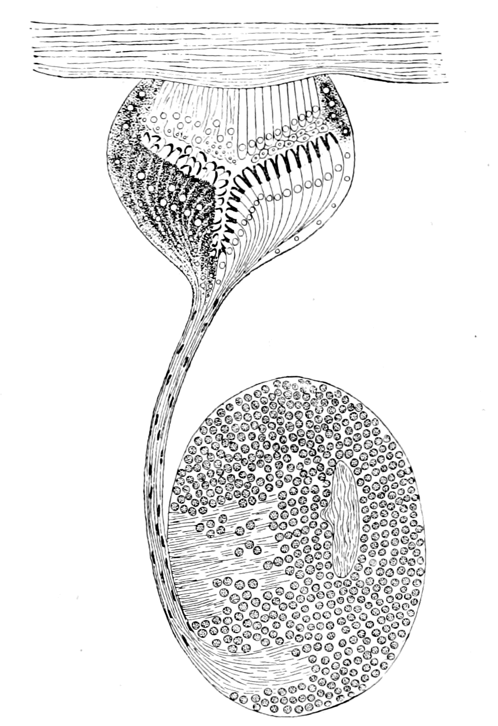
Fig. 37.—Pineal Eye of Ammocœtes, with its Ganglion Habenulæ.
In order to complete the dioptric apparatus a lens is necessary. Where, then, is the lens in these pineal eyes? In all the arachnid eyes, whether median or lateral, the lens is a single corneal lens composed of the external cuticle, which is thickened over the corneagen cells. This thickened cuticle is composed of chitin, and is not cellular, but is dead material formed out of the living underlying corneagen cells. Such a lens is in marked contrast to the lens of the lateral vertebrate eye, which is formed by living cells themselves. This thickening of the cuticular layer to form a lens could only exist as long as that layer is absolutely external, so that the light strikes immediately upon it; for, if from any cause the eye became situated internally, the place of such a lens must be filled by the structures situated between it and the surface, and the thickened cuticle would no longer be formed.
In all vertebrates these pineal eyes are separated from the external surface by a greater or less thickness of tissues; in the case of Ammocœtes, as is seen in Fig. 31, the eye lies within the membranous cranial wall, and is attached closely to it. The position, then, of the cuticular, or corneal lens, as it is often called, on the supposition that this is a median eye of the arachnid type, is taken by the membranous cranium, and, as I have described in my paper in the Quarterly Journal, on carefully lifting the eye in the fresh condition from the cranial wall, it can be seen under a dissecting microscope that the cranial wall often forms at this spot a lens-like bulging, which fits the shallow concavity of the surface of the eye, and requires some little force to separate it from the eye.
As will appear in a subsequent chapter, this cranial wall has been formed by the growth, laterally and dorsally, of a skeletal structure known by the name of the plastron. The last part of it to be completed would be that part in the mid-dorsal line, where apparently, in consequence of the insinking of the degenerating eyes, a dermal and subdermal layer already intervened between the source of light and the eyes themselves.
When the membranous cranium was completed in the mid-dorsal region, it was situated here as elsewhere just internally to the subdermal layer, and therefore enclosed the pineal eyes. This, to my mind, is the reason why the pineal eyes, which, in all other respects, conform to the type of the median eyes of an arachnid-like animal, do not possess a cuticular lens. Other observers have attempted to make a lens out of the elongated cells of the anterior wall of the eye (my corneagen layer), but without success.
Studniçka, who calls this layer the pellucida, does not look upon it as the lens, but considers, strangely enough, that the translucent appearances at the ends of each nerve end-cell represent a lens for that cell, so that every nerve end-cell has its own lens. Still more strange is it that, holding this view, he should yet consider these knobs to be joined by filaments to the cells in the anterior wall of the eye, a conception fatal to the action of such knobs as lenses.
The discovery that the vertebrate possesses, in addition to the lateral eyes, a pair of median eyes, which are most conspicuous in the lowest living vertebrate, together with the fact that such eyes are built up on the same plan as the median eyes of living crustaceans or arachnids, not only with respect to the eye itself but also to its nerve and optic ganglion, constitutes a fact of the very greatest importance for any theory of the origin of vertebrates; especially in view of the further fact, that similar eyes in the same position are found not only in all the members of the Palæostraca, but also in all those ancient forms (classed as fishes) which lived at that time. At one and the same moment it proves the utter impossibility of reversing dorsal and ventral surfaces, points in the very strongest manner to the origin of the vertebrate from some member or other of the palæostracan group, and insists that the advocates of the origin of vertebrates from the Hemichordata, etc., should give an explanation of the presence of these two median eyes of a more convincing character than that given here.
The Lateral Eyes.
Turning now to the consideration of the lateral eyes, we see that these eyes in the arachnids often possess an inverted retina, in the crustaceans always an upright retina. In the arachnids they possess a simple retina, while in the crustaceans their retina is compound; so that in the latter case the so-called optic nerve is in reality a tract of fibres connecting together the brain-region with a variable number of optic ganglia, which have been left at the periphery in close contact with the retinal cells, when the brain sunk away from the superficial epithelial covering.
There is, then, this difference between the lateral eyes of crustaceans and arachnids, that the retina of the former is compound, but never inverted, while that of the latter may be inverted, but is always simple.
The retina of the lateral eyes of the vertebrate resembles both of these, for it is compound, as in the crustacean, and inverted as in the arachnid.
It must always be borne in mind that in the palæostracan epoch the dominant race was neither crustacean nor arachnid, but partook of the characters of both; also, as is characteristic of dominance, there was very great variety of form, so that it seems more probable than not that some of these forms may have combined the arachnid and crustacean characteristics to the extent of possessing lateral eyes with an inverted yet compound retina. A certain amount of evidence points in this direction. As already stated, the compound retina which characterizes the vertebrate lateral eyes is characteristic of all facetted eyes, and in the trilobites facetted lateral eyes are commonly found. From this it may be concluded that many of the trilobites possessed eyes with a compound retina. There have, however, been found in certain species, e.g. Harpes vittatus and Harpes ungula, lateral eyes which were not facetted, and are believed by Korschelt and Heider to be of an arachnid nature. They say, "Palæontologists have appropriately described them as ocelli, although, from a zoological point of view, they do not deserve this name, having most probably arisen in a way similar to that conjectured in connection with the lateral eyes of scorpions." If this conjecture is right, then in these forms the retina may have been inverted, but because they belonged to the trilobite group, the retina was most probably compound, so that here we may have had the combination of the arachnid and crustacean characteristics. On the other hand, in some forms of Branchipus, and many of the Gammaridæ, a single corneal lens is found in conjunction with an eye of the crustacean type, so that a non-facetted lateral eye, found in a fossil form, would not necessarily imply the arachnid type of eye with the possibility of an inverted retina. Whatever may be the ultimate decision upon these particular forms, the striking fact remains, that both in the vertebrate and in the arachnid the median eyes possess a simple upright retina, while the lateral eyes possess an inverted retina, and that both in the vertebrate and the crustacean the median eyes possess a simple upright retina, while the lateral eyes possess a compound retina.
The resemblance of the retina of the lateral eyes of vertebrates to that of the lateral eyes of many arthropods, especially crustaceans, has been pointed out by nearly every one who has worked at these invertebrate lateral eyes. The foundation of our knowledge of the compound retina is Berger's well-known paper, the results of which are summed up by him in the following two main conclusions.
1. The optic ganglion of the Arthropoda consists of two parts, of which the one stands in direct inseparable connection with the facetted eye, and together with the layer of retinal rods forms the retina of the facetted eye, while the other part is connected rather with the brain, and is to be considered as an integral part of the brain in the narrower sense of the word.
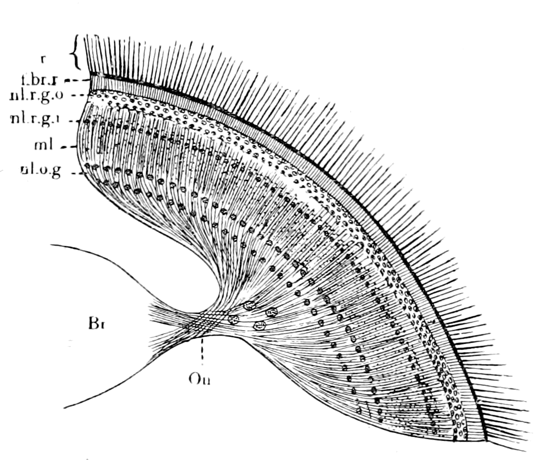
Fig. 38.—The Retina of Musca. (After Berger.)
Br., brain; O.n., optic nerve; n.l.o.g., nuclear layer of ganglion of optic nerve; m.l., molecular layer (Punktsubstanz); n.l.r.g.i. and n.l.r.g.o., inner and outer nuclear layers of the ganglion of the retina; f.br.r., terminal fibre-layer of retina; r., layer of retinal end-cells (indicated only).
2. In all arthropods examined by him, the retina consists of five layers, as follows:—
(1) The layer of rods and their nuclei. (2) The layer of nerve-bundles. (3) The nuclear layer. (4) The molecular layer. (5) The ganglion cell layer.
Berger passes under review the structure and arrangement of the optic ganglion in a large number of different groups of arthropods, and concludes that in all cases one part of the optic ganglion is always closely attached to the visual end-cells, and this combination he calls the retina. On the other hand, the nerve-fibres which connect the peripheral part of the optic ganglion with the brain, the so-called optic nerve, are by no means homologous in the different groups; for in some cases, as in many of the stalk-eyed crustaceans, the whole optic ganglion is at the periphery, while in others, as in the Diptera, only the retinal ganglion is at the periphery, and the nerve-stalk connects this with the rest of the optic ganglion, the latter being fused with the main brain-mass. In the Diptera, in fact, according to Berger, the optic nerve and retina are most nearly comparable to those of the vertebrate. For this reason I give Berger's picture of the retina of Musca (Fig. 38), in order to show the arrangement there of the retinal layers.
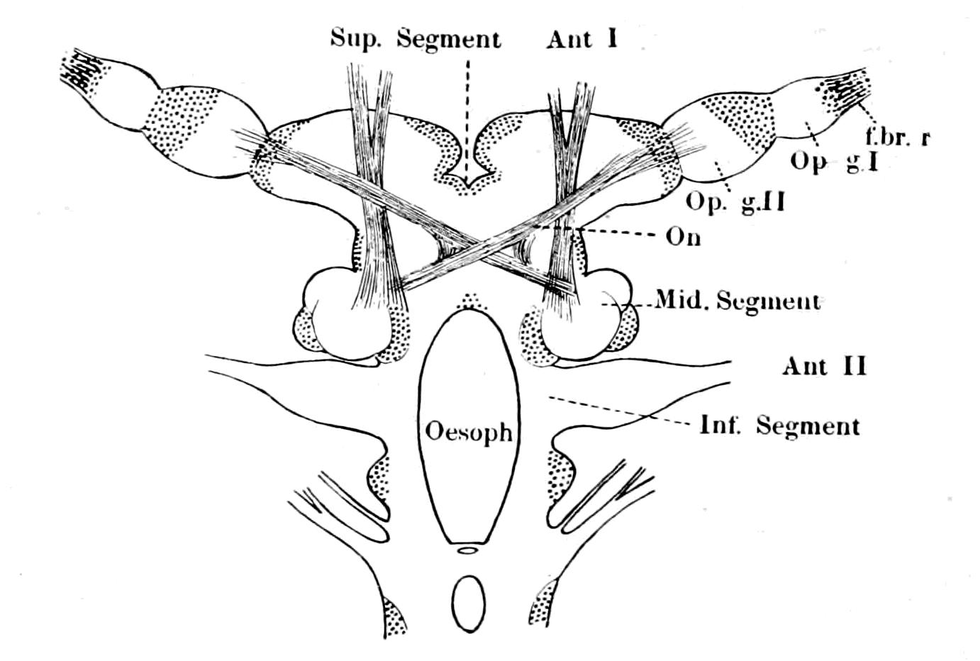
Fig. 39.—The Brain of Sphæroma serratum. (After Bellonci.)
Ant. I. and Ant. II., nerves to 1st and 2nd antennæ. f.br.r., terminal fibre-layer of retina; Op. g. I., first optic ganglion; Op. g. II., second optic ganglion; O.n., optic nerve-fibres forming an optic chiasma.
In Branchipus and other primitive Crustacea, Berger also finds the same retinal layers, but is unable to distinguish in the brain the rest of the optic ganglion. Judging from Berger's description of Branchipus, and Bellonci's of Sphæroma, it would almost appear as though the cerebral part of the retina in the higher forms originated from two ganglionic enlargements, an external and internal enlargement, as Bellonci calls them. The external ganglion (Op. g. I., Fig. 39) may be called the ganglion of the retina, the cells of which form the nuclear layer of the higher forms, and the internal ganglion (Op. g. II., Fig. 39), from which the optic nerve-fibres to the brain arise, may therefore be called the ganglion of the optic nerve. Bellonci describes how in this latter ganglion cells are found very different to the small ones of the external ganglion or ganglion of the retina. So also in Branchipus, judging from the pictures of Berger, Claus, and from my own observations (cf. Fig. 46, in which the double nature of the retinal ganglion is indicated), the peripheral part of the optic ganglion—i.e. the retinal ganglion—may be spoken of as composed of two ganglia. The external of these is clearly the ganglion of the retina; its cells form the nuclear layer, the striking character of which, and close resemblance to the corresponding layer in vertebrates, is shown by Claus' picture, which I reproduce (Fig. 40). The internal ganglion with which the optic nerve is in connection contains large ganglion cells, which, together with smaller ones, form the ganglionic layer of Berger.
The most recent observations of the structure of the compound retina of the crustacean eye are those of Parker, who, by the use of the methylene blue method, and Golgi's method of staining, has been able to follow out the structure of the compound retina in the arthropod on the same lines as had already been done for the vertebrate. These two methods have led to the conclusion that the arthropod central nervous system and the vertebrate central nervous system are built up in the same manner—viz. by means of a series of ganglia connected together, each ganglion being composed of nerve-cells, nerve-fibres, and a fine reticulated substance called by Leydig in arthropods 'Punktsubstanz,' and known in vertebrates and in invertebrates at the present time as 'neuropil.' A further analysis resolves the whole system into a combination of groups of neurones, the cells and fibres of which form the cells and fibres of the ganglia, while their dendritic connections with the terminations of other neurones, together with the neuroglia-cells form the 'neuropil.' As is natural to expect, that part of the central nervous system which helps to form the compound retina is built up in the same manner as the rest of the central nervous system.
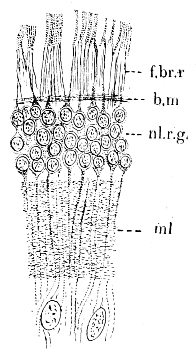
Fig. 40.—Bipolar Cells of Nuclear Layer in Retina of Branchipus. (After Claus.)
f.br.r., terminal fibre-layer of retina; n.l.r.g., bipolar cells of the ganglion of the retina = inner nuclear layer; m.l., Punktsubstanz = inner molecular layer; b.m., basement membrane formed by neurilemma round central nervous system.
Thus, according to Parker, the mass of nervous tissue which occupies the central part of the optic stalk in Astacus is composed of four distinct ganglia; the retina is connected with the first of these by means of the retinal fibres, and the optic nerve extends proximally from the fourth ganglion to the brain. Each ganglion consists of ganglion-cells, nerve-fibres, and 'neuropil,' and, in addition, supporting cells of a neuroglial type. By means of the methylene blue method and the Golgi method, it is seen that the retinal end-cells, with their visual rods, are connected with the fibres of the optic nerve by means of a system of neurones, the synapses of which take place in and help to form the 'neuropil' of the various ganglia. Thus, an impulse in passing from the retina to the brain would ordinarily travel over five neurones, beginning with one of the first order and ending with one of the fifth. He makes five neurones although there are only four ganglia, because he reckons the retinal cell with its elongated fibre as a neurone of the first order, such fibre terminating in dendritic processes which form synapses in the 'neuropil' of the first ganglion with the neurones of the second order.
Similarly the neurones of the second order terminate in the 'neuropil' of the second ganglion, and so on, until we reach the neurones of the fifth order, which terminate on the one hand in the 'neuropil' of the fourth ganglion, and on the other pass to the optic lobes of the brain by their long neuraxons—the fibres of the optic nerve.
He compares this arrangement with that of Branchipus, Apus, Estheria, Daphnia, etc., and shows that in the more primitive crustaceans the peripheral optic apparatus was composed, not of four but of two optic ganglia, not, therefore, of five but of three neurones, viz.—
1. The neurone of the first order—i.e. the retinal cell with its fibre terminating in the 'neuropil' of the first optic ganglion (ganglion of the retina).
2. The neurone of the second order, which terminates in the 'neuropil' of the second ganglion (ganglion of the optic nerve).