VOLUME XIV SLICE II Hydromechanics to Ichnography (continued 4)
Reading edition
The question is one intimately connected with the view taken as to the nature and individuality of polyp, medusa and gonophore respectively. On this point the following theories have been put forward.
1. The theory that the medusa is simply an organ, which has become detached and has acquired a certain degree of independence, like the well-known instance of the hectocotyle of the cuttle-fish. On this view, put forward by E. van Beneden and T. H. Huxley, the sporosac is the starting-point of an evolution leading up through the various types of gonophores to the free medusa as the culminating point of a phyletic series. The evidence against this view may be classed under two heads: first, comparative evidence; hydroids very different in their structural characters and widely separate in the systematic classification of these organisms may produce medusae very similar, at least so far as the essential features of medusan organization are concerned; on the other hydroids closely allied, perhaps almost indistinguishable, may produce gonophores in the one case, medusae in the other; for example, Hydractinia (gonophores) and Podocoryne (medusae), Tubularia (gonophores) and Ectopleura (medusae), Coryne (gonophores) and Syncoryne (medusae), and so on. If it is assumed that all these genera bore gonophores ancestrally, then medusa of similar type must have been evolved quite independently in a great number of cases. Secondly, there is the evidence from the development, namely, the presence of the entocodon in the medusa-bud, a structure which, as explained above, can only be accounted for satisfactorily by derivation from a medusan type of organization. Hence it may be concluded that the gonophores are degenerate medusae, and not that the medusae are highly elaborated gonophores, as the organ-theory requires.
2. The theory that the medusa is an independent individual, fully equivalent to the polyp in this respect, is now universally accepted as being supported by all the facts of comparative morphology and development. The question still remains open, however, which of the two types of person may be regarded as the most primitive, the most ancient in the race-history of the Hydromedusae. F. M. Balfour put forward the view that the polyp was the more primitive type, and that the medusa is a special modification of the polyp for reproductive purposes, the result of division of labour in a polyp-colony, whereby special reproductive persons become detached and acquire organs of locomotion for spreading the species. W. K. Brooks, on the other hand, as stated above, regards the medusa as the older type and looks upon both polyp and medusa, in the Hydromedusae, as derived from a free-swimming or floating actinula, the polyp being thus merely a fixed nutritive stage, possessing secondarily acquired powers of multiplication by budding.
The Hertwigs when they discovered the endoderm-lamella showed on morphological grounds that polyp and medusa are independent types, each produced by modification in different directions of a more primitive type represented in development by the actinula-stage. If a polyp, such as Hydra, be regarded simply as a sessile actinula, we must certainly consider the polyp to be the older type, and it may be pointed out that in the Anthozoa only polyp-individuals occur. This must not be taken to mean, however, that the medusa is derived from a sessile polyp; it must be regarded as a direct modification of the more ancient free actinula form, without primitively any intervening polyp-stage, such as has been introduced secondarily into the development of the Leptolinae and represents a revival, so to speak, of an ancestral form or larval stage, which has taken on a special role in the economy of the species.
Systematic Review of the Hydromedusae
Order I. Eleutheroblastea.—Simple polyps which become sexually mature and which also reproduce non-sexually, but without any medusoid stage in the life-cycle.
The sub-order includes the family Hydridae, containing the common fresh-water polyps of the genus Hydra. Certain other forms of doubtful affinities have also been referred provisionally to this section.
Hydra.—This genus comprises fresh-water polyps of simple structure. The body bears tentacles, but shows no division into hydrorhiza, hydrocaulus or hydranth; it is temporarily fixed and has no perisarc. The polyp is usually hermaphrodite, developing both ovaries and testes in the same individual. There is no free-swimming planula larva, but the stage corresponding to it is passed over in an enveloping cyst, which is secreted round the embryo by its own ectodermal layer, shortly after the germ-layer formation is complete, i.e. in the parenchymula-stage. The envelope is double, consisting of an external chitinous stratified shell, and an internal thin elastic membrane. Protected by the double envelope, the embryo is set free as a so-called “egg,” and in Europe it passes the winter in this condition. In the spring the embryo bursts its shell and is set free as a minute actinula which becomes a Hydra.
Many species are known, of which three are common in European waters. It has been shown by C. F. Jickeli (28) that the species are distinguishable by the characters of their nematocysts. They also show characteristic differences in the egg (Brauer [2]). In Hydra viridis the polyp is of a green colour and produces a spherical egg with a smooth shell which is dropped into the mud. H. grisea is greyish in tint and produces a spherical egg with a spiky shell, which also is dropped into the mud. H. fusca (= H. vulgaris) is brown in colour, and produces a bun-shaped egg, spiky on the convex surface, and attached to a water-weed or some object by its flattened side. Brauer found a fourth species, similar in appearance to H. fusca, but differing from the three other species in being of separate sexes, and in producing a spherical egg with a knobby shell, which is attached like that of H. fusca.
The fact already noted that the species of Hydra can be distinguished by the characters of their nematocysts is a point of great interest. In each species, two or three kinds of nematocysts occur, some large, some small, and for specific identification the nematocysts must be studied collectively in each species. It is very remarkable that this method of characterizing and diagnozing species has never been extended to the marine hydroids. It is quite possible that the characters of the nematocysts might afford data as useful to the systematist in this group as do the spicules of sponges, for instance. It would be particularly interesting to ascertain how the nematocysts of a polyp are related to those possessed by the medusa budded from it, and it is possible that in this manner obscure questions of relationship might be cleared up.
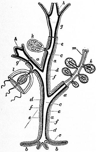
Fig. 49.—Diagram showing possible modifications of persons of a gymnoblastic Hydromedusa. (After Allman.)
a, Hydrocaulus (stem). b, Hydrorhiza (root). c, Enteric cavity. d, Endoderm. e, Ectoderm. f, Perisarc, (horny case). g, Hydranth (hydriform person) expanded. g′, Hydranth (hydriform person) contracted. h, Hypostome, bearing mouth at its extremity. k, Sporosac springing from the hydrocaulus. k′, Sporosac springing from m, a modified hydriform person (blastostyle): the genitalia are seen surrounding the spadix or manubrium. l, Medusiform person or medusa. m, Blastostyle.
Protohydra is a marine genus characterized by the absence of tentacles, by a great similarity to Hydra in histological structure, and by reproduction by transverse fission. It was found originally in an oyster-farm at Ostend. The sexual reproduction is unknown. For further information see C. Chun (Hydrozoa [1]. Pl. I.).
Polypodium hydriforme Ussow is a fresh-water form parasitic on the eggs of the sterlet. A “stolon” of unknown origin produces thirty-two buds, which become as many Polypodia; each has twenty-four tentacles and divides by fission repeated twice into four individuals, each with six tentacles. The daughter-individuals grow, form the full number of twenty-four tentacles and divide again. The polyps are free and walk on their tentacles. See Ussow [54].
Tetraplatia volitans Viguier is a remarkable floating marine form. See C. Viguier [56] and Delage and Hérouard (Hydrozoa [2]).
Haleremita Schaudinn. See F. Schaudinn [50] and Delage and Hérouard (Hydrozoa [2]).
In all the above-mentioned genera, with the exception of Hydra, the life-cycle is so imperfectly known that their true position cannot be determined in the present state of our knowledge. They may prove eventually to belong to other orders. Hence only the genus Hydra can be considered as truly representing the order Eleutheroblastea. The phylogenetic position of this genus has been discussed above.
Order II. Hydroidea seu Leptolinae.—Hydromedusae with alternation of generations (metagenesis) in which a non-sexual polyp-generation (trophosome) produces by budding a sexual medusa-generation (gonosome). The polyp may be solitary, but more usually produces polyps by budding and forms a polyp-colony. The polyp usually has the body distinctly divisible into hydranth, hydrocaulus and hydrorhiza, and is usually clothed in a perisarc. The medusae may be set free or may remain attached to the polyp-colony and degenerate into a gonophore. When fully developed the medusa is characterized by the sense organs being composed entirely of ectoderm, developed independently of the tentacles, and innervated from the sub-umbral nerve-ring.
The two kinds of persons present in the typical Hydroidea make the classification of the group extremely difficult, for reasons explained above. Hence the systematic arrangement that follows must be considered purely provisional. A natural classification of the Hydroidea has yet to be put forward. Many genera and families are separated by purely artificial characters, mere shelf-and-bottle groupings devised, for the convenience of the museum curator and the collector. Thus many subdivisions are diagnosed by setting free medusae in one case, or producing gonophores in another, although it is very obvious, as pointed out above, that a genus producing medusae may be far more closely allied to one producing gonophores than to another producing medusae, or vice versa, and that in some cases the production of medusae or gonophores varies with the season or the sex. Moreover, P. Hallez [22] has recently shown that hydroids hitherto regarded as distinct species are only forms of the same species grown under different conditions.
Sub-Order 1. Hydroidea Gymnoblastea (Anthomedusae).—Trophosome without hydrothecae or gonothecae, with monopodial type of budding. Gonosome with free medusae or gonophores; medusae usually with ocelli, never with otocysts. The gymnoblastic polyp usually has a distinct perisarc investing the hydrorhiza and the hydrocaulus, sometimes also the hydranth as far as the bases of the tentacles (Bimeria); but in such cases the perisarc forms a closely-fitting investment or cuticule on the hydranth, never a hydrotheca standing off from it, as in the next sub-order. The polyps may be solitary, or form colonies, which may be of the spreading or encrusting type, or arborescent, and then always of monopodial growth and budding. In some cases, any polyp of the colony may bud medusae; in other cases, only certain polyps, the blastostyles, have this power. When blastostyles are present, however, they are never enclosed in special gonothecae as in the next sub-order. In this sub-order the characters of the hydranth are very variable, probably owing to the fact that it is exposed and not protected by a hydrotheca, as in Calyptoblastea.
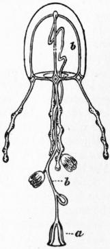
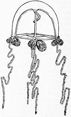
Fig. 50.—Sarsia (Dipurena) gemnifera. b, The long manubrium, bearing medusiform buds; a, mouth. Fig. 51.—Sarsia prolifera. Ocelli are seen at the base of the tentacles, and also (as an exception) groups of medusiform buds.
Speaking generally, three principal types of hydranth can be distinguished, each with subordinate varieties of form.
1. Club-shaped hydranths with numerous tentacles, generally scattered irregularly, sometimes with a spiral arrangement, or in whorls (“verticillate”).
(a) Tentacles filiform; type of Clava (fig. 5), Cordylophora, &c.
(b) Tentacles capitate, simple; type of Coryne and Syncoryne; Myriothela is an aberrant form with some of the tentacles modified as “claspers” to hold the ova.
(c) Tentacles capitate, branched, wholly or in part; type of Cladocoryne.
(d) Tentacles filiform or capitate, tending to be arranged in definite whorls; type of Stauridium (fig. 2), Cladonema and Pennaria.
2. Hydranth more shortened, daisy-like in form, with two whorls of tentacles, oral and aboral.
(a) Tentacles filiform, simple, radially arranged or scattered irregularly; type of Tubularia (fig. 4), Corymorpha (fig. 3), Nemopsis, Pelagohydra, &c.
(b) Tentacles with a bilateral arrangement, branched tentacles in addition to simple filiform ones; type of Branchiocerianthus.
3. Hydranth with a single circlet of tentacles.
(a) With filiform tentacles; the commonest type, seen in Bougainvillea (fig. 13), Eudendrium, &c.
(b) With capitate tentacles; type of Clavatella.
4. Hydranth with tentacles reduced below four; type of Lar (fig. 11), Monobrachium, &c.
The Anthomedusa in form is generally deep, bell-shaped. The sense organs are typically ocelli, never otocysts. The gonads are borne on the manubrium, either forming a continuous ring (Codonid type), or four masses or pairs of masses (Oceanid type). The tentacles may be scattered singly round the margin of the umbrella (“monerenematous”) or arranged in tufts (“lophonematous”); in form they may be simple or branched (Cladonemid type); in structure they may be hollow (“coelomerinthous”); or solid (“pycnomerinthous”). When sessile gonophores are produced, they may show all stages of degeneration.
Classification.—Until quite recently the hydroids (Gymnoblastea) and the medusae (Anthomedusae) have been classified separately, since the connexion between them was insufficiently known. Delage and Hérouard (Hydrozoa [2]) were the first to make an heroic attempt to unite the two classifications into one, to which Hickson (Hydrozoa [4]) has made some additions and slight modifications. The classification given here is for the most part that of Delage and Hérouard. It is certain, however, that no such classification can be considered final at present, but must undergo continual revision in the future. With this reservation we may recognize fifteen well-characterized families and others of more doubtful nature. Certain discrepancies must also be noted.
1. Margelidae (= medusa-family Margelidae + hydroid families Bougainvillidae, Dicorynidae, Bimeridae and Eudendridae). Trophosome arborescent, with hydranths of Bougainvillea-type; gonosome free medusae or gonophores, the medusae with solid tentacles in tufts (lophonematous). Common genera are the hydroid Bougainvillea (figs. 12, 13), and the medusae Hippocrene (budded from Bougainvillea), Margelis, Rathkea (fig. 24), and Margellium. Other hydroids are Garveia, Bimeria, Eudendrium and Heterocordyle, with gonophores, and Dicoryne with peculiar sporosacs.
After Haeckel, System der Medusen, by permission of Gustav Fischer.
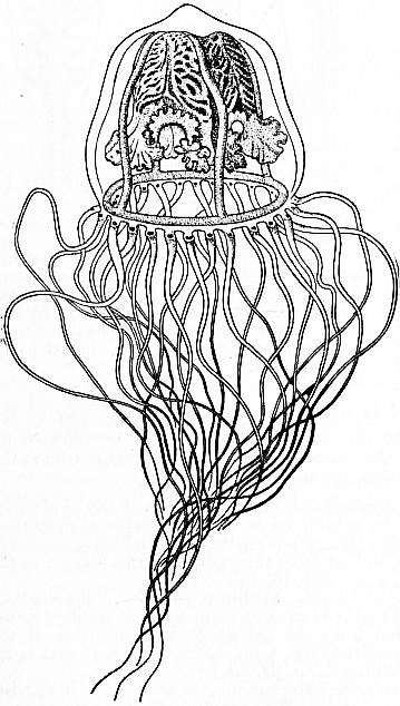
Fig. 52.—Tiara pileata, L. Agassiz.
2. Podocorynidae (= medusa-families Thamnostomidae and Cytaeidae + hydroid families Podocorynidae and Hydractiniidae). Trophosome encrusting with hydranths of Bougainvillea-type, polyps differentiated into blastostyles, gastrozoids and dactylozoids; gonosome free medusae or gonophores. The typical genus is the well-known hydroid Podocoryne, budding the medusa known as Dysmorphosa; Thamnostylus, Cytaeis, &c., are other medusae with unknown hydroids. Hydractinia (figs. 9, 10) is a familiar hydroid genus, bearing gonophores.
3. Cladonemidae.—Trophosome, polyps with two whorls of tentacles, the lower filiform, the upper capitate; gonosome, free medusae, with tentacles solid and branched. The type-genus Cladonema (fig. 20) is a common British form.
4. Clavatellidae.—Trophosome, polyps with a single whorl of capitate tentacles; gonosome, free medusae, with tentacles branched, solid. Clavatella (fig. 21), with a peculiar ambulatory medusa is a British form.
5. Pennariidae.—Trophosome, polyps with an upper circlet of numerous capitate tentacles, and a lower circlet of filiform tentacles. Pennaria, with a free medusa known as Globiceps, is a common Mediterranean form. Stauridium (fig. 2) is a British hydroid.
6. Tubulariidae.—Trophosome, polyps with two whorls of tentacles, both filiform. Tubularia (fig. 4), a well-known British hydroid, bears gonophores.
7. Corymorphidae (including the medusa-family Hybocodonidae).—Trophosome solitary polyps, with two whorls of tentacles; gonosome, free medusae or gonophores. Corymorpha (fig. 3), a well-known British genus, sets free a medusa known as Steenstrupia (fig. 22). Here belong the deep-sea genera Monocaulus and Branchiocerianthus, including the largest hydroid polyps known, both genera producing sessile gonophores.
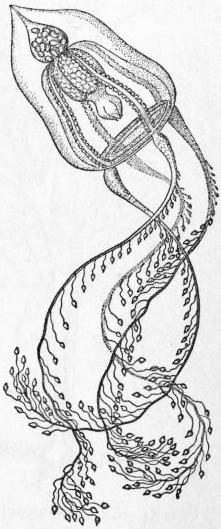
After Haeckel, System der Medusen, by permission of Gustav Fischer.
Fig. 53.—Pteronema darwinii. The apex of the stomach is prolonged into a brood pouch containing embryos.
8. Dendroclavidae.—Trophosome, polyp with filiform tentacles in three or four whorls. Dendroclava, a hydroid, produces the medusa known as Turritopsis.
9. Clavidae (including the medusa-family Tiaridae (figs. 27 and 51). Trophosome, polyps with scattered filiform tentacles; gonosome, medusae or gonophores, the medusae with hollow tentacles. Clava (fig. 5), a common British hydroid, produces gonophores; so also does Cordylophora, a form inhabiting fresh or brackish water. Turris produces free medusae. Amphinema is a medusan genus of unknown hydroid.
10. Bythotiaridae.—Trophosome unknown; gonosome, free medusae, with deep, bell-shaped umbrella, with interradial gonads on the base of the stomach, with branched radial canals, and correspondingly numerous hollow tentacles. Bythotiara, Sibogita.
11. Corynidae (= hydroid families Corynidae, Syncorynidae and Cladocorynidae + medusan family Sarsiidae).—Trophosome polyps with capitate tentacles, simple or branched, scattered or verticillate; gonosome, free medusae or gonophores. Coryne, a common British hydroid, produces gonophores; Syncoryne, indistinguishable from it, produces medusae known as Sarsia (fig. 51). Cladocoryne is another hydroid genus; Codonium and Dipurena (fig. 50) are medusan genera.
12. Myriothelidae.—The genus Myriothela is a solitary polyp with scattered capitate tentacles, producing sporosacs.
13. Hydrolaridae.—Trophosome (only known in one genus), polyps with two tentacles forming a creeping colony; gonosome, free medusae with four, six or more radial canals, giving off one or more lateral branches which run to the margin of the umbrella, with the stomach produced into four, six or more lobes, upon which the gonads are developed; the mouth with four lips or with a folded margin; the tentacles simple, arranged evenly round the margin of the umbrella. The remarkable hydroid Lar (fig. 11) grows upon the tubes of the worm Sabella and produces a medusa known as Willia. Another medusan genus is Proboscidactyla.
14. Monobrachiidae.—The genus Monobrachium is a colony-forming hydroid which grows upon the shells of bivalve molluscs, each polyp having but a single tentacle. It buds medusae, which, however, are as yet only known in an immature condition (C. Mereschkowsky [41]).
15. Ceratellidae.—Trophosome polyps forming branching colonies of which the stem and main branches are thick and composed of a network of anastomosing coenosarcal tubes covered by a common ectoderm and supported by a thick chitinous perisarc; hydranths similar to those of Coryne; gonosome, sessile gonophores. Ceratella, an exotic genus from the coast of East Africa, New South Wales and Japan. The genera Dehitella Gray and Dendrocoryne Inaba should perhaps be referred to this family; the last-named is regarded by S. Goto [16] as the type of a distinct family, Dendrocorynidae.
Doubtful families, or forms difficult to classify, are: Pteronemidae, Medusae of Cladonemid type, with hydroids for the most part unknown. The British genus Gemmaria, however, is budded from a hydroid referable to the family Corynidae. Pteronema (fig. 53).
Nemopsidae, for the floating polyp Nemopsis, very similar to Tubularia in character; the medusa, on the other hand, is very similar to Hippocrene (Margelidae). See C. Chun (Hydrozoa [1]).
Pelagohydridae, for the floating polyp Pelagohydra, Dendy, from New Zealand. The animal is a solitary polyp bearing a great number of medusa-buds. The body, representing the hydranth of an ordinary hydroid, has the aboral portion modified into a float, from which hangs down a proboscis bearing the mouth. The float is covered with long tentacles and bears the medusa-buds. The proboscis bears at its extremity a circlet of smaller oral tentacles. Thus the affinities of the hydranth are clearly, as Dendy points out, with a form such as Corymorpha, which also is not fixed but only rooted in the mud. The medusae, on the other hand, have the tentacles in four tufts of (in the buds) five each, and thus resemble the medusae of the family Margelidae. See A. Dendy [12].
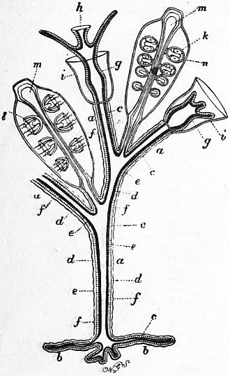
Fig. 54.—Diagram showing possible modifications of the persons of a Calyptoblastic Hydromedusa. Letters a to h same as in fig. 49. i, The horny cup or hydrotheca of the hydriform persons; l, medusiform person springing from m, a modified, hydriform person (blastostyle); n, the horny case or gonangium enclosing the blastostyle and its buds. This and the hydrotheca i give origin to the name Calyptoblastea. (After Allman.)
Perigonimus.—This common British hydroid belongs by its characters to the family Bougainvillidae; it produces, however, a medusa of the genus Tiara (fig. 52), referable to the family Clavidae; a fact sufficient to indicate the tentative character of even the most modern classifications of this order.
Sub-order II. Hydroidea Calyptoblastea (Leptomedusae).—Trophosome with polyps always differentiated into nutritive and reproductive individuals (blastostyles) enclosed in hydrothecae and gonothecae respectively; with sympodial type of budding. Gonosome with free medusae or gonophores; the medusae typically with otocysts, sometimes with cordyli or ocelli (figs. 54, 55).
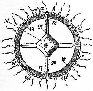
Fig. 55.—View of the Oral Surface of one of the Leptomedusae (Irene pellucida, Haeckel), to show the numerous tentacles and the otocysts.
ge, Genital glands. M, Manubrium. ot, Otocysts. rc, The four radiating canals. Ve, The velum.
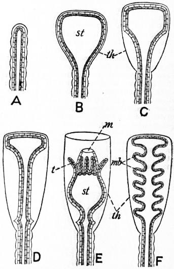
The calyptoblastic polyp of the nutritive type is very uniform in character, its tendency to variation being limited, as it were, by the enclosing hydrotheca. The hydranth almost always has a single circlet of tentacles, like the Bougainvillea-type, in the preceding sub-order; an exception is the curious genus Clathrozoon, in which the hydranth has a single tentacle. The characteristic hydrotheca is formed by the bud at an early stage (fig. 56); when complete it is an open cup, in which the hydranth develops and can be protruded from the opening for the capture of food, or is withdrawn into it for protection. Solitary polyps are unknown in this sub-order; the colony may be creeping or arborescent in form; if the latter, the budding of the polyps, as already stated, is of the sympodial type, and either biserial, forming stems capable of further branching, or uniserial, forming pinnules not capable of further branching. In the biserial type the polyps on the two sides of the stem have primitively an alternating, zigzag arrangement; but, by a process of differential growth, quickened in the 1st, 3rd, 5th, &c., members of the stem, and retarded in the 2nd, 4th, 6th, &c., members, the polyps may assume secondarily positions opposite to one another on the two sides of the stem. Other variations in the mode of growth or budding bring about further differences in the building up of the colony, which are not in all cases properly understood and cannot be described in detail here. The stem may contain a single coenosarcal tube (“monosiphonic”) or several united in a common perisarc (“polysiphonic”). An important variation is seen, in the form of the hydrotheca itself, which may come off from the main stem by a stalk, as in Obelia, or may be sessile, without a stalk, as in Sertularia.
After Allman, Gymnoblastic Hydroids, by permission of the council of the Ray Society.
Fig. 56.—Diagrams to show the mode of formation of the Hydrotheca and Gonotheca in Calyptoblastic Hydroids. A-D are stages common to both; from D arises the hydrotheca (E) or the gonotheca (F); th, theca; st, stomach; t, tentacles; m, mouth; mb, medusa-buds.
In many Calyptoblastea there occur also reduced defensive polyps or dactylozoids, which in this sub-order have received the special name of sarcostyles. Such are the “snake-like zoids” of Ophiodes and other genera, and as such are generally interpreted the “machopolyps” of the Plumularidea. These organs are supported by cuplike structures of the perisarc, termed nematophores, regarded as modified hydrothecae supporting the specialized polyp-individuals. They are specially characteristic of the family Plumularidae.
The medusa-buds, as already stated, are always produced from blastostyles, reduced non-nutritive polyps without mouth or tentacles. An apparent, but not real, exception is Halecium halecinum, in which the blastostyle is produced from the side of a nutritive polyp, and both are enclosed in a common theca without a partition between them (Allman [1] p. 50, fig. 24). The gonotheca is formed in its early stage in the same way as the hydrotheca, but the remains of the hydranth persists as an operculum closing the capsule, to be withdrawn when the medusae or genital products are set free (fig. 56).
The blastostyles, gonophores and gonothecae furnish a series of variations which can best be considered as so many stages of evolution.
Stage 1, seen in Obelia. Numerous medusae are budded successively within the gonotheca and set free; they swim off and mature in the open sea (Allman [1], p. 48, figs. 18, 19).
Stage 2, seen in Gonothyraea. Medusae, so-called “meconidia,” are budded but not liberated; each in turn, when it reaches sexual maturity, is protruded from the gonotheca by elongation of the stalk, and sets free the embryos, after which it withers and is replaced by another (Allman [1], p. 57, fig. 28).
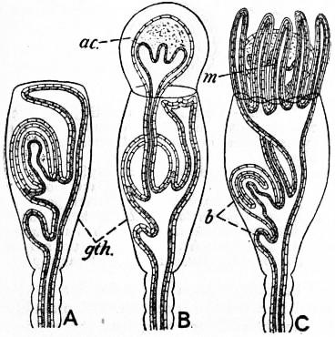
Stage 3, seen in Sertularia.—The gonophores are reduced in varying degree, it may be to sporosacs; they are budded successively from the blastostyle, and each in turn, when ripe, protrudes the spadix through the gonotheca (fig. 57, A, B). The spadix forms a gelatinous cyst, the so-called acrocyst (ac), external to the gonotheca (gth), enclosing and protecting the embryos. Then the spadix withers, leaving the embryos in the acrocyst, which may be further protected by a so-called marsupium, a structure formed by tentacle-like processes growing out from the blastostyle to enclose the acrocyst, each such process being covered by perisarc like a glove-finger secreted by it (fig. 57, C). (Allman [1], pp. 50, 51, figs. 21-24; Weismann [58], p. 170, pl. ix., figs. 7, 8.)
Stage 4, seen in Plumularidae.—The generative elements are produced in structures termed corbulae, formed by reduction and modification of branches of the colony. Each corbula contains a central row of blastostyles enclosed and protected by lateral rows of branches representing stunted buds (Allman [1], p. 66, fig. 30).
After Allman, Gymnoblastic Hydroids, by permission of the council of the Ray Society.
Fig. 57.—Diagrams to show the mode of formation of an Acrocyst and a Marsupium. In A two medusa-buds are seen within the gonotheca (gth), the upper more advanced than the lower one. In B the spadix of the upper bud has protruded itself through the top of the gonotheca and the acrocyst (ac) is secreted round it. In C the marsupium (m) is formed as finger-like process from the summit of the blastostyle, enclosing the acrocyst; b, medusa-buds on the blastostyle.
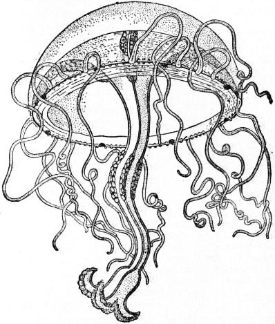
The Leptomedusa in form is generally shallow, more or less saucer-like, with velum less developed than in Anthomedusae (fig. 55). The characteristic sense-organs are ectodermal otocysts, absent, however, in some genera, in which case cordyli may replace them. When otocysts are present, they are at least eight in number, situated adradially, but are often very numerous. The cordyli are scattered on the ring-canal. Ocelli, if present, are borne on the tentacle-bulbs. The tentacles are usually hollow, rarely solid (Obelia). In number they are rarely less than four, but in Dissonema there are only two. Primitively there are four perradial tentacles, to which may be added four interradial, or they may become very numerous and are then scattered evenly round the margin, never arranged in tufts or clusters. In addition to tentacles, there may be marginal cirri (Laodice) with a solid endodermal axis, spirally coiled, very contractile, and bearing a terminal battery of nematocysts. The gonads are developed typically beneath the radial canals or below the stomach or its pouches, often stretching as long bands on to the base of the manubrium. In Octorchidae (fig. 58) each such band is interrupted, forming one mass at the base of the manubrium and another below the radial canal in each radius, in all eight separate gonad-masses, as the name implies. In some Leptomedusae excretory “marginal tubercles” are developed on the ring-canal.
Classification.—As in the Gymnoblastea, the difficulty of uniting the hydroid and medusan systems into one scheme of classification is very great in the present state of our knowledge. In a great many Leptomedusae the hydroid stage is as yet unknown, and it is by no means certain even that they possess one. It is quite possible that some of these medusae will be found to be truly hypogenetic, that is to say, with a life-cycle secondarily simplified by suppression of metagenesis. At present, ten recent and one extinct family of Calyptoblastea (Leptomedusae) may be recognized provisionally:
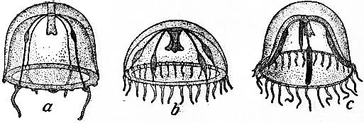
1. Eucopidae (figs. 55, 59).—Trophosome with stalked hydrothecae; gonosome, free medusae with otocysts and four, rarely six or eight, unbranched radial canals. Two of the commonest British hydroids belong to this family, Obelia and Clytia. Obelia forms numerous polyserial stems of the characteristic zigzag pattern growing up from a creeping basal stolon, and buds the medusa of the same name. In Clytia the polyps arise singly from the stolon, and the medusa is known as Phialidium (fig. 59).
2. Aequoridae.—Trophosome only known in one genus (Polycanna), and similar to the preceding; gonosome, free medusae with otocysts and with at least eight radial canals, often a hundred or more, simple or branched. Aequorea is a common medusa.
3. Thaumantidae.—Trophosome only known in one genus (Thaumantias), similar to that of the Eucopidae; gonosome, free medusae with otocysts inconspicuous or absent, with usually four, sometimes eight, rarely more than eight, radial canals, simple and unbranched, along which the gonads are developed, with numerous tentacles bearing ocelli and with marginal sense-clubs. Laodice and Thaumantias are representative genera.
4. Berenicidae.—Trophosome unknown; gonosome, free medusae, with four or six radial canals, bearing the gonads, with numerous tentacles, between which occur sense-clubs, without otocysts. Berenice, Staurodiscus, &c.
After Haeckel, System der Medusen, by permission of Gustav Fischer.
Fig. 58.—Octorchandra canariensis, from life.
5. Polyorchidae.—Trophosome unknown; gonosome, free medusae of deep form, with radial canals branched in a feathery manner, and bearing gonads on the main canal, but not on the branches, with numerous hollow tentacles bearing ocelli, and without otocysts. Polyorchis, Spirocodon.
6. Campanularidae.--Trophosome as in Eucopidae; gonosome, sessile gonophores. Many common or well-known genera belong here, such as Halecium, Campanularia, Gonothyraea, &c.
7. Lafoëidae.—Trophosome as in the preceding; gonosome, free medusae or gonophores, the medusae with large open otocysts. The hydroid genus Lafoëa is remarkable for producing gonothecae on the hydrorhiza, each containing a blastostyle which bears a single gonophore; this portion of the colony was formerly regarded as an independent parasitic hydroid, and was named Coppinia. Medusan genera are Mitrocoma, Halopsis, Tiaropsis (fig. 29, &c.).
(So far as the characters of the trophosome are concerned, the seven preceding families are scarcely distinguishable, and they form a section apart, contrasting sharply with the families next to be mentioned, in none of which are free medusae liberated from the colony, so that only the characters of the trophosome need be considered.)
After E. T. Browne, Proc. Zool. Soc. of London, 1896.
Fig. 59.—Three stages in the development of Phialidium temporarium. a, The youngest stage, is magnified about 22 diam.; b, older, is magnified about 8 diam.; c, the adult medusa, is magnified.
8. Sertularidae.—Hydrothecae sessile, biserial, alternating or opposite on the stem. Sertularia and Sertularella are two very common genera of this family.
9. Plumularidae.—Hydrothecae sessile, biserial on the main stem, uniserial on the lateral branches or pinnules, which give the colony its characteristic feathery form; with nematophores. A very abundant and prolific family; well-known British genera are Plumularia, Antennularia and Aglaophenia.
10. Hydroceratinidae.—This family contains the single Australian species Clathrozoon wilsoni Spencer, in which a massive hydrorhiza bears sessile hydrothecae, containing hydranths each with a single tentacle, and numerous nematophores. See W. B. Spencer [53].
11. Dendrograptidae, containing fossil (Silurian) genera, such as Dendrograptus and Thamnograptus, of doubtful affinities.
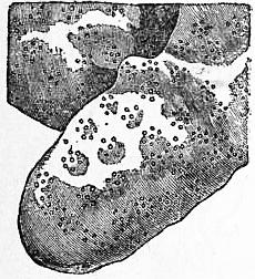
Fig. 60.—Portion of the calcareous corallum of Millepora nodosa, showing the cyclical arrangement of the pores occupied by the “persons” or hydranths. About twice the natural size. (From Moseley.)
Order III. Hydrocorallinae.—Metagenetic colony-forming Hydromedusae, in which the polyp-colony forms a massive, calcareous corallum into which the polyps can be retracted; polyp-individuals always of two kinds, gastrozoids and dactylozoids; gonosome either free medusae or sessile gonophores. The trophosome consists of a mass of coenosarcal tubes anastomosing in all planes. The interspaces between the tubes are filled up by a solid mass of lime, consisting chiefly of calcium carbonate, which replaces the chitinous perisarc of ordinary hydroids and forms a stony corallum or coenosteum (fig. 60). The surface of the coenosteum is covered by a layer of common ectoderm, containing large nematocysts, and is perforated by pores of two kinds, gastropores and dactylopores, giving exit to gastrozoids and dactylozoids respectively, which are lodged in vertical pore-canals of wider calibre than the coenosarcal canals of the general network. The coenosteum increases in size by new growth at the surface; and in the deeper, older portions of massive forms the tissues die off after a certain time, only the superficial region retaining its vitality down to a certain depth. The living tissues at the surface are cut off from the underlying dead portions by horizontal partitions termed tabulae, which are formed successively as the coenosteum increases in age and size. If the coenosteum of Millepora be broken across, each pore-canal (perhaps better termed a polyp-canal) is seen to be interrupted by a series of transverse partitions, representing successive periods of growth with separation from the underlying dead portions.
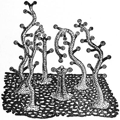
Fig. 61.—Enlarged view of the surface of a living Millepora, showing five dactylozooids surrounding a central gastrozooid. (From Moseley.)
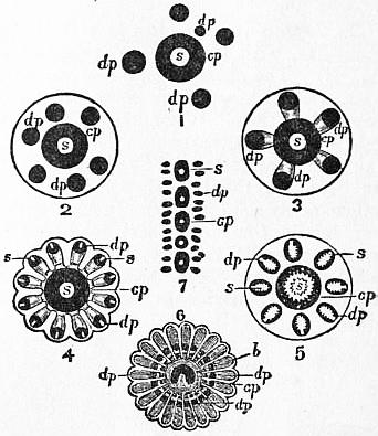
Fig. 62.—Diagrams illustrating the successive stages in the development of the cyclosystems of the Stylasteridae. (After Moseley.)
1, Sporadopora dichotoma. 2, 3, Allopora nobilis. 4, Allopora profunda. 5, Allopora miniacea. 6, Astylus subviridis. 7, Distichopora coccinea. s, Style. dp, Dactylopore. gp, Gastropore. b, In fig. 6, inner horseshoe-shaped mouth of gastropore.
Besides the wider vertical pore-canals and the narrower, irregular coenosarcal canals, the coenosteum may contain, in its superficial portion, chambers or ampullae, in which the reproductive zoids (medusae or gonophores) are budded from the coenosarc.
The gastropores and dactylopores are arranged in various ways at the surface, a common pattern being the formation of a cyclosystem (fig. 60), in which a central gastrozoid is surrounded by a ring of dactylozoids (fig. 61). In such a system the dactylopores may be confluent with the gastropore, so that the entire cyclosystem presents itself as a single aperture subdivided by radiating partitions, thus having a superficial resemblance to a madreporarian coral with its radiating septa (figs. 62 and 63).
The gastrozoids usually bear short capitate tentacles, four, six or twelve in number; but in Astylus (fig. 63) they have no tentacles. The dactylozoids have no mouth; in Milleporidae they have short capitate tentacles, but lack tentacles in Stylasteridae.
The gonosome consists of free medusae in Milleporidae, which are budded from the apex of a dactylozoid in Millepora murrayi, but in other species from the coenosarcal canals. The medusae are produced by direct budding, without an entocodon in the bud. They are liberated in a mature condition, and probably live but a short time, merely sufficient to spread the species. The manubrium bearing the gonads is mouthless, and the umbrella is without tentacles, sense-organs, velum or radial canals. In the Stylasteridae sessile gonophores are formed, always by budding from the coenosarc. In Distichopora the gonophores have radial canals, but in other genera they are sporosacs with no trace of medusoid structure.
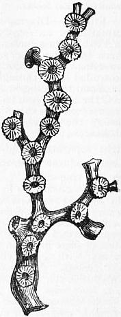
Fig. 63.—Portion of the corallum of Astylus subviridis (one of the Stylasteridae), showing cyclosystems placed at intervals on the branches, each with a central gastropore and zone of slit-like dactylopores. (After Moseley.)
Classification.—Two families are known:—
1. Milleporidae.—Coenosteum massive, irregular in form; pores scattered irregularly or in cyclosystems, without styles, with transverse tabulae; free medusae. A single genus, Millepora (figs. 60, 61).
2. Stylasteridae.—Coenosteum arborescent, sometimes fanlike, with pores only on one face, or on the lateral margins of the branches; gastropores with tabulae only in two genera, but with (except in Astylus) a style, i.e. a conical, thorn-like projection from the base of the pore, sometimes found also in dactylopores; sessile gonophores. Sporadopora has the pores scattered irregularly. Distichopora has the pores arranged in rows. Stylaster has cyclosystems. In Allopora the cyclostems resemble the calyces of Anthozoan corals. In Cryptohelia the cyclosystem is covered by a cap or operculum. In Astylus (fig. 63) styles are absent.
Affinities of the Hydrocorallinae.—There can be no doubt that the forms comprised in this order bear a close relationship to the Hydroidea, especially the sub-order Gymnoblastea, with which they should perhaps be classed in a natural classification. A hydrocoralline may be regarded as a form of hydroid colony in which the coenosarc forms a felt-work ramifying in all planes, and in which the chitinous perisarc is replaced by a massive calcareous skeleton. So far as the trophosome is concerned, the step from an encrusting hydroid such as Hydractinia to the hydrocoralline Millepora is not great.
Hickson considers that the families Milleporidae and Stylasteridae should stand quite apart from one another and should not be united in one order. The nearest approach to the Stylasteridae is perhaps to be found in Ceratella, with its arborescent trophosome formed of anastomosing coenosarcal tubes supported by a thick perisarc and covered by a common ectoderm. Ceratella stands in much the same relation to the Stylasteridae that Hydractinia does to the Milleporidae, in both cases the chitinous perisarc being replaced by the solid coenosteum to which the hydrocorallines owe the second half of their name.
Order IV. Graptolitoidea (Rhabdophora, Allman).—This order has been constituted for a peculiar group of palaeozoic fossils, which have been interpreted as the remains of the skeletons of Hydrozoa of an extinct type.
A typical graptolite consists of an axis bearing a series of tooth-like projections, like a saw. Each such projection is regarded as representing a cup or hydrotheca, similar to those borne by a calyptoblastic hydroid, such as Sertularia. The supposed hydrothecae may be present on one side of the axis only (monoprionid) or on both sides (diprionid); the first case may be conjectured to be the result of uniserial (helicoid) budding, the second to be produced by biserial (scorpioid) budding. In one division (Retiolitidae) the axis is reticulate. In addition to the stems bearing cups, there are found vesicles associated with them, which have been interpreted as gonothecae or as floats, that is to say, air-bladders, acting as hydrostatic organs for a floating polyp-colony.
Since no graptolites are known living, or, indeed, since palaeozoic times, the interpretation of their structure and affinities must of necessity be extremely conjectural, and it is by no means certain that they are Hydrozoa at all. It can only be said that their organization, so far as the state of their preservation permits it to be ascertained, offers closer analogies with the Hydrozoa, especially the Calyptoblastea, than with any other existing group of the animal kingdom.
See the treatise of Delage and Hérouard (Hydrozoa, [4]), and the article Graptolites.
Order V. Trachylinea.—Hydromedusae without alternation of generations, i.e. without a hydroid phase; the medusa develops directly from the actinula larva, which may, however, multiply by budding. Medusae with sense-organs represented by otocysts derived from modified tentacles (tentaculocysts), containing otoliths of endodermal origin, and innervated from the ex-umbral nerve-ring.
This order, containing the typical oceanic medusae, is divided into two sub-orders.
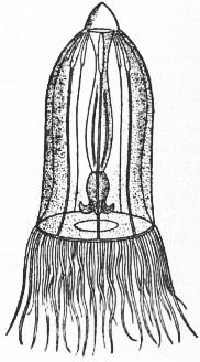
Sub-order 1. Trachomedusae.—Tentacles given off from the margin of the umbrella, which is entire, i.e. not lobed or indented; tentaculocysts usually enclosed in vesicles; gonads on the radial canals. The medusae of this order are characterized by the tough, rigid consistence of the umbrella, due partly to the dense nature of the mesogloea, partly to the presence of a marginal rim of chondral tissue, consisting of thickened ectoderm containing great numbers of nematocysts, and forming, as it were, a cushion-tyre supporting the edge of the umbrella. Prolongations from the rim of chondral tissue may form clasps or peronia supporting the tentacles. The tentacles are primarily four in number, perradial, alternating with four interradial tentaculocysts, but both tentacles and sense-organs may be multiplied and the primary perradii may be six instead of four (fig. 26). The tentacles are always solid, containing an axis of endoderm-cells resembling notochordal tissue or plant-parenchyma, and are but moderately flexible. The sense-organs are tentaculocysts which are usually enclosed in vesicles and may be sunk far below the surface. The gonads are on the radial canals or on the stomach (Ptychogastridae), and each gonad may be divided into two by a longitudinal sub-umbral muscle-tract. The radial canals are four, six, eight or more, and in some genera blindly-ending centripetal canals are present (fig. 26). The stomach may be drawn out into the manubrium, forming a proboscis (“Magenstiel”) of considerable length.
The development of the Trachomedusae, so far as it is known, shows an actinula-stage which is either free (larval) or passed over in the egg (foetal) as in Geryonia; in no case does there appear to be a free planula-stage. The actinula, when free, may multiply by larval budding, but in all cases both the original actinula and all its descendants become converted into medusae, so that there is no alternation of generations. In Gonionemus the actinula becomes attached and polyp-like and reproduces by budding.
After Haeckel, System der Medusen, by permission of Gustav Fischer.
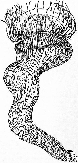
Fig. 64. Olindias mülleri.
The Trachomedusae are divided into the following families:
1. Petasidae (Petachnidae).—Four radial canals, four gonads; stomach not prolonged into the manubrium, which is relatively short; tentaculocysts free. Petasus and other genera make up this family, founded by Haeckel, but no other naturalist has ever seen them, and it is probable that they are simply immature forms of other genera.
2. Olindiadae, with four radial canals and four gonads; manubrium short; ring-canals giving off blind centripetal canals; tentaculocysts enclosed. Olindias mülleri (fig. 64) is a common Mediterranean species. Other genera are Aglauropsis, Gossea and Gonionemus; the last named bears adhesive suckers on the tentacles. Some doubt attaches to the position of this family. It has been asserted that the tentaculocysts are entirely ectodermal and that either the family should be placed amongst the Leptomedusae, or should form, together with certain Leptomedusae, an entirely distinct order. In Gonionemus, however, the concrement-cells are endodermal.
3. Trachynemidae.—Eight radial canals, eight gonads, stomach not prolonged into manubrium; tentaculocysts enclosed. Rhopalonema, Trachynema, &c.
After E. T. Browne, Proc. Zool. Soc. of London.
Fig. 65.—Aglantha rosea (Forbes), a British medusa.
4. Ptychogastridae (Pectyllidae).—As in the preceding, but with suckers on the tentacles. Ptychogastria Allman (= Pectyllis), a deep-sea form.
5. Aglauridae.—Eight radial canals, two, four or eight gonads; tentacles numerous; tentaculocysts free; stomach prolonged into manubrium. Aglaura, Aglantha (fig. 65), &c., with eight gonads; Stauraglaura with four; Persa with two. Amphogona, hermaphrodite, with male and female gonads on alternating radial canals.
6. Geryonidae.—Four or six radial canals; gonads band-like; stomach prolonged into a manubrium of great length; tentaculocysts enclosed. Liriope, &c., with four radial canals; Geryonia, Carmarina (fig. 26), &c., with six.
7. Halicreidae.—Eight very broad radial canals; ex-umbrella often provided with lateral outgrowths; tentacles differing in size, but in a single row. Halicreas.
Sub-order 2. Narcomedusae.—Margin of the umbrella-lobed, tentacles arising from the ex-umbrella at some distance from the margin; tentaculocysts exposed, not enclosed in vesicles; gonads on the sub-umbral floor of the stomach or of the gastric pouches.
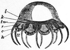
Fig. 66.—Cunina rhododactyla, one of the Narcomedusae. (After Haeckel.)
c, Circular canal. h, “Otoporpae” or centripetal process of the marginal cartilaginous ring connected with tentaculocyst. k, Stomach. l, Jelly of the disk. r, Radiating canal (pouch of stomach). tt, Tentacles. tw, Tentacle root.
The Narcomedusae exhibit peculiarities of form and structure which distinguish them at once from all other Hydromedusae. The umbrella is shallow and has the margin supported by a rim of thickened ectoderm, as in the Trachomedusae, but not so strongly developed. The tentacles are not inserted on the margin of the umbrella, but arise high up on the ex-umbral surface, and the umbrella is prolonged into lobes corresponding to the interspaces between the tentacles. The condition of things can be imagined by supposing that in a medusa primitively of normal build, with tentacles at the margin, the umbrella has grown down past the insertion of the tentacles. As a result of this extension of the umbrellar margin, all structures belonging to this region, namely, the ring-canal, the nerve-rings, and the rim of thickened ectoderm, do not run an even course, but are thrown into festoons, caught up under the insertion of each tentacle in such a way that the ring-canal and its accompaniments form in each notch of the umbrellar margin an inverted V, the apex of which corresponds to the insertion of the tentacle; in some cases the limbs of the V may run for some distance parallel to one another, and may be fused into one, giving a figure better compared to an inverted Y. Thus the ectodermal rim runs round the edge of each lobe of the umbrella and then passes upwards towards the base of the tentacle from the re-entering angle between two adjacent lobes, to form with its fellow of the next lobe a tentacle-clasp or peronium, i.e. a streak of thickened ectoderm supporting the tentacle. Similarly the ring-canal runs round the edge of the lobe as the so-called festoon-canal, and then runs upwards under the peronium to the base of the tentacle as one of a pair of peronial canals, the limbs of the V-like figure already mentioned. The nerve-rings have a similar course. The tentaculocysts are implanted round the margins of the lobes of the umbrella and may be supported by prolongations of the ectodermal rim termed otoporpae (Gehörspangen). The radial canals are represented by wide gastric pouches, and may be absent, so that the tentacles arise directly from the stomach (Solmaridae). The tentacles are always solid, as in Trachomedusae.
The development of the Narcomedusae is in the main similar to that of the Trachomedusae, but shows some remarkable features. In Aeginopsis a planula is formed by multipolar immigration. The two ends of the planula become greatly lengthened and give rise to the two primary tentacles of the actinula, of which the mouth arises from one side of the planula. Hence the principal axis of the future medusa corresponds, not to the longitudinal axis of the planula, but to a transverse axis. This is in some degree parallel to the cases described above, in which a planula gives rise to the hydrorhiza, and buds a polyp laterally.
In Cunina and allied genera the actinula, formed in the manner described, has a hypostome of great length, quite disproportionate to the size of the body, and is further endowed with the power of producing buds from a stolon arising from the aboral side of the body. In these species the actinula is parasitic upon another medusa; for instance, Cunoctantha octonaria upon Turritopsis, C. proboscidea upon Liriope or Geryonia. The parasite effects a lodgment in the host either by invading it as a free-swimming planula, or, apparently, in other cases, as a spore-embryo which is captured and swallowed as food by the host. The parasitic actinula is found attached to the proboscis of the medusa; it thrusts its greatly elongated hypostome into the mouth of the medusa and nourishes itself upon the food in the digestive cavity of its host. At the same time it produces buds from an aboral stolon. The buds become medusae by the direct method of budding described above. In some cases the buds do not become detached at once, but the stolon continues to grow and to produce more buds, forming a “bud-spike” (Knospenähre), which consists of the axial stolon bearing medusa-buds in all stages of development. In such cases the original parent-actinula does not itself become a medusa, but remains arrested in development and ultimately dies off, so that a true alternation of generations is brought about. It is in these parasitic forms that we meet with the method of reproduction by sporogony described above.
In other Narcomedusae, e.g. Cunoctantha fowleri Browne, buds are formed from the sub-umbrella on the under side of the stomach pouches, where later the gonads are developed.
Classification.—Three families of Narcomedusae are recognized (see O. Maas [40]):
After O. Maas, Craspedoten Medusen der Siboga Expedition, by permission of E. S. Brill & Co.
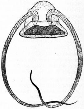
Fig. 67.—Solmundella bitentaculata (Quoy and Gaimard).
1. Cunanthidae.—With broad gastric pouches which are simple, i.e. undivided, and “pernemal,” i.e. correspond in position with the tentacles. Cunina (fig. 66) with more than eight tentacles; Cunoctantha with eight tentacles, four perradial, four interradial.
2. Aeginidae.—Radii a multiple of four, with radial gastric pouches bifurcated or subdivided; the tentacles are implanted in the notch between the two subdivisions of each (primary) gastric pouch, hence the (secondary) gastric pouches appear to be “internemal” in position, i.e. to alternate in position with the tentacles. Aegina, with four tentacles and eight pouches; Aeginura (fig. 25), with eight tentacles and sixteen pouches; Solmundella (fig. 67), with two tentacles and eight pouches; Aeginopsis (fig. 23), with two or four tentacles and sixteen pouches.
3. Solmaridae.—No gastric pouches; the numerous tentacles arise direct from the stomach, into which also the peronial canals open, so that the ring-canal is cut up into separate festoons. Solmaris, Pegantha, Polyxenia, &c. To this family should be referred, probably, the genus Hydroctena, described by C. Dawydov [11a] and regarded by him as intermediate between Hydromedusae and Ctenophora. See O. Maas [35].
Appendix to the Trachylinae.
Of doubtful position, but commonly referred to the Trachylinae, are the two genera of fresh-water medusae, Limnocodium and Limnocnida.
Limnocodium sowerbyi was first discovered in the Victoria regia tank in the Botanic Gardens, Regent’s Park, London. Since then it has been discovered in other botanic gardens in various parts of Europe, its two most recent appearances being at Lyons (1901) and Munich (1905), occurring always in tanks in which the Victoria regia is cultivated, a fact which indicates that tropical South America is its original habitat. In the same tanks a small hydroid, very similar to Microhydra, has been found, which bears medusa-buds and is probably the stock from which the medusa is budded. It is a remarkable fact that all specimens of Limnocodium hitherto seen have been males; it may be inferred from this either that only one polyp-stock has been introduced into Europe, from which all the medusae seen hitherto have been budded, or perhaps that the female medusa is a sessile gonophore, as in Pennaria. The male gonads are carried on the radial canals.
Limnocnida tanganyicae was discovered first in Lake Tanganyika, but has since been discovered also in Lake Victoria and in the river Niger. It differs from Limnocodium in having practically no manubrium but a wide mouth two-thirds the diameter of the umbrella across. It buds medusae from the margin of the mouth in May and June, and in August and September the gonads are formed in the place where the buds arose. The hydroid phase, if any, is not known.
Both these medusae have sense-organs of a peculiar type, which are said to contain an endodermal axis like the sense-organs of Trachylinae, but the fact has recently been called in question for Limnocodium by S. Goto, who considers the genus to be allied to Olindias. Allman, on the other hand, referred Limnocodium to the Leptomedusae.
In this connexion must be mentioned, finally, the medusae budded from the fresh-water polyp Microhydra. The polyp-stages of Limnocodium and Microhydra are extremely similar in character. In both cases the hydranth is extremely reduced and has no tentacles, and the polyp forms a colony by budding from the base. In Limnocodium the body secretes a gelatinous mucus to which adhere particles of mud, &c., forming a protective covering. In Microhydra no such protecting case is formed. In view of the great resemblance between Microhydra and the polyp of Limnocodium, it might be expected that the medusae to which they give origin would also be similar. As yet, however, the medusa of Microhydra has only been seen in an immature condition, but it shows some well-marked differences from Limnocodium, especially in the structure of the tentacles, which furnish useful characters for distinguishing species amongst medusae. The possession of a polyp-stage by Limnocodium and Microhydra furnishes an argument against placing them in the Trachylinae. Their sense-organs require renewed investigations. (Browne [10] and [10a].)
Order VI. Siphonophora.—Pelagic floating Hydrozoa with great differentiation of parts, each performing a special function; generally regarded as colonies showing differentiation of individuals in correspondence with a physiological division of labour.
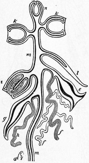
Fig. 68.—Diagram showing possible modifications of medusiform and hydriform persons of a colony of Siphonophora. The thick black line represents endoderm, the thinner line ectoderm. (After Allman.)
n, Pneumatocyst. k, Nectocalyces (swimming bells). l, Hydrophyllium (covering-piece). i, Generative medusiform person. g, Palpon with attached palpacle, h. e, Siphon with branched grappling tentacle, f. m, Stem.
A typical Siphonophore is a stock or cormus consisting of a number of appendages placed in organic connexion with one another by means of a coenosarc. The coenosarc does not differ in structure from that already described in colonial Hydrozoa. It consists of a hollow tube, or tubes, of which the wall is made up of the two body-layers, ectoderm and endoderm, and the cavity is a continuation of the digestive cavities of the nutritive and other appendages, i.e. of the coelenteron. The coenosarc may consist of a single elongated tube or stolon, forming the stem or axis of the cormus on which, usually, the appendages are arranged in groups termed cormidia; or it may take the form of a compact mass of ramifying, anastomosing tubes, in which case the cormus as a whole has a compact form and cormidia are not distinguishable. In the Disconectae the coenosarc forms a spongy mass, the “centradenia,” which is partly hepatic in function, forming the so-called liver, and partly excretory.
The appendages show various types of form and structure corresponding to different functions. The cormus is always differentiated into two parts; an upper portion termed the nectosome, in which the appendages are locomotor or hydrostatic in function, that is to say, serve for swimming or floating; and a lower portion termed the siphosome, bearing appendages which are nutritive, reproductive or simply protective in function.
Divergent views have been held by different authors both as regards the nature of the cormus as a whole, and as regards the homologies of the different types of appendages borne by it.
The general theories of Siphonophoran morphology are discussed below, but in enumerating the various types of appendages it is convenient to discuss their morphological interpretation at the same time.
After A. Agassiz, from Lankester’s Treatise on Zoology.
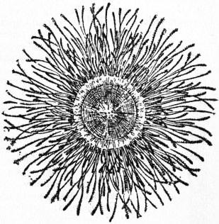
Fig. 69.—Porpita, seen from above, showing the pneumatophore and expanded palpons.
In the nectosome one or more of the following types of appendage occur:—
1. Swimming-bells, termed nectocalyces or nectophores (fig. 68, k), absent in Chondrophorida and Cystophorida; they are contractile and resemble, both in appearance, structure and function, the umbrella of a medusa, with radial canals, ring-canal and velum; but they are without manubrium, tentacles or sense-organs, and are always bilaterally symmetrical, a peculiarity of form related with the fact that they are attached on one side to the stem. A given cormus may bear one or several nectocalyces, and by their contractions they propel the colony slowly along, like so many medusae harnessed together. In cases where the cormus has no pneumatophore the topmost swimming bell may contain an oil-reservoir or oleocyst.
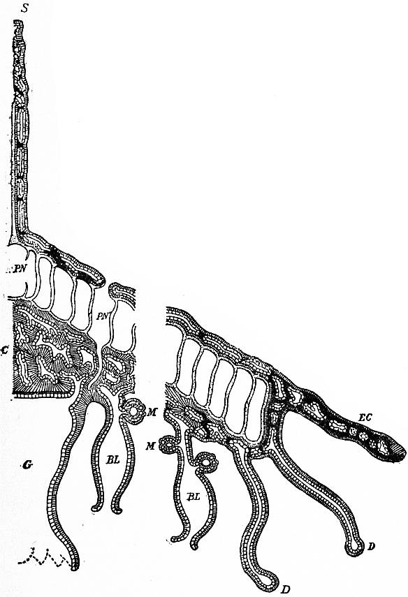
2. The pneumatophore or air-bladder (fig. 68, n), for passive locomotion, forming a float which keeps the cormus at or near the surface of the water. The pneumatophore arises from the ectoderm as a pit or invagination, part of which forms a gas-secreting gland, while the rest gives rise to an air-sack lined by a chitinous cuticle. The orifice of invagination forms a pore which may be closed up or may form a protruding duct or funnel. As in the analogous swim-bladder of fishes, the gas in the pneumatophore can be secreted or absorbed, whereby the specific gravity of the body can be diminished or increased, so as to cause it to float nearer the surface or at a deeper level. Never more than one pneumatophore is found in a cormus, and when present it is always situated at the highest point above the swimming bells, if these are present also. In Velella the pneumatophore becomes of complex structure and sends air-tubes, lined by a chitin and resembling tracheae, down into the compact coenosarc, thus evidently serving a respiratory as well as a hydrostatic function.
Divergent views have been held as to the morphological significance of the pneumatophore. E. Haeckel regarded the whole structure as a glandular ectodermal pit formed on the ex-umbral surface of a medusa-person. C. Chun and, more recently, R. Woltereck [59], on the other hand, have shown that the ectodermal pit which gives rise to the pneumatophore represents an entocodon. Hence the cavity of the air-sack is equivalent to a sub-umbral cavity in which no manubrium is formed, and the pore or orifice of invagination would represent the margin of the umbrella. In the wall of the sack is a double layer of endoderm, the space between which is a continuation of the coelenteron. By coalescence of the endoderm-layers, the coelenteron may be reduced to vessels, usually eight in number, opening into a ring-sinus surrounding the pore. Thus the disposition of the endoderm-cavities is roughly comparable to the gastrovascular system of a medusa.
The difference between the theories of Haeckel and Chun is connected with a further divergence in the interpretation of the stem or axis of the cormus. Haeckel regards it as the equivalent of the manubrium, and as it is implanted on the blind end of the pneumatophore, such a view leads necessarily to the air-sack and gland being a development on the ex-umbral surface of the medusa-person. Chun and Woltereck, on the other hand, regard the stem as a stolo prolifer arising from the aboral pole, that is to say, from the ex-umbrella, similar to that which grows out from the ex-umbral surface of the embryo of the Narcomedusae and produces buds, a view which is certainly supported by the embryological evidence to be adduced shortly.
In the siphosome the following types of appendages occur:—
1. Siphons or nutritive appendages, from which the order takes its name; never absent and usually present in great numbers (fig. 68, e). Each is a tube dilated at or towards the base and containing a mouth at its extremity, leading into a stomach placed in the dilatation already mentioned. The siphons have been compared to the manubrium of a medusa-individual, or to polyps, and hence are sometimes termed gastrozoids.
2. Palpons (fig. 68, g), present in some genera, especially in Physonectae; similar to the siphons but without a mouth, and purely tactile in function, hence sometimes termed dactylozoids. If a distal pore or aperture is present, it is excretory in function; such varieties have been termed “cystons” by Haeckel.
3. Tentacles (“Fangfäden”), always present, and implanted one at the base of each siphon (fig. 68, f). The tentacles of siphonophores may reach a great length and have a complex structure. They may bear accessory filaments or tentilla (f′), covered thickly with batteries of nematocysts, to which these organisms owe their great powers of offence and defence.
4. Palpacles (“Tastfäden”), occurring together with palpons, one implanted at the base of each palpon (fig. 68, h). Each palpacle is a tactile filament, very extensile, without accessory filaments or nematocysts.
5. Bracts (“hydrophyllia”), occur in Calycophorida and some Physophorida as scale-like appendages protecting other parts (fig. 68, l). The mesogloea is greatly developed in them and they are often of very tough consistency. By Haeckel they are considered homologous with the umbrella of a medusa.
From G. H. Fowler, after A. Agassiz, Lankester’s Treatise on Zoology.
Fig. 70.—Diagram of the structure of Velella, showing the central and peripheral thirds of a half-section of the colony, the middle third being omitted. The ectoderm is indicated by close hatching, the endoderm by light hatching, the mesogloea by thick black lines, the horny skeleton of the pneumatophore and sail by dotting.
BL, Blastostyle. C, Centradenia. D, Palpon. EC, Edge of colony prolonged beyond the pneumatophore. G, Cavity of the large central siphon. M, Medusoid gonophores. PN, Primary central chamber, and PN′, concentric chamber of the pneumatophore, showing an opening to the exterior and a “trachea.” S, Sail.
6. Gonostyles, appendages which produce by budding medusae or gonophores, like the blastostyles of a hydroid colony. In their most primitive form they are seen in Velella as “gonosiphons,” which possess mouths like the ordinary sterile siphons and bud free medusae. In other forms they have no mouths. They may be branched, so-called “gonodendra,” and amongst them may occur special forms of palpons, “gonopalpons.” The gonostyles have been compared to the blastostyles of a hydroid colony, or to the manubrium of a medusa which produces free or sessile medusa-buds.
7. Gonophores, produced either on the gonostyles already mentioned or budded, as in hydrocorallines, from the coenosarc, i.e. the stem (fig. 68, i.). They show every transition between free medusae and sporosacs, as already described, for hydroid colonies. Thus in Velella free medusae are produced, which have been described as an independent genus of medusae, Chrysomitra. In other types the medusae may be set free in a mature condition as the so-called “genital swimming bells,” comparable to the Globiceps of Pennaria. The most usual condition, however, is that in which sessile medusoid gonophores or sporosacs are produced.
From G. H. Fowler, after G. Cuvier, Lankester’s Treatise on Zoology.
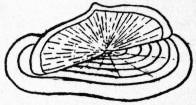
Fig. 71.—Upper surface of Velella, showing pneumatophore and sail.
The various types of appendages described in the foregoing may be arranged in groups termed cormidia. In forms with a compact coenosarc such as Velella, Physalia, &c., the separate cormidia cannot be sharply distinguished, and such a condition is described technically as one with “scattered” cormidia. In forms in which, on the other hand, the coenosarc forms an elongated, tubular axis or stem, the appendages are arranged as regularly recurrent cormidia along it, and the cormidia are then said to be “ordinate.” In such cases the oldest cormidia, that is to say, those furthest from the nectosome, may become detached (like the segments or proglottides of a tape-worm) and swim off, each such detached cormidium then becoming a small free cormus which, in many cases, has been given an independent generic name. A cormidium may contain a single nutritive siphon (“monogastric”) or several siphons (“polygastric”):
The following are some of the forms of cormidia that occur:—
1. The eudoxome (Calycophorida), consisting of a bract, siphon, tentacle and gonophore; when free it is known as Eudoxia.
2. The ersaeome (Calycophorida), made up of the same appendages as the preceding type but with the addition of a nectocalyx; when free termed Ersaea.
3. The rhodalome of some Rhodalidae, consisting of siphon, tentacle and one or more gonophores.
4. The athorome of Physophora, &c., consisting of siphon, tentacle, one or more palpons with palpacles, and one or more gonophores.
5. The crystallome of Anthemodes, &c., similar to the athorome but with the addition of a group of bracts.
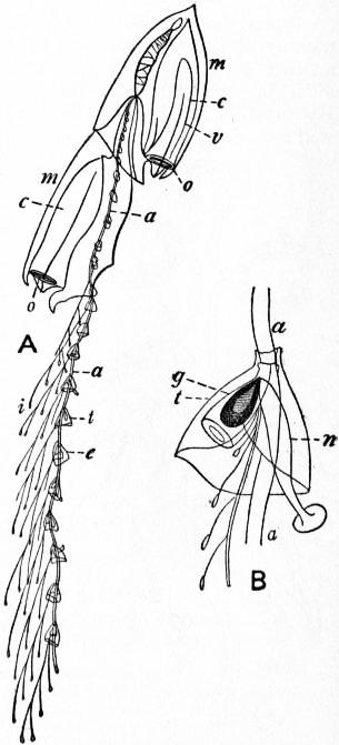
Fig. 72.—A, Diphyes campanulata; B, a group of appendages (cormidium) of the same Diphyes. (After C. Gegenbaur.)
a, Axis of the colony. m, Nectocalyx. c, Sub-umbral cavity of nectocalyx. v, Radial canals of nectocalyx. o, Orifice of nectocalyx. t, Bract. n, Siphon. g, Gonophore. i, Tentacle.
Embryology of the Siphonophora.—The fertilized ovum gives rise to a parenchymula, with solid endoderm, which is set free as a free-swimming planula larva, in the manner already described (see Hydrozoa). The planula has its two extremities dissimilar (Bipolaria-larva). The subsequent development is slightly different according as the future cormus is headed by a pneumatophore (Physophorida, Cystophorida) or by a nectocalyx (Calycophorida).
(i.) Physophorida, for example Halistemma (C. Chun, Hydrozoa [1]). The planula becomes elongated and broader towards one pole, at which a pit or invagination of the ectoderm arises. Next the pit closes up to form a vesicle with a pore, and so gives rise to the pneumatophore. From the broader portion of the planula an outgrowth arises which becomes the first tentacle of the cormus. The endoderm of the planula now acquires a cavity, and at the narrower pole a mouth is formed, giving rise to the primary siphon. Thus from the original planula three appendages are, as it were, budded off, while the planula itself mostly gives rise to coenosarc, just as in some hydroids the planula is converted chiefly into hydrorhiza.
(ii.) Calycophorida, for example, Muggiaea. The planula develops, on the whole, in a similar manner, but the ectodermal invagination arises, not at the pole of the planula, but on the side of its broader portion, and gives rise, not to a pneumatophore, but to a nectocalyx, the primary swimming bell or protocodon (“Fallschirm”) which is later thrown off and replaced by secondary swimming bells, metacodons, budded from the coenosarc.
From a comparison of the two embryological types there can be no doubt on two points; first, that the pneumatophore and the protocodon are strictly homologous, and, therefore if the nectocalyx is comparable to the umbrella of a medusa, as seems obvious, the pneumatophore must be so too; secondly, that the coenosarcal axis arises from the ex-umbrella of the medusa and cannot be compared to a manubrium, but is strictly comparable to the “bud-spike” of a Narcomedusan.
Theories of Siphonophore Morphology.—The many theories that have been put forward as to the interpretation of the cormus and the various parts are set forth and discussed in the treatise of Y. Delage and E. Hérouard (Hydrozoa [4]) and more recently by R. Woltereck [59], and only a brief analysis can be given here.
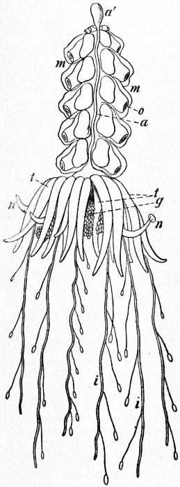
After C. Gegenbaur.
Fig. 73.—Physophora hydrostatica.
a′, Pneumatocyst. t, Palpons. a, Axis of the colony. m, Nectocalyx. o, Orifice of nectocalyx. n, Siphon. g, Gonophore. i, Tentacle.
In the first place the cormus has been regarded as a single individual and its appendages as organs. This is the so-called “polyorgan” theory, especially connected with the name of Huxley; but it must be borne in mind that Huxley regarded all the forms produced, in any animal, between one egg-generation and the next, as constituting in the lump one single individual. Huxley, therefore, considered a hydroid colony, for example, as a single individual, and each separate polyp or medusa budded from it as having the value of an organ and not of an individual. Hence Huxley’s view is not so different from those held by other authors as it seems to be at first sight.
In more recent years Woltereck [59] has supported Huxley’s view of individuality, at the same time drawing a fine distinction between “individual” and “person.” The individual is the product of sexual reproduction; a person is an individual of lower rank, which may be produced asexually. A Siphonophore is regarded as a single individual composed of numerous zoids, budded from the primary zoid (siphon) produced from the planula. Any given zoid is a person-zoid if equivalent to the primary zoid, an organ-zoid if equivalent only to a part of it. Woltereck considers the siphonophores most nearly allied to the Narcomedusae, producing like the buds from an aboral stolon, the first bud being represented by the pneumatophore or protocodon, in different cases.
Contrasting, in the second place, with the polyorgan theory are the various “polyperson” theories which interpret the Siphonophore cormus as a colony composed of more or fewer individuals in organic union with one another. On this interpretation there is still room for considerable divergence of opinion as regards detail. To begin with, it is not necessary on the polyperson theory to regard each appendage as a distinct individual; it is still possible to compare appendages with parts of an individual which have become separated from one another by a process of “dislocation of organs.” Thus a bract may be regarded, with Haeckel, as a modified umbrella of a medusa, a siphon as its manubrium, and a tentacle as representing a medusan tentacle shifted in attachment from the margin to the sub-umbrella; or a siphon may be compared with a polyp, of which the single tentacle has become shifted so as to be attached to the coenosarc and so on. Some authors prefer, on the other hand, to regard every appendage as a separate individual, or at least as a portion of an individual, of which other portions have been lost or obliterated.
A further divergence of opinion arises from differences in the interpretation of the persons composing the colony. It is possible to regard the cormus (1) as a colony of medusa-persons, (2) as a colony of polyp-persons, (3) as composed partly of one, partly of the other. It is sufficient here to mention briefly the views put forward on this point by C. Chun and R. Haeckel.
Chun (Hydrozoa [1]) maintains the older views of Leuckart and Claus, according to which the cormus is to be compared to a floating hydroid colony. It may be regarded as derived from floating polyps similar to Nemopsis or Pelagohydra, which by budding produce a colony of polyps and also form medusa-buds. The polyp-individuals form the nutritive siphosome or trophosome. The medusa-buds are either fertile or sterile. If fertile they become free medusae or sessile gonophores. If sterile they remain attached and locomotor in function, forming the nectosome, the pneumatophore and swimming-bells.
Haeckel, on the other hand, is in accordance with Balfour in regarding a Siphonophore as a medusome, that is to say, as a colony composed of medusoid persons or organs entirely. Haeckel considers that the Siphonophores have two distinct ancestral lines of evolution:
1. In the Disconanthae, i.e. in such forms as Velella, Porpita, &c., the ancestor was an eight-rayed medusa (Disconula) which acquired a pneumatophore as an ectodermal pit on the ex-umbrella, and in which the organs (manubrium, tentacles, &c.) became secondarily multiplied, just as they do in Gastroblasta as the result of incomplete fission. The nearest living allies of the ancestral Disconula are to be sought in the Pectyllidae.
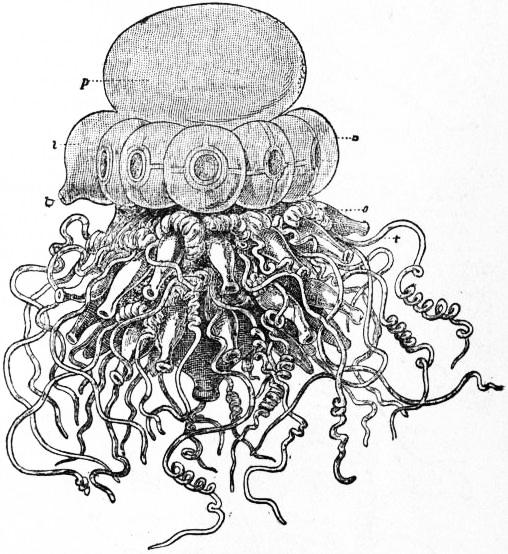
After Haeckel, from Lankester’s Treatise on Zoology.
Fig. 74.—Stephalia corona, a young colony.
p, Pneumatophore. n, Nectocalyx. l, Aurophore. lo, Orifice of the aurophore. s, Siphon. t, Tentacle.
2. In the Siphonanthae, i.e. in all other Siphonophores, the ancestral form was a Siphonula, a bilaterally symmetrical Anthomedusa with a single long tentacle (cf. Corymorpha), which became displaced from the margin to the sub-umbrella. The Siphonula produced buds on the manubrium, as many Anthomedusae are known to do, and these by reduction or dislocation of parts gave rise to the various appendages of the colony. Thus the umbrella of the Siphonula became the protocodon, and its manubrium, the axis or stolon, which, by a process of dislocation of organs, escaped, as it were, from the sub-umbrella through a cleft and became secondarily attached to the ex-umbrella. It must be pointed out that, however probable Haeckel’s theory may be in other respects, there is not the slightest evidence for any such cleft in the umbrella having been present at any time, and that the embryological evidence, as already pointed out, is all against any homology between the stem and a manubrium, since the primary siphon does not become the stem, which arises from the ex-umbral side of the protocodon and is strictly comparable to a stolon.
Classification.—The Siphonophora may be divided, following Delage and Hérouard, into four sub-orders:
I. Chondrophorida (Disconectae Haeckel, Tracheophysae Chun). With an apical chambered pneumatophore, from which tracheal tubes may take origin (fig. 70); no nectocalyces or bracts; appendages all on the lower side of the pneumatophore arising from a compact coenosarc, and consisting of a central principal siphon, surrounded by gonosiphons, and these again by tentacles.
Three families: (1) Discalidae, for Discalia and allied genera, deep-sea forms not well known; (2) Porpitidae for the familiar genus Porpita (fig. 69) and its allies; and (3) Velellidae, represented by the well-known genus Velella (figs. 70, 71), common in the Mediterranean and other seas.
II. Calycophorida (Calyconectae, Haeckel). Without pneumatophore, with one, two, rarely more nectocalyces.
Three families: (1) Monophyidae, with a single nectocalyx; examples Muggiaea, sometimes found in British seas, Sphaeronectes, &c.; (2) Diphyidae, with two nectocalyces; examples Diphyes (fig. 72), Praya, Abyla, &c.; and (3) Polyphyidae, with numerous nectocalyces; example Hippopodius, Stephanophyes and other genera.
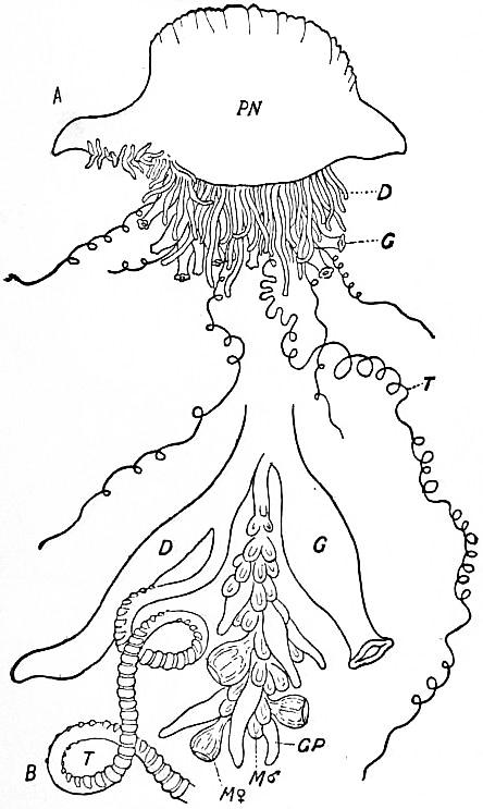
From G. H. Fowler, modified after G. Cuvier and E. Haeckel, Lankester’s Treatise on Zoology.
Fig. 75.—A. Physalia, general view, diagrammatic; B, cormidium of Physalia; D, palpon; T, palpacle; G, siphon; GP, gonopalpon; M ♂, male gonophore; M ♀, female gonophore, ultimately set free.
III. Physophorida (Physonectae + Auronectae, Haeckel). With an apical pneumatophore, not divided into chambers, followed by a series of nectocalyces or bracts.
A great number of families and genera are referred to this group, amongst which may be mentioned specially—(1) Agalmidae, containing the genera Stephanomia, Agalma, Anthemodes, Halistemma, &c.; (2) Apolemidae, with the genus Apolemia and its allies; (3) Forskaliidae, with Forskalia and allied forms; (4) Physophoridae, for Physophora (fig. 73) and other genera, (5) Anthophysidae, for Anthophysa, Athorybia, &c.; and lastly the two families (6) Rhodalidae and (7) Stephalidae (fig. 74), constituting the group Auronectae of Haeckel. The Auronectae are peculiar deep-sea forms, little known except from Haeckel’s descriptions, in which the large pneumatophore has a peculiar duct, termed the aurophore, placed on its lower side in the midst of a circle of swimming-bells.
IV. Cystophorida (Cystonectae, Haeckel). With a very large pneumatophore not divided into chambers, but without nectocalyces or bracts. Two sections can be distinguished, the Rhizophysina, with long tubular coenosarc-bearing ordinate cormidia, and Physalina, with compact coenosarc-bearing scattered cormidia.
A type of the Rhizophysina is the genus Rhizophysa. The Physalina comprise the families Physalidae and Epibulidae, of which the types are Physalia (figs. 74, 75) and Epibulia, respectively. Physalia, known commonly as the Portuguese man-of-war, is remarkable for its great size, its brilliant colours, and its terrible stinging powers.
Bibliography.—In addition to the works cited below, see the general works cited in the article Hydrozoa, in some of which very full bibliographies will be found.
1. G. J. Allman, “A Monograph of the Gymnoblastic or Tubularian Hydroids,” Ray Society (1871-1872); 2. A. Brauer, “Über die Entwickelung von Hydra,” Zeitschr. f. wiss. Zool. lii. (1891), pp. 169-216, pls. ix.-xii.; 3. “Über die Entstehung der Geschlechtsprodukte und die Entwickelung von Tubularia mesembryanthemum Allm.,” t.c. pp. 551-579, pls. xxxiii.-xxxv.; 4. W. K. Brooks, “The Life-History of the Hydromedusae: a discussion of the Origin of the Medusae, and of the significance of Metagenesis,” Mem. Boston Soc. Nat. Hist. iii. (1886), pp. 259-430, pis. xxxvii.-xliv.; 5. “The Sensory Clubs of Cordyli of Laodice,” Journ. Morphology, x. (1895), pp. 287-304, pl. xvii.; 6. E. T. Browne, “On British Hydroids and Medusae,” Proc. Zool. Soc. (1896), pp. 459-500, pls. xvi., xvii., (1897), pp. 816-835, pls. xlviii. xlix. 12 text-figs.; 7. “Biscayan Medusae,” Trans. Linn. Soc. x. (1906), pp. 163-187, pl. xiii.; 8. “Medusae” in Herdman, Rep. Pearl Oyster Fisheries, Gulf of Manaar, iv. (1905), pp. 131-166, 4 pls.; 9. “Hydromedusae with a Revision of the Williadae and Petasidae,” Fauna and Geogr. Maldive and Laccadive Archipelagos, ii. (1904), pp. 722-749, pls. liv.-lvii.; 10. “On the Freshwater Medusa liberated by Microhydra ryderi, Potts, and a Comparison with Limnocodium,” Quart. Journ. Micr. Sci. I (1906), pp. 635, 645, pl. xxxvii.; 10a. “On the Freshwater Medusa Limnocnida tanganicae” Budgett Memorial Volume (Cambridge, 1908, pp. 471-482, pl. xxviii.; 11. C. Claus, “Über die Struktur der Muskelzellen und über den Körperbau von Mnestra parasites Krohn,” Verhandl. zool. bot. Ges. Wien, xxv. (1876), pp. 9-12, pl. i.; 11a. C. Dawydov, “Hydroctena salenskii,” Mém. Acad. Imp. St. Pétersbourg (viii.) xiv. No. 9 (1903), 17 pp., 1 pl.; 12. A. Dendy, “On a Free-swimming Hydroid, Pelagohydra mirabilis,” n. gen. et sp., Quart. Journ. Micr. Sci. xlvi. (1903), pp. 1-24, pls. i. ii.; 13. H. Driesch, “Tektonische Studien an Hydroidpolypen,” (1) Jen. Zeitschr., xxiv. (1890), pp. 189-226, 12 figs.; (2) t.c. pp. 657-688, 6 figs.; (3) ibid. xxv. (1891), pp. 467-479, 3 figs.; 14. G. Duplessis, “On Campanularia volubilis,” Soc. Vaud. Bull. 13 (Lausanne, 1874-1875); 15. J. W. Fewkes, “On Mnestra,” Amer. Natural., xviii. (1884), pp. 197-198, 3 figs.; 16. S. Goto, “Dendrocoryne Inaba, Vertreterin einer neuen Familie der Hydromedusen,” Annot. Zool. Tokyo, i. (1897), pp. 93-104, pl. vi., figs. 106-113; 17. “The Craspe dote Medusa Olindias and some of its Natural Allies,” Mark Anniversary Volume (New York, 1903), pp. 1-22, 3 pls.; 18. H. Grenacher, “Über die Nesselkapseln von Hydra,” Zool. Anz. xviii. (1895), pp. 310-321, 7 figs.; 19. R. T. Günther, “On the Structure and Affinities of Mnestra parasites Krohn; with a revision of the Classification of the Cladonemidae,” Mitt. Stat. Neapel, xvi. (1903), pp. 35-62, pls. ii. iii.; 20. E. Haeckel, “Das System der Medusen,” Denkschr. med.-nat.-wiss. Ges. (Jena, 1879-1881); 21. “Deep Sea Medusae,” in Reports of the Challenger Expedition, Zool. iv. pt. 2 (London, 1882); 22. P. Hallez, “Bougainvillia fruticosa Allm. est le faciès d’eau agitée du Bougainvillia ramosa Van Ben.” C.-R. Acad. Sci. Paris, cxl. (1905), pp. 457-459; 23. O. & R. Hertwig, Der Organismus der Medusen (Jena, 1878), 70 pp., 3 pls.; 24. Das Nervensystem und die Sinnesorgane der Medusen (Leipzig, 1878), 186 pp., 10 pls.; 25. S. J. Hickson, “The Medusae of Millepora,” Proc. Roy. Soc. lxvi. (1899), pp. 6-10, 10 figs.; 26. T. Hincks, A History of British Hydroid Zoophytes (2 vols., London, 1868); 27. N. Iwanzov, “Über den Bau, die Wirkungsweise und die Entwickelung der Nesselkapseln von Coelenteraten,” Bull. Soc. Imp. Natural, Moscou (1896), pp. 323-355, 4 pls.; 28. C. F. Jickeli, “Der Bau der Hydroidpolypen,” (1) Morph. Jahrbuch, viii. (1883), pp. 373-416, pls. xvi.-xviii.; (2) t.c., pp. 580-680, pls. xxv.-xxviii.; 29. Albert Lang, “Über die Knospung bei Hydra und einigen Hydropolypen,” Zeitschr. f. wiss. Zool. liv. (1892), pp. 365-384, pl. xvii.; 30. Arnold Lang, “Gastroblasta Raffaelei. Eine durch eine Art unvollständiger Theilung entstehende Medusen-Kolonie,” Jena Zeitschr. xix. (1886), pp. 735-762, pls. xx., xxi.; 31. A. Linko, “Observations sur les méduses de la mer Blanche,” Trav. Soc. Imp. Nat. St Pétersbourg, xxix. (1899); 32. “Über den Bau der Augen bei den Hydromedusen,” Zapiski Imp. Akad. Nauk (Mém. Acad. Imp. Sci.) St Pétersbourg (8) x. 3 (1900), 23 pp., 2 pls.; 33. O. Maas, “Die craspedoten Medusen,” in Ergebn. Plankton Expedition, ii. (Kiel and Leipzig, 1893), 107 pp., 8 pls., 3 figs.; 34. “Die Medusen,” Mem. Mus. Comp. Zool. Harvard, xxiii. (1897), i.; 35. “On Hydroctena,” Zool. Centralbl. xi. (1904), pp. 240-243; 36. “Revision des méduses appartenant aux familles des Cunanthidae et des Aeginidae, et groupement nouveau des genres,” Bull. Mus. Monaco, v. (1904), 8 pp.; 37. “Revision der Cannotiden Haeckels,” SB. K. Bayer. Akad. xxxiv. (1904), pp. 421-445; 38. “Meduses,” Result. Camp. Sci. Monaco, xxviii. (1904), 71 pp., 6 pls.; 39. “Die craspedoten Medusen der Siboga-Expedition,” Uitkomst. Siboga-Exped. x. (1905), 84 pp., 14 pls.; 40. “Die arktischen Medusen (ausschliesslich der Polypomedusen),” Fauna arctica, iv. (1906), pp. 479-526; 41. C. Mereschkowsky, “On a new Genus of Hydroids (Monobrachium) from the White Sea, with a short description of other new Hydroids,” Ann. Mag. Nat. Hist. (4) xx. (1877), pp. 220-229, pls. v. vi.; 42. E. Metchinkoft, “Studien über die Entwickelung der Medusen und Siphonophoren,” Zeitschr. f. wiss. Zool. xxiv. (1874), pp. 15-83, pls. i.-xii.; 43. “Vergleichend-embryologische Studien” (Geryoniden, Cunina), ibid. xxxvi. (1882), pp. 433-458, pl. xxviii.; 44. Embryologische Studien an Medusen (Vienna, 1886), 150 pp., 12 pls., 10 figs.; 45. “Medusologische Mittheilungen,” Arb. zool. Inst. Wien, vi. (1886), pp. 237-266, pls. xxii. xxiii.; 46. L. Murbach, “Beiträge zur Kenntnis der Anatomie und Entwickelung der Nesselorgane der Hydroiden,” Arch. f. Naturgesch. lx. i. (1894), pp. 217-254, pl. xii.; 47. “Preliminary Note on the Life-History of Gonionemus,” Journ. Morph. xi. (1895), pp. 493-496; 48. L. Murbach and C. Shearer, “On Medusae from the Coast of British Columbia and Alaska,” Proc. Zool. Soc. (1903), ii. pp. 164-191, pls. xvii.-xxii.; 49. H. F. Perkins, “The Development of Gonionema murbachii,” Proc. Acad. Nat. Sci. Philadelphia (1902), pp. 750-790, pls. xxxi-xxxiv.; 50. F. Schaudinn, “Über Haleremita cumulans, n. g. n. sp., einen marinen Hydroidpolypen,” SB. Ges. natforsch. Freunde Berlin (1894), pp. 226-234, 8 figs.; 51. F. E. Schulze, “On the Structure and Arrangement of the Soft Parts in Euplectella aspergillum” (Amphibrachium), Tr. R. Soc. Edinburgh, xxix. (1880), pp. 661-673, pl. xvii.; 52. O. Seeliger, “Über das Verhalten der Keimblätter bei der Knospung der Cölenteraten,” Zeitschr. f. wiss. Zool. lviii. (1894), pp. 152-188, pls. vii.-ix.; 53. W. B. Spencer, “A new Family of Hydroidea (Clathrozoon), together with a description of the Structure of a new Species of Plumularia,” Trans. Roy. Soc. Victoria (1890), pp. 121-140, 7 pls.; 54. M. Ussow, “A new Form of Fresh-water Coelenterate” (Polypodium), Ann. Mag. Nat. Hist. (5) xviii. (1886), pp. 110-124, pl. iv.; 55. E. Vanhöffen, “Versuch einer natürlichen Gruppierung der Anthomedusen,” Zool. Anzeiger, xiv. (1891), pp. 439-446; 56. C. Viguier, “Études sur les animaux inférieurs de la baie d’Alger” (Tetraplatia), Arch. Zool. Exp. Gen. viii. (1890), pp. 101-142, pls. vii.-ix.; 57. J. Wagner, “Recherches sur l’organisation de Monobrachium parasiticum Méréjk,” Arch. biol. x. (1890), pp. 273-309, pls. viii. ix.; 58. A. Weismann, Die Entstehung der Sexualzellen bei den Hydromedusen (Jena, 1883); 59. R. Woltereck, “Beiträge zur Ontogenie und Ableitung des Siphonophorenstocks,” Zeitschr. f. wiss. Zool. lxxxii. (1905), pp. 611-637, 21 text-figs.; 60. J. Wulfert, “Die Embryonalentwickelung von Gonothyraea loveni Allm.,” Zeitschr. f. wiss. Zool. lxxi. (1902), pp. 296-326, pls. xvi.-xviii.
(E. A. M.)
1 In some cases hydroids have been reared in aquaria from ova of medusae, but these hydroids have not yet been found in the sea (Browne [10 a]).
2 The numbers in square brackets [] refer to the bibliography at the end of this article; but when the number is preceded by the word Hydrozoa, it refers to the bibliography at the end of the article Hydrozoa.
HYDROMETER (Gr. ὕδωρ, water, and μέτρον, a measure), an instrument for determining the density of bodies, generally of fluids, but in some cases of solids. When a body floats in a fluid under the action of gravity, the weight of the body is equal to that of the fluid which it displaces (see Hydromechanics). It is upon this principle that the hydrometer is constructed, and it obviously admits of two modes of application in the case of fluids: either we may compare the weights of floating bodies which are capable of displacing the same volume of different fluids, or we may compare the volumes of the different fluids which are displaced by the same weight. In the latter case, the densities of the fluids will be inversely proportional to the volumes thus displaced.
The hydrometer is said by Synesius Cyreneus in his fifth letter to have been invented by Hypatia at Alexandria,1 but appears to have been neglected until it was reinvented by Robert Boyle, whose “New Essay Instrument,” as described in the Phil. Trans. for June 1675, differs in no essential particular from Nicholson’s hydrometer. This instrument was devised for the purpose of detecting counterfeit coin, especially guineas and half-guineas. In the first section of the paper (Phil. Trans. No. 115, p. 329) the author refers to a glass instrument exhibited by himself many years before, and “consisting of a bubble furnished with a long and slender stem, which was to be put into several liquors, to compare and estimate their specific gravities.” This seems to be the first reference to the hydrometer in modern times.
In fig. 1 C represents the instrument used for guineas, the circular plates A representing plates of lead, which are used as ballast when lighter coins than guineas are examined. B represents “a small glass instrument for estimating the specific gravities of liquors,” an account of which was promised by Boyle in the following number of the Phil. Trans., but did not appear.
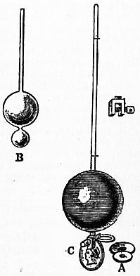
Fig. 1.—Boyle’s New Essay Instrument.
The instrument represented at B (fig. 1), which is copied from Robert Boyle’s sketch in the Phil. Trans. for 1675, is generally known as the common hydrometer. It is usually made of glass, the lower bulb being loaded with mercury or small shot which serves as ballast, causing the instrument to float with the stem vertical. The quantity of mercury or shot inserted depends upon the density of the liquids for which the hydrometer is to be employed, it being essential that the whole of the bulb should be immersed in the heaviest liquid for which the instrument is used, while the length and diameter of the stem must be such that the hydrometer will float in the lightest liquid for which it is required. The stem is usually divided into a number of equal parts, the divisions of the scale being varied in different instruments, according to the purposes for which they are employed.
Let V denote the volume of the instrument immersed (i.e. of liquid displaced) when the surface of the liquid in which the hydrometer floats coincides with the lowest division of the scale, A the area of the transverse section of the stem, l the length of a scale division, n the number of divisions on the stem, and W the weight of the instrument. Suppose the successive divisions of the scale to be numbered 0, 1, 2 ... n starting with the lowest, and let w0, W1, w2 ... wn be the weights of unit volume of the liquids in which the hydrometer sinks to the divisions 0, 1, 2 ... n respectively. Then, by the principle of Archimedes,
W = Vw0; or w0 = W / V. Also W = (V + lA) w1; or w1 = W/(V + lA), wp2 = W / (V + plA), and wn= W / (V + nlA),
or the densities of the several liquids vary inversely as the respective volumes of the instrument immersed in them; and, since the divisions of the scale correspond to equal increments of volume immersed, it follows that the densities of the several liquids in which the instrument sinks to the successive divisions form a harmonic series.
If V = NlA then N expresses the ratio of the volume of the instrument up to the zero of the scale to that of one of the scale-divisions. If we suppose the lower part of the instrument replaced by a uniform bar of the same sectional area as the stem and of volume V, the indications of the instrument will be in no respect altered, and the bottom of the bar will be at a distance of N scale-divisions below the zero of the scale.
In this case we have wp = W/(N + p)lA; or the density of the liquid varies inversely as N + p, that is, as the whole number of scale-divisions between the bottom of the tube and the plane of flotation.
If we wish the successive divisions of the scale to correspond to equal increments in the density of the corresponding liquids, then the volumes of the instrument, measured up to the successive divisions of the scale, must form a series in harmonical progression, the lengths of the divisions increasing as we go up the stem.
The greatest density of the liquid for which the instrument described above can be employed is W/V, while the least density is W/(V + nlA), or W/(V + v), where v represents the volume of the stem between the extreme divisions of the scale. Now, by increasing v, leaving W and V unchanged, we may increase the range of the instrument indefinitely. But it is clear that if we increase A, the sectional area of the stem, we shall diminish l, the length of a scale-division corresponding to a given variation of density, and thereby proportionately diminish the sensibility of the instrument, while diminishing the section A will increase l and proportionately increase the sensibility, but will diminish the range over which the instrument can be employed, unless we increase the length of the stem in the inverse ratio of the sectional area. Hence, to obtain great sensibility along with a considerable range, we require very long slender stems, and to these two objections apply in addition to the question of portability; for, in the first place, an instrument with a very long stem requires a very deep vessel of liquid for its complete immersion, and, in the second place, when most of the stem is above the plane of flotation, the stability of the instrument when floating will be diminished or destroyed. The various devices which have been adopted to overcome this difficulty will be described in the account given of the several hydrometers which have been hitherto generally employed.
The plan commonly adopted to obviate the necessity of inconveniently long stems is to construct a number of hydrometers as nearly alike as may be, but to load them differently, so that the scale-divisions at the bottom of the stem of one hydrometer just overlap those at the top of the stem of the preceding. By this means a set of six hydrometers, each having a stem rather more than 5 in. long, will be equivalent to a single hydrometer with a stem of 30 in. But, instead of employing a number of instruments differing only in the weights with which they are loaded, we may employ the same instrument, and alter its weight either by adding mercury or shot to the interior (if it can be opened) or by attaching weights to the exterior. These two operations are not quite equivalent, since a weight added to the interior does not affect the volume of liquid displaced when the instrument is immersed up to a given division of the scale, while the addition of weights to the exterior increases the displacement. This difficulty may be met, as in Keene’s hydrometer, by having all the weights of precisely the same volume but of different masses, and never using the instrument except with one of these weights attached.
Fig. 2.—Clarke’s Hydrometer.
The first hydrometer intended for the determination of the densities of liquids, and furnished with a set of weights to be attached when necessary, was that constructed by Mr Clarke (instrument-maker) and described by J. T. Desaguliers in the Philosophical Transactions for March and April 1730, No. 413, p. 278. The following is Desaguliers’s account of the instrument (fig. 2):—
“After having made several fruitless trials with ivory, because it imbibes spirituous liquors, and thereby alters its gravity, he (Mr Clarke) at last made a copper hydrometer, represented in fig. 2, having a brass wire of about 1 in. thick going through, and soldered into the copper ball Bb. The upper part of this wire is filed flat on one side, for the stem of the hydrometer, with a mark at m, to which it sinks exactly in proof spirits. There are two other marks, A and B, at top and bottom of the stem, to show whether the liquor be 1⁄10th above proof (as when it sinks to A), or 1⁄10th under proof (as when it emerges to B), when a brass weight such as C has been screwed on to the bottom at c. There are a great many such weights, of different sizes, and marked to be screwed on instead of C, for liquors that differ more than 1⁄10th from proof, so as to serve for the specific gravities in all such proportions as relate to the mixture of spirituous liquors, in all the variety made use of in trade. There are also other balls for showing the specific gravities quite to common water, which make the instrument perfect in its kind.”
Clarke’s hydrometer, as afterwards constructed for the purposes of the excise, was provided with thirty-two weights to adapt it to spirits of different specific gravities, and eleven smaller weights, or “weather weights” as they were called, which were attached to the instrument in order to correct for variations of temperature. The weights were adjusted for successive intervals of 5° F., but for degrees intermediate between these no additional correction was applied. The correction for temperature thus afforded was not sufficiently accurate for excise purposes, and William Speer in his essay on the hydrometer (Tilloch’s Phil. Mag., 1802, vol. xiv.) mentions cases in which this imperfect compensation led to the extra duty payable upon spirits which were more than 10% over proof being demanded on spirits which were purposely diluted to below 10% over proof in order to avoid the charge. Clarke’s hydrometer, however, remained the standard instrument for excise purposes from 1787 until it was displaced by that of Sikes.
Desaguliers himself constructed a hydrometer of the ordinary type for comparing the specific gravities of different kinds of water (Desaguliers’s Experimental Philosophy, ii. 234). In order to give great sensibility to the instrument, the large glass ball was made nearly 3 in. in diameter, while the stem consisted of a wire 10 in. in length and only 1⁄40in. in diameter. The instrument weighed 4000 grains, and the addition of a grain caused it to sink through an inch. By altering the quantity of shot in the small balls the instrument could be adapted for liquids other than water.
To an instrument constructed for the same purpose, but on a still larger scale than that of Desaguliers, A. Deparcieux added a small dish on the top of the stem for the reception of the weights necessary to sink the instrument to a convenient depth. The effect of weights placed in such a dish or pan is of course the same as if they were placed within the bulb of the instrument, since they do not alter the volume of that part which is immersed.
Fig. 3.—Nicholson’s Hydrometer.
The first important improvement in the hydrometer after its reinvention by Boyle was introduced by G. D. Fahrenheit, who adopted the second mode of construction above referred to, arranging his instrument so as always to displace the same volume of liquid, its weight being varied accordingly. Instead of a scale, only a single mark is placed upon the stem, which is very slender, and bears at the top a small scale pan into which weights are placed until the instrument sinks to the mark upon its stem. The volume of the displaced liquid being then always the same, its density will be proportional to the whole weight supported, that is, to the weight of the instrument together with the weights required to be placed in the scale pan.
Nicholson’s hydrometer (fig. 3) combines the characteristics of Fahrenheit’s hydrometer and of Boyle’s essay instrument.2 The following is the description given of it by W. Nicholson in the Manchester Memoirs, ii. 374:—
“AA represents a small scale. It may be taken off at D. Diameter 1½ in., weight 44 grains.
“B a stem of hardened steel wire. Diameter 1⁄100 in.
“E a hollow copper globe. Diameter 28⁄10 in. Weight with stem 369 grains.
“FF a stirrup of wire screwed to the globe at C.
“G a small scale, serving likewise as a counterpoise. Diameter 1½ in. Weight with stirrup 1634 grains.
“The other dimensions may be had from the drawing, which is one-sixth of the linear magnitude of the instrument itself.
“In the construction it is assumed that the upper scale shall constantly carry 1000 grains when the lower scale is empty, and the instrument sunk in distilled water at the temperature of 60° Fahr. to the middle of the wire or stem. The length of the stem is arbitrary, as is likewise the distance of the lower scale from the surface of the globe. But, the length of the stem being settled, the lower scale may be made lighter, and, consequently, the globe less, the greater its distance is taken from the surface of the globe; and the contrary.”
In comparing the densities of different liquids, it is clear that this instrument is precisely equivalent to that of Fahrenheit, and must be employed in the same manner, weights being placed in the top scale only until the hydrometer sinks to the mark on the wire, when the specific gravity of the liquid will be proportional to the weight of the instrument together with the weights in the scale.
In the subsequent portion of the paper above referred to, Nicholson explains how the instrument may be employed as a thermometer, since, fluids generally expanding more than the solids of which the instrument is constructed, the instrument will sink as the temperature rises.
To determine the density of solids heavier than water with this instrument, let the solid be placed in the upper scale pan, and let the weight now required to cause the instrument to sink in distilled water at standard temperature to the mark B be denoted by w, while W denotes the weight required when the solid is not present. Then W − w is the weight of the solid. Now let the solid be placed in the lower pan, care being taken that no bubbles of air remain attached to it, and let w1 be the weight now required in the scale pan. This weight will exceed w in consequence of the water displaced by the solid, and the weight of the water thus displaced will be W1 − w, which is therefore the weight of a volume of water equal to that of the solid. Hence, since the weight of the solid itself is W − w, its density must be (W − w)/(w1 − w).