VOLUME XIV SLICE II Hydromechanics to Ichnography (continued 3)
Reading edition
(3)
Y1 = (αM′U′ + ξ) sin Rt + βM′V′ cos Rt − βM′V = (α − β) M′Q cos (θ − Rt) sin Rt + ξ sin RT,
(4)
N1 = [ −(αM′U′ + ξ) sin (θ − Rt) + βM′V′ cos (θ − Rt) ] OO′ = [ −(α − β) M′Q cos (θ − Rt) sin (θ − Rt) − ξ sin (θ − Rt) ] Qt.
(5)
The components of force, X, Y, and N, acting on the liquid at O, and reacting on the body, are then
X = lt. X1/t = (α − β) M′QR sin θ = (α − β) M′VR,
(6)
Y = lt. Y1/t = (α − β) M′QR cos θ + ξR = (α − β) M′UR + ξR,
(7)
Z = lt. Z1/t = −(α − β) M′Q2 sin θ cos θ − ξQ sin θ = [ −(α − β) M′U + ξ ] V.
(8)
Now suppose the cylinder is free; the additional forces acting on the body are the components of kinetic reaction of the liquid
−αM′ ( dU − VR ), −βM′ ( dV + UR ), εC′ dR ,
dt dt dt
(9)
so that its equations of motion are
M ( dU − VR ) = −αM′ ( dU − VR ) − (α − β) M′VR,
dt dt
(10)
M ( dV + UR ) = −βM′ ( dV + UR ) − (α − β) M′UR − ξR,
dt dt
(11)
C dR = −εC′ dR + (α − β) M′UV + ξV;
dt dt
(12)
and putting as before
M + αM′ = c1, M + βM′ = c2, C + εC′ = C3,
(13)
c1 dU c2VR = 0,
dt
(14)
c2 dV + (c1U + ξ) R = 0,
dt
(15)
c3 dR − (c1U + ξ − c2U) V = 0;
dt
(16)
showing the modification of the equations of plane motion, due to the component ξ of the circulation.
The integral of (14) and (15) may be written
c1U + ξ = F cos θ, c2V = − F sin θ,
(17)
dx = U cos θ − V sin θ = F cos2 θ + F sin2 θ − ξ cos θ,
dt c1 c2 c1
(18)
dμ = U sin θ + V cos θ = ( F − F ) sin θ cos θ − ξ sin θ,
dt c1 c2 c1
(19)
C3 d2θ = ( F2 − F2 ) sin θ cos θ − Fξ sin θ = F dμ ,
dt2 c1 c2 c1 dt
(20)
C3 dθ = Fy = √ [ − F2 cos2 θ − F2 sin2 θ + 2 Fξ cos θ + H ];
dt c1 c2 c1
(21)
so that cos θ and y is an elliptic function of the time.
When ξ is absent, dx/dt is always positive, and the centre of the body cannot describe loops; but with ξ, the influence may be great enough to make dx/dt change sign, and so loops occur, as shown in A. B. Basset’s Hydrodynamics, i. 192, resembling the trochoidal curves, which can be looped, investigated in § 29 for the motion of a cylinder under gravity, when surrounded by a vortex.
The branch of hydrodynamics which discusses wave motion in a liquid or gas is given now in the articles Sound and Wave; while the influence of viscosity is considered under Hydraulics.
References.—For the history and references to the original memoirs see Report to the British Association, by G. G. Stokes (1846), and W. M. Hicks (1882). See also the Fortschritte der Mathematik, and A. E. H. Love, “Hydrodynamik” in the Encyklöpadie der mathematischen Wissenschaften (1901).
(A. G. G.)
HYDROMEDUSAE, a group of marine animals, recognized as belonging to the Hydrozoa (q.v.) by the following characters. (1) The polyp (hydropolyp) is of simple structure, typically much longer than broad, without ectodermal oesophagus or mesenteries, such as are seen in the anthopolyp (see article Anthozoa); the mouth is usually raised above the peristome on a short conical elevation or hypostome; the ectoderm is without cilia. (2) With very few exceptions, the polyp is not the only type of individual that occurs, but alternates in the life-cycle of a given species, with a distinct type, the medusa (q.v.), while in other cases the polyp-stage may be absent altogether, so that only medusa-individuals occur in the life-cycle.
The Hydromedusae represent, therefore, a sub-class of the Hydrozoa. The only other sub-class is the Scyphomedusae (q.v.). The Hydromedusae contrast with the Scyphomedusae in the following points. (1) The polyp, when present, is without the strongly developed longitudinal retractor muscles, forming ridges (taeniolae) projecting into the digestive cavity, seen in the scyphistoma or scyphopolyp. (2) The medusa, when present, has a velum and is hence said to be craspedote; the nervous system forms two continuous rings running above and below the velum; the margin of the umbrella is not lobed (except in Narcomedusae) but entire; there are characteristic differences in the sense-organs (see below, and Scyphomedusae); and gastral filaments (phacellae), subgenital pits, &c., are absent. (3) The gonads, whether formed in the polyp or the medusa, are developed in the ectoderm.
The Hydromedusae form a widespread, dominant and highly differentiated group of animals, typically marine, and found in all seas and in all zones of marine life. Fresh-water forms, however, are also known, very few as regards species or genera, but often extremely abundant as individuals. In the British fresh-water fauna only two genera, Hydra and Cordylophora, are found; in America occurs an additional genus, Microhydra. The paucity of fresh-water forms contrasts sharply, with the great abundance of marine genera common in all seas and on every shore. The species of Hydra, however, are extremely common and familiar inhabitants of ponds and ditches.
In fresh-water Hydromedusae the life-cycle is usually secondarily simplified, but in marine forms the life-cycle may be extremely complicated, and a given species often passes in the course of its history through widely different forms adapted to different habitats and modes of life. Apart from larval or embryonic forms there are found typically two types of person, as already stated, the polyp and the medusa, each of which may vary independently of the other, since their environment and life-conditions are usually quite different. Hence both polyp and medusa present characters for classification, and a given species, genus or other taxonomic category may be defined by polyp-characters or medusa-characters or by both combined. If our knowledge of the life-histories of these organisms were perfect, their polymorphism would present no difficulties to classification; but unfortunately this is far from being the case. In the majority of cases we do not know the polyp corresponding to a given medusa, or the medusa that arises from a given polyp.1 Even when a medusa is seen to be budded, from a polyp under observation in an aquarium, the difficulty is not always solved, since the freshly-liberated, immature medusa may differ greatly from the full-grown, sexually-mature medusa after several months of life on the high seas (see figs. 11, B, C, and 59, a, b, c). To establish the exact relationship it is necessary not only to breed but to rear the medusa, which cannot always be done in confinement. The alternative is to fish all stages of the medusa in its growth in the open sea, a slow and laborious method in which the chance of error is very great, unless the series of stages is very complete.
At present, therefore, classifications of the Hydromedusae have a more or less tentative character, and are liable to revision with increased knowledge of the life-histories of these organisms. Many groups bear at present two names, the one representing the group as defined by polyp-characters, the other as defined by medusa-characters. It is not even possible in all cases to be certain that the polyp-group corresponds exactly to the medusa-group, especially in minor systematic categories, such as families.
The following is the main outline of the classification that is Adopted in the present article. Groups founded on polyp-characters are printed in ordinary type, those founded on medusa-characters in italics. For definitions of the groups see below.
Sub-class Hydromedusae (Hydrozoa Craspedota). Order I. Eleutheroblastea. ” II. Hydroidea (Leptolinae). Sub-order 1. Gymnoblastea (Anthomedusae). ” 2. Calyptoblastea (Leptomedusae). Order III. Hydrocorallinae. ” IV. Graptolitoidea. ” V. Trachylinae. Sub-order 1. Trachomedusae. ” 2. Narcomedusae. Order VI. Siphonophora. Sub-order 1. Chondrophorida. ” 2. Calycophorida. ” 3. Physophorida. ” 4. Cystophorida.
Organization and Morphology of the Hydromedusae.
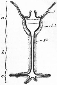
Fig. 1.—Diagram of a typical Hydropolyp.
a, Hydranth; b, Hydrocaulus; c, Hydrorhiza; t, Tentacle; ps, Perisarc, forming in the region of the hydranth a cup or hydrotheca(h, t),—which, however, is only found in polyps of the order Calyptoblastea.
As already stated, there occur in the Hydromedusae two distinct types of person, the polyp and the medusa; and either of them is capable of non-sexual reproduction by budding, a process which may lead to the formation of colonies, composed of more or fewer individuals combined and connected together. The morphology of the group thus falls naturally into four sections—(1) the hydropolyp, (2) the polyp-colony, (3) the hydromedusa, (4) the medusa-colonies. Since, however, medusa-colonies occur only in one group, the Siphonophora, and divergent views are held with regard to the morphological interpretation of the members of a siphonophore, only the first three of the above subdivisions of hydromedusa morphology will be dealt with here in a general way, and the morphology of the Siphonophora will be considered under the heading of the group itself.
1. The Hydropolyp (fig. 1)—The general characters of this organism are described above and in the articles Hydrozoa and Polyp. It is rarely free, but usually fixed and incapable of locomotion. The foot by which it is attached often sends out root-like processes—the hydrorhiza (c). The column (b) is generally long, slender and stalk-like (hydrocaulus). Just below the crown of tentacles, however, the body widens out to form a “head,” termed, the hydranth (a), containing a stomach-like dilatation of the digestive cavity. On the upper face of the hydranth the crown of tentacles (t) surrounds the peristome, from which rises the conical hypostome, bearing the mouth at its extremity. The general ectoderm covering the surface of the body has entirely lost the cilia present in the earlier larval stages (planula), and may be naked, or clothed in a cuticle or exoskeleton, the perisarc (ps), which in its simplest condition is a chitinous membrane secreted by the ectoderm. The perisarc when present invests the hydrorhiza and hydrocaulus; it may stop short below the hydranth, or it may extend farther. In general there are two types of exoskeleton, characteristic of the two principal divisions of the Hydroidea. In the Gymnoblastea the perisarc either stops below the hydranth, or, if continued on to it, forms a closely-fitting investment extending as a thin cuticle as far as the bases of the tentacles (e.g. Bimeria, see G. J. Allman [1],2 pl. xii. figs, 1 and 3). In the Calyptoblastea the perisarc is always continued above the hydrocaulus, and forms a cup, the hydrangium or hydrotheca (h, t), standing off from the body, into which the hydranth can be retracted for shelter and protection.
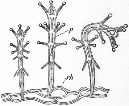
From Allman’s Gymnoblastic Hydroids, by permission of the Council of the Ray Society.
Fig. 2.—Stauridium productum, portion of the colony magnified; p, polyp; rh, hydrorhiza.
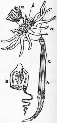
Fig. 3.—Diagram of Corymorpha. A, A hydriform person giving rise to medusiform persons by budding from the margin of the disk; B, free swimming medusa (Steenstrupia of Forbes) detached from the same, with manubrial genitalia, (Anthomedusae) and only one tentacle. (After Allman).
The architecture of the hydropolyp, simple though it be, furnishes a long series of variations affecting each part of the body. The greatest variation, however, is seen in the tentacles. As regards number, we find in the aberrant forms Protohydra and Microhydra tentacles entirely absent. In the curious hydroid Monobrachium a single tentacle is present, and the same is the case in Clathrozoon; in Amphibrachium and in Lar (fig. 11, A) the polyp bears two tentacles only. The reduction of the tentacles in all these forms may be correlated with their mode of life, and especially with living in a constant current of water, which brings food-particles always from one direction and renders a complete whorl or circle of tentacles unnecessary. Thus Microhydra lives amongst Bryozoa, and appears to utilize the currents produced by these animals. Protohydra occurs in oyster-banks and Monobrachium also grows on the shells of bivalves, and both these hydroids probably fish in the currents produced by the lamellibranchs. Amphibrachium grows in the tissues of a sponge, Euplectella, and protrudes its hydranth into the canal-system of the sponge; and Lar grows on the tubes of the worm Sabella. With the exception of these forms, reduced for the most part in correlation with a semi-parasitic mode of life, the tentacles are usually numerous. It is rare to find in the polyp a regular, symmetrical disposition of the tentacles as in the medusa. The primitive number of four in a whorl is seen, however, in Stauridium (fig. 2) and Cladonema (Allman [1], pl. xvii.), and in Clavatella each whorl consists regularly of eight (Allman, loc. cit. pl. xviii.). As a rule, however, the number in a whorl is irregular. The tentacles may form a single whorl, or more than one; thus in Corymorpha (fig. 3) and Tubularia (fig. 4) there are two circlets; in Stauridium (fig. 2) several; in Coryne and Cordylophora the tentacles are scattered irregularly over the elongated hydranth.
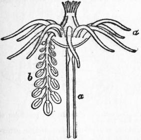
Fig. 4.—Diagram of Tubularia indivisa. A single hydriform person a bearing a stalk carrying numerous degenerate medusiform persons or sporosacs b. (After Allman.)
As regards form, the tentacles show a number of types, of which the most important are (1) filiform, i.e. cylindrical or tapering from base to extremity, as in Clava (fig. 5); (2) capitate, i.e. knobbed at the extremity, as in Coryne (see Allman, loc. cit. pl. iv.); (3) branched, a rare form in the polyp, but seen in Cladocoryne (see Allman, loc. cit. p. 380, fig. 82). Sometimes more than one type of form is found in the same polyp; in Pennaria and Stauridium (fig. 2) the upper whorls are capitate, the lower filiform. Finally, as regards structure, the tentacles may retain their primitive hollow nature, or become solid by obliteration of the axial cavity.
The hypostome of the hydropolyp may be small, or, on the other hand, as in Eudendrium (Allman, loc. cit. pls. xiii., xiv.), large and trumpet-shaped. In the curious polyp Myriothela the body of the polyp is differentiated into nutritive and reproductive portions.
Histology.—The ectoderm of the hydropolyp is chiefly sensory, contractile and protective in function. It may also be glandular in places. It consists of two regions, an external epithelial layer and a more internal sub-epithelial layer.
The epithelial layer consists of (1) so-called “indifferent” cells secreting the perisarc or cuticle and modified to form glandular cells in places; for example, the adhesive cells in the foot. (2) Sensory cells, which may be fairly numerous in places, especially on the tentacles, but which occur always scattered and isolated, never aggregated to form sense-organs as in the medusa. (3) Contractile or myo-epithelial cells, with the cell prolonged at the base into a contractile muscle-fibre (fig. 6, B). In the hydropolyp the ectodermal muscle-fibres are always directed longitudinally. Belonging primarily to the epithelial layer, the muscular cells may become secondarily sub-epithelial.
From Allman’s Gymnoblastic Hydroids, by permission of the Council of the Ray Society.
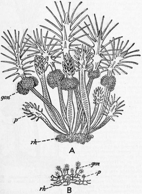
Fig. 5.—Colonies of Clava. A, Clava squamata, magnified. B, C. multicornis, natural size; p, polyp; gon, gonophores; rh, hydrorhiza.
The sub-epithelial layer consists primarily of the so-called interstitial cells, lodged between the narrowed basal portions of the epithelial cells. From them are developed two distinct types of histological elements; the genital cells and the cnidoblasts or mother-cells of the nematocysts. The sub-epithelial layer thus primarily constituted may be recruited by immigration from without of other elements, more especially by nervous (ganglion) cells and muscle-cells derived from the epithelial layer. In its fullest development, therefore, the sub-epithelial layer consists of four classes of cell-elements.
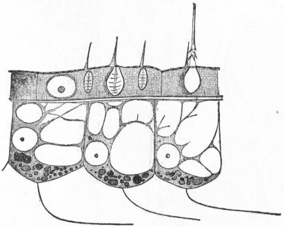
Fig. 6 A.—Portion of the body-wall of Hydra, showing ectoderm cells above, separated by “structureless lamella” from three flagellate endoderm cells below. The latter are vacuolated, and contain each a nucleus and several dark granules. In the middle ectoderm cell are seen a nucleus and three nematocysts, with trigger hairs projecting beyond the cuticle. A large nematocyst, with everted thread, is seen in the right-hand ectodermal cell. (After F. E. Schulze.)
The genital cells are simple wandering cells (archaeocytes), at first minute and without any specially distinctive features, until they begin to develop into germ-cells. According to Wulfert [60] the primitive germ-cells of Gonothyraea can be distinguished soon after the fixation of the planula, appearing amongst the interstitial cells of the ectoderm. The germ-cells are capable of extensive migrations, not only in the body of the same polyp, but also from parent to bud through many non-sexual generations of polyps in a colony (A. Weismann [58]).
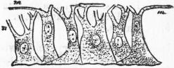
Fig. 6 B.—Epidermo-muscular cells of Hydra. m, muscular-fibre processes. (After Kleinenberg, from Gegenbaur.)
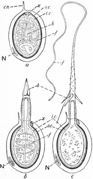
Fig. 7.—Diagrams to show the structure of Nematocysts and their mode of working. (After Iwanzov.)
a, Undischarged nematocyst. b, Commencing discharge. c, Discharge complete. cn, Cnidocil. N, Nucleus of cnidoblast. o.c, Outer capsule. x, Plug closing the opening of the outer capsule. i.c., Inner capsule, continuous with the wall of the filament, f. b, Barbs.
The cnidoblasts are the mother-cells of the nematocysts, each cell producing one nematocyst in its interior. The complete nematocyst (fig. 7) is a spherical or oval capsule containing a hollow thread, usually barbed, coiled in its interior. The capsule has a double wall, an outer one (o.c.), tough and rigid in nature, and an inner one (i.c.) of more flexible consistence. The outer wall of the capsule is incomplete at one pole, leaving an aperture through which the thread is discharged. The inner membrane is continuous with the wall of the hollow thread at a spot immediately below the aperture in the outer wall, so that the thread itself (f) is simply a hollow prolongation of the wall of the inner capsule inverted and pushed into its cavity. The entire nematocyst is enclosed in the cnidoblast which formed it. When the nematocyst is completely developed, the cnidoblast passes outwards so as to occupy a superficial position in the ectoderm, and a delicate protoplasmic process of sensory nature, termed the cnidocil (cn) projects from the cnidoblast like a fine hair or cilium. Many points in the development and mechanism of the nematocyst are disputed, but it is tolerably certain (1) that the cnidocil is of sensory nature, and that stimulation, by contact with prey or in other ways, causes a reflex discharge of the nematocyst; (2) that the discharge is an explosive change whereby the in-turned thread is suddenly everted and turned inside out, being thus shot through the opening in the outer wall of the capsule, and forced violently into the tissues of the prey, or, it may be, of an enemy; (3) that the thread inflicts not merely a mechanical wound, but instils an irritant poison, numbing and paralysing in its action. The points most in dispute are, first, how the explosive discharge is brought about, whether by pressure exerted external to the capsule (i.e. by contraction of the cnidoblast) or by internal pressure. N. Iwanzov [27] has brought forward strong grounds for the latter view, pointing out that the cnidoblast has no contractile mechanism and that measurements show discharged capsules to be on the average slightly larger than undischarged ones. He believes that the capsule contains a substance which swells very rapidly when brought into contact with water, and that in the undischarged condition the capsule has its opening closed by a plug of protoplasm (x, fig. 7) which prevents access of water to the contents; when the cnidocil is stimulated it sets in action a mechanism or perhaps a series of chemical changes by which the plug is dissolved or removed; as a result water penetrates into the capsule and causes its contents to swell, with the result that the thread is everted violently. A second point of dispute concerns the spot at which the poison is lodged. Iwanzov believes it to be contained within the thread itself before discharge, and to be introduced into the tissues of the prey by the eversion of the thread. A third point of dispute is whether the nematocysts are formed in situ, or whether the cnidoblasts migrate with them to the region where they are most needed; the fact that in Hydra, for example, there are no interstitial cells in the tentacles, where nematocysts are very abundant, is certainly in favour of the view that the cnidoblasts migrate on to the tentacles from the body, and that like the genital cells the cnidoblasts are wandering cells.
The muscular tissue consists primarily of processes from the bases of the epithelial cells, processes which are contractile in nature and may be distinctly striated. A further stage in evolution is that the muscle-cells lose their connexion with the epithelium and come to lie entirely beneath it, forming a sub-epithelial contractile layer, developed chiefly in the tentacles of the polyp. The evolution of the ganglion-cells, is probably similar; an epithelial cell develops processes of nervous nature from the base, which come into connexion with the bases of the sensory cells, with the muscular cells, and with the similar processes of other nerve-cells; next the nerve-cell loses its connexion with the outer epithelium and becomes a sub-epithelial ganglion-cell which is closely connected with the muscular layer, conveying stimuli from the sensory cells to the contractile elements. The ganglion-cells of Hydromedusae are generally very small. In the polyp the nervous tissue is always in the form of a scattered plexus, never concentrated to form a definite nervous system as in the medusa.
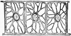
From Gegenbaur’s Elements of Comparative Anatomy.
Fig. 8.—Vacuolated Endoderm Cells of cartilaginous consistence from the axis of the tentacle of a Medusa (Cunina).
The endoderm of the polyp is typically a flagellated epithelium of large cells (fig. 6), from the bases of which arise contractile muscular processes lying in the plane of the transverse section of the body. In different parts of the coelenteron the endoderm may be of three principal types—(1) digestive endoderm, the primitive type, with cells of large size and considerably vacuolated, found in the hydranth; some of these cells may become special glandular cells, without flagella or contractile processes; (2) circulatory endoderm, without vacuoles and without basal contractile processes, found in the hydrorhiza and hydrocaulus; (3) supporting endoderm (fig. 8), seen in solid tentacles as a row of cubical vacuolated cells, occupying the axis of the tentacle, greatly resembling notochordal tissue, particularly that of Amphioxus at a certain stage of development; as a fourth variety of endodermal cells excretory cells should perhaps be reckoned, as seen in the pores in the foot of Hydra and elsewhere (cf. C. Chun, Hydrozoa [1], pp. 314, 315).
The mesogloea in the hydropolyp is a thin elastic layer, in which may be lodged the muscular fibres and ganglion cells mentioned above, but which never contains any connective tissue or skeletogenous cells or any other kind of special mesogloeal corpuscles.
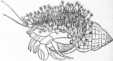
From Allman’s Gymnoblastic Hydroids, by permission of the Council of the Ray Society.
Fig. 9.—Colony of Hydractinia echinata, growing on the Shell of a Whelk. Natural size.
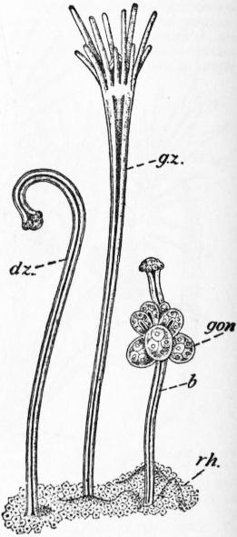
From Allman’s Gymnoblastic Hydroids, by permission of the Council of the Ray Society.
Fig. 10.—Polyps from a Colony of Hydractinia, magnified. dz, dactylozoid; gz, gastrozoid: b, blastostyle; gon, gonophores; rh, hydrorhiza.
2. The Polyp-colony.—All known hydropolyps possess the power of reproduction by budding, and the buds produced may become either polyps or medusae. The buds may all become detached after a time and give rise to separate and independent individuals, as in the common Hydra, in which only polyp-individuals are produced and sexual elements are developed upon the polyps themselves; or, on the other hand, the polyp-individuals produced by budding may remain permanently in connexion with the parent polyp, in which case sexual elements are never developed on polyp-individuals but only on medusa-individuals, and a true colony is formed. Thus the typical hydroid colony starts from a “founder” polyp, which in the vast majority of cases is fixed, but which may be floating, as in Nemopsis, Pelagohydra, &c. The founder-polyp usually produces by budding polyp-individuals, and these in their turn produce other buds. The polyps are all non-sexual individuals whose function is purely nutritive. After a time the polyps, or certain of them, produce by budding medusa-individuals, which sooner or later develop sexual elements; in some cases, however, the founder-polyp remains solitary, that is to say, does not produce polyp-buds, but only medusa-buds, from the first (Corymorpha, fig. 3, Myriothela, &c.). In primitive forms the medusa-individuals are set free before reaching sexual maturity and do not contribute anything to the colony. In other cases, however, the medusa-individuals become sexually mature while still attached to the parent polyp, and are then not set free at all, but become appanages of the hydroid colony and undergo degenerative changes leading to reduction and even to complete obliteration of their original medusan structure. In this way the hydroid colony becomes composed of two portions of different function, the nutritive “trophosome,” composed of non-sexual polyps, and the reproductive “gonosome,” composed of sexual medusa-individuals, which never exercise a nutritive function while attached to the colony. As a general rule polyp-buds are produced from the hydrorhiza and hydrocaulus, while medusa-buds are formed on the hydranth. In some cases, however, medusa-buds are formed on the hydrorhiza, as in Hydrocorallines.
In such a colony of connected individuals, the exact limits of the separate “persons” are not always clearly marked out. Hence it is necessary to distinguish between, first, the “zooids,” indicated in the case of the polyps by the hydranths, each with mouth and tentacles; and, secondly, the “coenosarc,” or common flesh, which cannot be assigned more to one individual than another, but consists of a more or less complicated network of tubes, corresponding to the hydrocaulus and hydrorhiza of the primitive independent polyp-individual. The coenosarc constitutes a system by which the digestive cavity of any one polyp is put into communication with that of any other individual either of the trophosome or gonosome. In this manner the food absorbed by one individual contributes to the welfare of the whole colony, and the coenosarc has the function of circulating and distributing nutriment through the colony.
The hydroid colony shows many variations in form and architecture which depend simply upon differences in the methods in which polyps are budded.
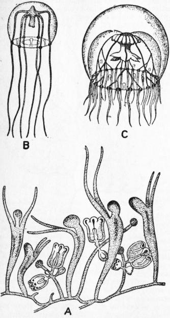
After Hincks, Forbes, and Browne. A and B modified from Hincks; C modified from Forbes’s Brit. Naked-eyed Medusae.
Fig. 11.—Lar sabellarum and two stages of its Medusa, Willia stellata. A, colony of Lar; B and C, young and adult medusae.
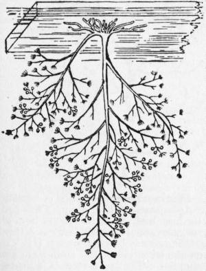
Fig. 12.—Colony of Bougainvillea fruticosa, natural size, attached to the underside of a piece of floating timber. (After Allman.)
In the first place, buds may be produced only from the hydrorhiza, which grows out and branches to form a basal stolon, typically net-like, spreading over the substratum to which the founder-polyp attached itself. From the stolon the daughter-polyps grow up vertically. The result is a spreading or creeping colony, with the coenosarc in the form of a root-like horizontal network (fig. 5, B; 11, A). Such a colony may undergo two principal modifications. The meshes of the basal network may become very small or virtually obliterated, so that the coenosarc becomes a crust of tubes tending to fuse together, and covered over by a common perisarc. Encrusting colonies of this kind are seen in Clava squamata (fig. 5, A) and Hydractinia (figs. 9, 10), the latter having the perisarc calcified. A further very important modification is seen when the tubes of the basal perisarc do not remain spread out in one plane, but grow in all planes forming a felt-work; the result is a massive colony, such as is seen in the so-called Hydrocorallines (fig. 60), where the interspaces between the coenosarcal tubes are filled up with calcareous matter, or coenosteum, replacing the chitinous perisarc. The result is a stony, solid mass, which contributes to the building up of coral reefs. In massive colonies of this kind no sharp distinction can be drawn between hydrorhiza and hydrocaulus in the coenosarc; it is practically all hydrorhiza. Massive colonies may assume various forms and are often branching or tree-like. A further peculiarity of this type of colony is that the entire coenosarcal complex is covered externally by a common layer of ectoderm; it is not clear how this covering layer is developed.
In the second place, the buds may be produced from the hydrocaulus, growing out laterally from it; the result is an arborescent, tree-like colony (figs. 12, 13). Budding from the hydrocaulus may be combined with budding from the hydrorhiza, so that numerous branching colonies arise from a common basal stolon. In the formation of arborescent colonies, two sharply distinct types of budding are found, which are best described in botanical terminology as the monopodial or racemose, and the sympodial or cymose types respectively; each is characteristic of one of the two sub-orders of the Hydroidea, the Gymnoblastea and Calyptoblastea.
In the monopodial method (figs. 12, 14) the founder-polyp is, theoretically, of unlimited growth in a vertical direction, and as it grows up it throws out buds right and left alternately, so that the first bud produced by it is the lowest down, the second bud is above the first, the third above this again, and so on. Each bud produced by the founder proceeds to grow and to bud in the same way as the founder did, producing a side branch of the main stem. Hence, in a colony of gymnoblastic hydroids, the oldest polyp of each system, that is to say, of the main stem or of a branch, is the topmost polyp; the youngest polyp of the system is the one nearest to the topmost polyp; and the axis of the system is a true axis.
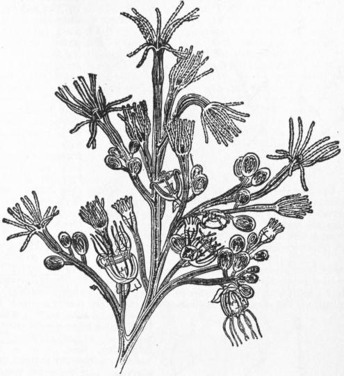
Fig. 13.—Portion of colony of Bougainvillea fruticosa (Anthomedusae-Gymnoblastea) more magnified. (From Lubbock, after Allman.)
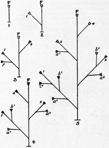
Fig. 14.—Diagrams of the monopodial method of budding, shown in five stages (1-5). F, the founder-polyp; 1, 2, 3, 4, the succession of polyps budded from the founder-polyp; a′, b′, c′, the succession of polyps budded from 1; a2, b2, polyps budded from 2; a3, polyp budded from 3.
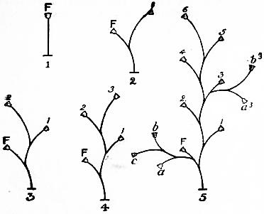
Fig. 15.—Diagram of sympodial budding, biserial type, shown in five stages (1-5). F, founder-polyp; 1, 2, 3, 4, 5, 6, succession of polyps budded from the founder; a, b, c, second series of polyps budded from the founder; a3, b3, series budded from 3.
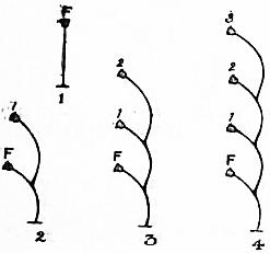
Fig. 16.—Diagram of sympodial budding, uniserial type, shown in four stages (1-4). F, founder-polyp; 1, 2, 3, succession of polyps budded from the founder.
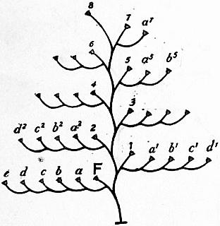
Fig. 17.—Diagram of sympodial budding, simple unbranched Plumularia-type. F, founder; 1-8, main axis formed by biserial budding from founder; a-e, pinnule formed by uniserial budding from founder; a¹-d¹, branch formed by similar budding from 1; a2-d2 from 2, and so forth.
In the sympodial method of budding, on the other hand, the founder-polyp is of limited growth, and forms a bud from its side, which is also of limited growth, and forms a bud in its turn, and so on (figs. 15, 16). Hence, in a colony of calyptoblastic hydroids, the oldest polyp of a system is the lowest; the youngest polyp is the topmost one; and the axis of the system is a false axis composed of portions of each of the consecutive polyps. In this method of budding there are two types. In one, the biserial type (fig. 15), the polyps produce buds right and left alternately, so that the hydranths are arranged in a zigzag fashion, forming a “scorpioid cyme,” as in Obelia and Sertularia. In the other, the uniserial type (fig. 16), the buds are formed always on the same side, forming a “helicoid cyme,” as in Hydrallmania, according to H. Driesch, in which, however, the primitively uniserial arrangement becomes masked later by secondary torsions of the hydranths.
In a colony formed by sympodial budding, a polyp always produces first a bud, which contributes to the system to which it belongs, i.e. continues the stem or branch of which its parent forms a part. The polyp may then form a second bud, which becomes the starting point of a new system, the beginning, that is, of a new branch; and even a third bud, starting yet another system, may be produced from the same polyp. Hence the colonies of Calyptoblastea may be complexly branched, and the budding may be biserial throughout, uniserial throughout, or partly one, partly the other. Thus in Plumularidae (figs. 17, 18) there is formed a main stem by biserial budding; each polyp on the main stem forms a second bud, which usually forms a side branch or pinnule by uniserial budding. In this way are formed the familiar feathery colonies of Plumularia, in which the pinnules are all in one plane, while in the allied Antennularia the pinnules are arranged in whorls round the main biserial stem. The pinnules never branch again, since in the uniserial mode of budding a polyp never forms a second polyp-bud. On the other hand, a polyp on the main stem may form a second bud which, instead of forming a pinnule by uniserial budding, produces by biserial budding a branch, from which pinnules arise as from the main stem (fig. 18—3, 6). Or a polyp on the main stem, after having budded a second time to form a pinnule, may give rise to a third bud, which starts a new biserial system, from which uniserial pinnules arise as from the main stem—type of Aglaophenia (fig. 19). The laws of budding in hydroids have been worked out in an interesting manner by H. Driesch [13], to whose memoirs the reader must be referred for further details.
Individualization of Polyp-Colonies.—As in other cases where animal colonies are formed by organic union of separate individuals, there is ever a tendency for the polyp-colony as a whole to act as a single individual, and for the members to become subordinated to the needs of the colony and to undergo specialization for particular functions, with the result that they simulate organs and their individuality becomes masked to a greater or less degree. Perhaps the earliest of such specializations is connected with the reproductive function. Whereas primitively any polyp in a colony may produce medusa-buds, in many hydroid colonies medusae are budded only by certain polyps termed blastostyles (fig. 10, b). At first not differing in any way from other polyps (fig. 5), the blastostyles gradually lose their nutritive function and the organs connected with it; the mouth and tentacles disappear, and the blastostyle obtains the nutriment necessary for its activity by way of the coenosarc. In the Calyptoblastea, where the polyps are protected by special capsules of the perisarc, the gonothecae enclosing the blastostyles differ from the hydrothecae protecting the hydranths (fig. 54).
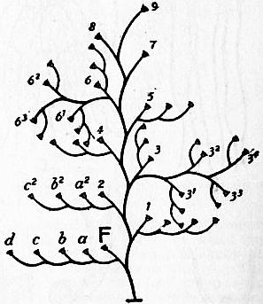
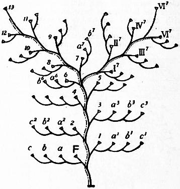
Fig. 18.—Diagram showing method of branching in the Plumularia-type; compare with fig. 17. Polyps 3 and 6, instead of producing uniserial pinnules, have produced biserial branches (31, 32, 33, 34; 61-63), which give off uniserial branches in their turn. Fig. 19.—Diagram showing method of branching in the Aglaophenia-type. Polyp 7 has produced as its first bud, 8; as its second bud, a7, which starts a uniserial pinnule; and as a third bud I7, which starts a biserial branch (II7-VI7) that repeats the structure of the main stem and gives off pinnules. The main stem is indicated by-·-·-·, the new stem by ······.
In other colonies the two functions of the nutritive polyp, namely, capture and digestion of food, may be shared between different polyps (fig. 10). One class of polyps, the dactylozoids (dz), lose their mouth and stomach, and become elongated and tentacle-like, showing great activity of movement. Another class, the gastrozoids (gz), have the tentacles reduced or absent, but have the mouth and stomach enlarged. The dactylozoids capture food, and pass it on to the gastrozoids, which swallow and digest it.
Besides the three types of individual above mentioned, there are other appendages of hydroid colonies, of which the individuality is doubtful. Such are the “guard-polyps” (machopolyps) of Plumularidae, which are often regarded as individuals of the nature of dactylozoids, but from a study of the mode of budding in this hydroid family Driesch concluded that the guard-polyps were not true polyp-individuals, although each is enclosed in a small protecting cup of the perisarc, known as a nematophore. Again, the spines arising from the basal crust of Podocoryne have been interpreted by some authors as reduced polyps.
3. The Medusa.—In the Hydromedusae the medusa-individual occurs, as already stated, in one of two conditions, either as an independent organism leading a true life in the open seas, or as a subordinate individuality in the hydroid colony, from which it is never set free; it then becomes a mere reproductive appendage or gonophore, losing successively its organs of sense, locomotion and nutrition, until its medusoid nature and organization become scarcely recognizable. Hence it is convenient to consider the morphology of the medusa from these two aspects.
(a) The Medusa as an Independent Organism.—The general structure and characteristics of the medusa are described elsewhere (see articles Hydrozoa and Medusa), and it is only necessary here to deal with the peculiarities of the Hydromedusa.
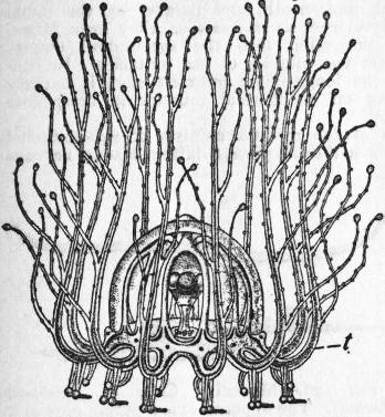
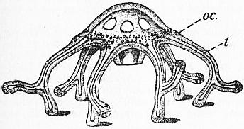
From Allman’s Gymnoblastic Hydroids, by permission of the Council of the Ray Society. From Allman’s Gymnoblastic Hydroids, by permission of the Council of the Ray Society.
Fig. 20.—Cladonema radiatum, the medusa walking on the basal branches of its tentacles (t), which are turned up over the body. Fig. 21.—Clavatella prolifera, ambulatory medusa. t, tentacles; oc, ocelli.
As regards habit of life the vast majority of Hydromedusae are pelagic organisms, floating on the surface of the open sea, propelling themselves feebly by the pumping movements of the umbrella produced by contraction of the sub-umbral musculature, and capturing their prey with their tentacles. The genera Cladonema (fig. 20) and Clavatella (fig. 21), however, are ambulatory, creeping forms, living in rock-pools and walking, as it were, on the tips of the proximal branches of each of the tentacles, while the remaining branches serve for capture of food. Cladonema still has the typical medusan structure, and is able to swim about, but in Clavatella the umbrella is so much reduced, that swimming is no longer possible. The remarkable medusa Mnestra parasites is ecto-parasitic throughout life on the pelagic mollusc Phyllirrhoe, attached to it by the sub-umbral surface, and its tentacles have become rudimentary or absent. It is interesting to note that Mnestra has been shown by J. W. Fewkes [15] and R. T. Günther [19] to belong to the same family (Cladonemidae) as Cladonema and Clavatella, and it is reasonable to suppose that the non-parasitic ancestor of Mnestra was, like the other two genera, an ambulatory medusa which acquired louse-like habits. In some species of the genus Cunina (Narcomedusae) the youngest individuals (actinulae) are parasitic on other medusae (see below), but in later life the parasitic habit is abandoned. No other instances are known of sessile habit in Hydromedusae.
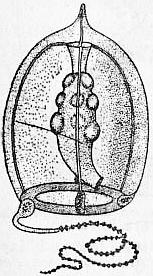
After E. T. Browne, from Proc. Zool. Soc. of London.
Fig. 22.—Corymorpha nutans, adult female Medusa. Magnified 10 diameters.
The external form of the Hydromedusae varies from that of a deep bell or thimble, characteristic of the Anthomedusae, to the shallow saucer-like form characteristic of the Leptomedusae. It is usual for the umbrella to have an even, circular, uninterrupted margin; but in the order Narcomedusae secondary down-growths between the tentacles produce a lobed, indented margin to the umbrella. The marginal tentacles are rarely absent in non-parasitic forms, and are typically four in number, corresponding to the four perradii marked by the radial canals. Interradial tentacles may be also developed, so that the total number present may be increased to eight or to an indefinitely large number. In Willia, Geryonia, &c., however, the tentacles and radial canals are on the plan of six instead of four (figs. 11 and 26). On the other hand, in some cases the tentacles are less in number than the perradii; in Corymorpha (figs. 3 and 22) there is but a single tentacle, while two are found in Amphinema and Gemmaria (Anthomedusae), and in Solmundella bitentaculata (fig. 67) and Aeginopsis hensenii (fig. 23) (Narcomedusae). The tentacles also vary considerably in other ways than in number: first, in form, being usually simple, with a basal bulb, but in Cladonemidae they are branched, often in complicated fashion; secondly, in grouping, being usually given off singly, and at regular intervals from the margin of the umbrella, but in Margelidae and in some Trachomedusae they are given off in tufts or bunches (fig. 24); thirdly, in position and origin, being usually implanted on the extreme edge of the umbrella, but in Narcomedusae they become secondarily shifted and are given off high up on the ex-umbrella (figs. 23 and 25); and, fourthly, in structure, being hollow or solid, as in the polyp. In some medusae, for instance, the remarkable deep-sea family Pectyllidae, the tentacles may bear suckers, by which the animal may attach itself temporarily. It should be mentioned finally that the tentacles are very contractile and extensible, and may therefore present themselves, in one and the same individual, as long, drawn-out threads, or in the form of short corkscrew-like ringlets; they may stream downwards from the sub-umbrella, or be held out horizontally, or be directed upwards over the ex-umbrella (fig. 23). Each species of medusa usually has a characteristic method of carrying its tentacles.
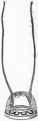
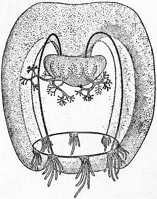
After O. Maas, Die craspedoten Medusen der Plankton Expedition, by permission of Lipsius and Tischer. After O. Maas, Craspedoten Medusen der Siboga-Expedition, by permission of E. S. Brill & Co.
Fig. 23.—Aeginopsis hensenii, slightly magnified, showing the manner in which the tentacles are carried in life. Fig. 24.—Rathkea octonemalis.
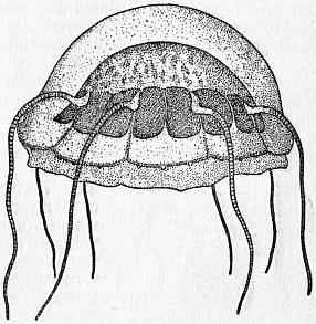
After O. Maas, Medusae, in Prince of Monaco’s series.
Fig. 25.—Aeginura grimaldii.
The sub-umbrella invariably shows a velum as an inwardly projecting ridge or rim at its margin, within the circle of tentacles; hence the medusae of this sub-class are termed craspedote. The manubrium is absent altogether in the fresh-water medusa Limnocnida, in which the diameter of the mouth exceeds half that of the umbrella; on the other hand, the manubrium may attain a great length, owing to the centre of the sub-umbrella with the stomach being drawn into it, as it were, to form a long proboscis, as in Geryonia. The mouth may be a simple, circular pore at the extremity of the manubrium, or by folding of the edges it may become square or shaped like a Maltese cross, with four corners and four lips. The corners of the mouth may then be drawn out into lobes or lappets, which may have a branched or fringed outline (fig. 27), and in Margelidae the subdivisions of the fringe simulate tentacles (fig. 24).
The internal anatomy of the Hydromedusae shows numerous variations. The stomach may be altogether lodged in the manubrium, from which the radial canals then take origin directly as in Geryonia (Trachomedusae); it may be with or without gastric pouches. The radial canals may be simple or branched, primarily four, rarely six in number. The ring-canal is drawn out in Narcomedusae into festoons corresponding with the lobes of the margin, and may be obliterated altogether (Solmaris). In this order the radial canals are represented only by wide gastric pouches, and in the family Solmaridae are suppressed altogether, so that the tentacles and the festoons of the ring-canal arise directly from the stomach. In Geryonia, centripetal canals, ending blindly, arise from the ring-canal and run in a radial direction towards the centre of the umbrella (fig. 26).
Histology of the Hydromedusa.—The histology described above for the polyp may be taken as the primitive type, from which that of the medusa differs only in greater elaboration and differentiation of the cell-elements, which are also more concentrated to form distinct tissues.
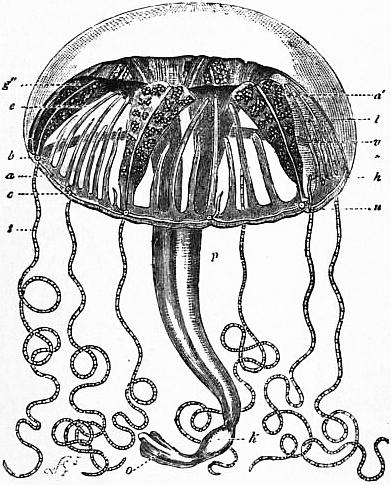
Fig. 26.—Carmarina (Geryonia) hastata, one of the Trachomedusae. (After Haeckel.)
a, Nerve ring. a′, Radial nerve. b, Tentaculocyst. c, Circular canal. e, Radiating canal. g″. Ovary. h, Peronia or cartilaginous process ascending from the cartilaginous margin of the disk centripetally in the outer surface of the jelly-like disk; six of these are perradial, six interradial, corresponding to the twelve solid larval tentacles, resembling those of Cunina. k, Dilatation (stomach) of the manubrium. l, Jelly of the disk. p, Manubrium. t, Tentacle (hollow and tertiary, i.e. preceded by six perradial and six interradial solid larval tentacles). u, Cartilaginous margin of the disk covered by thread-cells. v. Velum.
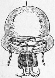
After O. Maas in Results of the “Albatross” Expedition, Museum of Comparative Zoology, Cambridge, Mass., U.S.A.
Fig. 27.—Stomotoca divisa, one of the Tiaridae (Anthomedusae).
The ectoderm furnishes the general epithelial covering of the body, and the muscular tissue, nervous system and sense-organs. The external epithelium is flat on the ex-umbral surface, more columnar on the sub-umbral surface, where it forms the muscular tissue of the sub-umbrella and the velum. The nematocysts of the ectoderm may be grouped to form batteries on the tentacles, umbrellar margin and oral lappets. In places the nematocysts may be crowded so thickly as to form a tough, supporting, “chondral” tissue, resembling cartilage, chiefly developed at the margin of the umbrella and forming streaks or bars supporting the tentacles (“Tentakelspangen,” peronia) or the tentaculocysts (“Gehörspangen,” otoporpae).
The muscular tissue of the Hydromedusae is entirely ectodermal. The muscle-fibres arise as processes from the bases of the epithelial cells; such cells may individually become sub-epithelial in position, as in the polyp; or, in places where muscular tissue is greatly developed, as in the velum or sub-umbrella, the entire muscular epithelium may be thrown into folds in order to increase its surface, so that a deeper sub-epithelial muscular layer becomes separated completely from a more superficial body-epithelium.
In its arrangement the muscular tissue forms two systems: the one composed of striated fibres arranged circularly, that is to say, concentrically round the central axis of the umbrella; the other of non-striated fibres running longitudinally, that is to say, in a radial direction from, or (in the manubrium) parallel to, the same ideal axis. The circular system is developed continuously over the entire sub-umbral surface, and the velum represents a special local development of this system, at a region where it is able to act at the greatest mechanical advantage in producing the contractions of the umbrella by which the animal progresses. The longitudinal system is discontinuous, and is subdivided into proximal, medial and distal portions. The proximal portion forms the retractor muscles of the manubrium, or proboscis, well developed, for example, in Geryonia. The medial portion forms radiating tracts of fibres, the so-called “bell-muscles” running underneath, and parallel to, the radial canals; when greatly developed, as in Tiaridae, they form ridges, so-called mesenteries, projecting into the sub-umbral cavity. The distal portions form the muscles of the tentacles. In contrast with the polyp, the longitudinal muscle-system is entirely ectodermal, there being no endodermal muscles in craspedote medusae.
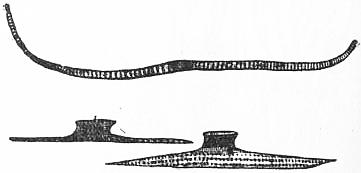
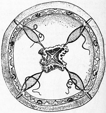
Fig. 28.—Muscular Cells of Medusae (Lizzia). The uppermost is a purely muscular cell from the sub-umbrella; the two lower are epidermo-muscular cells from the base of a tentacle; the upstanding nucleated portion forms part of the epidermal mosaic on the free surface of the body. (After Hertwig.) After O. Maas, Craspedoten Medusen der Siboga Expedition, by permission of E. S. Brill & Co.
Fig. 29.—Tiaropsis rosea (Ag. and Mayer) showing the eight adradial Statocysts, each close to an Ocellus. Cf. fig. 30.
The nervous system of the medusa consists of sub-epithelial ganglion-cells, which form, in the first place, a diffuse plexus of nervous tissue, as in the polyp, but developed chiefly on the sub-umbral surface; and which are concentrated, in the second place, to form a definite central nervous system, never found in the polyp. In Hydromedusae the central nervous system forms two concentric nerve-rings at the margin of the umbrella, near the base of the velum. One, the “upper” or ex-umbral nerve-ring, is derived from the ectoderm on the ex-umbral side of the velum; it is the larger of the two rings, containing more numerous but smaller ganglion-cells, and innervates the tentacles. The other, the “lower” or sub-umbral nerve-ring, is derived from the ectoderm on the sub-umbral side of the velum; it contains fewer but larger ganglion-cells and innervates the muscles of the velum (see diagram in article Medusae). The two nerve-rings are connected by fibres passing from one to the other.
The sensory cells are slender epithelial cells, often with a cilium or stiff protoplasmic process, and should perhaps be regarded as the only ectoderm-cells which retain the primitive ciliation of the larval ectoderm, otherwise lost in all Hydrozoa. The sense-cells form, in the first place, a diffuse system of scattered sensory cells, as in the polyp, developed chiefly on the manubrium, the tentacles and the margin of the umbrella, where they form a sensory ciliated epithelium covering the nerve-centres; in the second place, the sense-cells are concentrated to form definite sense-organs, situated always at the margin of the umbrella, hence often termed “marginal bodies.” The possession of definite sense-organs at once distinguishes the medusa from the polyp, in which they are never found.
The sense-organs of medusae are of two kinds—first, organs sensitive to light, usually termed ocelli (fig. 29); secondly, organs commonly termed otocysts, on account of their resemblance to the auditory vesicles of higher animals, but serving for the sense of balance and orientation, and therefore given the special name of statocysts (fig. 30). The sense-organs may be tentaculocysts, i.e. modifications of a tentacle, as in Trachylinae, or developed from the margin of the umbrella, in no connexion with a tentacle (or, if so connected, not producing any modification in the tentacle), as in Leptolinae. In Hydromedusae the sense-organs are always exposed at the umbrellar margin (hence Gymnophthalmata), while in Scyphomedusae they are covered over by flaps of the umbrellar margin (hence Steganophthalmata).
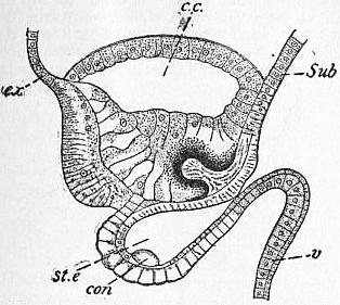
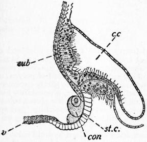
Modified after Linko, Traveaux Soc. Imp. Nat., St. Petersbourg, xxix. Modified after O. and R, Hertwig, Nervensystem und Sinnesorgane der Medusen, by permission of F. C. W. Vogel.
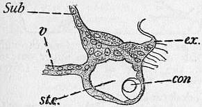
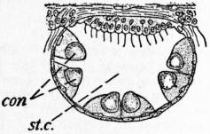
Fig. 30.—Section of a Statocyst and Ocellus of Tiaropsis diademata; cf. fig. 29. Fig. 31.—Section of a Statocyst of Mitrocoma annae.
ex, Ex-umbral ectoderm. sub, Sub-umbral ectoderm. c.c, Circular canal. v, Velum. st.e, Cavity of statocyst. con, Concrement-cell with otolith. sub, Sub-umbral ectoderm. c.c, Circular canal. v, Velum. st.c, Cavity of statocyst. con, Concrement-cell with otolith.
The statocysts present in general the structure of either a knob or a closed vesicle, composed of (1) indifferent supporting epithelium: (2) sensory, so-called auditory epithelium of slender cells, each bearing at its free upper end a stiff bristle and running out at its base into a nerve-fibre; (3) concrement-cells, which produce intercellular concretions, so-called otoliths. By means of vibrations or shocks transmitted through the water, or by displacements in the balance or position of the animal, the otoliths are caused to impinge against the bristles of the sensory cells, now on one side, now on the other, causing shocks or stimuli which are transmitted by the basal nerve-fibre to the central nervous system. Two stages in the development of the otocyst can be recognized, the first that of an open pit on a freely-projecting knob, in which the otoliths are exposed, the second that of a closed vesicle, in which the otoliths are covered over. Further, two distinct types of otocyst can be recognized in the Hydromedusae: that of the Leptolinae, in which the entire organ is ectodermal, concrement-cells and all, and the organ is not a tentaculocyst; and that of the Trachylinae, in which the organ is a tentaculocyst, and the concrement-cells are endodermal, derived from the endoderm of the modified tentacle, while the rest of the organ is ectodermal.
Modified after O. and R, Hertwig, Nervensystem und Sinnesorgane der Medusen, by permission of F. C. W. Vogel. Modified after O. and R, Hertwig, Nervensystem und Sinnesorgane der Medusen, by permission of F. C. W. Vogel.
Fig. 32.—Section of a Statocyst of Phialidium. Fig. 33.—Optical Section of a Statocyst of Octorchis.
ex, Ex-umbral ectoderm. sub, Sub-umbral ectoderm. v, Velum. st.c, Cavity of statocyst. con, Concrement-cell with otolith. con, Concrement-cell with otolith. st.c, Cavity of statocyst.
In the Leptolinae the otocysts are seen in their first stage in Mitrocoma annae (fig. 31) and Tiaropsis (figs. 29, 30) as an open pit at the base of the velum, on its sub-umbral side. The pit has its opening turned towards the sub-umbral cavity, while its base or fundus forms a bulge, more or less pronounced, on the ex-umbral side of the velum. At the fundus are placed the concrement-cells with their conspicuous otoliths (con) and the inconspicuous auditory cells, which are connected with. the sub-umbral nerve-ring. From the open condition arises the closed condition very simply by closing up of the aperture of the pit. We then find the typical otocyst of the Leptomedusae, a vesicle bulging on the ex-umbral side of the velum (figs. 32, 33). The otocysts are placed on the outer wall of the vesicle (the fundus of the original pit) or on its sides; their arrangement and number vary greatly and furnish useful characters for distinguishing genera. The sense-cells are innervated, as before, from the sub-umbral nerve-ring. The inner wall of the vesicle (region of closure) is frequently thickened to form a so-called “sense-cushion,” apparently a ganglionic offshoot from the sub-umbral nerve-ring. In many Leptomedusae the otocysts are very small, inconspicuous and embedded completely in the tissues; hence they may be easily overlooked in badly-preserved material, and perhaps are present in many cases where they have been said to have been wanting.
After O. and R, Hertwig, Nervensystem und Sinnesorgane der Medusen, by permission of F. C. W. Vogel. After O. and R, Hertwig, Nervensystem und Sinnesorgane der Medusen, by permission of F. C. W. Vogel.
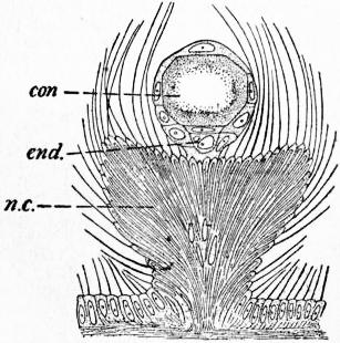
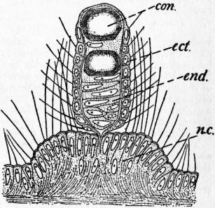
Fig. 34.—Tentaculocyst (statorhabd) of Cunina solmaris. n.c, Nerve-cushion; end, endodermal concrement-cells; con, otolith. Fig. 35.—Tentaculocyst of Cunina lativentris.
ect, Ectoderm. n.c, Nerve-cushion. end, Endodermal concrement-cells. con, Otolith.
In the Trachylinae the simplest condition of the otocyst is a freely projecting club, a so-called statorhabd (figs. 34, 35), representing a tentacle greatly reduced in size, covered with sensory ectodermal epithelium (ect.), and containing an endodermal core (end.), which is at first continuous with the endoderm of the ring-canal, but later becomes separated from it. In the endoderm large concretions are formed (con.). Other sensory cells with long cilia cover a sort of cushion (n.c.) at the base of the club; the club may be long and the cushion small, or the cushion large and the club small. The whole structure is innervated, like the tentacles, from the ex-umbral nerve-ring. An advance towards the second stage is seen in such a form as Rhopalonema (fig. 36), where the ectoderm of the cushion rises up in a double fold to enclose the club in a protective covering forming a cup or vesicle, at first open distally; finally the opening closes and the closed vesicle may sink inwards and be found far removed from the surface, as in Geryonia (fig. 37).
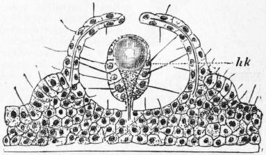
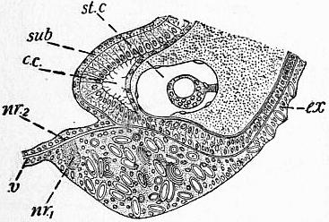
Fig. 36.—Simple tentaculocyst of Rhopalonema velatum. The process carrying the otolith or concretion hk, formed by endoderm cells, is enclosed by an upgrowth forming the “vesicle,” which is not yet quite closed in at the top. (After Hertwig.) After O. and R, Hertwig, Nervensystem und Sinnesorgane der Medusen, by permission of F. C. W. Vogel.
Fig. 37.—Section of statocyst of Geryonia (Carmarina hastata).
st.c, Statocyst containing the minute tentaculocyst. nr1, Ex-umbral nerve-ring. nr2, Sub-umbral nerve-ring. ex, Ex-umbral ectoderm. sub, Sub-umbral ectoderm. c.c, Circular canal. v, Velum.
The ocelli are seen in their simplest form as a pigmented patch of ectoderm, which consists of two kinds of cells—(1) pigment-cells, which are ordinary indifferent cells of the epithelium containing pigment-granules, and (2) visual cells, slender sensory epithelial cells of the usual type, which may develop visual cones or rods at their free extremity. The ocelli occur usually either on the inner or outer sides of the tentacles; if on the inner side, the tentacle is turned upwards and carried over the ex-umbrella, so as to expose the ocellus to the light; if the ocellus be on the outer side of a tentacle, two nerves run round the base of the tentacle to it. In other cases ocelli may occur between tentacles, as in Tiaropsis (fig. 29).
The simple form of ocellus described in the foregoing paragraph may become folded into a pit or cup, the interior of which becomes filled with a clear gelatinous secretion forming a sort of vitreous body. The distal portion of the vitreous body may project from the cavity of the cup, forming a non-cellular lens as in Lizzia (fig. 28). Beyond this simple condition the visual organs of the Hydromedusae do not advance, and are far from reaching the wonderful development of the eyes of Scyphomedusae (Charybdaea).
Besides the ordinary type of ocellus just described, there is found in one genus (Tiaropsis) a type of ocellus in which the visual elements are inverted, and have their cones turned away from the light, as in the human retina (fig. 30). In this case the pigment-cells are endodermal, forming a cup of pigment in which the visual cones are embedded. A similar ocellus is formed in Aurelia among the Scyphomedusae (q.v.).
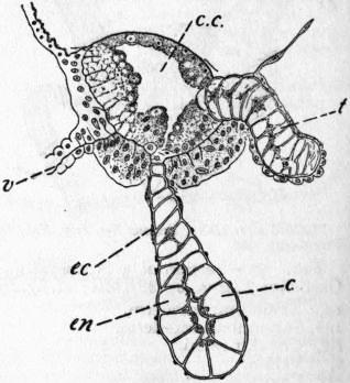
Other sense organs of Hydromedusae are the so-called sense-clubs or cordyli found in a few Leptomedusae, especially in those genera in which otocysts are inconspicuous or absent (fig. 39). Each cordylus is a tentacle-like structure with an endodermal axis containing an axial cavity which may be continuous with the ring-canal, or may be partially occluded. Externally the cordylus is covered, by very flattened ectoderm, and bears no otoliths or sense-cells, but the base of the club rests upon the ex-umbral nerve-ring. Brooks regards these organs as sensory, serving for the sense of balance, and representing a primitive stage of the tentaculocysts of Trachylinae; Linko, on the other hand, finding no nerve-elements connected with them, regards them as digestive (?) in function.
The sense-organs of the two fresh-water medusae Limnocodium and Limnocnida are peculiar and of rather doubtful nature (see E. T. Browne [10]).
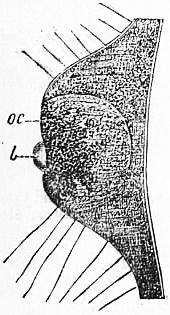
Fig. 38.—Ocellus of Lizzia koellikeri. oc, Pigmented ectodermal cells; l, lens. (After Hertwig.)
The endoderm of the medusa shows the same general types of structure as in the polyp, described above. We can distinguish (1) digestive endoderm, in the stomach, often with special glandular elements; (2) circulatory endoderm, in the radial and ring-canals; (3) supporting endoderm in the axes of the tentacles and in the endoderm-lamella; the latter is primitively a double layer of cells, produced by concrescence of the ex-umbral and sub-umbral layers of the coelenteron, but it is usually found as a single layer of flattened cells (fig. 40); in Geryonia, however, it remains double, and the centripetal canals arise by parting of the two layers; (4) excretory endoderm, lining pores at the margin of the umbrella, occurring in certain Leptomedusae as so-called “marginal tubercles,” opening, on the one hand, into the ring-canal and, on the other hand, to the exterior by “marginal funnels,” which debouch into the sub-umbral cavity above the velum. As has been described above, the endoderm may also contribute to the sense-organs, but such contributions are always of an accessory nature, for instance, concrement-cells in the otocysts, pigment in the ocelli, and never of sensory nature, sense-cells being in all cases ectodermal.
The reproductive cells may be regarded as belonging primarily to neither ectoderm nor endoderm, though lodged in the ectoderm in all Hydromedusae. As described for the polyp, they are wandering cells capable of extensive migrations before reaching the particular spot at which they ripen. In the Hydromedusae they usually, if not invariably, ripen in the ectoderm, but in the neighbourhood of the main sources of nutriment, that is to say, not far from the stomach. Hence the gonads are found on the manubrium in Anthomedusae generally; on the base of the manubrium, or under the gastral pouches, or in both these situations (Octorchidae), or under the radial canals, in Trachomedusae; under the gastral pouches or radial canals, in Narcomedusae. When ripe, the germ-cells are dehisced directly to the exterior.
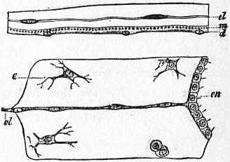
After W. K. Brooks, Journal of Morphology, x., by permission of Ginn & Co. Fig. 40.—Portions of Sections through the Disk of Medusae—the upper one of Lizzia, the lower of Aurelia. (After Hertwig.)
Fig. 39.—Section of a Cordylus of Laodice.
c.c, Circular canal. v, Velum. t, Tentacle. c, Cordylus, composed of flattened ectoderm ec covering a large-celled endodermal axis en. el, Endoderm lamella. m, Muscular processes of the ectoderm-cells in cross section. d, Ectoderm. en, Endoderm lining the enteric cavity. e, Wandering endoderm cells of the gelatinous substance.
Hydromedusae are of separate sexes, the only known exception being Amphogona apsteini, one of the Trachomedusae (Browne [9]). Moreover, all the medusae budded from a given hydroid colony are either male or female, so that even the non-sexual polyp must be considered to have a latent sex. (In Hydra, on the other hand, the individual is usually hermaphrodite.) The medusa always reproduces itself sexually, and in some cases non-sexually also. The non-sexual reproduction takes the form of fission, budding or sporogony, the details of which are described below. Buds may be produced from the manubrium, radial canals, ring-canal, or tentacle-bases, or from an aboral stolon (Narcomedusae). In all cases only medusa-buds are produced, never polyp-buds.
The mesogloea of the medusa is largely developed and of great thickness in the umbrella. The sub-epithelial tissues, i.e. the nervous and muscular cells, are lodged in the mesogloea, but in Hydromedusae it never contains tissue-cells or mesogloeal corpuscles.
(b) The Medusae as a Subordinate Individuality.—It has been shown above that polyps are budded only from polyps and that the medusae may be budded either from polyps or from medusae. In any case the daughter-individuals produced from the buds may be imagined as remaining attached to the parent and forming a colony of individuals in organic connexion with one another, and thus three possible cases arise. The first case gives a colony entirely composed of polyps, as in many Hydroidea. The second case gives a colony partly composed of polyp-individuals, partly of medusa-individuals, a possibility also realized in many colonies of Hydroidea. The third case gives a colony entirely composed of medusa-individuals, a possibility perhaps realized in the Siphonophora, which will be discussed in dealing with this group.
The first step towards the formation of a mixed hydroid colony is undoubtedly a hastening of the sexual maturity of the medusa-individual. Normally the medusae are liberated in quite an immature state; they swim away, feed, grow and become adult mature individuals. From the bionomical point of view, the medusa is to be considered as a means of spreading the species, supplementing the deficiencies of the sessile polyp. It may be, however, that increased reproductiveness becomes of greater importance to the species than wide diffusion; such a condition will be brought about if the medusae mature quickly and are either set free in a mature condition or remain in the shelter of the polyp-colony, protected from risks of a free life in the open sea. In this way the medusa sinks from an independent personality to an organ of the polyp-colony, becoming a so-called medusoid gonophore, or bearer of the reproductive organs, and losing gradually all organs necessary for an independent existence, namely those of sense, locomotion and nutrition.
In some cases both free medusae and gonophores may be produced from the same hydroid colony. This is the case in Syncoryne mirabilis (Allman [1], p. 278) and in Campanularia volubilis; in the latter, free medusae are produced in summer, gonophores in winter (Duplessis [14]). Again in Pennaria, the male medusae are set free in a state of maturity, and have ocelli; the female medusae remain attached and have no sense organs.
Modified from Weismann, Entstehung der Sexualzellen bei den Hydromedusen.
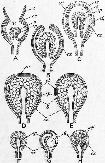
Fig. 41.—Diagrams of the Structure of the Gonophores of various Hydromedusae, based on the figures of G. J. Allman and A. Weismann.
A, “Meconidium” of Gonothyraea. B, Type of Tubularia. C, Type of Garveia, &c. D, Type of Plumularia, Agalma, &c. E, Type of Coryne, Forskalia, &c. F, G, H, Sporosacs. F, With simple spadix. G, With spadix prolonged (Eudendrium). H, With spadix branched (Cordylophora). s.c, Sub-umbral cavity. t, Tentacles. c.c, Circular canal, g, Gonads. sp, Spadix. e.l, Endoderm-lamella. ex, Ex-umbral ectoderm. ect, Ectotheca.
After Allman, Gymnoblastic Hydroids, by permission of the Council of the Ray Society.
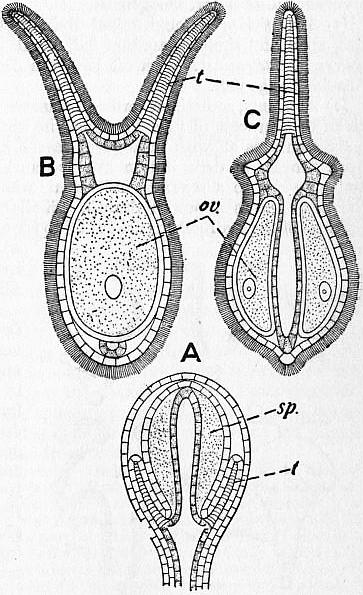
Fig. 42.—Gonophores of Dicoryne conferta.
A, A male gonophore still enclosed in its ectotheca. B and C, Two views of a female gonophore after liberation. t, Tentacles. ov, Ova, two carried on each female gonophore. sp, Testis.
The gonophores of different hydroids differ greatly in structure from one another, and form a series showing degeneration of the medusa-individual, which is gradually stripped, as it were, of its characteristic features of medusan organization and finally reduced to the simplest structure. A very early stage in the degeneration is well exemplified by the so-called “meconidium” of Gonothyraea (fig. 41, A). Here the medusoid, attached by the centre of its ex-umbral surface, has lost its velum and sub-umbral muscles, its sense organs and mouth, though still retaining rudimentary tentacles. The gonads (g) are produced on the manubrium, which has a hollow endodermal axis, termed the spadix (sp.), in open communication with the coenosarc of the polyp-colony and serving for the nutrition of the generative cells. A very similar condition is seen in Tubularia (fig. 41, B), where, however, the tentacles have quite disappeared, and the circular rim formed by the margin of the umbrella has nearly closed over the manubrium leaving only a small aperture through which the embryos emerge. The next step is illustrated by the female gonophores of Cladocoryne, where the radial and ring-canals have become obliterated by coalescence of their walls, so that the entire endoderm of the umbrella is in the condition of the endoderm-lamella. Next the opening of the umbrella closes up completely and disappears, so that the sub-umbral cavity forms a closed space surrounding the manubrium, on which the gonads are developed; such a condition is seen in the male gonophore of Cladocoryne and in Garveia (fig. 41, C), where, however, there is a further complication in the form of an adventitious envelope or ectotheca (ect.) split off from the gonophore as a protective covering, and not present in Cladocoryne. The sub-umbral cavity (s.c.) functions as a brood-space for the developing embryos, which are set free by rupture of the wall. It is evident that the outer envelope of the gonophore represents the ex-umbral ectoderm (ex.), and that the inner ectoderm lining the cavity represents the sub-umbral ectoderm of the free medusa. The next step is the gradual obliteration of the sub-umbral cavity (s.c.) by disappearance of which the sub-umbral ectoderm comes into contact with the ectoderm of the manubrium. Such a type is found in Plumularia and also in Agalma (fig. 41, D); centrally is seen the spadix (sp.), bearing the generative cells (g), and external to these (1) a layer of ectoderm representing the epithelium of the manubrium; (2) the layer of sub-umbral ectoderm; (3) the endoderm-lamella (e.l.); (4) the ex-umbral ectoderm (ex.); and (5) there may or may not be present also an ectotheca. Thus the gonads are covered over by at least four layers of epithelium, and since these are unnecessary, presenting merely obstacles to the dehiscence of the gonads, they gradually undergo reduction. The sub-umbral ectoderm and that covering the manubrium undergo concrescence to form a single layer (fig. 41, E), which finally disappears altogether, and the endoderm-lamella disappears. The gonophore is now reduced to its simplest condition, known as the sporosac (fig. 41, F, G, H), and consists of the spadix bearing the gonads covered by a single layer of ectoderm (ex.), with or without the addition of an ectotheca. It cannot be too strongly emphasized, however, that the sporosac should not be compared simply with the manubrium of the medusa, as is sometimes done. The endodermal spadix (sp.) of the sporosac represents the endoderm of the manubrium; the ectodermal lining of the sporosac (ex.) represents the ex-umbral ectoderm of the medusa; and the intervening layers, together with the sub-umbral cavity, have disappeared. The spadix, as the organ of nutrition for the gonads, may be developed in various ways, being simple (fig. 41, F) or branched (fig. 41, H); in Eudendrium (fig. 41, G) it curls round the single large ovum.
The hydroid Dicoryne is remarkable for the possession of gonophores, which are ciliate and become detached and swim away by means of their cilia. Each such sporosac has two long tentacle-like processes thickly ciliated.
It has been maintained that the gonads of Hydra represent sporosacs or gonophores greatly reduced, with the last traces of medusoid structure completely obliterated. There is, however, no evidence whatever for this, the gonads of Hydra being purely ectodermal structures, while all medusoid gonophores have an endodermal portion. Hydra is, moreover, bisexual, in contrast with what is known of hydroid colonies.
In some Leptomedusae the gonads are formed on the radial canals and form protruding masses resembling sporosacs superficially, but not in structure. Allman, however, regarded this type of gonad as equivalent to a sporosac, and considered the medusa bearing them as a non-sexual organism, a “blastocheme” as he termed it, producing by budding medusoid gonophores. As medusae are known to bud medusae from the radial canals there is nothing impossible in Allman’s theory, but it cannot be said to have received satisfactory proof.
Reproduction and Ontogeny of the Hydromedusae.
Nearly every possible method of reproduction occurs amongst the Hydromedusae. In classifying methods of generation it is usual to make use of the sexual or non-sexual nature of the reproduction as a primary difference, but a more scientific classification is afforded by the distinction between tissue-cells (histocytes) and germinal cells, actual or potential (archaeocytes), amongst the constituent cells of the animal body. In this way we may distinguish, first, vegetative reproduction, the result of discontinuous growth of the tissues and cell-layers of the body as a whole, leading to (1) fission, (2) autotomy, or (3) vegetative budding; secondly, germinal reproduction, the result of the reproductive activity of the archaeocytes or germinal tissue. In germinal reproduction the proliferating cells may be undifferentiated, so-called primitive germ-cells, or they may be differentiated as sexual cells, male or female, i.e. spermatozoa and ova. If the germ-cells are undifferentiated, the offspring may arise from many cells or from a single cell; the first type is (4) germinal budding, the second is (5) sporogony. If the germ-cells are differentiated, the offspring arises by syngamy or sexual union of the ordinary type between an ovum and spermatozoon, so-called fertilization, of the ovum, or by parthenogenesis, i.e. development of an ovum without fertilization. The only one of these possible modes of reproduction not known to occur in Hydromedusae is parthenogenesis.
(1) True fission or longitudinal division of an individual into two equal and similar daughter-individuals is not common but occurs in Gastroblasta, where it has been described in detail by Arnold Lang [30].
(2) Autotomy, sometimes termed transverse fission, is the name given to a process of unequal fission in which a portion of the body separates off with subsequent regeneration. In Tubularia by a process of decapitation the hydranths may separate off and give rise to a separate individual, while the remainder of the body grows a new hydranth. Similarly in Schizocladium portions of the hydrocaulus are cut off to form so-called “spores,” which grow into new individuals (see Allman [1]).
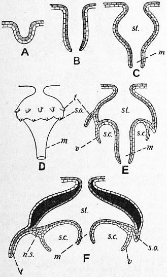
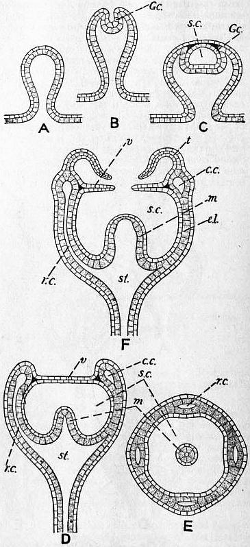
Much modified from C. Chun, “Coelenterata,” in Bronn’s Tierreich. Fig. 44.—Diagrams of Medusa budding with the formation of an entocodon. The endoderm is shaded, the ectoderm left clear.
Fig. 43.—Direct Budding of Cunina.
A, B, C, E, F, In vertical section. D, Sketch of external view. st, Stomach. m, Manubrium. t. Tentacle. s.o, Sense organ. v, Velum. s.c, Sub-umbral cavity. n.s, Nervous system. A, B, C, D, F, Successive stages in vertical section. E, Transverse section of a stage similar to D. Gc, Entocodon. s.c, Cavity of entocodon, forming the future sub-umbral cavity. st, Stomach. r.c, Radial canal. c.c, Circular canal. e.l, Endoderm lamella. m, Manubrium. v, Velum. t, Tentacle.
(3) Vegetative budding is almost universal in the Hydromedusae. By budding is understood the formation of a new individual from a fresh growth of undifferentiated material. It is convenient to distinguish buds that give rise to polyps from those that form medusae.
(a) The Polyp.—The buds that form polyps are very simple in mode of formation. Four stages may be distinguished; the first is a simple outgrowth of both layers, ectoderm and endoderm, containing a prolongation of the coelenteric cavity; in the second stage the tentacles grow out as secondary diverticula from the side of the first outgrowth; in the third stage the mouth is formed as a perforation of the two layers; and, lastly, if the bud is to be separated, it becomes nipped off from the parent polyp and begins a free existence.
(b) The Medusae.—Two types of budding must be distinguished—the direct, so-called, palingenetic type, and the indirect, so-called coenogenetic type.
The direct type of budding is rare, but is seen in Cunina and Millepora. In Cunina there arises, first, a simple outgrowth of both layers, as in a polyp-bud (fig. 43, A); in this the mouth is formed distally as a perforation (B); next the sides of the tube so formed bulge out laterally near the attachment to form the umbrella, while the distal undilated portion of the tube represents the manubrium (C); the umbrella now grows out into a number of lobes or lappets, and the tentacles and tentaculocysts grow out, the former in a notch between two lappets, the latter on the apex of each lappet (D, E); finally, the velum arises as a growth of the ectoderm alone, the whole bud shapes itself, so to speak, and the little medusa is separated off by rupture of the thin stalk connecting it with the parent (F). The direct method of medusa-budding only differs from the polyp-bud by its greater complexity of parts and organs.
The indirect mode of budding (figs. 44, 45) is the commonest method by which medusa-buds are formed. It is marked by the formation in the bud of a characteristic structure termed the entocodon (Knospenkern, Glockenkern).
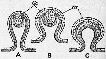
Fig. 45.—Modifications of the method of budding shown in fig. 44, with solid Entocodon (Gc.) and formation of an ectotheca (ect.).
The first stage is a simple hollow outgrowth of both body-layers (fig. 44, A); at the tip of this is formed a thickening of the ectoderm, arising primitively as a hollow ingrowth (fig. 44, B), but more usually as a solid mass of ectoderm-cells (fig. 45, A). The ectodermal ingrowth is the entocodon (Gc.); it bulges into, and pushes down, the endoderm at the apex of the bud, and if solid it soon acquires a cavity (fig. 44, C, s.c.). The cavity of the entocodon increases continually in size, while the endoderm pushes up at the sides of it to form a cup with hollow walls, enclosing but not quite surrounding the entocodon, which remains in contact at its outer side with the ectoderm covering the bud (fig. 44, D, v). The next changes that take place are chiefly in the endoderm-cup (fig. 44, D, E); the cavity between the two walls of the cup becomes reduced by concrescence to form the radial canals (r.c.), ring-canal (c.c.), and endoderm-lamella (e.l., fig. 44, E), and at the same time the base of the cup is thrust upwards to form the manubrium (m), converting the cavity of the entocodon into a space which is crescentic or horse-shoe-like in section. Next tentacles (t, fig. 44, F) grow out from the ring-canal, and the double plate of ectoderm on the distal side of the entocodon becomes perforated, leaving a circular rim composed of two layers of ectoderm, the velum (v) of the medusa. Finally, a mouth is formed by breaking through at the apex of the manubrium, and the now fully-formed medusa becomes separated by rupture of the stalk of the bud and swims away.
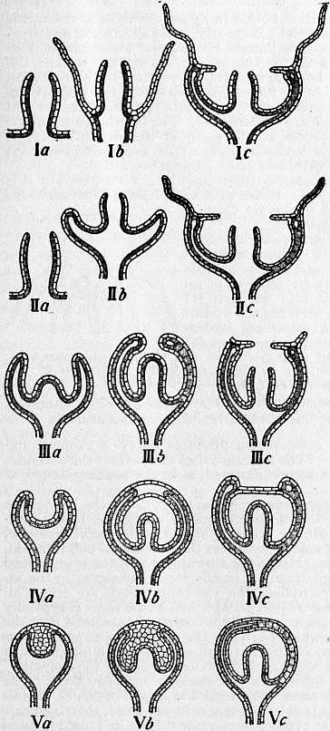
Fig. 46.—Diagrams to show the significance of the Entocodon in Medusa-buds. (Modified from a diagram given by A. Weismann.)
I, Ideally primitive method of budding, in which the mouth is formed first (Ia), next the tentacles (Ib), and lastly the umbrella. II, Method. of Cunina; (a) the mouth arises, next the umbrella (b), and lastly the tentacles (c). III, Hypothetical transition from II to the indirect method with an entocodon; the formation of the manubrium is retarded, that of the umbrella hastened (IIIa, b). IV, a, b, c, budding with an entocodon (cf. fig. 44). V, Budding with a solid entocodon (cf. fig. 45).
If the bud, however, is destined to give rise not to a free medusa, but to a gonophore, the development is similar but becomes arrested at various points, according to the degree to which the gonophore is degenerate. The entocodon is usually formed, proving the medusoid nature of the bud, but in sporosacs the entocodon may be rudimentary or absent altogether. The process of budding as above described may be varied or complicated in various ways; thus a secondary, amnion-like, ectodermal covering or ectotheca (fig. 45, C, ect.) may be formed over all, as in Garveia, &c.; or the entocodon may remain solid and without cavity until after the formation of the manubrium, or may never acquire a cavity at all, as described above for the gonophores.
Phylogenetic Significance of the Entocodon.—It is seen from the foregoing account of medusa-budding that the entocodon is a very important constituent of the bud, furnishing some of the most essential portions of the medusa; its cavity becomes the sub-umbral cavity, and its lining furnishes the ectodermal epithelium of the manubrium and of the sub-umbral cavity as far as the edge of the velum. Hence the entocodon represents a precocious formation of the sub-umbral surface, equivalent to the peristome of the polyp, differentiated in the bud prior to other portions of the organism which must be regarded as antecedent to it in phylogeny.
If the three principal organ-systems of the medusa, namely mouth, tentacles and umbrella, be considered in the light of phylogeny, it is evident that the manubrium bearing the mouth must be the oldest, as representing a common property of all the Coelentera, even of the gastrula embryo of all Enterozoa. Next in order come the tentacles, common to all Cnidaria. The special property of the medusa is the umbrella, distinguishing the medusa at once from other morphological types among the Coelentera. If, therefore, the formation of these three systems of organs took place according to a strictly phylogenetic sequence, we should expect them to appear in the order set forth above (fig. 46, Ia, b, c). The nearest approach to the phylogenetic sequence is seen in the budding of Cunina, where the manubrium and mouth appear first, but the umbrella is formed before the tentacles (fig. 46, IIa, b, c). In the indirect or coenogenetic method of budding, the first two members of the sequence exhibited by Cunina change places, and the umbrella is formed first, the manubrium next, and then the tentacles; the actual mouth-perforation being delayed to the very last (fig. 46, IVa, b, c). Hence the budding of medusae exemplifies very clearly a common phenomenon in development, a phylogenetic series of events completely dislocated in the ontogenetic time-sequence.
The entocodon is to be regarded, therefore, not as primarily an ingrowth of ectoderm, but rather as an upgrowth of both body-layers, in the form of a circular rim (IVa), representing the umbrellar margin; it is comparable to the bulging that forms the umbrella in the direct method of budding, but takes place before a manubrium is formed, and is greatly reduced in size, so as to become a little pit. By a simple modification, the open pit becomes a solid ectodermal ingrowth, just as in Teleostean fishes the hollow medullary tube, or the auditory pit of other vertebrate embryos, is formed at first as a solid cord of cells, which acquires a cavity secondarily. Moreover, the entocodon, however developed, gives rise at first to a closed cavity, representing a closing over of the umbrella, temporary in the bud destined to be a free medusa, but usually permanent in the sessile gonophore. As has been shown above, the closing up of the sub-umbral cavity is one of the earliest degenerative changes in the evolution of the gonophore, and we may regard it as the umbrellar fold taking on a protective function, either temporarily for the bud or permanently for the gonophore.
To sum up, the entocodon is a precocious formation of the umbrella, closing over to protect the organs in the umbrellar cavity. The possession of an entocodon proves the medusa-nature of the bud, and can only be explained on the theory that gonophores are degenerate medusae, and is inexplicable on the opposed view that medusae are derived from gonophores secondarily set free. In the sporosac, however, the medusa-individual has become so degenerate that even the documentary proof, so to speak, of its medusoid nature may have been destroyed, and only circumstantial evidence of its nature can be produced.
4. Germinal Budding.—This method of budding is commonly described as budding from a single body-layer, instead of from both layers. The layer that produces the bud is invariably the ectoderm, i.e. the layer in which, in Hydromedusae, the generative cells are lodged; and in some cases the buds are produced in the exact spot in which later the gonads appear. From these facts, and from those of the sporogony, to be described below, we may regard budding to this type as taking place from the germinal epithelium rather than from ordinary ectoderm.
(a) The Polyp.—Budding from the ectoderm alone has been described by A. Lang [29] in Hydra and other polyps. The tissues of the bud become differentiated into ectoderm and endoderm, and the endoderm of the bud becomes secondarily continuous with that of the parent, but no part of the parental endoderm contributes to the building up of the daughter-polyp. Lang regarded this method of budding as universal in polyps, a notion disproved by O. Seeliger [52] who went to the opposite extreme and regarded the type of budding described by Lang as non-existent. In view, however, both of the statements and figures of Lang and of the facts to be described presently for medusae (Margellium), it is at least theoretically possible that both germinal and vegetative budding may occur in polyps as well as in medusae.
(b) The Medusa.—The clearest instance of germinal budding is furnished by Margellium (Rathkea) octopunctatum, one of the Margelidae. The budding of this medusa has been worked out in detail by Chun (Hydrozoa, [1]), to whom the reader must be referred for the interesting laws of budding regulating the sequence and order of formation of the buds.
The buds of Margellium are produced on the manubrium in each of the four interradii, and they arise from the ectoderm, that is to say, the germinal epithelium, which later gives rise to the gonads. The buds do not appear simultaneously but successively on each of the four sides of the manubrium, thus: and secondary buds may be produced on the medusa-buds before the latter are set free as medusae. Each bud arises as a thickening of the epithelium, which first forms two or three layers (fig. 47, A), and becomes separated into a superficial layer, future ectoderm, surrounding a central mass, future endoderm (fig. 47, B). The ectodermal epithelium on the distal side of the bud becomes thickened, grows inwards, and forms a typical entocodon (fig. 37, D, E, F). The remaining development of the bud is just as described above for the indirect method of medusa-budding (fig. 47, G, H). When the bud is nearly complete, the body-wall of the parent immediately below it becomes perforated, placing the coelenteric cavity of the parent in secondary communication with that of the bud (H), doubtless for the better nutrition of the latter.

Especially noteworthy in the germinal budding of Margellium is the formation of the entocodon, as in the vegetative budding of the indirect type.
5. Sporogony.—This method of reproduction has been described by E. Metchnikoff in Cunina and allied genera. In individuals either of the male or female sex, germ-cells which are quite undifferentiated and neutral in character, become amoeboid, and wander into the endoderm. They divide each into two sister-cells, one of which—the spore—becomes enveloped by the other. The spore-cell multiplies by division, while the enveloping cell is nutrient and protective. The spore cell gives rise to a “spore-larva,” which is set free in the coelenteron and grows into a medusa. Whether sporogony occurs also in the polyp or not remains to be proved.
6. Sexual Reproduction and Embryology.—The ovum of Hydromedusae is usually one of a large number of oögonia, and grows at the expense of its sister-cells. No regular follicle is formed, but the oöcyte absorbs nutriment from the remaining oögonia. In Hydra the oöcyte is a large amoeboid cell, which sends out pseudopodia amongst the oögonia and absorbs nutriment from them. When the oöcyte is full grown, the residual oögonia die off and disintegrate.
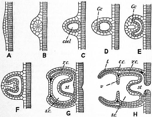
Fig. 47.—Budding from the Ectoderm (germinal epithelium) in Margellium. (After C. Chun.)
A, The epithelium becomes two-layered. B, The lower layer forms a solid mass of cells, which (C) becomes a vesicle, the future endoderm, containing the coelenteric cavity (coel), while the outer layer furnishes the future ectoderm. D, E, F, a thickening of the ectoderm on the distal side of the bud forms an entocodon (Gc). G,H, Formation of the medusae. s.c, Sub-umbral cavity. r.c, Radial canal. st, Stomach, which in H acquires a secondary communication with the digestive cavity of the mother. cc, Circular canal. v, Velum. t, Tentacle.
The spermatogenesis and maturation and fertilization of the germ-cells present nothing out of the common and need not be described here. These processes have been studied in detail by A. Brauer [2] for Hydra.
The general course of the development is described in the article Hydrozoa. We may distinguish the following series of stages: (1) ovum; (2) cleavage, leading to formation of a blastula; (3) formation of an inner mass or parenchyma, the future endoderm, by immigration or delamination, leading to the so-called parenchymula-stage; (4) formation of an archenteric cavity, the future coelenteron, by a splitting of the internal parenchyma, and of a blastopore, the future mouth, by perforation at one pole, leading to the gastrula-stage; (5) the outgrowth of tentacles round the mouth (blastopore), leading to the actinula-stage; and (6) the actinula becomes the polyp or medusa in the manner described elsewhere (see articles Hydrozoa, Polyp and Medusa). This is the full, ideal development, which is always contracted or shortened to a greater or less extent. If the embryo is set free as a free-swimming, so-called planula-larva, in the blastula, parenchymula, or gastrula stage, then a free actinula stage is not found; if, on the other hand, a free actinula occurs, then there is no free planula stage.
The cleavage of the ovum follows two types, both seen in Tubularia (Brauer [3]). In the first, a cleavage follows each nuclear division; in the second, the nuclei multiply by division a number of times, and then the ovum divides into as many blastomeres as there are nuclei present. The result of cleavage in all cases is a typical blastula, which when set free becomes oval and develops a flagellum to each cell, but when not set free, it remains spherical in form and has no flagella.
The germ-layer formation is always by immigration or delamination, never by invagination. When the blastula is oval and free-swimming the inner mass is formed by unipolar immigration from the hinder pole. When the blastula is spherical and not set free, the germ-layer formation is always multipolar, either by immigration or by delamination, i.e. by tangential division of the cells of the blastoderm, as in Geryonia, or by a mixture of immigration and delamination, as in Hydra, Tubularia, &c. The blastopore is formed as a secondary perforation at one spot, in free-swimming forms at the hinder pole. Formation of archenteron and blastopore may, however, be deferred till a later stage (actinula or after).
The actinula stage is usually suppressed or not set free, but it is seen in Tubularia (fig. 48), where it is ambulatory, in Gonionemus (Trachomedusae), and in Cunina (Narcomedusae), where it is parasitic.
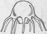
Modified from a plate by L. Agassiz, Contributions to Nat. Hist. U.S., iv.
Fig. 48.—Free Actinula of Tubularia.
In Leptolinae the embryonic development culminates in a polyp, which is usually formed by fixation of a planula (parenchymula), rarely by fixation of an actinula. The planula may fix itself (1) by one end, and then becomes the hydrocaulus and hydranth, while the hydrorhiza grows out from the base; or (2) partly by one side and then gives rise to the hydrorhiza as well as to the other parts of the polyp; or (3) entirely by its side, and then forms a recumbent hydrorhiza from which a polyp appears to be budded as an upgrowth.
In Trachylinae the development produces always a medusa, and there is no polyp-stage. The medusa arises direct from the actinula-stage and there is no entocodon formed, as in the budding described above.
Life-cycles of the Hydromedusae.—The life-cycle of the Leptolinae consists of an alternation of generations in which non-sexual individuals, polyps, produce by budding sexual individuals, medusae, which give rise by the sexual process to the non-sexual polyps again, so completing the cycle. Hence the alternation is of the type termed metagenesis. The Leptolinae are chiefly forms belonging to the inshore fauna. The Trachylinae, on the other hand, are above all oceanic forms, and have no polyp-stage, and hence there is typically no alternation in their life-cycle. It is commonly assumed that the Trachylinae are forms which have lost the alternation of generations possessed by them ancestrally, through secondary simplification of the life-cycle. Hence the Trachylinae are termed “hypogenetic” medusae to contrast them with the metagenetic Leptolinae. The whole question has, however, been argued at length by W. K. Brooks [4], who adduces strong evidence for a contrary view, that is to say, for regarding the direct type of development seen in Trachylinae as more primitive, and the metagenesis seen in Leptolinae as a secondary complication introduced into the life-cycle by the acquisition of larval budding. The polyp is regarded, on this view, as a form phylogenetically older than the medusa, in short, as nothing more than a sessile actinula. In Trachylinae the polyp-stage is passed over, and is represented only by the actinula as a transitory embryonic stage. In Leptolinae the actinula becomes the sessile polyp which has acquired the power of budding and producing individuals either of its own or of a higher rank; it represents a persistent larval stage and remains in a sexually immature condition as a neutral individual, sex being an attribute only of the final stage in the development, namely the medusa. The polyp of the Leptolinae has reached the limit of its individual development and is incapable of becoming itself a medusa, but only produces medusa-buds; hence a true alternation of generations is produced. In Trachylinae also the beginnings of a similar metagenesis can be found. Thus in Cunina octonaria, the ovum develops into an actinula which buds daughter-actinulae; all of them, both parent and offspring, develop into medusae, so that there is no alternation of generations, but only larval multiplication. In Cunina parasitica, however, the ovum develops into an actinula, which buds actinulae as before, but only the daughter-actinulae develop into medusae, while the original, parent-actinula dies off; here, therefore, larval budding has led to a true alternation of generations. In Gonionemus the actinula becomes fixed and polyp-like, and reproduces by budding, so that here also an alternation of generations may occur. In the Leptolinae we must first substitute polyp for actinula, and then a condition is found which can be compared to the case of Cunina parasitica or Gonionemus, if we suppose that neither the parent-actinula (i.e. founder-polyp) nor its offspring by budding (polyps of the colony) have the power of becoming medusae, but only of producing medusae by budding. For further arguments and illustrations the reader must be referred to Brooks’s most interesting memoir. The whole theory is one most intimately connected with the question of the relation between polyp and medusa, to be discussed presently. It will be seen elsewhere, however, that whatever view may be held as to the origin of metagenesis in Hydromedusae, in the case of Scyphomedusae (q.v.) no other view is possible than that the alternation of generations is the direct result of larval proliferation.
To complete our survey of life-cycles in the Hydromedusae it is necessary to add a few words about the position of Hydra and its allies. If we accept the view that Hydra is a true sexual polyp, and that its gonads are not gonophores (i.e. medusa-buds) in the extreme of degeneration, then it follows from Brooks’s theory that Hydra must be descended from an archaic form in which the medusan type of organization had not yet been evolved. Hydra must, in short, be a living representative of the ancestor of which the actinula-stage is a transient reminiscence in the development of higher forms. It may be pointed out in this connexion that the fixation of Hydra is only temporary, and that the animal is able at all times to detach itself, to move to a new situation, and to fix itself again. There is no difficulty whatever in regarding Hydra as bearing the same relation to the actinula-stage of other Hydromedusae that a Rotifer bears to a trochophore-larva or a fish to a tadpole.
The Relation of Polyp and Medusa.—Many views have been put forward as to the morphological relationship between the two types of person in the Hydromedusae. For the most part, polyp and medusa have been regarded as modifications of a common type, a view supported by the existence, among Scyphomedusae (q.v.), of sessile polyp-like medusae (Lucernaria, &c.). R. Leuckart in 1848 compared medusae in general terms to flattened polyps. G. J. Allman [1] put forward a more detailed view, which was as follows. In some polyps the tentacles are webbed at the base, and it was supposed that a medusa was a polyp of this kind set free, the umbrella being a greatly developed web or membrane extending between the tentacles. A very different theory was enunciated by E. Metchnikoff. In some hydroids the founder-polyp, developed from a planula after fixation, throws out numerous outgrowths from the base to form the hydrorhiza; these outgrowths may be radially arranged so as to form by contact or coalescence a flat plate. Mechnikov considered the plate thus formed at the base of the polyp as equivalent to the umbrella, and the body of the polyp as equivalent to the manubrium, of the medusa; on this view the marginal tentacles almost invariably present in medusae are new formations, and the tentacles of the polyp are represented in the medusa by the oral arms which may occur round the mouth, and which sometimes, e.g. in Margelidae, have the appearance and structure of tentacles. Apart from the weighty arguments which the development furnishes against the theories of Allman and Mechnikov, it may be pointed out that neither hypothesis gives a satisfactory explanation of a structure universally present in medusae of whatever class, namely the endoderm-lamella, discovered by the brothers O. and R. Hertwig. It would be necessary to regard this structure as a secondary extension of the endoderm in the tentacle-web, on Allman’s theory, or between the outgrowths of the hydrorhiza, on Mechnikov’s hypothesis. The development, on the contrary, shows unequivocally that the endoderm-lamella arises as a local coalescence of the endodermal linings of a primitively extensive gastral space.