VOLUME XII SLICE VII Gyantse to Hallel (continued 2)
Reading edition
The roots of many conifers possess a narrow band of primary xylem-tracheids with a group of narrow spiral protoxylem-elements at each end (diarch). A striking feature in the roots of several genera, excluding the Abietineae, is the occurrence Anatomy. of thick and somewhat irregular bands of thickening on the cell-walls of the cortical layer next to the endodermis. These bands, which may serve to strengthen the central cylinder, have been compared with the netting surrounding the delicate wall of an inflated balloon. It is not always easy to distinguish a root from a stem; in some cases (e.g. Sequoia) the primary tetrarch structure is easily identified in the centre of an old root, but in other cases the primary elements are very difficult to recognize. The sudden termination of the secondary tracheids against the pith-cells may afford evidence of root-structure as distinct from stem-structure, in which the radial rows of secondary tracheids pass into the irregularly-arranged primary elements next the pith. The annual rings in a root are often less clearly marked than in the stem, and the xylem-elements are frequently larger and thinner. The primary vascular bundles in a young conifer stem are collateral, and, like those of a Dicotyledon, they are arranged in a circle round a central pith and enclosed by a common endodermis. It is in the nature of the secondary xylem that the Coniferales are most readily distinguished from the Dicotyledons and Cycadaceae; the wood is homogeneous in structure, consisting almost entirely of tracheids with circular or polygonal bordered pits on the radial walls, more particularly in the late summer wood. In many genera xylem-parenchyma is present, but never in great abundance. A few Dicotyledons, e.g. Drimys (Magnoliaceae) closely resemble conifers in the homogeneous character of the wood, but in most cases the presence of large spring vessels, wood-fibres and abundant parenchyma affords an obvious distinguishing feature.
The abundance of petrified coniferous wood in rocks of various ages has led many botanists to investigate the structure of modern genera with a view to determining how far anatomical characters may be used as evidence of generic distinctions. There are a few well-marked types of wood which serve as convenient standards of comparison, but these cannot be used except in a few cases to distinguish individual genera. The genus Pinus serves as an illustration of wood of a distinct type characterized by the absence of xylem-parenchyma, except such as is associated with the numerous resin-canals that occur abundantly in the wood, cortex and medullary rays; the medullary rays are composed of parenchyma and of horizontal tracheids with irregular ingrowths from their walls. In a radial section of a pine stem each ray is seen to consist in the median part of a few rows of parenchymatous cells with large oval simple pits in their walls, accompanied above and below by horizontal tracheids with bordered pits. The pits in the radial walls of the ordinary xylem-tracheids occur in a single row or in a double row, of which the pits are not in contact, and those of the two rows are placed on the same level. The medullary rays usually consist of a single tier of cells, but in the Pinus type of wood broader medullary rays also occur and are traversed by horizontal resin-canals. In the wood of Cypressus, Cedrus, Abies and several other genera, parenchymatous cells occur in association with the xylem-tracheids and take the place of the resin-canals of other types. In the Araucarian type of wood (Araucaria and Agathis) the bordered pits, which occur in two or three rows on the radial walls of the tracheids, are in mutual contact and polygonal in shape, the pits of the different rows are alternate and not on the same level; in this type of wood the annual rings are often much less distinct than in Cupressus, Pinus and other genera. In Taxus, Torreya (California and the Far East) and Cephalotaxus the absence of resin-canals and the presence of spiral thickening-bands on the tracheids constitute well-marked characteristics. An examination of the wood of branches, stems and roots of the same species or individual usually reveals a fairly wide variation in some of the characters, such as the abundance and size of the medullary rays, the size and arrangement of pits, the presence of wood-parenchyma—characters to which undue importance has often been attached in systematic anatomical work. The phloem consists of sieve-tubes, with pitted areas on the lateral as well as on the inclined terminal walls, phloem-parenchyma and, in some genera, fibres. In the Abietineae the phloem consists of parenchyma and sieve-tubes only, but in most other forms tangential rows of fibres occur in regular alternation with the parenchyma and sieve-tubes. The characteristic companion-cells of Angiosperms are represented by phloem-parenchyma cells with albuminous contents; other parenchymatous elements of the bast contain starch or crystals of calcium oxalate. When tracheids occur in the medullary rays of the xylem these are replaced in the phloem-region by irregular parenchymatous cells known as albuminous cells. Resin-canals, which occur abundantly in the xylem, phloem or cortex, are not found in the wood of the yew. Cephalotaxus (Taxeae) is also peculiar in having resin-canals in the pith (cf. Ginkgo). One form of Cephalotaxus is characterized by the presence of short tracheids in the pith, in shape like ordinary parenchyma, but in the possession of bordered pits and lignified walls agreeing with ordinary xylem-tracheids; it is probable that these short tracheids serve as reservoirs for storing rather than for conducting water. The vascular bundle entering the stem from a leaf with a single vein passes by a more or less direct course into the central cylinder of the stem, and does not assume the girdle-like form characteristic of the cycadean leaf-trace. In species of which the leaves have more than one vein (e.g. Araucaria imbricata, &c.) the leaf-trace leaves the stele of the stem as a single bundle which splits up into several strands in its course through the cortex. In the wood of some conifers, e.g. Araucaria, the leaf-traces persist for a considerable time, perhaps indefinitely, and may be seen in tangential sections of the wood of old stems. The leaf-trace in the Coniferales is simple in its course through the stem, differing in this respect from the double leaf-trace of Ginkgo. A detailed account of the anatomical characters of conifers has been published by Professor D. P. Penhallow of Montreal and Dr. Gothan of Berlin which will be found useful for diagnostic purposes. The characters of leaves most useful for diagnostic purposes are the position of the stomata, the presence and arrangement of resin-canals, the structure of the mesophyll and vascular bundles. The presence of hypodermal fibres is another feature worthy of note, but the occurrence of these elements is too closely connected with external conditions to be of much systematic value. A pine needle grown in continuous light differs from one grown under ordinary conditions in the absence of hypodermal fibres, in the absence of the characteristic infoldings of the mesophyll cell-walls, in the smaller size of the resin-canals, &c. The endodermis in Pinus, Picea and many other genera is usually a well-defined layer of cells enclosing the vascular bundles, and separated from them by a tissue consisting in part of ordinary parenchyma and to some extent of isodiametric tracheids; but this tissue, usually spoken of as the pericycle, is in direct continuity with other stem-tissues as well as the pericycle. The occurrence of short tracheids in close proximity to the veins is a characteristic of coniferous leaves; these elements assume two distinct forms—(1) the short isodiametric tracheids (transfusion-tracheids) closely associated with the veins; (2) longer tracheids extending across the mesophyll at right angles to the veins, and no doubt functioning as representatives of lateral veins. It has been suggested that transfusion-tracheids represent, in part at least, the centripetal xylem, which forms a distinctive feature of cycadean leaf-bundles; these short tracheids form conspicuous groups laterally attached to the veins in Cunninghamia, abundantly represented in a similar position in the leaves of Sequoia, and scattered through the so-called pericycle in Pinus, Picea, &c. It is of interest to note the occurrence of precisely similar elements in the mesophyll of Lepidodendron leaves. An anatomical peculiarity in the veins of Pinus and several other genera is the continuity of the medullary rays, which extend as continuous plates from one end of the leaf to the other. The mesophyll of Pinus and Cedrus is characterized by its homogeneous character and by the presence of infoldings of the cell-walls. In many leaves, e.g. Abies, Tsuga, Larix, &c., the mesophyll is heterogeneous, consisting of palisade and spongy parenchyma. In the leaves of Araucaria imbricata, in which palisade-tissue occurs in both the upper and lower part of the mesophyll, the resin-canals are placed between the veins; in some species of Podocarpus (sect. Nageia) a canal occurs below each vein; in Tsuga, Torreya, Cephalotaxus, Sequoia, &c., a single canal occurs below the midrib; in Larix, Abies, &c., two canals run through the leaf parallel to the margins. The stomata are frequently arranged in rows, their position being marked by two white bands of wax on the leaf-surface.
The chief home of the Coniferales is in the northern hemisphere, where certain species occasionally extend into the Arctic circle and penetrate beyond the northern limit of dicotyledonous trees. Wide areas are often exclusively occupied by Distribution. conifers, which give the landscape a sombre aspect, suggesting a comparison with the forest vegetation of the Coal period. South of the tree-limit a belt of conifers stretches across north Europe, Siberia and Canada. In northern Europe this belt is characterized by such species as Picea excelsa (spruce), which extends south to the mountains of the Mediterranean region; Pinus sylvestris (Scottish fir), reaching from the far north to western Spain, Persia and Asia Minor; Juniperus communis, &c. In north Siberia Pinus Cembra (Cembra or Arolla Pine) has a wide range; also Abies sibirica (Siberian silver fir), Larix sibirica and Juniperus Sabina (savin). In the North American area Picea alba, P. nigra, Larix americana, Abies balsamea (balsam fir), Tsuga canadensis (hemlock spruce), Pinus Strobus (Weymouth pine), Thuja occidentalis (white cedar), Taxus canadensis are characteristic species. In the Mediterranean region occur Cupressus sempervirens, Pinus Pinea (stone pine), species of juniper, Cedrus atlantica, C. Libani, Callitris quadrivalvis, Pinus montana, &c. Several conifers of economic importance are abundant on the Atlantic side of North America—Juniperus virginiana (red cedar, used in the manufacture of lead pencils, and extending as far south as Florida), Taxodium distichum (swamp cypress), Pinus rigida (pitch pine), P. mitis (yellow pine), P. taeda, P. palustris, &c. On the west side of the American continent conifers play a still more striking rôle; among them are Chamaecyparis nutkaensis, Picea sitchensis, Libocedrus decurrens, Pseudotsuga Douglasii (Douglas fir), Sequoia sempervirens, S. gigantea (the only two surviving species of this generic type are now confined to a few localities in California, but were formerly widely spread in Europe and elsewhere), Pinus Coulteri, P. Lambertiana, &c. Farther south, a few representatives of such genera as Abies, Cupressus, Pinus and juniper are found in the Mexican Highlands, tropical America and the West Indies. In the far East conifers are richly represented; among them occur Pinus densiflora, Cryptomeria japonica, Cephalotaxus, species of Abies, Larix, Thujopsis, Sciadopitys verticillata, Pseudolarix Kaempferi, &c. In the Himalaya occur Cedrus deodara, Taxus, species of Cupressus, Pinus excelsa, Abies Webbiana, &c. The continent of Africa is singularly poor in conifers. Cedrus atlantica, a variety of Abies Pinsapo, Juniperus thurifera, Callitris quadrivalvis, occur in the north-west region, which may be regarded as the southern limit of the Mediterranean region. The greater part of Africa north of the equator is without any representatives of the conifers; Juniperus procera flourishes in Somaliland and on the mountains of Abyssinia; a species of Podocarpus occurs on the Cameroon mountains, and P. milanjiana is widely distributed in east tropical Africa. Widdringtonia Whytei, a species closely allied to W. juniperoides of the Cedarberg mountains of Cape Colony, is recorded from Nyassaland and from N.E. Rhodesia; while a third species, W. cupressoides, occurs in Cape Colony. Podocarpus elongata and P. Thunbergii (yellow wood) form the principal timber trees in the belt of forest which stretches from the coast mountains of Cape Colony to the north-east of the Transvaal. Libocedrus tetragona, Fitzroya patagonica, Araucaria brasiliensis, A. imbricata, Saxegothaea and others are met with in the Andes and other regions in South America. Athrotaxis and Microcachrys are characteristic Australian types. Phyllocladus occurs also in New Zealand, and species of Dacrydium, Araucaria, Agathis and Podocarpus are represented in Australia, New Zealand and the Malay regions.
Gnetales.—These are trees or shrubs with simple leaves. The flowers are dioecious, rarely monoecious, provided with one or two perianths. The wood is characterized by the presence of vessels in addition to tracheids. There are no resin-canals. The three existing genera, usually spoken of as members of the Gnetales, differ from one another more than is consistent with their inclusion in a single family; we may therefore better express their diverse characters by regarding them as types of three separate families—(1) Ephedroideae, genus Ephedra; (2) Welwitschioideae, genus Welwitschia; (3) Gnetoideae, genus Gnetum. Our knowledge of the Gnetales leaves much to be desired, but such facts as we possess would seem to indicate that this group is of special importance as foreshadowing, more than any other Gymnosperms, the Angiospermous type. In the more heterogeneous structure of the wood and in the possession of true vessels the Gnetales agree closely with the higher flowering plants. It is of interest to note that the leaves of Gnetum, while typically Dicotyledonous in appearance, possess a Gymnospermous character in the continuous and plate-like medullary rays of their vascular bundles. The presence of a perianth is a feature suggestive of an approach to the floral structure of Angiosperms; the prolongation of the integument furnishes the flowers with a substitute for a stigma and style. The genus Ephedra, with its prothallus and archegonia, which are similar to those of other Gymnosperms, may be safely regarded as the most primitive of the Gnetales. In Welwitschia also the megaspore is filled with prothallus-tissue, but single egg-cells take the place of archegonia. In certain species of Gnetum described by Karsten the megaspore contains a peripheral layer of protoplasm, in which scattered nuclei represent the female reproductive cells; in Gnetum Gnemon a similar state of things exists in the upper half of the megaspore, while the lower half agrees with the megaspore of Welwitschia in being full of prothallus-tissue, which serves merely as a reservoir of food. Lotsy has described the occurrence of special cells at the apex of the prothallus of Gnetum Gnemon, which he regards as imperfect archegonia (fig. 17, C, a); he suggests they may represent vestigial structures pointing back to some ancestral form beyond the limits of the present group. The Gnetales probably had a separate origin from the other Gymnosperms; they carry us nearer to the Angiosperms, but we have as yet no satisfactory evidence that they represent a stage in the direct line of Angiospermic evolution. It is not improbable that the three genera of this ancient phylum survive as types of a blindly-ending branch of the Gymnosperms; but be that as it may, it is in the Gnetales more than in any other Gymnosperms that we find features which help us to obtain a dim prospect of the lines along which the Angiosperms may have been evolved.
Ephedra.—This genus is the only member of the Gnetales represented in Europe. Its species, which are characteristic of warm temperate latitudes, are usually much-branched shrubs. The finer branches are green, and bear a close resemblance to the stems of Equisetum and to the slender twigs of Casuarina; the surface of the long internodes is marked by fine longitudinal ribs, and at the nodes are borne pairs of inconspicuous scale-leaves. The flowers are small, and borne on axillary shoots. A single male flower consists of an axis enclosed at the base by an inconspicuous perianth formed of two concrescent leaves and terminating in two, or as many as eight, shortly stalked or sessile anthers. The female flower is enveloped in a closely fitting sac-like investment, which must be regarded as a perianth; within this is an orthotropous ovule surrounded by a single integument prolonged upwards as a beak-like micropyle. The flower may be described as a bud bearing a pair of leaves which become fused and constitute a perianth, the apex of the shoot forming an ovule. In function the perianth may be compared with a unilocular ovary containing a single ovule; the projecting integument, which at the time of pollination secretes a drop of liquid, serves the same purpose as the style and stigma of an angiosperm. The megaspore is filled with tissue as in typical Gymnosperms, and from some of the superficial cells 3 to 5 archegonia are developed, characterized by long multicellular necks. The archegonia are separated from one another, as in Pinus, by some of the prothallus-tissue, and the cells next the egg-cells (tapetal layer) contribute food-material to their development. After fertilization, some of the uppermost bracts below each flower become red and fleshy; the perianth develops into a woody shell, while the integument remains membranous. In some species of Ephedra, e.g. E. altissima, the fertilized eggs grow into tubular proembryos, from the tip of each of which embryos begin to be developed, but one only comes to maturity. In Ephedra helvetica, as described by Jaccard, no proembryo or suspensor is formed; but the most vigorous fertilized egg, after undergoing several divisions, becomes attached to a tissue, termed the columella, which serves the purpose of a primary suspensor; the columella appears to be formed by the lignification of certain cells in the central region of the embryo-sac. At a later stage some of the cells in the upper (micropylar) end of the embryo divide and undergo considerable elongation, serving the purpose of a secondary suspensor. The secondary wood of Ephedra consists of tracheids, vessels and parenchyma; the vessels are characterized by their wide lumen and by the large simple or slightly-bordered pits on their oblique end-walls.
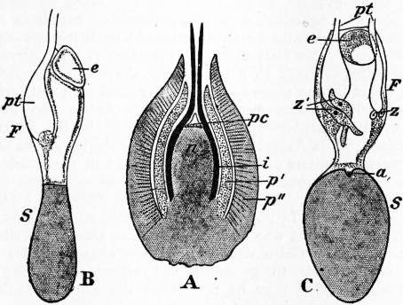
Fig. 17.—Gnetum Gnemon. (After Lotsy.)
A, Female Flower. a, Imperfect Archegonia. n, Nucellus. e, Partially developed Megaspore. pc, Pollen-chamber. F, Fertile half. i, Integument. S, Sterile half. p′, Inner Perianth. pt, Pollen-tube. p″, Outer Perianth. z, Zygote. B, C, Megaspore. z′, Prothallus. a, Imperfect Archegonia. e, Partially developed Megaspore. F, Fertile half. S, Sterile half. pt, Pollen-tube. z, Zygote. z′, Prothallus.
Gnetum.—This genus is represented by several species, most of which are climbing plants, both in tropical America and in warm regions of the Old World. The leaves, which are borne in pairs at the tumid nodes, are oval in form and have a Dicotyledonous type of venation. The male and female inflorescences have the form of simple or paniculate spikes. The spike of an inflorescence bears whorls of flowers at each node in the axils of concrescent bracts accompanied by numerous sterile hairs (paraphyses); in a male inflorescence numerous flowers occur at each node, while in a female inflorescence the number of flowers at each node is much smaller. A male flower consists of a single angular perianth, through the open apex of which the flower-axis projects as a slender column terminating in two anthers. The female flowers, which are more complex in structure, are of two types, complete and incomplete; the latter occur in association with male flowers in a male inflorescence. A complete female flower consists of a nucellus (fig. 17, A, n), surrounded by a single integument (fig. 17, A, i), prolonged upwards as a narrow tube and succeeded by an inner and an outer perianth (fig. 17, A, p′ and p″). The whole flower may be looked upon as an adventitious bud bearing two pairs of leaves; each pair becomes concrescent and forms a perianth, the apex of the shoot being converted into an orthotropous ovule. The incomplete female flowers are characterized by the almost complete suppression of the inner perianth. Several embryo-sacs (megaspores) are present in the nucellus of a young ovule, but one only attains full size, the smaller and partially developed megaspores (fig. 17, B and C, e) being usually found in close association with the surviving and fully-grown megaspore. In Gnetum Gnemon, as described by Lotsy, a mature embryo-sac contains in the upper part a large central vacuole and a peripheral layer of protoplasm, including several nuclei, which take the place of the archegonia of Ephedra; the lower part of the embryo-sac, separated from the upper by a constriction, is full of parenchyma. The upper part of the megaspore may be spoken of as the fertile half (fig. 17, B and C, F) and the lower part, which serves only as food-reservoir for the growing embryo, may be termed the sterile half (fig. 17, B and C, S). (Coulter, Bot. Gazette, xlvi., 1908, regards this tissue as belonging to the nucellus.) At the time of pollination the long tubular integument secretes a drop of fluid at its apex, which holds the pollen-grains, brought by the wind, or possibly to some extent by insect agency, and by evaporation these are drawn on to the top of the nucellus, where partial disorganization of the cells has given rise to an irregular pollen-chamber (fig. 17, A, pc). The pollen-tube, containing two generative and one vegetative nucleus, pierces the wall of the megaspore and then becomes swollen (fig. 17, B and C, pt); finally the two generative nuclei pass out of the tube and fuse with two of the nuclei in the fertile half of the megaspore. As the result of fertilization, the fertilized nuclei of the megaspore become surrounded by a cell-wall, and constitute zygotes, which may attach themselves either to the wall of the megaspore or to the end of a pollen-tube (fig. 17, C, z and z′); they then grow into long tubes or proembryos, which make their way towards the prothallus (C, z′), and eventually embryos are formed from the ends of the proembryo tubes. One embryo only comes to maturity. The embryo of Gnetum forms an out-growth from the hypocotyl, which serves as a feeder and draws nourishment from the prothallus. The fleshy outer portion of the seed is formed from the outer perianth, the woody shell being derived from the inner perianth. The climbing species of Gnetum are characterized by the production of several concentric cylinders of secondary wood and bast, the additional cambium-rings being products of the pericycle, as in Cycas and Macrozamia. The structure of the wood agrees in the main with that of Ephedra.
Welwitschia (Tumboa).—This is by far the most remarkable member of the Gnetales, both as regards habit and the form of its flowers. In a supplement to the systematic work of Engler and Prantl the well-known name Welwitschia, instituted by Hooker in 1864 in honour of Welwitsch, the discoverer of the plant, is superseded by that of Tumboa, originally suggested by Welwitsch. The genus is confined to certain localities in Damaraland and adjoining territory on the west coast of tropical South Africa. A well-grown plant projects less than a foot above the surface of the ground; the stem, which may have a circumference of more than 12 ft., terminates in a depressed crown resembling a circular table with a median groove across the centre and prominent broad ridges concentric with the margin. The thick tuberous stem becomes rapidly narrower, and passes gradually downwards into a tap-root. A pair of small strap-shaped leaves succeed the two cotyledons of the seedling, and persist as the only leaves during the life of the plant; they retain the power of growth in their basal portion, which is sunk in a narrow groove near the edge of the crown, and the tough lamina, 6 ft. in length, becomes split into narrow strap-shaped or thong-like strips which trail on the ground. Numerous circular pits occur on the concentric ridges of the depressed and wrinkled crown, marking the position of former inflorescences borne in the leaf-axil at different stages in the growth of the plant. An inflorescence has the form of a dichotomously-branched cyme bearing small erect cones; those containing the female flowers attain the size of a fir-cone, and are scarlet in colour. Each cone consists of an axis, on which numerous broad and thin bracts are arranged in regular rows; in the axil of each bract occurs a single flower; a male flower is enclosed by two opposite pairs of leaves, forming a perianth surrounding a central sterile ovule encircled by a ring of stamens united below, but free distally as short filaments, each of which terminates in a trilocular anther. The integument of the sterile ovule is prolonged above the nucellus as a spirally-twisted tube expanded at its apex into a flat stigma-like organ. A complete and functional female flower consists of a single ovule with two integuments, the inner of which is prolonged into a narrow tubular micropyle, like that in the flower of Gnetum. The megaspore of Welwitschia is filled with a prothallus-tissue before fertilization, and some of the prothallus-cells function as egg-cells; these grow upwards as long tubes into the apical region of the nucellus, where they come into contact with the pollen-tubes. After the egg-cells have been fertilized by the non-motile male cells they grow into tubular proembryos, producing terminal embryos. The stem is traversed by numerous collateral bundles, which have a limited growth, and are constantly replaced by new bundles developed from strands of secondary meristem. One of the best-known anatomical characteristics of the genus is the occurrence of numerous spindle-shaped or branched fibres with enormously-thickened walls studded with crystals of calcium oxalate. Additional information has been published by Professor Pearson of Cape Town based on material collected in Damaraland in 1904 and 1906-1907. In 1906 he gave an account of the early stages of development of the male and female organs and, among other interesting statements in regard to the general biology of Welwitschia, he expressed the opinion that, as Hooker suspected, the ovules are pollinated by insect-agency. In a later paper Pearson considerably extended our knowledge of the reproduction and gametophyte of this genus.
Authorities.—General: Bentham and Hooker, Genera Plantarum (London, 1862-1883); Engler and Prantl, Die natürlichen Pflanzenfamilien (Leipzig, 1889 and 1897); Strasburger, Die Coniferen und Gnetaceen (Jena, 1872); Die Angiospermen und die Gymnospermen (Jena, 1879); Histologische Beiträge, iv. (Jena, 1892); Coulter and Chamberlain, Morphology of Spermatophytes (New York, 1901); Rendle, The Classification of Flowering Plants, vol. i. (Cambridge, 1904); “The Origin of Gymnosperms” (A discussion at the Linnean Society; New Phytologist, vol. v., 1906). Cycadales: Mettenius, “Beiträge zur Anatomie der Cycadeen,” Abh. k. sächs. Ges. Wiss. (1860); Treub, “Recherches sur les Cycadées,” Ann. Bot. Jard. Buitenzorg, ii. (1884); Solms-Laubach, “Die Sprossfolge der Stangeria, &c.,” Bot. Zeit. xlviii. (1896); Worsdell, “Anatomy of Macrozamia,” Ann. Bot. x. (1896) (also papers by the same author, Ann. Bot., 1898, Trans. Linn. Soc. v., 1900); Scott, “The Anatomical Characters presented by the Peduncle of Cycadaceae,” Ann. Bot. xi. (1897); Lang, “Studies in the Development and Morphology of Cycadean Sporangia, No. I.,” Ann. Bot. xi. (1897); No. II., Ann. Bot. xiv. (1900); Webber, “Development of the Antherozoids of Zamia,” Bot. Gaz. (1897); Ikeno, “Untersuchungen über die Entwickelung, &c., bei Cycas revoluta,” Journ. Coll. Sci. Japan, xii. (1898); Wieland, “American Fossil Cycads,” Carnegie Institution Publication (1906); Stopes, “Beiträge zur Kenntnis der Fortpflanzungsorgane der Cycadeen,” Flora (1904); Caldwell, “Microcycas Calocoma,” Bot. Gaz. xliv., 1907 (also papers on this and other Cycads in the Bot. Gaz., 1907-1909); Matte, Recherches sur l’appareil libéro-ligneux des Cycadacées (Caen, 1904). Ginkgoales: Hirase, “Études sur la fécondation, &c., de Ginkgo biloba,” Journ. Coll. Sci. Japan, xii. (1898); Seward and Gowan, “Ginkgo biloba,” Ann. Bot. xiv. (1900) (with bibliography); Ikeno, “Contribution à l’étude de la fécondation chez le Ginkgo biloba,” Ann. Sci. Nat. xiii. (1901); Sprecher, Le Ginkgo biloba (Geneva, 1907). Coniferales: “Report of the Conifer Conference” (1891) Journ. R. Hort. Soc. xiv. (1892); Beissner, Handbuch der Nadelholzkunde (Berlin, 1891); Masters, “Comparative Morphology of the Coniferae,” Journ. Linn. Soc. xxvii. (1891); ibid. (1896), &c.; Penhallow, “The Generic Characters of the North American Taxaceae and Coniferae,” Proc. and Trans. R. Soc. Canada, ii. (1896); Blackman, “Fertilization in Pinus sylvestris,” Phil. Trans. (1898) (with bibliography); Worsdell, “Structure of the Female Flowers in Conifers,” Ann. Bot. xiv. (1900) (with bibliography); ibid. (1899); Veitch, Manual of the Coniferae (London, 1900); Penhallow, “Anatomy of North American Coniferales,” American Naturalist (1904); Engler and Pilger, Das Pflanzenreich, Taxaceae (1903); Seward and Ford, “The Araucarieae, recent and extinct,” Phil. Trans. R. Soc. (1906) (with bibliography); Lawson, “Sequoia sempervirens,” Annals of Botany (1904); Robertson, “Torreya Californica,” New Phytologist (1904); Coker, “Gametophyte and Embryo of Taxodium,” Bot. Gazette (1903); E. C. Jeffrey, “The Comparative Anatomy and Phylogeny of the Coniferales, part i. The Genus Sequoia,” Mem. Boston Nat. Hist. Soc. v. No. 10 (1903); Gothan, “Zur Anatomie lebender und fossiler Gymnospermen-Hölzer,” K. Preuss. Geol. Landes. (Berlin, 1905) (for more recent papers, see Ann. Bot., New Phytologist, and Bot. Gazette, 1906-1909). Gnetales: Hooker, “On Welwitschia mirabilis.” Trans. Linn. Soc. xxiv. (1864); Bower, “Germination, &., in Gnetum,” Journ. Mic. Sci. xxii. (1882); ibid. (1881); Jaccard, “Recherches embryologiques sur l’Ephedra helvetica,” Diss. Inaug. Lausanne (1894); Karsten, “Zur Entwickelungsgeschichte der Gattung Gnetum,” Cohn’s Beiträge, vi. (1893); Lotsy, “Contributions to the Life-History of the genus Gnetum,” Ann. Bot. Jard. Buitenzorg, xvi. (1899); Land, “Ephedra trifurca,” Bot. Gazette (1904); Pearson, “Some observations on Welwitschia mirabilis,” Phil. Trans. R. Soc. (1906); Pearson, “Further Observations on Welwitschia,” Phil. Trans. R. Soc. vol. 200 (1909).
(A. C. Se.)
GYMNOSTOMACEAE, an order of Ciliate Infusoria (q.v.), characterized by a closed mouth, which only opens to swallow food actively, and body cilia forming a general or partial investment (rarely represented by a girdle of membranellae), but not differentiated in different regions. With the Aspirotrochaceae (q.v.) it formed the Holotricha of Stein.
GYMPIE, a mining town of March county, Queensland, Australia, 107 m. N. of Brisbane, and 61 m. S. of Maryborough by rail. Pop. (1901) 11,959. Numerous gold mines are worked in the district, which also abounds in copper, silver, antimony, cinnabar, bismuth and nickel. Extensive undeveloped coal-beds lie 40 m. N. at Miva. Gympie became a municipality in 1880.
GYNAECEUM (Gr. γυναικεῖον, from γυνή, woman), that part in a Greek house which was specially reserved for the women, in contradistinction to the “andron,” the men’s quarters; in the larger houses there was an open court with peristyles round, and as a rule all the rooms were on the same level; in smaller houses the servants were placed in an upper storey, and this seems to have been the case to a certain extent in the Homeric house of the Odyssey. “Gynaeconitis” is the term given by Procopius to the space reserved for women in the Eastern Church, and this separation of the sexes was maintained in the early Christian churches where there were separate entrances and accommodation for the men and women, the latter being placed in the triforium gallery, or, in its absence, either on one side of the church, the men being on the other, or occasionally in the aisles, the nave being occupied by the men.
GYNAECOLOGY (from Gr. γυνή, γυναικός, a woman, and λόγος, discourse), the name given to that branch of medicine which concerns the pathology and treatment of affections peculiar to the female sex.
Gynaecology may be said to be one of the most ancient branches of medicine. The papyrus of Ebers, which is one of the oldest known works on medicine and dates from 1550 B.C., contains references to diseases of women, and it is recorded that specialism in this branch was known amongst Egyptian medical practitioners. The Vedas contain a list of therapeutic agents used in the treatment of gynaecological diseases. The treatises on gynaecology formerly attributed to Hippocrates (460 B.C.) are now said to be spurious, but the wording of the famous oath shows that he was at least familiar with the use of gynaecological instruments. Diocles Carystius, of the Alexandrian school (4th century B.C.), practised this branch, and Praxagoras of Cos, who lived shortly after, opened the abdomen by laparotomy. While the Alexandrine school represented Greek medicine, Greeks began to practise in Rome, and in the first years of the Christian era gynaecologists were much in demand (Häser). A speculum for gynaecological purposes has been found in the ruins of Pompeii, and votive offerings of anatomical parts found in the temples show that various gynaecological malformations were known to the ancients. Writers who have treated of this branch are Celsus (50 B.C.-A.D. 7) and Soranus of Ephesus (A.D. 98-138), who refers in his works to the fact that the Roman midwives frequently called to their aid practitioners who made a special study of diseases of women. These midwives attended the simpler gynaecological ailments. This was no innovation, as in Athens, as mentioned by Hyginus, we find one Agnodice, a midwife, disguising herself in man’s attire so that she might attend lectures on medicine and diseases of women. After instruction she practised as a gynaecologist. This being contrary to Athenian law she was prosecuted, but was saved by the wives of some of the chief men testifying on her behalf. Besides Agnodice we have Sotira, who wrote a work on menstruation which is preserved in the library at Florence, while Aspasia is mentioned by Aetius as the author of several chapters of his work. It is evident that during the Roman period much of the gynaecological work was in the hands of women. Martial alludes to the “feminae medicae” in his epigram on Leda. These women must not be confounded with the midwives who on monuments are always described as “obstetrices.” Galen devotes the sixth chapter of his work De locis affectis to gynaecological ailments. During the Byzantine period may be mentioned the work of Oribasius (A.D. 325) and Moschion (2nd century A.D.) who wrote a book in Latin for the use of matrons and midwives ignorant of Greek.
In modern times James Parsons (1705-1770) published his Elenchus gynaicopathologicus et obstetricarius, and in 1755 Charles Perry published his Mechanical account and explication of the hysterical passion and of all other nervous disorders incident to the sex, with an appendix on cancers. In the early part of the 19th century fresh interest in diseases of women awakened. Joseph Récamier (1774-1852) by his writings and teachings advocated the use of the speculum and sound. This was followed in 1840 by the writings of Simpson in England and Huguier in France. In 1845 John Hughes Bennett published his great work on inflammation of the uterus, and in 1850 Tilt published his book on ovarian inflammation. The credit of being the first to perform the operation of ovariotomy is now credited to McDowell of Kentucky in 1809, and to Robert Lawson Tait (1845-1899) in 1883 the first operation for ruptured ectopic gestation.
Menstruation.—Normal menstruation comprises the escape of from 4 to 6 oz. of blood together with mucus from the uterus at intervals of twenty-eight days (more or less). The flow begins at the age of puberty, the average age of which in England is between fourteen and sixteen years. It ceases between forty-five and fifty years of age, and this is called the menopause or climacteric period, commonly spoken of as “the change of life.” Both the age of puberty and that of the menopause may supervene earlier or later according to local conditions. At both times the menstrual flow may be replaced by haemorrhage from distant organs (epistaxis, haematemesis, haemoptysis); this is called vicarious menstruation. Menstruation is usually but not necessarily coincident with ovulation. The usual disorders of menstruation are: (1) amenorrhoea (absence of flow), (2) dysmenorrhoea (painful flow), (3) menorrhagia (excessive flow), (4) metrorrhagia (excessive and irregular flow). Amenorrhoea may arise from physiological causes, such as pregnancy, lactation, the menopause; constitutional causes, such as phthisis, anaemia and chlorosis, febrile disorders, some chronic intoxications, such as morphinomania, and some forms of cerebral disease; local causes, which include malformations or absence of one or more of the genital parts, such as absence of ovaries, uterus or vagina, atresia of vagina, imperforate cervix, disease of the ovaries, or sometimes imperforate hymen. The treatment of amenorrhoea must be directed towards the cause. In anaemia and phthisis menstruation often returns after improvement in the general condition, with good food and good sanitary conditions, an outdoor life and the administration of iron or other tonics. In local conditions of imperforate hymen, imperforate cervix or ovarian disease, surgical interference is necessary. Amenorrhoea is permanent when due to absence of the genital parts. The causes of dysmenorrhoea are classified as follows: (1) ovarian, due to disease of the ovaries or Fallopian tubes; (2) obstructive, due to some obstacle to the flow, as stenosis, flexions and malpositions of the uterus, or malformations; (3) congestive, due to subinvolution, chronic inflammation of the uterus or its lining membrane, fibroid growths and polypi of the uterus, cardiac or hepatic disease; (4) neuralgic; (5) membranous. The foremost place in the treatment of dysmenorrhoea must be given to aperients and purgatives administered a day or two before the period is expected. By this means congestion is reduced. Hot baths are useful, and various drugs such as hyoscyanus, cannabis indica, phenalgin, ammonol or phenacetin have been prescribed. Medicinal treatment is, however, only palliative, and flexions and malpositions of the uterus must be corrected, stenosis treated by dilatation, fibroid growths if present removed, and endometritis when present treated by local applications or curetting according to its severity. Menorrhagia signifies excessive bleeding at the menstrual periods. Constitutional causes are purpura, haemophilia, excessive food and alcoholic drinks and warm climates; while local causes are congestion and displacements of the uterus, endometritis, subinvolution, retention of the products of conception, new growths in the uterus such as mucous and fibroid polypi, malignant growths, tubo-ovarian inflammation and some ovarian tumours. Metrorrhagia is a discharge of blood from the uterus, independent of menstruation. It always arises from disease of the uterus or its appendages. Local causes are polypi, retention of the products of conception, extra uterine gestation, haemorrhages in connexion with pregnancy, and new growths in the uterus. In the treatment of both menorrhagia and metrorrhagia the local condition must be carefully ascertained. When pregnancy has been excluded, and constitutional causes treated, efforts should be made to relieve congestion. Uterine haemostatics, as ergot, ergotin, tincture of hydrastis or hamamelis, are of use, together with rest in bed. Fibroid polypi and other new growths must be removed. Irregular bleeding in women over forty years of age is frequently a sign of early malignant disease, and should on no account be neglected.
Diseases of the External Genital Organs.—The vulva comprises several organs and structures grouped together for convenience of description (see Reproductive System). The affections to which these structures are liable may be classified as follows: (1) Injuries to the vulva, either accidental or occurring during parturition; these are generally rupture of the perinaeum. (2) Vulvitis. Simple Vulvitis is due to want of cleanliness, or irritating discharges, and in children may result from threadworms. The symptoms are heat, itching and throbbing, and the parts are red and swollen. The treatment consists of rest, thorough cleanliness and fomentations. Infective vulvitis is nearly always due to gonorrhoea. The symptoms are the same as in simple vulvitis, with the addition of mucopurulent yellow discharge and scalding pain on micturition; if neglected, extension of the disease may result. The treatment consists of rest in bed, warm medicated baths several times a day or fomentations of boracic acid. The parts must be kept thoroughly clean and discharges swabbed away. Diphtheritic vulvitis occasionally occurs, and erysipelas of the vulva may follow wounds, but since the use of antiseptics is rarely seen. (3) Vascular disturbances may occur in the vulva, including varix, haematoma, oedema and gangrene; the treatment is the same as for the same disease in other parts. (4) The vulva is likely to be affected by a number of cutaneous affections, the most important being erythema, eczema, herpes, lichen, tubercle, elephantiasis, vulvitis pruriginosa, syphilis and kraurosis. These affections present the same characters as in other parts of the body. Kraurosis vulvae, first described by Lawson Tait in 1875, is an atrophic change accompanied by pain and a yellowish discharge; the cause is unknown. Pruritis vulvae is due to parasites, or to irritating discharges, as leucorrhoea, and is frequent in diabetic subjects. The hymen may be occasionally imperforate and require incision. Cysts and painful carunculae may occur on the clitoris. Any part of the vulva may be the seat of new growths, simple or malignant.
Diseases of the Vagina.—(1) Malformations. The vagina may be absent in whole or in part or may present a septum. Stenosis of the vagina may be a barrier to menstruation. (2) Displacements of the vagina; (a) cystocele, which is a hernia of the bladder into the vagina; (b) rectocele, a hernia of the rectum into the vagina. The cause of these conditions is relaxation of the tissues due to parturition. The palliative treatment consists in keeping up the parts by the insertion of a pessary; when this fails operative interference is called for. (3) Fistulae may form between the vagina and bladder or vagina and rectum; they are generally caused by injuries during parturition or the late stages of carcinoma. Persistent fistulae require operative treatment. The vagina normally secretes a thin opalescent acid fluid derived from the lymph serum and the shedding of squamous epithelium. This fluid normally contains the vagina bacillus. In pathological conditions of the vagina this secretion undergoes changes. For practical purposes three varieties of vaginitis may be described: (a) simple catarrhal vaginitis is due to the same causes as simple vulvitis, and occasionally in children is important from a medico-legal aspect when it is complicated by vulvitis. The symptoms are heat and discomfort with copious mucopurulent discharge. The only treatment required is rest, with vaginal douches of warm unirritating lotions such as boracic acid or subacetate of lead. (b) Gonorrhoeal vaginitis is most common in adults. The patient complains of pain and burning, pain on passing water and discharge which is generally green or yellow. The results of untreated gonorrhoeal vaginitis are serious and far-reaching. The disease may spread up the genital passages, causing endometritis, salpingitis and septic peritonitis, or may extend into the bladder, causing cystitis. Strict rest should be enjoined, douches of carbolic acid (1 in 40) or of perchloride of mercury (1 in 2000) should be ordered morning and evening, the vagina being packed with tampons of iodoform gauze. Saline purgatives and alkaline diuretics should be given, (c) Chronic vaginitis (leucorrhoea or “the whites”) may follow acute conditions and persist indefinitely. The vagina is rarely the seat of tumours, but cysts are common.
Diseases of the Uterus.—The uterus undergoes important changes during life, chiefly at puberty and at the menopause. At puberty it assumes the pear shape characteristic of the mature uterus. At the menopause it shares in the general atrophy of the reproductive organs. It is subject to various disorders and misplacements. (a) Displacements of the Uterus.—The normal position of the uterus, when the bladder is empty, is that of anteversion. We have therefore to consider the following conditions as pathological: anteflexion, retroflexion, retroversion, inversion, prolapse and procidentia. Slight anteflexion or bending forwards is normal; when exaggerated it gives rise to dysmenorrhoea, sterility and reflex nervous phenomena. This condition is usually congenital and is often associated with under-development of the uterus, from which the sterility results. The treatment is by dilatation of the canal or by a plastic operation. Retroflexion is a bending over of the uterus backwards, and occurs as a complication of retroversion (or displacement backwards). The causes are (1) any cause tending to make the fundus or upper part of the uterus extra heavy, such as tumours or congestion, (2) loss of tone of the uterine walls, (3) adhesions formed after cellulitis, (4) violent muscular efforts, (5) weakening of the uterine supports from parturition. The symptoms are dysmenorrhoea, pain on defaecation and constipation from the pressure of the fundus on the rectum; the patient is often sterile. The treatment is the replacing of the uterus in position, where it can be kept by the insertion of a pessary; failing this, operative treatment may be required. Retroversion when pathological is rarer than retroflexion. It may be the result of injury or is associated with pregnancy or a fibroid. The symptoms are those of retroflexion with feeling of pain and weight in the pelvis and desire to micturate followed by retention of urine due to the pressure of the cervix against the base of the bladder. The uterus must be skilfully replaced in position; when pessaries fail to keep it there the operation of hysteropexy gives excellent results.
Inversion occurs when the uterus is turned inside out. It is only possible when the cavity is dilated, either after pregnancy or by a polypus. The greater number of cases follow delivery and are acute. Chronic inversions are generally due to the weight of a polypus. The symptoms are menorrhagia, metrorrhagia and bladder troubles; on examination a tumour-like mass occupies the vagina. Reduction of the condition is often difficult, particularly when the condition has lasted for a long time. The tumour which has caused the inversion must be excised. Prolapse and procidentia are different degrees of the same variety of displacement. When the uterus lies in the vagina it is spoken of as prolapse, when it protrudes through the vulva it is procidentia. The causes are directly due to increased intra-abdominal pressure, increased weight of the uterus by fibroids, violent straining, chronic cough and weakening of the supporting structures of the pelvic floor, such as laceration of the vagina and perinaeum. Traction on the uterus from below (as a cervical tumour) may be a cause; advanced age, laborious occupations and frequent pregnancies are indirect causes. The symptoms are a “bearing down” feeling, pain and fatigue in walking, trouble with micturition and defaecation. The condition is generally obvious on examination. As a rule the uterus is easy to replace in position. A rubber ring pessary will often serve to keep it there. If the perinaeum is very much torn it may be necessary to repair it. Various operations for retaining the uterus in position are described. (b) Enlargements of the Uterus (hypertrophy or hyperplasia). This condition may sometimes involve the uterus as a whole or may be most marked in the body or in the cervix. It follows chronic congestion or inflammatory prolapse, or any condition interfering with the circulation. The symptoms comprise local discomfort and sometimes dysmenorrhoea, leucorrhoea or menorrhagia. When the elongation occurs in the cervical portion the only possible treatment is amputation of the cervix. Atrophy of the uterus is normal after the menopause. It may follow the removal of the tubes and ovaries. Some constitutional diseases produce the same result, as tuberculosis, chlorosis, chronic morphinism and certain diseases of the central nervous system.
(c) Injuries and Diseases resultant from Pregnancy.—The most frequent of these injuries is laceration of the cervix uteri, which is frequent in precipitate labour. Once the cervix is torn the raw surfaces become covered by granulations and later by cicatricial tissue, but as a rule they do not unite. The torn lips may become unhealthy, and the congestion and oedema spread to the body of the uterus. A lacerated cervix does not usually give rise to symptoms; these depend on the accompanying endometritis, and include leucorrhoea, aching and a feeling of weight. Lacerations are to be felt digitally. As lacerations predispose to abortion the operation of trachelorraphy or repair of the cervix is indicated. Perforation of the uterus may occur from the use of the sound in diseased conditions of the uterine walls. Superinvolution means premature atrophy following parturition. Subinvolution is a condition in which the uterus fails to return to its normal size and remains enlarged. Retention of the products of conception may cause irregular haemorrhages and may lead to a diagnosis of tumour. The uterus should be carefully explored.
(d) Inflammations Acute and Chronic.—The mucous membrane lining the cervical canal and body of the uterus is called the endometrium. Acute inflammation or endometritis may attack it. The chief causes are sepsis following labour or abortion, extension of a gonorrhoeal vaginitis, or gangrene or infection of a uterine myoma. The puerperal endometritis following labour is an avoidable disease due to lack of scrupulous aseptic precautions.
Gonorrhoeal endometritis is an acute form associated with copious purulent discharge and well-marked constitutional disturbance. The temperature ranges from 99° to 105° F., associated with pelvic pain, and rigors are not uncommon. The tendency is to recovery with more or less protracted convalescence. The most serious complications are extension of the disease and later sterility. Rest in bed and intrauterine irrigation, followed by the introduction of iodoform pencils into the uterine cavity, should be resorted to, while pain is relieved by hot fomentations and sitz baths. Chronic endometritis may be the sequela of the acute form, or may be septic in origin, or the result of chronic congestion, acute retroflection or subinvolution following delivery or abortion. The varieties are glandular, interstitial, haemorrhagic and senile. The symptoms are disturbance of the menstrual function, headache, pain and pelvic discomfort, and more or less profuse thick leucorrhoeal discharge. The treatment consists in attention to the general health, with suitable laxatives and local injections, and in obstinate cases curettage is the most effectual measure. The disease is frequently associated with adenomatous disease of the cervix, formerly called erosion. In this disease there is a new formation of glandular elements, which enlarge and multiply, forming a soft velvety areola dotted with pink spots. This was formerly erroneously termed ulceration. The cause is unknown. It occurs in virgins as well as in mothers, but it often accompanies lacerations of the cervix. The symptoms are indefinite pain and leucorrhoea. The condition is visible on inspection with a speculum. The treatment is swabbing with iodized phenol or curettage. The body of the uterus may also be the seat of adenomatous disease. Tuberculosis may attack the uterus; this usually forms part of a general tuberculosis.
(e) New Growths in the Uterus.—The uterus is the most common seat of new growths. From the researches of von Gurlt, compiled from the Vienna Hospital Reports, embracing 15,880 cases of tumour, females exceed males in the proportion of seven to three, and of this large majority uterine growths account for 25%. When we consider its periodic monthly engorgements and the alternate hypertrophy and involution it undergoes in connexion with pregnancy, we can anticipate the special proneness of the uterus to new growths. Tumours of the uterus are divided into benign and malignant. The benign tumours known as fibroids or myomata are very common. They are stated by Bayle to occur in 20% of women over 35 years of age, but happily in a great number of cases they are small and give rise to no symptoms. They are definitely associated with the period of sexual activity and occur more frequently in married women than in single, in the proportion of two to one (Winckel). It is doubtful if they ever originate after the menopause. Indeed if uncomplicated by changes in them they share in the general atrophy of the sexual organs which then takes place. They are divided according to their position in the tissues into intramural, subserous and submucous (the last when it has a pedicle forms a polypus), or as to the part of the uterus in which they develop into fibroids of the cervix and fibroids of the body. Intramural and submucous fibroids give rise to haemorrhage. The menses may be so increased that the patient is scarcely ever free from haemorrhage. The pressure of the growth may cause dysmenorrhoea, or pressure on the bladder and rectum may cause dysuria, retention or rectal tenesmus. The uterus may be displaced by the weight of the tumour. Secondary changes take place in fibroids, such as mucous degeneration, fatty metamorphosis, calcification, septic infection (sloughing fibroid) and malignant (sarcomatous) degeneration.
The modes in which fibroids imperil life are haemorrhage (the commonest of all), septic infection, which is one of the most dangerous, impaction when it fits the true pelvis so tightly that the tumour cannot rise, twisting of the pedicle by rotation, leading to sloughing and intestinal and urinary obstruction. When fibroids are complicated by pregnancy, impaction and consequent abortion may take place, or a cervical myoma may offer a mechanical obstacle to delivery or lead to serious post partem haemorrhage. In the treatment of fibroids various drugs (ergot, hamamelis, hydrastis canadensis) may be tried to control the haemorrhage, and repose and the injection of hot water (120° F.) are sometimes successful, together with electrical treatment. Surgical measures are needed, however, in severe recurrent haemorrhage, intestinal obstruction, sloughing and the co-existence of pregnancy. An endeavour must be made if possible to enucleate the fibroid, or hysterectomy (removal of the uterus) may be required. The operation of removal of the ovaries to precipitate the menopause has fallen into disuse.
(f) Malignant Disease of the Uterus.—The varieties of malignant disease met with in the uterus are sarcoma, carcinoma and chorion-epithelioma malignum. Sarcomata may occur in the body and in the neck. They occur at an earlier age than carcinomata. Marked enlargement and haemorrhage are the symptoms. The differential diagnosis is microscopic. Extirpation of the uterus is the only chance of prolonging life. The age at which women are most subject to carcinoma (cancer) of the uterus is towards the decline of sexual life. Of 3385 collected cases of cancer of the uterus 1169 occurred between 40 and 50, and 856 between 50 and 60. In contradistinction to fibroid tumours it frequently arises after the menopause. It may be divided into cancer of the body and cancer of the neck (cervix). Cancer of the neck of the uterus is almost exclusively confined to women who have been pregnant (Bland-Sutton). Predisposing causes may be injuries during delivery. The symptoms which induce women to seek medical aid are haemorrhage, foetid discharge, and later pain and cachexia. An unfortunate belief amongst the public that the menopause is associated with irregular bleeding and offensive discharges has prevented many women from seeking medical advice until too late. It cannot be too widely understood that cancer of the cervix is in its early stages a purely local disease, and if removed in this stage usually results in cure. So important is the recognition of this fact in the saving of human life that at the meeting of the British Medical Association in April 1909 the council issued for publication a special appeal to medical practitioners, midwives and nurses, and directed it to be published in British and colonial medical and nursing journals. It will be useful to quote here a part of the appeal directed to midwives and nurses: “Cancer may occur at any age and in a woman who looks quite well, and who may have no pain, no wasting, no foul discharge and no profuse bleeding. To wait for pain, wasting, foul discharge or profuse bleeding is to throw away the chance of successful treatment. The early symptoms of cancer of the womb are:—(1) bleeding which occurs after the change of life, (2) bleeding after sexual intercourse or after a vaginal douche, (3) bleeding, slight or abundant, even in young women, if occurring between the usual monthly periods, and especially when accompanied by a bad-smelling or watery blood-tinged discharge, (4) thin watery discharge occurring at any age.” On examination the cervix presents certain characteristic signs, though these may be modified according to the variety of cancer present. Hard nodules or definite loss of substance, extreme friability and bleeding after slight manipulation, are suspicious. Epithelial cancer of the cervix may assume a proliferating ulcerative type, forming the well-known “cauliflower” excrescence. The treatment of cancer of the cervix is free removal at the earliest possible moment. Cancer of the body of the uterus is rare before the 45th year. It is most frequent at or subsequent to the menopause. The majority of the patients are nulliparae (Bland-Sutton). The signs are fitful haemorrhages after the menopause, followed by profuse and offensive discharges. The uterus on examination often feels enlarged. The diagnosis being made, hysterectomy (removal of the uterus) is the only treatment. Cancer of the body of the uterus may complicate fibroids. Chorion-epithelioma malignum (deciduoma) was first described in 1889 by Sänger and Pfeiffer. It is a malignant disease presenting microscopic characters resembling decidual tissue. It occurs in connexion with recent pregnancy, and particularly with the variety of abortion termed hydatid mole. In many cases it destroys life with a rapidity unequalled by any other kind of growth. It quickly ulcerates and infiltrates the uterine tissues, forming metastatic growths in the lung and vagina. Clinically it is recognized by the occurrence after pregnancy of violent haemorrhages, progressive cachexia and fever with rigors. Recent suggestions have been made as to chorion-epithelioma being the result of pathological changes in the lutein tissue of the ovary. The growth is usually primary in the uterus, but may be so in the Fallopian tubes and in the vagina. A few cases have been recorded unconnected with pregnancy. The virulence of chorion-epithelioma varies, but in the present state of our knowledge immediate removal of the primary growth along with the affected organ is the only treatment.
Diseases of the Fallopian Tubes.—The Fallopian tubes or oviducts are liable to inflammatory affections, tuberculosis, sarcomata, cancer, chorion-epithelioma and tubal pregnancy. Salpingitis (inflammation of the oviducts) is nearly always secondary to septic infection of the genital tract. The chief causes are septic endometritis following labour or abortion, gangrene of a myoma, gonorrhoea, tuberculosis and cancer of the uterus; it sometimes follows the specific fevers. When the pus escapes from the tubes into the coelom it sets up pelvic peritonitis. When the inflammation is adjacent to the ostium it leads to the matting together of the tubal fimbriae and glues them to an adjacent organ. This seals the ostium. The occluded tube may now have an accumulation of pus in it (pyosalpinx). When in consequence of the sealing of the ostium the tube becomes distended with serous fluid it is termed hydrosalpinx. Haematosalpinx is a term applied to the non-gravid tube distended with blood; later the tubes may become sclerosed. Acute septic salpingitis is ushered in by a rigor, the temperature rising to 103°, 104° F., with severe pain and constitutional disturbance. The symptoms may become merged in those of general peritonitis. In chronic disease there is a history of puerperal trouble followed by sterility, with excessive and painful menstruation. Acute salpingitis requires absolute rest, opium suppositories and hot fomentations. With urgent symptoms removal of the inflamed adnexa must be resorted to. Chronic salpingitis often renders a woman an invalid. Permanent relief can only be afforded by surgical intervention. Tuberculous salpingitis is usually secondary to other tuberculous infections. The Fallopian tubes may be the seat of malignant disease. This is rarely primary. By far the most important of the conditions of the Fallopian tubes is tubal pregnancy (or ectopic gestation). It is now known that fertilization of the human ovum by the spermatozoon may take place even when the ovum is in its follicle in the ovary, for oosperms have been found in the ovary and Fallopian tubes as well as in the uterus. Belief in ovarian pregnancy is of old standing, and had been regarded as possible but unproved, no case of an early embryo in its membranes in the sac of an ovary being forthcoming, until the remarkable case published by Dr Catherine van Tussenboek of Amsterdam in 1899 (Bland-Sutton). Tubal pregnancy is most frequent in the left tube; it sometimes complicates uterine pregnancy; rarely both tubes are pregnant. When the oosperm lodges in the ampulla or isthmus it is called tubal gestation; when it is retained in the portion traversing the uterine wall it is called tubo-uterine gestation. Wherever the fertilized ovum remains and implants its villi the tube becomes turgid and swollen, and the abdominal ostium gradually closes. The ovum in this situation is liable to apoplexy, forming tubal mole. When the abdominal ostium remains pervious the ovum may escape into the coelomic cavity (tubal abortion); death from shock and haemorrhage into the abdominal cavity may result. When neither of these occurrences has taken place the ovum continues to grow inside the tube, the rupture of the distended tube usually taking place between the sixth and the tenth week. The rupture of the tube may be intraperitoneal or extraperitoneal. The danger is death from haemorrhage occurring during the rupture, or adhesions may form, the retained blood forming a haematocele. The ovum may be destroyed or may continue to develop. In rare cases rupture may not occur, the tube bulging into the peritoneal cavity; and the foetus may break through the membranes and lie free among the intestines, where it may die, becoming encysted or calcified. The tubal placenta possesses foetal structures, the true decidua forming in the uterus. The signs suggestive of tubal pregnancy before rupture are missed periods, pelvic pains and the presence of an enlarged tube. When rupture takes place it is attended in both varieties with sudden and severe pain and more or less marked collapse, and a tumour may or may not be felt according to the situation of the rupture. There is a general “feeling of something having given way.” If diagnosed before rupture, the sac must be removed by abdominal section. In intraperitoneal rupture immediate operation affords the only chance of saving life. In extraperitoneal rupture the foetus may occasionally remain alive until full term and be rescued by abdominal section, if the condition is recognized, or a false labour may take place, accompanied by death of the foetus.
Diseases of the Ovaries and Parovarium.—The ovaries undergo striking changes at puberty, and again at the menopause, after which there is a gradual shrinkage. One or both may be absent or malformed, or they are subject to displacements, being either undescended, contained in a hernia or prolapsed. Either of these conditions, if a source of pain, may necessitate their removal. The ovary is also subject to haemorrhage or apoplexy. Acute inflammations (oöphorites) are constantly associated with salpingitis or other septic conditions of the genital tract or with an attack of mumps. The relation of oöphoritis to mumps is at present unknown. Acute oöphoritis may culminate in abscess but more usually adhesions are formed. The surgical treatment is that of pyosalpinx. Chronic inflammation may follow acute or be consequent on pelvic cellulitis. Its constant features are more or less pain followed by sterility. The ovary may be the seat of tuberculosis, which is generally secondary to other lesions. Suppuration and abscess of the ovary also occur. Perioöphoritis, or chronic inflammation in the neighbourhood, may also involve the gland. The cause of cirrhosis of the ovaries is unknown, though it may be associated with cirrhotic liver. The change is met with in women between 20 and 40 years of age, the ovaries being in a shrunken, hard, wrinkled condition. Under ovarian neuralgia are grouped indefinite painful symptoms occurring frequently in neurotic and alcoholic subjects, and often worse during menstruation. The treatment, whether local or operative, is usually unsatisfactory. The ovary is frequently the seat of tumours, dermoids and cysts. Cysts may be simple, unilocular or multilocular, and may attain an enormous size. The largest on record was removed by Dr Elizabeth Reifsnyder of Shanghai, and contained 100 litres of fluid, and the patient recovered. The operation is termed ovariotomy. Dermoid cysts containing skin, bones, teeth and hair, are of frequent growth in the ovary, and have attained the weight of from 20 to 40 kilogrammes. In one case a girl weighed 27 kilogrammes and her tumour 44 kilogrammes (Keen). Papillomatous cysts also occur in the ovary. Parovarian and Gärtnerian cysts are found, and adenomata form 20% of all ovarian cysts. Occasionally the tunic of peritoneum surrounding the ovary becomes distended with serous fluid. This is termed ovarian hydrocele. Ovarian fibroids occur, and malignant disease (sarcoma and carcinoma) is fairly frequent, sarcoma being the most usual ovarian tumour occurring before puberty. Carcinoma of the ovary is rarely primary, but it is a common situation for secondary cancer to that of the breast, gall-bladder or gastro-intestinal tract. The treatment of all rapidly-growing tumours of the ovary is removal.
Diseases of the Pelvic Peritoneum and Connective Tissue.—Women are excessively liable to peritoneal infections. (1) Septic infection often follows acute salpingitis and may give rise to pelvic peritonitis (perimetritis), which may be adhesive, serous or purulent. It may follow the rupture of ovarian or dermoid cysts, rupture of the uterus, extra uterine pregnancy or extension from pyosalpinx. The symptoms are severe pain, fever, 103° F. and higher, marked constitutional disturbances, vomiting, restlessness, even delirium. The abdomen is fixed and tympanitic. Its results are the formation of adhesions causing abnormal positions of the organs, or chronic peritonitis may follow. The treatment is rest in bed, opium, hot stupes to the abdomen and quinine. (2) Epithelial infections take place in the peritoneum in connexion with other malignant growths. (3) Hydroperitoneum, a collection of free fluid in the abdominal cavity, may be due to tumours of the abdominal viscera or to tuberculosis of the peritoneum. (4) Pelvic cellulitis (parametritis) signifies the inflammation of the connective tissue between the folds of the broad ligament (mesometrium). The general causes are septic changes following abortion, delivery at term (especially instrumental delivery), following operations on the uterus or salpingitis. The symptoms are chill followed by severe intrapelvic pain and tension, fever 100° to 102° F. There may be nausea and vomiting, diarrhoea, rectal tenseness and dysuria. If consequent on parturition the lochia cease or become offensive. On examination there is tenderness and swelling in one flank and the uterus becomes fixed and immovable in the exudate as if embedded in plaster of Paris. The illness may go to resolution if treated by rest, opium, hot stupes or icebags and glycerine tampons, or may go on to suppuration forming pelvic abscess, which signifies a collection of pus between the layers of the broad ligament. The pus in a pelvic abscess may point and escape through the walls of the vagina, rectum or bladder. It occasionally points in the groin. If the pus can be localized an incision should be made and the abscess drained. The tumours which arise in the broad ligament are haematocele, solid tumours (as myomata, lipomata and sarcomata), and echinnococcus colonies (hydatids).
Bibliography.—Albutt, Playfair and Eden, System of Gynaecology (1906); McNaughton Jones, Manual of Diseases of Women (1904); Bland-Sutton and Giles, Diseases of Women (1906); C. Lockyer, “Lutein Cysts in association with Chorio-Epithelioma,” Journal of Obstetrics and Gynaecology (January, 1905); W. Stewart McKay, History of Ancient Gynaecology; Hart and Barbour, Diseases of Women; Howard Kelly, Operative Gynaecology.
(H. L. H.)
GYÖNGYÖSI, ISTVÁN [Stephen] (1620-1704), Hungarian poet, was born of poor but noble parents in 1620. His abilities early attracted the notice of Count Ferencz Wesselényi, who in 1640 appointed him to a post of confidence in Fülek castle. Here he remained till 1653, when he married and became an assessor of the judicial board. In 1681 he was elected as a representative of his county at the diet held at Soprony (Oedenburg). From 1686 to 1693, and again from 1700 to his death in 1704, he was deputy lord-lieutenant of the county of Gömör. Of his literary works the most famous is the epic poem Murányi Venus (Caschau, 1664), in honour of his benefactor’s wife Maria Szécsi, the heroine of Murány. Among his later productions the best known are Rózsa-Koszorú, or Rose-Wreath (1690), Kemény-János (1693), Cupidó (1695), Palinodia (1695) and Chariklia (1700).
The earliest edition of his collected poetical works is by Dugonics (Pressburg and Pest, 1796); the best modern selection is that of Toldy, entitled Gyöngyösi István válogatott poétai munkái (Select poetical works of Stephen Gyöngyösi, 2 vols., 1864-1865).
GYÖR (Ger. Raab), a town of Hungary, capital of a county of the same name, 88 m. W. of Budapest by rail. Pop. (1900) 27,758. It is situated at the confluence of the Raab with the Danube, and is composed of the inner town and three suburbs. Györ is a well-built town, and is the seat of a Roman Catholic bishop. Amongst its principal buildings are the cathedral, dating from the 12th century, and rebuilt in 1639-1654; the bishop’s palace; the town hall; the Roman Catholic seminary for priests and several churches. There are manufactures of cloth, machinery and tobacco, and an active trade in grain and horses. Twenty miles by rail W. S. W. of the town is situated Csorna, a village with a Premonstratensian abbey, whose archives contain numerous valuable historical documents.
Györ is one of the oldest towns in Hungary and occupies the site of the Roman Arabona. It was already a place of some importance in the 10th century, and its bishopric was created in the 11th century. It was a strongly fortified town which resisted successfully the attacks of the Turks, into whose hands it fell by treachery in 1594, but they retained possession of it only for four years. Montecucculi made Györ a first-class fortress, and it remained so until 1783, when it was abandoned. At the beginning of the 19th century, the fortifications were re-erected, but were easily taken by the French in 1809, and were again stormed by the Austrians on the 28th of June 1849.
About 11 m. S.E. of Györ on a spur of the Bakony Forest lies the famous Benedictine abbey of Pannonhalma (Ger. St Martinsberg; Lat. Mons Sancti Martini), one of the oldest and wealthiest abbeys of Hungary. It was founded by King St Stephen, and the original deed from 1001 is preserved in the archives of the abbey. The present building is a block of palaces, containing a beautiful church, some of its parts dating from the 12th century, and lies on a hill 1200 ft. high. The church has a tower 130 ft. high. In the convent there are a seminary for priests, a normal school, a gymnasium and a library of 120,000 vols. The chief abbot has the rank of a bishop, and is a member of the Upper House of the Hungarian parliament, while in spiritual matters he is subordinate immediately to the Roman curia.
GYP, the pen name of Sibylle Gabrielle Marie Antoinette Riqueti de Mirabeau, Comtesse de Martel de Janville (1850- ) French writer, who was born at the château of Koetsal in the Morbihan. Her father, who was the grandson of the vicomte de Mirabeau and great-nephew of the orator, served in the Papal Zouaves, and died during the campaign of 1860. Her mother, the comtesse de Mirabeau, in addition to some graver compositions, contributed to the Figaro and the Vie parisienne, under various pseudonyms, papers in the manner successfully developed by her daughter. Under the pseudonym of “Gyp” Madame de Martel, who was married in 1869, sent to the Vie parisienne, and later to the Revue des deux mondes, a large number of social sketches and dialogues, afterwards reprinted in volumes. Her later work includes stories of a more formal sort, essentially differing but little from the shorter studies. The following list includes some of the best known of Madame de Martel’s publications, nearly seventy in number: Petit Bob (1882); Autour du mariage (1883); Ce que femme veut (1883); Le Monde à côté (1884), Sans voiles (1885); Autour du divorce (1886); Dans le train (1886); Mademoiselle Loulou (1888); Bob au salon (1888-1889); L’Education d’un prince (1890); Passionette (1891); Ohé! la grande vie (1891); Une Élection à Tigre-sur-mer (1890), an account of “Gyp’s” experiences in support of a Boulangist candidate; Mariage civil (1892); Ces bons docteurs (1892); Du haut en bas (1893); Mariage de chiffon (1894); Leurs âmes (1895); Le Cœur d’Ariane (1895); Le Bonheur de Ginette (1896); Totote (1897); Lune de miel (1898); Israël (1898); L’Entrevue (1899); Le Pays des champs (1900); Trop de chic (1900); Le Friquet (1901); La Fée (1902); Un Mariage chic (1903); Un Ménage dernier cri (1903); Maman (1904); Le Cœur de Pierrette (1905). From the first “Gyp,” writing of a society to which she belonged, displayed all the qualities which have given her a distinct, if not pre-eminent, position among writers of her class. Those qualities included an intense faculty of observation, much skill in innuendo, a mordant wit combined with some breadth of humour, and a singular power of animating ordinary dialogues without destroying the appearance of reality. Her Parisian types of the spoiled child, of the precocious schoolgirl, of the young bride, and of various masculine figures in the gay world, have become almost classical, and may probably survive as faithful pictures of luxurious manners in the 19th century. Some later productions, inspired by a violent anti-Semitic and Nationalist bias, deserve little consideration. An earlier attempt to dramatize Autour du mariage was a failure, not owing to the audacities which it shares with most of its author’s works, but from lack of cohesion and incident. More successful was Mademoiselle Ève (1895), but indeed “Gyp’s” successes are all achieved without a trace of dramatic faculty. In 1901 Madame de Martel furnished a sensational incident in the Nationalist campaign during the municipal elections in Paris. She was said to have been the victim of a kidnapping outrage or piece of horseplay provoked by her political attitude, but though a most circumstantial account of the outrages committed on her and of her adventurous escape was published, the affair was never clearly explained or verified.
GYPSUM, a common mineral consisting of hydrous calcium sulphate, named from the Gr. γύψος, a word used by Theophrastus to denote not only the raw mineral but also the product of its calcination, which was employed in ancient times, as it still is, as a plaster. When crystallized, gypsum is often called selenite, the σεληνίτης of Dioscorides, so named from σελήνη, “the moon,” probably in allusion to the soft moon-like reflection of light from some of its faces, or, according to a legend, because it is found at night when the moon is on the increase. The granular, marble-like gypsum is termed alabaster (q.v.).
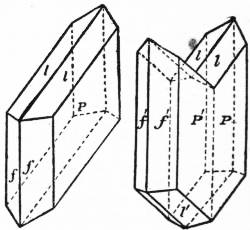
Fig. 1. Fig. 2.
Gypsum crystallizes in the monoclinic system, the habit of the crystals being usually either prismatic or tabular; in the latter case the broad planes are parallel to the faces of the clinopinacoid. The crystals may become lenticular by curvature of certain faces. In the characteristic type represented in fig. 1, f represents the prism, l the hemi-pyramid and P the clinopinacoid. Twins are common, as in fig. 2, forming in some cases arrow-headed and swallow-tailed crystals. Cleavage is perfect parallel to the clinopinacoid, yielding thin plates, often diamond-shaped, with pearly lustre; these flakes are usually flexible, but may be brittle, as in the gypsum of Montmartre. Two other cleavages are recognized, but they are imperfect. Crystals of gypsum, when occurring in clay, may enclose much muddy matter; in other cases a large proportion of sand may be mechanically entangled in the crystals without serious disturbance of form; whilst certain crystals occasionally enclose cavities with liquid and an air-bubble. Gypsum not infrequently becomes fibrous. This variety occurs in veins, often running through gypseous marls, with the fibres disposed at right angles to the direction of the vein. Such gypsum when cut and polished has a pearly opalescence, or satiny sheen, whence it is called satin-spar (q.v.).
Gypsum is so soft as to be scratched even by the finger-nail (H = 1.5 to 2). Its specific gravity is about 2.3. The mineral is slightly soluble in water, one part of gypsum being soluble, according to G. K. Cameron, in 372 parts of pure water at 26° C. Waters percolating through gypseous strata, like the Keuper marls, dissolve the calcium sulphate and thus become permanently hard or “selenitic.” Such water has special value for brewing pale ale, and the water used by the Burton breweries is of this character; hence the artificial dissolving of gypsum in water for brewing purposes is known as “burtonization.” Deposits of gypsum are formed in boilers using selenitic water.
Pure gypsum is colourless or white, but it is often tinted, especially in the alabaster variety, grey, yellow or pink. Gypsum crystallizes with two molecules of water, equal to about 21% by weight, and consequently has the formula CaSO4·2H2O. By exposure to strong heat all the water may be expelled, and the substance then has the composition of anhydrite (q.v.). When the calcination, however, is conducted at such a temperature that only about 75% of the water is lost, it yields a white pulverulent substance, known as “plaster of Paris,” which may readily be caused to recombine with water, forming a hard cement. The gypsum quarries of Montmartre, in the north of Paris, were worked in Tertiary strata, rich in fossils. Gypsum is largely quarried in England for conversion into plaster of Paris, whence it is sometimes known as “plaster stone,” and since much is sent to the Staffordshire potteries for making moulds it is also termed “potter’s stone.” The chief workings are in the Keuper marls near Newark in Nottinghamshire, Fauld in Staffordshire and Chellaston in Derbyshire. It is also worked in Permian beds in Cumberland and Westmorland, and in Purbeck strata near Battle in Sussex.
Gypsum frequently occurs in association with rock-salt, having been deposited in shallow basins of salt water. Much of the calcium in sea-water exists as sulphate; and on evaporation of a drop of sea-water under the microscope this sulphate is deposited as acicular crystals of gypsum. In salt-lagoons the deposition of the gypsum is probably effected in most cases by means of micro-organisms. Waters containing sulphuretted hydrogen, on exposure to the air in the presence of limestone, may yield gypsum by the formation of sulphuric acid and its interaction with the calcium carbonate. In volcanic districts gypsum is produced by the action of sulphuric acid, resulting from the oxidation of sulphurous vapours, on lime-bearing minerals, like labradorite and augite, in the volcanic rocks: hence gypsum is common around solfataras. Again, by the oxidation of iron-pyrites and the action of the resulting sulphuric acid on limestone or on shells, gypsum may be formed; whence its origin in most clays. Gypsum is also formed in some cases by the hydration of anhydrite, the change being accompanied by an increase of volume to the extent of about 60%. Conversely gypsum may, under certain conditions, be dehydrated or reduced to anhydrite.
Some of the largest known crystals of selenite have been found in southern Utah, where they occur in huge geodes, or crystal-lined cavities, in deposits from the old salt-lakes. Fine crystals, sometimes curiously bent, occur in the Permian rocks of Friedrichroda, near Gotha, where there is a grotto called the Marienglashöhle, close to Rheinhardsbrunn. Many of the best localities for selenite are in the New Red Sandstone formation (Trias and Permian), notably the salt-mines of Hall and Hallein, near Salzburg, and of Bex in Switzerland. Excellent crystals, usually of a brownish colour arranged in groups, are often found in the brine-chambers and the launders used in salt-works. Selenite also occurs in fine crystals in the sulphur-bearing marls of Girgenti and other Sicilian localities; whilst in Britain very bold crystals are yielded by the Kimeridge clay of Shotover Hill near Oxford. Twisted crystals and rosettes of gypsum found in the Mammoth Cave, Kentucky, have been called “oulopholites” (οὖλος, “woolly”; φωλεός, “cave”).
In addition to the use of gypsum in cement-making, the mineral finds application as an agricultural agent in dressing land, and it has also been used in the manufacture of porcelain and glass. Formerly it was employed, in the form of thin cleavage-plates, for glazing windows, and seems to have been, with mica, called lapis specularis. It is still known in Germany as Marienglas and Fraueneis. Delicate cleavage-plates of gypsum are used in microscopic petrography for the determination of certain optical constants in the rock-forming minerals.
(F. W. R.*)
GYROSCOPE AND GYROSTAT. These are scientific models or instruments designed to illustrate experimentally the dynamics of a rotating body such as the spinning-top, hoop and bicycle, and also the precession of the equinox and the rotation of the earth.
The gyroscope (Gr. γῦρος, ring, σκοπεῖν, to see) may be distinguished from the gyrostat (γῦρος, and στατικός, stationary) as an instrument in which the rotating wheel or disk is mounted in gimbals so that the principal axis of rotation always passes through a fixed point (fig. 1). It can be made to imitate the motion of a spinning-top of which the point is placed in a smooth agate cup as in Maxwell’s dynamical top (figs. 2, 3). (Collected Works, i. 248.) A bicycle wheel, with a prolongation of the axle placed in a cup, can also be made to serve (fig. 4).
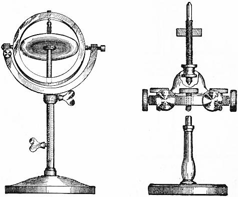
Fig. 1. Fig. 2.
The gyrostat is an instrument designed by Lord Kelvin (Natural Philosophy, § 345) to illustrate the more complicated state of motion of a spinning body when free to wander about on a horizontal plane, like a top spun on the pavement, or a hoop or bicycle on the road. It consists essentially of a massive fly-wheel concealed in a metal casing, and its behaviour on a table, or with various modes of suspension or support, described in Thomson and Tait, Natural Philosophy, serves to illustrate the curious reversal of the ordinary laws of statical equilibrium due to the gyrostatic domination of the interior invisible fly-wheel, when rotated rapidly (fig. 5).
The toy shown in figs. 6 and 7, which can be bought for a shilling, is acting as a gyroscope in fig. 6 and a gyrostat in fig. 7.
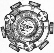
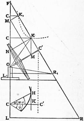
Fig. 3. Fig. 4.
The gyroscope, as represented in figs. 2 and 3 by Maxwell’s dynamical top, is provided with screws by which the centre of gravity can be brought into coincidence with the point of support. It can then be used to illustrate Poinsot’s theory of the motion of a body under no force, the gyroscope being made kinetically unsymmetrical by a setting of the screws. The discussion of this movement is required for Jacobi’s theorems on the allied motion of a top and of a body under no force (Poinsot, Théorie nouvelle de la rotation des corps, Paris, 1857; Jacobi, Werke, ii. Note B, p. 476).
To imitate the movement of the top the centre of gravity is displaced from the point of support so as to give a preponderance. When the motion takes place in the neighbourhood of the downward vertical, the bicycle wheel can be made to serve again mounted as in fig. 8 by a stalk in the prolongation of the axle, suspended from a universal joint at O; it can then be spun by hand and projected in any manner.
Fig. 5. Fig. 6. Fig. 7.
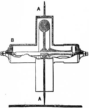
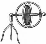
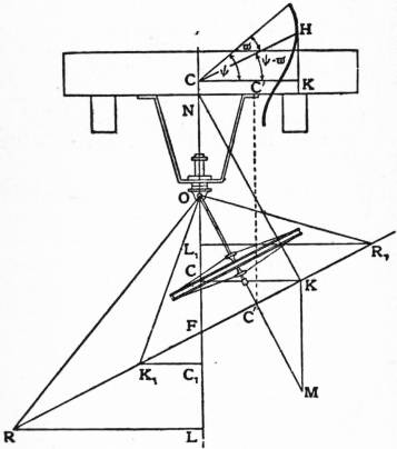
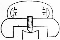
Fig. 8. Fig. 9.
The first practical application of the gyroscopic principle was invented and carried out (1744) by Serson, with a spinning top with a polished upper plane surface for giving an artificial horizon at sea, undisturbed by the motion of the ship, when the real horizon was obscured. The instrument has been perfected by Admiral Georges Ernest Fleuriais (fig. 9), and is interesting theoretically as showing the correction required practically for the rotation of the earth. Gilbert’s barogyroscope is devised for the same purpose of showing the earth’s rotation; a description of it, and of the latest form employed by Föppl, is given in the Ency. d. math. Wiss., 1904, with bibliographical references in the article “Mechanics of Physical Apparatus.” The rotation of the fly-wheel is maintained here by an electric motor, as devised by G. M. Hopkins, and described in the Scientific American, 1878. To demonstrate the rotation of the earth by the constancy in direction of the axis of a gyroscope is a suggestion that has often been made; by E. Sang in 1836, and others. The experiment was first carried out with success by Foucault in 1851, by a simple pendulum swung in the dome of the Pantheon, Paris, and it has been repeated frequently (Mémoires sur le pendule, 1889).
A gyroscopic fly-wheel will preserve its original direction in space only when left absolutely free in all directions, as required in the experiments above. If employed in steering, as of a torpedo, the gyroscope must act through the intermediary of a light relay; but if direct-acting, the reaction will cause precession of the axis, and the original direction is lost.
The gyrostatic principle, in which one degree of freedom is suppressed in the axis, is useful for imparting steadiness and stability in a moving body; it is employed by Schlick to mitigate the rolling of a ship and to maintain the upright position of Brennan’s monorail car.
Lastly, as an application of gyroscopic theory, a stretched chain of fly-wheels in rotation was employed by Kelvin as a mechanical model of the rotary polarization of light in an electromagnetic field; the apparatus may be constructed of bicycle wheels connected by short links, and suspended vertically.
Theory of the Symmetrical Top.
1. The physical constants of a given symmetrical top, expressed in C.G.S. units, which are employed in the subsequent formulae, are denoted by M, h, C and A. M is the weight in grammes (g) as given by the number of gramme weights which equilibrate the top when weighed in a balance; h is the distance OG in centimetres (cm.) between G the centre of gravity and O the point of support, and Mh may be called the preponderance in g.-cm.; Mh and M can be measured by a spring balance holding up in a horizontal position the axis OC in fig. 8 suspended at O. Then gMh (dyne-cm. or ergs) is the moment of gravity about O when the axis OG is horizontal, gMh sin θ being the moment when the axis OG makes an angle θ with the vertical, and g = 981 (cm./s2) on the average; C is the moment of inertia of the top about OG, and A about any axis through O at right angles to OG, both measured in g-cm.2.
To measure A experimentally, swing the top freely about O in small plane oscillation, and determine the length, l cm., of the equivalent simple pendulum; then
l = A/Mh, A = Mhl.
(1)
Next make the top, or this simple pendulum, perform small conical revolutions, nearly coincident with the downward vertical position of equilibrium, and measure n, the mean angular velocity of the conical pendulum in radians / second; and T its period in seconds; then
4π2/T2 = n2 = g/l = gMh/A;
(2)
and f = n/2π is the number of revolutions per second, called the frequency, T = 2π/n is the period of a revolution, in seconds.
2. In the popular explanation of the steady movement of the top at a constant inclination to the vertical, depending on the composition of angular velocity, such as given in Perry’s Spinning Tops, or Worthington’s Dynamics of Rotation, Steady motion of the top. it is asserted that the moment of gravity is always generating an angular velocity about an axis OB perpendicular to the vertical plane COC′ through the axis of the top OC′; and this angular velocity, compounded with the resultant angular velocity about an axis OI, nearly coincident with OC′, causes the axes OI and OC′ to keep taking up a new position by moving at right angles to the plane COC′, at a constant precessional angular velocity, say μ rad./sec., round the vertical OC (fig. 4).
If, however, the axis OC′ is prevented from taking up this precessional velocity, the top at once falls down; thence all the ingenious attempts—for instance, in the swinging cabin of the Bessemer ship—to utilise the gyroscope as a mechanical directive agency have always resulted in failure (Engineer, October 1874), unless restricted to actuate a light relay, which guides the mechanism, as in steering a torpedo.
An experimental verification can be carried out with the gyroscope in fig. 1; so long as the vertical spindle is free to rotate in its socket, the rapidly rotating wheel will resist the impulse of tapping on the gimbal by moving to one side; but when the pinch screw prevents the rotation of the vertical spindle in the massive pedestal, this resistance to the tapping at once disappears, provided the friction of the table prevents the movement of the pedestal; and if the wheel has any preponderance, it falls down.
Familiar instances of the same principles are observable in the movement of a hoop, or in the steering of a bicycle; it is essential that the handle of the bicycle should be free to rotate to secure the stability of the movement.
The bicycle wheel, employed as a spinning top, in fig. 4, can also be held by the stalk, and will thus, when rotated rapidly, convey a distinct muscular impression of resistance to change of direction, if brandished.
3. A demonstration, depending on the elementary principles of dynamics, of the exact conditions required for the Elementary demonstration of the condition of steady motion. axis OC′ of a spinning top to spin steadily at a constant inclination θ to the vertical OC, is given here before proceeding to the more complicated question of the general motion, when θ, the inclination of the axis, is varying by nutation.
It is a fundamental principle in dynamics that if OH is a vector representing to scale the angular momentum of a system, and if Oh is the vector representing the axis of the impressed couple or torque, then OH will vary so that the velocity of H is represented to scale by the impressed couple Oh, and if the top is moving freely about O, Oh is at right angles to the vertical plane COC′, and
Oh = gMh sin θ.
(1)
In the case of the steady motion of the top, the vector OH lies in the vertical plane COC′, in OK suppose (fig. 4), and has a component OC = G about the vertical and a component OC′ = G′, suppose, about the axis OC; and G′ = CR, if R denotes the angular velocity of the top with which it is spun about OC′.
If μ denotes the constant precessional angular velocity of the vertical plane COC′ the components of angular velocity and momentum about OA are μ sin θ and Aμ sin θ, OA being perpendicular to OC′ in the plane COC′; so that the vector OK has the components
OC′ = G′, and C′K = Aμ sin θ,
(2)
and the horizontal component
CK = OC′ sin θ − C′K cos θ = G′ sin θ − Aμ sin θ cos θ.
(3)
The velocity of K being equal to the impressed couple Oh,
gMh sin θ = μ·CK = sin θ (G′μ − Aμ2 cos θ),
(4)
and dropping the factor sin θ,
Aμ2 cos θ − G′μ + gMh = 0, or Aμ2 cos θ − CRμ + An2 = 0,
(5)
the condition for steady motion.
Solving this as a quadratic in μ, the roots μ1, μ2 are given by
μ1, μ2 = G′ sec θ [1 ± √ (1 − 4A2n2 cos θ) ];
2A G′2
(6)
and the minimum value of G′ = CR for real values of μ is given by
G′2 = cos θ, CR 2√(cos θ);
4A2n2 An
(7)
for a smaller value of R the top cannot spin steadily at the inclination θ to the upward vertical.
Interpreted geometrically in fig. 4
μ = gMh sin θ/CK = An2/KN, and μ = C′K/A sin θ = KM/A,
(8)
KM·KN = A2 n2,
(9)
so that K lies on a hyperbola with OC, OC′ as asymptotes.
4. Suppose the top or gyroscope, instead of moving freely about the point O, is held in a ring or frame which is compelled Constrained motion of the gyroscope. to rotate about the vertical axis OC with constant angular velocity μ; then if N denotes the couple of reaction of the frame keeping the top from falling, acting in the plane COC’, equation (4) § 3 becomes modified into
gMh sin θ − N = μ·CK = sin θ G′μ − Aμ2 cos θ,
(1)
N = sin θ (Aμ2 cos θ − G′μ + gMh) = A sin θ cos θ (μ − μ1) (μ − μ2);
(2)
and hence, as μ increases through μ2 and μ1, the sign of N can be determined, positive or negative, according as the tendency of the axis is to fall or rise.
When G′ = CR is large, μ2 is large, and
μ1 ≈ gMh/G′ = An2/CR,
(3)
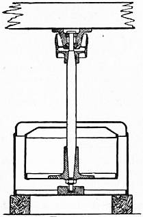
Fig. 10.
the same for all inclinations, and this is the precession observed in the spinning top and centrifugal machine of fig. 10 This is true accurately when the axis OC′ is horizontal, and then it agrees with the result of the popular explanation of § 2.
If the axis of the top OC′ is pointing upward, the precession is in the same direction as the rotation, and an increase of μ from μ1 makes N negative, and the top rises; conversely a decrease of the procession μ causes the axis to fall (Perry, Spinning Tops, p. 48).
If the axis points downward, as in the centrifugal machine with upper support, the precession is in the opposite direction to the rotation, and to make the axis approach the vertical position the precession must be reduced.
This is effected automatically in the Weston centrifugal machine (fig. 10) used for the separation of water and Centrifugal machine. molasses, by the friction of the indiarubber cushions above the support; or else the spindle is produced downwards below the drum a short distance, and turns in a hole in a weight resting on the bottom of the case, which weight is dragged round until the spindle is upright; this second arrangement is more effective when a liquid is treated in the drum, and wave action is set up (The Centrifugal Machine, C. A. Matthey).
Similar considerations apply to the stability of the whirling bowl in a cream-separating machine.
We can write equation (1)
N = An2 sin θ − μ·CK = (A2n2 − KM·KN) sin θ/A,
(4)
so that N is negative or positive, and the axis tends to rise or fall according as K moves to the inside or outside of the hyperbola of free motion. Thus a tap on the axis tending to hurry the precession is equivalent to an impulse couple giving an increase to C′K, and will make K move to the interior of the hyperbola and cause the axis to rise; the steering of a bicycle may be explained in this way; but K1 will move to the exterior of the hyperbola, and so the axis will fall in this second more violent motion.
Friction on the point of the top may be supposed to act like a tap in the direction opposite to the precession; and so the axis of a top spun violently rises at first and up to the vertical position, but falls away again as the motion dies out. Friction considered as acting in retarding the rotation may be compared to an impulse couple tending to reduce OC′, and so make K and K1 both move to the exterior of the hyperbola, and the axis falls in both cases. The axis may rise or fall according to the direction of the frictional couple, depending on the shape of the point; an analytical treatment of the varying motion is very intractable; a memoir by E. G. Gallop may be consulted in the Trans. Camb. Phil. Soc., 1903.
The earth behaves in precession like a large spinning top, of which the axis describes a circle round the pole of the ecliptic of mean angular radius θ, about 23½°, in a period of 26,000 years, so that R/μ = 26000 × 365; and the mean couple producing precession is