PART SECOND. OF PARTICULAR SURGICAL SUBJECTS. (continued 2)
Reading edition
When purulent matter forms, fluctuation becomes more distinct, the pain increases, and there is slight headache and fever. The integuments inflame more and more, and, if the case is neglected, ultimately give way by sloughing. A small ragged opening, often indirect, is formed, and the contents of the sac are not thereby all discharged; the thinner fluid only escaping, whilst the more viscid remains and clogs the aperture. The swelling is not much diminished; the margins of the aperture thicken, become indurated, and contract, the purulent contents of the sac are gradually discharged, and the tears afterwards flow through the opening. The parts are now in that condition to which the term Fistula lachrymalis is with propriety applied. The swelling of the canal may gradually subside, the tears resume their wonted course, and the opening may then contract, and the parts cicatrise; but frequently the fistula remains open for a long period, gradually diminishing in diameter, and only a small passage, almost imperceptible, ultimately remaining, through which a few drops of lachrymal fluid are occasionally discharged. Sometimes the fistula closes entirely without the obstruction of the nasal duct having been removed, and the lachrymal sac remains in consequence distended; then the tears or mucus, either clear or turbid, can generally be squeezed through the puncta.
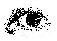
It frequently happens that the meibomian glands are the seat of morbid action, along with the lachrymal passages; their secretion is changed, becoming in some cases thick and caseous, in others puriform. By some, affection of the meibomian glands has been considered as the cause of inflammation and abscess of the lachrymal sac. This opinion, however, cannot be agreed to, for the diseases are not always coexistent; and besides, the affection of the surface of the lachrymal sac and ductus ad narem is as likely to be the consequence of morbid action, extending upwards from the nostrils, as of morbid secretion from the eyelids blocking up and irritating the puncta and the lachrymal passages. Disease of the meibomian glands in the under eyelid often exists along with disease of the lachrymal passages, but the latter is generally the primary affection; the conjunctival covering of the eyelid is at the same time inflamed, swollen, and often granulated.
In some cases of abscess in the lachrymal sac, before the integuments give way, the subjacent bone becomes diseased in consequence of the pressure of the confined matter; portions are affected by necrosis, and after their separation considerable deformity is produced. The exfoliation is often very tedious, and is attended with discharge of fetid thin fluid from the nostril, and from the ill-conditioned lachrymal fistula.
Fistula lachrymalis is often merely one of the symptoms of disease in the bones of the nose, with obstruction of the nasal duct,—as in patients who have suffered from mercury.
Treatment.—In the treatment of epiphora or blenorrhœa—that is, chronic collection of a mucous fluid in the lachrymal sac, with weeping of the eye—a primary object of attention is the state of the general health. The habit of the patient will commonly be found weak, and, if not decidedly strumous, at least inclining towards that diathesis. In such cases the digestive organs must, if possible, be brought into a vigorous state by tonics and nourishing regimen. The local treatment chiefly consists in applying stimulants to the internal surfaces of the palpebræ and lachrymal sac. For this purpose, solutions of stimulating and astringent substances, termed collyria, and various ointments, are employed. At first they ought to be used of rather a mild nature, and their stimulating power must be afterwards increased gradually. The applications are placed between the eyelids, and, becoming mixed with the natural secretion, pervade the diseased surfaces; and, being taken up by the puncta lachrymalia, are afterwards conveyed into the sac. It was formerly the custom to inject the fluids into the sac; but this is unnecessary so long as the puncta and canaliculi remain pervious, and the permeability of these can be readily ascertained by means of a small probe. Permanent pressure on the sac can be productive of no good effect, and is extremely liable to do harm. The repeated application of very small blisters over the sac has been found useful.
Introducing minute gold probes through the puncta has been much recommended, but in the generality of cases can be of little service. The probes are too limber for removing mechanical obstruction, or for affecting in any way the contracted or strictured duct. But passing of the probe may tend to remove the irritability of the passage, as happens in the urethra, and thence some relief may follow. Much dexterity is required in using either the probe or syringe. The puncta are often very small, and it is in general necessary to dilate them by means of the point of a common pin, before any instrument can be passed through them into the sac. The point of the probe being introduced into the punctum, either superior or inferior, must first be carried towards the nose for about 2-10ths of an inch, the instrument being lightly held betwixt the fore and middle fingers of the right hand. It is then directed downwards and backwards. Care must be taken to prevent entanglement in folds of the membrane. Should obstruction be felt, the instrument is withdrawn a little, and then carefully and gently carried in the right direction. The small syringe is managed with one hand, whilst, with the forefinger of the other, the punctum not occupied by the pipe is compressed.
Neither can much or any benefit be expected to follow attempts to force obstruction in the lachrymal passages, by the weight of a column of mercury. A plan of dilating and rectifying the nasal duct by styles introduced through the puncta has been proposed, but scarcely deserves to be mentioned as a means of cure.
When suppuration is threatened, with increase of the swelling, inability of the patient to empty the sac by pressure, redness of the integuments, &c., an early opening should be made into the tumour, in order to prevent further and more serious mischief. A small opening into the sac cannot be productive of so much injury as forcible dilatation of the canaliculi, followed by and causing ulceration. The point of a straight narrow bistoury is to be entered into the sac, and carried on into the nasal duct, the knife being pushed downwards, backwards, and a little inwards, in the direction of that passage. The point to be punctured can always be readily ascertained by feeling for the firm ligament which attaches the orbicularis palpebrarum to the nasal process of the superior maxillary bone, as the upper orifice of the ductus ad narem is situated immediately below this tendon; by introducing the knife below the ligament, and within the sharp edge of the orbit, and then carrying it forward in the direction already mentioned, the surgeon cannot fail to enter the nasal duct. The knife should be followed by a probe, and ought not to be entirely withdrawn till the probe is fairly lodged in the duct, otherwise the surgeon will experience much difficulty in the after proceedings. If the knife be not pushed into the duct, a blunt instrument can scarcely be introduced afterwards. Some force is required, but is not hurtful, provided it be made in the proper direction, so as to remove the obstruction in the duct without injuring the bones and other parts in the neighbourhood. After the operation, some drops of blood should escape from the corresponding nostril, showing that it has fairly entered this passage; or the patient being made to expire forcibly, the nostrils being at the same time compressed with the fingers, air, blood, and mucus are forced upwards through the opening made.
Many and various modes have been pursued with a view of securing a pervious state of the nasal duct. Instruments of different kinds have been introduced through the puncta, through the opening in the sac, and through the termination of the duct under the spongy bone, and have been retained for a longer or shorter period, according to the fancy, or theory, or plan of the surgeon. The first of the methods of introduction is abandoned, as already stated. By the ancients the passages in fault were got rid of altogether, being either cauterised or destroyed by escharotics.
The passing of probes into the duct from its lower aperture is useful in removing trifling obstructions caused by concretion of deteriorated mucus, or slight thickening of the lining membrane, and in chronic dilatation of the sac with probable contraction of the duct. But, at the same time, it is an operation requiring much dexterity, and which ought not to be attempted till after much practice on the dead body. The first introduction of the instrument is always the most difficult, from obstruction by a valvular projection of the membrane at the lower orifice, the use of which in the healthy state of the parts must be apparent. Destruction of it renders after-introduction of instruments much more easy.
But the preferable practice is making an opening into the sac, and then introducing instruments from the upper orifice of the duct; more especially in cases where the swelling and pain are considerable. The instruments employed for dilatation of the passage are tubes and styles. The tubes are made either of silver or gold, of equal calibre throughout, and of the same length as the passage. For some time after their introduction they cause much irritation; this gradually diminishes, and the wound heals over them. But, according to my experience, the effects are not satisfactory. The irritation which they at first occasion generally subsides, but abscess again occurs, with much swelling, and it becomes necessary to remove the foreign body. Again, the tube sometimes becomes obstructed by thickening and concretion of the discharge, and then, when it is necessary to remove it, the process is found to be by no means an easy one; a free incision is required; a screw must be fastened into the tube, or, when that cannot be accomplished, the foreign body must be laid firmly hold of with strong forceps; altogether the extraction is very painful, and often extremely tedious. In short, the practice of introducing tubes does not appear to be founded on sound surgical principles.
After extensive and impartial trial of both the tubes and style, I decidedly prefer the use of the latter. On the point of the bistoury being fairly lodged in the lachrymal duct, a probe is passed along it; the knife is then withdrawn, and the passage is gently dilated by the probe. The probe again is followed by the style, which should be made of silver, of the same thickness throughout, of the same length as the duct, and with a flattened head placed obliquely to the body of the style. The size of the style should be at first small, and gradually increased. The irritation caused by the first introduction is in many cases very severe, but the parts soon accommodate themselves to the presence of the foreign body; the pain and swelling diminish, as also the discharge. If a large style be pushed forcibly in at first, violent inflammatory action will ensue, and much mischief may be produced. After irritation has gone off, the tears pass readily down in the nose by the sides of the style, according to the laws of capillary attraction, little or no fluid escapes from the external opening, the wound contracts around the instrument, and, its head being covered with black wax, no deformity is produced. The instrument should be removed from time to time, cleaned, and replaced. When, by the continued use of styles gradually increased in size, the duct has been dilated to its full extent, and appears restored to a sound condition, the instrument may be withdrawn, and afterwards introduced only occasionally. The external aperture, which has become fistulous from the long presence of the foreign body, then begins to contract, and, on its completely closing, the tears continue to follow their usual course, and the disease is overcome. But sometimes a small fistulous aperture remains, and there appears to be a disposition towards the renewal of the affection; in such a case, a small style, not exceeding a thin gold probe in diameter, should be introduced every evening, and retained for some hours: this causes little or no inconvenience to the patient, and insures the permeability of the canal.
Such is the method by which a permanent cure may often be obtained, and which, in my opinion, is preferable to the use of tubes. If these are to be employed, they should, as already mentioned, be nearly of equal calibre throughout; the external opening must not be allowed to close for a considerable time after the introduction of the instrument; and the tube must be kept pervious for some time by a style introduced through it. But by these means, which are essential for the success of the practice, the main advantage arising from the use of a tube, viz., little irritation being produced at first, and the parts being allowed to close soon over it, are completely done away with.
The practice of perforating the os unguis never can be required; it is cruel, unnecessary, and unsurgical.
Sometimes the lachrymal passages are entirely destroyed. In such cases, it has been found that no great inconvenience arises from their obliteration, as the lachrymal gland ceases, in a great measure, to secrete fluid, and the conjunctival secretion, after having performed its office, evaporates from the surface. In truth, the lachrymal gland always enjoys long periods of repose, and is only called into active exercise of its functions occasionally, as the eye in its ordinary condition is sufficiently lubricated by secretion from its conjunctival covering.
The treatment of fistula lachrymalis, as has been well remarked by an eminent author, must be varied and regulated according to circumstances;—by the degree of obstruction in the duct, by the state of the coverings of the sac, of the sac itself, and of the subjacent bone, and by the general state and habit of the patient.
Encanthis is a tumour situated in the corner of the eye. The caruncula lachrymalis appears to be the original seat of the disease, at least it is involved at an early period. The growth is at first small, and appears to be simple enlargement of the caruncle: it is of a reddish colour, and its surface is studded with numerous granulations. It often attains a very considerable size; and, on account of its propinquity to the lachrymal passages, is accompanied with watering of the eye, the puncta being either involved in the growth, or compressed or displaced by it. Sometimes the whole inner corner of the eye, from the margin of the cornea to the inner junction of the eyelids, is occupied by the granulated swelling; and in such cases it is not uncommon for the tumour to extend itself outwards, in the form of a lunated appendage, on the under surface of each lid; thereby the motions and functions of the ball are much impeded, and a prominent deformity is occasioned. In most instances the growth seems to be a simple enlargement of structure, and is of a benign nature; but sometimes it is firm, hard, of rather a livid hue, with a smooth slimy surface, and is decidedly malignant,—enlarging, and gradually involving the surrounding parts.
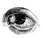
Cancerous ulceration, attacking and destroying the eyelids, and the parts around the ball of the eye, often commences in the situation of the caruncle, or in a wart on the edge of the lid. Cancer, though a rare and uncommon disease of the eyeball, frequently seizes on the appendages of the eye, extending rapidly in all directions, and often completely detaching the ball by ulceration. Warty tumours also occur on the conjunctiva of the lids, or of the ball, and are inconvenient as a source of much irritation to the neighbouring parts, even though of a benign nature in themselves.
Extirpation, by means of a small pointed knife, or curved scissors, is the only means to be relied on for the cure of such warty tumours, and of encanthis. The growth must be fixed and pulled outwards with a small hook, and carefully dissected away; the eyelids, and, if necessary, the ball of the eye, being kept fixed with the fingers, or by means of a speculum: the fingers are generally sufficient, and more convenient than any instrument. If from the appearance of the parts, and from induration surrounding the tumour, malignant action has evidently taken place or is dreaded, then the incisions must be made wide of the base of the swelling. For malignant, open, and extensive ulcerations, nothing can be done farther than to allay the pain, and soothe the constitutional disturbance. On the whole, encanthis is a rare disease; however, I have seen, and operated on, several instances of it.
Encysted Tumours of the Eyelids.—These occur beneath the conjunctival lining of either the upper or under lid, but most frequently in the former. They form rapidly, but seldom attain any very considerable size; and may be found to contain, along with glairy fluid, a mixture of pus, or curdy matter. The contents, however, are generally glairy, rarely atheromatous. The cysts are very thin and adherent, and the tumour projects externally, forming a dusky red elevation of the integuments. They cause considerable deformity, watering of the eye, and stiffness and difficulty in moving the lids. On everting the eyelid, the contents of the tumour are seen shining through the distended conjunctiva, and present a bluish appearance. They are seldom single, and are not remediable but by operation. It is improper to attempt their extirpation from without, as there is a certainty of cutting completely through the eyelid, the inner covering of the cyst being merely attenuated conjunctiva. The lid is to be everted, and an incision made into the prominent and thin cyst with the point of a cataract knife; the contents can then be readily scooped out with the end of a probe. It is impossible to dissect out the tender cyst entire, and, when this is attempted, the cure can seldom be permanent. If, after incision and discharge of the contents, nothing farther is done, the disease will almost certainly return, in consequence of the remaining cyst reassuming a secreting action. The only effectual and radical cure is the application of a finely-pointed piece of caustic potass to the interior of the cyst, after discharge of the contents and cessation of bleeding. The cyst is thereby completely destroyed. A slip of soft lint, dipped in oil, is interposed betwixt the lid and eyeball, for an hour or two, in order to protect that delicate organ from the caustic. The wound suppurates and heals kindly, and no mark is visible, the incision having been made from within. I have had no instance of return of the disease since adopting this practice; and I have operated on many which had been previously treated by other and ineffectual means. The laceration of the cyst with a pointed probe is sometimes followed by a permanent cure, but it cannot be depended upon.
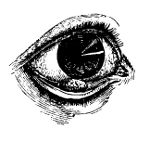
Closure of the Eyelids may be either congenital, or a consequence of injuries, as burns of the parts. The closure may be complete or partial. In general it is partial, though perhaps extensive; and the adhesions can be readily separated by the point of a knife, or small probe having been previously introduced beneath; or a small and narrow probe-pointed bistoury may be conveniently used for the purpose. In the after-treatment means must, of course, be taken to prevent the lids from again adhering.
Ectropion, or eversion of the eyelids, may be produced, merely by swelling of the conjunctival lining protruding the lid: or the lid may be relaxed, and the conjunctiva may swell in consequence of repeated inflammation of the parts, caused by frequent and careless exposure; or the disease may be the result of contraction, by cicatrisation of the integuments of the face, as after burns, extensive superficial wounds in the neighbourhood of the eye, or the effect of periosteal disease of the orbit. The affection may exist to a greater or less degree, being in some instances scarcely visible, and not troublesome, whilst in others, the eyelashes lie on the upper part of the cheek, and the swollen granulated conjunctiva is exposed. The lower lid is generally the one which is affected. The disease may exist in both eyes, or only in one. In strumous habits both are frequently affected in a slight degree; and the upper lid, too, is sometimes turned a little outwards. When eversion is of long continuance, and complete or almost so, the conjunctival covering of the ball of the eye, and of the cornea, becomes dry and wrinkled; in short, the membrane completely changes its character, and becomes cuticular. In a lad who laboured eleven years under eversion of the upper and lower lids—arising from abscess and exfoliation of the external angular process of the os frontis, following a blow received when a boy—the conjunctiva was hard, wrinkled, scaly, and exactly similar to cuticle: this change of the membrane also extended over the whole cornea. The surface of the eye had lost its lustre, and vision was much impaired, the patient being able to distinguish only very bright objects. By such cases, continuity of the conjunctiva with the outer layer of the cornea is beautifully demonstrated.
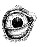
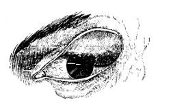
Some of the most intractable of all cases of eversion are the result of burns. The constantly increasing contraction of the cicatrix draws either the upper or the lower lid far from its natural situation, and produces frightful deformity. The tarsal cartilages are greatly extended, and in any operation for the relief of the patient it is necessary to remove a portion before the lid can be properly adapted.
Great inconvenience is caused by the state of eversion: the surface of the eyeball is subject to inflammation, in consequence of being insufficiently protected; the change of its investing membrane is a serious evil; and in some cases the cornea becomes extensively ulcerated, unusually vascular, and opaque.
When the conjunctiva only is in fault, the deformity is slight, and the state of matters is readily ameliorated by excision of the relaxed portion. This is done by sharp curved scissors. As the wound gradually contracts, the eyelid is drawn inwards, and, on cicatrisation taking place, the parts have become restored to their healthy condition. Care, however, should be taken that too much of the swollen conjunctiva is not removed, otherwise the subsequent contraction may cause inversion of the lid. Combined with the above practice, relaxation of the lid itself will in many cases be remedied by removal of a portion of it in the form of the letter V, by means of a sharp-pointed bistoury: the edges of the incisions are afterwards put together by a point of interrupted suture. When eversion arises from a cicatrix of the integuments, the part in fault may be divided; but a temporary benefit only can be procured. For, during the healing of the wound, the parts again contract; and, though a portion of the conjunctiva is at the same time removed, the contraction internally will hardly counteract that which is going on externally. In order fully to obviate the evil of this contraction of the cicatrix in inveterate cases of ectropion, a form of plastic operation may be successfully resorted to. The cicatrix being dissected out, and the tarsal cartilage brought neatly into position, a piece of integument from the temple or cheek may be adapted, and a portion of a new eyelid formed. The parts may sometimes be brought into a good position without the necessity of borrowing any portion of integument. A V-shaped incision can be made, the apex pointing downwards, so as to loosen the under lid; and after it has been drawn upwards and put straight, the edges of the lower part of the exposed space are united by suture.
Entropion, or inversion, consists in the turning in of the tarsal margins of the lids, and generally takes place during inflammation and swelling of the conjunctival lining of the lid. During violent inflammation of the lid the conjunctiva and integuments are much swollen, and bulge out externally; by the projection the margin is forced mechanically towards the ball, and entropion takes place. But in this state of matters, should the lid be by any chance everted, and not replaced, then the bulging is from the conjunctival surface, and prevents the margin from regaining its former site, and permanent eversion or ectropion occurs. In fact, inversion and eversion, like phymosis and paraphymosis, exist from the same parts being put in different relation to each other. More permanent entropion is caused by the contraction which follows removal of tumours from the under surface of the lids, or destruction of large portions of the conjunctiva. The disease is most frequently met with in the upper lid.
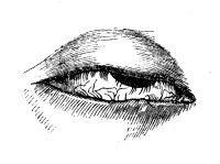
Trichiasis consists in a vicious bend of the eyelashes, or in a supernumerary growth in the rows or numbers of individual cilia, whereby they are inverted, and sweep the surface of the conjunctiva covering the cornea; thus great distress is caused by the friction of the hairs and edge of the lid on the sensible surface of the eyeball, and inflammation is frequently kindled and kept up by the continued irritation; it is accompanied by its usual distressing symptoms when seated in that organ, and too often followed by a greater or less number of untoward consequences. Sometimes only one or two hairs are at fault; in other instances, the half of the eyelash grows inwards; and sometimes there is a double row of cilia; one set being in the usual position, while the other projects against the eyeball. If proper means are not taken to remedy the evil, and moderate the irritation which it produces, the cornea becomes thickened and changed in structure; and vision, at first impaired and indistinct, may be entirely lost.
The symptoms may be for a time palliated by plucking out the faulty hairs, abstracting blood from the loaded vessels, and subsequently using ointments or collyria,—the best of which, perhaps, is the solution of nitrate of silver. In some cases it may be necessary to employ counter-irritation, as blistering the nape of the neck; and in all the general health must be strictly attended to. Other means may be required, and will be mentioned when treating of chronic ophthalmia.
The permanent cure of the disease is effected either by removal or by destruction of the roots of the cilia. The whole edge of the eyelid, or the offending part of it, is removed with a sharp narrow bistoury, the operator steadying the parts by laying hold of the cilia with the fingers of his left hand. It is necessary to remove the mere edge only, the cilia and their roots, and not the whole of the tarsal cartilage, as has been proposed.
Inversion of the lid, from contraction of a cicatrix in the conjunctiva, may be counteracted, by destroying with caustic, or removing with cutting instruments, a portion of the outer integuments, corresponding to the internal cicatrix. Forceps with broad points are used for taking up a fold of the skin, and an oval portion is then excised with a knife or scissors, cutting instruments being less painful and more precise than caustics. Of the latter, the sulphuric acid has been particularly recommended for this purpose. The contraction of the wound releases the cilia from the power of the internal cicatrix, and the parts are restored to their healthy state.
The term Pterygium is employed to denote a thickened and vascular state of part of the conjunctiva. The diseased portion is generally of a triangular form, commencing at the inner corner of the eye, extending towards the cornea, gradually diminishing in breadth, and terminating in a sharp apex, either at the margin of the cornea, or somewhere between its margin and centre. The thickening is seldom great, but the vessels which traverse the thickened part are numerous, enlarged, and tortuous—are, in fact, varicose. The base of the pterygium is always on the circumference of the eye, generally at the inner corner, and its apex is seldom, if ever, situated beyond the centre of the cornea: frequently the sclerotic conjunctiva alone is affected. The motions of the eye are little disturbed by the disease, but vision is materially impaired when a considerable part of the cornea is covered. Pterygium is in general single, but sometimes, though very rarely, there are two or more pterygia in one eye; and, in such cases, the patient’s vision is seriously affected, in consequence of the apices of the different pterygia uniting and coalescing on the cornea, and investing the greater part of that organ with a thick and dark shade. When several occur, they sometimes unite throughout their whole extent, and cover the half or more of the eye. This disease is very common amongst negroes and persons residing in equatorial climates.
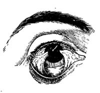
When the pterygium is of considerable size, extending over the cornea, the only remedy is excision. The apex of the web is laid hold of and pulled outwards by forceps or a hook, and the whole diseased part is then carefully dissected off with scissors, the incisions commencing at the apex, and being carried on to the base. The wound gradually contracts; and though an opaque cicatrix must form on the corneal surface, the speck is of much less dimensions than the space formerly occupied by the pterygium. If the web be thin and not exceedingly vascular, it may be sufficient to make a semicircular section of it transversely, by means of a hook and scissors, between its base and the margin of the cornea; its growth is thereby arrested, and there is a probable chance of its beginning to diminish, and ultimately disappearing. When it is small, and so situated as to cause no impairment of vision, it is prudent and good practice not to interfere with it at all.
DISEASES OF THE EYEBALL
Diseases of the eyeball are numerous, and various in their nature. Some are acute, others chronic; and their attack is either sudden, or slow and insidious. Most of them are attended with pain and other annoying symptoms, and some cause loss of vision. Some are cured by internal means; others require surgical operations; and the cure is either complete and permanent, or palliative and temporary. Some destroy the organ, and others, still more malignant, cause extinction of life. All require much attention and care.
Of Ophthalmia, or Inflammation of the Eye.—The symptoms and appearances of ophthalmia vary much according to the particular texture or textures affected. They require to be minutely attended to, that the treatment may be varied in such a way as to obviate any bad consequences which may be threatened. The great importance of the organ, and the danger to its structure and functions which is likely to occur from any other termination of the affection than resolution, must never be lost sight of.
We shall first treat of inflammation of the more external parts of the ball, an affection generally less dangerous than inflammation of the interior, but at the same time of more frequent occurrence, and produced by slighter causes.
Inflammation of the conjunctiva occurs in many individuals during very warm and sunny weather. At such a period, the eye is often excited by reflection of intense light from the surface of the earth; and is irritated by sudden exposure to a degree of light to which it has not been previously accustomed. Different directions of the sun’s rays, and different kinds of light, seem to exert different influences on the organ. The rays are most hurtful when they do not fall in a perpendicular direction on the eye, but slopingly or horizontally. Strong light from the moon, and light reflected from scarlet, are also particularly injurious. Undue exertion of the eye weakens it, and renders it prone to become inflamed. The eyes of infants are often violently inflamed, in consequence of imprudent exposure to light before they have been gradually accustomed to its stimulus. Again, inflammation is caused by imprudent exposure of the eye directly to cold, or by exposure of other parts causing suppression of their discharges, whether natural or not. Inflammation of the conjunctiva often follows suppression, however occasioned, of the menstrual or hemorrhoidal discharges, as also suppression of discharges from the urethra, from the Schneiderian membrane, or from behind the ears. Irritations in the neighbouring parts, as in the mouth during dentition, may also excite the disease. Immediate irritations, however, are the most frequent cause, as the lodgement of extraneous bodies on the surface of the organ—particles of sand, dust, snuff, pepper, or gunpowder, minute insects, loose or inverted eyelashes. By the presence of such substances, the eye is often kept in a very irritated state for a long period. The most violent conjunctival inflammation is sometimes produced by contact of gonorrhœal matter through carelessness. Occasionally metastasis of inflammation takes place from one eye to another; so that a person may be seen one day with severe inflammation of the right, and on the following day with a similar affection of the left, and the right entirely free from disease. Another cause, sometimes met with, of inflammatory action in the conjunctiva, is the lodgement of large foreign bodies in the orbit, with or without destruction of the eye; as splinters of wood, straws, rusty iron nails, sharp portions of stone, &c., penetrating the globe of the eye, or parts in the immediate neighbourhood. Upon removal of the cause, the redness, discharge of tears, pain, &c., sometimes subside without inflammation having been established, the vessels of the part regaining their contractility; but if the cause is continued for any considerable time the effects do not rapidly abate. Wounds and other injuries of the organ are generally followed by inflammation. But a simple clean wound or puncture made with a fine instrument, as in many operations, and in a favourable constitution, frequently produces little or no excitement of the part. The degree of excitement must of course depend upon the nature of the wound, the structure of the parts involved, the lodgement or not of the body by which the wound is inflicted, and many accidental circumstances. The eye may be injured by acids or by lime, and the textures acted upon chemically; again, the membrane may be wounded by pieces of hot metal, and then the destructive action is both chemical and mechanical: in both cases active inflammation of the injured conjunctiva is kindled. The state of the patient’s constitution modifies very much inflammatory action of the eye, however induced; and it has been observed, that dark eyes bear injury or incited action better than those of a light hue. Not unfrequently conjunctivitis is a secondary affection, accompanying eruptile diseases, as measles or small-pox.
In considering the disease, it is necessary to keep in mind the loose connection of the membrane with the subjacent parts, as well as its own texture and functions.
In conjunctival inflammation, the patient first feels a degree of pain and stiffness in moving the organ; and has always a feeling as if a foreign body were present, whether such is the case or not. There is also a degree of itching with a sensation of fulness in the part, and this is followed by redness of the membrane, becoming more and more intense. If the disease gain ground, the colour changes to a darkish red or purple hue. To the redness succeeds heat, with profuse and hot lachrymation. Then swelling supervenes, often to a great extent: the vessels, both veins and arteries, are much gorged, and effusion of serum or blood takes place into the loose cellular tissue which connects the conjunctiva to the sclerotic.
In some cases, the effusion in this situation is very considerable; lymph as well as blood is deposited, and a bulging forwards of the conjunctiva is produced; the stretched membrane becomes thickened, of a raw granulated appearance, and a bright scarlet hue, and the cornea appears sunk in the midst of the swelling, and almost hid by it: this state of matters is termed Inflammatory Chemosis, and only occurs when the excitement is very intense.
Blood is frequently effused beneath the conjunctiva in small quantity, in consequence of a bruise or other injury of the eye,—from violent exertion, as during coughing,—or from a less degree of inflammatory action than in the preceding case; but the swelling thereby occasioned is comparatively trifling, and the effusion is, in general, speedily absorbed. To this affection the term Ecchymosis is attached.
In inflammation of the external parts of the eye, the redness begins from the margins of the organ, and gradually diffuses itself towards the cornea. Such is not the case in inflammation more deeply seated. There is intolerance of light in a slight degree, and the patient is inclined to keep the eyelids shut. At first the discharge from the conjunctiva and meibomian glands is increased and changed, and flows occasionally over the cheek, producing a scalding sensation. When the eyelids are at rest, as during the night, they become glued together by the viscid fluid from the meibomian follicles; but, if the inflammation increases in intensity, the discharge is arrested.
In external inflammation there is more or less constitutional disturbance, proportioned to the violence of the action and the irritability of the system. In most instances the patient complains of headache.
The above symptoms subside along with the inflammation; but, if this has been at all severe or protracted, distension of the vessels to a considerable degree continues, and the ophthalmia becomes chronic. This change from acute to chronic takes place at various periods of the affection, according to the intensity of the action, the nature of the cause, and the irritability of the constitution. And again, the second stage of ophthalmia may revert to the first, acute inflammation being rekindled by fresh irritation of the organ.
Purulent Ophthalmia most frequently occurs in warm climates, and is attended from the first with profuse puriform discharge from the conjunctiva. In the natural state of the organ, the conjunctival discharge is pellucid, and so small in quantity as to be indiscernible; but in this disease it possesses all the external characters of pus, and is secreted in large quantity. The affection commences generally in the under eyelid, with a feeling as if sand or foreign bodies were lodged in the eye. The parts swell very much, and the eyelids become more or less inverted, in consequence of serous effusion into their cellular texture. Frequently the patient experiences an exacerbation of the complaint about three or four hours after each meal. Though the disease usually commences in the conjunctival lining of the eyelids, the external coverings of the ball are often secondarily affected. In some cases the bulb becomes the seat of lancinating pains; its coats give way; the humours are discharged; and the eye sinks, with immediate relief to the patient from the more urgent symptoms, but at the same time with irreparable loss of vision. In other instances the effects are less injurious to the structure of the organ, but equally so to the sense of vision: the cornea becomes dull, and ultimately opaque, or ulcerates, or partially sloughs; the swollen conjunctival surface of the lids is covered with granulations, and secretes a copious puriform discharge, with or without eversion, according to the degree of swelling. At first the lids are more or less inverted, on account of œdematous swelling of the cellular tissue: in the latter stages they are everted by thickening and turgescence of the conjunctiva. This membrane is at first villous and of a dull red colour, relaxed, and its vessels enlarged and loaded; afterwards it becomes hard, almost warty, and continues to discharge puriform fluid. The latter state of the lining of the lid produces disease of the cornea, opacity of a greenish colour, or an ulcer with intolerance of light, and other symptoms of disorganisation proceeding in that tissue. The disease is supposed to be contagious, and was the scourge of the British army for many years after the campaign in Egypt. In that country it seems to be caused by exposure to cold and damp during the night, and the intense rays of light during the day, more especially when these causes act on eyes which have not been accustomed to such vicissitudes. After its invasion, it is communicable to others by contact of the morbid secretion; and in individuals who have been once affected the disease is very apt to recur when they are crowded together in unhealthy situations.
A disease of equal malignity, and resembling in all respects the Egyptian ophthalmia, occurs from the application of gonorrhœal matter to the conjunctiva, or on sudden suppression of the gonorrhœal discharge,—metastasis of the action sometimes takes place from the urethral membrane to the conjunctiva. The eye is seldom saved from the destructive effects of the violent inflammation which follows the contact of the morbid fluid. Of all forms of purulent ophthalmia, the gonorrhœal is the most rapid in its course and destructive in its effects.
Children are not unfrequently the victims of purulent ophthalmia—the ophthalmia neonatorum. Immediately after birth the conjunctival lining of the eyelids seems unusually red and turgid, and a great degree of swelling soon takes place, so as to render separation of the eyelids very difficult. Occasionally eversion of the lids occurs, when the child cries, from sudden and forcible contraction of the strong external fibres of the orbicular muscle. In general, the lids soon relapse into their former situation; but sometimes the eversion remains, if the internally projecting tumour of the conjunctiva is allowed to become still more swelled from strangulation, caused by the outer margin of the reflected lid. The inflammation spreads over the ball; and, in general, the swelling of the conjunctiva, being greatest at the circumference of the eye, bulges out the eyelids, and turns in their margins. Puriform matter is secreted copiously, and is confined, more especially when, from inattention, the margins of the lids are allowed to become glued together. They often adhere so firmly as to require a very considerable force for their separation, and when opened the matter gushes out as if from the cavity of an abscess. From confinement of the matter the inflammation is still more increased, and the cornea involved. Whitish specks form on it, or it ulcerates, and the ulcers make their way into the anterior chamber of the eye; or portions of it slough, causing partial loss of the organ and openings into the chamber, in consequence of which the aqueous humour is discharged, and the cornea sinks and becomes flaccid. In many instances the cornea becomes opaque, changed in texture, and increased in thickness, so as to form a convex projection from betwixt the eyelids, termed Staphyloma; the sclerotic coat also is occasionally affected in a similar manner. A frequent cause of purulent ophthalmia in children is imprudent exposure of the eyes to strong light, the parent or nurse not remembering that the organ must be gradually accustomed to the stimulus. Exposure to cold may also induce the inflammatory action. The application of leucorrhœal or gonorrhœal matter to the eyes of the child, whilst passing through the vagina of the mother, is perhaps the most common cause of the disease. A very unhealthy state of the constitution accompanies the affection: the scalp and other parts of the surface are frequently covered with eruptions. A singular result sometimes follows the purulent ophthalmia of infants. A small opaque spot is observed on the capsule of the lens, which remains through life a central spurious capsular cataract.
Inflammation of the Cornea supervenes on simple conjunctival inflammation, and frequently on the purulent. The vessels of the part, both veins and arteries, previously carrying single and therefore invisible blood corpuscules, become much dilated, are filled with numerous globules, and hence are rendered red and conspicuous to the unassisted eye. Writers on ophthalmic surgery, in their rage for refinement, speak of three kinds of this inflammation—inflammation of the external or conjunctival covering, of the middle tunics or cornea propria, and, lastly, of the third coat, the capsule of the aqueous humour: such distinctions, however, are found to effect no good practical end, and it is unnecessary to follow them. One particular layer of the cornea may be first attacked, but the whole structure soon becomes involved. The inflammation generally commences in the conjunctival covering. Vision is necessarily much obscured from even slight inflammatory affection of the cornea. Part only of the organ may be affected, but frequently the whole is involved. Sometimes only one or two vessels remain dilated; but still they, passing over the centre of the cornea, render vision indistinct. Opacity of the cornea, to a greater or less degree, always attends dilatation of its vessels.
In inflammation of the internal and middle tunics of the cornea, most of the enlarged vessels which traverse it are seen to be continuations of those that ramify in the conjunctival covering; while the anastomotic vessels derived from the sclerotic coat are smaller and less apparent than those of the conjunctiva. The cornea, and the sclerotic immediately surrounding it, frequently appear to be almost entirely covered with meshes of their dilated capillaries. At first the whole cornea has a clouded appearance, but as the disease advances portions become distinctly opaque, and at these points either lymph or pus is effused. Sometimes matter collects between the laminæ, distends them, and, causing ulceration, discharges itself either into the anterior chamber or externally. Inflammation of the cornea arises frequently from lodgement of a foreign body in it: and ulcers of it are often produced by a similar cause. If the extraneous matter is not removed soon after its insertion, nature commences her endeavours to detach it, and the process employed is ulceration. Sometimes, however, a sac is formed around the foreign body as in other parts, and no ulcer is produced.
Ulceration of the cornea also takes place in order to afford an exit to matter formed between its layers deeply or superficially. Deep abscess of the cornea is by no means a rare consequence of violent inflammatory action in the part. A minute opaque spot is at first seen; this extends, assumes a yellow colour, and does not change its situation on the head being moved. The internal lamellæ may ulcerate in consequence of the pressure; but this seldom happens; the matter is discharged externally. Suppuration in this situation is often attended with much pain. Abscess of the surface of the cornea is of more frequent occurrence than one more deeply seated: from its external covering yielding readily to the pressure of the accumulating matter, it generally assumes a pustular form. The fluid in such cases is sometimes absorbed, and no vestige of disease remains in the part; but more frequently the apex of the pustule gives way, and an ulcer is the consequence. A similar result takes place if an artificial opening is made for evacuation of the matter; and it may be considered as a good rule in practice not to interfere with collections in the cornea, as there is a probable chance of the matter being absorbed, and the cornea regaining its transparency; while it is certain that breach of its surface, in such cases, though made by the most delicate instrument, will give rise to ulceration.
Pustular Opthalmia is at some seasons frequently met with: small pustules, sometimes numerous, form on the conjunctiva, whilst that membrane is turgid and its vessels dilated; the sclerotic conjunctiva around the cornea is their most common situation, but sometimes almost the whole conjunctival surface appears studded with them. When the cornea is affected, the pustules frequently give way, and produce ulceration; and when the pustules are numerous, and surrounded by much vascularity, the part becomes opaque as well as ulcerated.
In weak constitutions Ulcers of the Cornea occur from slight causes,—exposure to strong light, intemperance, inverted or irregular ciliæ, a granulated state of the lining of the lids, or from momentary irritation of the part by extraneous matter. The ulcer appears at first circular, but during its progress it often becomes of an irregular form; its surface is depressed and ragged, and can readily be seen by directing the patient to fix the eye, and then looking at the part from one side. The edges are elevated; and the surface, which is of an ash colour, discharges an acrid colourless fluid, as in similar affections of all surfaces that are covered with a delicate, tense, and exquisitely sensible expansion. Sometimes the ulcer is very minute and superficial, and enlarges very slowly, if at all; but in other instances it extends rapidly in depth and size, with great pain and irritability of the organ, and intolerance of light. Occasionally their increase is expedited by partial sloughing. At first, when the ulcer is minute, the part often retains its natural transparency. But as the disease advances, when the sore spreads superficially either by the sloughing or the ulcerative process, or by both, the cornea becomes opaque, often to a considerable extent, around the ulcerated part; and if the ulcer extends deeply, so as to perforate the tunics, the aqueous humour escapes, the iris falls forward, and the pupil becomes distorted: in either case vision is impaired or destroyed. In some cases great relief follows discharge of the humour, and the consequent flaccidity of the cornea, the ulcers seeming to have been prolonged and irritated by the fulness of the chamber. Sometimes an ulcer will penetrate the laminæ of the cornea, even to the aqueous membrane. This latter tissue may resist the ulcerative process, and will then be pushed forward into the opening by the pressure of the aqueous fluid. This is the hernia of the aqueous membrane, so called, instances of which have been known to acquire a considerable size before the bag has given way.
Abrasion of the conjunctival covering of the cornea is produced by accident, or follows incited action of the vessels. The abraded surface either ulcerates, or contracts and heals kindly, with or without opacity of the part. Breach of surface in the cornea,—whether an ulcer, an abrasion, or a raw surface, caused by the giving way of a pustule, or of a small abscess,—is constantly liable to irritation, on account of not being protected by mucous membrane and mucous discharge: even the contact of the tears irritates, and keeps up inflammatory action in the membranes. When the ulcerative process ceases, lymph is effused, and a grayish halo forms around the sore; the ash colour of the surface of the sore disappears, and is succeeded by florid granulations, extremely minute, which fill up the cavity; cicatrisation follows in due time, with subsidence of all the symptoms and appearances of inflammation. There remains, however, an opaque speck of a pearly hue corresponding to the sore, but occupying rather less space. When the cornea is perforated by ulceration, the sore sometimes shows no disposition to heal, becoming a fistulous aperture through which the aqueous humour is from time to time discharged. By this condition of parts vision is much impaired, the cornea being always more or less flaccid. Touching the fistulous opening with the nitrate of silver, reduced by scraping to a very fine point, will often promote a healthy action in the tissue, and effect adhesion of its sides.
The pearly speck which remains after cicatrisation of a corneal sore is termed Leucoma, and is permanent. It is generally of an uniform colour, but occasionally a black speck is perceptible in some part of it. For, when an ulcer lays open the anterior chamber, part or the whole of the aqueous humour is evacuated, and the iris falls forward; a portion of the iris falling into the opening, provided this is not in the centre of the cornea, closes it up, and becomes adherent to that part. If the opening is large, the prolapsus of the iris is considerable; and in some cases this membrane, being pressed on by the humours, is forced through the opening in the form of a small bag. This change of position is termed Hernia of the Iris; and the dark sacculated portion of the iris which projects from the surface of the cornea is called Myocephalon, from its resemblance to the head of a fly. The myocephalon may remain for a considerable time, or may sphacelate and drop away. The pupil is thus rendered irregular, is perhaps nearly obliterated, or is drawn down behind the opaque part, and thereby rendered totally useless to the patient. The impairment of vision caused by Leucoma depends on the size and situation of the speck. The disease is irremediable, though the thin cloudy opacity, which frequently surrounds the leucoma, may be dissipated. The operation of artificial pupil is sometimes required, in order to afford a degree of vision in this affection of the cornea,—as well as in the speck of a similar appearance occasioned by effusion and organisation of lymph betwixt the deep lamellæ of the cornea, and which is termed Albugo.
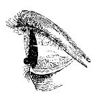
Albugo occurs during the intensity of inflammatory attacks. It also is surrounded occasionally by thinner opacity, but not depressed and unequal on the surface, as leucoma sometimes is. Large and tortuous vessels are generally seen passing into albugines, but meshes of dilated vessels are seldom present. When the affection is recent, it sometimes disappears under proper treatment, especially in young subjects; but the albugo is by no means so readily removed as the Nebula, or thin cloudy opacity which is the frequent consequence of obstinate chronic dilatation of the conjunctival vessels. Nebula is superficial, and consists of mere thickening of the conjunctival covering, from lymph having been effused. It impairs vision, but does not destroy it, for the affected part remains semitransparent.
In strumous constitutions specks of the cornea are often accompanied with ulceration of the edges of the palpebræ, and destruction of the ciliæ—the ophthalmia tarsi. The margins of the eyelids are red and slightly tumid, and discharge an acrid fluid; the ciliæ are matted together; pustules form at their roots; the bags which secrete them are laid open and destroyed, and they consequently fall out. The affection is often of long duration, and may be in part prolonged by vitiated secretion from the meibomian glands. During its progress it excites very considerable irritation in the whole eye, and, as has been already stated, opacities of the cornea not unfrequently accompany it. Veins become enlarged, and varicose on the conjunctiva, as also their minute ramifications on the clear part of the ball; small reddish lines appear on the cornea, and around them is “diffused a thin, milky, or albuminous humour,” which destroys its transparency at that part. Such spots may be solitary or numerous, and darken the cornea either partially or entirely. They are always surrounded with a fasciculus of enlarged veins.
In elderly people a dim opaque ring, of a greyish colour, sometimes encircles the margins of the cornea, and is called Arcus Senilis; but this can scarcely be looked upon as a disease.
Sometimes the cornea presents a spotted appearance; and this state of the organ is generally attended by obstinate inflammatory action in the part. The affection, however, is rare. I have seen several instances of it: in one, both corneæ were spotted, and sight was almost destroyed, without much irritability of the organ. The disease yielded to external stimulants, and the internal use of the bichloride of mercury. It is met with in a chronic and very intractable form.
The cornea may sometimes be rendered dim by over-distension, the aqueous humour being unusually copious.
Occasionally sloughing takes place in the cornea from over-action. It is dangerous to the structure and functions of the organ, according to the extent to which it occurs.
Ossification of the cornea is said to take place; but few cases are on record, and these were in very old people.
The cornea sometimes becomes conical to a great degree in persons considerably advanced in life. The cone has its apex in the centre of the organ, seems thick and crystalline when viewed laterally, and when looked on from the front has a sparkling appearance. In some cases it is opaque in the centre, and occasionally its surface is irregular. Vision of objects at any distance is very indistinct; those placed within an inch or two of the eye are most distinctly seen, especially if looked on through a small aperture. The disease usually affects both eyes, though not always in an equal degree. The patients cannot judge accurately of distance, and see objects multiplied and disfigured.
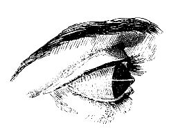
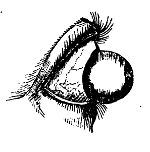
Staphyloma has been already alluded to as an occasional consequence of purulent ophthalmia in children. The cornea is thickened, prominent, and opaque; and in most cases vision is either much impaired or entirely lost. The prominence varies in different cases, being sometimes very little elevated beyond the natural state of the part, while in other instances it protrudes from between the eyelids. After having attained a certain size it often becomes stationary; but very frequently it continues to enlarge gradually. When the prominence is large, much inconvenience arises from the eyelids not being allowed to close; and the eye, being thereby deprived of its natural covering, is extremely liable to become inflamed from external irritation. When one eye is affected with staphyloma, the other not unfrequently becomes similarly diseased.
Dropsy of the anterior chamber, or Hydrophthalmia, occasionally takes place in persons of weak constitutions. The aqueous humour is either secreted in greater abundance than it usually is, or absorption is diminished. The cornea gradually accommodates itself to the increase of the fluid behind, and becomes wider and more prominent, but retains its transparency; in looking at the eye, the anterior chamber is seen evidently enlarged, and occasionally the aqueous humour is of a turbid appearance. There is little or no pain in the eyeball, but the patient complains of an annoying sense of fulness and tension in the part. In consequence of the vitreous humour also accumulating, the whole organ is ultimately enlarged considerably, and its motions are thereby much impeded. At first, vision of near objects is impaired, whilst the patient sees very distinctly those placed at a distance; ultimately sight is entirely lost.
Exophthalmia, or protrusion of the eye, attends the preceding disease, and is also a consequence of various other morbid actions in the globe and its neighbourhood, especially from the pressure of tumours in the orbit. The chronic enlargement of the bulb is noticed more fully in the succeeding chapters.
Treatment of External Ophthalmia, and its Consequences.—The exciting cause, if such exist and can be discovered, ought in the first place to be removed. The surface of the organ and of the palpebræ should be carefully examined, either with the naked eye or with a magnifying glass, in order to detect any small extraneous body which may be lodged in the part. In examining the inner surfaces of the palpebræ, it is necessary, to produce complete eversion, to bring the parts completely into view; and the most convenient method of accomplishing this is to lay hold of the ciliæ between the finger and thumb, and reflect the lid over a silver probe placed along its base. This can, by a little practice, be accomplished readily without using a probe, and even by the fingers of one hand only. This is the more necessary, as small particles of foreign matter lodge more frequently on the palpebral conjunctiva than on any other part. If a particle of glass, metal, stone, &c., be discovered, it should be gently removed by the flattened extremity of a silver probe, or by a scoop, a fine needle, or a delicate brush. In some cases washing the surface by means of a small syringe, filled with a bland fluid, is extremely useful; as when an impalpable powder has been thrown into the eye, and can with difficulty be removed, in consequence of spasmodic contraction of the eyelids preventing exposure of the parts. The application of an emollient poultice, with the addition of hyoscyamus, is beneficial when it has been found impossible to remove the whole of a fine powder. When particles of lunar caustic have, by accident, come in contact with the eye, they are to be removed, as soon after insertion as possible, by a fine hair pencil dipped in oil or fresh butter,—not in water. Small loose bodies are generally carried, by the increased lachrymal secretion, along the sulcus formed by the apposition of the eyelids, to the inner canthus, and there discharged. And, in order to favour this natural process for removing extraneous matter, the patient should be directed to keep the eyelids shut, and as quiet as possible, to cover them with his hands, and to blow his nose forcibly: thus the greater number of the extraneous particles will be got rid of. Those which remain lodged in the membranes must be speedily removed by those artificial means which have been already enumerated. If entropion is the cause of the inflammation, the eyelashes are either plucked out, or completely destroyed by removal of their roots. The inflamed organ should be carefully protected from the stimulus of strong light; the patient is to be placed in a darkened room, and the eye protected by a thin green shade. The shade, however, may be worn too long, so as to induce an extremely weak and tender state of the organ.
If there be good grounds for believing that the incited action has been caused by suppression of any discharge, that should be encouraged to return, and the cause of the suppression must be avoided. If a gonorrhœa have been suddenly arrested by the employment of stimulating injections, these must be instantly discontinued; and some have even gone so far, in such cases, as to introduce bougies impregnated with gonorrhœal matter, in order to procure a renewal of the discharge. In cases of suppression of purulent discharge from the ears, or the surrounding parts, followed by external ophthalmia, a blister or sinapism should be applied in the neighbourhood of the part from which the discharge formerly issued. When the menstrual evacuation has been arrested, leeches and fomentations should be applied to the pudenda, or around the anus, and emenagogues administered internally; the patient should be placed in a quiet and well ventilated apartment, and kept free from any emotions of the mind; all noise and other sources of irritation should be studiously avoided.
If the incited action in the eye do not subside, as it often will not, on removal of the exciting cause, recourse must immediately be had to very active means for its subjugation; for in no other organ does inflammatory action proceed more rapidly to an unfavourable termination. By timely use of antiphlogistic means, those consequences of external ophthalmia, which we have already treated of, may be avoided; and, with respect to most of them, it is much better to prevent their occurrence, than combat them after they have been allowed to take place. The eye is more valuable to a great proportion of people than a limb; and the surgeon is very culpable if he be not master of this part of his profession, and able to undertake the management of every disease and accident to which the eye is liable. “In cases of inflammation the general treatment is the same; but each variety requires peculiar attention during the cure, depending on the structure and function of the tissue affected.”
In the first stage of external ophthalmia, active antiphlogistic measures must be put in force. In full habits, and cases of intense action, general bleeding must be employed, even to fainting, from the veins of the arm or of the neck, or from the temporal artery,—and repeated, if necessary, according to circumstances. Blood is sometimes abstracted by cupping from the temples or the nape of the neck; but it is a painful and uncertain mode of emptying the vessels. Local bloodletting, in many cases, suffices to moderate the action; in all it is most beneficial and important, after the employment of general depletion. The application of leeches to the inner canthus is the most effectual method of abstracting blood locally, as at that point the venous return is made from the eye. If placed on the temples, they can produce very little benefit; if on the outer surface of the eyelids, ecchymosis follows, on account of the extreme looseness of the cellular tissue in that situation. Or the angular vein, at the inner canthus, may be opened with a lancet, and a considerable quantity of blood thereby abstracted from the seat of the disease. Leeches applied to the conjunctiva of the lower lid are sometimes advantageous; but leeching and scarification are more useful in the chronic stage: and the latter is injurious in acute ophthalmia. Saline purgatives, and antimonial medicines, a very material part of the antiphlogistic regimen, must not be omitted. Enemata, with occasional pediluvia, are much recommended by some Continental writers. In bilious habits emetics, followed by mercurial purges, will be found very useful. With respect to topical treatment, warm applications are found to afford decided relief in the first stage, and are, in consequence, generally used. By some, however, cold water, or water with vinegar, is applied from the first. Poultices, whether warm or cold, prove annoying from their weight. Warm fomentations, simple or anodyne, are preferable, and may be repeated according to the feelings of the patient; or the eye may be exposed to the steam of water.
When by these means the violence of the symptoms has abated, as usually happens in the course of a very few days, the organ must be gradually accustomed to its natural stimulus, light. The shade must be discontinued, and the room no longer darkened; and now leeching becomes of great service, while the evacuation is to be followed by gently stimulating or astringent applications, so as to produce contraction of the still dilated, though partially emptied, vessels. Various collyria may be employed for this purpose. Solutions of the sulphate of zinc, of muriate of mercury, of sulphate of alum, of acetate of lead, or of the lapis divinus—wine of opium—the citrine ointment, or the unguentum oxydi hydrargyri rubri, &c.—or stimulating vapours of various kinds. Camphor is a good addition to many of the applications. The collyria may be cold, or slightly warmed; and maybe dropped into the outer canthus, flowing over the eye, and escaping by the inner canthus, according to the natural course of the fluids of the eye; or they may be inserted at the inner canthus, the head being immediately afterwards inclined so as to allow the fluid to pass towards the external canthus; or they may be applied by means of an eyeglass. Warm fomentations, and other relaxing remedies, however useful during the first stage, are worse than useless, are hurtful in the highest degree, when the affection has passed into a chronic state; as also are antiphlogistic means, and exclusion of light,—remedies so essentially necessary in the first stage.
In ophthalmia, attended with profuse purulent discharge, the structure of the eye is in great danger of being destroyed, from the intensity of the action, and its liability to extend to the deep parts of the organ; the most active practice is required from the first. Copious general depletion, ad deliquium, must be quickly had recourse to; and the patient must be freely purged, and kept in a state of partial nausea for some time, by exhibition of antimonials. After general bloodletting, the repeated application of leeches to the inner canthus is necessary, in order to empty sufficiently the vessels of the part. Where the chemosis is so extensive as to bury the cornea, as it were, beneath the folds of the swollen conjunctiva, sloughing of the transparent tunic is frequently threatened. In order to arrest this fatal result, much good is often obtained by division of the chemosis. A sharp-pointed bistoury is passed through the swollen membrane, and radiating incisions practised, commencing at the corneal margins, and directing them towards the circumference of the globe. Sometimes four or even five of such divisions are called for, while care is taken not to wound the sclerotic coat beneath. A considerable quantity of blood is sometimes lost by this procedure, and, the chemosis subsiding, the cornea is saved. Infusion of tobacco, solutions of acetate of lead, and nitrate of silver, æther and laudanum, have been used as applications to the eye from the very commencement of the affection; but the propriety of the practice appears very questionable. Blistering the nape of the neck proves highly beneficial, after the employment of the antiphlogistic measures; and in many cases it is necessary to keep up discharge from the blistered surface for some time. On subsidence of the violent symptoms, the swelled conjunctiva is to be attacked with escharotics and stimulants, as the nitras argenti, sulphas cupri, or various collyria: then only can such applications be advantageous; at an earlier period they must do harm. They repress the exuberant granulations which may have formed, or may be forming, on the conjunctiva of the eyelids, promote contraction of the dilated vessels, diminish the relaxation of all the tissues, and stimulate the now dormant action of the part into a healthy state of excitement. Gently stimulating collyria may be injected betwixt the lids, by means of a small syringe. In granulated conjunctiva, it is sometimes necessary to remove a greater or less part of the diseased membrane by escharotics, the knife, or scissors; and after this has been accomplished it is well to encourage bleeding to a slight extent. In removing part of the palpebral conjunctiva, care must be taken to avoid injuring the cartilage of the tarsus; and, in the lower lid, not to take away too large a portion, lest entropion should occur during cicatrisation. In hospital practice, the infected should be separated from the healthy; and promiscuous use of towels and sponges must not be allowed.
In Purulent Ophthalmia of Children, antiphlogistic means must be pursued, if the patient is seen during the first stage of the disease; but children do not bear depletion well. After the discharge is established, the surface of the eye must be kept free of matter, by frequent injection of a bland, tepid fluid; and stimulating or astringent collyria should be applied three or four times a-day.
When Inflammation of the Cornea is established, it is exceedingly difficult to procure contraction of the vessels. Active antiphlogistic measures must be employed in the acute stage; and in the chronic, stimulant applications are to be had recourse to. As, however, corneitis is frequently kept up in its chronic form, from deficient constitutional power in strumous habits, strict attention must be paid to the diet and secretions of the patient. Mild mercurial alteratives, diaphoretics, and tonics combined, will often effect a cure, where all local treatment has been tried in vain. When a large plexus of vessels remain dilated on the part, the most effectual method of removal is to divide them, as they ramify on the sclerotic, by means of scissors, or a fine knife, and afterwards to employ stimulating applications.
The irritability of ulcers on the cornea is diminished by the application of nitrate of silver, in solution or substance. If in solution, the application is used in the proportion of three to ten grains of the salt to the ounce of distilled water; if in substance, a portion, finely pointed, is gently applied to the sore, which may be afterwards besmeared with a little oil or simple ointment, in order to confine the action of the nitrate to the ulcerated part. It is not necessary, but, on the contrary, hurtful, to rub the caustic freely on the sore; a very slight application is sufficient to coagulate the secretions on the part, and form a covering for protection of the surface. In two or three days afterwards, when the temporary covering has become detached, and when the irritability of the sore has in consequence returned, it will be necessary to repeat the application, but not till then. On each application, and few are in general required, the sore is found reduced in size considerably. The collyrium nitratis argenti is very useful in many obstinate cases of affections of the eye and eyelids, the strength of the solution being varied, according to circumstances.
In Albugo and Leucoma, proposals have been made for excising, scraping, or perforating the opaque part; but the cure by such means is worse than the disease, as a raw surface is left larger than the previous opacity, and the cicatrix which must inevitably form also occupies a larger space, and is equally opaque. Leucoma and Albugo are incurable diseases, though the opacities may become somewhat thinner, by natural processes, after the lapse of a long period. Nebulæ, however, are often removable. During the treatment of them, or, rather, before beginning to treat them, it is of the utmost importance to attend to the state of the surface of the eye, and of the lids and eyelashes. Stimulating substances may be applied in cases where the opacities are thin: powders of calomel, aloes, sugar, &c., have been blown into the eye; stimulating lotions or ointments are preferable; one containing the nitrate of silver, with the addition of a proportion of the liq. sub-acet. plumbi, is sometimes used with advantage. These, however, are often of no avail, unless the dilated vessels, when such exist, are divided, or a portion dissected out; afterwards stimulants will be efficacious, and must be used assiduously. The vessels may require to be divided again and again.
In Ophthalmia tarsi, gently stimulating ointments or lotions are to be used, and in obstinate cases much advantage will result from the application of blisters behind the ears and to the nape of the neck, or from the insertion of a seton in the latter situation. In children it is necessary to correct the state of the bowels, scarify teeth, and remove other irritating causes to which that tender age is liable.
Sloughing of the Cornea should, of course, be prevented, if possible, by subduing the incited action before it has attained such intensity as to overcome the power of the part. The slough is slow in separating when the constitution has been much weakened; and sometimes tonics and stimulants, both external and internal, are required to hasten the process of separation. When the surface has at length become clean, the same treatment is required as to an ulcer of the part.
Conical Cornea.—This deformity can scarcely be cured, nor can any optical contrivance effectually remedy the disturbance of vision. When the apex of the cone is opaque, the removal of the pupil to the circumference by operation affords the best chance of assisting the sight of the patient.
When staphyloma is small, neither impeding the motions of the eye, nor preventing its being protected by the lids, no surgical interference is called for, as the loss or impairment of vision cannot be remedied, and as no other inconvenience than blindness is produced by the change of form in the part. But when the diseased cornea projects from between the eyelids the prominence must be diminished, on account of the deformity which it occasions, and in consequence of the eye being deprived of its natural protection of the lids, and being thereby exposed to constant irritation. In such cases it is necessary to take away a portion of the cornea, that the eye may be so diminished in bulk as to retract within the eyelids; the size of the part removed must be proportioned to the degree of protrusion. A cornea-knife is passed into the prominence, and carried forwards so as to transfix the part, in a direction from the external to the inner canthus; and by the knife being carried on, with its cutting edge looking downwards, a flap of the cornea is made. This flap is then laid hold of by means of forceps, and removed either with the knife or with scissors. The aqueous humour immediately escapes, and in most cases the crystalline lens and vitreous humour are also discharged. The eye consequently shrinks, and retracts within the palpebræ. The cut margins of the cornea soon assume a reddish appearance—they form granulations, the wound contracts gradually, and ultimately closes; but the eye is necessarily much shrunk, and totally useless as an organ of vision. Generally suppuration takes place, causing complete disorganisation of the parts; and the preceding inflammatory action may be so intense, and attended with so much constitutional disturbance, as to require active measures for its moderation. Deformity may be in a great measure removed by adapting an artificial eye to the shrivelled remains of the natural one. When it is necessary to remove only a small part of the cornea, the aqueous humour alone escapes, and during the cure of the wound the patient not unfrequently enjoys a tolerable degree of vision; but after the wound has completely closed, vision is again lost completely.
Hydrophthalmia, also, is a disease in which little hope can be entertained of materially benefiting the patient. In the slighter cases, in which no very annoying symptoms accompany the affection, vision may be improved by the use of optical instruments; mercurial preparations may be employed in moderation, with the view of promoting absorption of the superabundant fluid. When the disease has made considerable progress, temporary relief may be obtained from puncturing the cornea at its lower part, so as to discharge the accumulated aqueous humour; but a cure can scarcely be expected from such a practice, however often the paracentesis may be repeated. In the worst cases the pain is so excruciating, and the system so much disturbed by the local affection, as almost to warrant the destruction of the organ, in order to relieve the patient; but, after all, even such severe measures will most probably prove unavailing.
In Exophthalmos the treatment must vary according to the circumstances which cause the protrusion of the eyeball.
Of Internal Ophthalmia.—Inflammation of the internal parts of the eyeball sometimes supervenes on conjunctival inflammation, and then the distinctive characters of the two affections are not perceptible. When inflammatory action attacks the deep parts primarily, the external ciliary vessels ramifying on the sclerotic coat are seen, enlarged, shining through the conjunctiva; and, as they advance towards the clearer part of the eye, they form a zone of a pink colour, whose vessels run in a straight direction towards the margin of the cornea; but between the zone and the cornea a distinct white line is often interposed. Then large arborescent and reticulated vessels soon appear on the white part of the eye; and from their being more superficial than the first, and of a brighter hue, it is obvious that they belong to the conjunctiva. They also approach the clear part of the eye, and, if numerous, obscure the former vessels—as also the red zone and white line—for they pass over them, and reach the corneal margins, and often go beyond it, in continuous ramifications. The sclerotic, in consequence, assumes a pink-red colour, and the cornea becomes dim.
The iris may be primarily and principally affected, and, if so, the disease is termed Iritis; but in most cases all the other internal parts suffer more or less. The iris changes its appearance, becomes of a dusky hue, either in part or throughout, and red vessels are sometimes distinctly seen in it; from grey or blue it changes to a greenish colour, and when formerly black or brown it becomes reddish. The size of the pupil diminishes, and the contraction is often irregular, when the inflammatory action is intense. The iris swells perceptibly, and the pupil loses its dark colour, or is almost entirely closed, either from effusion of lymph, or from inflammation and consequent opacity of the crystalline lens and its capsule. The iris projects forwards, and diminishes the capacity of the anterior chamber; the pupil is irregular, and often assumes an angular appearance; and the irregularity becomes permanent from adhesion of the pupillary margin of the iris to the capsule of the lens, lymph being effused and organised, and forming a firm uniting medium between the parts. Occasionally adhesions form at the middle of the iris, and cause so great contraction as to give the pupil an appearance of being double. Of course irregularities of the pupil are most distinct when the part is dilated, either spontaneously or by the application of belladonna. Tubercles sometimes form on the iris, and not unfrequently it presents a granulated appearance. From the commencement of the inflammatory attack the patient feels great pain in the organ and in the forehead, and there is great intolerance of light. There is a feeling of tension of the eyeball, followed by deep throbbing pain increasing every instant. As the disease advances, the cornea is rendered opaque by the fulness of the chambers, and the aqueous humour becomes turbid and of a milky appearance; or lymph is effused into the anterior chamber, and floats about in flaky portions. Occasionally the vessels of the iris are so distended as to give way, causing effusion of blood into the chamber, often in considerable quantity.
More frequently, however, puriform fluid is deposited, occasioning the appearance termed Hypopium. The pus is either fluid or of a thick curdy consistence: when fluid and thin, it mixes with the aqueous humour, rendering it white and opaque; if of firmer consistence, it lodges in the lower part of the chamber, but changes its position, and mixes partially with the humour, on the head being moved; when dense and curdy, it remains separate from the humour, and its position is not altered by motions of the head.
During the progress of the inflammatory action, all the symptoms increase; the pain shoots to the top of the head, and is much aggravated by pressure on the eyeball. Of course vision is materially impaired. Constitutional disturbance always accompanies the affection, and exists in a greater or less degree according to the extent of the disease. The iris may be primarily affected, but the other textures, both external and more deeply seated, too often become involved; and in aggravated cases the whole eyeball suffers. When the most internal parts, as the choroid coat, the retina, and the vitreous humour, are affected, sudden and bright flashes of light disturb the patient, whilst vision is rapidly lost, and for ever. Occasionally the intense over-action terminates in suppuration of all the affected textures, and the eyeball soon becomes completely disorganised.
In Rheumatic Ophthalmia the appearances of the diseased eye are similar to those in ophthalmia produced by any other cause. But the affection is accompanied with, and seems to arise from rheumatic diathesis. There is pain in many of the joints, and frequently in the scalp and portions of the face, increased on hanging the head, and by pressing the parts. The pains are remittent, supervene at night, and subside in the morning. In general the ophthalmia is external; but in severe cases the internal parts become affected, and the eye is sometimes lost by giving way of the cornea.
Internal ophthalmia is often occasioned by wounds inflicted either accidentally or by operation. Laceration of the iris in the extraction of cataract, or an improper performance of the operation for cataract with a needle, is by no means an unfrequent cause of the affection. Iritis often occurs during the exhibition of mercury in undue quantities, and is said also to be a symptom of syphilitic taint. It is, in many cases, preceded by cutaneous eruption, and seems to be the consequence of the eruption being repelled, or interfered with in its progress.
Choroiditis.—The choroid membrane is sometimes primarily affected: but more frequently the inflammation of this tunic is the consequence of sclerotitis, or the disease last described. When the result of the former cause, it generally takes on the rheumatic type. The early symptoms are zonular redness of the sclerotic, accompanied by a general impairment of vision, so that the patient expresses himself as if looking through gauze or some dark network. Presently the sight becomes more and more impaired, until a complete amaurosis results. The pupil is generally in a semi-dilated state, and, instead of presenting the intense black hue of the healthy eye, it reflects a greenish-grey colour, dependent upon the effusion of a turbid fluid between the choroid and retina. The nervous structures, becoming thus pressed upon, lose their sensibility to light, and are paralysed. This form of inflammation is generally chronic, and imperceptibly advances to the iris anteriorly, and to the retina within; the ultimate termination being complete glaucoma. Various dull and heavy pains accompany this affection; and, in the latter stages, acute circum-orbitar neuralgia is the most distressing concomitant. By long-continued chronic inflammation the sclerotic coat appears to lose its powers of resistance—the accumulating fluid pushes before it the weakened tunic, and Staphyloma Scleroticæ is produced. This protrusion of the external tunic sometimes takes place in various parts, and to a considerable extent, so that the figure of the globe is entirely lost. The thinning of the sclerotic at these points allows the dark hue of the choroid to shine through, and this, together with the bunched-like appearance of the protruded portions, has entailed upon it the name of Staphyloma Racemosum.
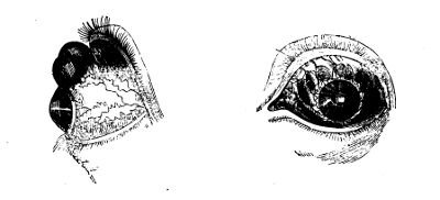
Treatment.—In the first stage of internal ophthalmia, active treatment, properly conducted, should be successful in averting the progress of the disease; in the latter stages, there is every chance of vision being entirely lost. The treatment must be actively antiphlogistic, consisting of general and local bleeding, the internal use of purgatives and antimonial medicines, and strict abstinence. A free use of mercury internally is said to check the disease, and, in its advanced stages, to procure absorption of effused lymph. But the inflammation can be subdued without the aid of that mineral, though its effects are often powerful; and a recollection of the bad effects which are so apt to follow its employment renders a prudent surgeon cautious in having recourse to it. Mercurial ointment, with opium, rubbed on the forehead, immediately above the affected eye, gives great relief. The same relief follows friction with oil, in which the muriate or other salts of morphia is dissolved. When the incited action declines, the extracts of belladonna, hyoscyamus, or stramonium, rubbed on the eyelids and brow, procure dilatation of the pupil, and thereby tend to prevent its further contraction; but whilst acute inflammation exists, the pupil is not dilatable; and it is consequently an encouraging symptom when the pupil begins to yield to the influence of these medicines. In hypopium it is sometimes necessary to evacuate the pus when effused in large quantity, in order to prevent the injurious effects that its pressure might occasion; but, if the quantity be small, there is a good chance of its being removed by absorption. In suppuration of the eyeball, whilst the other eye remains sound, it may be prudent to open the cornea, and allow a free exit for the matter, in order to prevent the healthy eye from becoming affected. In the staphyloma of the sclerotic coat, when the eye, as it were, is affected by a sort of chronic dropsy, (and this disease is met with at various periods of life,) the tension and bulk of the organ may be diminished by occasional puncture. The opening may be kept pervious by the introduction of a conical probe from time to time. I have more than once introduced a silk thread through the most dependent and prominent part of the globe with good effect. The organ ultimately shrinks.
Amaurosis implies an impairment of vision more or less complete, arising from disease in the brain, in the optic nerve, or in the retina, whether consisting of change or destruction of structure, or derangement of function. Vision may be diminished or lost by organic disease in the coats or humours of the eye, or by morbid formations in the orbit; but to such the term Amaurosis does not strictly apply. But, after establishment of the disease, other textures of the eye may, and often do, become affected. Usually one eye at first is amaurotic; but the other soon participates, and ultimately vision is impaired or entirely lost in both. The disease may occur idiopathically, or be symptomatic of other affections.
The general symptoms of amaurosis are the following. Headache is felt for some time, either constant, or, as is most commonly the case, occasional, and most severe in the forehead: in many cases the pain is at times most excruciating. The eyesight gradually becomes weak; distant objects are unusually obscure, or not at all observed; and those which are near cannot be accurately discerned. For a short time vision may seem to be restored, but soon it diminishes more and more, all objects seem to be enshrouded in a mist, at first thin and shadowy, but gradually becoming opaque and impenetrable; or a feeling is communicated of a dark network obstructing the view. Unnatural impressions are made on the retina; flashes of strong light, or luminous sparks, appear to dart across the eyes; darkened spots are seen where none exist; gnats, flies, or other minute bodies, various in colour and brilliancy, seem to flutter before the face; or a single dark speck intercepts the vision. Usually the pupil is dilated and the iris insensible to the stimulus of light; and the former has not its natural translucent aspect, but is dull and cloudy. But the state of the pupil cannot be accurately determined in amaurosis, for not unfrequently it is much contracted, and in many cases the iris retains both its natural appearance and the full exercise of its functions. The disease either advances to complete blindness, or stops in its destructive progress, leaving the patient with vision impaired to a greater or less degree. When the disease is established, pain in the head and eyes usually either ceases quickly and entirely, or gradually abates.
Amaurosis is sometimes temporary, occurring at regular intervals; and, during its accession, it often varies in intensity. With some patients strong light is intolerable, and vision is best in the twilight; others court sunshine, finding their eyesight thereby much improved; accordingly the former are said to labour under nyctalopia, the latter under hemeralopia. Some can discern the shape of objects, but either have no perception of the colours, or mistake the individual colours; others not only see all objects indistinctively, but conceive them distorted, double, or extensively multiplied: in some one-half of the object looked upon is obscured—and frequently there is strabismus, in consequence of the paralysis being only partial.
Organic amaurosis (that depending on organic disease) may arise from the change of structure consequent on inflammatory action in the retina, whether chronic or acute—from atrophy of that membrane and of the optic nerve—from extravasation into the substance of the nerve, or compression of it by morbid formations—from softening or suppuration of the nerve and its connexions—or from various diseases of the encephalon. Functional amaurosis may proceed from temporary plethora about the optic nerve and retina—from intense and long continued use of the organ—from derangement of the digestive apparatus—from general debility, however induced—from excessive influence on the system of poisons or powerful medicines—from concussion of the nervous and cerebral substance, or from long continued irritation in the neighbourhood of the eye. Amaurosis may also follow injuries of various kinds.
In the treatment of organic amaurosis but little can be done, and that little is unsatisfactory. In the functional form, however, vision may be improved, if not wholly restored, by removal of the exciting cause, and the carefully avoiding of such circumstances as seem to predispose to the affection. After due constitutional treatment, considerable benefit is often derived from counter-irritation; and I have in many cases witnessed the good effects of blistering the temples and besprinkling the raw surface with the powder of strychnine,—a practice very far from nugatory. On removing the blister, the cuticle and lymphatic effusion beneath are carefully scraped away, and from one-eighth to one-half of a grain of the powder dusted over the exposed cutis. The sprinkling is repeated daily, and the dose gradually increased. When the surface dries, a fresh blister is applied, and the use of the powder resumed. It may be employed, when gradually increased, to the extent of two grains on each temple; but, if spasmodic twitchings and constitutional disturbance begin to show themselves, it must be immediately abandoned, and not resumed till after some days, and even then in diminished doses. In not a few cases, both of complete amaurosis, and of vision impaired to such an extent that the patient could merely distinguish light from darkness, I have by this practice succeeded in restoring the sight completely; in others, vision has been very much improved. Still, by far the greater number of amaurotic patients are incurable; and even those who have derived benefit from strychnine are, I am strongly inclined to suspect, exceedingly liable to relapse.
In the treatment of functional amaurosis, it will be necessary to investigate minutely the causes on which the defective vision may depend. Thus we may find a congestive state of the retina or brain, arising from suppressed natural discharges, as the menstrual flux, or the sudden suppression of habitual but morbid discharges, as the healing of an old ulcer, &c.
Again, amaurosis maybe the result of irritation in some portion of the alimentary canal, as from the presence of worms. Patients who have long laboured under imperfect amaurosis have occasionally been suddenly relieved by the discharge of a tape-worm. Difficult and painful dentition in children not unfrequently gives rise to this disease. Hence the treatment of functional amaurosis will necessarily vary with the cause; and no general rule can, with any propriety, be laid down as to our selection of remedial measures.
Glaucoma, or green cataract, is a disease of the hyaloid membrane and vitreous humour, probably depending on a varicose state of the bloodvessels. The pupil is usually dilated, irregularly oblong, the iris being narrowed towards the upper and inner side. There is a dull shining appearance at the bottom of the eye, not fixed as in cataract, but varying according to the position of the light. The lens becomes opaque and greenish as the disease advances, vision gradually diminishes, and the iris is immovable. After sight is lost, the patient has a perception of a luminous appearance in the organ when pressed upon. Both eyes are generally affected, one after the other; headache, often violent, attends the disease; many remedies, both external and internal, may be tried on recommendation, though without effect: the disease seldom, if ever, admits of cure.
Of Cataract, or opacity of the crystalline lens and its capsule, attended with partial loss of vision.—The disease is, in general, gradual in its progress: but sometimes it advances rapidly, as when occasioned by a blow or wound. When slow, the opacity commences in the centre of the lens, and extends gradually towards the circumference. Before any change can be perceived in the organ, the patient sees objects as if covered with a mist or veil; and, as the opacity becomes distinct, vision is gradually impaired. During the day, vision is very indistinct, as the pupil is contracted, and the rays of light reach the retina only through the opaque centre of the cataract. But during twilight vision improves, as then the pupil becomes dilated, and admits of transmission of light through a portion of the transparent vitreous humour, as well as through the semi-opaque margins of the crystalline lens: for a similar reason, it is also more distinct after the application of belladonna or hyoscyamus either to the eye or to its neighbourhood. In the ordinary state of the parts, a clear black ring is often visible around the opacity, either from the margins of the lens being unaffected, or from the posterior surface of the pupillary portion of the iris being pushed forwards by enlargement of the lens. Patients, having become aware of the great improvement of vision caused by dilatation of the pupil, are often contented to use narcotic remedies externally, so long as they retain their dilating influence—and, strange to say, they do not soon lose it—instead of submitting to any operation. As cataract advances, even luminous bodies cannot be accurately distinguished, though the situation from which the light proceeds is perceived; thus the patient in a clear light may have an indistinct perception of a candle or window, and in some cases even of the bars of the window. The motions of the iris are not affected, unless, in rare cases, when the cataract is large and compresses the iris; or when the functions of the third pair of nerves have been in any way impaired; or when the iris has been the seat of acute inflammation.