PART FIRST. OF INFLAMMATION. (continued 3)
Reading edition
ON GROWTHS FROM THE SYNOVIAL MEMBRANE, AND LOOSE SUBSTANCES IN THE JOINTS.
The synovial membrane is sometimes studded on its inner aspect with loose fleshy or semicartilaginous substances projecting into the cavity of the joint. The entire surface of the membrane is occasionally covered with these bodies, which are of a white or yellowish colour, and very variable in size and shape; the smallest presenting the form of villi not much larger than those of the jejunum, the largest having somewhat the magnitude and appearance of the appendices epiploicæ of the large intestine, while many of an intermediate size approach in appearance to a lemon-seed. In some instances the membrane is only partially pervaded by them, and they are not unfrequently arranged like a fringe around the edge of the articular surfaces. They are generally very smooth on the surface, which appears to be perfectly continuous, if not identical, with the synovial membrane. Their attachment is frequently broad, and occasionally very narrow and pedunculated, often merely filamentous, so that a little further thinning of the part, or slight force acting on the body, would remove it from the capsule, and throw it loose into the cavity of the articulation. The disease has been most frequently seen in the knee, and sometimes in the elbow.
The affection is obscure in its nature; it is slow in its progress; the joint is the seat of pain after and during exercise, probably from the morbid processes interfering with the motions of the articular surfaces. As the disease advances, the joint becomes swelled and elastic, unattended generally by ulceration of the tissues within or around it. In examining the part, when the articular surfaces are moved on each other, it will be found that their motion is more or less interfered with; and considerable irregularity in their action may be felt by the hand placed firmly on the joint during the procedure.
Loose bodies may be found in the articulations, particularly in the knee and elbow, under the circumstances just described; but we often find others of a different structure loose within the capsule, which may be throughout smooth and apparently healthy. These bodies are extremely variable in size, generally rounded or oval, with a polished surface; many of the smallest present the character of fibro-cartilage or cartilage; the larger are often more or less modulated, very firm and gristly, and sometimes contain a considerable nucleus of bony matter. The articular ends of the bones are occasionally more or less misshapen by the formation around them of knobs of adventitious osseous substance.
The disease is common in the knee; and patients frequently go about for years, who will tell you that they have something rolling within the joint, which will often be found on examination to be actually the case. It is obvious that loose bodies in the joint must prove an obstacle to easy progression; and the smaller ones seem to be particularly so, probably from becoming easily entangled by different parts of the articular apparatus.
However difficult it may be to account for the cause of these formations, the means by which they may get within the articulation seems obvious enough. If a small knob of coagulated lymph should form on the inner aspect of the membrane, the point of attachment, as has already been noticed, might gradually become narrower, from the motions of the joint or other causes, till the connection were severed. But there is reason to believe that many of the substances found loose in the joints are formed altogether in the cellular tissue without the capsule; for it is difficult to conceive that they can increase in size, or take on the ossific action in their centre, by an act of independent vitality, after they have been separated from the surrounding parts. Accordingly, dissection has occasionally disclosed loose bodies within the knee-joint, and others of just the same structure outside the synovial membrane, which was however protruded by their pressure, and appeared to connect some of them by a pedicle only, ready to break and allow the foreign body, with its covering of the membrane, to fall into the cavity of the articulation.
AFFECTIONS OF SYNOVIAL POUCHES OR BURSÆ.
Bursæ are lined by a membrane, greatly resembling the synovial in appearance, function, and disease. They are frequently the seat of inflammation of an acute character; but in most instances the action is of short duration, and generally terminates in an increase and accumulation of the secreted fluid. The attendant pain is very severe, and much increased by any motion of the neighbouring parts.
Occasionally a portion of the cellular substance, which is exposed to pressure of motion, as over a prominent portion of bone, assumes the appearance of a bursa, secretes a similar fluid, and is similarly affected in consequence of inflammation. These adventitious bursæ are met with in various situations. Bunion is a good example of such a bursa thickened from long-continued pressure. They are seen on the outer ankles of tailors, on the shins of boot-closers, on the forehead, point of the elbow, &c.
Disease of the bursæ may occur from external injury, and they often become affected subsequently to disease of the neighbouring joint. If the action is violent, lymph is effused on the inner surface, or external to its cavity, causing considerable thickening. The sac is thus sometimes all but obliterated. Tumours, originally housemaid’s knee, aggravated by continued pressure, are met with over the ligament of the patella in almost a solid state. Occasionally the action terminates in suppuration, pus being effused to a greater or less extent into the cavity; and if allowed to remain or accumulate, the abscess extends, and frequently communicates with the neighbouring joint, which may not have been previously diseased. In chronic cases of enlarged bursæ, especially of the bursal thecæ of the tendons of the wrist or ankle, we not unfrequently meet with loose cartilaginous bodies of various sizes, and of a flattened oval form, floating in the accumulated fluid. These have been also supposed to be formed by portions of lymph which have been deposited on the surface, condensed in structure, and afterwards become detached.
When inflammatory action has commenced in a bursa, it must be subdued by copious topical bleeding, along with the exhibition of purgatives; in most cases general bloodletting will not be required. After the inflammation has subsided, the parts remain swollen, from the effusion either of serum or lymph; stimulating applications may then be employed with advantage. In general, the ammoniacal plaster, or the brushing over the surface with tincture of iodine will answer. Blisters are sometimes employed with the same view. In obstinate cases, when the tumour is of no large size, a seton may be passed, retained till suppuration has taken place, and gradually withdrawn. Great risk attends interference with bursæ of large size near joints or cysts containing serous or glairy fluid in any situation. Even trifling punctures into such have been sometimes followed by inflammation of the inner secreting surface and violent constitutional disturbance. When suppuration has occurred, it will in many instances be prudent to evacuate the matter by one or more incisions, in order to prevent farther mischief, especially if the bursa, a superficial one, is in the neighbourhood of a joint. After the matter has been evacuated, the cavity gradually contracts, and ultimately the bursa is completely obliterated. Diseased bursæ, near the surface, and unconnected with important parts, have been dissected out. The operation is not often necessary, and in some situations attended with considerable risk. Tumours, solid or nearly so, arising from diseased bursa of long standing, may sometimes require to be so treated.
OF COXALGIA, MORBUS COXARIUS, OR HIP-JOINT DISEASE.
This disease has been supposed to commence in the cartilages; it appears, however, to originate indiscriminately in the cartilage and the bone, as well as in the membrane lining the capsule and investing the cartilage and the ligaments; but whether it begins in one or other of these tissues, it soon, if neglected, involves them all. It affects patients of all ages, though children under twelve are most generally its victims; and in these it often makes considerable progress without its existence being suspected. The patient is observed to be a little lame, and to be awkward in the use of the affected limb, but he experiences little or no pain in the first instance; and if he does, it is of a dull kind, and generally referred to other parts. Thus, pain in the knee is generally the prominent symptom of this affection, and occasionally pain is also referred to the ankle, or to the sole of the foot: careful study and considerable experience are here required, to guard the young practitioner from error in diagnosis. Parts remote from the seat of morbid action have often been made the subject of treatment in this and other affections; the knee, in morbus coxarius, has been leeched, poulticed, blistered, and burnt, and that, too, when this joint was not at all altered in appearance, and showed no symptom of disease.
Again, and particularly in adults, the limb is easy only in certain positions, and cannot be moved without great suffering; pain is also complained of in the groin, and often immediately behind the trochanter major. If an examination is made when the patient is thus halting, and even though he complains of no pain, the limb is found shrunk, wasted, and lengthened. The elongation of the limb occurs mainly in consequence of the inclination of the pelvis towards that side. When the disease has made progress, it has been supposed that swelling of the apparatus of the joint, and effusion into its cavity, might separate the head of the bone from the acetabulum, when pressure from the trunk was not applied. The lengthening is often great, and its extent and cause are ascertained by accurate comparison of the two limbs, laid in contact when the patient is in the recumbent posture.
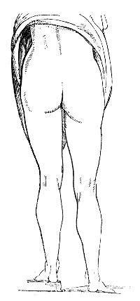
The degree of lengthening is here carefully represented from a recent case. But occasionally, even in the first stage, before destructive ulceration has set in, in consequence of the pain and spasms, the limb becomes remarkably shortened and retracted. This also will be found, on careful examination, to depend upon the relative positions of the two ossa innominata.
When the patient stands, the affected limb is considerably advanced before the other, on which the weight of the trunk is chiefly, or entirely, supported; the knee is generally bent, and the toes only rest on the ground. In the advanced stages of the disease, and when there is reason to suppose that ulceration of the cartilages has set in, the patient, during progression, moves the affected limb with the hands grasped round the thigh, and in bed it is moved by the aid of the sound one. The spine is frequently affected, becoming bent in different directions, to preserve the equilibrium of the body; and a deformity of the trunk to a certain degree occurs, which, however, may be in general easily remedied. The nates are much altered; they become flattened, and those parts which are naturally most prominent are reduced to the level of the others; the usual niche between the buttock and thigh, in the erect position, is effaced, and the upper part of the thigh is often considerably swollen. The alteration is at once manifest on contrasting the healthy with the diseased side. Even from the first, locomotion is difficult: in the morning, the movements of the joint are constrained and stiff; afterwards, however, the patient walks with more ease, though still by very slight exertion the limb is speedily tired, and he is unwilling to use it. Pain is produced by pressing on the groin, or by tapping on the trochanter, and by pushing the head of the femur forcibly against the acetabulum. The inguinal glands occasionally become enlarged. As the disease advances, the lameness is more apparent; pain is produced and increased by motion, and by any attempt to stretch, and more especially to abduct the limb whilst in the recumbent posture. The emaciation of the member becomes more and more visible. The muscles, as it were, are paralysed from inaction and pain, abscesses form, and the constitution then sympathises remarkably; hectic fever supervenes, with its usual train of symptoms.
The circumstances attending the first stage of the disease in childhood, in which the limb is lengthened, and there is no decrease, but rather an enlargement of the parts composing the joint, have been already described and illustrated. When, however, absorption occurs, and the articulation begins to be destroyed, the second stage of the disease commences, and the limb becomes then sensibly shortened; the toes are turned inwards or outwards; in many cases there is every appearance of dislocation of the thigh upward and backward; and in others the limb is much bent, the toes only reaching the ground. The ultimate position of the limb and degree of shortening will depend much upon the extent to which the head and neck of the femur is destroyed, upon the inclination of the pelvis, and also upon the portion of the acetabulum which is most diseased. The joint becomes tender, the slightest motion causing much pain, and the parts around appear swollen. The patient retains the limb in the most comfortable position, and it is generally bent upon the pelvis and inverted. This may arise from relief being afforded when the psoas is relaxed, and the pressure thus removed from the fore part of the joint. In many cases matter forms behind, or rather below, the trochanter major, and the collection often attains a large size. When the presence of matter has been ascertained in this situation, it has been recommended that an early opening should be made, on the supposition that the disease arises from an acrimonious discharge into and round the joint, and that, by the matter being allowed to escape, the cause of the disease may be removed. The synovia has been compared by one old author to bland oil, the vitiated secretion to oil of vitriol. Though the principle is incorrect, still the rule of practice is important; for in consequence of the long-continued presence of matter, accumulating in a cavity which is not dilatable in proportion to the increase of purulent secretion, the original affection will be much aggravated, and disease induced in the neighbouring parts. But the existence of matter in the joint could only be ascertained to exist in a very emaciated person.
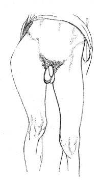
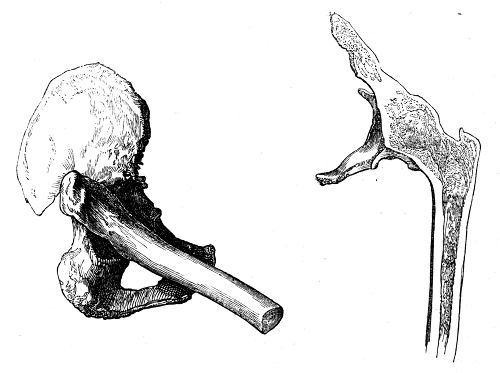
The formation of matter is preceded by great pain, and frequent startings of the limb during sleep, accompanied with fever, and other symptoms of severe constitutional disturbance. On the escape of matter by the natural process from the capsule the painful feelings usually subside. The abscess may appear, as already stated, near the trochanter major, or in the back part of the thigh. Matter sometimes makes its way into the pelvis, through a perforation in the acetabulum, thence it may fall through the sacro-ischiatic notch into the thigh, and find its way under the fascia, nearly to the knee; or again, it may present to the side of the rectum, or even, as I have seen, burst into the bowel and continue to be discharged thus for a long period. If the treatment is neglected, abscess succeeds abscess; and in consequence of the profuse discharge, which may be evacuated from one or many openings round the joint, the patient is at length exhausted, and sinks. In some instances the spontaneous cure by anchylosis occurs, as in the instances from which these sketches are taken. In the one, the head and neck of the bone had been almost entirely destroyed by ulceration, before anchylosis had begun; in the other, the change is very slight, but the head of the femur and os innominatum are inseparably united by bone, and their cancellated texture runs into each other. Or when the femur has been dislocated, which is a very rare occurrence, the disease sometimes gradually abates, and a sort of new joint is formed; the limb, after some time, may thus again become so far useful to the patient.
In many cases, the appearance which the various parts of the diseased joint present, are similar to those which have been already described when treating of affections of the joints generally. Frequently, however, the osseous tissue in this situation is much more extensively affected. Often the whole cartilage on the head of the femur is completely removed, exposing the bone in an ulcerated condition; and when the system has long borne up under the disease, the greater portion of the head, neck, and even of the trochanter, is destroyed, the extremity of the bone being completely altered in form, and composed of a loose and spongy structure. A similar disorganisation occurs in the acetabulum; the mucous gland is destroyed, the cartilage is often wholly removed, and the margins of the acetabulum absorbed, a large and flat ulcerated depression merely being left for the reception of the diseased femur; in other instances the margins remain unaffected, whilst the ulceration proceeds in the centre, and the cavity is thereby much deepened. Not unfrequently the ulceration proceeds farther, and an aperture is formed in the acetabulum, so that matter accumulates within the pelvis. The opening is sometimes so large that the femur is protruded through it. When matter has formed in the soft parts round the joint, portions of the bones of the pelvis, in contact with the pus, are ulcerated to a greater or less extent, and sometimes these ulcers are surrounded by deposits of new bony matter.
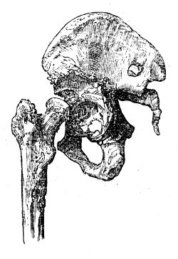
From such changes in the osseous parts of the articulation the limb is shortened, sometimes to a great degree, though no dislocation has occurred. Indeed, dislocation is by no means so frequent a cause of the shortening as is generally believed.
If the head of the femur has been dislocated, and if the disease in the joint has afterwards subsided, the acetabulum is found to be much contracted, with its margins smooth and little elevated, and, if the patient survive for a number of years, it will be almost wholly obliterated. But a portion of the dorsum of the ilium, upward and backward, which is the most frequent dislocation in this disease, is gradually absorbed, so as to form a sort of glenoid cavity for the reception of the femur, the extremity of which becomes more solid in texture, and more smooth on its articular surface. The remaining neck of the bone is in the sketch here given turned forwards, and must have given rise to great eversion of the toes. I have seen one other specimen of this form of luxation. The limb is generally, however, inverted; and what remains of the head of the bone consequently points backwards. The consecutive luxation occasionally, also, though rarely, takes place upon the pubis. Whilst a depression is thus formed, new bone is sometimes deposited round its margins, whereby the cavity is increased in depth, so as to resemble somewhat the original acetabulum, the new deposit having become smooth and of a regular form.
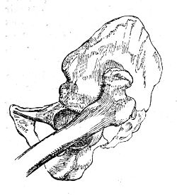
ON CERTAIN ALTERATIONS OF FORM IN THE HEAD AND NECK OF THE THIGH BONE.
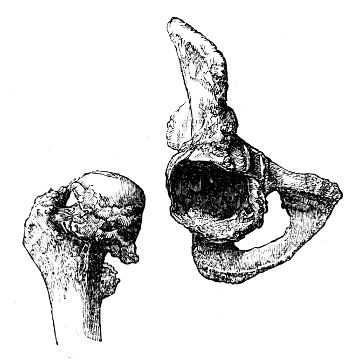
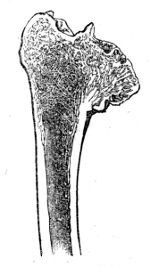
When treating of atrophy of the articular cartilages, it was observed that, in the latter stages of the disease, the subjacent bone was liable to become denuded, and its articular extremity more or less deformed. Now one of the most interesting of these changes occurs in the head of the thigh bone, which becomes polished on its surface, flattened and expanded, with a corresponding alteration in the acetabulum. In other cases the head of the thigh bone is somewhat elongated, and the acetabulum becomes deepened in proportion, by a deposition of new bone around its margin, as shown in the preceding page, frequently to such an extent as to render the removal of the head of the bone, even after the removal of all the soft parts by maceration, almost impossible without fracture of the edge of the socket. The head of the bone may also become still further misshapen, and anchylosis result; while in some instances the new bony matter presents in the form of nodosities, sometimes projecting towards the cavity of the articulation. This cut represents a section of the upper end of the femur of a labouring man, aged 49, who had fallen on the trochanter ten years before death, and became gradually lame in consequence, with shortening of the limb and anchylosis of the joint, although he had never been confined more than a day or two on account of the injury.
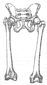
But there is another affection in which the femur may be shortened in a comparatively brief space of time from absorption of the intra-capsular portion of its neck, generally succeeding to a fall on the trochanter, and sometimes occurring in young and middle-aged subjects. In treating of fractures of the cervix femoris, it will be observed that retraction of the limb is occasionally delayed for a while: hence the importance of an accurate knowledge of those cases in which shortening of the neck of the thigh bone may succeed to an injury short of fracture. The deficiency of accurate anatomical details concerning such cases led to a doubt as to their existence; but this question has been completely put at rest since the history and dissection of two unequivocal examples by Mr. Gulliver; and the annexed cut represents the changes of form in the head of the bone, the shortening of the neck and comparative length of the femora, in a young man from whom these bones were obtained, and who walked about as many others have done, during the progress of the disease.
In the above, and in many other cases, the shortening of the neck of the bone is unaccompanied by any absorption of the articular cartilage of its head. A section of another well marked specimen may be here added. This bone also exhibits approximation of the head to the shaft, from absorption of the neck. The head is somewhat flattened and expanded, but the articular cartilage is entire, and of its natural thickness. The acetabalum was diminished in depth, but enlarged laterally, so as to correspond with the alteration of shape in the head of the thigh-bone. The preparation was obtained from a man at 32, who died at the General Hospital at Chatham of pulmonary consumption, after a confinement of two years in hospital. Previously to his admission, he had regularly performed his duty as a light infantry soldier, from which it is plain that his limbs were then of equal length, although, when the body was examined, the affected femur was upwards of one inch and a half shorter than the other. From a careful inquiry after his death, it appeared that he had, five years previously, fallen on the trochanter, in consequence of which he often complained of pain in the hip, but continued to do his duty long after, never having been confined on account of the accident. It therefore results, that morbid change had taken place during his long confinement in hospital, a circumstance not very favourable to the recommendation by some surgeons of the horizontal posture, as a remedy in such cases, and equally adverse to the opinion of certain continental pathologists, who inform us, that shortening of the neck of the femur is to be attributed to the gradual operation of the superincumbent weight of the body.
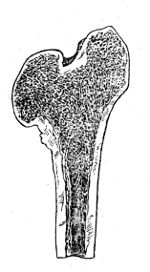
In old subjects, particularly in fat women, the neck of the femur is often shortened, and becomes more transverse and brittle than natural, from a true interstitial absorption or atrophy of the osseous tissue. This has sometimes been described as a natural effect of age, but it is undoubtedly disease, for in the greater number of old people the neck of the thigh-bone presents its usual length and obliquity. The affection is obviously a very serious one, as predisposing to fracture of the part, under circumstances which render its reunion almost hopeless.
Treatment of Affections of the Joints.—After the infliction of a wound, accidental or not, in order to prevent inflammation of a joint from becoming violent, it is of the utmost importance to bring the edges of the wound into close apposition. Sometimes neither local action, nor disturbance of the constitution, supervene on an opening, even of considerable size, being made into a joint, while the slightest puncture often gives rise to the most dreadful symptoms, both local and general. An incised wound, of no great extent, will be sufficiently closed by the careful application of adhesive plaster, and attention to the position of the limb; but if it is extensive stitches become indispensable. The parts must be kept in a state of complete relaxation and rest; cold cloths or iced water, allowed to drop or run over the surface by capillary attraction, should be assiduously applied. The patient’s bowels must be freely opened, and he is to be kept on low diet. If inflammatory action occur, bleeding, locally and generally, must be had recourse to energetically, accompanied with warm fomentations to the parts; and, at the same time, preparations of antimony, and other saline medicines, are to be administered internally. If there be reason, from the symptoms, to suspect the formation of matter, the parts ought to be attentively examined, in order to detect its presence; and, when discovered to exist, it must be evacuated without delay. In chronic cases, even local bleeding to any great extent is inadmissible. In some a few leeches may be applied, followed by counter-irritation, with advantage. The employment of counter-irritation is, perhaps, chiefly to be trusted to in the treatment of those more chronic affections of the joints in which, from the painful feelings, there is reason to dread disease of cartilage or bone. Of these, blisters are most in use; though, from my own experience, I cannot much recommend them. Their constant repetition is exceedingly annoying, and the slow progress which is made towards a cure under their use is far from encouraging. Tartrate of antimony, applied in the form either of ointment or of plaster, is generally productive of much advantage, in the first instance, by causing a great degree of superficial irritation, and relieving the internal parts. After the pustules have been fully developed, its use is to be discontinued till the surface be nearly whole, when it is again to be resumed, if the recurrence of painful feelings should demand it. Caustic issues have been much praised, and are occasionally beneficial. An eschar is made by the caustic potass applied to the skin, or by rubbing the bichloride of mercury, or any other caustic, into scarifications made by the lancet: the slough separates, and pus is discharged. Instead of promoting a continuation of the discharge, by applying savine ointment, and inserting foreign bodies, or employing other irritating dressings, it is better, when the sore begins to heal, to repeat the application of the caustic to another part; or, when the issue begins to dry up, to apply for a few hours a pledget of strong antimonial ointment.
The employment of the bichloride of mercury, though a most efficient escharotic, appears not to be unattended with danger, as in many instances violent purging, with tenesmus and bloody stools, follow its application. When swelling exists without pain, considerable advantage may be derived from frictions and liniments of various kinds, with careful bandaging. No applications can be of the least avail unless the joint be kept completely at rest.
When there is no pain in the joint, when swelling exists, with or without fluctuation, and there is every reason to believe that the extremities of the bones are not much diseased, a cure may be, in general, obtained by the employment of rest, support, and slight superficial excitement. A practice which has been extensively and rather indiscriminately pursued in diseases of joints and of other parts, in affections of synovial membrane, ulceration of cartilage, disease of bone, and even in cases of necrosis, is here detailed. It is useful in proper cases, very hurtful in others. The joint is well washed with soap and water, and afterwards rubbed with camphorated spirits of wine: it is then covered with an ointment spread on lint, and composed of equal parts of the unguentum hydrargyri cum camphora and the ceratum saponis—in the majority of cases the mercurial ointment would be better omitted—strips of lead, soap, or adhesive plaster are then applied with a moderate degree of tightness round, from two or three inches below the joint to the same distance above it: these are covered with soap-plaster spread on thick leather, and the whole is surrounded with a bandage, which extends from the extremity of the limb. The bandage should be put on as far as the joint, before the plasters are applied. The irritation produced on the surface tends to check the deep-seated action, whilst the compression excites the action of the absorbents to remove the effused fluids, and thus to reduce the swelling: by the joint being kept completely motionless, not the least salutary indication of the treatment, the cartilage and bones, if ulcerated, are placed in a condition tending much to expedite their restoration to a natural state. If they are diseased to such an extent that the process of cicatrisation cannot be expected, a cure by anchylosis is, by these means, much more likely to occur. The dressings may be left undisturbed for the space of a week or two; at which period, and, in many cases, sooner, they will be found much slackened, in consequence of the swelling being greatly reduced. By repetition of the application at intervals, the disease will, in a great many instances, rapidly cease, and the joint resume almost its natural appearance; but the period at which this takes place will be found to vary much according to circumstances. If, however, the plasters produce such irritation as to cause a return of inflammatory action, their use must be discontinued till such action has been subdued by the usual means. If the thickening of the external parts has occurred to such an extent as to cause immobility of the joint, or if partial anchylosis has ensued, the limb may be brought to the most convenient position, the knee nearly, but not quite, straight, the elbow half bent, and so on, by the cautious use of a jointed splint, fitted with an extending screw. The practitioner is not to be deterred from having recourse to the above practice, even when matter has collected and burst externally, for sometimes under its employment the cavities of abscesses contract rapidly. After the swelling and other symptoms of disease have subsided, the joint is to be slightly moved, but with great care; and, by a gradual increase of the movement, the natural motion of the part may be ultimately restored. The great object in treating chronic affections of joints must be to give them support, and prevent motion as much as possible. This is effected pretty well in the articulations, removed from the trunk, by the process above described, modified according to circumstances. The filth and smell arising from the plasters often becomes annoying, and, above all, the eruptions and excoriations produced by them prove so troublesome that the application requires to be discontinued. The principal and most essential indication, that of securing repose to the affected parts, is fully as well obtained by the application of well-adjusted splints. These may be formed of lint soaked in a mucilage of gum acacia, as described in the Practical Surgery, p. 150, or coarse linen may be smeared over thickly with a coating of mucilage mixed with common whiting, and another layer of linen spread over that. This is laid out smooth and allowed to dry; portions of this sheet are cut to the form of the affected joint, and, after being moistened, are applied and retained by a roller. A firm case is thus formed, which may be afterwards lined with lint or cotton wadding, and so reapplied. The most suitable splints of all are made of leather, prepared without oil, softened in warm water, and put on as above described. They are, when they have become dry, pared and well fitted, then lined with wash-leather, and padded as may be required. The employment of these splints gives great relief in cases where further excited action has been lighted up in the joint in consequence of the surface of the articulating cartilage having become ulcerated from its free or attached surfaces, or in consequence of matter finding its way into the synovial cavity from the cancelli in the head of the bone. In cases, also, where the painful sensations have existed from the first, and even before any great alteration in the external form of the joint has taken place, indicating primary ulceration of cartilage, this practice affords immediate relief. These splints are easily applicable to the shoulder or hip-joints; and, in the majority of instances, nothing further requires to be done. It appears that Dr. Physick of Philadelphia had been for many years in the habit of confining the motions of the hip-joint by means of hollowed wooden splints, and his practice was marked by very favourable results.9
It is seldom that local abstraction of blood is at all required, and its employment in cases of morbus coxarius in weak constitutions, which it generally seizes upon, is very questionable. Neither is counter-irritation called for; and it is only in extreme cases, when the suffering is intense, that small blisters over the fore part of the joint, or a slight drain in that situation, or behind the trochanter, is admissible. The nitrate of silver rubbed on the surface causes vesication and discharge; and its early repetition is often followed by good effects. The tincture of iodine, or a liniment containing sulphuric acid, may sometimes be applied with the same view. There is no doubt whatever, that much of the benefit that attends upon counter-irritation, both in the human body and in the lower animals, is to be attributed to the rest that is at the same time enjoined, and often indispensable. It is thus that the firing and blistering of horses does good in the majority of the diseases of tendons, bursæ, bones, and joints. In some instances, it may be necessary to have recourse to soothing applications, as fomentations to affected joints, and to employ general antiphlogistic means suited to the age, strength, and constitution of the patient. In the greater number of cases in young subjects, after the stomach and bowels are unloaded, the system must be supported by tonics and nutritious food; none are more beneficial than the preparations of iron.
When the occurrence of anchylosis affords the only hope, this process ought to be encouraged by rest, and the limb at the same time kept in that position which will be most useful in after life; this will be effected here also by the employment of splints.
In cases of disease of the knee, ankle, elbow, or wrist joints, notwithstanding everything that can be done, the disease often runs its course, abscesses form and give way, the patient’s health declines, and he becomes emaciated and hectic. In such cases amputation, when not contraindicated by internal disease, must be had recourse to, as the only remedy. In some few instances, the excision of the diseased extremities of the bones may supersede this operation; but this will be discussed in a future part of the work. Every circumstance must be well weighed before mutilation be resorted to; and there can be little doubt that thousands of limbs have been saved by the employment of the means above mentioned, which would have been otherwise doomed to amputation.
Hydrops articuli will in general be got rid of by the use of friction, either dry or with liniments; by stimulating plasters or by blisters; and by the proper application of bandages. Mercurial ointments are used in this affection, and often with very great benefit. Electricity has been recommended.10
In neuralgic affections of joints, it is clear that the symptoms are to be combated by general and not by local means, at least of a severe nature. The patient must be amused and occupied, exercise in the open air must be enjoined, and attention paid to diet. The state of the digestive organs and secretions should be looked to, and corrected if need be. Medicines directed to the regulation of the functions of the uterus are essential in the majority of cases. When there is reason to suspect the lodgement of sordes or worms in the intestinal canal of children so affected, the balsam of copaiba, in doses of Ʒss., or more, on an empty stomach, followed, if need be, by brisk purgatives, will be found to answer admirably. Tonics, such as preparations of iron, of quina, &c., are then given with advantage: many apparently bad cases yield at once to such treatment.
As local applications, anodynes, fomentations, or cold lotions are employed, according to circumstances. Frictions with gently stimulating liniments, belladonna, veratria, &c., may be used as occasion demands. The patient must be encouraged to use the affected limb as much as possible; and the use of all severe and heroic remedies, as bleeding, blistering, counter-irritation, setons, issues, or moxas, discountenanced.
OF INFLAMMATION OF BONE, AND DISEASES THENCE ARISING.
Bones grow and are nourished by the same means, and are subject to the same laws, with other parts of the system. Like all the tissues of a white colour, particularly when their growth is completed, they are less freely supplied with bloodvessels and nerves than other parts. When incited action of the bloodvessels occurs in the harder textures, sensibility is roused to an exquisite degree, and the healthy and perverted processes often advance with great vigour and amazing rapidity.
Inflammation of bone often arises from external injury, and in some constitutions from very slight causes. Its occurrence is supposed to be favoured by a syphilitic taint, but the inflammatory disposition is much more frequently produced in a system vitiated by the abuse of mercury. From the unyielding nature of the tissue, the pain attendant on inflammatory action is dreadfully excruciating; it is also most violent during the night, even in chronic cases, a circumstance which does not admit of satisfactory explanation. The integuments over the inflamed bone are swollen, and the tumour is œdematous; whilst a hard and solid tumefaction exists in the more deeply-seated parts, caused partly by enlargement of the osseous tissue and partly by effusion of lymph into the cellular substance. The bone is imbedded in a gelatinous or lymphatic effusion, situated mostly beneath the periosteum. This membrane is more vascular than in its natural condition, thickened, and at the same time opened out in texture. The bloodvessels of the affected bone are much increased, both in activity and in size; and, in consequence of enlargement of the vessels, and thickening of the naturally delicate membrane on which the vessels ramify, the bone is swollen and increased in size; its texture, as shown in the annexed cut, is loose, somewhat resembling the cancellated structure, and its surface is occupied by numerous foramina, which are enlarged in proportion to the size of the vessels which they contain. The limb is often enormously swollen and indurated. The gelatinous effusion beneath the periosteum speedily becomes organised, nodules of osseous matter project into it, and adhere to the surface of the bone frequently by a narrow neck; these increase in number, gradually assume a solid appearance: the bone is thus thickened, often to a very great extent.
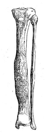
It has been supposed that the new osseous matter is deposited by the vessels of the soft parts and of the periosteum; but there can be little doubt but that it is secreted principally by the vessels which ramify within the substance of the bone, and by the vessels of the periosteum after they have entered the osseous tissue. Thus, in the case of fracture, the new osseous particles lie between the periosteum and bone at a distance from the broken ends, where the vessels are enlarged and increased in activity, or adhere to fragments which have been detached in part and retain their vitality, but not to the under surface of the periosteum. There is no doubt that thin laminæ of bone are now and then found attached to the periosteum, or impacted within its substance; but this is to be attributed to that morbid action of the tissue, to which this as well as several other membranes is subject.
When bone is extensively affected with inflammation, motion is impaired; the muscles being displaced and retarded in their action by the swelling and irregularity of the bone, by effusion of lymph into their tissue and intermuscular spaces, and, perhaps, also, by their partaking, in some measure, of the inflammatory action. Any attempt to move the parts very much aggravates the patient’s suffering. Occasionally inflammation attacks almost all the bones in the body, and causes great constitutional disturbance, by exhausting the powers of life: it sometimes terminates fatally. Bones become inflamed from various causes. However it originates, the action ends, as in the other tissues, in resolution, suppuration, or mortification. The effusion by which the diseased vessels naturally relieve themselves in softer textures cannot here take place so readily, or to such an extent as to prove beneficial: the intensity of the action is with difficulty subdued, and, consequently, resolution is comparatively rare. When it does occur, the parts do not soon regain their natural condition, but often remain considerably swollen and indurated, as is seen in nodes, which continue during the life of the patient, without causing pain or much inconvenience.
Suppuration on the surface, or in the centre, and partial or total death of a portion of bone are the most frequent consequences of external injury and incited vascular action; but suppuration in the cancellated texture frequently follows very slight incited action in those of a scrofulous habit. Tubercular matter exists, in all probability, previously, and it leads very often to long continued disease, curable only by operation. The matter may find its way to the surface after long suffering and great constitutional disturbance. Again, it may be confined for months, or even years, the patient suffering from time to time the most excruciating agony. The bone becomes thickened towards the surface by new deposit, as the cavity is increased by ulcerative absorption, and relief is only afforded, a correct diagnosis having been formed, by artificial evacuation of the matter. In many cases small sequestra lie in the cavity; and though the matter escapes naturally, or is evacuated, the patient’s strength becomes worn out, and he perishes, unless the limb is removed. So long as dead portions of bone remain, the discharge cannot cease permanently. Fresh collections are apt to form in the soft parts if the original openings close. It is no easy matter to discover or remove sequestra from deeply seated bones. A section of the femur is represented on the next page, showing a cloaca leading to the cavity of an abscess in the medullary canal. The bone is very dense in consistence, and irregular on its surface.
SUPPURATION IN BONE
Suppuration in bones is necessarily connected with loss of substance, and condensation of the surrounding parts; and purulent collections exteriorly, if allowed to press long, or if bound down by unyielding sheaths, will sooner or later produce a breach of continuity, by causing absorption of the outer lamella and the subjacent cancellated texture. A similar effect is produced by aneurismal and some other tumours. Such loss of substance is, in some instances, speedily repaired, after removal of the cause, by effusion of new matter from the surrounding bloodvessels of the bone; thus, in disease in consequence of pressure from large aneurism, there is reason to suppose that the healing process commences as soon as the aneurismal sac begins to diminish, as after operation. But, as has been already observed, the healthy actions are more vigorous in the softer tissues than in bone; and when ulceration has occurred in the latter, it is generally attended with weak action, and presents the same general characters as an ulcer in the soft parts, connected with a feeble action of the bloodvessels; the discharge is thin and fetid, absorption gradually proceeds, and there is little or no effort towards reparation. Cavities in bones are necessarily slower in healing than those in the soft parts; the vitality and power of reparation are lower; and there being no elasticity in the parts, the walls cannot come rapidly together, contract and coalesce. It may tend to prevent confusion of the two different morbid states, if we confine the term ulceration to suppuration in, and absorption of, bone, whilst the vessels retain a considerable power of action, throw out new matter, and procure a reparation of the breach; and this condition of the osseous tissue exists when the disease is situated in the surface of the bone, and when it has been produced by an external cause. On the contrary, the term caries will denote that peculiar kind of ulceration in which reparation is hardly attempted by nature, and is with difficulty obtained by the most active interference; and this disease will most generally be found to affect the cancellated structure. The comparative frequency of one or other of the terminations of inflammation depends much on the kind of bone implicated.
CARIES
Caries most frequently occurs in the heads of long and in the cancellated structure of the short bones, as unhealthy suppuration most frequently takes place in the loose, fatty, and cellular tissues of the soft parts. The formation of abscess in the cancelli is generally preceded by deposit of tubercular matter, isolated or collected in masses, and by the softening of this cheesy substance. When pus has formed in the substance of a bone, the outer lamella, in the end, becomes absorbed, and the effusion undermines the periosteum, which, from the distension, also ulcerates: the matter then spreads into the neighbouring cellular tissue, or makes its way to the surface, and is evacuated, or, what is still worse, it escapes into an articulation. The discharge is often continued, as already stated, in consequence of a dead portion of the cancellated structure being imbedded either in the carious cavity, or in the soft parts, where they sometimes are lodged for a long period.
It was formerly remarked that bones become highly sensible from incited action; hence, during the progress of this disease, which is attended with more or less inflammation, the patient generally suffers most excruciating agony—so great, in general, as to prevent him, perhaps for weeks, from enjoying the least repose. The affected part is considerably swollen, but the enlargement is seldom so general, or so great as in the diseased state of the ligaments and other apparatus of a joint. White swelling, however, may be the precursor of caries; or, in other words, a disease commencing in the bursæ, ligaments, synovial membrane, or cartilage, may extend to the bone; and breach of continuity, attended with weak action, be the consequence. In caries the affected portion appears neither to possess vitality enough to enable it to repair the breach, nor to be sufficiently deprived of vitality to be thrown off by the surrounding parts. Considerable portions of dead bone are occasionally found in carious cavities, in the heads of bones, and even in the vertebral column. Small portions are also detached piecemeal in the progress of ulceration, and discharged; but it is seldom that the whole surface is thrown off, so as to give place to a healthy and reparative action. When the parietes of the cavity have remained a considerable time in this inactive state, the surrounding vessels become more active, and the surface of the bone in the vicinity is studded with nodules of new osseous matter. The disease here delineated affected only a small portion of the cancellated texture of the condyle. An ashy looking substance fills the cavity, and this again was concealed in the recent state by lardaceous matter. The elbow joint is unaffected, excepting only that, from the deposit of new bone in spiculæ and nodules, and the condensation of the soft parts, almost complete anchylosis had taken place. This deposit is not always limited to the affected bone, if one only be the seat of the disease, but frequently extends to those articulated with it. The soft parts are commonly more or less thickened, and rendered exceedingly dense by effusion of lymph into the cellular texture; and so great is this thickening sometimes, that the knife is resisted as if by cartilage. The discharge which proceeds from the carious part is generally highly fetid, very profuse, is often poured through several openings, and the surrounding skin is excoriated and generally of a livid colour. The ichorous discharge occasionally dries up for a short period and again breaks out more violently. The surface of the ulcer is, in some cases, occupied by soft unhealthy granulations; in others the earthy part of the bone is most prominent. When the parts have been macerated and dried, the disease is often found to have proceeded more in width than in depth, and the absorption has not reduced all points of the diseased surface to the same level, thin portions remaining somewhat elevated, and giving the part a cancellated appearance; and there often project numerous minute osseous fibrillæ of considerable length, which intermix with one another, and form a most delicate network. In other instances, the ulceration has extended more deeply and uniformly, and a considerable cavity is formed, with irregular margins and surface; not unfrequently it contains dead portions of the cancellated structure, in some of a dark, in others of a light colour; or it is occupied, in the recent state, by a substance resembling lard. The surrounding bone is much softened, and, after maceration, becomes exceedingly light. The disease is generally confined to one or two bones, but occasionally involves a whole chain. It may be limited to a part of one bone in a joint, or may embrace the whole of it. Its extent will depend on the severity of the primary action, or on the degree and duration of the pressure of fluid which has been allowed to exist, whether from the nature of the superincumbent texture or the carelessness of the surgeon.
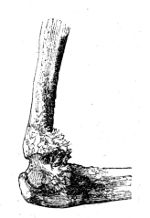
Interstitial absorption of those bones which are in the neighbourhood of the carious ulceration often occurs in the tarsus and carpus. The superincumbent integuments are livid and cold, and pain is felt in the situation of the bones; yet they are not affected with continuous ulceration, but portions of their substance are gradually removed by absorption, so that they are much loosened in texture, and may be altogether destroyed, or come to consist merely of a thin and reticulated osseous shell, whilst at the same time their cartilaginous surfaces often remain in their healthy condition.
The constitutional disorder attendant on caries is at first very great; the sympathetic fever is followed by hectic, under which, and the discharge, many patients sink. The general affection in some degree keeps pace with the local in violence and duration. The irritation is in some cases so great as to destroy the patient in a very few months or weeks; but not unfrequently a constitution, by no means strong, will be enabled to bear up for a long period under very extensive disease of a bone. The paroxysms of pain and inflammation occasion fresh attacks of constitutional derangement: this occurs till the patient’s health and strength are exhausted, and he sinks under the disease, or is relieved by the spontaneous or artificial removal of the cause.
A natural cure of caries may occur in consequence of the diseased parts so far recovering their natural degree of vascular action as to form granulations and repair the breach; but most frequently it is necessary, for the accomplishment of this purpose, that incited action occur to a very considerable degree; and the diseased parts, already extremely weak, have not sufficient power to withstand the action, but perish; whilst the action of the surrounding parts, not being increased to such a degree as to overcome their powers, throws off the dead, secretes a more bland discharge, and deposits healthy granulations, which gradually fill up the cavity.
Treatment.—In inflammation of bone, resolution must be brought about, if possible; the other terminations are to be prevented by all possible means, since they frequently endanger the limb, and even the life, of the patient, and, at best, never admit of a speedy cure. To promote resolution, blood must be drawn copiously from the part; and general bleeding may also be required, though in some constitutions it cannot be safely carried to any great extent. After local bleeding, fomentations assiduously applied will tend much to relieve the sufferings of the patient. Purgatives, nauseating doses of antimony, and all safe measures likely to subdue the vascular action, must at the same time be adopted. Free incisions through the periosteum sometimes relieve the pain, and cut short the disease, the distended vessels being thereby emptied; but such practice is only a last resource, when the action has resisted all other means, and threatens an unfavourable termination. If, notwithstanding the resolutive means employed, the inflammation proceeds unabated, and suppuration occurs, the effused pus ought never to be allowed to remain on the surface of the bone, but must be evacuated by early incision. Otherwise the pressure of the extraneous fluid will cause absorption of the bone, or detachment of periosteum and superficial necrosis; the absorbed surface will, in its turn, secrete pus, and thus an ulcer will be produced; and, from the vascular action becoming debilitated in consequence of the previous incitation, that ulcer will in all probability degenerate into caries. Much mischief is produced by squeezing and bandaging tightly the inflamed parts, as can readily be understood; yet such practice is frequently adopted after suppuration. By it the inflammatory action is excited anew, the formation of matter is very much increased, and however useful such manipulation may be in stiffness of a joint, or mere swelling of bursæ, and sheaths of tendons, still, in inflammation and abscess of bones or joints it is extremely prejudicial, and from its indiscriminate employment by those ignorant of the profession, many limbs have been destroyed. General chronic periostitis, which is produced by exposure to cold, or occurs after or during mercurial courses, and is often supposed to be a symptom of syphilis, is relieved by the internal exhibition of the bichloride of mercury, or other mercurial preparations, combined with sarsaparilla and diaphoretics. In many instances such an affection will yield to no other treatment; and thus the practitioner is occasionally obliged to have recourse to a somewhat paradoxical practice, that of giving mercury for a disease which seems to have been produced by that mineral.
In inflammation of the short bones or heads of the long bones, if the action does not yield to topical bleeding and becomes chronic, counter-irritants must be employed. Blisters repeated are often useful in subduing the remaining action, and in obstinate cases small caustic issues are sometimes of service. During the adhesion of the eschar, the best application is a common poultice or water dressing, which, on the separation of the dead part, may be exchanged for any simple ointment, it not being at all desirable in general to check the discharge and heal up the breach of surface. Moxa is sometimes employed to make an issue in these and other cases, but it is not superior in any respect to the potential cautery, whilst its employment is generally very alarming to the patient. The sores following the use of the moxa are in some instances tedious in healing; and this may be ascribed to the vitality of the surrounding parts having been diminished by the application.
In cases of atrophy of bone, and where there is reason to suspect the scrofulous or tubercular deposit to be going on, the affected part should not be much used, and means taken to give tone to the system. Preparations of iron are often exhibited with advantage. The combination of iodine with iron may sometimes answer. Abscesses should be opened early, so as to prevent extension of the mischief. In abscess in the shafts of the long bones, it is occasionally necessary to make an opening through the outer lamella by the trephine, so as to evacuate its contents. Some instructive cases have been given by Sir B. Brodie, illustrative of the good effects of this practice. I subjoin one out of many from my own hospital practice.
“W. A., aged 22, was admitted Oct. 26, 1837, under the care of Mr. Liston. He is a policeman of weak conformation. He states that when about six or seven years of age he was first attacked with an aching pain in the right leg, near its middle, and since that period has been subject to three or four attacks every year. These usually were experienced in the spring, during rough, windy, and cold weather, and continued from one to three weeks. The pain was always aggravated at night, and so trifling in the day that he was always able to go about. These attacks ceased to occur when he was between 15 and 16 years of age, and since that time, until last May, he has been free from them. He states that up to about his 16th year the bone of the leg gradually enlarged in its middle portion, but then became stationary, and at the period of entering the police the difference between the bones of both legs was not great; this was about two years ago. Since then he has been accustomed to walk for a considerable length of time daily. During some months he was obliged to do the night work, and then he was much exposed to cold and wet. Last May, while thus engaged, he experienced again an aching pain in the middle of the right tibia; this was aggravated at night; and after a fortnight’s duration, during which it became gradually worse, he was obliged to give up his duties in the police, being unable to continue them any longer. At this time he consulted the surgeon of the subdivision with which he was connected; his treatment was fomentations to the leg and aperient medicine occasionally. Not being much relieved by this, he afterwards ordered the application of leeches on three several occasions, and mercury to salivation. At the end of three weeks he returned to his duty. In the latter part of September he was again attacked with pain during night duty; this pain increased rapidly in severity, and after three nights he was again obliged to leave duty. He was now treated again with the frequent and copious application of leeches at different intervals, and likewise took some pills, which did not affect his mouth. This treatment, with frequent fomentations, was persevered in for a month, but without permanent benefit, and then he was brought to this hospital. Has never had any syphilitic complaint.
“Present state.—Has an enlargement of the tibia about its middle third, of a diffused character, and which seems to extend a good deal inwards and backwards; in this part he experiences a throbbing and lancinating pain at night; during the day he is in general easy; at night there is considerable heat and swelling in the leg; tongue whitish; appetite good. The following pills were ordered:—℞. Bichloride of mercury, two grains; powder of gum guiacum, two scruples; oil of sassafras, ten minims; extract of sarsaparilla, four scruples. To be divided into twenty-four pills, of which let two be taken three times a day. Apply eighteen leeches to the affected part.
“Nov. 4. The leeches were repeated; much the same.
“8. Symptoms as before. As he complains of pain over the eyebrow, with nausea, let the mercury be left off. A variety of constitutional and local treatment has been pursued during Mr. Liston’s absence from the hospital, but without affording any relief; the patient’s nights were passed in great agony, and his general health began to suffer. On the 27th, after consultation with Mr. Fisher, the surgeon to the Police Force, Mr. Liston had the patient carried into the operating theatre. He made an incision along the spine of the tibia of about three inches in extent; another shorter one was made to fall on this at right angles from the inner side. The surface of the bone thus exposed was perforated to the depth of fully half an inch by a small trephine. A very dense circle was removed from the perforation; still the fluid, which was suspected to exist, did not appear. Mr. Liston, encouraged by the intense pain complained of as the process of perforation proceeded, again applied the crown of the instrument, and after a few more turns, brought out a further circle of considerable thickness, and this was forthwith followed by a flow of well-digested purulent matter.
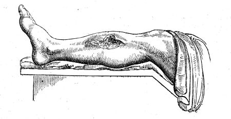
“28. Complains of no pain comparatively; slept well last night after an anodyne draught; has felt much relieved since the operation; a good deal of matter was discharged from the sore during the night; water-dressing to be applied to the wound.
“30. Slept well without any anodyne the last two nights. The relief afforded by the operation is felt more now than it was at first. The wound discharges a good deal. A tonic mixture, containing infusion of gentian, sulphate of magnesia, and sulphuric acid, was ordered.
“Dec. 2. The wound looks florid and clean; granulations are springing up; general health very much improved.
“4. The edges of the wound are thickened by the granulations; the discharge from the wound still considerable; feels quite well in health.
“12. The wound continues much the same; the rollers have been discontinued.
“14. Feels himself gaining strength daily; can walk without pain; wound is contracting and hard; granulations seem to lessen the depth of the opening in the bone; there appears to be no sequestrum.”
When caries is fairly established, and the integuments have given way, the best and most successful proceeding is that pointed out by nature—destruction of the diseased portion; and the means must vary according to the particular circumstances. In many cases, nature seems to wait but for the separation of the sickly parts, either by accident, or by the interference of art. The means are to remove, partially or wholly, the diseased part, or to effect such a change of action as will throw it off. The first indication will be accomplished by trephines, scoops, saws, and forceps; the second by active escharotics; frequently both are required.
If there be extensive disease in the medullary canal of a bone, several perforations may require to be made, and these may be connected by the use of a small saw, or the cutting forceps. The diseased cancelli, thus exposed, can be readily removed by the scoop or graver, as recommended by Mr. Hey, primus, in his excellent work. If, with the probe, it is ascertained that a portion of the cancellated texture has become dead and loose, it is to be removed after dilatation of the external opening. It may frequently be difficult to distinguish in the effused blood, between what is diseased and what is not; it will often be necessary afterwards to cauterise freely the exposed surface, and for this purpose the oxidum hydrargyri rubrum ought generally to be preferred. The slough will soon be thrown off, and healthy granulations fill up the breach. The application of the actual cautery may be by some considered necessary: at one time I employed this remedy very extensively in caries, and occasionally with very good success; I have since, however, been led to change my opinion, and am now inclined to prefer the potential cautery already mentioned. By the application of the red-hot iron, the diseased portion is destroyed effectually, but at the same time the vitality of the surrounding parts is often very much weakened and their power of reparation diminished, so that they are incapable of assuming a sufficient degree of action for throwing off the dead part; their action being increased whilst their power is diminished, they may become affected with caries, and thus, instead of being arrested, the original disease will either be increased, or extensive necrosis may take place. The red oxide of mercury is not calculated to produce such bad effects; it does not spread or insinuate itself into the bony tissue, as liquid caustics are apt to do; and it is sufficiently powerful for complete destruction of the diseased parts. It will be necessary to keep the wound open, by proper dressings, till all the dead portions of bone be discharged, and every part of the parietes of the cavity produce healthy granulations: if the discharge be offensive, its fetor may be corrected by the use of spirituous applications, such as the tinctures of myrrh, opium, or aloes, separately or combined. After healthy granulations have appeared, and the cavity has begun to contract, light dressing is all that is necessary.
In operating on the carpal and tarsal bones for removal of caries, the surgeon must be well acquainted with the connexions and relations of the parts. If one bone is diseased, its removal will be sufficient; if several, the operation becomes more painful and difficult. When one only of the tarsal or carpal bones is almost completely destroyed, and the surfaces of those articulated with it are also more or less affected, it is not sufficient or safe merely to remove the loosely attached portions of the one primarily attacked; the diseased parts of those surrounding it must also be taken away, and it will often be necessary to apply the caustic afterwards. In caries of the distal range of bones, the bases of those supported by them are in general involved, and must also be removed. If one only is diseased, with the base of the metacarpal or metatarsal bone attached to it, the removal of these will be enough, and can be accomplished without difficulty. Some have recommended the total extirpation of a metacarpal or metatarsal bone, leaving the finger or toe appended; but the member, when thus left unsupported, can never become of any service to the patient, and may be productive of much inconvenience; whilst removal of them, along with the diseased bone, renders the operation much more easy of execution. If the whole disease can be extirpated, leaving the surfaces of the surrounding bones covered with healthy cartilage, the use of the caustic is not required, and would be productive of harm; but wherever it is impossible to avoid encroaching on the cancellated texture, such as of the os calcis, which it would be unsafe or imprudent to take away entirely, its use is then indispensable. After the removal of carious bones, the symptoms soon disappear, and the patient obtains a rapid, and often permanent cure. The instruments for these operations, and the method of performing them, will be afterwards mentioned. In conclusion, it may be remarked that the temporary cicatrix of a sore leading to a diseased bone has a very different appearance from the sound scar which is formed after its removal. The former is bluish, soft, on a level with the surrounding parts, and moveable; the latter is depressed, white, and firmly adherent to the bone.
NECROSIS
Death of bone, or Necrosis, is an effect of violent inflammation, particularly of the medullary web, or external injury; a termination of inflammatory action in bone corresponding to sphacelation in the softer tissues. It has been observed, that the bones are not extensively supplied with bloodvessels, and that their natural powers are inferior to those of the softer parts; and from this circumstance the frequency of necrosis can be readily accounted for. The short bones and the heads of the long bones, are more vascular than the flattened bones and the shafts of the long ones. Hence necrosis most frequently occurs in the latter. Necrosis, fortunately, seldom occurs in the heads of the long bones, or penetrates the separation betwixt the cancelli of the shaft and the epiphysis. Bits of dead bone in the articular ends, however, very often lead to disease in the joint. There are in my private collection a few specimens of necrosis, in which matter found its way into the neighbouring joint, leading to disease of the tissues composing it, and rendering amputation necessary for the preservation of the patient’s existence. External injury may produce this disease by causing a violent increase of action, or it may be so severe as at once to deprive part of the bone of its vitality. Destruction of the periosteum, and of the vessels which enter the surface of the bone, frequently gives rise to superficial necrosis or exfoliation. Such a result, however, does not always follow; for we not unfrequently find, when the periosteum has been forcibly torn off, to a considerable extent, by external injury, that the part still retains its vitality. When, however, the bone has been at the same time contused, it is extremely probable that external necrosis may occur. Again, when the periosteum has been removed in the most careful manner possible, exfoliation occasionally takes place. If the exposed bone remain of a brownish hue, it will generally retain its vigour; if, on the other hand, the colour is white, it will most probably be cast off. Necrosis may come on at various periods of life, but is most commonly met with in young subjects, in whom the inflammatory action is allowed to make progress before it is noticed or attended to. It may affect the external or the internal part of a bone, or nearly its whole thickness. The whole of a bone seldom or ever dies in consequence of increased action, and it is not often that the entire thickness of any part of it is found to be necrosed. If the entire thickness dies to a great extent, there is no reproduction; the epiphyses approximate, and the limb, if there is only a single bone, must be lost. A large portion of a bone, or numerous small irregular portions, may die; but still a part of the original shaft remains, and by its vessels reproduction is accomplished. The articulating extremity is very rarely destroyed by this disease. Many writers have talked of death of a bone throughout its whole extent, and, in fact, the term necrosis was originally adopted on this supposition.
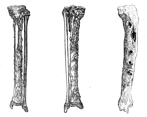
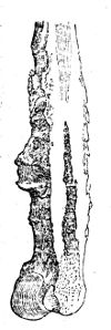
The progress of necrosis is, as has been said, similar to that of sphacelation. The affected bone gradually changes its colour, and loses its sensibility; a line of demarcation is formed, and ultimately the dead portion is completely detached from the living. Previous to its separation, the surrounding parts, the portions of bone which are not doomed to perish, have commenced forming new osseous matter, which is secreted in nodules, and from continued deposition soon becomes consolidated. The commencement of the process is well seen in the following sketches from specimens in my collection. The disease, as represented in the two first cuts, was of the most acute kind, and a great part of the shaft of the tibia had perished. This is seen at various points through the sort of cortical deposit of new bone. The new bone, in its turn, secretes a texture similar to itself, whereby the deposit becomes more and more extended, and not unfrequently affords an almost complete encasement to the dead portion, or sequestrum, as represented in the cut on the right-hand side of the page. In general bone dies irregularly, so that the sequestrum presents an uneven surface, and its margins are rough and serrated by numerous sharp projections, as seen in the one taken from the tibia, and represented here. From the appearance of the dead bone, it was imagined that after its separation, portions of it were removed by absorption; and this opinion was strengthened by the thin exfoliations of the external lamina being found perforated at several points by minute apertures,—worm-eaten, as it was called. These cases of death of inner or medullary shell are irregularly separated, like any other slough; the remaining living outer shell is enlarged by inflammatory action and deposit. But a dead portion of bone, detached from the surrounding parts, is in every respect an extraneous body, and is not, and cannot be, acted on by the absorbents, any more than a piece of metal, wood, or stone. Some have gone so far as to affirm that portions of foreign bodies, ligatures, &c., are absorbed; but this opinion is altogether too absurd to require any contradiction; the knots of ligatures, like portions of glass, or other foreign substance, become surrounded with a dense cyst, and often remain in the body for a long time; so do portions of dead bone separated by the process here described. A series of experiments were made by Mr. Gulliver, in order to put this question at rest, many of which I witnessed and assisted at, and several I also repeated. Setons of bone were inserted and worn for a long time; thin plates of bone were confined on suppurating surfaces; pieces of bone were inserted in the medullary canal of various animals, and kept there for months, and in one instance for more than a year. These foreign bodies were weighed with the greatest care and accuracy before and after they were so exposed to the absorbents, and were found unaltered in any respect. A paper, detailing these experiments, is published in the Medico-Chir. Transactions.
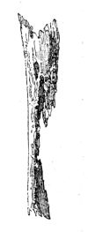
The separation of the dead part from the living is accomplished with greater or less ease, according to the bone which is affected, the state of the constitution, and the general health; in the bones of the superior extremity, this, as well as every other action, proceeds more rapidly than in those of the inferior. It occurs in consequence of absorption of the living part of the bone, which is in close proximity to the dead. The sequestrum, if large, is not pushed off, as some have supposed, by granulations, deposited on the living margin of the bone. A small portion of the inner shell, when completely detached, may sometimes be observed to be extruded from a cloaca by granulations from the living bone. During its progress, matter forms, makes its way to the surface, and is discharged through minute, and often numerous apertures, which afterwards become fistulous. The soft parts are thickened and indurated, and the integuments are red, and sometimes of a livid colour.
Formation of matter upon the bone is occasionally the cause of necrosis, the periosteum being destroyed or separated from its connections by the pressure or insinuation of the pus. I have seen several instances in which it followed neglected erysipelas of the lower extremity.
The matter is in general thick and laudable; at first it is secreted profusely, but afterwards in smaller quantity. The external openings, or papillæ, through which it is discharged, are found to lead to cloacæ, or apertures in the new and living bone, which encase the dead, and through these the dead portions can be discovered by the probe; and it will thus be ascertained whether the sequestrum is fixed or detached: when loose, it can sometimes be moved upward and downward in the cavity. When the shaft of a bone is much affected, the whole limb is enlarged, by the inflammation having extended to a considerable distance above and below the portion about to become necrosed. The unshapely appearance of the limb continues until the sequestra are discharged; for by their presence incited action is still continued, and subsides only after their removal. Some time before any portion of bone has become dead, or begun to be separated, great effusion of new bone has, in general, occurred; thus a preparation has been made for the strengthening of the limb, which, after a considerable portion of the bone has been detached, would otherwise be incapable of supporting the weight of the body. The unnatural bulk of the limb is afterwards much diminished, for the new bone gradually becomes consolidated, and smooth on the surface by the action of the absorbents. Nature seems to construct her substitute after the model of the original, and in some instances but very little change can afterwards be observed in the limb.
In external necrosis, or death of the outer lamella, reparation is chiefly made by the subjacent parts; and this species of necrosis occurs most frequently in the flat bones. In necrosis involving a greater thickness of the bone, the new matter is also furnished by the subjacent parts, which, however, are materially assisted in the process by the living bone, which forms the margins of the void caused by the absorbent process for the detachment of the dead portion. The bony matter is deposited with great activity, and frequently columns of the new deposit cross over the sequestrum, binding it firmly down, and rendering it almost immovable, although it may be completely detached from the living parts.
It has already been stated, that those vessels which ramify within the substance of the periosteum have no share in the reproduction of bone, but plastic matter is effused by the ramifications extending from the membrane to the bone: this effusion becomes organised, and greatly assists in forming the substitute.
It has been formerly remarked, that a limited, and, on after examination, an apparently trifling necrosis of the cancellated structure, may produce the most violent local symptoms; the painful feelings, the discharge, and the thickening of the bone, continue, as long as the cancellated sequestrum remains; severe symptomatic fever is induced, endangering the life of the patient, and often rendering removal of the limb absolutely necessary.
Occasionally abscesses form at a considerable distance from the necrosed part, and terminate in sinuses, which communicate with the diseased bone, and are consequently long and tortuous, so that examination by the probe is rendered difficult. When necrosis is extensive, there is a risk of fracture occurring, if motion of the limb be permitted before a sufficient quantity of matter has been effused, before nature has had sufficient time for the consolidation of her substitute, and consequently before the new bone has come to resemble the old in thickness and cohesion.
Violent inflammatory fever attends the incited action of the vessels of the bone and periosteum which precedes necrosis. But after the abscesses have given way the painful symptoms subside, and the health seldom suffers to any great extent, the system becoming gradually accustomed, as it were, to the new condition of the parts. Hectic supervenes only when the disease is very extensive, and joints become involved. Frequently fresh collections of matter form as each piece of bone approaches the surface. When the effusion of new bone has extended to the neighbourhood of a joint, its motion may be very much impeded, and, from the limb being kept in a state of rest for the cure of the necrosis, anchylosis may even occur.
Treatment.—The means of preventing inflammatory action from running high and ending in death of bone have been already alluded to—abstraction of blood, rest, purgatives, and antimonials. When necrosis has occurred, no interference with the bone is allowable, unless the sequestrum is quite loose, or unless the patient’s health is suffering severely under the discharge and irritation. When the sequestrum can be readily moved about, or when, projecting through the external opening, it can be laid hold of by the fingers or forceps, attempts must be made to remove it. The surgeon ought not, however, to allow it to approach the surface, and project externally, for the natural discharge of the sequestrum is a much more tedious process than the removal of it by art, and by the irritation produced during its spontaneous ejection the inflammatory action is continued, and may prove alarming. Long before it has appeared externally, it must have been completely separated from the living parts, so as to admit of ready extraction by the proper means. When it has been ascertained that the sequestrum is separated, it ought to be laid hold of by forceps, and moved freely upward and downward, so that any slight attachments by which it is connected to the neighbouring parts may be destroyed, whether these be minute filaments which still in some degree retain their vitality, or small portions of newly deposited bone, which are so situated as to prevent the free movement of the sequestrum. In general, no impediment of this nature exists, and the dead bone is easily removed. Before extraction can be accomplished, it is generally necessary to enlarge freely the external opening, in all cases where the dead portion of bone is of considerable size. If, on thus exposing the parts, the sequestrum be found detached, but still firmly bound down by the substitute bone, deposited over it either in one continuous sheet, or in irregular columns, this must be divided by a trephine, a small saw, or cutting pliers, before the sequestrum can be extracted. When a dead portion of bone, of considerable length, is exposed at its centre, whilst its extremities are entangled by the old or substitute bone, the division of the exposed part of sequestrum, by means of the cutting pliers, will often be sufficient for its removal, the cut ends being seized by the forceps, and one half removed after the other; thus the perforation or removal of any portion of the substitute will be rendered unnecessary. The instruments, and especially those for extraction, ought to be very powerful, and suited to the purpose; for in the employment of inefficient means there is much folly and cruelty. Incisions into a necrosed limb are attended with profuse hemorrhage from the enlarged and excited vessels; and in some cases it is with difficulty arrested, in consequence of retraction of the cut ends of the vessels not taking place within the condensed and indurated parts. Pressure, and an elevated position of the part, will generally be found to answer. When necrosis has been extensive, the limb must be carefully supported by the application of splints and bandage, till the process of reparation be completed, in order to prevent fracture of the recently formed substitute. This proceeding is seldom, however, necessary.
The treatment may be summed up in a very few words. Prevent the necrosis, if possible; open abscesses whenever they appear; encourage the patient to move the neighbouring joints; support the strength; remove sequestra when loose, but do not interfere till they are ascertained to be so; give the limb proper support and rest, when a large sequestrum is formed. When fracture has taken place, when the health has been undermined, or when neighbouring joints have become diseased, amputate, in order to save the life, if it be impossible to save the limb.
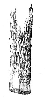
It is almost superfluous to remark, that leeching and blistering are worse than useless after necrosis has occurred, however useful they may be in preventing it; and that the adoption of measures to promote the dissolution and absorption of the sequestra are glaringly absurd.
Necrosis, after amputation, was formerly frequent; but in the present improved state of this operation it is so rare as scarcely to demand separate consideration.
Such specimens as here depicted are common enough in the collections of those who have practised the old round-about operation; in fact, it is only by this painful and tedious interference of nature that a tolerable stump is formed in many of these cases. Death of a small portion will sometimes, though very rarely, follow even a very well performed amputation, if through any mischance the recovery is slow, and wasting discharge takes place with emaciation. It happens sometimes, as when secondary hemorrhage (that is to say, bleeding after the fourth day) has taken place, that the flaps are separated by the coagula, and it may be impossible to bring the parts together and give them due support; then the muscles, wasted and shrunk, may leave the bone a little, but the exfoliation is but very trifling.
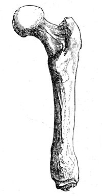
The inner shell of bone, as may be seen in the above sketch, perishes more extensively than the outer; and this arises probably from inflammation of the medullary membrane, in consequence of exposure, or, perhaps, from its being sometimes injured by the operator or assistants seizing the bone rudely to steady the stump, in order to facilitate the ligature of the vessels. In experiments on animals, the disturbance and injury of the medullary membrane is followed by internal necrosis, thickening of the outer living shell, and effusion betwixt the periosteum and bone. New bone is also furnished from the medullary canal, as is also shown in the sketch.
FRAGILITAS OSSIUM
Occurs chiefly in old people, whose bones contain an undue proportion of earthy matter, are endowed with little vascularity, and filled with an oleaginous fluid. They contain an undue quantity of phosphate of lime compared to the gelatin; and the liability to fracture is further increased by the interstitial absorption of the outer shell. They are in an atrophied state, and this is often in part attributable to disuse of the limbs. This state of the osseous system very often follows upon an attack of rheumatic fever, and is met with in patients who have laboured under cancerous affections.
The bones, when in this condition, often break from the slightest force applied; as from the action of the muscles when the patient turns himself in bed, whilst walking across the room, or when endeavouring to attain the erect posture when seated on a chair. After fracture the process of reunion is extremely slow, and it does not take place at all in patients very old and of worn-out constitution. With a view to prevent the occurrence of fracture when the bones are in this condition, for it is impossible by any treatment to prevent the change in the texture of the bones, the only rational indications seem to be to keep the patient on a generous diet, and to prohibit him from making any great muscular exertion—to avoid, in fact, all circumstances likely to produce a sudden action of any particular set of muscles.
OF MOLLITIES OSSIUM, RACHITIS, ETC.
These affections differ only in this, that in the latter the earthy matter is not deposited originally, whilst in the former it is absorbed after having been deposited; in both the result is the same. The latter is peculiar to the very young, the former to those of an advanced age.11
Rickets and mollities ossium seem to differ also in this. In the latter there is seldom, if ever, any reparative action. The diseased process of deposit continues in the bone, the softening increases, and the patient ultimately perishes. Whereas, in rickets, the softened and yielding state of the bones is only temporary: after a time earthy matter is deposited in due quantity, and the bones become compact, firm, and solid, capable of supporting the weight of the body, though necessarily permanently bent and deformed, if proper means have not been employed during the softened condition. The thickness of the rickety bone, as Mr. Stanley has shown, takes place on the concavity, which is the situation where the greatest strength may be added with the least expense of new matter. In the same way the reparation of fractures not accurately adjusted goes on most actively in the concavity formed by their displacement.
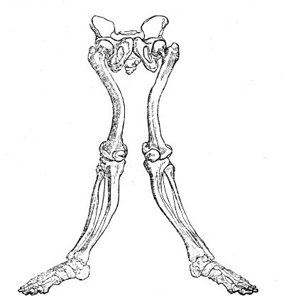
Softening of the bones is met with at all ages, and in different degrees. It seems sometimes to be congenital, and combined with hydrocephalus. It often follows dentition, measles, hooping-cough, or other infantile diseases inducing debility. In females it seems to be produced, or at least often accompanied, by the debilitating effects of leucorrhœa, miscarriages, and floodings. Loss of blood, in any way, predisposes to it. Mercury, given in immoderate quantities, produces a softening of the bones; and, in some most remarkable instances on record, the free use of common salt was the only cause assigned. When the disease affects children, all the bones generally suffer, those of the extremities as well as those of the trunk; the limbs become bent in an extraordinary manner, and the heads of the bones are swollen, and appear to be much more so in consequence of the wasted and flabby state of the muscles. The child walks with difficulty, and in many cases the legs are utterly incapable of supporting the weight of the body, so that he cannot remain in the erect position. The chest and pelvis become deformed, breathing is oppressed, the digestive organs are deranged, and the belly is tumid. The bones of the limbs become flattened as well as bent, and in their concavities, as remarked in the preceding cut, new bone is effused, in order that the column of support may be thereby strengthened. The new deposit is of extremely dense consistence, and is effused in greater or less quantity, according to the degree of curvature.
The bones of rickety subjects are soft, cellular, and of a brown colour, contain a dark fluid, and are very deficient in earthy matter. As a simple proof of the latter circumstance, it may be mentioned that distortions of the pelvis can be, and often are, accurately imitated by soaking the bones for some time in acid, whereby the earthy matter is extracted. In many instances this component of the bones is almost entirely removed, and soft matter deposited instead; they then consist merely of an extremely thin external osseous shell, covered by thickened periosteum, and containing a pulpy substance resembling fatty matter. During the progress of the disease, the urine deposits, often in great quantity, a white sediment, which, on analysis, is found to be the phosphate of lime. In rickets the head is generally enlarged to a greater or less degree, and the bones of the cranium are thickened and spongy; not unfrequently the intellectual faculties remain acute. In adolescents and adults the limbs seldom become affected; the bones composing the spinal column are the seat of the disease, and, along with the distortion of the spine, the position of the ribs is necessarily altered. Certain rare cases have occurred, in which all the bones of the adult were softened to a very great degree. In one remarkable instance, the patient complained of an annoying sense of tightness in the limb most affected, and, on examination, the softened bone was found greatly depressed at that point, as if a strong ligature had been drawn tightly round it. Mr. Howship, who attended the patient, was so kind as to present me some years ago with a portion of the altered femur, which consists of a fatty-looking substance, and appears to contain little or no earthy matter.13
Though incurvation of the spine occurs in boys, and even in adults, still it is most frequently met with in young females; and in them it is often induced by their having assumed a bad habit by sitting long in one constrained and awkward posture, as in writing or drawing, without, perhaps, the bones being unnaturally soft in the first instance. It often follows affections of the lower limbs, as of the knee or hip-joint; and is also caused by shortening of a limb, which has been negligently or ineffectually treated after fracture during childhood, or by the patient being allowed to continue a custom of standing awkwardly on one leg. In a very remarkable specimen in my possession, the curvature seems to have resulted from the tremendous enlargement and consequent weight of the head. The whole skeleton (head, thorax, pelvis, and extremities) is deformed, flattened, and twisted. This may have arisen more readily in consequence of the atrophy of the bones, and retardation of their growth, produced by the long confinement to bed. The number of ossa triquetra in the lambdoidal suture was unusually great. The patient attained the age of twenty-five. The affection commenced from birth.
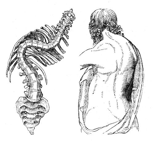
At first, during slight curvature from such causes, the spine can be brought into its original straight position by the voluntary action of its muscles. After some time, however, the curve cannot be remedied by any effort; interstitial absorption of the bodies of the vertebræ towards the concavity of the curve occurs; they become changed in form, and accommodated to their altered position, as shown in the accompanying sketch; the muscles also accommodate themselves to the new position, as do also the various ligaments connected with the spinal column. When the curvature is seated in the dorsal vertebræ it is generally to the right side; this shoulder is raised, and the chest is protruded, whilst the opposite side is depressed and flattened. The clothes hang loose, or fall off on the left side—the patient rests the weight of his body chiefly on the left leg—on stooping the right scapula projects, and, on examination, is found to be nearer to the spinous processes of the vertebræ than the left. The left cavity of the chest is diminished, and the ribs press upon the heart and lungs, causing difficulty of breathing. To preserve the balance of the body, a curvature occurs below the former, and in the opposite direction; and not unfrequently there is a third incurvation situated above the primary one.
The bones of the pelvis become distorted, and are twisted to one side; or, when the softening is great, and the patient confined to the recumbent position, the introitus of the cavity becomes diminished in the antero-posterior diameter; and, if the patient walk about, the ossa pubis are squeezed together, in consequence of the pressure of the ossa femora against the acetabula. The crests of the ilia are often bent inwards, in consequence of the pressure of steel apparatus injudiciously applied with a view of removing deformity. When the bones become consolidated after such distortion, they present most serious obstacles to parturition; and, most unfortunately, crooked and deformed women possess, it is said, “great aptitude for conception.” When, in such females, the untoward circumstance of pregnancy has occurred, it has been necessary, in some, to have recourse to the Cæsarean operation, and others have been delivered with the greatest difficulty and danger; notwithstanding which, many of these latter have, after recovering from a long and tedious illness, again become pregnant.
In some cases the softened ribs not only compress the organs of the chest, but are also pressed down upon the abdominal viscera, or even into the pelvis. The symptoms arising from such displacement are at first urgent, as can readily be imagined, and are often treated as inflammatory, to the detriment of the patient.
After some time, as the state of the patient’s health improves, the bones in some degree regain their original firmness, and the curvatures are rendered permanent. New bone is deposited in the concavity of the curve, at first in irregular masses, but afterwards becoming condensed, and assuming a more regular form, the column is thereby supported and strengthened.
Bending of the spine backward, with depression of the spinous processes, is extremely rare. But curvature forward, with projection of these processes, is by no means uncommon, and is generally supposed to be caused by caries of the bodies of the vertebræ; in many instances, however, it arises from interstitial absorption only.
Curvature from caries of the vertebræ, though not so frequent as the curvature from other causes, is met with pretty often. In adults, the curvature from ulcerative absorption is more common than that from softening of the bones. It is attended with the formation of purulent matter, which presents in the loins, at the top of the thigh, or near the anus; the bones may become affected secondarily, though much less frequently, in consequence of the formation and accumulation of purulent matter in their neighbourhood. There is pain in the loins; the patient walks in a stooping posture, and often complains of pain in the knee or thigh. The lower limbs sometimes become paralytic, as also the sphincters and extremities of the hollow viscera; this, however, may arise, without curvature, from softening of, or effusion on, the chord, or diseased thickening of its membranes. In some cases the palsy supervenes slowly; at first the patient has an awkward gait; he lifts his feet high to avoid stumbling, and afterwards puts them down clumsily and suddenly; the foot, in some cases, is extended, so that the patient is unable to plant the sole on the ground. Retention of urine occurs, and is followed by incontinence, with copious deposits.