PART FIRST. OF INFLAMMATION. (continued 5)
Reading edition
The larger nervous trunks sometimes become diseased, being affected with an enlargement resembling the structure and appearance of the preceding tumour, and such enlargements are termed Neuromata. Occasionally a portion of a nervous trunk is thus enlarged, from a blow or wound; and sometimes there can be no cause assigned. Nerves when divided, become bulbous on that extremity towards their centre. In stumps this is well seen; whilst all the tissues entering into it, after a time shrink, and become more or less atrophied (bone, muscles, vessels, &c.), the extremities of the nerves swell out and present a bulbous appearance. When these tumours are connected to, and only covered by, integument, or when they are adherent to the ligamentous substance covering the bone, and become exposed to pressure, as in badly made stumps, the patient often suffers excruciating agony. The nervous trunk above is, in general, slightly enlarged, sometimes has a tortuous course; and in some instances the neurilemmal bloodvessels are considerably increased in size. The same symptoms exist, though in a less acute form, as in the painful tubercle.
OF POLYPUS.
A common species of tumour is that which is attached to a mucous surface, and is called POLYPUS. Polypi vary in structure and disposition; some are simple and benign, others are most malignant. The simple mucous polypus has a shining appearance, being invested by an extremely delicate membrane, in some degree resembling the mucous, and moistened by a fluid similar to the mucous secretion; it is of soft consistence and homogeneous structure. They are generally light brown, sometimes greyish, and in some degree diaphanous. They are connected to the mucous membrane by a narrow pedicle; generally occur in clusters, and are of pyriform shape; one or more are often suspended from one narrow base, and they seldom attain a large size. They possess but little vascularity, though occasionally minute vessels are seen ramifying pretty freely on their surface, and may be considered as almost devoid of sensibility. The malignant polypus, on the contrary, is always attached to the mucous membrane, and also to the subjacent parts, by means of a broad base; and its form and structure vary according to the particular action which it has assumed. Most frequently it is encephaloid, of an irregular form, and often presenting a cauliflower appearance, its surface being studded with numerous excrescences of medullary consistence and colour. Such tumours will be afterwards treated of, as occurring in different situations.
OF ENCYSTED TUMOURS.
Along with these have been classed the enlargements of bursæ, sheaths of tendons, &c., but with equal propriety might we include hydrocele and other collections in natural cavities. Encysted tumours are almost always situated superficially. The skin is distended, seldom inflamed, and often contains enlarged bloodvessels, which give it a streaked appearance. They consist of an external cyst, which is in some instances extremely thin and delicate, in others dense, of considerable thickness, and composed of fibrous looking substance, occasionally it is almost cartilaginous; the internal structure may be said to be almost always more or less fluid, but varies much in consistence. The tumour is surrounded with condensed cellular substance, which is of greater or less thickness and strength according to the size of the tumour. Some of these tumours are supposed, and on good grounds, to be mere enlargements of the natural mucous follicles, in consequence of obstruction in their ducts, by hardened and vitiated secretion; the cyst, therefore, will be at first thin and delicate; its contents will resemble the natural secretion of the follicle, and in many cases may be readily squeezed out. There is a black point on the most prominent part, marking the obstructed orifice of the follicle, and the sac is found at this point to adhere firmly to the skin. Even after all other marks of its original formation have disappeared, the situation of the orifice is sometimes indicated by small dark spots, by depression, or by a minute ulcer.
Encysted tumours, or wens, as they have been called, often appear to be hereditary; seldom occur single, and are met with under the surface of all parts of the body. They have been divided into different classes, according to the nature of their contents: Atheromatous; containing curdy matter; Meliceritous, containing a substance like honey; and Steatomatous, containing fatty matter, generally in a semifluid condition. But such terms are not adequate to express the nature of all encysted tumours; they are extremely various in their actions, and their contents vary according to the particular secretory action which the lining membrane of the cyst assumes; for the same reason, also, the contents of a tumour will differ in the different stages of its progress. Some contain a thin, fetid, brown fluid, mixed with solid particles, resembling half-dissolved fibrinous matter; in some the contents are serous, or sero-purulent,—in others they are gelatinous; whilst in those which have become inflamed from external irritation, the contents are altogether purulent, or contain a very considerable proportion of that fluid; not unfrequently the cyst is covered internally by a layer of calcareous matter, to which similar particles are loosely attached. Sometimes, in consequence of irritation, organisable matter is poured out on the inner surface of the cyst, adheres firmly to its inner surface, and is often disposed in concentric laminæ.
Sometimes, though rarely, the most prominent parts of the tumour ulcerate, and on the exposed surface is deposited a substance of semifluid consistence and gelatinous appearance, which afterwards increases in density, and ultimately assumes all the characters of horn. This hard excrescence in some instances increases only to a slight degree, and afterwards remains stationary; in others it attains a large size, and occasionally assumes a curved or tortuous form, like that of the horns of inferior animals. Horns are generally met with on the forehead, and the scalp may be said to be their seat. The largest which I have seen, measured seven inches in length, and two in circumference; but others have been removed still larger.
In many encysted tumours, hairs grow from the internal membrane of the cyst, often numerous, and are generally found in those situated on the eyelids; in some the hairs are destitute of bulbs, lie loose within the tumour, and are often rolled into a globular form. The adhesions of these tumours are in general very slight and easily broken up, but when seated amongst tendons, or in unyielding parts, they are often extremely firm. Violent inflammatory action may follow injuries of the tumours, or the making of even minute openings into them; suppuration occurs, the discharge is thin, fetid, and often bloody; there is much pain, and frequently severe constitutional irritation. Occasionally a fungus, bleeding or not, is protruded through the aperture; more frequently, however, the opening heals, and the tumour remains as before. External injuries sometimes appear to check the secreting action, and to excite the absorbents to remove the morbid growths, and this with or without rupture of the cyst. Thus, in the case of an encysted tumour the size of a hen’s egg, on the external lateral ligament of the knee joint, free and pretty rough manipulation was necessary to ascertain its nature and exact situation; in consequence of which, the tumour gradually disappeared, and no traces of it remained twelve days afterwards. Others of less size, I have known to disappear in a much shorter period.
OF TUMOURS OF BONES.
The vascular action of bones, in their healthy state, is feeble, but, as in other feeble parts, it is easily excited, and disease of an obstinate and unyielding nature is apt to follow.
The morbid growths vary much in texture. The most frequent are the osseous, or those of the same structure with the original bone; but even these differ much in the density and arrangement of their particles: they have been termed exostoses. They may be of great density, and are then called the hard, or ivory; these never attain a large size, seldom exceed that of a bean, have a smooth and polished surface, and are of a flattened and hemispheroidal form, their greatest circumference being at the base; they occur in many of the bones, but generally in those of the cranium and face.
Others, being of a more loose and spongy texture, have been called cancellated. These are commonly formed by the bones of the extremities, and often attain a very considerable size; they grow from the periosteum, or from the outer surface of the bone, and are then covered by an expansion of this membrane. Sometimes they adhere by a narrow neck, and expand into a bulbous form, so that they can be very readily removed by operation, and are very apt to be broken off by external injury. Others have a broad and firmer attachment, and are of an irregular shape, often projecting in the form of a large spicula, and at other times assuming a somewhat stalactical appearance. Such frequently prove the source of much inconvenience, by impeding the motions of the muscles, or disturbing the functions of any adjoining organ. They possess no malignant disposition, but are under the same laws, though perhaps in a less degree, with their parent trunk.
On making sections of exostoses, and of the bones from which they arise, some appear to be mere enlargements or processes of the parent bone, the cancellated tissue extending itself so as to form the interior of the exostoses, whilst the exterior resembles a proportionate extension of the outer lamina. Others are evidently formed by the deposition of osseous matter externally to the outer lamina, many being dense and compact throughout, others containing an internal cancellated structure, but which is not continuous with that of the bone, as it is separated by the natural outer lamina. Their formation appears similar to that of the fœtal bones: a glutinous matter is effused, becomes dense, and is converted into cartilage; bloodvessels shoot into it, ramify throughout its whole structure in a radiated form, and deposit osseous matter. This deposit increases, and extends from a central nucleus towards the circumference; the cartilage is in part absorbed, and the new structure becomes osseous, and similar to the original tissue by the vessels of which it was formed. These tumours, even when they have attained a large size, contain a mixture of bone and cartilage, covered by a dense fibrous investment. The bony matter is arranged in spicula, adhering to the surface of the shaft or head of the bone, and projecting into the morbid mass: the spaces are filled up by a cartilaginous substance. The growth is sometimes rapid, and the tumour soon becomes troublesome from its immense size.
Frequently a bone is much enlarged throughout its whole extent, or the greater part of it, and presents a cancellated texture: sometimes, also, it is much thickened, and, at the same time, of great solidity; but such enlargements cannot be considered as tumours of bones, or exostoses, any more than those nodules of new osseous matter, which are effused in consequence of inflammation of the osseous tissue. The most frequent cause of exostoses appears to be external injury; their progress is slow, attended with slight dull pain, and often accompanied with no inconvenience; their existence can be readily ascertained, a hard and immoveable body being felt where no bone exists in the natural state of parts; but when the tumour projects into an internal cavity, the diagnosis is rendered obscure. Most frequently, they remain stationary, after having attained a certain size, and are productive of little inconvenience, the surrounding parts having accommodated themselves to the new formation. Occasionally, suppuration occurs in the soft parts, the matter comes to the surface, and a troublesome abscess is formed.
To this class of tumours would I confine the term exostosis, not including those consisting of softer materials, and possessed of a less benign action.
OF OSTEOSARCOMA.
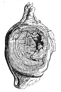
By this term is meant, an enlargement and alteration in the structure of a bone, accompanied with the deposition of a morbid sarcomatous substance internally. This morbid change appears to be the consequence of inflammation, and its origin is frequently attributed to some mechanical injury or local irritation. In the commencement of the disease, the bone is slightly enlarged, perhaps somewhat thickened in its outer laminæ; and on a section of it being made, is found to contain a brown fleshy substance instead of its cancelli. This appears to be formed in consequence of a morbid action, perhaps inflammatory, of its internal structure. By the pressure of the new formation, the parietes of the bone are pushed outwards, in some cases attenuated, in others thickened by deposition of new osseous matter, inflammatory action having been induced by the pressure. As the internal formation increases, the parietes are extended, and are generally much attenuated, becoming in some places thin as paper, and diaphanous; they also would seem to lose a portion of their earthy matter, for they are flexible, somewhat elastic, and not of their usual density. Frequently they are in several places deficient, and their situation occupied by a membranous expansion, sometimes thin and delicate, but mostly thick and ligamentous; in some cases, the external lamina appears to be converted into a substance resembling the internal growth, with which it is continuous. The investing periosteum is much thickened, and its bloodvessels are enlarged. Occasionally, the deficiency of the bone is not supplied by any membranous expansion, and the morbid growth protrudes, fungous. The internal structure varies much in appearance; generally it is brown and soft, in some places broken down and mixed with a dark-coloured fluid, or with gelatiniform matter; sometimes it is much more dense, and resembles cartilage; in others, the cavity contains an ichorous fluid, mixed with small portions of more solid matter; in the advanced stage of the disease, the contents are often of the encephaloid nature, either in its homogeneous and solid form, or softened, broken down, and mixed with blood, or with a lard-like substance. Sometimes the cancelli of the bone are not destroyed, but extended, forming numerous cavities of considerable size, in which the morbid matter is deposited; in other instances, there is no appearance of cancellated structure, and the diseased mass contains rough osseous spicula, some detached, some loosely connected with each other, and others projecting from the inner surface of the bony parietes of the tumour. At the commencement of the disease, the patient feels acute pain in the part, the constitution is disturbed; afterwards, the pain becomes more dull, and there is a considerable swelling externally, which feels hard, and slightly elastic; in the advanced stage, the pain again becomes severe, and is of a lancinating kind, and the system is much deranged, the tumour is softer, often presents a sense of distinct fluctuation, and on being freely handled, is found to crepitate, in consequence of the loose spicula of bone rubbing upon each other. Ultimately, the integuments become tense, livid, or dark-red, ulcerate, and allow a portion of the softened tumour to protrude, in the form of a frightful fungus; there is profuse discharge, thin, and sometimes bloody; there is much constitutional irritation, and the patient is greatly exhausted. Not unfrequently, during the progress of the disease, especially in the long bones, fracture occurs at the diseased part, either from external injury, or sudden muscular exertion. This occurred in the case from which the specimen here delineated was obtained some months before the patient submitted to amputation. The morbid structure had not broken through its periosteal investment. The muscles and their interfilamentous tissue were sound. The patient remained free from any return of the local disease. Bones so affected, when broken, do not unite, the movement of the loose and rough ends is a cause of much irritation: inflammatory action is kindled in the morbid structure, suppuration occurs, the integuments give way, and ulcerate to a greater or less extent, and the advancement of the disease is thus much hastened. The tumour may be safely pronounced malignant; it is true, that for some time it shows no tendency to involve the adjoining soft parts, further than by the effects of inflammation induced by its pressure; but then it is limited by the external lamina of the bones, which confines it to the tissue in which it originated; but after this barrier has given way, the tumour projects through the aperture, contaminating the adjacent soft parts, imparting to them a morbid action, and extending also in the cancellated tissue of the shaft of the bone. In some cases, the integuments are tense and discoloured, with large vessels running on their surface; the tumour feels soft and fluctuating, though the skin may not ulcerate till long afterwards. Perhaps the most common seat of this disease is the under-jaw, but it may occur in any of the bones; when it has been of chronic duration, not one bone but several are affected; and in one case which I saw, the disease commenced in the under-jaw, which it deformed to a frightful degree; almost every bone in the body was similarly, though less extensively diseased; this could be readily observed during the life of the patient, and was confirmed by dissection. From this, it appears, that the affection is not only dependent on local causes, but connected with a morbid state of the constitution, predisposing to it, and cooperating with its exciting cause.
There are other tumours of bones in some degree resembling, which do not strictly come under the term Osteosarcoma. Some are wholly cartilaginous, the disease commencing in their centre, and involving their entire substance, emitting a gelatinous fluid when cut, but containing no cells; others are not uncommon, partly osseous, and partly cartilaginous, containing cells filled with a glairy fluid; others are composed of cartilage, intersected with dense fibrous matter, in a greater or less proportion. In fact, the individual tumours of bones vary as much from one another as those of the softer tissues; scarcely two are alike in their progress, action, or anatomical characters. Irregular spicula of bone are found in many parts of their structure; in the same way that portions of bone often exist in tumours having no connection with the bones; in many places they are softened and broken down, the partitions between the cells are destroyed, and these contain a pulpy mass of a dark sanious appearance. On making a section, they are observed to be continuous with the interior of the bone, which is converted into a substance similar to themselves, or is of a more soft and medullary character. The external surface becomes tuberculated, the integuments are painful, and changed in appearance; they ulcerate, the tubercles burst, the discharge is thin and bloody, the ulceration extends; not unfrequently a fungus protrudes, and occasionally bleeds; this may slough, the tumour becomes farther exposed, portions of it die, and are discharged, unhealthy fungous granulations project from amongst the cancelli of the tumour, and emit a sanious putrid discharge, often bloody; severe constitutional irritation accompanies this state, the patient becomes hectic, is much exhausted, and sinks, unless the morbid parts be removed. There are also tumours of bones, composed partly of cartilaginous or fleshy, or partly of osseous matter, arising from the periosteum and outer shell of the bone, and these often acquire a great magnitude before the diseased mass reaches the interior or medullary part of the bone.
SPINA VENTOSA.
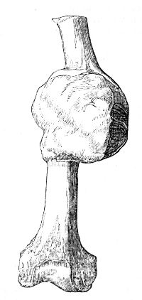
By this term is understood a mere expansion of a bone from a collection of matter in its substance. The disease may be produced by external injury, exciting inflammation, and consequent suppuration, in the cancellated tissue; or in a weakened and unhealthy constitution, the action may be of a chronic nature. The fluid accumulates, the cancelli are broken down, and the much-attenuated parietes of the bone are pressed outwards. Occasionally inflammatory action is excited on the external surface, from the pressure of the contained fluid, and minute nodules of bony matter are effused, as if nature endeavoured to strengthen those walls which are daily becoming thinner, and more incapable of supporting the weight of those parts which they encircle. The disease differs from Osteosarcoma in the contents being uniformly fluid, generally purulent, though often mixed with more liquid and dark-coloured matter, or with a curdy substance—in the gradual extension of the bone—in no fungus protruding after a portion of the attenuated bone has given way, matter being discharged as from a common abscess; and in the tumour not possessing a malignant disposition. At first there is considerable pain in the part whilst the matter is forming, but afterwards it becomes much less acute, and in many instances there is no inconvenience, except from the bulk of the tumour. Often after having reached no very large size, it becomes stationary, neither recedes or enlarges, and all painful sensations cease; in other cases it enlarges gradually, attains an enormous size, and produces much disturbance of the constitution; but in such instances the patient is generally weak and cachectic. The largest tumour of this species which I have seen, occurred in the lower part of the femur. It measured, in breadth, seven inches, in length, seven and one-fourth. The parietes were composed of an extremely thin lamina of bone, and in this there were numerous deficiencies supplied by delicate ligamentous matter; its cavity was divided into several compartments by thin septa, partly osseous and partly membranous. A representation of the femur so affected is given in the Practical Surgery, p. 350. The patient was a boy of twelve years of age; amputation of the limb was earnestly advised, the friends objected, he died hectic.
ANEURISMAL TUMOURS.
Besides these tumours a species of an anomalous character is sometimes met with, appearing to arise from an aneurismal or varicose state of the venous radicles or capillaries, and partaking somewhat also of the nature of fungus hæmatodes.21 I shall detail shortly the more important circumstances of one case. The patient, a lad aged sixteen, was admitted into a public hospital on the 7th of November, 1819, on account of a tumour over the left scapula. It was there deemed imprudent and inadvisable to attempt operation; and, after the application of leeches, he was dismissed, at the end of eight days. He then applied to me. The tumour was very large, hard, inelastic, firmly attached to the left scapula, and extending from its spine over all its lower surface. It also stretched into the axilla to within half an inch of the nervous and vascular plexus, and a large arterial trunk could be felt along its under surface. The arm hung useless, and, from the wasting of its muscles, was hardly half the size of the other. According to his own account, the uneasiness produced by the tumour was trifling when compared to the lancinating and excruciating pains in the limb. On attempting to move the tumour independently of the scapula, crepitation was distinctly perceived, as if from fracture of osseous spicula. A tumour was first perceived about three months previous, situated immediately below the spine of the scapula, about the size of a filbert, of a flat form, and attended with distinct pulsation; it had subsequently increased with great rapidity. About ten days before his admission into the hospital, it had been punctured; nothing but blood escaped. It was evident, from the rapid growth of the tumour, and the severity of the symptoms, that the patient would soon be destroyed if no operation were attempted. There were no signs of evil in the thoracic viscera, the ribs and intercostal muscles were unaffected; though the tumour was firmly fixed to the scapula, yet that bone was moveable as the one on the opposite side, and the vessels and nerves in the axilla were quite unconnected with the swelling. The operation was commenced by making an incision from the axilla to the lower and posterior part of the tumour. The latissimus dorsi was then cut across at about two inches from its insertion, so as to expose the inner edge of the tumour, with a view to tie the subscapular artery in the first instance; in this, however, I was foiled, owing to its depth. The dissection was proceeded with to where the branches from the supra-scapular were expected to enter. In detaching the tumour from the spine of the scapula, the knife and fingers suddenly dipped into its substance. This was attended with a profuse gush of florid blood, with coagula; by a sponge thrust into the cavity, the hemorrhage was in a great degree arrested; at the same time an attempt made to compress the subclavian failed, on account of the arm being much raised to facilitate the dissection in the axilla. The patient, exhausted, made some efforts to vomit, and dropped his head from the pillow, pale, cold, and almost lifeless. Then only the nature of the case became apparent. The sponge being withdrawn, one rapid incision completely separated the upper edge of the tumour, so as to expose its cavity; and, directed by the warm gush of blood, a large vessel in the upper corner, which with open mouth was pouring its contents into the sac, was immediately secured. The coagula being removed, by dissecting under the finger, the subscapular artery was then separated, so that an aneurism needle could be passed under it at its origin from the axillary, and about an inch from the sac. After securing this and two other large vessels which supplied the cavity, the tumour was dissected from the ribs without further hemorrhage, cutting the diseased scapula and the under part of the sac. It was then found necessary to saw off the ragged and spongy part of the scapula, leaving only about a fourth part of that bone, containing the glenoid cavity, processes, and half of its spine. The edges of the wound were brought together, and the patient lifted cautiously to bed. At this time he was pale, almost insensible, and without any pulsation perceptible through the integuments in the greater arteries, though the ends of the vessels in the wound beat very forcibly. Stimuli were employed externally and internally; in the evening his pulse at the wrist was ninety, and soft.
The sac of the tumour was composed of bony matter, containing little earth, and arranged in strata of short fibres pointing to the cavity. Its outer surface was smooth, and covered by a dense membrane; whereas the inner, to which so equable a resistance was not afforded, was studded with projecting spicula. The lower part of the scapula, partially absorbed, lay in the middle of the sac, covered by the remains of its muscles and coagula. Very large vessels were perceived ramifying on the surface of the tumour.
The patient made a rapid recovery, and the wound all but healed. A fungus, however, began to appear in about six weeks, which grew rapidly. This was removed, and the bone cauterized with little good effect. The tumour was soon reproduced. It was proposed to remove the remainder of the scapula with the extremity, as the only chance, though perhaps a slight one. This was objected to, and he died about five months after the operation, worn out by hemorrhage and profuse discharge.
The diseased parts presented the following appearances. Portions of the acromion process, superior costa, and spine of the scapula, were of their natural appearance. But the coracoid process, the glenoid cavity, and the cervix, were entirely destroyed, and their situation occupied by an irregular broken-down tumour, consisting of osseous spiculæ, and cancelli, irregularly disposed, and forming cavities which were filled with blood, partly fluid and partly coagulated. The head of the humerus was extensively absorbed. The articulating cartilage was almost entirely destroyed, particularly on the inner side, where a large portion of the bony matter had also been removed. The ulcerated surfaces were of a dark, bloody colour.
HYDATIC TUMORS.
Another disease of the bones which ought to be introduced here is the development of hydatids, which I described twelve years ago under the name of osteo-hydatidic tumours. The seat of this affection is not confined to any particular class of bones; though the long are perhaps most prone to it. Its precise nature and origin have not yet been determined; nor are its symptoms such as to enable us, in the present state of our knowledge, to distinguish it from exostosis, osteosarcoma, and other maladies. Its progress is commonly slow, the surface of the tumour is smooth and regular, the skin exhibits no unusual appearance, and the adjacent textures rarely participate in the morbid action. The hydatids, usually of a globular figure, vary greatly in size and number, and are generally filled with a thin, limpid fluid. They appear to be of the nature of acephalocysts. The prognosis unfavourable, owing to the difficulty of destroying these parasites, and their constant tendency, when interfered with, to reappear.
As soon as the true nature of this tumour is ascertained it should be laid freely open, its contents turned out, and the sac destroyed. For this purpose the sides of the cavity should be seared with the actual cautery, or touched with some of the more powerful escharotics, as the nitrate of silver, or the caustic potash. If these measures fail, and the disease involve the whole circumference of the bone, nothing short of amputation will suffice. This was successfully resorted to in one of the cases which came under the observation of Mr. Lucas of London.]
TREATMENT OF TUMOURS.
It may be observed generally, that no benefit can be derived from external applications to tumours, such as friction with liniments or ointments containing iodine, mercury, &c.; and that, therefore, it is injudicious to employ such temporising measures; for though a tumour at its commencement appear to be of a very harmless nature, yet it may soon assume a most malignant character. If an apparently simple tumour increase, and exhibit symptoms of inflammation, it will perhaps be advantageous to apply leeches, to arrest that incited action which affords the accession of new materials; this, however, cannot check the morbid activity inherent in the new formation, though it may hold the growth in check a little. If a tumour is to be removed by external applications, it is evident that these must be such as shall prevent the deposit of new matter, and allow the absorbents to remove that which already exists; for absorption is always going on in a tumour, though it leaves no evidence of its progress, on account of the deposition of new matter exceeding the removal of the old. I must say that I am unacquainted with any remedies capable of performing the above indication. The removal of a swelled gland may occasionally be accelerated by such means when stationary, or on the decline, and before cheesy tubercular unorganized matter is infiltrated into its texture; but to trifle so with a new and independent growth is altogether absurd. The knife only is to be depended on.
Many of the tumours first described have no malignant disposition originally, and only require surgical interference when they produce deformity or inconvenience from their bulk. Yet even these ought not to be allowed to attain any great size, however indolent they may appear at first, and however little pain they may produce; for there is always a danger of their assuming a malignant tendency, or forming connections with important parts, so as to render their removal either altogether impracticable, or at least attended with much difficulty. Tumours of every kind, when seated near important organs, must be early removed. Glandular tumours, however, even when of great size and long continuance, are not to be rashly interfered with, when they arise from irritation in the neighbourhood.
Those in which it is feared that malignant action has commenced cannot be trifled with; and the only means which afford a chance of the patient’s being effectually delivered from them is an operation. With a view to their complete extirpation, the external incisions ought always to be free, so as to admit of the after-dissection being easily and rapidly performed: they ought also to be made in the direction of the muscular fibres, whether these lie above or beneath the tumour. In this way the margins of the wound are easily brought into apposition, and there retained; whereas, if the fibres be divided transversely, the wound will gape, and union by the first intention be rendered absolutely impossible. If there is no reason to suppose that the tumour is malignant, little or no integument ought to be removed, unless the growth is of a large size; but, when malignity is dreaded, all the discoloured, tense, and adherent integument, all that is permeated by dilated and tortuous vessels, ought to be taken away, and the incisions made at a distance from the disease. In all cases they ought to commence at the point where the principal vessels enter; these are thus divided at the outset, can be readily secured by ligature, or by the fingers of an assistant, and the dissection is proceeded in without risk or interruption from farther hemorrhage. If the opposite course be pursued, the vessels will be divided two or more times during the operation, and thus the performance of it will either be delayed by the application of numerous ligatures, or will be attended with a considerable loss of blood. After the tumour has been exposed it ought to be principally detached in one direction, as in this way its removal will be sooner accomplished, and not first cut on one side and then on another. If malignant, great care should be taken that all the diseased mass be removed, for a minute portion remaining will form a nucleus in which similar diseased actions are certain to arise; in most instances, it will be prudent not only to remove the parts actually diseased, but those also which are in immediate connection with the tumour, though at the time they appear healthy. All important parts must be carefully avoided. After removal of the mass, and the complete cessation of bleeding, the edges of the wound must be approximated, so as to favour union by the first intention; if this fail, granulation must be promoted, and the wound dressed according to the particular circumstances of the case. All operations on malignant tumours, in their advanced stages, are unwarrantable; they are necessarily painful and severe, and cruel because unavailing; they often, indeed, expedite the dissolution of the patient. If the integuments over the tumour have ulcerated, and if the lymphatics in the neighbourhood are diseased, the disease if removed will certainly be reproduced, and the succeeding tumour will be still more malignant. The operation ought, if at all, to be performed when the disease is in its incipient state, for then only can success be expected.
Exostoses need not to be interfered with, unless they are the source of much inconvenience, either from their size and form, or from their having been detached, and lodged amongst the adjacent soft parts. If loose, they can be removed in the same way as any other extraneous body; if firm, their attachment must be divided by a saw, or by cutting pliers, close to the bone from which they spring. Sometimes, as in the scapula or other flat bone, a portion of the original bony tissue can be cut out along with the new growth, and this renders the chance of any return of disease much less likely.
Osteo-sarcomatous tumours are to be taken away, along with the part of the bone in which they are imbedded, and, if possible, before the integuments have ulcerated. The incisions must be made, and the bone sawn, at a healthy part. The removal of the entire bone in which the disease has commenced, when practicable, will afford a still better chance of immunity from farther disease.
In spina ventosa more is seldom required than to lay open the cavity, give vent to the matter, and then treat the case on the same principles as in abscess of the soft parts. The cellular tumours, partly cartilaginous, partly osseous, ought not to be permitted to remain; the operation can generally be done without much difficulty; and thus the danger of their degenerating avoided. Frequently, however, a considerable part of the bone must be removed along with the tumour, since the neighbouring tissue is generally softened, and intimately adherent to the diseased part, which it somewhat resembles in structure.
In general, regular dissection is unnecessary in the removal of encysted tumours. An incision is made, or an elliptical portion cut out; the contents escape, and the cysts, being then laid hold of by dissecting forceps, is readily separated. In some situations, as on the eyelids, under the tongue, or amongst tendons, the sac, which is thin, is not so easy of extraction; it is then inseparable, either naturally, or from previous inflammation. Caustic is used with safety to destroy those parts which cannot be detached, and for this purpose the potass is to be preferred. When, however, the tumour is large, a part of the integuments covering it must be removed, otherwise a large cavity will be left, in which pus might accumulate. In this case, the base of the tumour is to be surrounded by two elliptical incisions, and the cyst dissected out entire, leaving only integument sufficient to cover the exposed surface. In the smaller tumours, it is vain to attempt regular dissection; a portion of the cyst will be left, and the disease reproduced: whereas, by using the potass, the operation is much more speedy, and always successful. The making a minute aperture, and squeezing out the contents, is at best but a palliative measure, and is often followed by severe constitutional disturbance.
OF WOUNDS.
These vary in extent and nature. The instrument by which they are inflicted, the violence attending the injury, and the nature and importance of the parts divided, or in the neighbourhood of the wound, must all be attended to, for, from an accurate knowledge of these circumstances, the treatment of the case comes to be conducted accordingly. Wounds are divided into incised, punctured, bruised, and lacerated; that is, into such as are inflicted by a sharp-edged, sharp-pointed, or an obtuse body. In the first kind, there is greater or less effusion of blood, according to the size and number of the vessels divided. Some extend but a little way beyond the subcutaneous cellular tissue, and are consequently attended with but slight bleeding; others penetrate to a greater depth, and occasion hemorrhage from a large vessel, or other alarming symptoms, by having reached some important organ; others, though not of so great a depth as the former, may still, on account of their mere extent, be accompanied with very considerable loss of blood from a number of small branches. It is seldom that fatal effects immediately follow external wounds; but they may and do occur when bloodvessels of the first class only are cut. They are most likely to prove suddenly fatal when the arteries are only partially divided, and when the large veins accompanying them are also involved. When the artery is cut through, its extremities retract, effusion takes place into the sheath and compresses the orifice; the formation of a coagulum within the vessel is thus promoted, and the hemorrhage arrested. But, when a portion only of the circumference is divided, the blood continues to flow through the aperture and onwards, as if into a smaller ramification of itself, no retraction or contraction of the vessel can occur, coagulation is slow, and the bleeding profuse. I have seen a wound of so small a vessel as the internal mammary prove almost instantaneously fatal. Wounds of the large internal vessels for the most part prove immediately fatal; as wounds of the heart, or the large vessels passing to and from its cavities, at the root of the lungs, or at the upper part of the liver. When the heart, or the vessels within the pericardium, have been divided, it can be readily understood how life should be immediately destroyed, since the blood effused into the cavity of the pericardium by its pressure completely arrests the action of the heart. But occasionally punctured wounds, in such situations, have not been followed by instant death. In such cases, alarming symptoms occur at the time, but subside, and the patient may for some time suffer no uneasiness, but afterwards expires suddenly during muscular exertion, or perhaps in a fit of violent passion. Blood must have been effused into the pericardium at the first, causing symptoms of, or actual, syncope; but then the aperture in the vessel had become obstructed by coagulum before blood had been poured out in such quantity as to effectually prevent the actions of the heart; at a future period the coagulum gives way, and the subsequent effusion is limited only by the pericardial cavity being completely filled. In wounds, hemorrhage is the symptom which most alarms the bystanders, and which demands immediate attention; but, to operate successfully, the surgeon must divest himself of all fear, and learn to look boldly on the open and bleeding mouths of arteries. Effusion of blood ceases spontaneously, even from considerable vessels, on faintness supervening, and thus many lives are saved; but as soon as reaction commences it generally recurs, and may prove fatal, unless proper measures be resorted to.
When an artery is divided, its extremity retracts within the sheath, it also contracts, and coagulation occurs; thus the orifice is obstructed, and a temporary barrier formed to further hemorrhage. The tube, however, is permanently closed by effusion of lymph from its orifice, and consolidation of the surrounding parts.
The circumstances which follow division of an artery are these:—The immediate effect is retraction of its ends within the investing sheath, and a simultaneous contraction of the coats, so as to diminish the calibre. From the superior orifice there is necessarily a profuse flow of blood, which is discharged through the sheath that formerly enclosed that part of the vessel which has retracted. After considerable effusion of blood, the flow becomes slower and less profuse; particles of blood adhere to those filaments which previously connected the artery to the sheath, but which were lacerated by the sudden retraction of the divided extremity; these particles coagulate, and lessen the canal through which the blood is discharged, whilst they present an irregular surface, on which the blood continues to be deposited and to coagulate; and thus the aperture in the sheath is ultimately closed. This external coagulum is found to commence at the extremity of the artery, where it is of a cylindrical form, and shuts up the mouth of the vessel; it then extends along the canal in the sheath, frequently assuming a conical form; and, if a free discharge has been allowed for the blood, it will terminate at the cut margin of the sheath, otherwise it will be found continuous with the coagulum blocking up the external wound. Also, when hemorrhage has been resisted by the shutting of the external wound, blood is infiltrated into the cellular tissue around the bleeding point, and there coagulates; but this circumstance can be productive of little or no pressure on the parietes of the vessel, so as to assist those other natural means which obstruct it. The flow of blood through the divided vessel being prevented, the circulating fluid necessarily passes through the nearest collateral branches, leaving the blood in the extremity of the larger trunk in a state of comparative rest; consequently, coagulation occurs in this situation. The internal coagulum, however, is small, and not sufficient to occupy completely the cavity of the vessel; it is of a conical form, its apex being towards the heart, and opposite to the first collateral branch, and its base resting on the external coagulum, and there adhering to the internal surface of the artery. But, whilst this latter process is advancing, the capillary vessels supplying the cut margins of the artery have begun to act; they throw out coagulating lymph, and continue to do so until their secretion has completely filled the vessel immediately opposite to its divided margins; thus a third and more effectual coagulum is formed,—one of plastic matter, situated between the external and internal coagula of blood, and in general closely adherent to them. Lymph is also effused externally to the artery and its sheath, forming a dense stratum, which separates the extremity of the vessel from the external wound; it becomes organised, forms granulations, and thus the parts are consolidated, and the wound cicatrised. When the artery is permanently obstructed by the adhesion of its cut margins, the external coagulum can be dispensed with, and is gradually absorbed. Afterwards all the newly formed parts are condensed, and diminish in size; the artery contracts, its internal surface finally embraces the coagulated blood which lay loose in its canal; its coats appear to be thickened, and it is firm and hard. Ultimately, in consequence of the continuance of absorption, it becomes much more attenuated, so as scarcely to differ from the surrounding cellular tissue. Similar changes occur in the lower extremity of the divided artery; in general it retracts farther, its orifice is more contracted, and, the flow of blood being much less profuse than in the superior, the natural means for its temporary closure are sooner accomplished. When an artery has been divided close to the origin of a collateral branch, no bloody coagulum can form internally, for the blood in that situation is necessarily in a state of constant motion.
If the hemorrhage is suppressed artificially, either by ligature, or by otherwise well-applied pressure, no external coagulum is formed; there appears only the internal bloody coagulum, the lymphatic effusion, and consolidation of the compressed part. The natural contraction and retraction cannot occur in vessels partially divided; hemorrhage, therefore, is more violent and dangerous from a partial than from a complete section. Again, transverse wounds are more dangerous than longitudinal; in the latter, the edges of the wound are spontaneously approximated on account of the structure of the vessels, whilst, from the same cause, the margins of the latter continue separate, and, in fact, the aperture is a complete circle; the lips of an oblique wound will be more or less apart, in proportion as it approaches to the transverse direction. When an artery has been punctured, the wound in the sheath perhaps does not correspond with that in the vessel; blood, therefore, accumulates between the vessel and its sheath, and there coagulates. The wound is thus compressed, its edges kept in contact, and the farther escape of blood prevented; the lips of the incision are then agglutinated by effused lymph, and cicatrisation occurs. This, however, cannot be expected to take place unless methodical pressure has been applied from the first. Even from small punctures blood is effused under the sheath and into the neighbouring cellular tissue, rapidly, and in such quantity as to prevent adhesion. The effusion continues, and a false aneurism is formed. If a considerable part of the circumference has been divided, the lymph may be, and generally is, superabundant, and often to such an extent as to close up the canal of the artery at that point; but, if the aperture is minute and in a longitudinal direction, lymph will seldom be effused in greater quantity than is sufficient for the cicatrisation; and, though it should be superabundant, it is afterwards removed by the absorbents. In all cases, the cellular tissue round the wounded point is much thickened and condensed by the deposition of lymph, but this gradually disappears after cicatrisation has been completed. Sometimes, and generally when the wound has been transverse and large, the process of adhesion is disturbed, and suppuration occurs; in this case the wound in the vessel communicates with the fistulous track in the externally effused lymph, and may be the source of troublesome hemorrhage. In other instances of extensive transverse wounds, the undivided slip ulcerates, and the artery becomes obliterated, by means of the same natural processes that occur in complete division. In cases of laceration of an artery, when its coats have been forcibly torn rather than divided, little or no bleeding takes place. The vessel retracts; the lacerated margins of its inner coat become puckered up, so as to contract greatly the orifice of the vessel; the lacerated sheath is pulled out to a point, and closed at a little distance from the divided inner coats. If a large artery is torn asunder in the dead body, this stretching out and contraction of the sheath will prevent injection passing; in short, the immediate effects of the injury are such as to favour the instant formation of coagula, by which the hemorrhage is arrested until the orifices of the vessel be permanently closed by the adhesive process. Thus, in instances where the whole of an extremity has been torn off, the patients have generally lost but a very small quantity of blood.
From wounds of veins the blood flows, not in a sub-saltatory but in a uniform stream: its colour is dark, and the flow is easily suppressed. The common opinion is, that to place a ligature on a vein is dangerous, and to be scrupulously avoided. The process of reparation, besides, in a wounded vein, is different from that in an artery. Veins are less disposed to the secreting action by which adhesion is perfected; and, when inflamed, the inflammation is extremely apt to extend along the coats of the vessel; which latter circumstance has been ascribed to the great proportion of cellular tissue in their coats. When punctured longitudinally, the lips of the wound remain in contact, and cicatrisation, by means of effused lymph, is soon effected; in fact, the wound heals by the first intention. But if opened obliquely or transversely, not to a great extent, the immediate result is discharge of blood, and, when this has ceased, a coagulum forms in the wound, the margins of which remain separate; and this coagulum generally communicates with blood effused into the sheath of the vessel. After some time, the lips of the wound, encircling the coagulum which occupies the aperture, and which has temporarily averted the hemorrhage, become somewhat turgid, and increased in vascularity; they then appear to assume a secreting action, by which a membranous substance, of extreme delicacy, is produced; and the extent of this membrane is increased until it form an expansion, investing the outer surface of the clot; it then becomes thickened, by addition of matter, similar to itself, from the recent vessels which ramify in it. At the same time it forms adhesions to the surrounding cellular tissue, and resembles the original tunics of the vein. After being consolidated, so as to prevent the flow of blood through that part, the coagulum, formed to arrest the hemorrhage until a more complete barrier should be furnished, is gradually absorbed. But the membrane long remains smooth, thin, and diaphanous, and can be thereby readily distinguished from the original coats. This reparative process is much longer in being finished than the corresponding one in arteries; and, from what has been stated, it is evident that the two actions differ in other respects than the time requisite to complete them. When a vein has been completely divided, the extremities are closed by means similar to those which have been already detailed in regard to arteries.
In many, nay in most, instances of hemorrhage from a wounded artery, the surgeon cannot wait for the natural processes by which the flow of blood is arrested, but must have recourse to immediate and certain means. In division of the smaller arteries, or in minute wounds of the larger, pressure, well applied, will often be sufficient. In both cases it immediately stops the flow: in the former, it prevents the blood from penetrating into that portion of the sheath which has been vacated by the retracted artery; and it being thereby confined, and kept in a state of rest, coagulation soon takes place. At the same time, the compression brings the divided margins of the vessel into close apposition, and thereby permanent closure, by adhesion, is quickly accomplished. In the latter, the mere circumstance of the escape of blood being prevented, naturally hastens the closure of the minute aperture by the natural process; and, if the compression be accurate and very firm, the opposite surfaces of the vessel, being brought in contact, may adhere, and the canal be obliterated at the wounded point. It is obvious that, in this latter class, pressure can only be of advantage immediately after the infliction of the wound, and not when blood is extravasated to a great extent.
Pressure may be used along with styptics, or along with escharotics, actual or potential. They may be often employed when pressure ought not; styptics promote the contraction and retraction of the divided extremities, and thereby expedite the formation of a coagulum. Escharotics form a slough, which, adhering to the extremity of the vessel, stops the flow of blood, and the cut margins of the vessel, being stimulated by the application, soon cohere. Active stimulating applications merely cause effusion quickly of coagulated lymph, and thus often arrest hemorrhage from very vascular surfaces better than the so-called styptics. Not unfrequently, after the separation of the slough, it is found that union has not taken place, and hemorrhage is renewed; from this circumstance, the remedy cannot be trusted to, except when the divided vessels or vessel are of small size. It may be stated, generally, that these means are of little avail without methodical pressure. In oozing from small vessels, pressure may be applied by means of agaric, sponge, or lint. In bleeding from small vessels, where there is general oozing from the surface, and pressure cannot readily be made, applications tending to produce effusion of lymph—stimulants, such as turpentine or creosote, are often remarkably efficacious, and very speedily so; but in wounds of the larger vessels, the most efficient mean is a graduated compress of lint placed immediately on the external wound, and supported by a firmly-applied bandage. The bandage ought to encircle not only the wounded part, but every part of the limb with a uniform tightness, not so great as to arrest the general circulation; the parts are thereby supported, and engorgement prevented. This method, when employed previously to the effusion of much blood into the cellular tissue, has proved effectual in wounds even of the brachial, femoral, and carotid arteries. When blood has been extensively injected into the limb, when the aperture in the vessel has remained pervious, and when a large diffused aneurism exists, bandaging is worse than useless. By its application in such a case the limb becomes discoloured and swells extensively; there is a risk of mortification from impeded circulation. If a small quantity only of blood has escaped, its diffusion and increase may be prevented by the bandage: but a cyst will nevertheless be formed in the cellular tissue; its parietes will communicate with the margins of the aperture in the artery, its cavity with the canal of the vessel; an aneurism of the false kind will be established, and will run the course of one arising spontaneously.
A ligature, well applied, is the only means that can be relied on. The immediate effect of a tightly-drawn ligature is to avert the flow of blood, to divide the internal and middle coats at the deligated point, the cellular coat remaining entire, and to narrow the canal for some extent above the point at which it is applied. Coagulation then occurs within the vessel above the ligature, provided there is no collateral branch in the immediate vicinity. The ruptured margins of the internal coat effuse lymph and cohere; lymph is effused also in the cellular tissue, exterior to the artery and to the ligature; by the compression of the ligature, ulceration occurs in those parts which it envelopes, and the foreign body is discharged; but before this occurs the canal of the vessel has been obliterated by an internal coagulum, and by the effused lymph. Afterwards, the same absorption and consolidation occur as in a divided artery, the orifice of which has closed permanently and spontaneously.
When from a punctured wound profuse hemorrhage ensues, there is reason to suspect that an important vessel has been hurt, and the bleeding point must be sought for. After the artery giving out the blood has been discovered, the external wound must be enlarged, so as to expose the vessel, and admit of the convenient application of a ligature. It will not be sufficient to include the vessel above the wounded point, for the lower part will, after some time, be supplied with blood by the collateral branches almost as freely as by the large trunk, and, consequently, bleeding will be renewed. Two ligatures are to be employed, one above, the other below, the wound. The wounded vessel must be exposed, as already stated, but not detached more than is sufficient for the application of the ligature; and at the same time the ligatures ought to enclose nothing but the vessel. Neither ought the ligatures to be placed at any considerable distance, but as close to the wounded point as possible; otherwise circulation in the included part may be restored. The ligature, round, narrow, and firm, ought to be tightly applied. Cases of hemorrhage have occurred in which the tying of the vessel immediately above the wound has been successful; but these are few, and by no means afford any authority for the general adoption of such a measure. If the vessel is merely punctured, it is necessary to apply the ligature by means of a blunt pointed needle, and the parts are to be disturbed as little as possible. If, however, the artery is completely divided, its cut extremities are to be drawn out of their sheath by a hook or forceps, and the ligatures applied close to the connections of the vessel; the vasa vasorum, in the immediate vicinity of the deligated point, being left to carry on those processes by which obliteration is accomplished. In punctured or partial wounds of arteries, it deserves consideration whether the hemorrhage may not be restrained by the application of slight pressure, so regulated as to prevent the flow of blood laterally through the wound, but not so forcibly applied as to stop the onward current of the blood along the vessel, from the part of the tube above to that below the puncture. Some experiments made by Dr. Davy seem favourable to this view; as bleeding from the carotid arteries, partially divided transversely, in dogs was easily arrested by the means above-mentioned, the wound of the vessel readily healing, so as to preserve its tube entire; whereas, when the pressure was increased, the hemorrhage became violent. The subject is mentioned as one worthy of a further experimental investigation. The instrument which will generally be found most useful for laying hold of the vessel is the common dissecting forceps, but a tenaculum will, in certain circumstances, be more convenient. By far the most convenient machine is that here represented.
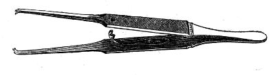
When no assistant is at hand, and in cases of emergency, the surgeon provided with this little instrument can tie vessels without the least difficulty; and in operation, when many vessels spring, several of these forceps can be applied; there is besides this great advantage in their employment, that a clumsy assistant can scarcely include the point of the instrument with the vessel. Hemorrhage from the smaller vessels soon ceases; and, before reaction occurs, their orifices have generally become so obstructed as to resist the effusion of blood.
The effects of ligature on a vein are somewhat different from those on an artery. The inner coat of the former is more dense and elastic, and remains entire, whilst the external and middle are divided. It is puckered by the ligature, and its opposite surfaces are placed in immediate contact; but there is no breach of surface, and adhesion does not occur till the tunic has been divided by ulceration; then the opposite margins cohere, the vessel is obliterated, and undergoes changes resembling those in an artery similarly circumstanced. The coagulum between the ligature and extremity is of considerable extent, dense, and completely filling the canal of the vein, and consequently, of a cylindrical form.
The edges of the wound, in the soft parts, ought not to be approximated till the bleeding has entirely ceased, and the surface become glazed, for the interposition of the slightest quantity of blood prevents union by the first intention. When bleeding has ceased, the divided edges are to be brought together as accurately as possible, and adhesion promoted. The minute vessels assume an action greater than in the healthy state, though not equal to inflammatory action; they effuse coagulating lymph, by which the opposed surfaces are agglutinated, though the union is at first feeble and easily broken up. Soon the lymph is firmly attached, by newly-formed vessels, to the surface from which it was secreted; in effect, it becomes organised, and rendered capable of undergoing, through its inherent powers, the changes necessary for its perfection and stability. Similar processes go on in it as in any sound part of the body; new matter is deposited, and the superfluous is absorbed. The process of nutrition, however, is not the same in all parts of its substance, that is, the new matter deposited is not exactly similar at all points; but, according as the new secreting vessels proceed from the different tissues of which the margins of the recent wound are composed, so, in various parts of the new formation, these vessels assume peculiar modes of action, one set forming muscular, another cellular tissue, and a third a substitute for skin, formations corresponding to the primary tissues from which the secreting vessels proceed.
Thus the vasa vasorum, ramifying on the divided ends of the minute vessels, secrete a substance which is transformed into a set of minute capillaries, and these also, assuming a secretive action, produce an arterial or venous tube, similar to that nourished by the original vasa vasorum. By this process the lymph becomes well supplied with bloodvessels, those from the opposite surfaces meeting, and freely inosculating with each other. These bloodvessels, as already stated, have been produced from arteries possessing different powers, and hence the newly-formed assume actions similar to those of the primary, and thereby interstitial matter is deposited of its proper kind and in its proper place, a cuticular membrane superficially, then cellular tissue, afterwards muscular, and so on according to the primary tissues which had been divided; these parts do not at first resemble exactly the corresponding natural tissues, but, by the continued action of the new vessels and capillaries, they are moulded and prepared for the due performance of their respective functions. If the degree of action necessary for the accomplishment of these processes increase to inflammation, adhesion is interrupted till the action be lowered to its previous standard.
From this view it is evident, that, besides a certain excitement of the bloodvessels, it is necessary that the raw margins be in close apposition, and carefully retained so, for, by ever so slight movement of the parts, the recent and delicate bond of union will be ruptured; and, if this motion be allowed to continue, adhesion may be at divers times begun, but can never be perfected. Whereas, if the necessary precautions are adopted, union is often completed in thirty or forty hours, sometimes sooner, seldom later. From a knowledge of the astonishing powers of nature in repairing injuries, many and important improvements have of late years been made in the practice of surgery. In the majority of instances it is also requisite that the parts be brought in contact soon after division, otherwise granulation will have commenced in the different parts of the wound, and the surfaces then approximated will not so readily adhere: pus is formed, and, having lodged between the surfaces, acts as an extraneous substance, keeping them apart, and separating them still farther by its accumulation. All foreign bodies in the wound must be removed before adhesion can occur; and, on the same principle, care ought to be taken that no effused blood be interposed. In many cases the margins of the wound can be accurately adjusted by careful attention to the position of the part, or by the application of a few strips of adhesive plaster; but the latter, from indiscriminate use, often prove the source of much irritation, and totally frustrate the end for which they are designed. When employed, they ought to be narrow and few. If such means be considered insufficient, recourse must be had to a few points of interrupted suture, and these are not productive of bad consequences which have been by some attributed to them. When neatly applied, they can produce but little irritation, more especially if removed as soon as their presence is unnecessary, that is, as soon as adhesion has fairly commenced, and the natural bond of union is of such strength as to need no artificial assistance. By these the edges of the wound are more neatly and suitably placed than by any other means; they meet easily, without the puckering or overlapping of each other; and, from the circumstance of sutures obtaining a more just coaptation, they can be sooner discontinued. In most wounds no other dressing is required; but in some a combination of sutures, adhesive plaster, and compress, is necessary.
Of late, I have greatly dispensed with stitches and the common adhesive plaster, using, instead of the latter, slips of glazed riband smeared with a saturated solution of isinglass in brandy, which is much less irritating and more tenacious than the common adhesive compost. The parts are fixed temporarily with a single stitch, or two at most, and cloths dipped in cold water are placed over the wound; the ribands are not applied till the adhesive substance has partly congealed, and the oozing of blood ceased. The divided margins being approximated by the fingers of an assistant, the ribands are laid gently over, and held for a few seconds. Soon after a sufficient number have been applied the stitches are withdrawn, being no longer necessary. No other dressing is required unless suppuration occur; the ribands will adhere firmly till the completion of the cure, and thus the pain and irritation caused by frequent dressing is avoided. Even the largest wounds, as after amputation, are treated in this manner with the most satisfactory results. Of late years a plaster, made by coating oiled silk with a solution of isinglass, has been used instead; the glazed surface of the slips is moistened, and applied as here directed.
If at any part adhesion fail, suppuration and granulation must follow. The adhesion may be prevented by any of the circumstances already mentioned, or by an unfavourable state of the constitution, the nature of the wound, exposure to bad air, the occurrence of fever, or of a flux natural or not. The wound may contain foreign matter; blood or the contents of canals may be effused into it, and many other obstacles may exist to retard, or prevent adhesion. Notwithstanding, in all cases, though the chance of union be but small, the parts should be approximated. A great point is gained if certain parts only are brought to adhere, for by their natural attachments the opposite surfaces are preserved in more direct contact than they could otherwise be, and thereby but little space remains to be filled up with granulations. If, on the contrary, the surfaces are not approximated, the flap is diminished in size, and when afterwards placed in contact with the surface from which it was detached, it is found not to correspond, leaving considerable deficiency to be repaired by the comparatively slow process of granulation. Whereas, if it had been early replaced, partial attachments would probably have been formed by adhesion, the flap thereby retained in situ, and prevented from shrinking, so that but little new matter would be required.
Sometimes union does occur, and that speedily, after the flap has remained separate for a considerable time; and in such cases it may be doubted, whether union is accomplished by adhesion, strictly so called. Most probably it is by this process that the rapid union occurs in such circumstances: the divided parts have assumed an excited action, and effused lymph; during their state of separation, the lymph will become organised when it is connected with the original parts, just as well as if the surfaces had been in contact; and when they are at length so placed, they will be agglutinated to each other by the outer part of the effused lymph, which still continues soft and unorganised. If motion be then prevented, organisation, which has already commenced in the connecting medium, will proceed undisturbed, converting the agglutination of soft unorganised lymph into firm and permanent union by means of organised tissue. In these circumstances, it is not to be wondered at if adhesion should be completed in a shorter time than when the surfaces are brought together immediately after their division; in each instance the process is the same, only in the one it has to proceed from its very commencement, whilst in the other it is previously all but perfected, and after the parts have been put together, the last stage only requires completion. It is only in a particular stage of a granulating surface, that adhesion will take place speedily, when the discharge is diminished, but healthy, and the granulations florid and firm.
When a wound is to heal by granulation, the exposed surface at first is dry, painful, and slightly swelled, and afterwards a thin discharge of bloody serum is poured out, with relief to the painful sensations; the surface is at this time covered by a thin layer of coagulated lymph, and the parts, if approximated, are in the most favourable state for adhesion. Soon, however, the vessels assume a different mode of action, and secrete a fluid which becomes purulent; the effused lymph has been organised, forms a living part of the surface from which it was deposited, and is covered and protected, in its yet delicate condition, by the purulent fluid. This new matter is disposed in numerous small conical projections of a florid colour; and these, by their own power, form others similar to themselves, at the same time discharging purulent matter; so that, in a healthy constitution, the cavity is soon filled by the granulations, which come to the same level as the surrounding integument. Sometimes they are exuberant, soft, and spongy, and in this state possessed of little sensibility, and but ill supplied with bloodvessels. At others, they are slow in approaching the surface, and then often morbidly sensible. In all cases, the new matter is very apt to be absorbed, either from the state of the patient’s health, or from the nature of the applications; and foreign substances, in a state of solution or minute division, are more readily taken into the system from the raw surface than from the sound skin. When, then, the granulations approach the skin, the sore contracts, the newly formed parts being modelled into a more firm and dense condition by the action of the absorbents. Sometimes, in superficial sores, the skin is seen spreading from several parts near the centre; but at these points portions of the original skin must have remained uninjured, though the others were destroyed, and have formed cutaneous matter as soon as they were on the same level with the surrounding granulations; for these insulated portions of skin are not a product of granulations, as some suppose, but of a substance similar to themselves. Skin is formed from skin. Thus, where a part of the integuments has been completely removed by operation, or destroyed by accident, no islands of skin are observed during the cure, but the sore is uniformly covered by skin proceeding from its margins. The margins of a healing sore are of a white colour, and adherent to the subjacent parts; but in an unhealthy one the margins are often unsupported, the subjacent granulations are absorbed, and their place is occupied by thin purulent matter; the new skin is unable to maintain its independent existence, continues of a dark colour, perhaps for a long period, then wastes away or sloughs. The recent cutaneous matter covering a sore contracts, and the neighbouring old skin is extended; the new surface is thus diminished; it assumes a slightly puckered appearance, and is termed cicatrix. This is at first pretty vascular, the vessels running straight; after a time they contract and become invisible, and scarcely admit fine injections. Frequently the scar is so far absorbed after some time, as to leave only a firm line, whiter and more dense than the surrounding integument.
If union by the first intention does not take place, then all the application to procure it must be abandoned, all sutures, plasters, compresses, &c., must be dismissed, for they now can do no good, and may be productive of harm; the attention, on the contrary, must be directed to effecting union by granulation; with this view, other means are to be chosen, so that to continue those which were formerly used to promote adhesion would be absurd, when adhesion can no longer be expected. The stitches must then be taken out, when inflammation has gone off, and œdema remains, the parts are to be supported; and by attention to position, and gentle bandaging, the size of the wound will be diminished. Inflammation must be subdued by the usual means, and suppuration encouraged by fomentation and poulticing, or warm water dressing. After inflammation has subsided, tension disappeared, purulent discharge occurred, and granulations formed, the edges of the wound are to be gently brought together, so as to render the quantity of new matter requisite for filling up the cavity as small as possible. Nature will then accomplish the union in her own way, and we can only assist and minister to her; for who thinks now of healing wounds by pure force of surgery? The dressings ought to be light, the ointment, if any is used, scanty; in some cases the application may be dry; but in many cases various lotions will be found of much advantage. These latter are used of different qualities, according to the nature of the sore; and these can be of little avail unless evaporation be prevented, by a piece of oil-silk laid on the outer surface of the dressing. In most granulating wounds, they require to be of a mildly stimulating nature, and the one which I have most frequently employed is a weak solution of the sulphate of zinc. The integuments round the wound may be occasionally washed, to prevent excoriation, but no good can accrue from washing the sore; its natural discharge is its best protection, and if superabundant, it can be removed by means of dry lint or tow from the surrounding parts.
From bruised and lacerated wounds there is little or no hemorrhage, but in proportion to the severity of the bruise, is the bleeding slight, and the danger great. The bloodvessels are so torn and twisted as to permit the spontaneous and temporary suppression of hemorrhage to occur almost immediately; and the larger arteries may escape entirely, owing to their elasticity. Sometimes after bruised wounds, such as those inflicted by gunshot, the large vessels bleed instantly and violently; often, however, hemorrhage occurs only after the sloughs separate, many days after the infliction of the injury, and then it is generally very profuse; in some instances, limbs are torn, bruised, or shot away, without hemorrhage occurring to any great degree, or at any period. Frequently the vitality of the parts surrounding the wound is much diminished; and the whole limb is apt to become gangrenous, either immediately, on account of the extreme violence of the injury, or consecutively, from greatly excited action going on in parts whose power of resistance has been much impaired: it often arises also from constitutional peculiarity. The gangrene extends often rapidly, in consequence of the infiltration of putrid serosity into the cellular tissue. In the treatment of bruised wounds, the position of the parts must be carefully attended to; they must be placed in a state of relaxation. In general, it is unnecessary to retain the margins of the wound in contact, for adhesion cannot occur—suppuration must ensue, and is to be desired—and the dead and dying parts must be loosened and discharged before union can take place. Sometimes, as when a large flap has been detached, and the parts not much injured otherwise, approximation ought to be accomplished, for the reasons already mentioned. In almost all cases, and most certainly in those in which the mechanical injury has been severe, and its effects extensive, violent inflammatory action is to be dreaded, and measures must be taken to ward it off: notwithstanding the prophylactic treatment, violent inflammation often comes on, and then recourse must be had to the means proper for the subduing of it. Blood is to be taken from the part, if necessary, and soothing applications used, in the form of fomentation and poultice. The main indications are to prevent extravasation into the substance of the limb, and strongly excited action. When the sloughs begin to separate, emollient poultices promote the suppuration and discharge of dead matter, and afterwards the sore must be dressed, according to the nature of the case, with the applications most fit for granulating sores in their different degrees of action and advancement. During the after treatment, the sides of the sore ought to be well supported, so as to prevent, as much as possible, suppuration from extending along the neighbouring cellular tissue; but, at the same time, the dressing must not be so tightly applied as to cause irritation. When abscesses have formed in the neighbourhood, the cavities should be freely exposed by incision; thus a free discharge will be given to the matter, and the cavity brought to granulate from the bottom. During the suppurating stage, the patient’s strength must be maintained by generous diet.
Punctured wounds are dangerous, from the deep and internal effusion of blood and serum which usually attends them. The effusion, which in open wounds is poured out externally, and moderates and prevents the excited action from exceeding what is salutary, is, in punctured wounds, poured into the substance of the limb to its detriment. It is followed by severe inflammatory action and profuse suppuration. In order to prevent these untoward results, it was formerly the practice immediately to dilate the wound; but this is hurtful, for if the wound be deep, as it generally is, dilatation of its whole extent is a proceeding severe in itself, and in its consequences; whilst, if the external part only of the wound is dilated, the operation will entirely fail to effect what is intended. The knife will be used in great good time where a foreign body is found to be lodged in the wound, when tension has occurred, or matter has formed. Sometimes the wound heals throughout its whole track by adhesion, without any bad symptoms being so much as threatened. Setons, recommended in this class of wounds, are of no service. It is not the narrowness of the external opening, as is sometimes supposed, that is the cause of all the mischief, but the injury and consequent inflammation of deep-seated parts.
Poisoned wounds are rare in this country. Wounds by the stings of certain trifling insects produce considerable swelling in some constitutions, and when the injury has been inflicted on a loose texture. In some parts of this country, the bite of a small adder causes pain, swelling, and unhealthy suppuration of the part, with some constitutional disturbance, but the results are seldom serious, and never fatal. In warmer climates, the bites of some snakes are followed by the most violent symptoms; in some cases proving fatal in a few hours, in others after a day or two. Great swelling occurs almost immediately, attended with excruciating pain, and extends upward along the limb; vision becomes impaired, the patient lies in a state of stupor, and ultimately succumbs under convulsions and delirium; the symptoms vary in particular instances according to idiosyncrasies and the state of the constitution when the injury is inflicted.22 In this country, the bites of rabid animals are more dangerous than those of animals naturally poisonous. Rabies most frequently occurs in dogs, and others of that species, such as wolves, foxes, &c. They become dull, sluggish, and irritable; have unnatural appetites and cravings, devouring their excrement and urine; the stomach is generally found full of chopped straw, pieces of wood, &c. Derangement of the cerebral functions is not complete,—they know and obey their master. They are often not afraid of water, but lap it and go into it readily. From them the disease is communicated to the human subject, and to the lower animals, such as cats, sheep, cattle, and even fowls; the virus is not communicable, except by the deposition of the saliva on an abraded surface, or into a wound. It is not produced by eating the flesh of a rabid animal. During the first days of the attack, pustules are, it is said, observed under the tongue, but there is no apparent change in the glands. The symptoms of hydrophobia in man seldom appear before the twentieth day after the infliction of the wound, and in some instances they have not presented themselves till after the lapse of months. The most prominent are great restlessness, much irritability and anxiety, and convulsions of the muscles concerned in deglutition, produced either by attempts to swallow, or by fluid being presented to the patient. Ultimately, the spasms become general, are induced by the most trifling exertions or noises, and prove fatal in a few days. Frequently the patient retains his senses throughout, and is fully aware of his lamentable situation; in other instances, he soon becomes delirious, raves, and threatens his attendants. For this horrible disease we are unacquainted with any cure. In general, profuse bleedings are employed, and large quantities of opium given internally; every powerful antispasmodic, as well as every violent medicine, has been made trial of, but in vain: some certainly mitigate the symptoms, but none cure the patient. It has been even proposed to suspend or destroy the function of the nervous system for a time, by the employment of the Wourali poison, keeping up at the same time respiration by artificial means, under the expectation that thus the impression on the system might be got over. The morbid appearances usually observed after death are marks of inflammation of the pharynx and air-passages, and of the mucous surface of the stomach and intestines. It is evident that the disease ought to be prevented if possible, and for its prevention the most efficient measure is timely excision of the affected parts; and they should be cut out a long period before the constitutional affection comes on: when excision is dangerous, or wholly impracticable, and when the patient does not apply soon after the accident, the injured parts may be destroyed by some active caustic, as the potassa fusa. The nitrate of silver has been strongly recommended and extensively employed by Mr. Youatt, whose experience in this disease is very great. This application should scarcely be trusted to alone. The removal of parts wounded by snakes, even after violent symptoms have appeared, has proved successful, ammonia having been at the same time administered internally. In some instances, arsenic has been found efficacious when given in large doses, and frequently repeated.23
Wounds received during dissection occasionally have unpleasant consequences from the absorption of animal matter. The absorbents leading from the wounded part become swelled and painful, and in slight cases there are shivering and general indisposition for some days. The more violent symptoms arise from examining bodies which are rather recent, and in which putrefaction is just commencing, and very frequently from inspecting the bodies of females who have died of puerperal diseases. The absorption may take place from punctures made by scissors, the point of a knife, or spiculæ of bone, or from old scratches, or chops by the side of the nail or on the hand. There is little or no danger from an open and bleeding wound, as by the flow of blood the part is completely cleaned; it is generally from slight punctures that untoward symptoms need be apprehended. Effects similar to those resulting from wounds in dissection often occur in nurses and others who have pricked themselves with pins while washing foul clothes, or from handling poultices or dressings removed from bad, putrid, or sloughing surfaces. The symptoms already mentioned are soon followed by others more severe: shivering continues, and the patient is seized with vomiting; the part affected, and often the greater part of the arm, becomes red and much swollen; the cellular tissue is infiltrated with serum often dark and putrescent, abscesses form at various points along the limb to the axilla, and purulent matter is diffused throughout the unhealthy cellular tissue, which in many instances sloughs, and gives rise to extensive sores. Typhoid symptoms soon appear, and in the more aggravated cases speedily prove fatal. When such local and constitutional symptoms arise, it will generally be found that the patient was of a broken-up constitution previously to the infliction of the wound; did they solely depend on the inoculation of virus, they would be of very common occurrence, considering that wounds are so frequently received during dissection; but it is seldom comparatively that any unpleasant symptoms follow such an accident. In all cases, however, it is prudent to adopt measures in order to prevent absorption of the virus. With this view, the wound is made to bleed by means of pressure or suction, and by the latter method the exposed surface is most effectually purified; afterwards nitrate of silver maybe applied to deaden the surface, and protect it by an eschar. If such means be unavailing, the after symptoms must be encountered as they appear, local inflammation subdued, tension relieved, abscesses opened, sloughs removed, &c. General bleeding is seldom admissible, but purgatives and antimonials will prove beneficial at the commencement; afterwards the strength is to be supported, and, if the patient be much reduced, stimulants are to be liberally administered.
We shall next treat briefly of gunshot wounds; under this head are included the contused and the lacerated, caused by splinters, &c. The vitality of the injured surface is generally destroyed at once, whence bleeding seldom occurs, even after whole limbs have been shot away; in some cases, however, hemorrhage is profuse, as when a large artery has been wounded by small shot. The effects on the system are extremely various; some persons are affected with tremors, anxiety, and depression from slight wounds; while the most severe injuries are often unaccompanied by any disturbance of the nerves. The shock is generally of short duration, disappearing soon, on the patient’s being reassured and encouraged, or after his taking a little wine or opium. In gunshot wounds, those inflicted from a distance or close at hand can in general be readily distinguished. In the latter, the wound is large and lacerated, portions of the wadding are impacted, and the skin around is marked with grains of gunpowder. In the former, the wound is small and clean. When a ball passes through a fleshy part, the opening at its entrance is small and depressed; whilst that by which it escaped is open, with everted edges. When it follows a superficial course, its track is marked by a wheal, or elevation of the skin with discoloration. At one time, it was believed that the most serious consequences resulted from a ball passing close past the body without even touching it—that in this way violent concussion of the brain, proving instantly fatal, was produced; but this notion has been disproved by experience; part of the head accoutrements, of the hair, of the nose, and of the ears, have been shot away by cannon-balls, and yet no disturbance of the brain has followed either immediately or consecutively. The opinion originated from the circumstance of soldiers having been found dead without any evidence of injury; but bones are often broken and comminuted by an indirect blow, or by a spent ball, without any breach of surface or external sign remaining; internal injuries indeed—rupture of viscera—more than sufficient to cause instant death, are thus inflicted without any apparent external læsion.
The course of a ball in the body is often very strange, depending on the force with which it is projected, or the resistance which is opposed to it, and on the position of the struck part. Balls often pass under the integument almost completely round the head or chest, having first struck the bone at a very oblique angle. Frequently they remain, lodged along with part of the clothing which they thrust before them. In such cases, they may be immediately removed, their exact situation being previously ascertained by external examination, or by means of a probe. They can generally be extracted through the aperture by which they entered; but if situated superficially, and at a distance from the opening, this will be more readily accomplished through an incision made upon them; if allowed to remain, suppuration will occur in the neighbourhood, the surrounding cellular tissue will be condensed, an abscess will be formed containing the foreign body, and by the process of absorption proceeding in the parts external to the abscess, the ball will at last reach the surface, and be discharged. The track is often so extremely tortuous, as to render it impossible to ascertain the situation, or even the existence of the foreign body, which greatly impedes the operation; and, in other instances, it may be necessary to allow the ball to remain undisturbed, on account of its being placed near important parts, which might be injured by any attempt at removal. Foreign bodies often remain lodged in fleshy or membranous parts for years, having become enveloped in a dense cyst, and having ceased to produce any great irritation. In consequence of the force with which they have been projected, and the resistance which has been opposed to their progress in the body, musket-balls, when extracted, either immediately after the infliction of the injury, or after a considerable time has elapsed, are seldom found to retain their globular form, but are flattened and ragged, and not unfrequently completely divided by the bones on which they have impinged. A bullet may be divided into numerous fragments on a bone, and part may enter into the osseous substance, whilst other portions penetrate in all directions into the soft parts, and, though sharp and irregular, may remain long in the dense cellular cyst which forms over them, without producing pain or inconvenience. There will necessarily be suppuration, and generally discharge of dead matter, from gunshot wounds, in consequence of the bruising of the parts by the ball, which may be expected to injure the superficial layer of parts in its tract so severely, that it must slough more or less.
Besides, the bones are often splintered by the force with which they are struck, and loose portions of them lodge amongst the muscles; then they are the cause of much mischief, for, on account of their long, sharp, and irregular form, they occasion great irritation, suppuration ensues in various parts, sinuses form, and the cure is rendered very tedious. In other cases, the bone is split in a longitudinal direction, and, in the cylindrical bones, these fissures are often of great extent.
Considering the nature of the body which inflicts the injury in a gunshot wound, and the velocity with which it is impelled, it is evident that the cure must be in all cases tedious, in consequence of the sloughing and suppuration which is induced, particularly at the aperture through which the ball passed. The foreign body ought always to be removed as early as possible, provided it can be accomplished without much violence, or injury to the parts. Dilatation of gunshot wounds is now had recourse to only to facilitate the removal of balls, splinters, &c., and even with this view, it ought to be employed but to a very slight extent, if at all; for foreign substances, when deeply seated, can be much more easily taken out when the sloughs are separating, and the parts relaxed by suppuration; then, too, they can be more readily reached through a counter-opening, when their situation renders this expedient. In short, the surgeon is not justified in cutting for balls, unless they are easily felt, and not deeply lodged. In order to discover the foreign body, probes will sometimes be required; the finger often answers the purpose best, unless when the wound is of considerable depth. If, on examination, the ball cannot be discovered, and if there is reason to think that it has followed an indirect course, the surgeon will, sometimes, be assisted in his search by placing the patient in that position in which he received the wound, and then judging of the circumstances most likely to affect the foreign body in its passage. In many cases, extraction can be accomplished by means of the finger alone; in others, forceps and scoops, various in length and size, are indispensable. Afterwards, light dressings are to be employed; and in the first instance, cold applications may prove advantageous in keeping down the inflammatory action; but when inflammation has commenced, and to encourage suppuration, warm fomentations and poultices are to be preferred; they will in many cases be both more grateful to the patient and more beneficial in their results, when used even from the commencement. Afterwards, it will be necessary to afford sufficient support to the parts by bandaging, and to change the applications according to the particular circumstances:—soothing, if the wound be irritable, stimulating, if inert, and gently escharotic, if the granulations be exuberant.
In severe injuries of the limbs, the surgeon must be guided by the state of the part, and of the constitution, by the circumstances in which he is placed, as to accommodation, and mode of transportation, &c., in deciding on the removal of the part by amputation,—or on making an attempt to save it, by trusting to, and assisting, the processes of nature. The question whether to amputate immediately, and on the spot, merely allowing the shock, if any exist, to pass away, or to delay till suppuration occur, is now scarcely a matter of dispute. When it is evident, from the extent, violence, and nature of the injury, that there is danger of speedy mortification, or of extensive and severe inflammation and suppuration, amputation is to be instantly performed,—delay is inadmissible. In comminution of the hard, with contusion and laceration of the soft parts—where limbs have been shattered and completely detached, or nearly so—in lacerations of parts, including the principal bloodvessels and nerves—fractures of the heads of bones, with openings into the joints—and in bad compound fractures, more especially of the thigh (for all compound fractures of the upper part of the thigh are dangerous), amputate at once. When the limb has been retained, and gangrene arises in consequence of the external injury, and when there is no reason to suppose that any internal cause is also in existence; or when the violence of inflammatory action has subsided, and the patient is become hectic, with profuse purulent discharge, and with disunited bones, then amputate. But, in this latter case, the chance of recovery is not so great, and the proportion of recoveries small; whereas, in the immediate or primary operations, the very reverse holds true. Such is the experience of the greater number of military surgeons. In civil practice, the results are somewhat different; a greater proportion of primary amputations are unsuccessful, and the secondary turn out more favourably than the statements of military surgeons would lead one to believe. In all cases, the judgment of the surgeon must guide him in his determination. The circumstances of the case, and the probable contingencies, must be all duly considered, and he must not proceed with his knife where there exists even a slight chance of preserving the limb.