PART SECOND. OF PARTICULAR SURGICAL SUBJECTS. (continued 13)
Reading edition
[Congenital Luxation of the Hip-joint is sometimes met with, though on the whole a very rare affection, especially in this country. Female children are more apt to suffer from it than males, and it is also more common in such as are of a scrofulous habit than in such as are endowed with a good constitution. Of twenty-six cases of this malformation observed by Dupuytren, not above three or four were males; a disproportion probably not altogether dependent upon chance. The immediate causes of this variety of displacement are, first, shortness, total absence, or extreme obliquity of the neck of the thigh-bone; secondly, partial or entire obliteration of the cotyloid cavity; thirdly, deficiency, extraordinary elongation, or complete absence of the round ligament.
The characters of this malformation are, shortening of the affected limb, unnatural projection of the great trochanter, ascent of the head of the femur into the iliac fossa, inversion of the leg, and obliquity of the pelvis. The motions of the joint, particularly those of abduction and rotation, are constrained and imperfect; the muscles of the upper part of the thigh are retracted, or drawn towards the iliac crest; the limb is thin, wasted, and out of all proportion to the rest of the body; the tuberosity of the ischium is almost uncovered, and consequently unusually prominent; the upper part of the trunk is thrown backwards, while the lumbar portion of the spine projects forwards, being concave behind; the pubes is placed almost horizontally on the thighs; and the ball of the foot alone touches the ground when the child stands erect.
In the recumbent posture, when the weight of the trunk is taken off, and the muscles are relaxed, most of the symptoms of the luxation disappear, and the limb may be shortened or elongated at pleasure. In walking, the body is inclined towards the sound side, and the head of the dislocated bone sinks towards the cotyloid cavity by its own weight. As age advances, the limb becomes shorter, in consequence of the femur ascending higher and higher on the ilium; the obliquity of the pelvis augments; and the power of locomotion, already so much impaired, is completely destroyed.
Congenital dislocation of the hip-joint may, in general, be easily distinguished from other accidents or maladies, by the affection being observed at or soon after birth, by the obliquity of one or both thighs; by the absence of pain, swelling, and ulceration; by the head of the femur being displaced without any external violence; and by the ability of the surgeon to lengthen or shorten the limb at pleasure. In disease of the hip there is always more or less pain, with a feverish state of the system, and gradual failure of the strength; the parts about the joint are tense and swollen; the limb, at first somewhat lengthened, becomes afterwards shortened, and cannot be extended without the greatest suffering; and the motions of the ileo-femoral articulation are forever impaired.
The post-mortem appearances vary. In general the cotyloid cavity is partially obliterated, or entirely deficient, being replaced by a small, irregular, osseous prominence, devoid of cartilage and synovial membrane; the head of the femur, often flattened at its antero-internal aspect, rests in a sort of superficial fossa on the dorsal surface of the ilium; the round ligament, as was before remarked, is elongated, partially worn away, or even altogether absent; and the surrounding muscles are either atrophied, transformed into a species of yellowish fibrous tissue, or preternaturally developed. In the latter case, their action is preserved; in the former, it is very much restricted, or totally impeded.
The prognosis is always unfavourable, as the patient dies either young, or remains permanently lame and deformed.
The treatment can be only palliative; and as the weight of the trunk is the main agent in aggravating the displacement, repose is obviously indicated: but it is not necessary to confine the patient to the recumbent posture; since, in the act of sitting, there is no stress on the femur, the body resting principally on the tuberosities of the ischia. Dupuytren thought favourably of the cold bath: it should be strongly impregnated with salt, and the body immersed for three or four minutes at a time. He was also in the habit of using a well-stuffed belt, about four inches wide, for surrounding the pelvis and fixing the great trochanters; thus binding the ill-adapted parts together, keeping them at the same height, and preventing that continued motion to which they are otherwise so much exposed.]
Luxation of the Bones of the Leg.—separation of them from the end of the femur—seldom occurs. It can be the effect only of great violence and great laceration. Most frequently fracture is concomitant, perhaps with wound; and such accidents require amputation, either primarily or secondarily. Subluxation, from laceration of the internal lateral ligament, is not so unfrequent. It is most common in females, the natural conformation of their thigh-bones disposing them to bend inwards; and from falling awkwardly, particularly if carrying a weight, the ligament is apt to give way. The limb is pained, deformed, and unable to support the body, and swelling to a considerable extent soon follows. Reduction is extremely easy; and the parts are retained in site by the application of a wooden splint, to either the outer or the posterior side of the joint, the leg and foot being previously bandaged. The joint remains long weak, and never recovers entirely; a sustaining apparatus, fitted on the outside, retained by straps, and with a joint opposite to the articulation, is required to be constantly worn when the patient wishes to use the limb.
Luxation of the Patella is spoken of by some as common. Others of much experience have not met with a single instance of it. I have never seen this accident. The bone, it is said, may be displaced outwards, inwards, or upwards. The first form of luxation is the most frequent, and is caused by a severe fall, with the foot twisted outwards and the knee inwards. Displacement inwards is produced by direct violence applied to the outer part of the bone, or by the foot being turned inwards in a fall. Displacement upwards can occur only after laceration of the ligamentum patellæ, the bone being then drawn up by the unresisted action of the muscles on the fore part of the thigh. In dislocation outwards, the bone has been found “resting with its inner edge upon the outer surface of the condyle, the fore part facing obliquely forwards and inwards.” In this last form of accident, sudden, forcible, and complete flexion of the limb is said to produce immediate reduction. In dislocation outwards or inwards, the muscles are to be relaxed by raising the heel, extending the limb, flexing the thigh, and then forcing the bone to its proper site by manipulation. In the dislocation upwards with rupture, the limb is to be kept extended and raised, and the bone is brought as nearly into its place as possible by bandaging. When a peculiar laxity of the apparatus about the joint exists, whether as a cause of luxation or not, the support of a well-made knee-cap is required.
As formerly stated, Dislocation of the Ankle cannot take place inwards or outwards, without fracture of the end of the tibia or of the fibula, either above the articulation, or where they project by the sides of the astragalus for the greater security and strength of the joint. Subluxation, however, or sprain, may occur without injury of the bones: in this accident, should the parts not have spontaneously resumed their original situation, no difficulty is experienced in putting them to rights; simple manipulation is sufficient. Occasionally, the foot is luxated forwards, by force applied either to the heel or to the fore part of the leg whilst the limb is fixed. The heel is shortened, the foot elongated; indeed, the marks of the injury are so distinct, that comparison of the limbs is sufficient for diagnosis. Luxation may also take place backwards; and in this case the heel is elongated and the foot shortened. In these accidents it is not unfrequently found that one or other malleolus has given way, or that the lower end of the tibia is split. Reduction is sometimes difficult. Extension is to be made by grasping the foot and pulling whilst the limb is fixed, at the same time making pressure either backwards or forwards, as may be required. To retain the bones in their proper situations, it is always necessary, at least prudent, to apply a paste-board or leathern splint to each side of the limb, particularly when fracture of the malleoli is conjoined.
Displacement of the Bones of the Tarsus may result from great force; for example, when the foot is squeezed under a heavy weight, one or more bones may escape from their connections, and project. Reduction of such displacement is exceedingly difficult at any period, and becomes almost impossible when inflammatory action is allowed to supervene previously to attempts being made. The astragalus is sometimes pushed out of its place; though it is difficult to conceive how, to a bone so hid and so firmly connected, such force should be applied as to cause protrusion of it from its natural situation. It has been found lying on the dorsum of the foot, causing swelling, lameness, great pain, shortening and deformity of the limb; and the shape of the bone can, in such circumstances, be distinctly felt and seen through the integument. As already observed, reduction is almost impracticable, and, with the view of remedying deformity, it has been proposed to cut out the displaced bone; but as to the expediency of such practice I can give no opinion.
I have seen but one instance of displacement of this bone backwards, and most probably another will never occur to me. A heavy young man, in a state of utter intoxication, fell backwards down a stair, and in the fall his foot became entangled in the railing. The astragalus was found lying betwixt the back of the tibia and the tendo-Achillis, its upper articulating surface facing forwards, the lower in contact with the tendon. All attempts to reduce the bone proved fruitless. Violent inflammatory action followed, but was reduced by active measures; and the limb ultimately became very useful; in fact, though not till after many months, little lameness or shortening was perceptible.
By Sprain is understood subluxation or partial displacement of a joint, with stretching, and more or less laceration of the articulating apparatus—ligaments, tendons, sheaths, and bursæ, being all involved in the injury. Sometimes small portions of the processes of bone are separated, being torn away, attached to ligament or tendon. All joints, both large and small, are liable to the accident. In the proximal, or in the middle joints of the finger, for example, one or other lateral ligament is stretched or torn; the finger is twisted to a side; the joint is swelled; and this swelling, with pain, is of long continuance, perhaps increased by repeated twists, or by imprudent use of the joint. The elbow and shoulder are frequently sprained, as also the hip and knee; but the injury most frequently occurs in the wrist and ankle. It is generally occasioned by a fall, the foot or hand coming awkwardly to the ground, the muscles being at the time relaxed and unprepared; by over-exertion in lifting heavy weights; by entanglement and twisting of the limb, &c. The ankle is often sprained by what is called a false step; the fore part of the foot comes in contact with an obstacle unexpectedly, the foot is twisted under the limb, the weight of the body is thrown on the apparatus of one side of the joint, and this is in consequence immoderately and unnaturally stretched. Violent pain immediately occurs, and the patient is sick and faint. Discoloration and rapid swelling take place from extravasation of blood into the cellular tissue, into the sheaths of the tendons, and perhaps into the synovial pouches, in consequence of laceration of the bloodvessels. Effusion of serum and increased secretion of synovia afterwards occur, from incited action of the vessels. Thus the joint is deformed. Attentive examination is required to guard against mistakes; the existence or non-existence either of displacement or of fracture must be at once ascertained by determined and perfect manipulation; the parts must be pressed and moved, to such an extent as is necessary, notwithstanding the pain thereby occasioned, and notwithstanding the resistance afforded by the patient. It has been already stated that luxation of the wrist is not uncommon; that separation of the one bone of the forearm from the other, and transverse fracture or splitting of the radius, at the distal extremity, are accidents by no means rare. Great disfiguration follows simple sprain, much swelling taking place on the fore part of the limb from effusion under the fascia, and there is also much serous and bloody infiltration of the cellular tissue on the back of the hand and forearm. In the ankle, the ends of the bones must be carefully examined, and also the fibula in its whole extent, that the existence or non-existence of fracture may be ascertained, and that the surgeon may be guided to a correct mode of treatment. If the joint is not put at rest immediately, the extravasation is increased, and, in consequence, the pain and inflammatory swelling also; and parts of the joint at first not involved in the injury may thus be made to suffer. Many diseases of synovial membrane and articulating cartilages are attributable, and can be traced, to badly managed sprains; and in some constitutions, but slight injury, combined with a little bad treatment, suffices to destroy a joint. When, the case is well managed, the pain is never great, and soon abates; the swelling after a few days slackens; the discoloration becomes greater, the serum being absorbed, and the effused blood shining through the skin; the integuments appear green, blue, red, purple—these hues either being present all at the same time, or occurring successively; the discoloration often extends far from the joint. The mobility and strength of the joint are recovered gradually.
Perhaps no injury is more frequently mismanaged, by those both in and out of the profession. Every old woman thinks she can cure a sprain; most absurd and hurtful measures are resorted to; the injured parts are kept in motion; cold lotions and cold effusions are employed, and at the same time stimulating frictions: probably attempts are made, either by leeching or by puncturing, to extract the effused blood; and many similar follies are committed. The proper treatment certainly appears to consist principally in absolute rest and position. If there is any displacement it must be rectified immediately. If there is any fracture; or if there is a tendency to redisplacement after reduction; or if the patient is restless either from folly or from insensibility, as when the head has been injured by the accident, when the patient is under the influence of strong liquors, or when he labours under delirium tremens,—a splint or splints must be applied to secure immobility of the parts, at the same time without such compression as may interfere with swelling from effusion; the effusion is a salutary process, and should be encouraged, not repressed. By absolute rest and elevation of the limb, the extent of the swelling is limited, and inflammation warded off. Fomentations, properly employed, afford much relief; at first they probably encourage the serous effusion. The integuments soon become relaxed, during the regular use of fomentation, and tension and vascular action subside, as also pain. The swelling then abates, and is no longer hard; it pits on pressure, and the skin has a puckered appearance. Then gentle friction becomes advantageous, and uniform support should be afforded by the application of a flannel roller. The longer the limb is disused, the more perfect and rapid is the recovery, provided the rest of the cure be properly conducted. In general nothing more than what has been stated is required. But if the limb be moved, or stimulated in any way, early, then necessity will arise for antiphlogistic measures—perhaps venesection, certainly copious and repeated abstraction of blood by leeches, accompanied with fomentations, and the internal exhibition of antimonials, purgatives, &c. When such is the case the cure is tedious, the joint long remains swelled and stiff, the patient is lame and incapable of exertion.
Leeching or puncturing at an early period, with the view of allowing extravasated blood to escape, is useless and hurtful. The effused and coagulated blood cannot be evacuated, and suppuration, followed by destruction of the cellular tissue, has often been the consequence of such ill-advised proceedings. Friction with stimulating liniments, or even simple friction, at an early period, is also hurtful, as tending to excite vascular action, and to convert simple swelling into inflammatory. The application of cold at any period is of little use, and ought certainly to be avoided immediately after the injury, as adding to the sufferings of the patient, and interfering with the natural processes which have commenced for the reparation of that injury.
In limbs that have remained stiff after severe and mismanaged sprain, the dashing of water, either cold or tepid, has been strongly recommended. The practice is not ineffectual; the vessels of the surface are excited, perhaps as by other friction, and perhaps by the reaction which follows the chill. But the limb is apt to become rheumatic; and, on this account, the state of matters will not be improved by this proceeding, unless it be resorted to with proper precautions.
In severe sprains there is reason to think that sometimes even the tendons yield a little—that many of the fibres give way, and that thus the tendon is thinned and elongated. Such injury happens often in horses, in what is called breaking down. In them the tendon is occasionally snapped entirely through, and the ends widely separated. The same occurs in the human subject. Separation of the muscular fibres, however, is rare; laceration of the tendon itself, or separation of the tendon from the muscle, is more common. The yielding of the broad tendons on the upper and fore, lateral and under parts of the abdomen, affords an example of laceration of tendinous fibre from violent exertion. The tendons of the limbs are more frequently injured, and in the lower oftener than in the upper. I have more than once seen the tendon of the biceps torn in violent exertion. In the thigh, too, some fibres occasionally give way from a similar cause. The supra-muscular fascia in the arm and thigh is apt to give way at one or more points during powerful exertion of the muscles, causing deformity by protrusion of muscle through the torn space. But it is in the apparatus for extending the foot, and raising the weight of the body, that laceration of tendon most frequently takes place. The accident is uncommon till after the middle period of life, when the body has become heavier, when muscular exertions have been less habitually practised, and when the fibre has grown more rigid. The person in raising himself over some slight obstruction in walking, perhaps attempting to pass a small ditch or stile, suddenly “breaks down.” Or in dancing,—an amusement which he has long discontinued—a sudden snap is felt, with immediate lameness and slight pain in the back of the limb; swelling and discoloration follow; and these symptoms and signs vary in intensity according to the extent of the injury. Laceration may have been slight; the pain, swelling, and lameness are proportional, and at first an inconsiderable void can be felt at the upper part of the tendinous termination of the gastrocnemii. Sometimes no change is perceptible, and in such cases some have been of opinion that the slender tendon of the plantaris has given way and caused the lameness; but this is doubtful, and it seems more probable that stretching and yielding has taken place in some part of the tendon of the gastrocnemii, which had been in powerful action—probably, the tendinous and muscular tissues have been separated to a slight extent. Occasionally the tendo-Achillis is found completely torn through, and its upper end retracted; in such cases a large space is occasioned at the injured part, when the knee is extended and the foot bent. Sometimes the tendo-Achillis is cut through; I have seen both completely divided in the same individual—he received a wound by a cutlass across the back of both limbs, while endeavouring to escape from the mate of a vessel, in which he had been stealing. In rupture without breach of surface, the torn bloodvessels pour out their contents into the cellular tissue to a considerable extent, and if a proper mode of cure be not adopted immediately, inflammation quickly supervenes; and this is apt to terminate very unfavourably in the infiltrated tissue.
The tendon is united by the deposition of new matter, and the conversion of this into substance resembling the original structure from the vessels of which the deposit has taken place. The quantity of new formation necessarily depends on the extent of laceration and the space thereby occasioned. That such reparation of tendon does take place, and that to a very great extent occasionally, is placed beyond all doubt by the results of veterinary practice. “Knuckling over” in horses is occasioned by contraction of the flexor tendon; the heel does not reach the ground, and in order to effect this the tendon has been in many instances completely divided. The cut ends immediately separate, to the extent of some inches, and after a time this large space is filled up by a substance similar to tendon; so similar, indeed, that on post mortem examination, some years afterwards, a careless observer could scarcely distinguish any difference in the appearance of the various portions of the tendon. The same is observed after the operations for deformity of the foot in the human subject.
The treatment of lacerated tendon consists in placing the parts so as to relax the muscles whose tendons have suffered. In rupture of the tendo-Achillis, the knee should be bent and the foot extended, relaxing the muscle and approximating the separated ends. This is readily and conveniently affected by placing a slipper on the foot, and attaching to its heel a firm band, which is then fastened to a ring or strap placed on the thigh. This apparatus must be worn for six or eight weeks. Afterwards a high-heeled shoe should be used for some time; or if the union be still weak and imperfect, a splint may be placed on the fore part, resting on the dorsum of the foot and the fore part of the leg.
Bruise.—The effects of bruises or contusions are, separation of the cellular connexions, rupture of bloodvessels, and effusion of their contents into the cells; a cavity, often large, is thus formed partly by the direct injury, and partly by the subsequent effusion, and this is quickly filled with blood, partly fluid and partly coagulated. Immediate tumour forms; and the integument is discoloured, often beyond the principal swelling. The injury may, or may not, be attended with division of the integuments, or with fracture or displacement of the bones; but all injuries of the hard parts are attended with more or less bruising of the soft. Bruise is most frequently produced by a blow, and is most severe when the violence is resisted by an unyielding part, as by bone; a squeeze between two bodies, particularly if they be in motion, also inflicts extensive contusion. The swelling continues to increase for some time, and then gradually disappears along with the pain. As the tumour subsides, the discoloration increases; the thinner parts of the effusion have been absorbed, and the clot then shines through the skin, imparting to it various hues.
Bruise may be followed by inflammatory action. Then effusion is increased, bloody fluid is poured both into the cavity and into the unbroken cellular tissue, the whole parts become extremely tender, the surface inflames, and the excited action is apt to terminate unfavourably in the various tissues. Not unfrequently sloughing takes place, both of the skin and of the cellular tissue and fatty matter, with unhealthy suppuration and infiltration; and constitutional disturbance accompanies. All this is likely, nay, certain, to follow admission of air into the cavity filled with effused blood, whether by accidental wound or by intentional division of the integument. Meddlesome surgery is unfortunate here, as well as in many other cases. After scarifications, punctures, leechings, or incisions, the blood often seems to undergo a putrefactive process, and unhealthy suppuration is quickly established.
Sometimes the clot is not entirely absorbed, and considerable swelling remains for a long time, perhaps with slight tenderness of the part; a foundation is thus laid for abscess, either chronic or acute. Frequently the inflammatory action following on bruise is not so violent and rapid as that above described, but is limited in its consequences chiefly to the effusion of coagulable lymph. This may not be altogether absorbed along with the other effusion, it may become organised, and be the nucleus or germ of a new growth, of a tumour contrary to nature—deposit increases in and around the nucleus, and this formation, though at first of a simple nature, may become rapid in its growth, and may assume a troublesome or even a malignant action; and sometimes all this may occur at an early period, before the attention either of the patient or of the practitioner has been drawn to the action or to its effects. Many tumours can be traced to the effects of a bruise.
In the treatment of bruise, the parts should be placed in a state of absolute rest, and methodically fomented. Local bleeding is seldom required, and is of little use; at first it is hurtful. When, from the extent or number of the bruises, fever follows, general antiphlogistic measures must be resorted to. Cold and astringent applications, and other repercussives, as also stimulants, are pernicious in the first stage, and are not very useful at any time. Opening of the cavity must be carefully avoided, excepting when absorption has ceased, when the tumour has increased and become painful, and when the effused blood is putrescent, and unhealthy suppuration has commenced. Then the cavity should be opened freely, and by poulticing the clots and sloughs are got quit of; afterwards the parts must be supported, as also the strength of the patient. When from long want of use, in tedious cases, the parts have become cold, shrunk, and weak, as also happens in sprain, friction, champooing, tepid affusion, passive motion, and voluntary motion short of giving pain, will all be of use as tending to restore the circulation, the nervous energy, and the muscular development. If œdema remain, bandaging or a laced support will be required.
Amputation.—Every endeavour, which skill and experience can suggest, must be made before mutilation of the body, by the removal of even the smallest portion of one of its members, is resorted to. But there are cases in which mutilation, though a harsh remedy, is still indispensable for the saving of life. There are others in which it is prudent and proper to resort to operation, in consequence of a member becoming perfectly unserviceable, and likely to impair the usefulness of the individual. Such are very bad and complicated fractures and luxations—laceration of the soft parts of a limb to such an extent as to impress the experienced surgeon with a certainty that in a short time gangrene must ensue, and render the success of any attempt to save life very problematical. When the extent of injury is such that, though gangrene may not be dreaded, yet it is plain that extensive suppurations and exfoliations must necessarily take place, a question may arise as to whether immediate amputation is to be performed or not. This will be decided by the circumstances in which the patient is placed, and often also by his own feelings upon the subject. He may choose to run some risk, and endure much suffering, with even a very slight chance of ultimately preserving his limb. In cases of traumatic gangrene of the chronic form, amputation is not only justifiable, but imperative; as also in those cases of severe fracture in which the patient is sinking under profuse discharge, with disunited bones. And the same absolute necessity for operation exists in many diseased joints, and in some diseased bones, when the patient’s safety would otherwise be endangered, or when, on mature consideration, it is evident that the member, if retained, must for ever be an encumbrance, and worse than useless. Certain tumours of bones, tumours involving joints, tumours and ulcers of the soft parts of a malignant nature, and without appreciable disease of the lymphatic system, will also demand recourse to the amputating knife. Patients, too, will be met with, who, after undergoing all the suffering attendant on disease of long duration—as exfoliation of bone and sloughing of tendons, following deep suppuration—will, to get rid of the annoyance of the stiff and deformed member, or part of a member, not only submit to, but urge and insist on, the removal of the offending part. Amputation will also occasionally be required for badly-formed stumps, as those in which the end of the bone protrudes through ulcer of the integument, and is necrosed—or those in which the bone has been sawn of an inconvenient length.
Many precautions are to be observed in this operation. It is not to be commenced without due consideration as to the position of the operator, and of his assistants—their several duties—the form of incision—the length of the stump—the difficulties, if any, which may be expected, and the best means of obviating them. The most prominent objects are, to save undue effusion of blood, to effect the incisions with as little suffering to the patient as possible, and to make them of such a form as to cover the end of the bone effectually— so that pressure may, after a time, be borne without risk of ulceration of the soft parts, or exfoliation of the bone.
In all cases, and in all situations and circumstances, hemorrhage can be restrained during completion of the incisions, and during the employment of means to close the cut ends of the vessels, by means of very slight but exact pressure on the trunk of the principal vessel. The point at which this is to be applied should be at as short a distance as possible above the place of incision, and at the same time above the origin of any branches which must be cut. Not the slightest pressure should be made until the instant when the incisions are about to be commenced, so that no venous congestion may take place in the limb. All the blood in the limb, below the incisions, must necessarily be lost. The veins are more easily compressed than the arteries, and pressure, made a short time before the operation, may arrest the return of the blood, whilst it may not completely stop its influx; thus engorgement of the lower part of the limb is produced, and the quantity of blood that must be lost is increased. For a similar reason, pressure, sufficiently firm to stop arterial hemorrhage, is to be continued till the principal branches are tied, and then entirely removed; for the continuance of even slight pressure will increase the flow from the surface of the stump—blood, flowing in, and being arrested in its venous return, trickles out through the open ends of the veins. If a circular band be used for the compression, such as the screw tourniquet, it should be put on quickly, and screwed up at once, and then the incisions should not be delayed one instant after; there should be no relaxation of the pressure at any part of the operation; and as soon as the principal vessels have been secured, the apparatus should be altogether removed—otherwise, as already stated, rapid oozing will continue from the face of the stump. It is my confirmed opinion, that much more blood is lost from the use of a tourniquet than without it. I would rather trust to a no very efficient assistant, than put on a tourniquet. It is evident that compression on the whole circumference of a limb must completely interrupt venous return, and cause the increase of hemorrhage already mentioned; whereas pressure on only two points of the same circumference, as is effected by the hand of an assistant, is not liable to this objection. Besides, the latter mode is more quickly applied, and more readily removed, causes infinitely less pain to the patient, and is equally effectual in arresting the flow in the main arterial trunk. Neither does it interfere with the due contraction and retraction of the divided tissues. The incisions should always be made rapidly; and after their completion, the surgeon, if distrustful of his assistant, or if his hand has become cramped and tired, may himself grasp the limb and compress the vessel, giving the forceps or ligatures to another.
The first step in the operation is to arrange the measures for temporary arrestment of the bleeding. The patient is placed in a favourable position, either sitting or lying, as may be most convenient for the particular amputation, and is firmly secured by one or more assistants; all the apparatus must be in good order and conveniently placed, and an assistant should be stationed to attend to them, and hand those required. The compressor and the operator are each at their post, and ready to act in concert. The incisions may be made either from without inwards, or from within outwards, after transfixion of the limb. The latter mode is to be preferred when practicable, as requiring less pressure; the parts are more stretched than in the former method, are therefore more easily and rapidly cut, and consequently less pain is inflicted. To the inexperienced transfixion may appear cruel, it may appal them, but in reality it is almost unattended with pain; it is rapidly executed, and renders the operator capable of completing his work with great quickness and little suffering, and at the same time with neatness and precision. The knife should be of a size and length proportioned to the incisions, straight-backed, and with a good point; of a form to pass through readily, yet strong, and not too broad. With one sweep of this, the incisions are made at once, through the muscles, through the cellular and fatty tissues, and through the integument—or vice versâ if the mode from without inwards be preferred. By these parts being cut rapidly and at once, their connexions with each other are not separated, the cut surface is smooth, and the parts are in the most favourable state for becoming agglutinated and consolidated; the bone is more deeply covered, and the stump of a handsomer and more useful shape, than when the parts are cut successively and with detachment.
The operator places himself so that he may grasp the part to be removed, during the sawing of the bone, without change of position. The incisions are made with the left hand free; but as soon as the saw is in the right, the left should take firm hold of the limb below the wound. During the operation, the limb is supported by an assistant, either sitting or kneeling before the patient; but the regulating of the position of the limb, during sawing, is not to be intrusted to him alone. He may, from anxiety to facilitate the action of the saw, snap the bone and splinter it when it has been little more than half divided; or, from dread of this, he may lock the instrument, and so delay completion of the operation. The management of the lower part of the limb should always be by the person using the saw. This instrument should have its teeth well set, and be provided with a workmanlike handle. It is worked steadily and not hurriedly, with very slight pressure, and that pressure employed only when pushing forwards. Before its application, all the soft parts must necessarily be divided completely; and this is done by carrying the knife, after formation of the flaps, round the bone, with its edge rasping on it, and as high up as possible. The instrument is then placed accurately on the point thus exposed, close to the soft parts, and during the sawing the flaps are well retracted by the hands of an assistant. The saw may be worked either horizontally or vertically; the latter direction is to be preferred, for thus, when the section is nearly completed, the uncut part of bone is deep, and less likely to snap on the weight of the limb being allowed to operate, or when undue pressure is made downwards. If splintering of the bone have occurred, whether from neglect of the foregoing precautions, or by other accident, the sharp projecting parts should be taken away, and the cut surface made quite smooth by means of the bone pliers; and with this instrument also, the sharp edge of the bone may be rounded off, in cases where subsequent pressure might cause ulceration or sloughing of part of the integument of the stump.
The arteries are tied close to their connexions. Their cut ends are laid hold of with the dissecting forceps, or by those represented at page 170, and pulled out; a small firm thread, either of linen or silk, is then applied tightly, and one end immediately cut away close to the reef-knot. Separation of the ligatures generally takes place from the sixth to the tenth or twelfth day; they produce little discharge or irritation during their presence, and no source of irritation connected with them is left behind. But when both ends are cut away close to the knot, separation is often long of taking place, and though the parts may heal over them kindly enough, the stump never can be considered sound till all are discharged. Probably several of these knots remain deeply imbedded after cicatrisation of the integument, and when the patient considers himself cured, and is moving about the room or ward, actively and cheerfully, painful hardness forms deeply, part of the stump reddens and swells, matter forms, and at length the insignificant origin of the mischief is discharged; and this may occur more than once. Generally such suppurations are limited, and soon cease; but occasionally the abscess formed round the knot is extensive, deep and free incision is required, the filling up of the cavity is necessarily slow, the cure is long protracted, and both practitioner and patient are disappointed and annoyed. Besides, the suppurations thus occasioned, though slight in extent, may, when in the neighbourhood of a principal arterial branch, cause ulceration of the coats of the vessel, producing troublesome hemorrhage at a late period. All these untoward consequences of cutting off both ends I have experienced in a series of cases, and from the results of a faithful comparative trial of both methods, I am now fully determined always to leave one end of the ligature hanging from the lips of the wound.
No one now, it is presumed, dreams of the absorption of ligatures, whether composed of animal substance or not; therefore the catgut ligature, at one time much recommended, has no superiority over the linen or silk thread,—besides it is not so convenient of application.
Twisting or bruising the cut ends of arteries has been long known as effectual in arresting bleeding. Vessels of a large size can be so treated with sufficient facility, and they may not bleed after; but well-tied ones are much more secure. The smaller cannot be pulled out and twisted, ligatures must be used for them; and the application of one or two more ligatures, namely, to the large arteries as well as the small, will add to the patient’s safety, and to the operator’s comfort and peace of mind, and can have little effect in increasing irritation. I have made trial of the method of torsion after amputation, and for the above reasons, and because the manipulations are more tedious, I disapprove of the plan, and decidedly prefer the ligatures. I am not aware that the proposal of leaving the vessels both untwisted and without ligature has been tried in this country; one would think that it must always be troublesome, and not unfrequently hazardous.
In some cases, as when the incisions are made in the neighbourhood of diseased bone, the soft parts are so condensed that the vessels cannot be pulled out by means of the forceps; they are to be transfixed by a sharp hook or tenaculum, and a ligature is then applied round the parts which the instrument holds; or the vessels may be encircled by a thread passed round by means of a curved needle; in both methods more or less of the surrounding tissues must necessarily be included in the noose, though always as little as possible. Sometimes an artery of the bone, whether sound or inflamed, bleeds sharply; in such circumstances the application of ligature is impossible, and I have occasionally been obliged to insert a wooden peg into the opening; to this a chord is attached by which it can be removed after a few days.
When bleeding has been satisfactorily arrested, the surface of the wound is to be cleaned of coagula, either with the fingers or with a warm and soft sponge, the ligatures are brought to the margin at convenient points, and the edges of the integument are then put together by interrupted sutures—two, three, or more, according to the extent of the wound. They need not be numerous, for they are only temporary, effecting partial approximation, and showing the line in which the parts are to be brought together by the after dressing. The stump is then covered with lint soaked in cold water, and this application is renewed frequently so long as any trickling of blood continues. Farther dressing is delayed for six or eight hours, when the oozing has entirely ceased, and the visible cut surface become glazed. Under this management, there is less chance of bleeding breaking out afresh than when the limb is encompassed by bandages and pledgets of lint, perhaps compressed so as to interfere with the return of the blood, and heated by superfluous dressings. If bleeding to any extent should occur, as there is always a risk of, after the patient has become warm and comfortable in bed, and reaction has been established, there is but little pain or annoyance in reaching the bleeding point, and taking measures to stop the flow; the few stitches are soon clipped away, and then the surface of the wound is completely exposed, and ligatures can be applied to those vessels which require them. Then, after removing all coagula, sutures are placed in the same perforations, and the stump is in as favourable a state as previously. I now generally leave the wound quite open, until all risk of hemorrhage has ceased, and, if one or two stitches are required, these are put in at the time of applying the plaster.
After six or eight hours, as already stated, any clots that have formed are to be taken away gently, and the glazed edges of the wound are then brought accurately and neatly together by the adhesive composition already recommended,—with the difference of its being spread upon slips of oiled silk, which I have found to be more pliable, and altogether preferable to the glazed riband. Interstices are left for the sutures and the ends of the ligatures, and the latter may now be abridged slightly. This mode of keeping the edges in contact I can confidently recommend from experience. The plasters are much more adhesive than those in common use, do not irritate, and are not loosened by discharge. After twelve or twenty-four hours, often much earlier, the sutures are clipped through and removed. No other dressing is required till the end of the cure, provided this proceed favourably. The part is kept cool, and the slight discharge which occurs in a day or two is wiped up from time to time, if it be in such abundance as to reach the oiled cloth over the pillow on which the stump is laid. No disturbance of the parts is necessary as when ointments, bandages, and compresses are employed, or straps that require frequent removal and reapplication. The patient suffers comparatively nothing; and the surgeon is saved much troublesome and dirty work,—for union by the first intention seldom fails.
Bleeding within a few hours after the operation, before excited action of the vessels has commenced, is easily arrested by exposure of the surface, removal of all clots, for by these hemorrhage is encouraged, and by including the open vessels in ligatures. For the accomplishment of this, the period and mode of dressing, above recommended, afford great facility, as has been already observed. Hemorrhage at a later period is not common. It happens occasionally in consequence of the stump having acquired an unhealthy condition, from sloughing,—or from abscess, as when this occurs round ligatures which have been retained, along with the slough of the vessel and cellular tissue, both ends having been cut away. The matter formed during separation of the ligature in the usual way, escapes readily along the protruding end, but when there is merely a knot, the integuments have most probably closed over, there is no direct outlet; the matter is confined, and causes ulceration of the coats of the vessel as well as of the surrounding tissues. In this kind of hemorrhage, it is needless to attempt finding the bleeding point by tearing open the stump, separating any adhesions that may have formed, causing much pain and retardation of cure. And even though the bleeding vessel or vessels could be found, they are not in a state to hold a ligature. The artery is surrounded by sloughing cellular substance, its coats are tender, and in no condition to assume a healthy action necessary for permanent closure after deligation. If ligature is applied, the included part quickly separates, and then the vessel is as open as before. Astringents, and even the cautery, are useless. Ligature of the main arterial trunk, above the origin of branches supplying the stump, so as to weaken for a time the circulation, is found to be effectual. I have had recourse to this in many cases, and uniformly with success. Some years ago, several occurred in the Royal Infirmary, within a very short time of one another; it was during rather an unhealthy season, and at the time I was making trial of cutting off both ends of the ligatures. They were all after removal of the lower limb; one patient died—the amputation was high, through the trochanter minor, and the vessel tied secondarily was the common iliac; this had the effect of completely arresting the hemorrhage, but the previous loss of blood proved too much for the system to recover from; transfusion was had recourse to, and produced temporary benefit. The others, cases of amputation below the knee, made most favourable recoveries after ligature of the superficial femoral, and in more than one the stump healed very rapidly after its readjustment. Indeed, it is not unfrequently found that when the flaps are separated, from whatever cause, and replaced when the granulations have appeared, there follows a rapid union and cure.
When healing by the first intention has failed, fomentation and poultices are generally the most grateful and beneficial applications for a day or two. Afterwards, when suppuration has been fairly established, and the stump begins to be flabby and œdematous, simple dressing and uniform support by bandaging are required, sometimes along with compresses on particular points to prevent lodgement of matter.
Sometimes the secondary hemorrhage is not an arterial and rapid flow, but a slow and continued oozing from a cavity, ulcerated, dark, and angry, round the end of the bone; this seems to arise from diseased action in the cancellated tissue of the bone. Removal of the coagula, stuffing the cavity with dry lint, and the application and continuance of firm pressure, generally suffice for its arrestment.
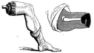
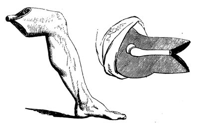
Hitherto, these general observations on amputation have regarded the operation by flaps only; the circular method has not been mentioned. The reason is, that the circular amputation has been, it is hoped, in a great measure abandoned in this country. And its inferiority to the method by flaps is so obvious, and so generally acknowledged, that detail of the different steps of the operation is, I conceive, here altogether unnecessary. It is more tedious in performance, more painful to the patient, does not afford so good a covering for the end of the bone, and consequently not so convenient and useful a support for an artificial limb, and the cure of the wound is protracted. The stump is almost always conical, the end of the bone is, ultimately at least, covered only by integument, and from even very slight pressure this is apt to ulcerate; exfoliation of the bone follows to a greater or less extent, or unhealthy nicer of the soft parts continues, along with caries of the bone, and partial death of its surface; and at length it becomes necessary either to perform a second amputation or to curtail the length of the bone. It may sometimes succeed tolerably well when there is but one bone: when there are two, it is altogether inadmissible. In very muscular limbs, when amputation is demanded on account of destruction of the bones and joints, with laceration of the soft parts, as when the patient is not required to have pressure made on the stump, it suits well to make the flaps of integument only, and to cut the muscles short, as will be noticed more fully afterwards. The advocates for the circular amputation, my excellent friend Sir George Ballingall, and others wish it to be believed, (and this is their main argument,) that the exposed surface of the flaps is much greater than that in their favourite method. Some of the philosophers of the Modern Athens have been appealed to, and have measured, it is said, the area of the one and the other, and given their verdict in favour of the round about incision. The accompanying drawings from nature, and the corresponding diagrams, speak pretty plainly in favour of the other method. In the first there is a cone formed by the cut skin and muscles, with a corresponding hollow and ragged cavity; and the second set shows two smooth, nearly triangular surfaces, which the said philosophers may measure and report upon at their leisure.
Various accidents and diseases require removal, either primary or secondary, of the fingers, or of parts of them. Amputation is most easily accomplished at the articulations, and ought therefore always to be performed at these points, when the circumstances of the case permit. The last phalanx may require removal either on account of severe injury, or from incurable disease, as onychia maligna, necrosis, caries, &c. The operation is one by single flap, and may be conducted in one of two ways. The doomed phalanx is grasped, on its anterior and posterior aspects, by the fore and middle fingers of the left hand; and the articulation is flexed almost to the full extent, in order that incision into it may be facilitated. A straight, narrow, and sharp-pointed bistoury is carried in a semicircular sweep over the back part of the joint, so as to divide the integuments, and open completely the articulating cavity. The remaining ligamentous investments of the joint are divided by one or more additional touches of the knife, so as to loosen the base of the phalanx. The fingers of the left hand are then changed from the fore and back parts of the phalanx to its sides, the edge of the knife is passed behind the base of the bone, and the surgeon, by carrying the blade forwards and downwards, forms a flap of sufficient dimensions to cover the wound, and removes the offending part. There is seldom any trouble from hemorrhage; no ligatures are required. The flap is turned up so as to form a cushion over the exposed surface of the middle phalanx, and is retained so by the adhesive composition formerly mentioned, or by one or two turns of a linen bandage. The other method of operation is the reverse of the preceding. The joint is extended, the bistoury is made to transfix close to the joint and at its anterior part, and by then carrying it downwards and forwards, as before, a similar flap is formed; this is retracted by an assistant, and with one sweep of the knife the articulation is divided from before backwards. By either method the flap is the same. By similar procedure the amputation at the middle articulation is performed.
It is sometimes an object to save as much as possible of the proximal phalanx, when amputation is rendered necessary by disease of the middle articulation, or of the distal extremity of the bone. In such cases, two semicircular flaps are made by cutting from without, either on the lateral, or on the thenal and anconal aspect, and the bone is divided either by a small saw or by the cutting pliers. The flaps are retained in apposition, and the bleeding arrested by bandaging.
Amputation at the proximal articulation is also performed by double flap. In the previous operations an assistant steadies and supports the hand; in this he has likewise to bend the rest of the fingers, and to separate as widely as possible those neighbouring to the one about to be removed. The operator seats himself before the patient, grasps the finger so as to manage its movements with the left hand, and holding the knife perpendicularly, with its point upwards, lays it over the knuckle, and carries it obliquely upwards so as to open that side of the articulation. He then pushes the finger towards the opposite side, and with the point of the knife completes the loosening of the articulation; for this the blade of the instrument should never be employed, otherwise the integument will be cross-cut and mangled. After separation of the base of the phalanx by the point, the blade is passed behind, and carrying it downwards and outwards, a flap is formed similar to the first—both proportioned to the size of the wound which they are to cover, and the bone which they are to protect. The fingers may also be removed by the oval method, as described and delineated in the Practical Surgery. The flaps are retained in contact by bringing the neighbouring fingers towards each other. This also suffices, in general, to suppress the bleeding, but sometimes one or both digital arteries require ligature. At first, cold cloths are probably the best applications, with the view of stopping the oozing, and warding off inflammatory action. Vascular excitement is very apt to follow this amputation, when performed for disease of the finger, as after neglected or severe whitlow; the soft parts in which the incisions are made are generally infiltrated and condensed, and prone to inflammation. The surface around is red, tense, and shining, on the second or third day; the back of the hand, the palm, and perhaps the forearm, are then involved in inflammation of the surface, and infiltration of the cellular tissue; and in all probability, free incisions, followed by poultice and fomentation, will be necessary to restore the parts to quietude. Such consequences are to be guarded against as much as possible, by attention to the system, and by avoiding all irritating dressing.
The phalanges of the thumb are removed in the same manner as those of the fingers. Amputation of the metacarpal bone is accomplished thus. The thumb is grasped by the fingers of the left hand, and so managed. The bistoury, held in the same manner as for amputation at the proximal articulation of the finger, is placed with its point on the web betwixt the thumb and forefinger, and carried in one sweep rapidly upwards in a slightly oblique direction, till it is stopped by the os trapezium. The point is used to effect disarticulation, the member being at the same time pushed steadily outwards; the blade is then placed behind the base, carried downwards close to the side of the bone, and is not to be brought out till sufficiently low down for forming a flap to cover the whole wound. The flaps may be formed otherwise, and much more handsomely, by transfixion of the ball of the thumb, as shown in the Practical Surgery, p. 360. After arresting the hemorrhage, the flap is laid smoothly down, and retained in its proper position by bandage or slips of the adhesive plaster. The metacarpal bone of the little finger is removed by the same method of incision as for the thumb.
In amputation of the fingers, the incisions sometimes require to extend beyond the proximal articulation, on account of disease having involved that part; in other words, it may be necessary to remove more or less of the metacarpal bone along with the finger. The method of incision will vary according to the extent to which the bone is diseased. When the operation is required for disease of merely the distal extremity, the incisions are made in the same form as for amputation of the joint, only they are on each side sufficiently high to be beyond diseased bone. They may either be made from below upwards in the usual way, or be commenced at their highest point on the dorsum of the metacarpal bone. After the soft parts have been separated from the bone, by a few touches of the knife after formation of the principal incisions, removal is completed either by the cutting pliers or the metacarpal bow-saw—the former I have found the more convenient instrument. It is applied perpendicularly, and should always have its smooth surface in contact with the part to be retained, otherwise the cut part will be rough and irregular. Section is completed more rapidly than by the saw, and, by attention to the above precaution, the stump is equally smooth, if not more so. The wound is brought and retained in contact by approximation of the fingers. But in taking away any considerable portion of the metacarpal bone, it is of importance to preserve the palm uninjured. With this view, the knife is entered over the centre of the bone on its dorsal aspect, above the diseased part, and carried straight downwards till near the articulation, when it is made to diverge for the formation of lateral flaps; the integuments in the track of the longitudinal wound are then dissected backwards, so as to expose the bone completely, and the bistoury is passed round the bone throughout its whole extent, the edge being kept close to it,—in order that the soft parts may be separated, and that without unnecessary width of wound or implication of the palmar arches and branches proceeding from them. Then the bone is clipped at the proper point by the cutting pliers; or the section of the bone may be performed before separation of the soft parts from its under surface, as, by raising the cut end, this part of the operation may be facilitated. Here the metacarpal saw is inapplicable.
Before quitting this part of the amputations, it may be observed, that no good can result from taking away a metacarpal bone and leaving a finger, or from removing a proximal phalanx and leaving the distal. The parts so left can be of no use, they have no support, and the muscles cannot act upon them: they must prove an incumbrance, and as such will either form the subject of a second amputation, or remain a proof of the unsuccessful result of the first.
Amputation in the forearm, may be necessary at various points, on account of accident or disease, but should never be resorted to, in either case, so long as part of the hand can be saved. The preservation of even a small portion of this useful member, even of one finger or a part of one, is of great importance to the patient. When, however, this is hopeless, all must be removed; and, if the wrist be sound, amputation may be performed at that joint. Hitherto, no mention has been made of the temporary suppression of bleeding, for in the amputations detailed there is scarcely ever any necessity for adopting such measures; but in those of the forearm and arm, a steady assistant must be placed ready to compress the humeral artery as soon as the incisions are commenced; in the case of the forearm, the pressure is made on the lower portion of the artery; in the arm, the point of pressure will necessarily depend on the point of removal. The patient may be either seated or recumbent. One assistant compresses, another steadies the limb in the supine position. The surgeon holds the hand in his left, standing on the inside of the right limb, and on the outside of the left. Suppose the right wrist is to be operated on,—the end of the ulna is felt for, and at that point the knife is entered, and drawn across the wrist on its dorsal aspect in a semilunar direction, the convexity of the curve of course pointing towards the fingers. The joint is opened by retracting and dissecting back the flap so formed; the knife is then passed behind the scaphoid and lunar bones, which are exposed and turned out by division of the ligaments, and by rapid and gentle sawing motion downwards and forwards, a flap of sufficient size is then formed on the palmar aspect. The dorsum of the hand may be so diseased or injured as not to afford sufficiency of sound parts for a posterior flap; in such circumstances the anterior must be made proportionally large, that it may alone cover the ends of the bones. After suppression of the bleeding, the flaps are approximated by one or two points of suture; these are afterwards removed, and their place supplied by the adhesive plaster.
Amputation, at any point between the wrist and elbow-joints, requires, in all, the same method of incision, but the nearer to the elbow the better is the stump; at the wrist the flaps are composed principally of integuments and tendons, and the cushion so formed for the bones is very inferior to one of muscle. Compression is made and the limb steadied as before; the surgeon with his left hand grasps the wrist, and places the forearm in the middle state between pronation and supination. In the right forearm, the knife, held perpendicularly, is entered over the centre of the radius, and its point, after reaching the bone, is inclined inwards so as to pass round it; transfixion is then made, the knife passing close to the palmar surface of the bones, and emerging at a point opposite to its entrance; and then by rapid motion downwards and inwards, a proper flap is formed. The instrument is again introduced over the radius, at a little distance from the upper part of the first wound, and passed on the opposite side of the bones, emerging also in the first incision and at a similar point; another flap is made. These are retracted, the knife is swept round the bones, and passed freely between them, to divide the remaining muscular substance, and after this has been completed, the saw is applied. During the sawing it is well to preserve the limb in the same position as during the incisions, and to apply the instrument perpendicularly; thus both bones will be divided at once, and the risk of splintering diminished. In transfixion, great care is required that the point of the knife pass across, not between, the bones, and with this view a slight change of position is useful; during the transfixion for the inner flap, the surgeon, as soon as he feels the knife rounding the radius, rotates the forearm gently inwards, and in transfixing for the outer flap similar rotation is made outwards. In this situation, and others where the soft parts are less thick in one aspect than the other, it is by much a preferable plan to make a flap first, by cutting from without inwards, as already described in regard to the wrist, and then to transfix for the formation of the second flap. This is delineated in the Practical Surgery, p. 367. Sometimes slight difficulty is experienced in tying the interosseal artery. The flaps are brought together, and treated in the way already mentioned.
In all amputations of the upper extremity, it is of importance, and indeed a rule scarcely to be departed from, to leave as much of the limb as possible; for here the longer the stump, the more useful is it to the patient. In accordance with this maxim, amputation at the elbow-joint may be required, when either disease or injury extends too high for amputation in the forearm, but not too high for the formation of an anterior flap over the joint. The limb is steadied in the supine position, compression being made near the middle of the humeral artery; the knife is passed horizontally across the condyles close to the bone, and brought downwards and forwards to a sufficient extent for the production of a flap, which is alone to form the protecting cushion. The joint is then cut through, the knife is passed down till obstructed by the olecranon, and with one sweep a semicircular incision of the posterior integument is made. All soft parts in this line are divided, and then the saw is applied to the olecranon process. This amputation is easy, rapid, and beautiful in execution; and, when the flap is sufficiently large,—as it always may be, for there is no want of soft parts in front,—the stump is well formed and useful. The flap is laid down, and attached by suture to the integument posteriorly; in due time the adhesive dressing is applied. The circumstance of a secreting surface forming the stump does not seem detrimental, either here, or in the wrist-joint. The synovial fluid soon ceases to distil, and union is not interrupted by it. There is no necessity for scarification, or scraping the cartilage, with the view of hastening cessation of secretion and granulation of the surface.
Amputation of the arm is performed by the formation of lateral flaps, at any point below the insertion of the deltoid muscle. Compression is made on the upper part of the humeral, or on the axillary artery. The limb is held well separated from the side; and care should be taken that, when so raised, its height be convenient to the operator. The knife is entered perpendicularly to the shaft of the bone, passed fairly down to it, and then inclined along its side; the operator now grasps the limb below the line of incision with the left hand, and pulls the muscles towards him—it is supposed that the right arm is being amputated, the surgeon standing on the inside, and an assistant supporting the forearm—and then completes transfixion, inclining the handle of the knife towards himself, in order that its point may pass round the bone and emerge at as distant a point in the circumference of the limb as possible. By a rapid sawing motion, downwards and inwards to the proper extent, the inner flap is formed; and by attention to the grasping of the muscles and the inclination of the knife, its base is so large as to admit of the more easy performance of accurate transfixion for the outer—that is, the instrument is more easily brought through the same incision. The assistant seizes the extremity of the inner flap as soon as formed, and retracts it, but only to a slight extent; it is simply held out of the way. If it be pulled backwards, as in retraction during sawing, the formation of the second will be much impeded. The knife is again entered about half an inch below the commencement of the first incision, and by inclining the handle the point is brought round the bone, and made to appear on the opposite side also in the first incision; this is facilitated by pulling the soft parts outwards with the left hand. Then the outer flap is completed. The knife is swept rapidly round the bone, so as to expose it completely at the upper part, the assistant at the same time retracting the flaps fully. The saw is applied, the arteries tied, the flaps approximated, and the operation completed.
In performing the second transfixion, the reason why the knife is entered lower than in the first, is, that cross-cutting of the corners of the wound is thereby avoided. For a long time I was surprised and annoyed to find many stumps present an unseemly cross-cutting of the integument at the upper parts, particularly after approximation of the flaps, although the incisions seemed to have been made smoothly and accurately. It is occasioned by the sawing process for making the second flap, and when this is commenced at the same height as for the first, irregularity of incision at the upper part is unavoidable. The precaution, however, of making the second transfixion considerably lower than the first I have found quite effectual. The disparity between the bases of the flaps is readily and quickly remedied, after their formation, by a sweep of the knife upwards on the lower side.
Irritable and painful stumps are more frequently met with after the amputations of the arm and forearm than any other. The occurrence, however, is less common than formerly, and this may be fairly attributed to the improvements in the operation—to the method by flaps having superseded the circular, and nothing but the arteries being surrounded in ligature. Still the affection is occasionally met with, and there can be none more painful and troublesome. Generally, no obvious cause can be found for the attack of this malady; but in some of the cases there would seem to be a constitutional and innate tendency, as it were, towards this irritability of the cut and bulbous extremities of the nerves—as in the following case:—A gentleman, aged 53, underwent amputation of the thumb, in consequence of laceration of the hand. Amputation was very soon afterwards performed at the middle of the forearm, on account of hemorrhage and infiltration of the hand, after fruitless attempts to secure the arteries. After cicatrisation of the wound, he complained of great pain in the stump, and in the situation where the tourniquet had been applied. Amputation of the arm was then performed, but the stump was not well made. The pain returned, and he applied to me, with the view of again submitting to amputation. It was performed nearer to the shoulder-joint, and in order to guard against recurrence of the disease, the nerves were laid hold of, pulled outwards, and cut across as high as possible. The patient was relieved of many of his sufferings, and continued tolerably comfortable for nearly two years; again, however, the painful symptoms have returned, though in a slighter degree. On examining the removed stump, all the nerves, particularly the musculo-spiral, were found greatly enlarged in their extremities, and intimately adherent to the cicatrix and the ligamentous covering of the rounded extremity of the humerus. More desperate operations have even been performed to free patients from irritable stump. The lower limb has been hacked off bit by bit, even to the coxo-femoral articulation, without much, if any, relief. Such operations are hardly warrantable.
Amputation at the shoulder-joint is more frequently required for accident than for disease. It has always been the custom to trust for suppression of bleeding, during this operation, to the hands of an assistant; and when the method has been found effectual in the case of the large axillary artery, why should it be objected to, on the score of inefficiency, in the minor amputations? particularly when it is evident, putting efficiency out of the question, that it is preferable to compression by the tourniquet, or any other circular apparatus;—more quickly applied and relaxed; less painful, less formidable; always ready, and independent of the instrument-maker. The compression is made above the clavicle, so as to secure the subclavian, where it passes over the first rib. The thumb of a steady assistant is placed deeply into the cavity of the lower triangular space of the neck, immediately above the first large sinuosity of the clavicle. The pressure thus made is quite effectual; but as it requires to be firm and uniform, and as there is always a risk of the thumb giving way from exhaustion, it is better to interpose some mechanical contrivance when the assistant is either diffident or inexperienced. The best apparatus for this purpose is a common door-key. A bit of lint or cloth is wrapped round the handle, and this part is then pressed down on the vessel. As in other amputations, the pressure is not commenced till the knife is about to enter.
The flaps may be double, on the outer and inner aspects, or one may be made sufficiently large to cover the whole wound. The patient is seated on a chair, and secured by a person placed behind. The arm is raised a little from the side, and supported by an assistant. Two oblique incisions are made, commencing high on each side of the shoulder, and converging gradually till they meet near the insertion of the deltoid. The triangular flap, so marked out, is dissected rapidly upwards, close to the bone, and kept raised by an assistant. The person supporting the arm then uses it as a lever, carrying it downwards and backwards; the joint, thus made more palpable, is cut into by a semicircular sweep of the knife across it; the head of the bone is now dislocated, and the rest of the capsule and fibrous tissue exterior to it divided; the blade of the knife is insinuated behind the head of the bone, and carried rapidly through the remaining soft parts. In no amputation can I conceive any necessity for suspending the incisions, in order to secure vessels, provided the pressure is well applied, and the knife used dexterously. Here very little blood should be lost. The vessels divided in the formation of the flap are small, and the axillary is not cut till the incisions are nearly completed; as soon as the limb has dropped, the surgeon places his finger on the mouth of the artery, and then applies his ligature as quickly as possible; the pressure may now be removed, and the minor vessels secured leisurely. The flap covers the wound completely, and is easily retained.
Or two flaps may be made by transfixion. In operating thus on the right limb, the surgeon, standing in front of the patient, enters the knife a little below the point of the acromion, passes it across the outer aspect of the joint, and by inclination of the handle outwards, makes its point to appear on the inside of the outer margin of the axilla; by carrying it downwards and outwards to a sufficient extent, the outer flap is formed. This is immediately elevated by an assistant, and then the arm is pushed upwards, and across the chest, so as to render the joint more accessible; the ligaments are cut, the bone disarticulated, the knife passed beyond its head, and placed with the edge parallel to the shaft of the humerus, and the arm restored to its former position; the inner flap is then made by carrying the instrument downwards and inwards. In the left limb, the knife is entered on the inside of the outer border of the axilla, and brought out below and in front of the extremity of the acromion, reversing the order of the former incisions; after the outer flap has been so formed, the joint is cut across, the knife passed beyond the head of the bone, and the inner flap made as before.
Accidental injury, as already stated, is the most frequent cause for amputation at this part, and this will always influence the method of incision. There is nothing peculiar in the after-treatment of the stump. But it ought always to be remembered, that the operation is one of great severity; that a large part of the body has been suddenly removed; that, consequently, there is risk of the mere shock being dangerous, and of an untoward constitutional condition supervening—and, therefore, the after-attendance should be zealous and careful.
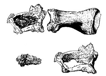
The phalanges of the toes are removed in the same way as those of the fingers. This latter operation may be required on account of bad onychia, large exostosis or injury. This and the other must also be removed on account of diseased bone, such as here represented with affection of the interposed joint. The osseous shell formed by the enlargement of the original tissue encasing a segmentum is well represented. In the amputation at the proximal joint, it is to be recollected that the extremity of the metatarsal bone lies more removed from the web of integument betwixt the toes than the metacarpal bone does from that of the fingers. The incision upwards, therefore, requires to be deeper; but in other respects the operations are precisely the same.
In amputating at the proximal articulation of the great toe, there is often a difficulty in obtaining a sufficient covering for the wound, on account of the presence of the sesamoid bones, and the general bulging of the heads of the bones. The knife is entered on the dorsum of the metatarsal bone, about half an inch above the joint, and then inclined to each side, marking out lateral flaps of considerable length; these are then reflected,—in making the outer, the instrument being dextrously passed round the sesamoid bone,—and the disarticulation completed.
In removing the great toe, along with the metatarsal bone, the bistoury is entered over the articulation of the metatarsal bone with the tarsus, and carried straight downwards, along the centre of the dorsum of the bone, till near its extremity; it is then inclined to each side, in the manner described for amputation of the metacarpal bones. The integuments are dissected off on each side of the longitudinal incision, and the knife run up along the inside of the bone, till stopped by the tarsus. The surgeon now presses the toe outwards, so as to assist the disarticulation; and after this has been completed, the bistoury is carried downwards, close to the outside of the bone, and not brought out till past the lower articulation. The external flap thus formed is then laid accurately down, so as to cover the wound, and retained. The preferable mode of making the flap, so as to expose the metatarsal bone for division or disarticulation, is well illustrated in the Practical Surgery, p. 375. The entire bone must be removed in such cases as that here sketched. In cases in which the shaft is comparatively sound, and the disease is principally seated in the articulation with its distant extremity, the shaft may be divided with the cutting forceps at a point sufficiently removed from the disease. The operations on the other metatarsal bones are the same as those on the metacarpal. They may be removed, either entirely or in part, along with the corresponding phalanges, by operative procedure similar to that practised on the hand.
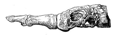
Sometimes disease of the foot is not so extensive as to require or justify removal of the whole organ; the metatarsal bones are not involved throughout their whole extent. The same remark applies to injury by accident. In such circumstances, amputation is performed at the articulation of the metatarsal bones with the tarsus. The operator first ascertains the exact site of these joints, and then transfixes the foot at that point, passing the knife close along the plantar aspect of the bones; carrying the instrument downwards, a sufficient flap is formed to cover the stump, or a semicircular flap may be made and reflected by cutting from without inwards. The integuments on the dorsum are then divided in the line of articulation, the joints divided successively, and the parts removed. The flap is raised, adjusted, and retained. In dividing the articulations, it is to be recollected, that the base of the second metatarsal bone, reckoning from that of the great toe, is lodged considerably higher than the others; and, therefore, the knife must be inclined upwards at that point, or else the use of the saw is requisite. The stump thus formed proves exceedingly useful: the subsequent lameness is not great; the heel and tarsus compose a very efficient support for the weight of the body, and the flexion is unimpaired; by attention, too, the deformity may be in a great measure concealed. In short, the surgeon who amputates above the ankle, for disease or injury not extending to the articulation of the metatarsus with the tarsus, is guilty of a serious error.
The disease may reach higher than is compatible with the preceding operation, and yet it may be possible to save the heel. In such cases amputation is performed in the articulations of the os calcis with the os cuboides, and of the astragalus with the os naviculare. The plan of the incisions is the same as that for the operation at the bases of the metatarsal bones.
No amputation is more frequently performed than that of the leg. Operation near the ankle is inadmissible; sufficiency of soft parts, for the protection of the stump, cannot be procured lower than the calf. Incision is completely limited to two points, either immediately below the tuberosity of the tibia, or in the bellies of the gastrocnemii. The former is the situation to be preferred in hospital practice, and amongst the lower orders generally; the latter is suitable to the better classes of society, that is, to those who can afford to purchase an expensive artificial support. The amputation below the tibial tuberosity being the most frequently required, will, with propriety, be described first. Suppose the right leg is to suffer:—The operator places himself on its inner side, according to the general rule formerly inculcated, and grasps the lower part of the limb with his left hand, an assistant supporting the foot at a proper height, and controlling motion. The knife is entered over the fibula, on its outer aspect, and carried upwards along that bone for an inch and a half, or two inches; it is then brought across the limb in a semilunar direction, the convexity of this incision pointing towards the foot, and after reaching the inner and lower part of the tibia transfixion is performed, the instrument being pushed along the posterior surface of the bones, and made to emerge at the upper part of the fibular incision. By then carrying the knife downwards, a posterior and larger flap is formed sufficient to cover the stump. All this is effected by uninterrupted sweeps of the knife, that is, without ever removing the point or edge from the track of incision. The integuments on the fore part are then dissected upwards a little, by a few touches of the knife, so as to form a small semilunar flap; at this part of the operation there is no necessity for laying down the knife and using a bistoury. The muscles in the interosseous space are then completely divided, and the knife swept round the bones to detach the soft parts still uncut. The saw is applied, either in a horizontal or perpendicular direction; I prefer the latter for reasons already assigned. The vessels are secured, and there are generally but three—the popliteal, and two sural. I now generally aim at cutting the vessel before it divides, and seldom fail in doing so. There is then possibly the popliteal only requiring ligature, and there is less chance of secondary hemorrhage. This has occurred, so as to prove fatal at a considerable period after the operation, in consequence of the posterior tibia being cut close to its origin, and no clot having formed in it. Before adjusting the flap, it is well to assist nature in rounding off the end of the tibia, and thereby prevent danger to the integument; with this view the sharp anterior ridge of the bone is cut away and rounded off by means of the pliers. This must be done sparingly if at all, and with great caution. The nipped surface is liable to exfoliation, or the medullary web is apt to be injured, and this is inevitably followed by more or less death of bone. To some the fibular incision may appear unnecessary; but I have long practised it from conviction of its advantage. It is an excellent mark for transfixion, and assists greatly in preventing entanglement of the knife betwixt the two bones; besides the soft parts in this situation must be divided at one or other step of the operation, and hence the procedure cannot be objected to on the ground of causing unnecessary wound. In operating on the left limb, there is not the same danger in transfixion, and consequently so long a preliminary incision on the inside is not requisite; in other respects the steps of the operation are the same as for removal of the right. In muscular subjects two semilunar flaps had better be made, one from the anterior aspect of the limb, the other from the posterior, the muscles being cut short in the ham, and the incision made to reach the popliteal artery.
It has been proposed to excise the head of the fibula after formation of the flap, instead of sawing it across at a corresponding point with the tibia. At one time I put this modification into extensive practice, with the effect of improving the appearance of the stump very considerably; but in several cases, untoward consequences took place. Discharge of synovia occurred on the second day, followed by very profuse suppuration, which proved of long continuance, and very exhausting; in more than one case, the joint became anchylosed, rendering the stump very inefficient as a means of support, in consequence of being fixed at an inconvenient angle; and one patient sank, exhausted by the profusion of the discharge. I then found, from repeated examination of the parts on the dead subject, that it was very difficult, nay impossible, to excise the head of the bone without dividing the capsular ligament, and wounding the synovial pouch, or opening a bursal cavity, beneath the popliteus muscle, communicating with that of the knee-joint. It is scarcely necessary to add, I have since wholly abandoned this method of operation.
It has been already observed that high amputation of the leg is preferable amongst the working classes. The limb is of much greater use to the patient than were the stump longer; he is able to follow his occupation with greater ease and security, and at less expense, by resting on the knee, than by using the artificial limb applied to the middle of the leg. The wealthier patient, however, can afford a more expensive support, and a less efficient, though more handsome continuation of the limb suffices. In such circumstances, amputation is performed at the middle of the leg; after cicatrisation, the artist supplies an artificial support resembling the natural limb; and thus the motion of the knee is preserved. The same directions apply to this operation as to that immediately below the knee.
In amputation of the thigh, the same method of incision is followed as in amputation of the arm. But, according to the point of removal, the direction of the flaps varies. If in operating high in the limb the flaps be made laterally, there will be imminent risk of the bone protruding through the upper part of the wound; for the patient uniformly raises the stump towards his abdomen. No antagonist muscular power is left to oppose the action of the muscles inserted into the trochanter minor, and the elevation of the stump is involuntary: it always occurs to a remarkable extent in young persons. On this account, anterior and posterior flaps are here far preferable to the lateral; for then the more the stump is raised, the better is the end of the bone covered—the anterior flap folds over it. Transfixion is therefore made horizontally; and the posterior flap should be a little longer than the anterior. But in the lower part of the limb, lateral flaps are not only not liable to the same objection, but preferable to the anterior and posterior. In the neighbourhood of the knee-joint, the soft parts consist almost entirely of ligamentous tissue on the fore and back part, and proper cushions can be obtained only from the sides. Transfixion is therefore made perpendicularly. Thus the bone will be well covered by parts likely soon to adhere; and there is no risk of protrusion, for muscles are left to counteract the elevators, and there is sufficient lever in the limb whereby to control its motions. And it may be here mentioned, that after all amputations, when startings of the muscles are not only painful, but disturb the position of the stump, the limb should be bound down by a broad band, passed across it a short way above the wound, and fastened firmly at each end to the bed or pillow; at the same time anodynes are to be administered. I have long since come to the conclusion that the femur in amputation should not be sawn lower than its middle; the method by anterior and posterior flap is therefore the only one applicable.
Amputation at the hip-joint is deservedly ranked amongst the most formidable operations in surgery. It ought, therefore, never to be performed but as a last and necessitous resource for the salvation of life. At the same time, when necessity for it is obvious and acknowledged, and no other means can be of any avail, hesitation and delay should never take place; otherwise the last and only chance of saving the patient will pass away, and the operation, when at length performed, will but hasten his exit from this world,—and besides inflict an injury to science, by intimidating practitioners, and affording subject of reproach and ridicule to the thoughtless and uninformed part of the public. I prefer the formation of anterior and posterior flaps,—as follows:—The patient is placed recumbent on a firm table, his nates resting on, or rather projecting a little over, the front edge. The sound limb is separated from the one to be removed, and held aside by an assistant. Or it is secured to the foot of the table by a towel, the necessity for an additional assistant being thus done away with, and more freedom in his movements afforded to the operator. Indeed, in all amputations of the lower extremity, this is the preferable method of fixing the sound limb. The other limb is supported by an experienced and intelligent assistant, who understands, and is able to perform, the movements to facilitate the different steps of the operation. The compression is intrusted also to an experienced and steady assistant, who, standing by the patient’s side, presses firmly with one or both thumbs on the femoral artery, where it passes over the pubes; and in this more than in any other operation should the pressure be delayed till the instant of incision, for otherwise the blood lost in the limb will be immense. Transfixion, by a knife proportioned in size to the dimensions of the limb, is made horizontally, the instrument being passed in a somewhat semicircular direction, so as to include as much of the soft parts as possible; an anterior flap is made by cutting downwards. During the passage of the knife across the joint, the assistant rotates the limb a little, so as to facilitate the bringing of the instrument out with its point well inwards; in the left limb the rotation will be inwards, in the right outwards. After formation of the flap, the assistant abducts forcibly, and presses downwards; the joint is opened, the round ligament cut, the capsule divided, and the blade of the knife placed behind the head of the bone and the large trochanter; the posterior flap is then made rapidly. After transfixion for the superior flap, and when the sawing motion downwards has advanced but a little way, the compressing assistant shifts one of his hands into the incision, immediately behind the back of the knife, and so obtains a firm grasp of the femoral artery previously to its division. He retains this hold, at the same time retracting the flap, during the rest of the operation. As soon as the limb has been separated, the surgeon secures the vessels on the posterior flap, partly by his fingers, partly by compression with a large sponge, and ligatures are applied as quickly as possible. The femoral is secured last; for, as long as the assistant retains his hold, hemorrhage from it is not to be dreaded. Thus, when both surgeon and assistants are quick and cool, the operation may be completed with the loss of much less blood than might be expected. I have had occasion more than once to perform this operation, and thus speak from experience. In cases of accidental injury requiring this operation, the lever use of the limb must frequently be wanting; and in such cases, too, the parts may be so injured as not to afford flaps anteriorly and posteriorly. In these circumstances, the surgeon must be guided by experience and judgment in adopting the mode of procedure which appears most applicable; in ordinary cases the operation above detailed appears the preferable.
Excision of diseased portions of bones, is practised occasionally with the view of removing a source of irritation and exhaustion from the system, without sacrifice of a limb. When the operation proves successful, the beneficial effect on the general health is as remarkable and rapid as after removal of the hectic cause by amputation; the pulse falls and grows firmer, diarrhœa and sweating cease, the hectic flush leave the cheek; in short, the constitution makes a complete and successful rally. It is had recourse to in order to take away disease in the following situations,—in the cancellated articulating extremity of a long bone, in part or the whole of a short bone, and in part or the whole of long bones. Even a long bone, from one articulating surface to the other, may be removed; the metacarpal bone of the thumb, and the metatarsal bone supporting the great toe, may, for example, be taken away in their whole extent. I have seen these bones so treated, but the result was unsatisfactory. As has already been observed, the part of the member that is left is without support, and not under the influence of muscle; it is consequently loose and useless.
Operation for the removal of necrosed, or softened and ulcerated portions of the carpal and tarsal bones, is sometimes successful. But operative interference, either with these, or with more extensive and formidable articulations, is not advisable unless the soft parts are not largely involved, and when the general health is tolerably good—the patient either having suffered less than usual, or having rallied and begun to gain strength after exhaustion by discharge and fever. If the ligaments, bursæ, and cellular tissue are much affected, as is often the case, there is no chance of discharge ceasing, and the patient regaining health, even though the bone be removed to any extent—a second operation will be required, namely, amputation above the diseased parts. And when this becomes requisite, after failure of the first to restore or even improve the health, the patient is apt, as has too often been the case, to sink under the accumulation of suffering. He might, even though much exhausted, have been able to bear up against the shock of one and a successful operation, but he cannot endure that of a second, or perhaps third, serious and protracted attack of the knivesman. The disappointed hope of a cure from the first operation is a secondary, though nevertheless a sure contributor towards the unfortunate issue.