PART SECOND. OF PARTICULAR SURGICAL SUBJECTS. (continued 11)
Reading edition
Popliteal aneurism is of more frequent occurrence than any of the preceding; and in regard to it, also, the old operation has deservedly fallen into disuse. It seems in most cases to be occasioned by partial laceration of the coats of the vessel; a sudden pain, and a feeling as of the receipt of an injury on the part, are generally felt, during some violent or unusual exertion; the pain continues, and an unwonted beating is soon perceived in the ham, along with inconsiderable swelling; the tumour with pulsation increases, and may ultimately attain a large size, causing pain, general uneasiness in the limb, and lameness, sometimes œdema. In cases of long duration, and when the patient is cachectic, the bones become diseased, absorption being caused by the pressure of the tumour, and deep extensive abscess may form in the soft parts.
The superficial femoral is to be tied, and the preferable point is where it is crossed by the sartorius muscle. This is always a better practice than removal of the limb, which has not unfrequently been resorted to in cases of large aneurism; there is great risk in such a proceeding, the anastomosing vessels in the thigh are all much enlarged, profuse hemorrhage takes place during the incisions, not completely arrested by any pressure, and probably twenty arteries or more require ligature, as I have witnessed; after all, the occurrence of secondary bleeding is not unlikely. I have tied the femoral artery, with a favourable result, in some cases of very large aneurismal tumour, and in one instance after the cyst had been imprudently punctured. An incision is made from three to four inches in length, and in an oblique direction in regard to the thigh, tracing the inner border of the sartorius muscle, and so placed that its middle may correspond with that part of the artery on which the ligature is to be put. In order to insure the wound being thus situated, there is no need for measurements; these are but a clumsy substitute for anatomical knowledge. The surgeon, well acquainted with the relative situation of the parts, finds it sufficient to ascertain the exact course of the muscle by manipulation, whilst the thigh is slightly bent, and then guides his knife by the eye, unfettered with mathematical diagrams. The muscle is exposed almost by the first incision; the dissection is then continued through the cellular tissue on its inner border, until the sheath of the bloodvessels is reached, the branches of the crural nerve on the fore part being carefully placed aside uninjured; the sheath is cautiously opened immediately above where the muscle conceals it, and the artery separated from its connections to a very slight extent; the needle is then passed, and the ligature applied. The operation, when thus conducted, is exceedingly simple. But embarrassment and delay have often been experienced from following an opposite method, cutting down on the outside of the sartorius; the muscle must either be dissected from its attachments and turned over, or cut across; or the artery cannot be found, and an additional external wound is necessary.
The artery may require ligature at a higher point, either in consequence of wound, or for the cure of femoral aneurism. This disease, however, is very unfrequent. When it does exist, it is usually so situated as not to admit of the favourable application of a ligature below the origin of the profunda; and it may be considered necessary to tie the common femoral. The course of this artery being superficial, is easily ascertained; an incision of convenient extent is made in the same line, penetrating the skin and fatty matter; the cellular tissue is carefully separated, and the sheath exposed; a limited opening is made, with corresponding detachment of the vessel, and the ligature applied, close to the lower edge of the ligament of Poupart. But ligature of the external iliac is in all cases to be preferred, for the reasons already given. This has proved successful in more than one case of double aneurism, one in the groin, the other in the ham.
In ligature of the common and of the superficial femoral, the vein is in more danger than the nerve, and the utmost caution is required lest it be punctured. It has been wounded—I witnessed one instance of it; the opening was drawn together and closed by ligature, inflammation of the vein supervened and proved fatal.
When secondary bleeding occurs, on the separation of the ligature, either after this operation or after that for popliteal aneurism, compression is not to be trusted to, nor should the vessel be tied higher in the thigh. From imprudent reliance on the former method I have known patients perish. An incision must be made in the same line as the former, and a ligature placed on the vessel both above and below the bleeding point, as may be necessary.
The arteries of the leg very seldom require ligature, except for wound. In such cases, the source of the bleeding must be the guide to the incisions, and these should be placed so as to interfere with the muscles as little as possible, always in the direction of their fibres. When the bleeding point is arrived at, the vessel is exposed to a short distance, and tied above and below the wound. During the dissection, it will in most cases be necessary to arrest the bleeding by pressure in the ham, either by the fingers of an assistant, or by means of a tourniquet.
The thigh may be the seat of aneurismal varix, the result of wound, as in the following case. Fourteen years ago, a young man wounded the lower part of his thigh deeply by the accidental thrust of a narrow chisel. The puncture was in the direction of the femoral artery; violent hemorrhage was the immediate consequence, and after he had fainted the wound was stuffed and compression applied. In eight days the parts had healed, and he returned to work as usual. But about twelve months afterwards, troublesome pulsation was perceived in the part, at the same time the veins of the leg became varicose, and a succession of ulcers formed on the lower and anterior portion of the limb. The affection attracted but little of his notice till about six months since, when he observed a considerable swelling in the site of the wound, beating strongly, and the pulsations accompanied with a peculiar thrilling sound and feel—not confined to the tumour, though strongest there, but extending to the groin along the course of the femoral vein, which was evidently much dilated throughout its whole course. At present the tumour is nearly equal to the fist in size, of regular surface and globular form, pulsating very strongly, and imparting to the hand the peculiar sensation of aneurismal varix, remarkably distinct and powerful. The pulsation and thrilling are continued, in a less degree, to Poupart’s ligament, and down to the calf of the leg. On applying the ear close to the tumour, or listening through the stethoscope, the peculiar noise is not only felt, but heard of almost startling intensity—somewhat resembling the noise of complicated and powerful machinery, softened and confused by distance. By making firm pressure on the tumour, the thrill is lost, and the regular pulsation alone perceived; at the same time, the turgescence of the femoral vein disappears, and on compressing the femoral artery in the middle of the thigh, both pulsation and thrilling are arrested, and the swelling much diminished,—but only temporarily, for the collateral circulation is free and complete. He feels little pain, but exercise and exertion of every kind are seriously impeded; constant and firm pressure on the swelling, with uniform compression of the whole limb, has been employed, with the effect of relieving all the symptoms, and rendering the limb much more useful, and by its continuance it is to be hoped that the disease will at least be considerably palliated.
In the lower extremity, as in the upper, the bursæ become enlarged, in consequence either of pressure or of external injury. The affection may be acute, following a blow or squeeze, but is most frequently chronic, enlarging gradually and with little or no pain, and caused by habitual pressure on the part. From this, it will at once be understood, why the bursa over the patella should be the one most commonly affected. Its vulgar name, housemaid’s-knee, marks its cause—the avocations of such persons requiring them to rest on one or both knees, frequently, and often for a long time. It also occurs in shop-keepers, and other persons accustomed to shut drawers with their knee, or in other ways to make frequent pressure on that part,—in gardeners, and those employed in similar pursuits. In the acute swelling from injury, local depletion, fomentation, and rest are required, and these are generally sufficient to arrest the swelling, and promote its subsidence; but, in some cases, the fluid is deteriorated and the surface inflames, free incision is required, followed by poultice, and afterwards by simple dressing. In the chronic collection of clear fluid, gentle and continued stimulation of the surface, as by the gum and mercurial plaster, causes gradual decrease by absorption; the causes of the affection being at the same time studiously avoided. The tumour sometimes, as here shown, attains a large size, and from repeated inflammatory attacks becomes consolidated. The cyst is thickened, and lymph is effused into the cavity so as to convert an encysted swelling into one of solid consistence. In such cases as these, the tumour may cause such inconvenience as to make the patient desirous to have it dissected out. This is easily and safely effected; the incisions are made in the direction of the limb, and it is kept at rest in the extended portion for some time, so as to favour the healing of the wound.
Unyielding parts, habituated to pressure, defend themselves by the interposition of a moveable bag containing fluid; betwixt them and the surface the cellular tissue condenses into a cyst, its internal surface assuming a serous appearance, and secreting a fluid resembling the synovial. Such adventitious bursæ are not unfrequent on the ankles and feet, as in tailors, or others usually sitting cross-legged. They may attain a considerable size, and so produce deformity; but they should not be interfered with unless they become inconvenient, as from excited action.
When the extremity of the metatarsal bone of the great toe is large, and consequently the seat of pressure, a bursal formation is produced in the soft parts covering it; this from increase of pressure, or other irritation, may inflame—forming the painful and troublesome disease termed Bunnion. Sometimes unhealthy abscess occurs, with thickening, infiltration, and condensation of the surrounding cellular tissue; in such cases, incision and poultice are required, and occasionally it is necessary to destroy the unsound cellular tissue and the degenerated cyst by free application of the caustic potass. The cyst is thus got rid of, healthy granulation takes place, and by afterwards avoiding undue pressure upon the part, a permanent cure is obtained.
It has been elsewhere mentioned, that cartilaginous bodies sometimes form within articulating cavities, occasionally attached by a narrow and slender connection with the secreting surface, but generally loose, seldom numerous, and usually of no great size. They are most commonly met with in the knee-joint, producing inconvenience by impeding progression. Sometimes they are neither painful nor annoying, being small, and seldom becoming interposed between the articulating extremities of the bones during motion; such ought not to be interfered with. But when large, they may be so troublesome as to warrant incision and removal. The foreign body is made to project on one side, and, having been made as superficial as possible at a favourable point, is fixed by the fingers of an assistant. The integuments are then drawn to one side, and an incision made over the body, the capsule is cut to as limited an extent as possible, and removal effected by pressure—or it may be laid hold of by a hook, and extracted; if the cartilaginous substance be attached by a pedicle, this must be divided, but with great caution. The integuments are immediately allowed to resume their natural situation, and so to close the wound of the capsule by overlapping it; the skin is then accurately approximated by adhesive plasters. The limb is kept extended, and not the slightest motion of the joint permitted. The patient is confined constantly to the recumbent posture, purged, and kept on low diet; the utmost vigilance is necessary to prevent inflammation of the synovial apparatus. In some patients on whom I have performed this operation, the wound closed by the first intention, and no untoward symptom threatened, motion and the erect position being resumed in a few weeks. But in the last case which came under my care, though the extirpation was performed with the utmost care, most violent inflammation supervened; the wound opened, synovial secretion flowed out in large quantity, profuse escape of unhealthy matter followed, and exhausting discharge continued for many weeks. At one time the constitutional disturbance was so great as to endanger life; the limb was saved with difficulty, the joint anchylosed. From the result of this case, I am disposed to dissuade operative interference, unless the patient strenuously urge it, and be willing to take the responsibility for the consequences on himself.
[The most common distortion to which the human body is liable is Club-foot; an affection which has at all times attracted the notice of the profession, but which has received unusual attention within the last ten years on account of the novel operation suggested for its cure by Dr. Stromeyer of Hanover, in Europe. The lesion is, for the most part, congenital. It may, however, be developed after birth, and even at an advanced period of life, from the foot being accidentally placed in a constrained position, and so retained until the soft structures—particularly the muscles and ligaments—are moulded into a new shape, or until they become fixed in their new situation. Various mechanical causes may give rise to this malady, such as splints and bandages, by which the parts to which they are applied are injuriously compressed, or thrown out of their natural relations. Similar results are produced by convulsions, dentition, nervous irritation, contusions, sprains, fractures, partial luxations, and preternatural laxity of the ligaments. In some instances the defect is occasioned by the presence of a corn, an ulcer, or some other disease which induces the person to walk on one side of the foot, the tip, or the heel, to ward off pressure from the tender parts. A vicious habit is thus established, which, if it be kept up, as it often is, for any length of time, leads to irregular action in the muscles, and to distortion of the bones into which they are inserted.
The formation of congenital club-foot has never been satisfactorily explained. By some—as Meckel, St. Hilaire, Serres, and Breschet—it has been ascribed to an arrest of development. This theory, however, for various reasons, is untenable, and has therefore not been generally adopted by surgical men. Mons. Martin, a recent French writer, thinks it is mainly occasioned by the pressure of the parietes of the uterus on the feet of the infant during gestation, owing to a deficiency of the amniotic fluid; an opinion in which he is joined by Professor Cruveilhier. That the disease may proceed from this source in some instances maybe readily supposed, but that this is the only cause, is what few will believe. The most plausible hypothesis, in my opinion, is that of Mons. Guerin of Paris. He supposes that the primary mischief is in the nervous system, and that the spasmodic and permanent shortening of the muscles of the affected limb is altogether consecutive. He sums up the results of his numerous observations in the following propositions:—1. Congenital club-foot is the effect of a convulsive contraction of the muscles of the leg and foot. 2. In the absence of general or direct traces of the convulsive affection we may almost always discover some immediate characters which indicate the nature of the exciting cause. 3. There are three constituent elements in the retraction of the muscles of the part: namely, the immediate shortening of their substance and tendons; a certain degree of paralysis; and, lastly, a consecutive arrest in the development of their substance. 4. There are no other causes of genuine congenital club-foot than convulsive muscular retraction. The pressure of the parietes of the uterus on the fœtus appears, indeed, in some cases, to produce a deformity of the limbs and feet, similar to but not identical with club-foot. The views of Mons. Guerin are confirmed, in some degree, by the history of those cases which occur after birth; but future observation must determine whether they are correct or otherwise.
The congenital variety of this distortion often affects both feet simultaneously, though rarely to the same extent. In one hundred and sixty-seven cases reported by Dr. Detmold of New-York, the disease was double in nine-three; in forty-one it occurred in the right foot only, and in thirty-three only in the left. Of eighty cases collected from various sources by Mons. Bouvier of Paris, or observed by himself, two-fifths were double; one-third affected the left limb, and one-fourth the right. Of sixty-one cases furnished by Martin, another French writer, twenty-six were double and thirty-five simple: of the latter, eighteen were of the right and seventeen of the left foot. Mons. Helt has published the results of thirty-one cases, in nineteen of which the disease was double; in two it was more distinctly marked on one leg than on the other; and in one instance the calcaneal form of the lesion was united with the inverted. In twenty-one cases observed by Scoutetten, both feet were deformed in nine; and in the other twelve the right limb was exclusively involved seven times; the left five times.
The disease would appear to be more frequent in males than in females, though the relative proportion has not been ascertained. The following table, embracing three hundred and twenty-nine cases, will throw some light on this subject:—
Authors.Number.Males.Females.
Detmold1679869
Bouvier804832
Martin614516
Scoutetten21138
——————
329204125
There are certain facts which would seem to show that club-foot is sometimes hereditary; or, at all events, that it may occur in several members of the same family. Thus, Dr. Detmold states that he has been able to trace the hereditary predisposition to this deformity in not less than eighteen cases, and in all excepting one, to the father’s side. Whether this was a mere coincidence, or obtains generally, it is impossible to say. Mons. d’Ivernois relates an instance in which four brothers were all born with the feet twisted inwards; and another writer, Mons. Helt, speaks of a family, which consisted of six children, all of whom were afflicted with congenital club-foot. In the latter case the disease was probably hereditary, as one of the parents was labouring under the same infirmity. It should be observed, however, that club-footed parents do not always produce club-footed children.
Club-foot may be conveniently divided into four varieties—the inverted, everted, phalangeal, and calcaneal—which differ from each other not only in regard to the character of the distortion and the accompanying phenomena, but likewise in relation to the frequency of their occurrence and the nature of their proximate causes. The most common form by far is the inverted, usually denominated varus, in which the patient walks upon the outer ankle, the great toe being directed inwards and upwards. The muscles of the calf and the adductors of the foot are contracted, and hence there is not only elevation of the heel, but a peculiar inward twist of the foot, analogous to supination of the hand. This alteration occasions the most serious impediment to progression, and when it reaches its highest point imparts a most disagreeable aspect to the affected limb. In the higher grades of the disorder, the sole of the foot is literally scooped out, as it were, as well as deeply furrowed; the instep, on the contrary, is unusually convex and prominent; the small toes generally present in a vertical position, while the big one, separated from the rest, looks upwards and inwards; the outer margin of the foot, which, in conjunction with the corresponding malleolus, chiefly sustains the weight of the body, is almost semicircular in its shape, rough, and callous; and the tendo-Achillis, forced obliquely towards the inner side of the leg, forms a tense, rigid chord beneath the skin.
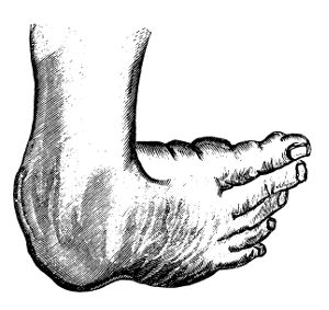
Sometimes both feet are affected with varus, so that their points form an acute angle with the leg; or approach so nearly as to touch, or even overlap one another. In the majority of cases the thigh and leg retain their natural conformation, being merely somewhat atrophied; occasionally, however, one or both knees project slightly inwards or outwards, owing to the contraction of the hamstring muscles.
The second variety of this deformity, anciently called valgus, may be regarded as the opposite of varus, the patient treading on the internal margin of the foot, while the external is entirely removed from the ground. The sole is directed outwards and slightly backwards, the toes are more or less elevated, and the outer ankle is in a state of semiflexion. The heel is drawn upwards and somewhat outwards, the internal malleolus is uncommonly prominent, the instep is flatter than natural, and the muscles of the calf, together with the adductors of the foot, are permanently contracted. When the disease has attained its highest point, the patient has an unsteady, vacillating gait, from the difficulty which he experiences in preserving his centre of gravity. Valgus is comparatively rare; and, like the first variety of the distortion, it may affect one or both limbs. It is seldom a congenital affection, but is almost always produced by some local injury—as a sprain or blow.
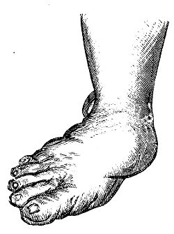
The phalangeal club-foot—the pes equinus of the older writers—is caused by a shortening of the gastrocnemial and soleal muscles, aided, in some cases, by the flexors of the toes. In this species of the deformity the individual walks upon the ball of the foot, the toes, or upon the metatarso-phalangeal articulations, without the heel or any other part of the sole touching the ground. The distance at which the heel is raised varies in different cases, from six lines to four or five inches, according to the extent of the contraction upon which the distortion depends. Considerable diversity is observed in regard to the manner in which the person treads on the ground; most commonly the ball of the little toe bears the brunt of the pressure, but in some instances the weight is thrown upon the great toe, or it is diffused over the whole of the fore part of the plantar surface. In the worst gradations, the heel is so much elevated that the foot forms nearly a straight line with the leg, the toes are much deformed, the instep is unnaturally convex, the plantar aponeurosis is greatly contracted, and the skin above the heel is thrown into dense wrinkles.
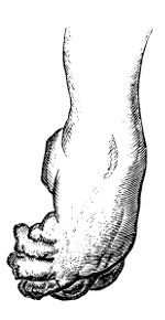
In the fourth variety—the calcaneal, recently described by Mons. Scoutetten—the limb rests upon the heel, the toes being drawn upwards, towards the anterior surface of the leg, with which they sometimes form an acute angle. The immediate cause of the deformity seems to be a contraction of the anterior tibial muscle and of the extensor of the great toe, assisted occasionally by that of the common extensor of the foot. The tendons of these muscles form an evident protuberance under the skin, where they present the appearance of tense, rigid chords, which powerfully resist the extension of the limb. The inner margin of the foot, as seen in the cut, is sensibly elevated above the outer, and there is always considerable atrophy of the leg. The distortion, which is almost always congenital, is exceedingly rare. Occasionally the foot inclines slightly outwards, owing to the inordinate contraction of the common extensor muscle.
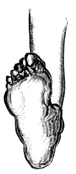
The changes which the bones, ligaments, and muscles undergo, vary, not only in the different species of club-foot, but in the different stages of the same case. The greatest alteration appears to exist on the part of the tarsal bones, which, although they are rarely completely dislocated, are generally somewhat separated from each other, twisted round their axis, variously distorted, atrophied, or marked by irregular spicula or exostoses. The calcaneum, cuboid, scaphoid, and astragalus, always suffer more than the other bones; which, however, as well as those of the metatarsus and of the toes, usually participate, more or less, in the deformity. The ligaments, in recent cases of club-foot, do not present any material changes, but in those of long standing, or in the higher grades of the affection, they are invariably stretched in the direction of extension, and relaxed in that of flexion. In some instances the original structures are partially replaced by bands of new formation, of a dense fibrous character—the volume and resistance of which vary according to the duration of the disease and the pressure of the parts which they serve to connect together. The muscles also are not much altered in the first instance, except that they deviate from their natural direction, and that, like the ligaments, they are elongated on the one hand and shortened on the other. In ancient cases the whole limb is always considerably wasted, and many of the muscles are remarkably thin and pale, or even transformed into soft, fatty bundles. The cellular substance is condensed and diminished in quantity; the adeps is absorbed; and even the vessels and nerves supplying the affected part are apparently reduced in volume. The skin of the foot, which receives the principal brunt of the pressure in standing and walking, is generally very much thickened and indurated, and large synovial bursæ are often formed beneath it, which are apt to inflame, and thus add to the suffering of the patient. Such is an outline of the more important changes experienced by the different textures in cases of club-foot: to enter more minutely into the subject would be foreign to the design of this article, the object of which is merely to present a general idea of the nature, causes, and treatment of this singular distortion.
The treatment of this affection should be delayed as little as possible. The sooner, indeed, it is attended to, the more probable will be the chances of effectually removing it. This is equally true, both of the congenital and of the accidental form of the disease. The bones in early life and in recent malformations are much more easily restored to their normal position than in youth and manhood, or in cases of long standing; and the muscles also regain much sooner, as well as more completely, their original power. In the worst grades of the disease it is often exceedingly difficult, if the treatment be delayed until after the age of puberty, to accomplish a cure without great carving of the tendons, and the constant employment for months of various kinds of apparatus.
It is still a disputed point, whether, in the treatment of this affection, particularly in infants and young subjects, it is necessary, or even justifiable, to divide, as a preliminary step, the tendons of the muscles which are instrumental in keeping up the distortion. Without endeavouring to settle this question, for which the time has not perhaps yet arrived, I must express my conviction that the present rage for tenotomy is calculated to do a vast deal of harm, not only in individual cases, many of which do not require it, but, what is worse and more deeply to be lamented, in bringing discredit upon an operation, which, if judiciously performed, cannot fail to be of the greatest benefit. In most of the cases occurring in children under two or three years of age, division of the tendons is altogether unnecessary; indeed, one of our most distinguished orthopedic surgeons, Dr. Chase of Philadelphia, seems to trust almost entirely to the employment of apparatus, and to resort to tenotomy only in the worst grades of the disease. Whether this practice will ultimately be adopted by the profession generally, or the division of the tendons be restricted to particular cases, it would be premature to predict; but my opinion is, that much more cutting is now done than is necessary, or than would be done if the treatment of the disease were better understood than it appears to be.
Different kinds of apparatus are in vogue for the cure of this deformity, and it is therefore impossible to determine which is the best, or which should be employed to the exclusion of the others. Every practitioner seems to have his own notions on the subject, and to adopt such measures as whim, fancy, or caprice may dictate. Whatever apparatus be resorted to, the great caution to be observed, on the part of the surgeon, is, that the extension be made in a slow and gradual manner, that the skin be protected from friction and uneven pressure, and that the dressings be steadily retained during the night, as well as during the day, until several weeks after all deformity has disappeared. The object of these directions is self-evident, and too important to be neglected in our curative procedures. The time required for restoring the limb to its normal position must necessarily vary in different cases, and depend upon so many circumstances as to render it impossible to lay down any specific rule. From six weeks to four months, however, may be considered as a fair average, though occasionally a much longer period will elapse. The division of the tendons of the contracted muscles generally expedites the cure by several weeks.
In the operation for dividing the tendo-Achillis the patient may either lie on his abdomen or sit on a chair, and the heel is to be drawn downwards by an assistant with the left hand, the right being placed upon the plantar surface of the toes. The necessary tension being thus given to the part that is to be cut, the surgeon passes a narrow, straight, sharp-pointed bistoury through the skin, from one to two inches above the internal malleolus, flatwise between the tendon and the deep-seated structures. The knife is then pushed on until it reaches the opposite side of the tendon, when its edge is brought in contact with the anterior surface of the chord, which is now completely divided by steady pressure upon the handle of the instrument. The separation of the parts is indicated by an audible snap, and by the immediate cessation of the tense resistance of the tendo-Achillis. Scarcely a drop of blood is lost during the operation, which is almost unattended with pain, and is accomplished in a few seconds. A strip of adhesive plaster is applied over the little puncture, which generally heals by union by the first intention; and the limb, laid in an easy position, should be supported by a paste-board splint and a common roller. The apparatus for keeping up permanent extension may be advantageously employed in three or four days after the operation.
The interval between the divided extremities of the tendon is filled up with coagulating lymph, which is often poured out in considerable quantities. As in other situations, it becomes gradually organised, and is finally converted into a firm, dense substance, not unlike the original structure.
The tendon of the posterior tibial muscle may be cut most advantageously about two inches above and behind the internal malleolus. The operation is conducted upon the same principles as in the preceeding case, and the only particular caution to be observed is to avoid the posterior tibial artery and nerve, which might be endangered by carrying the knife too deeply. The most favourable situation for dividing the anterior tibial muscle, is where it passes over the ankle-joint: the long flexor of the great toe may be cut in the sole of the foot, where, when it interferes with the rectification of the limb, it forms a tense, prominent chord.—ED.]
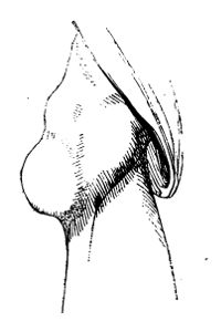
The phalanges of the toes in general resemble those of the fingers in their diseased actions. Exostosis of the extremity of the distal phalanx, however, has no analogy in the upper extremity; it is by no means an uncommon affection, and usually occurs in the great toe. The growth is generally globular and rough in its extremity, narrow at its origin, attached on the dorsal aspect, projecting obliquely upwards, and always of similar structure with the phalanx. Sometimes they are met with of a size nearly equal to that of the bone from which they spring, but the majority are considerably smaller. The only one I have met with springing from a small toe is here sketched. At first the patient complains merely of pain in the part while walking; soon the pain increases so as to impede progression very seriously; then the nail is found to be raised at its margin, and to cover a hard, unyielding, and tender swelling. The elevation of the nail increases, and the tumour becomes more apparent, covered by hardened cuticle, causing great uneasiness, and almost entirely preventing walking exercise.
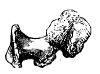
It has been recommended to expose the tumour by incision, and remove it at its origin. This affords temporary relief, but the disease is generally in no long time reproduced, and the incision must either be repeated, or the phalanx amputated. The preferable practice, according to my experience, is to remove the phalanx at once. It is less tedious and painful than the incision, produces very little, if any, impediment to progression, and of course is quite effectual in eradicating this most annoying though apparently simple disease.
Of Fractures.—Deformity, shortening, loss of power, unnatural motion on extending and moving the part, pain, and grating, mark solution of continuity in bone, or fracture. Swelling, with spasmodic of these symptoms may be wanting; there is little deformity, and no shortening, when one of two or more action of the muscles, soon takes place. One or several parallel bones is fractured. In fracture of the extremities, extrication of air into the cellular tissue, about the ends of the bone, is not unfrequent, though difficult to account for—giving rise to crepitation, superficial, and quite a distinct sensation from that imparted by the broken bone.
Bones become brittle as age increases, and fragility is also induced by certain disordered and debilitated states of the constitution. In some patients, the bones give way on very slight force being applied, after what may have been supposed a rheumatic attack; the thigh is broken by turning in bed, or by walking from the bed to a chair. In one instance, I had put up a fracture of the thigh with a long splint, and in three weeks afterwards the humerus was broken over the end of the splint during an attempt by the patient at change of posture. In many such cases union either does not take place, or is very imperfect.
In children, the bones frequently contain little earthy matter, bend easily, and often break partially on the convexity of the curve. Even at the age of twelve or thirteen, bending of the bones from injury sometimes occur to a great extent, as of the forearm from a fall on the palm of the hand; in adjusting the parts, a slight crackling is heard when they are brought nearly straight. Complete solution of continuity, though more rare, is occasionally met with in very young subjects.
Fractures are generally the result of great force applied directly to the shaft of a bone, or to its extremity; but they are also not unfrequently caused by twisting of the limb whilst the muscles are in a powerful action. Bones are broken transversely; but more frequently there is a degree of obliquity in the fracture, and the fragments are generally detached. A bone may be split longitudinally, as from a musket-ball striking its shaft in the centre; and fissures often extend from a cross break to a considerable extent, sometimes into joints.
Swelling is often rapid, from extravasation of blood; at other times it is slow, and of a serous character. At first it is soft and yielding, but after a time painful inflammatory tumescence supervenes, the violence and extent of which will depend on the severity of the injury, and very much also on the treatment to which the parts are subjected. If the bones be put as nearly as possible into their original position, and retained so, judiciously—the limb being laid in a comfortable and unconstrained posture, and the bandages, splints, &c., properly adapted—little or no pain or inflammatory swelling will occur; no more action ensues than is required for reparation of the injury. If, on the contrary, the bones are allowed to remain unreduced—perhaps after being well handled—their broken ends, laying among the soft parts, are pulled out by violent spasms, lacerations of the muscles and vessels is increased, effusion, swelling, and violent inflammatory action occur, the pain becomes excruciating, fever and delirium follow; there is an imminent risk of gangrene, and extensive suppuration among the muscles is almost inevitable. If the patient recover, the union is bad, and the limb deformed.
A fracture is said to be simple, where there is no wound of the superimposed integuments. The external parts may be bruised, or the deep structure much injured, with laceration of the vessels and rapid and great swelling; or there may be little or no injury of the soft parts. Great danger may exist without division of the integuments; these, yielding under the force, may remain entire, whilst by great and direct violence the bone is comminuted, the muscles broken up, and the vessels and nerves torn,—the limb is infiltrated with blood, and must become gangrenous as soon as reaction takes place. But usually these untoward circumstances do not exist in simple fractures, the soft parts being but slightly injured.
Fracture is compound when the integuments are divided by the external force, so as to expose the broken bone. But the wound may not penetrate to the bone; and then the accident is termed fracture with wound, not compound fracture. The soft parts are often divided by the sharp end of the bone; this is frequently the case in oblique fracture, occasioned by a fall from a height, the lower fractured extremity being pushed forcibly upwards. The muscles are usually much injured. The wound is either large or small, lacerated or clean.
Fracture, simple or compound, is comminuted when the bone is divided at the broken point into fragments, either small and loose, or large and adherent to the covering of the bone and other soft parts.
Fracture may be complicated with wound or displacement of a neighbouring joint, and with laceration of large bloodvessels and nerves.
Union of divided bones, as of soft parts, is preceded by incited circulation in the part, and effusion of organisable matter. The extent of action is regulated by that of the injury, whether inflicted by accident or by operation. If the soft parts have not been much bruised, if the bone and its covering are merely separated and slightly displaced, and then speedily put in contact, the incited action and the effusion are limited to the divided parts. There is no irregularity afterwards at the point of fracture, the new matter that is not required being absorbed soon after deposition; the bone is smooth and even as before. The deposit of new matter under the periosteum and into the medullary canal is here well exhibited. By this means only is the bone kept together for a very considerable period; afterwards the broken ends are united, and the temporary callus absorbed. If, on the contrary, there is much displacement, and if that is not entirely removed, intense action ensues both in the soft and hard parts, there is great effusion of new matter, or callus, soft and yielding at first, but gradually becoming hard and dense—bony particles being deposited from the vessels ramifying in the extremities, or in the attached fragments, of the old bone. When detached portions of callus are found lying in the soft parts, a piece of old bone which retained its vitality has generally formed the matrix of the deposit. When the ends of bones have been badly placed, and meet each other at an angle or curve, occasionally osseous deposit seems to form in the concavity. This increases in size, unites with the portions of the shaft, and forms a sort of bridge uniting them. This by M. Gulliver has been termed accidental callus.

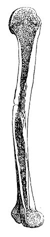
In badly reduced fracture the swelling is great and hard. The callus is exuberant, much being required for the union of the fractured ends that overlap, and are perhaps far from being in contact; the vascular action and accompanying effusion are great, according to the necessity for them. The bone at the united part is enlarged to perhaps double its original thickness, or even to a greater size. After some time, the ends of the old bone, and part of the new deposit, are rounded off by absorption of the protuberances, and the part becomes more shapely. The canal of the bone and the cancellated texture is again restored. The accompanying sketch of a section of the humerus shows a double fracture. The superior one near the neck, where there is still some thickening, had been well adapted, and the canal is quite perfect. In the other and more recent there is considerable overlapping. The portion of outer osseous shell projecting into the medullary canal would in the end have been removed by the absorbents, and the deformity much diminished.
When the ends of the bone are not well placed, or when they are moved occasionally whilst the uniting medium is still soft, there is danger of a false joint being formed—the callus either giving way, or being all along imperfect, and the extremities at the soft part becoming smooth and moveable on each other; or incited action may run high and terminate in suppuration, with death or ulceration of portions of the bone.
Fragments are sometimes entirely detached at the time of the accident, and perish at once; or are so slightly connected with the shaft that they lose their vitality on the first accession of inflammation, become surrounded by purulent matter, part from their slight attachments, and come towards the surface. Or the shaft itself may be so bruised by the violence of the injury as to be incapable of resisting incited action, though slight. By malpractice, such untoward consequences as the preceding, and many others beside, are frequently induced.
The uniting medium of separated bones remains soft for some time, as was already observed; and often, whether from the state of the constitution, or the circumstances connected with the fracture, the parts remain long moveable. Pregnancy is said to prevent union; but I have often seen fractures in pregnant women unite as speedily and firmly as if the patients had been in that state, and otherwise in robust health; profuse uterine or vaginal discharges, or determination to particular parts or organs, will certainly retard union.
In ordinary cases, the limb, if not lying altogether straight, can be moulded into a proper form after the lapse of eight or ten days from the time of injury, without the patient suffering any great degree of pain, without the process of union being at all interrupted, or the cure protracted; even at the late period of five or six weeks, badly united fractures may sometimes be much improved by gradual pressure and change of position. A gentleman fell from his horse, and sustained simple fracture of both bones of the leg, near the middle. It had been laid and retained on its side. I saw him exactly six weeks after the injury; the leg was much curved forwards, and the foot turned outwards. The limb was placed on the heel, and a long splint, with a foot-piece, applied on the outside; by attention to its position, and by gradually tightening of the bandages, it soon became quite handsome. Care should be taken not to allow the patient to rest too soon on the fractured limb; for though quite straight, symmetrical, and of the proper length, when the retentive apparatus is discontinued, it may become short and deformed in a few days from even slight weight being put upon it.
The period at which firm union takes place varies; the process is more rapid in young people than in those advanced in life, and will depend more on the extent of the injury, and its vicinity to the centre of the circulation than on the size of the broken bone. The requisite length of confinement is regulated by these circumstances, and by the use to which the part is to be afterwards put; the lower limbs require longer time for consolidation than the upper.
In the treatment of fracture, as in solution of continuity in the soft parts, great advantage is gained by placing the disjoined parts as nearly in their original position as possible, retaining them so, and allowing of no motion. These indications ought to be accomplished very soon after the accident; many evils are thus prevented—the further laceration of the soft parts, the inflammatory effusion into all the tissues, and the consequent startings and spasms of the muscles. This cannot be too much insisted on. There is much folly and absurdity in allowing a broken limb to lie unrestrained—leaving the ends of the bones displaced, the one riding over the other—whilst attempts are being made to keep down the inflammation, by applying leeches, cold lotions, or large poultices—all perfectly ineffectual so long as the palpable cause of incited action remains unheeded. The circumstances which kindle and keep up inflammation should always be understood; they are easily discovered in fracture, and when understood should never be lost sight of. If the parts be replaced there will seldom be inflammation; if they remain displaced, the inflammation is so great that it is impossible to subdue it by any means short of removal of the cause. There is also an impossibility,—not to mention the patient’s sufferings,—of reducing bones to a good position some weeks after the accident. Such practice has been extensively followed and recommended by some, even modern writers; they set about reducing a fracture at a period after the accident, at which, by proper treatment, union would have been completed, or at least far advanced. The confinement and suffering of the patient are increased threefold, and after all the cure is bad, and there is a risk of false joint.
In all fractures, whether simple or compound, comminuted or complicated, if an attempt is to be made to save the limb, let reduction be immediate; coaptation and retention of the separated parts cannot be made too soon. A neglected case may be met with, in which the intensity of inflammatory action in all the tissues may forbid immediate interference. But even though inflammatory action has taken place to some extent, there are no surer means of arresting it than removal of its cause—the irregular ends of the bones being taken away from among the soft parts—provided it can be done without violence or increase of tension. Reduction is facilitated by proper position of the limb, by relaxation of certain sets of muscles. Extension and counter-extension are made, and but very little force is required; the surgeon extends the limb with one hand, and resists with the other; when the system is excited, and the muscles act spasmodically, an assistant may be required to steady the limb, and to resist the extending power which the surgeon employs. Then the position of the limb and of the patient, when long confinement is required, must be considered, and rendered as easy as possible, though at the same time secure. The apparatus for retaining the bones in the right position must be varied according to circumstances.
In compound fractures, when the wound is so small and clean that adhesion readily takes place, the cure is as rapid as in the simplest form of accident; but when the soft parts are much lacerated, the breach in them must be repaired by granulation; there will be profuse discharge from the wound, with risk of deep suppuration, and union of the bone will be slow. To accomplish reduction, long and sharp pieces of bone may require to be removed by means either of the saw or of the forceps, or else the wound must be dilated; both proceedings may be necessary in some cases. Detached portions of bone, and foreign bodies, if any, must be taken away; and the edge of the wound may be approximated when a reasonable chance of adhesion exists. The limb must then be properly placed and secured. Inflammatory action, should it threaten, must be kept down, but bleeding and purging are to be employed with caution. The action and its consequences are moderated by one or two depletions, but these must not be had recourse to without due consideration of circumstances; strength is required to effect the action necessary for union, and to withstand the subsequent suppurations, though these may be prevented or at least moderated by timely depletion. Abscesses are to be opened early, the parts are fomented, and then perhaps poulticed. The limb must all along be kept in a correct position, dead portions of bone must be removed when detached, and the strength supported by generous diet and wine. Opiates are of great use in alleviating the pains and twitchings in the limb. Poulticing is to be continued only for a short time; in many cases it may be altogether superseded by fomentations; and the latter should be used only when abscess is threatened, or when the patient is much pained at one or more parts of the limb. Support and gentle pressure are indispensable soon after evacuation of the matter, when no fresh collection is threatened.
The injury is often so great as at once to demand removal of the limb. There is no alternative, when, from laceration of the soft parts, superficial, deep, or both—comminution of the bone to a great extent—rupture of large vessels—and opening of joints—either gangrene or an overpowering suppuration are rendered not only probable but almost certain. The period at which the operation is to be undertaken requires judicious selection. Some patients are not affected constitutionally even by great and violent injury, such as dreadful laceration of the limbs; whilst others, even after slight wounds, are seized with delirium, tremors, vomiting, lowness of spirits, depressed circulation, paleness of the surface, and appear on the eve either of rapid sinking or of immediate dissolution. In the first class of patients immediate amputation may be had recourse to with safety and advantage. In the second, the patient must be reassured, and stimulated both by external and internal means; in short, reaction must be brought about, and then let the surgeon operate. If he amputate before this, his patient will most probably die on the table, or very soon after his removal from it; reaction will never take place, and sinking of the vital powers be accelerated by the ill judged interference. A greater or less time is required for the occurrence of reaction in different individuals; the usual period is from two to six hours. Commencement of it is a sufficient warrant for operation; the surgeon must not delay till inflammatory fever has been lighted up, for then he will interfere with great disadvantage. He must then subdue the inordinate action as much as possible, and wait for the suppurative stage. When the patient has become hectic from profuse and long continued discharge, when, perhaps, no union has taken place—then also the limb must be removed. In civil practice, patients as often recover from secondary as from primary amputation. But according to the experience of military surgeons, the result is otherwise—many recover after primary and few after secondary; much may depend on the accommodation of the patient afterwards. A great deal must necessarily be left to the judgment, discretion, and conscientiousness of the surgeon.
Fractures of the cranium were treated of as connected with disturbance of the important organ which it protects.
The bones of the face are occasionally broken and displaced. The frontal sinus is sometimes opened by fracture of the external plate. No small degree of force is required to effect this injury:—I recollect an instance of it, with opening into the sinus, occasioned by an attempt at suicide; the man had struck his forehead violently with a large stone, wishing to knock his head to pieces. The integuments are generally divided, and, during expiration, blood, sometimes frothy, is poured out through the opening. When there is no wound of the integument, emphysema of the forehead and eyebrows has resulted from disruption of the bones that compose this cavity, or others connected with the nostrils.
The ossa nasi are fractured and displaced by direct violence. They may be broken and comminuted without much displacement, or separated from their connections and depressed without much fracture. Even slight cases are generally attended with laceration of the Schneiderian membrane, and with profuse hemorrhage from the nostrils. The soft parts over the bones are thin and tense, and consequently in many cases divided. Great swelling is apt to ensue, at first either bloody or œdematous. Inflammatory swelling to a great extent, both externally and internally, is to be dreaded and guarded against. Abscess of the Schneiderian membrane, frequently of the septum narium, occurs from slight injuries, if neglected; and, if not actively and properly treated, may terminate in loss of substance and consequent deformity of the features.
The existence of fracture of the ossa nasi is very readily ascertained; the part is distorted, being either uniformly depressed, or hollow at some points, and abruptly prominent and sharp at others. With the view of remedying deformity produced by displacement, and preventing the bad consequences already spoken of, the bones must be restored to their original position. They are to be raised by means of a strong probe or director, covered with lint, and introduced high into the cavity. Whilst, by means of this instrument, pressure outwards is made, the fingers of the surgeon are applied externally, so as to mould the organ into a proper shape. Unless force be again applied to the part, there is no risk of subsequent displacement; no apparatus is required to preserve the bones in situ.
In compound fracture the detached spiculæ are to be picked out, and the wound cleansed of blood and extraneous bodies; its edges are to be brought neatly together, and retained by one or more stitches, with slips of unirritating plaster. Inflammatory symptoms are to be warded off and combated by purgatives, antimonials, local abstraction of blood, and fomentations. Formation of matter in the nasal cavity is to be prevented, by scarification of the swollen membrane that fills the nostrils and precludes the passage of air; and if matter has been allowed to collect, it must be early discharged.
Opening into the frontal sinus, whether the result of accident or of exfoliation, may sometimes be closed by paring the edges of the integuments and bringing them together, or by covering the deficiency with a flap borrowed from a neighbouring part. Such measures should not be resorted to, in the case of opening from accident, till after all inordinate action has subsided, otherwise adhesion will fail.
Cases of fracture of the superior maxilla, os malæ, and zygoma, have been met with. Great displacement cannot occur, nor is any peculiarity of treatment required. If the fracture is compound, loose portions of bone may require removal.
The inferior maxilla is exposed to violence, but from its construction and consistence is capable of resisting a great degree of force. It may be broken at various points; the usual site of fracture is where the canine or the first small molar tooth is implanted; but it not unfrequently gives way at the symphysis, or near the angle. The alveolar processes are often detached, with loosening of one or more teeth. The fracture is frequently compound; being produced by a direct blow, as the kick of a horse. The bone sometimes breaks at a part not struck, as at the symphysis from a blow near the angle. The accident is easily recognised; in fact, the patient, if sensible, has himself discovered fracture before he applies for assistance. There is distortion of the part, and the broken extremities, when moved, are felt grating on each other; there is discharge of blood, perhaps of teeth, from the mouth; and in compound fracture the ends of the bone are visible. At the symphysis the parts are not much displaced; they are more so when the fracture is in the site of the first molar. In the latter situation it is occasionally difficult to replace the bone, and retain it in its proper position.
The face swells to a greater or less extent, according to the severity of injury done to the soft parts, and the time which has elapsed before reduction. The parts within the mouth swell; often there is great infiltration of the loose cellular tissue under the tongue. Sometimes extensive abscess forms, showing itself in the mouth or under the chin.
The bone is to be brought to its former shape by pressure of the fingers on the outside, and of the thumbs placed within the mouth on the corners of the teeth. Motion is prevented, and the parts are retained in their proper situation, by a wedge of cork or wood interposed on each side of the jaw, and grooved so as to receive the teeth both above and below. The wedges are placed with their thick ends anteriorly, and are retained by the lower jaw being firmly bound towards the upper; sufficient space for the introduction of food must be left between the wedges at the fore part of the mouth. Pasteboard or leather is applied externally, cut so as to fit exactly the fractured bone; it is previously softened in warm water, that it may adapt itself to the shape of the parts, and form a case over them; a thin layer of tow or wadding is placed between it and the skin, and the whole is retained by a roller, which is preferable to split cloths. The patient should not talk, or in any way attempt motion of the injured bone, and the food given should not require mastication. Inflammation is to be kept down by the usual means, and abscesses, if they form, must be early evacuated. Detached teeth and splinters of the jaw are to be extracted at the first; if teeth loosen much during the cure, they should be considered as foreign bodies, and removed, otherwise they will keep up the discharge, and tend to prevent union.61 From three to six weeks is generally sufficient time for consolidation of the fracture. In severe cases union may be prevented by necrosis of part of the bone; or, though the bone unite, the external wounds may not heal, and the discharge may continue till the dead portions separate and are discharged.
Fracture of the Spinal Column is attended with alarming symptoms, and often terminates fatally, from the pernicious effects necessarily produced on the spinal chord, either immediately or consecutively, when the bones forming the column are disjoined to any great extent. The injury is effected by great violence—by the body being projected and alighting awkwardly—by a fall on the breech from a height, the head and trunk being bent forcibly forwards—by direct blows on the spine.
Displacement of the bones forming the spine, seldom takes place without fracture to a greater or less extent. Pure dislocation of the spine, from the rupture of ligaments and fibro-cartilage, is a very rare accident; few cases of it are on record, and in them the injury was in the cervical region; I have only met with two instances of complete and pure dislocation. The ligaments are of great strength, and the bones yield sooner than they do; and in the greater number of severe injuries of joints this is the case more or less.
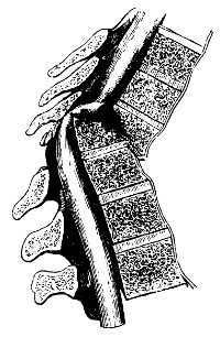
A very well marked specimen of luxation, without the slightest fracture of the fourth from the fifth cervical vertebra is delineated on the next page. The injury was occasioned by the person falling backwards over a high paling, on which he was sitting, and alighting on the back of the head: along with the proper ligaments, the spinal chord is seen to be torn. The patient, of course, did not survive many days, being almost perfectly paralysed.
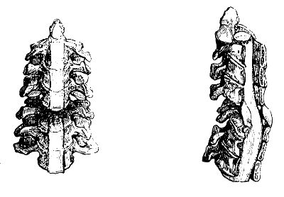
In general, either the bodies or the processes of the vertebræ are broken, and sometimes comminuted; occasionally the bodies are broken entirely through, with considerable displacement, the upper or lower end, as may be, projecting. There is twisting or bending of the trunk or neck, the articulating processes on one side only being displaced, whilst the ligaments on the other remain pretty entire. In some cases, either the spinous processes, or the articular, are separated without yielding of the bodies of the vertebræ, or of the interposed substance; then there is bending of the trunk forwards.
The symptoms vary according to the site of the injury, and the extent of violence inflicted on the spinal chord. This important organ may suffer concussion without fracture or displacement of the bones; its functions may be consequently more or less disturbed, and paralysis occasioned of those parts that are supplied with nerves from below the injured point. Without fracture, too, vessels may give way within the canal, and by compression from effused fluid urgent symptoms will be produced.
The power of motion may be lost whilst sensation is retained, and vice versâ; but in general both are either impaired or destroyed. In one case that came under my care, there was power of motion in one limb and no sensation, whilst in the other there was no motion but the usual sensation.
Patients may recover from the effects of a severe blow on the spinal column and consequent concussion of the chord, but very frequently they do not. Changes may take place at a late period in the chord or its membranes, in consequence of the injury—as thickening of the coverings—bloody, serous, or lymphatic purulent effusion—disorganisation and softening of the medullary matter. Inflammation of the membranes, or of the chord itself, may supervene, either very soon after the accident, or long afterwards; its intensity and period of accession will depend on the extent of the injury, and on the treatment. The muscles act spasmodically, the circulation is excited, the sensorium and nervous system are disordered, delirium ensues, and is followed by paralysis and coma.
In some cases of displacement, even to no small extent, the spinal chord escapes being bruised, torn, or compressed; no bad symptoms may ensue; or paralysis to a greater or less degree occurs and gradually goes off, probably occasioned by bloody effusion, which is afterwards absorbed. This I have witnessed in several instances—in a boy who fell from a high rock—in a woman who fell from a window; both lighted on the breech, and the trunk was bent forwards. The lad remained stout, but his trunk was somewhat deformed by an excurvation; the woman recovered perfectly. In these cases there was evidently laceration of the interspinal ligaments, though probably not of the ligamenta subflava, for the spinal chord must be stretched or otherwise injured when these are torn.
The chord is more or less injured in the majority of cases of fractured spine. If the injury occurs high in the cervical region, immediate death ensues, from compression or laceration of the medulla oblongata. Respiration is arrested by compression or destruction of the chord above the origin of the phrenic and other respiratory nerves. If the chord is injured in the middle of the cervical region, there is paralysis of the upper and lower extremities, with distention of the bowels, and inability to void the urine; the lower bowels have become insensible to the stimulus of distention from want of nervous influence, and the sphincter ani is paralysed. The bladder becomes distended, and then incontinence of urine follows; and frequently there is priapism. The quality of the urine is changed, the secretion of mucus from the bladder is vitiated and increased. Slow inflammation of that organ is induced, the urine becomes bloody and mixed with ropy mucus; lymph is deposited on the lining membrane.
Bruises of the loins often lay the foundation for degeneration and abscess of the kidney, with many of the symptoms of calculus vesicæ, attended with red tongue and hectic, ultimately terminating fatally.
The effects of concussion of the spine are frequently developed long after the infliction of the injury. There is formication, numbness, and difficulty of regulating the motions, in one or more limbs. Still the muscles are not shrunk, nor unable to perform powerful movements; but the patient cannot put his hand or foot to the place he wishes, and cannot support the weight of the body without assistance. Sensation in the limbs is lost to a greater or less degree, their heat is diminished, and it is found difficult to preserve their temperature equable. The symptoms increase till the limbs become totally useless. Along with the lower limbs the bladder is affected, though not always. The urine is not voided with force, and incontinence occurs from distention. Sometimes excitement of the viscus follows; the secretions from its surface are increased, and often mixed with blood. Yet patients survive long under such circumstances, digestion and the other important functions are well performed, and the intellect is unimpaired.
Prognosis in injuries of the spine is unfavourable, as well as in disease of the chord, whether the result of injury or not.
From the treatment much need not be expected; but still no chance is to be thrown away, even in the most unfavourable cases. The attention must be directed towards alleviation of the symptoms. The comfort of the patient must be looked to in regard to the situation of the injured bones and other parts, even where there is reason to believe that the chord is lacerated or completely divided, and that there is no chance of recovery. In less severe cases, by placing the injured parts in their proper position, and retaining them by splints placed along the sides of the spinous processes;—by keeping down inflammatory action, palliating all the symptoms as much as possible, and attending to the state of the bladder if necessary—unlooked for recoveries have taken place.
It has been proposed to treat the spine, in cases of severe and alarming fracture, in the same manner as the cranium—by trephining; and some have recommended this in almost all kinds of injuries. I allude to the practice only to condemn it. The spinal chord is generally displaced and compressed by the lower portion of the fractured body of the bones. One cannot easily comprehend what an operation is to effect in such cases. Further notice of this proceeding is unnecessary, seeing that, as far as I know, it has been unanimously discarded by the profession from amongst the list of surgical operations.
When the patient has borne up against the shock of the injury, and the more immediate consequences, and when partial loss of sensation and motion has supervened, great benefit is obtained from counter-irritation, by blisters, issues, or moxa. But these are not advisable, but to a certainty injurious, till after time has been allowed for subsidence of the immediate effects—for union of the divided parts, and disappearance of acutely excited vascular action. The endermoid application of strychnine is also efficacious in many cases where the injury has been slight—as in the following. A young man was struck on the back of the neck with a leaden plummet. The immediate effects were loss of power and sensation in the whole body. The use of the upper limbs was regained gradually and completely; and when he applied to me, the remaining symptoms were diminished sensation and irregular muscular action in the lower limbs. The mode of progression was very remarkable; supported on the points of the toes and assisted by a staff, he made two or three quick steps as if running, and then suddenly stopped, a few more rapid steps and another abrupt halt, and so on. A succession of small blisters was applied along the sides of the spine in the dorsal and lumbar regions, On the raw surface strychnine was sprinkled, commencing with half a grain daily, and gradually advancing to a grain and a half. He made a perfect recovery in less than three weeks.
In another patient, in whom sensation in one limb without motion, and in the other motion without sensation, remained after severe injury of the spine by a fall from a high window, complete recovery was obtained by the internal use of strychnine, and repeated application of the moxa.
Slow degenerations of the spinal chord are not easily combated with success. Considerable changes of structure have taken place, as shown by the symptoms, before the patient becomes alarmed and applies for relief. He has had a feeling of distention about the lower part of the bowels, and voids his urine with some difficulty; perhaps he suspects stricture of the urethra as the cause. He lifts his feet awkwardly, sets them down clumsily, and all of a piece; his knees totter, there is no feeling about his buttocks, and a numbness round the anus. At length he is for the first time alarmed by incontinence of urine having supervened, or by the limbs having sunk under the weight of the trunk, and by his coming to the ground with violence. The remedial means are local abstraction of blood from over the seat of the disease, followed by friction and counter-irritation. Strychnine may be tried in some cases. But it is indeed seldom that the progress of the case is satisfactory.
Fracture of the Clavicle.—This bone is liable to be broken by indirect violence, as by falls on the point of the shoulder, from horseback, or from the top of a carriage; or by a fall with a carriage, the person being inside—of this accident I have met with three or four instances. It may also be broken by direct violence, as by a blow on the bone, or by the person striking it against a hard substance in a fall. It generally gives way about the middle. The fracture, when occasioned by force applied to the acromial extremity, is usually oblique; transverse when the force is applied to the shaft of the bone. The displacement is in most cases great; but when the fracture is at the bend near the scapular extremity,—a not very uncommon accident,—disjunction of the fractured extremities is prevented by the attachments of the conoid and trapezoid ligaments. In ordinary cases, that fractured extremity projects which is attached to the sternum, whilst the scapular portion is depressed and carried inwards. In short, the scapular portion is displaced, the sternal is nearly in situ; though, from the depression of the former, the prominence of the latter appears to arise from displacement. The arm falls forwards and downwards.
The fracture is sometimes compound. The wound is generally small, and occasioned by the projection of the sternal portion; or the integument may be divided by the external force.
The nature of the accident is readily recognised. The deformity is very apparent. There is swelling, from extravasated blood, over the bone; the shoulder is unnaturally approximated to the chest, and depressed. The motions of the extremity, those above the shoulder, are impaired. Crepitation is felt on raising the arm, and carrying it backwards so as to bring the fractured surfaces into contact.
When the patient is seen immediately after the accident, the bones are to be placed in apposition, and retained, without delay, and before inflammatory swelling has come on. No complicated apparatus is required. A pad, firm, though of soft material, and large enough to fill the arm-pit completely, is rolled in a shawl and placed in the axilla; it is retained by tying the shawl over the opposite shoulder, a soft pad being interposed between the knot and the skin to prevent excoriation, and is farther secured by tying the ends under the axilla of the uninjured extremity, which should also be protected by a small cushion. A few turns of a roller, or a handkerchief, are placed round the arm and chest, so as to secure and fix the limb; so the retentive apparatus is completed. The shoulder is thus raised, and removed from its unnatural position; and the fractured extremities of the clavicle, previously placed in accurate contact, are prevented from being again displaced. The elbow and forearm should be supported by a sling, otherwise the unsupported weight of the limb dragging on the shoulder will cause considerable pain, and subsequent displacement will be apt to occur. In order to prevent swelling, it is sometimes advisable to support by a bandage the hand and forearm. The apparatus should be looked to occasionally, adjusted and tightened; and the cushions should be replaced by fresh ones, to prevent excoriation and uneasiness. The bone will be found to lie quite smooth, to remain of its proper length, to unite, generally within twenty days, and that without any unseemly exuberance of callus. No evaporating lotions are necessary. No compresses or splints need be applied over the bone. If the patient be bruised in other parts, and become feverish, it may be requisite to abstract blood and exhibit antimonials, purgatives, &c. But all inflammation, arising from the fracture, subsides on the accomplishment of reduction, adaptation, and retention of the portions. If the fracture be compound, the edges of the wound should be brought together and retained, so as to favour immediate union.
The body of the scapula is broken, generally by a severe injury of the chest, as by a hard and heavy body passing over it. There is little or no displacement; and the accident is not easily detected, more especially after swelling has taken place.
It is sufficient to restrain motion; and this is effected by passing a bandage round the chest, over the scapula, and round the arm.
The acromion process may be broken off; but the accident is rather uncommon. The fracture is produced by direct violence—a blow or a fall on that point. The spine of the bone also is sometimes broken by a like cause. Portions of the acromion may be separated along with the ligaments connecting the clavicle to it, in the accident of dislocation of the scapular extremity of that bone. The acromion is occasionally broken into fragments by heavy falls on the point of the shoulder.
There is a slight appearance of flattening of the shoulder at first, and then great swelling. Crepitation is felt by pressing gently and alternately with the points of the fingers over the fractured part.
The arm requires to be raised and supported by a sling.
Fracture of the Ribs.—One rib, or more, may be broken by injuries in various ways—by blows of the fist—falls on hard bodies—pressure on the chest by heavy bodies passing over or falling upon it. They generally give way anteriorly to the angles, at the most convex point; but sometimes near the spine or the sternum. At the same time they may be partially luxated at either of the extremities. The fracture is generally transverse; occasionally, and rarely, oblique. Sharp portions are seldom detached. The skin is sometimes divided, but more frequently the pleura and lungs are torn by the spiculæ projecting internally; hence effusion into the chest, and emphysema of the subcutaneous cellular tissue near the fracture, take place. The emphysema, if permitted, extends over the greater part of the chest, and even farther.
Fracture of the ribs is attended with pain, particularly during full inspiration; and if the injury is severe, the patient is incapable, without great pain and exertion, of accomplishing full inspiration. He uses his handkerchief, sneezes, and coughs, with the utmost difficulty. Crepitation is felt by the patient, and is easily detected by the surgeon, by placing the hand on the suspected point, and desiring the patient to attempt full inspiration so as to grate the surfaces on each other. Motions of the trunk, and often of the upper extremities also, are attended with aggravation of the symptoms. In some cases attentive examination is necessary to discover crepitus—in certain situations, and when perhaps one rib only has given way, especially if some time have elapsed betwixt the infliction of the injury and the application of the patient for relief.
In the slighter cases, it is sufficient to restrain the motions of the chest by a broad bandage applied firmly round it; and a split cloth, or a scapulary, may be passed over the shoulders and attached to the circular bandage to prevent its being displaced. Great and immediate relief is thus afforded. In those of a plethoric habit, blood may be taken from the arm, some hours after the injury, with relief and advantage; it may ward off an inflammatory attack—and it is absolutely necessary to adopt this practice on the slightest indication of such supervening. The appearance of the countenance, and the state of the pulse and respiration, must be watched; and on the first becoming anxious, the second strong and accelerated, and the third hurried and imperfect, active measures must be employed—venesection, antimony, purgatives, diaphoretics, anodynes—one or all according to circumstances. In the more severe injuries the same practice is pursued; and the symptoms are watched with great care. The air in the cellular tissue, if effused in great quantity about the neck and face, and interfering with the functions of the parts, is to be evacuated by punctures. If the emphysema is slight, and confined to the neighbourhood of the injured part, farther extrication is prevented by the timely and accurate application of a bandage; the air already in the cellular tissue speedily disappears. The effusion into the chest is also in general absorbed; but it may remain and increase, and from violence of action purulent secretion may be mixed with the serous. The breathing then becomes embarrassed, the chest swells, and the integuments are œdematous. The action of the lung is either much impaired or entirely arrested, as is ascertained by auscultation. In such circumstances, evacuation of the effused fluid may be required.
The Sternum is sometimes fractured, or, in young persons, the bones composing it disjoined; but the occurrence is exceedingly rare. The displacement is not great; and is rectified by changing the position of the trunk. The same treatment is required as for fracture of the ribs. Abscess has formed under the bone, as the result of the injury; but by antiphlogistic means, local and general, this may be in most cases prevented.
Fracture in the vicinity of the shoulder-joint requires to be most accurately examined, that a correct diagnosis may be formed, and the practice be judicious and decided.
Portions of the upper part of the humerus are torn off, along with the attachments of the short muscles, during violent exertions, particularly if the limb is in an awkward position. This is followed by want of power, great swelling, and considerable deformity. Some indistinct crepitation is perceived; the articulation is afterwards stiff, and the bone of an unnatural form. But these indications of the injury gradually disappear.
More extensive solution of the continuity of the bone takes place, generally in consequence of a direct and violent blow on the shoulder. The patient is unable to raise the arm, though with great pain it can be placed in any position that it occupies naturally; it can be abducted and raised, perhaps to a greater extent and more readily than when sound. The shoulder is flattened, and the limb apparently lengthened. The elbow is readily put to the side. On raising the humerus, rotating it, and moving it to and fro, crepitation is distinctly perceived—but not so readily after swelling has taken place. The swelling also obscures the appearances observed immediately after the infliction of the injury,—the flattening of the shoulder, and apparent elongation of the arm. By the fingers of one hand, pressed deep into the axilla, the head of the humerus can be discovered; and, on rotating the shaft of the bone with the other hand, grasping the elbow and pushing upwards at the same time, crepitation is perceived, and the upper portion of the bone is ascertained to be unaffected by the rotation of the shaft. The nature of the injury is then sufficiently apparent.
But the shaft of the humerus may, by such manipulation, be ascertained to be entire. Still, from the direction of the force which effected the injury, the flattening of the shoulder, the remarkable falling down of the arm, the loss of power, the free motion, and from the crepitation, though perhaps indistinct, it is evident that fracture has occurred. Then, by the fingers in the axilla, whilst the humerus is raised and moved in different directions, crepitation is recognised deeper and less distinct than in the former case; and the surgeon is warranted in believing that the glenoid cavity has suffered—that it is broken into fragments, or that it is separated from the body of the scapula by fracture of its neck; he is also warranted in adopting the means of cure suitable to such an accident. Many such injuries are supposed to occur, yet it is strange that preparations illustrative of it are scarcely to be met with in our collections of morbid specimens.
How both detachment and luxation of the head of the humerus should occur, can scarcely be explained. Luxation certainly cannot take place after fracture; no force can be applied to the head of the bone sufficient to displace it. It is barely possible, that after luxation, force may be applied to the bone so as to fracture its neck. This accident is of very rare occurrence, though by some supposed to be otherwise. I have had an opportunity of examining but one case, and that was very distinct; the head of the bone, completely detached from the shaft, lay in the axilla. Comminution of the head of the bone, with displacement of the fragments, is not uncommon.
Separation of the head of the bone occurs occasionally in young persons, presenting the same appearances and symptoms as fracture of the neck of the humerus in later life. Each, by a little care, is distinguishable from dislocation, even after swelling has supervened. And it is highly necessary that the diagnosis should be correct and prompt, otherwise atrociously cruel and unnecessary proceedings will be adopted, and irreparable mischief occasioned. Luxation is attended with flattening of the shoulder and elongation of the arm, to a greater or less degree, according to the position of the head of the bone. But the elbow does not come to the side, and the motions of the limb are abridged; it cannot be abducted to any extent, if the scapula is fixed. The head of the bone is felt under the pectoral muscle, or in the axilla; and on rotating the arm gently, by laying hold of the forearm, and using it, when bent, as a lever, the head and shaft are found to move simultaneously, all of a piece, and no crepitation is felt. Besides, the history of the accident is an excellent guide towards correctly ascertaining the nature of the injury. If the patient, in falling, have involuntarily stretched out his arm, in order to save himself, and alighted with his whole weight on the palm or elbow, dislocation will most probably have occurred. If, on the contrary, he have pitched upon the shoulder, without any intermediate breaking of the fall, fracture is to be expected.
The evil consequences of false diagnosis, and of treatment formed thereon, are very apparent. A dislocation may be put up and treated as a fracture, perhaps till too late for reduction; and the patient will possess but weak and imperfect motion of the limb, after having undergone long suffering. On the contrary, dreadful torments are inflicted on the patient when fracture is treated as luxation. The force applied with the view of reduction is in all circumstances very painful, but, when exerted on a fractured bone, must prove absolute torture; and during the whole treatment, the fragments are, perhaps, every now and then, by renewed attempts, torn separate, and union so prevented. Severe inflammatory action follows the reductive violence, and is kept alive or regenerated by the loose and projecting fractured ends of the bone; extensive suppurations, attended with fever, ensue, and may destroy the patient. Undetected fracture may also be treated as a bruise of the soft parts only; then every motion of the body and limb is productive of excruciating pain, and there is much risk of uncontrollable inflammation being excited—all which would have been warded off, by placing the bones in a proper and steady position in the first instance; the adaptation of a pad in the axilla is followed by immediate and great relief. Such mistakes are quite inexcusable. By one careful examination,—productive no doubt of considerable uneasiness to the patient in some conditions of the parts—the real state of matters should be ascertained; and then the practice founded on the knowledge so obtained will be followed with speedy cessation, or at least great diminution of pain, and with every probability of restoring the limb to strength and usefulness.
Fractures of the glenoid cavity, of the neck of the scapula, and of the neck of the humerus, are all treated by the same simple, though effectual, apparatus as employed for injuries of the clavicle. It requires to be re-adapted occasionally, to have the parts under the crossings of the bandage, and under the knots of the shawl retaining the pad, well protected by soft pads, and it must be worn for four or five weeks—perhaps, in some cases, even a short time longer. Then gentle passive motion of the limb is to be employed, gradually increased as the painful feelings abate. If the parts are at once placed in apposition, and accurately retained, no abstraction of blood, either general or local, is required at the time, and is not likely to be called for during any stage of the treatment. No cold evaporating lotions are necessary; fomentations are sometimes useful.
Fracture of the shaft of the humerus is either oblique or transverse, according to the direction of the force applied. There is considerable displacement. The limb is always shortened to a certain extent, and the natural contour destroyed; the arm is useless, and bent towards the trunk, and the muscles are in a state of spasmodic contraction. The nature of the injury is at once and readily recognised. There is unusual and unnatural mobility of the arm, and distinct crepitation at the fractured point. There is great pain from the pressure of the lower extremity of the bone upon the nervous trunks. The large vessels are seldom torn—though the branches of the humeral artery, and the vessel itself, have in a few cases been ruptured—but there is often considerable bloody swelling in this as in all fractures. Occasionally, when the violence has been great, either the upper or the lower fractured end is thrust through the skin.
When the inferior part of the shaft is broken, there is less displacement than when the fracture is towards the middle of the bone. Fracture above the condyles sometimes extends through them; and the one may be detached from the other either with or without fracture of the shaft. When such an accident is suspected, the position of the condyles in regard to the ends of the bones of the forearm should be accurately observed. Flexion and extension of the forearm can be readily performed, though with pain; not so, when the bones are luxated. Crepitation is detected along the line of fracture, during motion of the limb, and when the condyles are laid hold of and moved upon each other, or on the shaft.
In fracture of the middle of the shaft, coaptation is easily accomplished; slight extension is made by one hand grasping the elbow, whilst, by the other, the bones are brought together, and the straightness and outline of the limb restored. The proper position is readily maintained by two splints of bookbinders’ pasteboard, or of leather prepared for the purpose; one applied from over the acromion process to beyond the point of the elbow, the other from the axilla, and also passing over the elbow on the inside; thus the neighbouring joints are fixed, and the muscles rendered inactive. The conjoined breadth of the splints should be sufficient to embrace the limb almost entirely; some space being left, so that when the swelling subsides, they may neither meet, and consequently lie loose, nor overlap each other. They are softened by steeping in hot water, so that they may embrace every part of the limb to which they are applied; and the extremities should be rounded off, to prevent galling of the parts. They are padded with soft flannel, lint, or cotton wadding, or, what is better, with finely carded tow, and retained by a circular roller applied from the points of the fingers up to the shoulder. The binding should proceed from below upwards, to avoid swelling from obstructed circulation, and do away with the necessity of removing the apparatus arising from this cause. It is well to place a wooden splint on the outside, retained by an additional bandage, so as to steady the parts till the pasteboard or leather has dried, and formed a firm mould or case for the limb; then the wood is no longer necessary, and should be removed. The forearm is bent at right angles, and the humerus fixed to the trunk. In simple fracture, there is in general no necessity for interfering with the apparatus until the bandage slackens, in consequence of the swelling subsiding; then, usually at the end of eight days, it is to be reapplied. One splint is carefully raised, whilst the other is kept fixed and the parts steadied, and the limb is ascertained to be straight and of a proper length; if not, then, or even later, the position of the bones may be rectified without causing much uneasiness. The patient need not be confined to bed on account of a simple fracture; he may walk about with the arm supported in a sling.
In compound fracture similar splints are applied, after due attention has been paid to the wound and to the position of the bones. The patient is placed on his back in bed; and the splints are retained by slips of bandage, double, one end being passed through the loop and secured to the other by a running noose. This method of deligation affords facility for the removal of the splints, in order to examine into the state of the limb and dress the wound. It also permits the apparatus being slackened in the first instance during the swelling, and of being afterwards tightened, without lifting the limb or disturbing its position.
Fracture at the distal extremity of the humerus is managed most conveniently with the limb in the straight position. The fragments are placed accurately together, and one splint placed on the fore part, another posteriorly. The forearm is kept in a state of supination. At the end of about twenty days the apparatus should be removed, and the position of the articulation changed if possible. The forearm is to be bent slightly, and a splint applied,—made to fit accurately, and with a joint corresponding to the bend of the arm. This should be occasionally removed, provided consolidation of the fractured bones has advanced so far as to admit of it, and slight passive motion of the elbow-joint employed. Obstinate rigidity of the parts is thus guarded against.
Fracture of the condyles has been already alluded to. It may be farther observed, that the exact nature of the accident is often difficult to detect; in all cases accurate and careful manipulation is required. Displacement of one or other of the bones of the forearm almost uniformly attends this fracture, sometimes rendering diagnosis obscure.
Fracture of the olecranon process of the ulna is occasioned by falls on the point of the elbow; or the bone may be snapped asunder by powerful and sudden action of the triceps extensor cubiti, when the arm is much and quickly bent. The injury is readily recognised; there is inability to extend the forearm by its own muscular powers, a considerable space is felt between the separated portions of the bone, and the upper fragment is moveable as well as detached; these marks of the injury are rendered more conspicuous by bending the joint. Crepitation is produced by moving the limb when extended, and the separated parts thereby approximated. Bloody swelling soon takes place, large and extensive when bruising of the soft parts has been great—and this is usually the case, in consequence of the injury being almost always the result of direct violence. In some cases the process is comminuted.
Compound fracture is rare, and likely to be productive of serious consequences. I have treated and witnessed several cases. In one the process was cut off by the patient falling out of bed on an earthenware vessel, which broke under the limb. The joint is necessarily opened. Violent inflammation soon commences, and can very seldom be kept within moderate bounds. Discharge of increased and vitiated synovial secretion takes place, followed by profuse and unhealthy suppuration. The cartilages ulcerate, and then the bones. The cellular tissue around becomes infiltrated, the parts swell and are discoloured, and collections of matter form probably at more points than one; perhaps there is a succession of abscesses. The condyles, and often a portion of the shaft of the bone, are denuded by the suppuration, and superficial necrosis results. Ultimately the patient grows hectic. Amputation had to be resorted to in three of the cases which have come under my observation, at the end of some weeks or months from the receipt of the accident.
Union of the simple fracture will take place by bone, if the portions be retained accurately and permanently in contact; but there is a risk of the joint remaining stiff, and of re-separation being produced by even slight violence. Union by ligament is as rapid as that by osseous matter; and if the ligament is short, the arm is quite as useful. Approximation of the broken surfaces is favoured by extension of the elbow-joint, the triceps muscle being thus relaxed. The limb must not, however, be but perfectly straight. The position is preserved by a splint placed on the fore part of the limb, extending from the middle of the arm to the lower part of the forearm, and retained by a roller applied, not over-tight, from the fingers upwards. The application of apparatus to the separated portion, with the view of forcing it into contact with the shaft of the ulna, is useless. The figure of 8 bandage, and such like, are hurtful. Permanent relaxation of the triceps, with prevention of motion, is sufficient. This is continued for three or four weeks; by that time the fracture will in all probability have united, and then gentle and gradual passive motion of the joint is to be commenced.
In compound fracture the prognosis is always unfavourable. Means must be taken to avert incited action—the limb must be properly placed without delay, the edges of the wound accurately approximated, and antiphlogistic measures pursued. Purulent collections must be opened early. Rest of the joint is to be insured, and support afforded, by bandaging and by the application of a splint. The patient will be fortunate if he escape mutilation by the amputating knife; but when the wound is trifling, and the parts not much lacerated or bruised, and the treatment carefully conducted, a cure may be effected by the same process as the simple fracture.