Chapter II
Reading edition
MYOLOGY
The first point to decide in commencing this study is the order in which we shall consider the different muscles which we have to examine. It must not be forgotten that in the present work we compare the organization of animals with that of man, which we already know, and that it is on the construction of this latter that, in these studies, the thought must at each instant be carried back in order to establish this comparison. Now, the general tendency which we notice in our teaching of anatomy, when one regards the region of the trunk in the human figure (a living model or a figure in the round), is first to consider the anterior aspect. It is the latter that, for this reason, we study at the very beginning; we next deal with the posterior surface of the trunk, because it is opposite; lastly, the lateral surfaces, because they unite with the preceding surfaces, the one to the other.
In studying an animal, it is usually by one of its lateral aspects that one first observes it; it is, in fact, by these aspects that it presents its greatest dimensions, and that the morphological characters as a whole can be more readily appreciated. Hence, possibly, the order of description adopted in most texts, or in the figures which accompany them. The first representation of the human figure as a whole, in a treatise on anatomy, represents the anterior aspect; the first view of the horse as a whole, in a treatise on veterinary anatomy, for example, is, on the other hand, a lateral view.
We break with this latter custom, and, without taking into account the tendency above indicated, we will commence our analysis with the study of the aspect of the trunk, which corresponds to the anterior aspect of the same region in man.
The first muscles usually presented for study to artists being the pectorals, it is their homologues that we will first describe here. We will afterwards describe the abdominal region, then the muscles which occupy the dorsal aspect of the trunk. With regard to the lateral surfaces, they will be found, by this fact alone, almost completely studied, since the muscles of the two preceding (back and abdomen), spreading out, so to speak, over them, contribute to their formation. Nothing further will remain but to incorporate with them the muscles of the shoulder; but these will be studied in connection with the anterior limbs, from which they cannot be separated.
The neck, in man, may be considered in an isolated fashion, because, on account of its narrowness in proportion to the width of the shoulders, it is clearly differentiated from the trunk; for this reason we combine the study of it with that of the head. In animals, because of the absence or slight development of the clavicles, the neck is generally too much confounded with the region of the shoulders to make it legitimate to separate it from that region in too marked a fashion. It will, accordingly, be considered next.
We will then undertake the study of the muscles of the limbs, and end with the myology of the head.
THE MUSCLES OF THE TRUNK
We shall divide them into muscles of the thorax, of the abdomen, and of the back.
Muscles of the Thorax
The Pectoralis Major (Fig. 66, 1, 2; Fig. 67, 3, 4; Fig. 68, 7; Fig. 69, 10; Fig. 70, 11).—Further designated by the name of superficial pectoral, this muscle is described in treatises on veterinary anatomy as formed of two portions: an anterior one, called the sterno-humeral muscle; the other, situated below and behind the preceding, bearing the name of sterno-aponeurotic.
It occupies the region of the breast, and, as a whole, it takes origin from the median portion of the sternum, from which it is directed towards the arm and forearm.
The anterior portion (sterno-humeral muscle)—thick, forming an elevation under the skin, and really constituting the pectoral region—is directed downwards and outwards to be inserted into the anterior margin of the humerus—that is to say, to the ridge which limits in front the spiral groove of this bone.
The other part (sterno-aponeurotic muscle) is situated more posteriorly, and corresponds to the region known in veterinary anatomy as the inter-fore-limb space, which is limited laterally on each side by the superior portion of the forearm, of which the point of junction with the trunk bears the name ars. Arising from the sternum, as we have above indicated, this portion is directed outwards, to be joined with the terminal aponeurosis of the sterno-humeral, and with that which covers the internal surface of the forearm.
All things considered, the sterno-humeral muscle may be regarded as the representative of the upper fibres of the great pectoral of man, of which the attachments, owing to the more or less complete absence of the clavicle in the domestic mammals, the fibres must be concentrated on the sternum; the sterno-aponeurotic portion then representing the inferior fasciculæ of the same muscle.
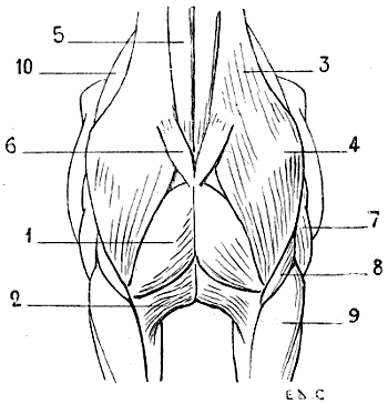
Fig. 66.—Myology of the Horse: Anterior Aspect of the Trunk.
1, Pectoralis major (sterno-humeral); 2, pectoralis major (sterno-aponeurotic); 3, mastoido-humeralis; 4, point of the shoulder; 5, sterno-mastoid or sterno-maxillary: 6, inferior portion of the platysma myoides of the neck, divided; 7, triceps cubiti; 8, brachialis anticus; 9, radialis (anterior extensor of the metacarpus); 10, scapular region.
The great pectoral muscle of one side is separated from that of the opposite side along the median line, and especially above and in front, by a groove which is more or less deep, according as the muscles are more or less developed. At the bottom of this groove, suggestive of that which exists in the corresponding region in man, is found, as in this latter, the median portion of the sternum.
The preceding description particularly applies to the arrangement which the great pectoral presents in the horse; in other animals it is marked by some distinctive characters. In the pig, it is inserted into the sternum as far only as the level of the third costal cartilage; in the ox and sheep, it extends as far as the sixth; in the dog, it is attached to the two first sternal pieces only—that is to say, as far as the third costal cartilage. Moreover, in the latter, as in the cat, the two portions which we have indicated are less readily distinguished.
The great pectoral, by its contraction, draws the fore-limb towards the middle line—that is to say, adducts it.
The Pectoralis Minor (Fig. 67, 6; Fig. 68, 8; Fig. 69, 11; Fig. 70, 12, 26).—This muscle, also called the deep pectoral, is, in animals, larger than the superficial pectoral, therefore certain authors prefer to give to this muscle and the preceding one the names of deep and superficial pectoral respectively. This nomenclature is evidently legitimate, and conforms more to reality, since it does not bring in the notion of dimensions which here is found in contradiction to nomenclature; but, in order to establish more clearly the parallelism with the corresponding muscles in man, we think it better, nevertheless, to give them the names by which it has been customary to designate them in connection with the latter.
We will recall at the outset that the lesser pectoral muscle in man is completely covered by the great. In animals this is not the case; the lesser pectoral being very highly developed, projects beyond the great pectoral posteriorly, and occupies to a greater or less extent the inferior surface of the abdomen.
It also consists of two parts: one anterior, which we designate by the name of sterno-prescapular; the other, posterior, bearing that of sterno-humeral.[20]
The sterno-prescapular muscle, being covered by the sterno-humeral, has little interest for us. It arises from the sternum, and is directed towards the angle formed by the junction of the scapula and humerus; then it is reflected upwards and backwards, to terminate on the anterior margin of the shoulder by insertion into the aponeurosis, which covers the supraspinatus muscle.
We can, especially in the horse after removal of the skin, recognise it, at the level of this region, in the interspace limited by the superficial muscles (Fig. 70, 26).
In the dog and cat this portion of the muscle does not exist. The other division of the muscle, the sterno-trochinian, is more interesting. It arises from the abdominal aponeurosis and the posterior part of the sternum; hence it passes forward, turns under the superficial pectoral, and is inserted into the lesser tuberosity (trochin) of the humerus.
In the pig, dog, and cat, it is inserted into the greater tuberosity (trochiter) of the bone of the arm.
The superior border of this muscle is in relation with a superficial vein, which is distinctly visible in the horse—the subcutaneous thoracic vein, which in this animal is called the vein of the spur.
The sterno-humeral muscle, in contracting, draws the shoulder and the whole anterior limb backwards.
Serratus Magnus (Fig. 67, 2; Fig. 69, 8; Fig. 70, 9).—This muscle, which is situated on the lateral aspect of the thorax, is covered to a considerable extent by the shoulder, the posterior muscular mass of the arm, and by the great dorsal muscle.
It arises by digitations from the external surface of the dorsal vertebræ; from the first eight in the horse, ox, and dog.
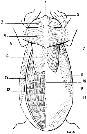
Fig. 67.—Myology of the Horse: Inferior Aspect of the Trunk.
1, Anterior extremity of the sternum; 2, point of the shoulder and inferior portion of the mastoido-humeral muscle; 3, pectoralis major (sterno-humeral); 4, pectoralis major (sterno-aponeurotic); 5, point of the elbow; 6, pectoralis minor (sterno-trochinian); 7, serratus magnus; 8, external oblique; 9, sheath of the rectus abdominis; 10, linea alba; 11, the umbilicus; 12, external oblique divided in order to expose the rectus abdominis; 13, rectus abdominis.
The muscular bundles, converging as they proceed, towards the scapula, pass under this bone, to be inserted into the superior portion of the subscapular fossa, near the spinal border. The inferior portion of its posterior digitations is visible in the ox and in the horse; these digitations are less visible in the pig. They are not seen at all in the dog (Fig. 68) or cat, for in these animals the great dorsal muscle covers them completely.
The great serratus muscle, by the position which it occupies and the arrangement that it presents, forms with the corresponding muscle of the opposite side a sort of girth, which supports the thorax, and at the same time helps to fix the scapula against the latter.
When it contracts, in taking its fixed point at the ribs, it draws the superior portion of the scapula downwards and backwards in such a way that this bone has its inferior angle directed forwards and upwards. If it takes its fixed point at the shoulder, it then acts on the ribs, raises them, and so becomes a muscle of inspiration.
Because of the connections of the serratus magnus with the levator anguli scapulæ, some authors consider it as united with the latter. But as the latter muscle is visible only in the region of the neck (see p. 157), and as it is separately described in man, we prefer to distinguish them from one another. We shall recall the connections to which we have just made allusion when describing the cervical region.
Muscles of the Abdomen
The abdominal wall is, as in man, formed by four large muscles: the external oblique, the internal oblique, and the transversalis, which form the lateral walls, and the rectus abdominis, situated on each side of the middle line of the abdomen. This latter, because of the general direction of the trunk in quadrupeds, has its superficial surface directed downwards.
The arrangement of these muscles closely corresponds to that which we find in the human species.
The External Oblique Muscle (Fig. 67, 8, 12; Fig. 68, 5; Fig. 69, 9; Fig. 70, 10).—This muscle arises, by digitations, from a number of ribs, which varies according to the species, the number of the ribs being itself variable for each of them, as we pointed out in connection with the osteology of the thorax. Indeed, the great oblique arises from the eight or nine posterior ribs in the dog and the ox, and from the thirteen or fourteen posterior in the horse. It is attached, besides, to the dorso-lumbar aponeurosis.
These attachments are arranged in a line directed obliquely upwards and backwards, and the first digitations—that is to say, the most anterior ones—dovetail with the posterior digitations of origin of the great serratus muscle.
The fleshy fibres are directed downwards and backwards, and terminate in an aponeurosis which covers the inferior aspect of the abdomen, and proceeds to form the linea alba by joining with that of the muscle of the opposite side, and also to be inserted into the crural arch.
This aponeurosis of the external oblique is covered by an expansion of elastic fibrous tissue, which doubles it externally, and which is known as the abdominal tunic. This latter is further developed as the organs of the digestive apparatus are more voluminous, and their weight, consequently, more considerable. For this reason, in the large herbivora, as the ox and the horse, this tunic is extremely thick, whereas in the pig, cat, and dog it is, on the contrary, reduced to a simple membrane. Indeed, in these latter, the abdominal viscera being less developed, the inferior wall of the abdomen does not require so strong a fibrous apparatus for supporting them. The great oblique, when it contracts, compresses the abdominal viscera in all circumstances under which this compression is necessary; it also acts as a flexor of the vertebral column.
The Internal Oblique Muscle.—This muscle, which is covered by the preceding, arises from the anterior superior iliac spine (external angle in ruminants and solipeds) and the neighbouring parts. From this origin its muscular fibres, the general direction of which is opposite to that of the fibres of the external oblique, diverging, proceed to terminate in an aponeurosis, which contributes to the formation of the linea alba, and to be attached superiorly to the internal surface of the last costal cartilages. It has the same action as the great oblique. What it presents of special interest is the detail of form which it determines in the region of the flank; this detail is the cord of the flank. It is characterized by an elongated prominence which, starting from the iliac spine, is directed obliquely downwards and forwards, to terminate near the cartilaginous border of the false ribs.
Often very apparent in the ox, and still more so in the cow, the cord in question contrasts with the depression which surmounts it; this depression is situated below the costiform processes of the lumbar vertebræ, and is called the hollow of the flank. It is so much the more marked as the mass of the intestinal viscera is of greater weight.
We sometimes meet with a case of the presence of this hollow in the horse. But when in the latter, the flank is well formed, the hollow is scarcely visible, and the cord but slightly prominent. It is only in emaciated subjects that these details are found clearly marked.
Transversalis Abdominis.—This muscle being deeply situated does not present any interest for us. We will, however, point out, in order to complete the series of muscles which form the abdominal wall, that the direction of its fibres is transverse, and that they extend from the internal surface of the cartilages of the false ribs, and the costiform processes of the lumbar vertebræ to the linea alba.
The Rectus Abdominis (Fig. 67, 13; Fig. 68, 6).—This muscle, enclosed, as it is in man, in a fibrous sheath (Fig. 67, 9) formed by the aponeuroses of the lateral muscles of the abdomen, is a long and wide fleshy band, which, as in the human species, reaches from the thorax to the pubis.
What distinguishes it in quadrupeds is that there are costal attachments which extend further on the sternal surface of the thorax, and the number of its aponeurotic insertions, which, in general, is more considerable. These are, indeed, six or seven in number in the pig and in ruminants, and about ten in the horse.
It is true that we may find but three in the cat and dog; still, we often find as many as six. These intersections are not marked on their exterior by transverse grooves, such as we find in the human species in individuals with delicate skin and whose adipose tissue is not very much developed.
The rectus abdominis is covered, in its anterior portion, by the sterno-trochinian muscle (posterior segment of the small pectoral). In contracting, this muscle brings the chest nearer the pelvis, and as a result flexes the vertebral column. It also contributes to the compression of the abdominal viscera.
Pyramidalis Abdominis.—This unimportant little muscle, which in man is situated at the lower part of the abdomen, extends from the pubis to the linea alba. It is not present in the domestic animals.
We consider it interesting, however, to point out, although the fact is not a very useful one as regards external form, that this muscle is distinctly developed in marsupials.
We know that in the opossum, the kangaroo, and the phalanger fox, the young are brought forth in an entirely incomplete state of development, and that, during a certain period, they are obliged to lodge in a pouch which is placed at the lower part of the abdomen of the mother. Now, this pouch contains the mammary glands; but the young, being too feeble to exercise the requisite suction, the pyramidal muscles come to their assistance. These muscles, in contracting, approximate to one another two bones which are placed above the pubis, the (so-called) marsupial bones (see Fig. 80); by their approximation the bones in question, which are placed behind and on the outer side of the mammary glands, compress the latter, and thus is brought about the result which the little ones, on account of their feebleness, would, without that intervention, be incapable of obtaining for themselves.
Muscles of the Back
Trapezius (Fig. 68, 1, 2; Fig. 69, 1, 2; Fig. 70, 1, 2).—This muscle, more or less well developed, according to the species, is divided into two portions, of which the names indicate the respective situations—a cervical and a dorsal.
These two parts, considered in the order in which we find them, take their origin from the superior cervical ligament and from the spinous processes of the first dorsal vertebræ. From these different points the fibres are directed towards the shoulder; the anterior are, consequently, oblique downwards and backwards, and the posterior are directed downwards and forwards. They are inserted into the scapula in the following manner: the fibres of the dorsal portion are attached to the tuberosity of the spine; those of the cervical region are also fixed into the same spine, but into a considerably larger surface.
The cervical portion occupies, in the region of the neck, an area relatively smaller than the corresponding portion of the trapezius in man. This diminished degree of development results from the absence, complete, or nearly so, of the clavicle in the animals which we are now considering. We remember, that the trapezius of man is partly inserted into the clavicle, and the disappearance of this latter cannot fail to bring modifications in the general disposition of the corresponding portion of the muscle. There results a disconnection of this latter, and it becomes united to other muscular fibres to form a muscle with which we shall soon have to deal—the mastoido-humeral (see p. 150).
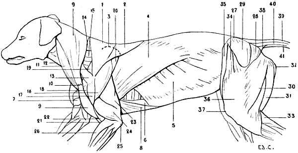
Fig. 68.—Myology of the Dog: Superficial Layer of Muscles.
1, Trapezius, cervical portion; 2, trapezius, dorsal portion; 3, superior outline of the scapula; 4, latissimus dorsi; 5, external oblique muscle; 6, rectus abdominis; 7, pectoralis major of the right side; 8, pectoralis minor (sterno-trochinian); 9, 9, mastoido humeral muscle; 10, tendinous intersection, at the level of which is found a rudimentary clavicle; 11, sterno-mastoid muscle; 12, infrahyoid muscles; 13, omo-tracheal or acromio-tracheal muscle; 14, splenius; 15, levator anguli scapulæ; 16, deltoid muscle, spinal portion; 17, deltoid, acromial portion; 18, superior extremity of the humerus; 19, supraspinatus; 20, infraspinatus; 21, biceps cubiti; 22, brachialis anticus; 23, triceps cubiti, long head; 24, triceps cubiti, external head; 25, olecranon process; 26, radialis (anterior extensor of the metacarpus); 27, iliac crest; 28, gluteus maximus; 29, gluteus medius; 30, biceps cruris; 31, semitendinosus; 32, semi-membranosus; 33, gastrocnemius; 34, tensor of the fascia lata; 35, sartorius; 36, fascia lata drawn up by the triceps; 37, the patella or knee-cap; 38, ischio-coccygeal muscle; 39, superior sacro-coccygeal; 40, lateral sacro-coccygeal; 41, inferior sacro-coccygeal.
As specific differences we should add that the trapezius occupies a more or less extensive portion of the median and superior regions of the neck; terminating at a considerable distance from the head in the dog and horse, it, on the contrary, approaches it in the pig and in ruminants. The cervical portion, when it contracts, draws the scapula upwards and forwards, the dorsal portion draws it upwards and backwards. When the trapezius acts as a whole the scapula is raised.
The Latissimus Dorsi (Fig. 68, 4; Fig. 69, 5; Fig. 70, 5).—This muscle arises by an aponeurosis, the so-called dorso-lumbar aponeurosis, from the spinous processes of the last dorsal vertebræ (the seven last in the dog, fourteen or fifteen last in the horse), from the spinous processes of the lumbar vertebræ, and from the last ribs. Its fleshy fibres are directed downwards and forwards, being more oblique in direction posteriorly, and pass on the inner side of the posterior muscular mass of the arm, to be inserted into the internal lip of the bicipital groove of the humerus, or, a little lower down, on the median portion of the internal surface of the same bone. This latter mode of insertion is met with in the horse and the ox.
The anterior fibres cover the posterior angle of the scapula (as in man, where the corresponding angle, but in this case inferior, is covered by the same muscle), and, a little higher up, are in their turn concealed by a portion of the dorsal fibres of the trapezius. It covers, to a greater or less extent, the great serratus muscle. These relations are similar to those found in the human species.
We find that the fleshy fibres of the great dorsal are prolonged more or less backwards if we examine this muscle in the dog, the ox, the pig, and the horse. Indeed, the fibres reach to the thirteenth rib in the dog and the cat (that is to say, the last rib), the eleventh in the ox, tenth in the pig, and twelfth only in the horse. We say ‘only’ in connection with this last because it is necessary to remember that the ribs are eighteen in number on each side of the thorax of this animal, and that, accordingly, the fleshy fibres of the great dorsal muscle are, relatively, of small extent.
When this muscle contracts it flexes the humerus upon the scapula, and helps to draw the whole of the anterior limb backwards and upwards.
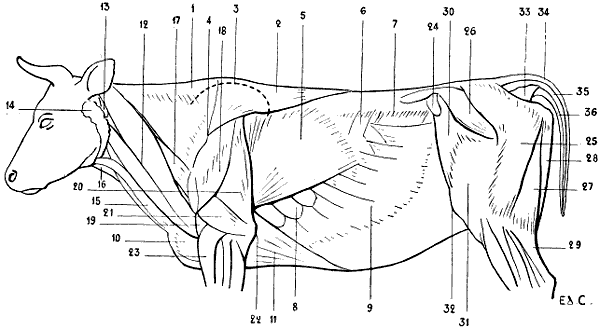
Fig. 69.—Myology of the Ox: Superficial Layer of Muscles.
1, Trapezius, cervical portion; 2, trapezius, dorsal portion; 3, outline of the scapula; 4, spine of the scapula; 5, latissimus dorsi; 6, small posterior serratus; 7, prominence caused by the costiform processes of the lumbar vertebræ; 8, serratus magnus; 9, external oblique; 10, pectoralis major (sterno-humeral); 11, mastoido-humeralis; 12, atlas; 13, atlas; 14, parotid gland; 15, sterno-mastoid muscle; 16, infrahyoid muscles; 17, omo-trachelian or acromio-trachelian muscle; 18, deltoid; 19, brachialis anticus; 20, triceps, long head; 21, triceps, external head; 22, olecranon; 23, radialis (anterior extensor of the metacarpus); 24, anterior iliac spine; 25, gluteus maximus; 26, gluteus medius; 27, biceps cruris; 28, semitendinosus; 29, gastrocnemius; 30, tensor of the fascia lata; 31, fascia lata covering the triceps of the thigh; 32, patella; 33, ischio-coccygeal muscle; 34, superior ischio-coccygeal; 35, lateral ischio-coccygeal; 36, inferior ischio-coccygeal.
There is a muscular fasciculus which, because of its relations with the muscle we have just been studying, is known as the supplementary muscle of the latissimus dorsi. But as, on the other hand, this fasciculus is in relation with the triceps, we shall in preference consider it in relation with this latter (see p. 173).
The aponeurosis by which the great dorsal arises from the vertebral column covers, as in man, the muscles which occupy the grooves situated on each side of the spinous processes—the spinal muscles or common muscular mass, if we regard them as a whole (Fig. 70, 7); the sacro-lumbar and the long dorsal muscles covering the transverse spinal, if we consider them as distinct.
It would be superfluous to enter here into a detailed examination of these muscles.
If they are but little developed the spinous processes become prominent under the skin; if they are more so they may by their thickness project beyond the level of these processes, and these latter thus come to lie in a groove more or less marked, which, on account of the division which is determined by its presence, has caused the regions which it occupies to be designated by the names double back and double loins.
The muscles are extensors of the vertebral column.
Under the aponeurosis of the great dorsal muscle there is found in man another muscle, the serratus posticus inferior, which, on account of being deeply placed and its slight thickness, offers nothing of interest in connection with the study of external form. It arises from the spinous processes of the three last dorsal vertebræ and those of the three first lumbar; it then passes upwards and outwards, and divides into four digitations, to be inserted into the inferior borders of the four last ribs. We repeat that it is covered by the great dorsal muscle.
In the pig, ox, and horse, which have this latter muscle less developed in its posterior portion, the same small serratus muscle, known as the posterior serratus, is visible in the superficial layer of muscles (Fig. 69, 6; Fig. 70, 6). The number of its digitations is more or less considerable according to the species examined.
The Rhomboid Muscle (Fig. 70, 21).—In order to make intelligible the position of the rhomboid in the superficial layer in quadrupeds, it appears to us necessary to recall the anatomical characters of the muscle as found in man. The rhomboid arises from the inferior portion of the posterior cervical ligament, from the spinous process of the seventh cervical vertebræ and the four or five upper dorsal; thence passing obliquely downwards and outwards, it is inserted into the spinal border of the scapula, into the portion of this border which is situated below the spine; it sometimes extends to the middle of the interval which separates this latter from the superior internal angle of the same bone.
The portion of the muscle which arises from the cervical ligament and the seventh cervical vertebra is often separated from the lower portion by a cellular interspace. For this cause some anatomists have described the rhomboid as consisting of two parts—the superior or small rhomboid and the inferior or large rhomboid, on account of the position occupied by each, and of their difference in volume.
This muscle can only be seen in the region of the back, in the space limited externally by the spinal border of the scapula, below by the latissimus dorsi, and internally by the trapezius, which covers it in the rest of its extent. It is not in this space that it is seen in certain quadrupeds. As we pointed out in the section on osteology, the spinal border of the scapula is short, and it seems to be due to this limitation in length that the trapezius and the latissimus dorsi muscle are, at this level, in contact the one with the other in such a way that they fill up the interval in which the rhomboid is seen in man.
In the horse we can partly see it in the superficial muscular layer, but in the region of the neck only, at the superior border of the shoulder. Indeed, as we have already pointed out, the trapezius does not reach the occipital protuberance; for this reason a part of the anterior portion of the rhomboid may be seen—that is, the portion which corresponds to the superior part of the human muscle.
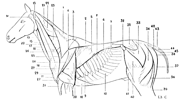
Fig. 70.—Myology of the Horse: Superficial Layer of Muscles.
1, Trapezius, cervical portion; 2, trapezius, dorsal portion; 3, superior outline of the scapula; 4, spine of the scapula; 5, latissimus dorsi muscle; 6, small posterior serratus; 7, spinal muscles, or common muscular mass; 8, ribs; 9, serratus magnus; 10, external oblique; 11, pectoralis major (sterno-humeral); 12, pectoralis minor (sterno-trochinian); 13, atlas; 14, parotid gland; 15, mastoido-humeralis; 16, point of the arm; 17, sterno-mastoid, or sterno-maxillary; 18, jugular groove; 19, infrahyoid muscles; 20, omo-trachelian muscle; 21, rhomboid; 22, splenius; 23, levator anguli scapulæ; 24, deltoid; 25, supraspinatus; 26, terminal part of the sterno-prescapular, a portion of the small pectoral muscle; 27, brachialis anticus; 28, triceps cubiti, middle or long head; 29, triceps cubiti, external head; 30, olecranon; 31, radial extensor (anterior extensor of the metacarpus); 32, anterior iliac spine; 33, anterior portion of the gluteus maximus—the aponeurosis of the muscle has been divided in order to expose the gluteus medius; 34, posterior portion of the gluteus maximus; 35, gluteus medius; 36, biceps cruris; 37, semitendinosus; 38, point of the buttock; 39, gastrocnemius; 40, tensor of the fascia lata; 41, triceps cruris; 42, ischio-coccygeal muscle; 43, superior sacro-coccygeal; 44, lateral sacro-coccygeal; 45, inferior sacro-coccygeal.
But whether it be covered by the trapezius, or, as we find in the cat and dog, by the mastoido-humeral muscle (see p. 150), which is very broad in this region, we do not the less recognise its presence; and in the horse and ox, in particular, it forms an elongated prominence beginning at the level of the scapula, and tapering as it ascends, towards the posterior part of the head.
Its origins are similar to those which we have already described in the human rhomboid. It arises from the cervical ligament and the spinous processes of the foremost dorsal vertebræ; its fibres converge and pass to the scapula, to be inserted into its superior or spinal border, or into the internal surface of the cartilage of prolongation.
It assists in keeping the scapula applied to the thoracic cage, and when it contracts, draws the scapula upwards and forwards.
Taking its fixed point at the scapula, it acts on the neck by its anterior fibres, and extends it.
We shall soon have occasion to mention this muscle again, in connection with the study of the muscles of the neck.
The Cutaneous Muscle of the Trunk (Fig. 71).—Immediately beneath the skin which covers the neck, shoulders, and trunk is found a vast cutaneous muscle, analogous to that which, in the human species, exists only in the cervical region.
This thin muscle, whose function is to move the skin which strongly adheres to it, and in this way to remove from it material causes of irritation (insects, for example), is of considerable thickness in the region of the trunk; where it constitutes what certain authors have designated by the name of panniculus carnosus. In this region it extends from the posterior border of the shoulder to the thigh, and, in the vertical direction, from the apices of the spinous process of the dorso-lumbar vertebræ to the median line of the abdomen.
Arising above from the supraspinous ligament of the dorso-lumbar and sacral regions (except in the carnivora; see below) by an aponeurosis which, posteriorly, covers the muscles of the hind-limbs, its fibres are directed to the elbow, on which they are arranged in two layers: a superficial, which becomes continuous with the panniculus muscle of the shoulder; and a deep, which passes on the inner side of the shoulder to be inserted into the internal surface of the humerus; this latter exists only in the dog and cat.
The most inferior fibres, behind, at the level of the knee-cap form a triangular process which in the horse receives the name of the stifle fold, from the name veterinarians give to the region of the articulation of the knee. This fold of skin, which commences on the antero-internal surface of this region, is directed upwards, and then forwards, to end by gradually disappearing over the corresponding part of the abdomen.
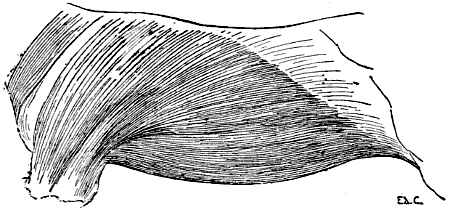
Fig. 71.—Myology of the Horse: Panniculus Muscle of the Trunk.
In the same animal the muscular fibres of the panniculus of the trunk arise along a line which connects the stifle-joint to the withers, a line which is, consequently, oblique upwards and forwards. Now, as the fleshy layer is thicker than the aponeurosis, the result is that the mode of constitution of this muscle can be recognised under the skin. Indeed, we can see in some animals, occasionally very distinctly, a slight elevation starting from the region of the abdomen in the neighbourhood of the knee, and thence directed obliquely upwards and forwards. This elevation is produced by the fleshy portion of the panniculus.
In the carnivora, the panniculus of the trunk is not attached to the supraspinous ligament; it is blended with the same muscle of the opposite side, passing over the spinous region of the vertebral column.
From this arrangement results a great mobility of the skin which covers the back. Further, it explains why it is possible to lift up this skin along with the panniculus which it covers, and to which it adheres, throughout the whole extent of the dorso-lumbar column. As we pointed out above, there is also a panniculus muscle of the shoulder and one of the neck. We will deal with them when treating of the regions to which those muscles belong.
The Coccygeal Region
As a sequel to the study of the muscles of the region of the trunk very naturally comes the description of those which, belonging to the region of the coccyx, are destined for the movements of the caudal appendix, of which this latter constitutes the skeleton. The muscles may not seem to be of much importance with regard to external form, but, as they form part of the superficial muscular layer, and as the mass of each is seen in the form of the tail in some animals (the lion, for example), they merit our attention for a moment. A few lines will suffice to give an idea of them. They are: the ischio-coccygeal, superior sacro-coccygeal, lateral sacro-coccygeal, and inferior sacro-coccygeal.
The Ischio-coccygeal (Fig. 18, 38; Fig. 69, 33; Fig. 70, 42).—This muscle, triangular in shape, better developed in the carnivora than in the horse, arises from the spine of the ischium, or from the supracotyloid crest, which replaces this latter in the solipeds and the ruminants. Thence its fleshy mass is directed upwards, expanding as it proceeds to be inserted into the transverse processes of the first two coccygeal vertebræ after insinuating itself between two of the following muscles, the lateral and inferior sacro-coccygeal.
In the dog and cat, the muscle is in great part covered by the great gluteal. In the ox, by a peculiar arrangement of the corresponding region of the muscles of the thigh—an arrangement which we will examine in connection with the study of the latter—it is more exposed than in the horse, and gives origin to an outline which corresponds to its general form in the region situated immediately below the root of the tail.
It is a depressor of the whole caudal appendix.
The Superior Sacro-coccygeal (Fig. 68, 39; Fig. 69, 34; Fig. 70, 43).—The fasciculi which form this muscle arise from the crest of the sacrum, and proceed thence to end successively on the coccygeal vertebræ. It is in contact in the middle line with the corresponding muscle of the opposite side.
It raises the tail and inclines it laterally; if the muscle of one side contracts at the same time as that of the other the tail is elevated directly.
The Lateral Sacro-coccygeal (Fig. 68, 40; Fig. 69, 35; Fig. 70, 44).—Situated on the lateral part of the caudal region, this muscle arises, in the dog, from the internal border of the iliac bone and the external border of the sacrum; in the horse, it arises from the crest of the sacrum. It is inserted into the coccygeal vertebræ.
It produces lateral movement of the tail.
The Inferior Sacro-coccygeal (Fig. 68, 41; Fig. 69, 36; Fig. 70, 43).—This muscle, which is fairly thick, arises from the inferior surface of the sacrum and the corresponding surface of the sacro-sciatic ligament; it is inserted into the coccygeal vertebræ.
It depresses the caudal appendix.
Muscles of the Neck
Mastoido-humeralis (Fig. 66, 3; Fig. 68, 9, 9, 10; Fig. 69, 12; Fig. 70, 15).—One of the most important muscles of the region of the neck in man is the sterno-cleido mastoid. We recollect that, in its inferior part, it is divided into two bundles, one of which arises from the manubrium of the sternum, and the other from the inner third of the clavicle, whence the denominations of the sternal portion and clavicular portion. The muscle formed by the union of these two portions is then directed obliquely outwards, backwards, and upwards, to be inserted into the mastoid process of the temporal bone and the two external thirds of the superior curved line of the occipital bone.
Now, the animals which we are here considering have but a rudimentary clavicle or are entirely without it. From the absence of this item of the skeleton there necessarily result modifications in the arrangement of the muscles of this region, which we must at the very outset explain, before undertaking the special study of the muscle which is the subject of the present paragraph.
Let us suppose, for the more definite arrangement of our ideas, that the clavicle is altogether absent, although we do find it in a rudimentary state in some animals and completely developed in others (marmot, bat), and we will proceed to indicate what this absence determines.
The great pectoral muscle in man arises in part from the clavicle; this origin not being possible in animals which have no clavicle, its attachments, as we have already seen, are concentrated on the sternum. The trapezius in man similarly arises in part from the clavicle; for the reasons above indicated its clavicular fasciculi cannot exist in distinct form in the animals which have no clavicle.
The sterno-cleido mastoid, whose inferior attachments we mentioned above, cannot have a clavicular portion.
It is the same in the case of the deltoid, which, we know, arises in part from the anterior bone of the shoulder.
Of the four muscles which have partial clavicular origins in man, two are known to us in connection with animals—the great pectoral and the trapezius. What has become of the other two, the sterno-cleido mastoid and the deltoid?
It is this which we now proceed to investigate. After a fashion simple enough, but which it is necessary to describe, the clavicular fasciculi of the trapezius and the corresponding fasciculi of the sterno-cleido mastoid are united the one to the other; the portion of the deltoid which in man arises from the clavicle, by reason of the absence of this latter, is also combined with the fleshy mass formed by the preceding muscles. From this fusion results the muscle known as the mastoido-humeral. This muscle, which consists of a long fleshy band situated on the lateral aspect of the neck, takes its origin, as a general rule, from the posterior surface of the skull and the upper part of the neck, from which it passes obliquely downwards and backwards, covering the scapulo-humeral angle—that is, the region known as the point of the shoulder or arm—and is inserted into the anterior border of the humerus, the border which, limiting anteriorly the musculo-spiral groove, forms a continuation of the deltoid impression. On account of the regions with which it is related, Bourgelat named this muscle the muscle common to the head, neck, and arm.
It is at the level of the scapulo-humeral angle that the vestiges of the clavicle are found.
This bone is represented in some animals—the pig, ox, and horse—by a single tendinous intersection, more or less apparent, which extends transversely from the scapula to the anterior extremity of the sternum. In the dog and the cat, we find, besides, on the deep surface of the muscle and at the level of this tendinous intersection, the rudiment of the clavicle of which we made mention in the section on Osteology (see p. 25).
It is beneath the intersection, the existence of which we have just pointed out, that is found that portion of the mastoido-humeral muscle which corresponds to the clavicular fasciculi of the deltoid; that portion which is situated above the intersection corresponds to the clavicular fibres of the sterno-cleido-mastoid and of the trapezius.
The mastoido-humeral presents certain varieties in different animals.
In the dog and the cat, this muscle, which is blended above with the sterno-mastoid (see p. 153), to be inserted with it into the mastoid process and the mastoid crest, covers the neck for a considerable extent from the superior curved line of the occipital bone to which it is attached, to the trapezius with which it unites posteriorly, but from which it separates below. Between these two extreme points of its superior portion it is attached to the cervical ligament.
In the pig and in ruminants, in which the trapezius approaches more closely to the head, the mastoido-humeral occupies, in consequence, a less extent of the cervical region.
In the horse, the mastoido-humeral neither covers the neck nor joins the trapezius; indeed, we have already shown that it is separated by a considerable distance from the head. In the limited interval between these two muscles a part of the rhomboid and parts of other muscles are seen with which we shall soon be occupied.
This muscle, as regards the horse, is described by some anatomists as consisting of two parts: one anterior, or superficial; the other posterior, or deep. In reality, the first only corresponds to the mastoido-humeral, which we are considering; the posterior may be more exactly regarded as representing a special muscle of quadrupeds, but which is here a little deformed, the omo-trachelian (see p. 155).
When the mastoido-humeral contracts, taking its fixed point above, it acts as an extensor of the humerus, and carries the entire fore-limb forwards. If it takes its fixed point below—that is to say, at the humerus—it inclines the head and neck to its own side. If it contracts at the same time as the mastoido-humeral of the opposite side, then the head and the neck are carried into the position of extension.
The Sterno-mastoid (Fig. 66, 5; Fig. 68, 11; Fig. 69, 15; Fig. 70, 17).—Having described the clavicular portion of the sterno-cleido-mastoid in connection with the mastoido-humeral, because it forms a part of the latter, we have, in order to complete the homologies of this muscle, to study now that which corresponds to its sternal portion. This is the sterno-mastoid muscle. In all the quadrupeds with which we are here concerned this muscle arises from the anterior extremity of the sternum; narrow and elongated in form, it passes towards the head in a direction parallel to the anterior border of the mastoido-humeral, from which it is separated by an interspace which, along its whole length, lodges superficially the jugular vein; hence the name of jugular groove, which is given to this part of the neck (Fig. 10, 18).
It is inserted, in the case of the dog and cat, into the mastoid process, where it is united with the mastoido-humeral; in the ox it is divided into two portions—one which goes to the base of the occipital bone, the other passing in front of the masseter is by the medium of the aponeurosis of this latter attached to the zygomatic crest. This latter part is considered by some writers as forming a portion of the panniculus muscle of the neck.
In the horse it is attached to the angle of the lower jaw by a tendon, which an aponeurosis that passes under the parotid gland binds to the mastoido-humeral muscle and the mastoid process.
By reason of this insertion into the jaw, in the case of the solipeds, this muscle is further named the sterno-maxillary.
When it contracts, it flexes the head, and inclines it laterally. This movement is changed to direct flexion when the two sterno-mastoid muscles contract simultaneously.
In man, the sterno-cleido-mastoid and the trapezius leave a triangular space between them, which, being limited inferiorly by the middle third of the clavicle, is known as the supraclavicular region; this region, being depressed, especially in its inferior part, has also been given the name of supraclavicular fossa—popularly called the ‘salt-cellar.’
The muscles which form the floor of this region, passing from above downwards, are: a very small portion of the complexus, splenius, levator anguli scapulæ, posterior scalenus, and anterior scalenus; then, crossing these latter, and most superficial, is the omo-hyoid muscle.
An analogous region, but of only slight depth, exists in quadrupeds; its borders are formed by the mastoido-humeral and trapezius muscles.
It is not limited below by the clavicle—we know, indeed, that this, or the intersection which represents it, belongs to the mastoido-humeral muscle—but by the inferior portion of the spine of the scapula.
It is of greater or less extent according to the species considered.
In the dog, cat, pig, and ox, it is narrow, for the muscles which bound it approach one another pretty closely. It has, as in man, the form of a triangle, with the apex above. In the horse it is much broader, and, contrary to the arrangement which it presents in the human species, the widest part is directed upwards.
The muscles which we find there are, consequently, more or less numerous. In the dog and cat they are: a portion of a muscle which we do not normally meet with in man—the omo-trachelian—then in a decreasing extent: supraspinatus, levator anguli scapulæ and splenius.
In the pig: the omo-trachelian, supraspinatus, and the terminal portion of the sterno-prescapular—the anterior part of the lesser or deep pectoral muscle.
In the ox: the omo-trachelian only.
But in the horse we find the omo-trachelian, the supraspinatus, and the terminal extremity of the sterno-prescapular; then in a larger extent of area the levator anguli scapulæ and the splenius; and, finally, the anterior portion of the rhomboid.
Among the muscles which we have just enumerated are some that we have already studied; these are the sterno-prescapular and the rhomboid. We will examine the supraspinatus muscle in connection with the region of the shoulder.
As to the scaleni muscles and the complexus, they are deeply situated, whereas the omo-hyoid is visible in the anterior region of the neck only.
There remain for us, accordingly, to examine, at the present juncture, but the omo-trachelian, levator anguli scapulæ, and splenius muscles.
The Omo-trachelian Muscle (Fig. 68, 13; Fig. 69, 17; Fig. 70, 20).—Also called the acromio-trachelian, levator ventri scapulæ,[21] the angulo-ventral muscle, and the transverso-scapular,[22] etc., this muscle is described by some hippotomists as belonging to the mastoido-humeral, of which it then forms its posterior or deep portion (see p. 153).
The omo-trachelian muscle is found in all mammalia, man alone excepted. It is, however, sometimes found in the human being; but it then constitutes an anomaly.
In the dog, pig, and ox, it arises from the inferior part of the spine of the scapula, in the region of the acromion, and terminates on the lateral portion of the atlas.
In the cat it is attached besides to the base of the occipital bone. It is visible in the space limited by the trapezius and the mastoido-humeral, the direction of which it crosses obliquely.
In the horse it appears to be blended in clearly defined fashion with the mastoido-humeral. Attached below, like this latter, to the anterior border of the humerus, it covers the scapulo-humeral angle; and is attached by its upper portion to the transverse processes of the first four cervical vertebræ.
We remember that the transverse processes are often, from their relation with the trachea, known as the tracheal processes. Hence the word ‘trachelian,’ which forms part of the name of the muscle with which we are now dealing.
By its contraction it helps to draw the anterior limb forwards.
When this muscle, as an abnormality, exists in man, it arises from the clavicle or the acromion process, traverses the supraclavicular fossa, and is inserted into the transverse processes of the atlas or axis, or of both these vertebræ, or of the cervical vertebræ below these latter. It is then known by the names of the elevator of the clavicle or elevator of the scapula, and, finally, as the cleido-omo-transversalis (Testut).[23]
The Levator Anguli Scapulæ (Fig. 68, 15; Fig. 70, 23).—As we have pointed out (p. 136), the levator anguli scapulæ, because of its connections with the great serratus, is sometimes described with it. But inasmuch as in human anatomy these two muscles are considered separately, and that, in the superficial layer of muscles, they are seen in different regions—the great serratus in the thoracic, and the levator anguli scapulæ in the cervical—we prefer to study them separately.
We remember that in man this muscle arises from the transverse processes of the upper cervical vertebræ and is inserted into the superior portion of the spinal border of the scapula, into the portion of this border which is situated above the spine; it also contributes to the formation of the floor of the supraclavicular region.
When it contracts, it draws the superior portion of the scapula forwards and upwards, and causes a see-saw movement, for at the same time the inferior angle of the scapula is directed backwards. Taking its fixed point at the shoulder, it directly extends the neck if the muscle of one side acts at the same time as that of the opposite; but if only one muscle contracts it inclines the neck to the corresponding side.
It is to be noticed that during movements a little more active than the ordinary the levator anguli scapulæ, as moreover the other muscles of the neck do, becomes very distinct. We have, indeed, often remarked that, apart from these movements, each time the support of one of the fore-limbs is brought into requisition a brusque contraction of the muscles of this region accompanies it.
This contraction gives the impression that, as on the one hand, each support determines a momentary arrest of progression, a jolt, and on the other hand, the head continues to be projected in the forward direction, the latter should be retained. But it cannot be so except by an effort in the opposite direction—that is to say, by the brusque contraction which we have just pointed out.
Analogous contractions also take place in a man while running at the beginning of each contact of the lower limbs with the ground.
We may add, apropos of this latter, that displacements of the head, sometimes in very pronounced fashion, take place during simple walking, and that every time one of the lower limbs is carried forwards the head is projected in the same direction. These displacements, which we also find take place in the horse in pacing, especially in the region of the neck and head, seem then to have the effect of aiding the progression of the body forwards.
They occur especially in animals when drawing a heavy load, and in individuals whose walking movements are executed with difficulty.
It is necessary to repeat that, in these cases, the individual appears to assist the movement of his body by the impetus which the projection of his head forward determines, in order to add—and it is for this that we have referred to the subject—that during the intervals between each projection the head is carried backwards by a muscular contraction similar to that above discussed.
The Splenius (Fig. 68, 14; Fig. 70, 22).—In man, this muscle is attached in the median line to the inferior half or two-thirds of the posterior cervical ligament, to the spinous processes of the seventh cervical, and four or five upper dorsal vertebræ; it passes obliquely upwards and outwards, becomes visible in the supraclavicular region, passes under the sterno-cleido-mastoid, and proceeds to duplicate the cranial insertions of this latter; and, further, the most external fasciculi of this muscle are inserted into the transverse processes of the atlas and the axis.
These separate superior attachments, and the division of the muscle which results, have caused the splenius to be regarded as formed of two portions: splenius of the head, and splenius of the neck.
In the horse, this muscle, which is of voluminous dimensions, arises from the superior cervical ligament, and the spinous processes of the first four or five dorsal vertebræ; thence it proceeds to be inserted into the mastoid crest, and the transverse processes of the atlas and three or four vertebræ following.
The region occupied superficially by the splenius is remarkable for the prominence which this muscle, with the deeply-seated complexus, which is equally bulky, determines at this level; it is situated above that region of the neck, in which are seen in part the fasciculi of the levator anguli scapulæ. It terminates above and in front in the ridge, which is sometimes very pronounced, which the transverse processes of the atlas make on each side of this part of the neck.
In the dog and the cat, the superior and anterior region of the neck is thick and of rounded form, on account of the development which the splenius presents in those animals; but it is covered by the mastoido-humeral.
This latter relation is also found in the ox, but the splenius in this case is but slightly developed.
When the splenius contracts it extends the head and neck, while inclining them to its own side.
If the splenius of one side contracts at the same time as that of the opposite, the extension takes place in a direct manner—that is to say, without any modifying lateral movement.
Infrahyoid Muscles
Having studied the lateral surfaces of the neck, we must now examine the anterior part of this region. Here, between the two sterno-mastoid muscles, we find a space broader above than below, in which are situated the larynx and the trachea, to the general arrangement of which is due the cylindrical form which this region presents. This space corresponds to that which in the neck of man is limited laterally by the sterno-cleido-mastoid muscles, below by the fourchette of the sternum, and above by the hyoid bone. In animals, as in man, it is called the infrahyoid region.
The hyoid bone in quadrupeds is situated between the two rami or branches of the lower jaw. Owing to this disposition, the region above this bone, instead of having its surface projecting a little beyond the inferior border of the maxillary bone, is depressed. This is especially so in the horse. It is there that we find in this animal the region known as the trough (auge); the larynx corresponds to that part known as the gullet.
The muscles which occupy the infrahyoid region are: the sterno-thyroid, the sterno-hyoid, and the omo-hyoid. There is also a thyro-hyoid, but because of its deep situation and its slight importance it offers no interest from our point of view.
Sterno-thyroid and the Sterno-hyoid Muscles.—These two muscles, long, narrow, and flat, arise from the anterior extremity of the sternum; then, covering the anterior surface of the trachea, they proceed to terminate, the one on the thyroid cartilage, and the other on the hyoid bone. The sterno-hyoid is superficial; it covers the sterno-thyroid, which, however, projects a little on its outer side.
Omo-hyoid.—This muscle does not exist in the dog or cat. It arises, in the horse, from the cervical border of the scapula, where it blends with the aponeurosis that envelops the subscapularis muscle, but in the pig and the ox it arises from the deep surface of the mastoido-humeral muscle. It is directed obliquely upwards and inwards, becoming superficial at the internal border of the sterno-mastoid, and is inserted into the hyoid bone.
The region in which are united the portion of the neck which we have just studied and the neighbouring part of the thorax—that is, the breast—has certainly, in our opinion, a form less expressive than the corresponding region in man.
In the latter, indeed, the fourchette of the sternum, with the hollow which it determines, the heads of the clavicles, and the sterno-cleido-mastoid muscles, by the elevations which they produce, and the trachea, by the situation which it occupies in the inferior part, constitute a whole in which are admirably indicated, not only the forms of the organs which constitute this region, but also the relations which these organs have one with another; and, to a certain extent, their respective functions.
In making an exception in the case of the ox, in which a fold of skin, the dewlap, which passes from the neck to the breast, constitutes an element of form which possesses some expressive value; in the horse and in the dog, which possess no sternal fourchette and no heads of clavicles, the bones and the muscles are found nearly on the same plane. This produces a uniformity which is evidently inferior, from an æsthetic point of view, to the modelling of the corresponding region of the human body. Such, at least, is our impression.
Suprahyoid Muscles
As their name indicates, these muscles are found above the hyoid bone; amongst those which should arrest our attention for a moment are the mylo-hyoid and the digastric.
Mylo-hyoid.—This muscle, forming a sort of fleshy sling which contributes in great measure to form the floor of the mouth, is situated between the lateral halves of the inferior maxillary bone. Arising on each side from the internal oblique line of the mandible, its fibres are directed towards the median line, to be inserted posteriorly into the hyoid bone, and, between this bone and the anterior part of the mandible, into a median raphe which unites these latter.
Digastric.—This muscle arises from the styloid process of the occipital bone and from the jugular process; it thence passes downwards and forwards, and terminates variously, in different species. In the ox and the horse it terminates in its anterior portion on the internal surface of the inferior maxillary bone, close to the chin. But in the horse a bundle of fibres is detached from the upper portion of the muscle, to be inserted into the recurved portion of the jaw. It is to this fasciculus that Bourgelat has given the name of ‘stylo-maxillary muscle.’
In the pig, dog, and cat, the digastric differs more from the corresponding muscle in man; it is not, as in the latter, formed of two parts. The anterior portion only exists. This consists of a thick muscular mass, which is inserted into the middle of the internal surface of the lower jaw.
In the dog and cat it is clearly recognisable in the superficial layer of muscles by the long and thick prominence which it produces below the masseter, against the inferior border of the mandible (see pp. 235 and 237, the two figures showing the myology of the head of the dog).
By its contraction, it draws the lower jaw downwards and backwards.
Panniculus of the Neck.—This very thin muscle, which cannot be recognised on the exterior, calls for little notice.
We shall merely point out that it duplicates the skin of the cervical region; but as the latter is only slightly adherent to it, the panniculus of this region seems rather destined to maintain in position the muscles which it covers than to displace the cutaneous covering.
We recall the fact that in man, on the contrary, the muscle is very evident at the instant of its contraction, and, for this reason, it presents a very great interest with regard to external modelling, and it plays an important part in the expression of the physiognomy.
MUSCLES OF THE ANTERIOR LIMBS
Muscles of the Shoulder
Deltoid (Fig. 68, 16, 17; Fig. 69, 18; Fig. 70, 24).—This is the first muscle we study in connection with the shoulder in human anatomy. Indeed, its wholly superficial position, and especially the manner in which it is separated from the surrounding muscles, its volume, and its characteristic modelling, give it such an importance that, from the didactic point of view, there is every indication for commencing with this muscle in studying the region to which it belongs. If, in regard to quadrupeds, we also commence with it, it is merely in deference to the spirit of method, and for the sake of symmetry; for it is far from presenting, in the latter, characters so distinctive and so clearly defined.
It is necessary to remark, at the outset, that in quadrupeds, on account of the absence or slight development of the clavicle, the clavicular portion of this muscle is, as we have shown, united to bundles of the same kind belonging to the sterno-cleido-mastoid and trapezius to form the mastoido-humeral (see p. 151). There exists, therefore, in an independent form, the scapular portion only.
It is this latter which, by itself alone, forms the deltoid of quadrupeds, a muscle known, in veterinary anatomy, as the long abductor of the arm.
In the dog and the cat it consists of two parts, one of which arises from the spine of the scapula; the other from the acromion process. Thence it passes to the crest of the humerus, which limits the musculo-spiral groove anteriorly, to be attached at a point which is found, as in other quadrupeds, to be the homologue of the human deltoid impression, or deltoid V, of the human humerus.
In the ox, in which the acromion process, which is very rudimentary, does not attain the level of the glenoid cavity, the acromion portion is but slightly marked off from that which takes its origin from the spine of the scapula.
Still, in the horse, which is completely deprived of an acromion process, the deltoid muscle is correspondingly divided into two parts, separated from one another by superficial interstices, but of which the arrangement differs from that of the portions above indicated; one part, the posterior, arises above from the superior part of the posterior border, and the postero-superior angle of the scapula (exactly as if, in man, certain fasciculi of the deltoid took their origin from the axillary border and inferior angle of the scapula); the other, anterior, arises from the tuberosity of the spine of the same bone. The two parts, united inferiorly, proceed to be inserted into the deltoid impression or infratrochiterian crest of the humerus.
It is necessary to add that the deltoid is inserted into the humerus, above the insertion of the mastoido-humeral.
This muscle flexes and abducts the humerus, and also rotates it outwards.
With regard to the other muscles of the human shoulder, subscapularis, supraspinatus, infraspinatus, teres minor and teres major, they are also present in quadrupeds, but in a form more elongated, as the scapula has its dimensions more extended from below upwards—that is, from the glenoid cavity towards the superior or spinal border.
Subscapularis.—This muscle occupies the subscapular fossa, from which it takes its origin, leaving free the superior part where the surface is found, to which are attached the serratus magnus and the levator anguli scapulæ. It passes towards the arm, to be inserted into the small tuberosity of the humerus. It is an adductor of the arm.
The subscapularis does not offer any interest from the point of view of external form, for it is completely covered by the scapula.
We speak of it, however, because we mention it in human anatomy, and that it affords us here a new opportunity of bringing into prominence the differences which exist in connection with the mobility of the shoulder.
We remember that in man, when the arm is abducted, and then raised a little above the horizontal, the scapula see-saws, is separated, to a certain extent, from the thoracic cage inferiorly and externally, and that, on the superficial layer of muscles, we are then able to see in the bottom of the armpit, at the level of the deep portion of the posterior wall of the latter, a small part of the subscapularis muscle.
In the animals with which we are here occupied it is not the same; for they are incapable of performing with their fore-limbs a movement analogous to that to which we have just referred, the humerus in their case being retained in contact with the trunk by the muscular masses which surround it.
Supraspinatus (Fig. 68, 19; Fig. 70, 25; Fig. 72, 7).—This muscle, as its name indicates, occupies the supraspinous fossa—that is to say, that which, by reason of the direction of the scapula in quadrupeds, is situated in front of rather than above the spine. It arises from this fossa; and, further, from the external surface of the cartilage which prolongs the scapula upwards in solipeds and ruminants. It projects more or less beyond the supraspinous fossa in front.
After passing downwards towards the humerus, it is inserted into the summit of the great tuberosity or trochiter—that is to say, to a part of this osseous prominence which represents the anterior facet of the great tuberosity of the human humerus, into which, as we know, the corresponding muscle is inserted.
In solipeds and ruminants it is inserted, by a second fasciculus, into the small tuberosity.
In the pig and the horse its anterior border is in relation with the terminal portion of the sterno-prescapular anterior portion of the small or deep pectoral.
The supraspinatus, which in man is completely covered by the trapezius, is partly visible in the superficial layer of the cat, dog, pig, and horse, in the lower part of the space limited by the mastoido-humeral and the trapezius. It is crossed by the scapulo-trachelian.
It is, in the ox, completely covered by these muscles, but its form, notwithstanding this, is easily discerned by the prominence which it produces. When it contracts, the supraspinatus muscle carries the humerus into the position of extension.
Infraspinatus (Fig. 68, 20; Fig. 72, 8).—This muscle, which occupies the infraspinous fossa, which, in quadrupeds, is situated behind the spine of the scapula, arises from the whole extent of this fossa, and in solipeds and ruminants encroaches on the cartilage of prolongation. Its fibres are directed downwards and forwards, to be inserted into the great tuberosity of the humerus—the trochiter—below the insertion of the supraspinatus.
It is completely covered (ox and horse), or in part only (cat and dog), by the portion of the deltoid which arises from the spine of the scapula; nevertheless, its presence is revealed by the prominence which it produces.
It is an abductor and external rotator of the humerus.
In connection with this muscle, which, as we have just pointed out, is less seen in the superficial muscular layer than the supraspinatus, we will draw attention to the fact that this arrangement is exactly the reverse of that which is found in the human shoulder. In this latter it is the supraspinatus which is not visible; while, on the contrary, the infraspinatus is uncovered in a considerable part of its extent. We further notice that it is accompanied by the teres minor, and that the teres major, situated inferiorly, forms with these two muscles a fleshy mass which, below, ends on the superior border of the great dorsal muscle.
In quadrupeds, in which the infraspinatus is so slightly visible, the teres major and minor are not found at all in the superficial muscular layer.
Accordingly, we will say but few words about them.
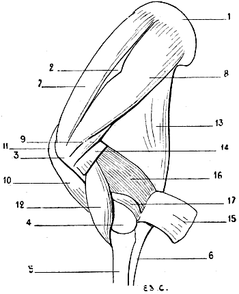
Fig. 72.—Myology of the Horse—Shoulder and Arm: Left Side, External Surface.
1, Cartilage of prolongation of the scapula; 2, tuberosity of the spine of the scapula; 3, superior extremity of the humerus; 4, inferior extremity of the humerus; 5, radius; 6, ulna; 7, supraspinatus muscle; 8, infraspinatus; 9, teres minor; 10, biceps; 11, tendon of the biceps passing over the anterior surface of the superior extremity of the humerus; 12, brachialis anticus; 13, triceps, long head; 14, external head of the triceps divided; 15, external head of the triceps reflected, in order to expose the anconeus; 16, region normally occupied by the external head of the triceps; 17, anconeus.
Teres Minor (Fig. 72, 9).—This muscle, also called in veterinary anatomy the short abductor of the arm, arises from the posterior border of the scapula (the external border in man), and is inserted below the great tuberosity of the humerus, between the attachments of the infraspinatus and deltoid.
It is covered by the deltoid and the infraspinatus.
Teres Major.—This muscle is known to veterinarians as the abductor of the arm; it arises from the postero-superior angle of the scapula (the inferior angle of the human scapula), from which it passes to be inserted into the internal surface of the humerus.
It is covered by the latissimus dorsi and the posterior muscular mass of the arm.
In brief, for the better understanding of the relations of the teres major and minor muscles in quadrupeds, we may fancy the corresponding muscles in man modified in the following manner: The infraspinatus, thicker, covering the teres minor; latissimus dorsi, more extended in its superior part, covering a large proportion of the teres major. As to the relations of the teres minor with the deltoid, they exist in man, seeing, in this case, the same muscle is, in its external portion, covered by this latter. With regard to the relations of the teres major with the posterior muscular mass of the arm, they also exist in man, since the external surface of this muscle is covered by the triceps.
These modifications are sufficient to render the small and large teres muscles completely invisible in the superficial layer.
The muscles of the shoulder which we have just been studying fulfil, with regard to the articulation which they surround, the function of active ligaments. This rôle is made necessary by the laxity of the scapulo-humeral capsule—a laxity which renders it incapable by itself of maintaining the bones in contact at this joint.
The same condition exists in man.
Panniculus Muscle of the Shoulder.—This thin muscle covers, as its name implies, the region of the shoulder, and is the continuation forward of the panniculus muscle of the trunk.
It arises, by its superior part, from the region of the withers and from the superior cervical ligament; thence its fibres descend directly towards the elbow, to terminate at the level of the region of the forearm.
The muscle is not found in the pig or in the carnivora.
Muscles of the Arm
We should remember, at the outset, that in man the muscles of the arm are divided into two groups: one anterior, which contains the biceps, brachialis anticus, and the coraco-brachialis; the other, posterior, which is constituted by a single muscle, the triceps.
In animals, we find them in the same number and arranged in analogous fashion—that is to say, in two groups—with respect to the bone of the arm. But then we find that they have undergone a transformation with regard to their length, and it is the change of general aspect which results from this modification that we proceed to examine.
We know that in quadrupeds, and especially in the domestic animals, the humerus is relatively short in proportion to the forearm. We have already seen, in dealing with the bones, that whilst in the human species the humerus is longer than the forearm, in the dog and cat these two segments of the fore-limb are of equal length, and that the humerus of the horse is, on the contrary, much shorter. Now, let us suppose the human humerus to be shorter than it is in reality; the anterior muscles undergoing, very naturally, the same reduction, will be uncovered only slightly by those above—the deltoid and the great pectoral—or will remain completely hidden by them. Thus would be found realized the disposition which we meet with in quadrupeds of the muscles of this region.
With regard to the posterior muscular mass of the arm, it does not undergo the same change. The muscle which constitutes it—the triceps cubiti—occupies, on the contrary, a greater area. Let us suppose, further—for it is the best method of comprehending the homologies which now occupy our attention—the humerus of man to be shortened as before, and directed downwards and backwards (as in quadrupeds), this bone would form an acute angle with the axillary border of the scapula. Let us suppose also that the long portion of the triceps, instead of arising solely from the superior part of this axillary border, is attached to the whole length of the latter, and that the triceps fills the whole interior of the angle formed by the arm and the shoulder. We then shall have an idea of what the triceps is in quadrupeds. It is necessary to add that the general resemblance would be still more complete if the arm were firmly supported by the side of the thorax, because in quadrupeds it occupies an analogous position, determined by the arrangement of the muscles which, proceeding from the trunk and neck, are attached to it.
Anterior Region
Biceps Cubiti (Fig. 68, 21; Fig. 72, 10, 11).—This muscle, also called the long flexor of the forearm, does not merit the name except by its analogy with the corresponding muscle in man. Indeed, in the domestic animals it is not divided into two parts; it is represented by a single fasciculus, long and fusiform, situated on the front of the humerus, and directed obliquely downwards and backwards, as the latter, on its part, is also inclined.
It arises above from a tubercle at the base of the coracoid process, which surmounts the glenoid cavity of the scapula. Its tendon, which is highly developed in the solipeds, occupies the bicipital groove. We remember that in these latter the groove in question is divided into two channels by a median prominence.
The tendon in which the muscle ends is inserted into a tuberosity, situated on the internal surface of the superior extremity of the radius—the bicipital tuberosity. In the pig, the cat, and the dog, there is detached from the tendon to which we have just referred a fasciculus of the same nature, which, after having wound round the radius, is inserted into the internal surface of the ulna, towards the base of the olecranon process. From the inferior part of the muscle arises a fibrous band, comparable to the aponeurotic expansion of the human biceps; but, instead of passing downwards and inwards, as does the latter, it terminates on the muscular mass which constitutes the antero-external part of the forearm.
The biceps is not seen in the superficial layer, except in the dog and cat (in which the humerus is, in fact, proportionately long); and even in them only to the slightest extent. It is covered partly in these latter, and completely in other animals, by the great pectoral and the inferior portion of the mastoido-humeral—that is to say, that part of the latter which represents the whole of the clavicular fibres of the human deltoid.
The biceps is a flexor of the forearm on the arm. It also contributes to the movement of extension of the humerus.
Brachialis Anticus (Fig. 68, 22; Fig. 69, 19; Fig. 70, 27; Fig. 72, 12).—In veterinary anatomy further designated as the short flexor of the forearm, this muscle, which is thick, occupies the musculo-spiral groove, and arises from it, reaching upwards to just below the head of the humerus. But it does not, as in man, extend to the internal surface of the bone.
Situated on the outside of the biceps, it is directed towards the forearm, and terminates by a flattened tendon, which, dividing into two slips, passes below the bicipital tuberosity, on the internal surface of the radius, into which one of these slips is inserted, while the other proceeds to terminate on the ulna.
The inferior half of this muscle is visible on the superficial layer, in the space limited posteriorly by the triceps brachialis, and below by the muscles of the forearm, which correspond to the external muscles of the human forearm, and in front by the great pectoral and the mastoido-humeral. It is in the upper part of the interspace which separates these latter from the brachialis anticus that the deltoid insinuates itself to proceed to its insertion into the humerus.
These relations precisely recall those which we meet with when we examine the external surface of the human arm, with this difference, however—that in the latter the anterior brachialis anticus is extensively related, in front, to the biceps. However, in animals it is not absolutely the same, since, as we have shown above, the biceps is covered, more or less completely, by the mastoido-humeral and the great pectoral.
The brachialis anticus flexes the forearm on the arm.
Coraco-brachialis.—In man this muscle, which occupies the superior half, or third, of the internal surface of the humerus, is visible only when the arm is abducted, and then especially when it approaches the vertical position; indeed, it is only in this attitude that the region which it occupies is accessible to view.
But an analogous attitude not being possible in domestic animals, in which the arm is fixed along the corresponding parts of the trunk, the result is that the coraco-brachialis is always covered, and that, consequently, it presents nothing of interest from our point of view. We speak of it, then, merely in order to complete the series of the muscles of the anterior surface of the arm, among which we rank it, in spite of the fact that in veterinary anatomy it is described as a muscle of the shoulder.
It arises above from the coracoid process, and thence passes downwards towards the internal surface of the humerus into which it is inserted, more or less high up, according to the species. The coraco-brachialis is an adductor of the arm.
Posterior Region
Triceps Cubiti (Fig. 68, 23, 24; Fig. 69, 20, 21; Fig. 70, 28, 29; Fig. 72, 13, 14, 15, 16).—This muscle, which is voluminous in the quadrupeds with which we are here concerned, fits more or less completely the angular space between the scapula and the humerus. Its bulk forms a thick prominence, which surmounts the elbow and the forearm.
We should say, with regard to this mass, that if the deltoid does not constitute in quadrupeds a prominence sufficient to remind one of that which this muscle produces in man, the triceps, in producing an analogous elevation, seems to replace in the general form of the body the relief which the deltoid is incapable of producing.
The triceps is divided into three portions, which, as in man, have the names middle, or long head; external and internal heads. But that which renders the nomenclature a little complicated is that veterinary anatomists have given other names to these three parts: that of great extensor of the forearm (caput magnum) to the long head; the short extensor of the forearm (caput parvum) to the external head; and of medium extensor of the forearm (caput medium) to the internal.[24]
It is more especially the long portion and the external head which, being visible on the external surface of the arm, contribute to the external form.
The long portion, which is triangular in shape and of considerable development, arises in the cat and the dog from the inferior half or two-thirds of the posterior border of the scapula (axillary border); from the whole extent of that border as far as the superior posterior angle in the pig, the ox, and the horse; it then passes downwards towards the articulation of the elbow, to terminate in a tendon which is inserted into the olecranon process. The portion of this muscle which is next the scapula is covered by the deltoid.
The external head, situated below the long portion, is directed obliquely downwards and backwards. It arises from the curved crest which, from the deltoid impression of the humerus, is directed upward to meet the articular head of the same bone. This crest limiting the musculo-spiral groove superiorly, and the brachialis anticus arising from the whole extent of this groove, the result is that at this level the external head is in relation with the brachialis anticus. From this origin it is directed towards the elbow, to be inserted into the olecranon, either directly or by the medium of the tendon of the long portion. The part of this muscle which arises from the humerus is covered by the deltoid.
As for the internal head (Fig. 76, 4), which, in the superficial layer, is only visible in its inferior part, on the internal aspect of the arm in those animals in which the elbow is free of the lateral wall of the thorax (the dog and the cat, for example), it arises from the internal surface of the humerus, and thence proceeds to be inserted into the olecranon.
The triceps extends the forearm on the arm.
A fourth muscle exists, which veterinary anatomists include in the study of the three portions of the triceps which we have just been discussing, in giving it the name of small extensor of the forearm. But, as this muscle is no other than the anconeus, and as, in human anatomy, we describe the latter, according to custom, in connection with the forearm, it is when on the subject of the latter that we will concern ourselves with it. This grouping of muscles cannot fail to give greater clearness to the description of the muscles of these regions.
The Supplemental or Accessory Muscle of the Latissimus Dorsi (Fig. 76, 2; Fig. 77, 1).—Because of the relations, to which we have already referred (see p. 142), of this muscle with the triceps cubiti, its description very naturally follows that of the latter.
Indeed, this supplementary muscle of the great dorsal is further designated in zoological anatomy under the name of long extensor of the forearm; and this name indicates that its study may be united to that of the triceps.
Situated on the internal surface of the arm, it arises from the external aspect of the tendon of the latissimus dorsi; it is very highly developed in the horse, in which it also arises from the posterior border (axillary) of the scapula; then, covering in part the internal head of the triceps and also the long portion, on the superior border of which it is folded, it proceeds to be inserted into the olecranon process and the anti-brachial aponeurosis.
It extends the forearm on the arm. Further, it makes tense the aponeurosis into which it is inserted; this explains the name of tensor of the fascia of the forearm, which is sometimes given to it.
It seems to us interesting to add that, abnormally, we sometimes find in man an analogue of this muscle. It is given off from the latissimus dorsi, near the insertion of the latter into the humerus; it accompanies the long head of the triceps and becomes fused with it. Sometimes it is inserted into the olecranon process, at other times into the antibrachial aponeurosis or the epitrochlea. It is on account of its insertion into the last-mentioned, in some cases, that it is also designated by the name of dorso-epitrochlear muscle.[25]
Muscles of the Forearm
Before commencing the special examination of each of the muscles of this region, it is absolutely indispensable to consider their general arrangement, and to determine very clearly how we should study them. We are too well convinced of the importance of this preliminary examination to dismiss it without entering rather fully into it. Indeed, the region on the myological study of which we are now entering is, unquestionably, one of the most complicated with which we have to deal. We know besides, in regard to the study of the forearm in man, how much a definite method is necessary in order that the arrangement of the muscles of this region be fixed in the memory, and that we are unable to obtain this result otherwise than by grouping the twenty muscles which constitute it in clearly defined regions.
We also know that these muscles are first studied with the forearm in the position of supination, and that it is only when they are well known after having considered them in this position that we are able to analyze and comprehend their forms when it is in pronation.
Now, as we have pointed out in the section on osteology (see p. 34), the forearm in quadrupeds is always in the position of pronation. Should we, then, in order to maintain the symmetry with human anatomy, first study the forearm in the position of supination? Evidently not. Besides the fact that this would in some cases be impossible since—as in the horse, for example—the radius and ulna are fused together, we should not gain any advantage; this position being never completely realizable even in those quadrupeds which have the radius relatively movable—as, for example, in the cat.
Accordingly, it is pronation which here, in connection with animals, becomes the standard attitude from the point of view of description. This is why, supposing that the reader knows well the muscles of the human forearm in the position of supination, we should recall what is the general arrangement occupied by these muscles when it is in pronation.
The fore-limb, being viewed on its anterior surface, presents above the anterior aspect of the region of the elbow; but below, it is the posterior surface of the wrist which is seen. Consequently, in the superior part, we see the external and anterior muscles limiting the hollow in front of the elbow; interiorly are found the posterior muscles.
The long supinator, passing obliquely downwards and inwards, divides, in fact, the forearm into two parts: one supero-internal, the other infero-external. In the first we see, but to an extent less and less considerable, the pronator teres, the flexor carpi radialis, the palmaris longus, and the flexor ulnaris; as to the flexors of the digits, on account of the rotation of the radius, they are only visible on the opposite surface—that is to say, on the surface of the wrist, which is now posterior. In the second part we see the two radial extensors, the common extensor of the fingers, the proper extensor of the little finger, and the ulnar extensor which, inferiorly, remains behind, by reason of the position of the ulna being unchanged, whilst the anconeus is wholly posterior, since the direction of the elbow is not modified. We also find, in this region, the long abductor of the thumb, the short extensor of the thumb, the long extensor of the thumb, and the special extensor of the index-finger, in the region where these deep muscles become superficial.
So that, to summarize, the external and posterior muscles occupy the anterior and external regions of the forearm, whilst the anterior muscles occupy rather the internal and posterior. It is in regarding them after this manner—that is to say, arranged in these two regions—that we proceed to study these muscles in quadrupeds.
Anterior and External Region
Supinator Longus.—We know that this muscle, which is especially a flexor of the forearm on the arm, plays, notwithstanding the name which has been given it, a part of but little importance in the movement of supination.
It acts slightly, however, as a supinator, for, being very oblique downwards and inwards at the time of pronation, it is able, while tending to resume its vertical direction, to carry the radius outwards; it places, in fact, the forearm in a position midway between pronation and supination.
We have just recalled these details, in order that it may be more easy to understand why it does not exist in animals in which the radius and ulna are fused together (horse, ox); and why, on the other hand, we find traces of it in the cat and the dog, in which the radius—to a slight extent, it is true—is able to rotate on the ulna. This displacement being a little more considerable in the felide, the long supinator is a little further developed than it is in the canine species; but, notwithstanding, it is only rudimentary.
The long supinator arises, above, from the external border of the humerus; thence, in the form of a very narrow fleshy band, it passes obliquely downwards and inwards, to be inserted into the inferior part of the internal surface of the radius.
It assists in turning the radius outwards and placing it in front of the ulna, the movement of supination being capable of being but little further extended.
First and Second External Radial Muscles: Extensor carpi radialis longior and brevior (Fig. 73, 8; Fig. 74, 8, 9; Fig. 75, 8, 9).—Fused together, these muscles form by their union what veterinary anatomists call the anterior extensor of the metacarpus. But we should add that these two muscles are united so much the more intimately as we examine them in passing successively from the cat to the dog, pig, ox, and horse. Thus, in the cat they are often distinct; in the dog, they unite only at the level of the middle third of the radius, and interiorly they have two tendons; in the pig, the ox, and the horse they are completely united, and there exists but a single tendon.
The anterior extensor of the metacarpus, which is situated behind the long supinator when the latter exists, occupies the external aspect of the forearm; its well-defined form absolutely recalls the prominence on the superior part of the external margin of the human forearm.
It arises superiorly from the portion of the external border of the humerus which is situated above the epicondyle and behind the musculo-spiral groove. Its fleshy mass appears in the angular space bounded by the brachialis anticus and the triceps. The superior portion is covered by the external head of the triceps; yet, in the dog, the superior portion of its humeral attachment is the only part so covered. This muscle is directed forward and downwards; it is also inclined a little inwards in such manner as to proceed to occupy the anterior aspect of the forearm.
Its fleshy belly is narrowed below, and, towards the inferior part of the forearm, is continued by a tendinous portion which is situated on the anterior surface of the carpus, after having traversed the median groove of the inferior extremity of the radius.
In the cat and the dog, in which the union of the two radial extensors is incomplete, the two tendons are inserted into the front of the base of the second and third metacarpal bones; consequently, as in man, into the metacarpals of the index and middle fingers.
In the ox, the tendon, which is single, is inserted into the internal and anterior half of the superior extremity of the principal metacarpal.
In the pig, this tendon is attached to the base of the large internal metacarpal.
In the horse, the corresponding tendon is attached to a tubercle which is situated on the anterior surface of the base of the principal metacarpal, a little internal to the median plane of the latter.
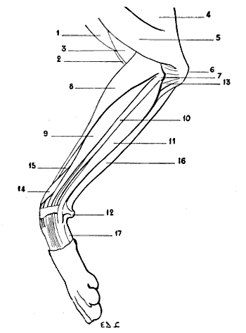
Fig. 73.—Myology of the Dog: Left Anterior Limb, External Aspect.
1, Mastoido-humeralis; 2, biceps; 3, brachialis anticus; 4, triceps, long portion; 5, triceps, external head; 6, olecranon process; 7, epicondyle; 8, radialis muscles (anterior extensor of the metacarpus); 9, extensor communis digitorum (anterior extensor of the phalanges); 10, extensor minimi digiti (lateral extensor of the phalanges, or common extensor of the three external digits); 11, posterior ulnar (external flexor of the metacarpus); 12, pisiform bone; 13, anconeus; 14, extensor ossis metacarpi pollicis and extensor primi internodii pollicis (oblique extensor of the metacarpus); 15, radius; 16, anterior ulnar (oblique flexor of the metacarpus); 17, external border of the hypothenar eminence (abductor of the little finger).
In order to properly understand and remember the respective positions occupied by these inferior insertions, it must be remembered that the human forearm being in the position of pronation, the tendons of the radials are attached to the bases of the metacarpals nearest to the thumb—that is to say, those occupying an internal position as regards the fourth and fifth metacarpals.
As its name indicates, this muscle extends the metacarpus. Consequently it is, in the horse, an extensor of the canon-bone.
It is also an adductor of the hand in those animals (cat, dog) in which the radio-carpal articulation, analogous in form to the corresponding articulation in man, permits lateral movements of the hand on the forearm. The union of the fleshy bodies of the two radials is sometimes found in the human species.
Supinator Brevis.—As in the case of the long supinator, the short supinator is found only in animals in which the radius can be rotated to a greater or less extent around the ulna; therefore this muscle is not found in the pig, the ox, or the horse; but it forms part of the forearm of the cat and the dog.
Deeply situated at the region of the elbow, the short supinator has little interest for us. All that we will say of it is that it goes from the external part of the inferior extremity of the humerus to the superior part of the radius; and that it is, in carnivora, the essential agent in the production of the movement of supination.
Extensor Communis Digitorum (Fig. 73, 9, 10, 11; Fig. 74, 10, 11, 12).—Also named in veterinary anatomy the anterior extensor of the phalanges, this muscle is situated external to and behind the anterior extensor of the metacarpus already described.
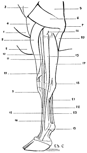
Fig. 74.—Myology of the Ox: Left Anterior Limb, External Aspect.
1, Mastoido-humeralis; 2, pectoralis major; 3, deltoid; 4, brachialis anticus; 5, triceps; 6, triceps, external head; 7, olecranon; 8, radial extensors (anterior extensor of the metacarpus); 9, insertion of the tendon of the anterior extensor of the metacarpus to the tubercle of the superior extremity of the principal metacarpal; 10, 11, extensor communis digitorum (10, proper extensor of the inner digits; 11, common extensor of the two digits); 12, tendon of the common extensor of the two digits; 13, band of reinforcement from the suspensory ligament of the fetlock; 14, external tuberosity of the superior extremity of the radius; 15, extensor minimi digiti (proper extensor of the external digit); 16, tendon of the proper extensor of the external digit; 17, posterior ulnar (external flexor of the metacarpus); 18, pisiform; 19, extensor ossis metacarpi pollicis and extensor primi internodii pollicis (oblique extensor of the metacarpus); 20, ulnar portion of the deep flexor of the toes; 21, tendon of the superficial flexor of the toes (superficial flexor of the phalanges); 22, tendon of the deep flexor of the toes (deep flexor of the phalanges); 23, suspensory ligament of the fetlock.
In the human being, the common extensor of the fingers springs, in its superior part, from the bottom of a depression, situated on the outer side of and behind the elbow, and limited in front by the muscular prominence which the long supinator and the first radial extensor form at that level. At the bottom of this hollow or fossette is found the epicondyle, which gives origin, amongst other muscles, to the common extensor of the fingers. It is necessary to add that it is most prominently visible during supination, and that it tends to be effaced during pronation.