Chapter II · (continued 2)
Reading edition
An analogous arrangement is met with in animals. But the muscular prominence is formed by the united radial extensors, and the fossette, because of the permanent pronation of the forearm, is scarcely recognisable. Likewise, with regard to the dog, we may say that it does not exist, on account of the prominence which the epicondyle forms in that animal (Fig. 73, 7).
In connection with this prominence of the epicondyle, it is interesting to add that this detail recalls the relief which the same process produces on the external aspect of the human elbow when the forearm is flexed on the arm. We know that, in this case, the epicondyle is exposed, because the muscles which mask it in supination (long supinator and long radial extensor) are displaced and set it free during flexion. But, in the dog, as in other quadrupeds besides, the forearm is, in the normal state, flexed on the arm; the latter being oblique downwards and backwards, and the former being vertical. Further, the epicondyle is well developed.
The muscle with which we are now occupied, long and vertical in direction, arises from the inferior part of the external border of the humerus (there it is covered by the anterior extensor of the metacarpus, from which it is freed a little lower down) and from the external and superior tuberosity of the radius. In the carnivora, it arises from the epicondyle. Its fleshy body is fusiform in shape, becomes tendinous in the lower half of the forearm, and then divides into a number of slips, varying in number according to the species; this division is correlated to that of the hand—that is to say, with the number of the digits. Before reaching this latter, the common extensor of the digits passes through the most external groove on the anterior surface of the inferior extremity of the radius.
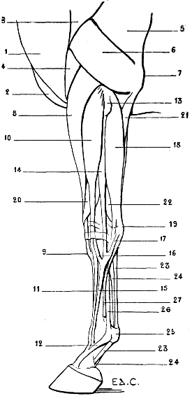
Fig. 75.—Myology of the Horse: Left Anterior Limb, External Aspect.
1, Mastoido-humeral; 2, pectoralis major; 3, deltoid; 4, brachialis anticus; 5, triceps, long head; 6, triceps, external head; 7, olecranon; 8, radial extensors (anterior extensor of the metacarpus); 9, insertion of the tendon of the anterior extensor of the metacarpus into the tubercle of the superior extremity of the principal metacarpal; 10, extensor communis digitorum (anterior extensor of the phalanges); 11, tendon of the anterior extensor of the phalanges; 12, reinforcing band arising from the suspensory ligament of the fetlock; 13, external tuberosity of the superior extremity of the radius; 14, extensor minimi digiti (lateral extensor of the phalanges); 15, tendon of the lateral extensor of the phalanges; 16, fibrous band which this latter receives from the tendon of the anterior extensor of the phalanges; 17, fibrous band which the same tendon receives from the carpal region; 18, posterior ulnar (external flexor of the metacarpus); 19, pisiform; 20, extensor ossis metacarpi pollicis and extensor primi internodii pollicis (oblique flexor of the metacarpus); 21, ulnar portion of the deep flexor of the digits; 22, flexor digitorum profundus; 23, 23, tendon of the flexor digitorum sublimis (superficial flexor of the phalanges); 24, 24, tendon of the flexor digitorum profundus (deep flexor of the phalanges); 25, sesamoid prominence; 26, suspensory ligament of the fetlock; 27, external rudimentary metacarpal.
In the cat and the dog, the four tendons which result from the division of the principal tendon go to the four last digits, and each of them is inserted, as in the human species, to the second and third phalanges.
In the pig, the anterior extensor of the phalanges is rather complicated in its arrangement. Its fleshy body is divided into four bundles terminated by tendons, which in turn divide and join certain digits; whence the special names given to each of these fasciculi, commencing with the most internal, of: proper extensor of the great inner toe; common extensor of the two inner toes; common extensor of the two outer toes; and proper extensor of the great outer toe.
In the ox, the same muscle is divided into two bundles: the internal proceeds to the internal toe, the external is common to the two toes.
In the horse, the tendon of the anterior extensor of the phalanges is divided into two parts of unequal bulk. The smaller of these tendinous slips, which is the more external, unites at the level of the superior part of the metacarpus with the tendon of the muscle which we are about to study in the following paragraph (Fig. 75, 16). The larger, after having reached the anterior surface of the digit, is attached to the anterior aspect of the first and second phalanges, and then forms a terminal expansion which is inserted into the pyramidal eminence of the third.
At the level of the first phalanx this tendon receives on each of its lateral aspects a strengthening band, which proceeds from the terminal extremity of the suspensory ligament of the fetlock,[26] and crosses obliquely downwards and forwards over the surface of the first phalanx to join the extensor tendon (Fig. 75, 12).
A similar arrangement is found in the ox.
This band is noticeable under the skin which covers the lateral aspects of the ham.
As the name indicates, this muscle extends the phalanges, one upon the other. It also contributes to the extension of the hand, as a whole, on the forearm.
Extensor Minimi Digiti (Fig. 73, 10; Fig. 74, 15, 16; Fig. 75, 14, 15).—This muscle, the lateral extensor of the phalanges of veterinary anatomy, situated on the external surface of the forearm, behind the common extensor of the digits, arises, as a rule, from the epicondyle (dog, cat), or from the external surface of the superior extremity of the radius (horse). The tendon succeeding to the fleshy body appears towards the lower third of the forearm, and at the level of the wrist lies in a groove analogous to that which in man is hollowed out for the passage of the corresponding tendon at the level of the inferior radio-ulnar articulation. This groove corresponds to the same articulation in animals in which the ulna is well developed, such as the dog and the cat; but it belongs to the radius when the inferior extremity of the ulna does not exist—for example, in the horse. Indeed, in this animal the groove in question is found on the external surface of the carpal extremity of the radius.
In the dog, the tendon is divided into three parts, which, crossing obliquely the tendons of the common extensor of the digits, pass to the three external digits, to be inserted by blending with the corresponding tendons of the latter into the third phalanges of those digits.
Thus is explained the name of common extensor of the three external digits which is sometimes given to this muscle.
In the cat, there is a fourth tendon, which passes to the index-finger, so that the name common extensor of the four external digits is in this case legitimate, and the lateral extensor of the phalanges is also a common extensor, as is the anterior extensor of the phalanges, or common extensor of the digits.
In the pig, the tendon, which is single, is inserted into the external digit, for which reason it has received the name of the proper extensor of the small external digit. This muscle is, then, really the homologue of that which exists in the human species.
In the ox, it is called the proper extensor of the external digit; it is as thick as the common extensor.
Finally, in the horse, the muscle is little developed. Its fleshy body, thin and flattened from before backwards, becomes distinctly visible only below the middle of the forearm. Above, it is enclosed in a limited space, bounded in front by the common extensor of the digits, and behind by the posterior ulnar; there these two muscles approach each other so closely that from the point of view of external form they seem to be nearly in contact.
The tendon, after receiving the small fasciculus from the common extensor (Fig. 75, 16), as well as a fibrous band emanating from the external surface of the carpus (Fig. 75, 17), is situated at the external side of the tendon of the anterior extensor of the phalanges, and is inserted into the anterior surface of the superior extremity of the first phalanx.
This muscle extends the digit or digits into which it is inserted. It also assists in the movement of extension of the hand as a whole.
Posterior Ulnar (Extensor carpi ulnaris) (Fig. 73, 11; Fig. 74, 17; Fig. 75, 18).—Designated by veterinary anatomists as the external flexor of the metacarpus,[27] or external cubital, this muscle is situated in the posterior region of the external surface of the forearm, behind the lateral extensor of the phalanges.
It arises from the epicondyle; its fleshy body, thick but flattened, is directed vertically towards the carpus, and its tendon is inserted into the external part of the superior extremity of the metacarpus, after having given off a fibrous band, which takes its attachment on the pisiform.
It is inserted, in the cat and the dog, into the superior extremity of the fifth metacarpal; in the pig to the external metacarpal; in the ox to the external side of the canon-bone; in the horse to the superior extremity of the external rudimentary metacarpal.
This muscle flexes the hand on the forearm, and in animals in which the radio-carpal articulation permits, by its formation, it inclines the hand slightly outwards—that is, abducts it.
Anconeus (Fig. 72, 17; Fig. 73, 13).—We have already stated (p. 174) that the anconeus is included with the triceps brachialis in zoological anatomy, and that veterinary anatomists give it the name of small extensor of the forearm.[28]
In the dog it recalls, as to position, the human anconeus, but with this difference—that, in the latter, the anconeus, triangular in outline, has one of its angles turned outwards (the epicondyloid attachment) and one of its sides turned towards the olecranon. Here it is entirely the opposite. The anconeus, similarly triangular, is broader externally. At this level it takes its origin from the external border of the humerus, the epicondyle, and the external lateral ligament of the articulation of the elbow; thence its fibres converge towards the external surface of the olecranon, to be there inserted.
It is in relation, anteriorly and inferiorly, with the posterior ulnar muscle. It is covered superiorly by the external head of the triceps. In the cat the disposition of the anconeus is analogous. But in the other quadrupeds with which we are here concerned it is completely covered by the external head of the triceps. It really participates in the formation of the triceps; and seeing that it takes origin from the posterior surface of the humerus at the margin of the olecranon fossa (Fig. 72), and proceeds thence towards the olecranon to be inserted, we can understand why veterinary anatomists have connected its study with that of the posterior muscular mass of the arm.
This muscle is an extensor of the forearm on the arm.
We proceed now to inquire what the deep muscles of the posterior region of the human forearm become in quadrupeds: the long abductor of the thumb, the short extensor of the thumb, the long extensor of the thumb, the proper extensor of the index. We know that in every instance these muscles, which are deeply seated at their origin, become superficial afterwards.
In quadrupeds, on account of the position in which the forearm is placed—viz., pronation—the corresponding muscles occupy the anterior aspect of this region.
Long Abductor of the Thumb (Extensor ossis metacarpi pollicis) and Short Extensor of the Thumb (Extensor primi internodii pollicis) (Fig. 73, 14; Fig. 74, 19; Fig. 75, 20).—United one to the other in man, blended in quadrupeds, they form in the latter the muscles to which veterinary anatomists give the name of oblique extensor of the metacarpus.
This muscle arises from the median portion of the skeleton of the forearm. There it is covered by the common extensor of the digits and that of the small digit (anterior extensor and lateral extensor of the phalanges). Then, at the internal border of the first of these muscles, it becomes superficial, passes downwards and inwards, crosses superficially the anterior extensor of the metacarpus, reaches the inferior extremity of the radius, and becomes lodged in the most internal of the grooves situated on the anterior surface of this extremity, passes on the internal side of the carpus, and is inserted into the superior extremity of the most internal metacarpal—that is, to the first metacarpal, or metacarpal of the thumb—in the dog and cat; to the internal rudimentary metacarpal in the horse.
It is an extensor of the metacarpal into which it is inserted; but as, if we recall the extreme examples given above, in the dog the first metacarpal is not very mobile, and in the horse the internal rudimentary metacarpal is absolutely fixed to the bone which it accompanies, it is more exact to add that this muscle is principally an extensor of the metacarpus as a whole.
And yet, in the cat and the dog, it is also able to adduct the first metacarpal bone. It must be understood that this movement would be abduction, if the hand could be placed in the position of complete supination, as in the human species.
Long Extensor of the Thumb (Extensor secundi internodii pollicis) and Proper Extensor of the Index (Extensor indicis).—These two muscles are blended together by their fleshy bodies, so that the single name of proper extensor of the thumb and index is preferable. This muscle is but of slight importance from our point of view, for it is extremely atrophied, and so much the more as the number of the digits is lessened.
It arises, as the preceding, from the skeleton of the forearm, and there it is deeply placed. Below, towards the carpus, its tendinous part becomes superficial, to end in the following manner:
In the carnivora, the tendon divides into two very slender parts, which are inserted into the thumb and the index. In the pig, the tendon is blended with that of the common extensor of the internal digits. Finally, in the ox and the horse, it is sometimes regarded as being blended with the common or anterior extensor of the phalanges. But to us it appears more rational to say that it does not exist, which, moreover, is explained by the digital simplification of the hand.
Internal and Posterior Region
Pronator Teres (Fig. 76, 8).—This muscle, as may easily be understood, undergoes, as do the supinators, a degree of degeneration in proportion to the loss of mobility of the radius on the ulna. In animals in which the bones of the forearm are not fused it exists; in those, on the other hand, in which this segment has become simply a supporting column, it is not developed—at least, in a normal manner.
It is, consequently, found best marked in the dog and the cat.
Forming, as in man, the internal limit of the hollow of the elbow, the pronator teres has a disposition analogous to that which characterizes the corresponding muscle in the human species. It arises from the epitrochlea (internal condyle), proceeds downwards and outwards, and is inserted into the middle portion of the body of the radius.
It is into the hollow in front of the elbow, which this muscle contributes to limit, that the biceps and the brachialis anticus dip.
In the pig and the ox it is atrophied.
In the horse it does not exist. We may, however, sometimes find it, but in an abnormal form. We were able to demonstrate its presence in the form of a fleshy tongue situated on the internal side of the elbow (Fig. 78) in a horse which we dissected many years ago in the laboratory of the School of Fine Arts. Moreover—and the fact seemed to us an interesting one—the forearm to which the muscle belonged had an ulna of relatively considerable development (Figs. 79 and 80).[29]
This muscle is a pronator.
Flexor Carpi Radialis (Fig. 76, 10; Fig. 77, 7).—Called by veterinary anatomists the internal flexor of the metacarpus, this muscle, which is found on the internal aspect of the forearm, is situated behind the pronator teres when this muscle exists, whilst in the animals which are deprived of the latter the flexor carpi radialis has in front of it the internal border of the radius, which separates it from the anterior extensor of the metacarpus.
It is necessary to add that the flexor carpi radialis is similarly separated from the anterior extensor of the metacarpus by the internal border of the radius in animals in which the pronator teres exists, but then only in that part of the forearm which is situated below this latter.
The flexor carpi radialis arises from the epitrochlea. Its fleshy body, fusiform in shape, descends vertically, and terminates in a tendon on the posterior surface of the bases of the second and third metacarpals in the dog and the cat, on the metacarpal of the large internal digit in the pig, on the internal side of the metacarpus in the ox, and on the superior extremity of the internal rudimentary metacarpal in the horse.
We see clearly, in this latter, a superficial vein which, in the shape of a strong cord, passes along the anterior border of the flexor carpi radialis; it is the subcutaneous median or internal vein, which, forming the continuation of the internal metacarpal vein, joins the venous system of the arm, after having crossed obliquely the corresponding part of the radius.
Palmaris Longus.—This muscle, which exists distinctly in some animals, but whose absence is far from being rare in the human species, is not developed as a distinct muscle in any of the domestic quadrupeds.
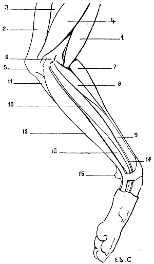
Fig. 76.—Myology of the Dog: Left Anterior Limb, Internal Aspect.
1, Biceps; 2, long extensor of the forearm (supplementary or accessory muscle of the great dorsal); 3, triceps, long head; 4, triceps, internal head; 5, olecranon; 6, epitrochlea (internal condyle); 7, radial extensors (anterior extensor of the metacarpus); 8, pronator teres; 9, radius; 10, flexor carpi radialis (internal flexor of the metacarpus); 11, anterior ulnar (oblique flexor of the metacarpus); 12, superficial flexor of the digits; 13, deep flexor of the digits; 14, flexor longus pollicis (radial fasciculus of the deep flexor of the digits); 15, pisiform bone.
And yet some authors announce its presence in the dog, and describe it as becoming detached, in the form of a cylindrical bundle, from the anterior surface of the fleshy mass of the deep flexor of the digits (see p. 196) to proceed then by a tendon which divides into two parts, to terminate in the palm of the hand, where it blends with the tendons of the superficial flexor, which are destined for the third and fourth digits.
These authors give to this muscle the name of palmaris longus, and attribute to it the action of flexing the hand.
Anterior Ulnar (Flexor carpi ulnaris) (Fig. 73, 16; Fig. 76, 11; Fig. 77, 8).—Called by veterinary anatomists the oblique flexor of the metacarpus, or internal ulnar, this muscle occupies the internal part of the posterior aspect of the forearm in the ox and the horse, while in the dog it occupies rather the external part.
This difference arises from the fact that in this latter, as in man, the anterior ulnar is separated from the flexor carpi radialis by an interval in which we see, on the internal aspect of the forearm, just at the level of the elbow, the flexors of the digits. This interval is so much the wider as there is no palmaris muscle to subdivide its extent (Fig. 81). In the horse, the interval in question does not exist. In this animal, indeed, the anterior ulnar is in contact with the radial flexor, so that this muscle can occupy only a region belonging rather to the internal surface of the forearm (Fig. 82).
In the dog the anterior ulnar is in contact with the posterior ulnar. This relation recalls that which is found in man, where the two muscles are merely separated by the crest of the ulna (Fig. 81). But in the horse, in which the anterior ulnar has, so to speak, slid towards the internal aspect, this muscle is separated above from the posterior ulnar, and it is in the interval separating these two muscles that we are able to perceive, but this time at the back of the forearm, the muscular mass of the flexors of the digits (Fig. 82).
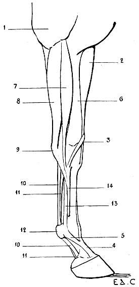
Fig. 77.—Myology of the Horse: Anterior Limb, Left Side, Internal Aspect.
1, Long extensor of the forearm (supplementary or accessory muscle of the latissimus dorsi); 2, radialis muscles (anterior extensor of the metacarpus); 3, tendons of extensor ossis metacarpi pollicis extensor primi internodii pollicis united (oblique extensor of the metacarpus); 4, tendon of extensor communis digitorum (anterior extensor of the phalanges); 5, strengthening band from the suspensory ligament of the fetlock; 6, internal surface of the radius; 7, flexor carpi radialis (internal flexor of the metacarpus); 8, anterior ulnar (oblique flexor of the metacarpus); 9, pisiform bone; 10, 10, tendon of the superficial flexor of the digits (superficial flexor of the phalanges); 11, 11, tendon of the deep flexor of the digits (deep flexor of the phalanges); 12, sesamoid prominence; 13, suspensory ligament of the fetlock; 14, internal rudimentary metacarpal.
The anterior ulnar arises above from the epitrochlea and the olecranon; thence it is directed towards the carpus, to be inserted into the pisiform bone. It proceeds therefore from the inner side of the elbow to the outer side of the upper part of the hand; it consequently crosses the posterior surface of the forearm obliquely. This is why, as we have pointed out above, it receives the name of the oblique flexor of the metacarpus.
It is not unprofitable to recall in this connection that there is an internal flexor of the metacarpus, which is the flexor carpi radialis; and an external flexor of the metacarpus, which is the posterior ulnar (in human anatomy, extensor carpi ulnaris). It is between these two muscles that we find the oblique flexor—the anterior ulnar which we have just been studying.
This muscle flexes the hand on the forearm.
Superficial Flexor of the Digits (Flexor digitorum sublimis) (Fig. 76, 12; Fig. 77, 10, 10).—This muscle arises from the epitrochlea; thence it passes towards the hand, becomes tendinous, passes in a groove on the posterior aspect of the carpus, and terminates on the palmar surface of the phalanges in furnishing a number of tendons proportioned to the digital division of the hand. Whatever the number, to which we will again refer, each tendon is attached to the second phalanx, after bifurcating at the level of the first, so as to form a sort of ring, destined to give passage to the corresponding tendon of the deep flexor. This ring and this passage have gained for the muscle the name of perforated flexor.
In the dog and the cat the principal tendon is divided into four parts, which go to the four last digits.
In the ox it is divided into two parts only; as, moreover, in the pig, whose superficial flexor is destined for the two large digits only, the lateral digits receiving no part of it.
Finally, in the horse the tendon is single.
We have previously pointed out that in the carnivora this muscle is visible on the internal and posterior aspects of the forearm, in the interval which is limited in front by the flexor carpi radialis and behind and outside by the anterior ulnar.
Certain details are still to be added to the description of this muscle. We will enter on an analysis of them after we have given some indications relative to the following muscle:
Deep Flexor of the Digits (Flexor digitorum profundus) (Fig. 75, 21, 22; Fig. 76, 12; Fig. 77, 11, 11).—This muscle is covered by the superficial flexor. It arises from the epitrochlea, from the radius, and from the ulna, either from the olecranon process—as in the ox, pig, and horse—or from almost the whole extent of the shaft of the same bone, as in the cat and dog.
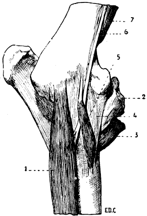
Fig. 78.—Left Anterior Limb of the Horse: Internal Aspect.
1, Internal flexor of the metacarpus or great palmar; 2, inferior part of the biceps; 3, inferior part of the brachialis anticus; 4, internal lateral ligament of the elbow; 5, pronato teres muscle.
The radial fasciculus represents in the domestic quadrupeds the long proper flexor muscle of the thumb in man. For this reason we shall describe the muscle afresh in the following paragraph:
The fleshy bundles of which we have just spoken terminate in a tendon which afterwards divides into slips, the number of which is in proportion to the digital division of the hand. These slips then pass through the slit or buttonhole in the tendon of the superficial flexor, and proceed to terminate on the third phalanx; hence the name of perforating, which is also given to the deep flexor of the digits.
In the dog and the cat the tendon is divided into five portions, each of which proceeds to one of the digits. The internal tendon, which is destined for the thumb, terminates on the second phalanx of this digit.
In the pig the tendon divides into four tendons destined for the four digits.
In the ox there are but two tendons.
In the horse the tendon is single.
As their names indicate, these muscles, both superficial and deep, flex the digits. In addition to this, they flex the hand on the forearm.
We mentioned above that certain details relative to the superficial flexor must be analyzed in a special way. We now add that this should also be done with regard to the deep flexor. The point in question is the arrangement which the tendons of these muscles present at the level of the palmar region of the hand.
It is easy, in the case of the dog or the cat, to picture to one’s self this arrangement, especially if we recollect that which exists in the human species. The tendons of the flexors are placed on a kind of muscular bed formed by the union of the muscles of the region, but, moreover, from the point of view of external form, these tendons are not of very great importance.
But in the ox and the horse it is quite otherwise. From the simplification of the skeleton of the hand, and the reduction of the number of movements which the bones that form it are able to execute, there naturally results a diminution of its muscular apparatus. Apart from the existence of muscular vestiges of but little importance, we can say that, in reality, the hand does not possess any muscles. On its palmar aspect are found only the tendons of the flexors of the digits, and as these tendons are large, and the hand long, they give origin to external forms which it is necessary to examine.
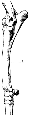
Fig. 79.—Left Anterior Limb of the Horse: External Aspect.
1, Ulna of abnormal length.
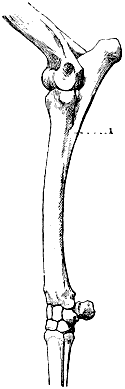
Fig. 80.—Left Anterior Limb of the Horse: External Aspect.
1, Normal ulna.
In the horse, which we take as a type, the tendons of the flexors, after being retained in position at the carpus by a fibrous band, the carpal sheath, which recalls the anterior annular ligament of the human carpus, and having passed this region, descend vertically, remaining separated from the posterior surface of the metacarpus, so that the skin sinks slightly on the lateral parts in front of the thick cord which these tendons form. This cord is known by the name of tendon.
The flexors then reach the fetlock, and occupy the groove formed by the peculiar arrangement of the two large sesamoid bones. They are retained in position at this level by a fibrous structure, which forms the metacarpo-phalangeal sheath. They then reach the phalanges, being directed obliquely downwards and forwards, as, moreover, the latter are also inclined. Then the tendon of the superficial flexor divides into two slips, which are inserted into the second phalanx, between which slips passes the tendon of the deep flexor, which in its turn goes to be inserted, in the form of an expansion, into the semilunar crest, by which the inferior surface of the third phalanx is divided into two parts.[30]
The part which these tendons play is of great importance in the large quadrupeds.
These tendons, in fact, in addition to the action determined by the contraction of the fleshy fibres to which they succeed, maintain the angle formed by the canon-bone and the phalangeal portion of the hand, and prevent its effacement under the weight of the body during the time of standing. Their strong development, and the position they occupy, make this understood, without it being necessary to insist on it further.
We mentioned above that the ‘tendon’ descends vertically from the carpus towards the fetlocks. This is as it should be. But, in some horses, it is oblique downwards and backwards, so that the canon, instead of being of equal depth from before backwards in its whole length, is a little narrower in its upper part.
This results from the fact that the tendons of the flexors, too firmly bound by the carpal sheath, gradually separate as they pass from the metacarpus, going to join the fetlock; hence the obliquity pointed out above. This abnormality producing a deleterious result, in the sense that the tendinous apparatus acts with less strength as an organ of support, it constitutes a defect of conformation which is expressed by saying that the tendon has ‘failed.’
Long Proper Flexor of the Thumb (Flexor longus pollicis) (Fig. 76, 14).—As we have already pointed out, this muscle is represented in quadrupeds by the radial bundle of the deep flexor of the digits, so that the two muscles are in reality blended the one to the other. This union is sometimes found, but only as an abnormality, in the human species. We have met some examples of this in the course of our dissections.
Pronator Quadratus.—This muscle conforms to the general law which we have already pointed out in connection with those which have for their action the rotation of the radius around the ulna. We remember, indeed, that when the bones of the forearm are fused with one another, the muscles which are destined to produce a mobility which has then become impossible disappear at the same blow.
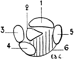
Fig. 81.—Diagram of the Posterior Part of a Transverse Section passing through the Middle of the Left Fore-limb of the Dog: Surface of the Inferior Segment of the Section.
1, Radius; 2, ulna; 3, posterior ulnar; 4, anterior ulnar; 5, great palmar (flexor carpi radialis); 6, flexors of the digits.
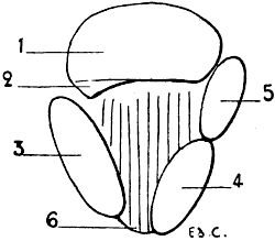
Fig. 82.—Diagram of a Horizontal Section of the Middle of the Forearm of the Left Leg of the Horse: Surface of the Inferior Segment of the Section.
1, Radius; 2, ulna; 3, posterior ulnar; 4, anterior ulnar; 5, great palmar (flexor carpi radialis); 6, flexors of the digits.
For this cause we do not find the square pronator in either the ox or the horse, but can demonstrate its presence in the dog and the cat.
It is very deeply situated. This is why, and also on account of the plan which we have traced for ourselves, we will simply say that it is situated on the postero-internal aspect of the skeleton of the forearm, and that it extends from the ulna to the radius.
It seems to us, however, sufficiently interesting to add that, instead of occupying, as in the human species, the inferior fourth of the two bones, it extends, particularly in the dog, over their whole length, with the exception of their superior and inferior extremities.
Muscles of the Hand
We will first recall that, in man, the palm of the hand is divided into three regions: a median palmar region, which is occupied by the tendons of the flexors of the digits, the lumbricales, and, deeply, by the interosseous muscles; an external region, or thenar eminence, formed by the muscles destined for the movements of the thumb; an internal region, or hypothenar eminence, which contains the muscles proper to the small digit and the palmar cutaneous muscle.
These muscles are found, more or less developed, in the dog and the cat.
In the ox and the horse we meet with no vestige of the muscles of the thenar or hypothenar eminences. Nevertheless, in these animals we find the muscles which belong to the central region of the palm. We refer to the lumbricales and the interosseous.
Although this fact has no relation to the object of our study, it appears to us interesting to announce that there are traces of the lumbricales found in the solipeds. These muscles are represented by two fleshy bundles, situated one on each side of the tendon of the deep flexor, above the ring of the tendon of the superficial flexor. These small muscles are continued as slender tendons, which become lost in the fibrous tissue of the spur, which is the horny process situated at the posterior part of the fetlock, and which is covered by the hairs, more or less abundant, which constitute the wisp.
As for the interosseous muscles, they are represented by the suspensory ligament of the fetlock, and by two other small muscles, tendinous throughout, which are situated between the principal metacarpal and the rudimentary ones.
The suspensory ligament of the fetlock is considered an interosseous muscle, on account of the red fleshy striations which it contains, and from certain relations which it forms with the tendon of the common extensor of the digits or anterior extensor of the phalanges. This ligament (Fig. 75, 26; Fig. 77, 13), which plays an important part in the standing position as a support of the foot, is a fibrous band situated between the tendons of the flexors of the digits and the principal metacarpal. It arises above, from the second row of the carpals, descends towards the fetlock, where it divides into two branches, which are inserted into the large sesamoid bones. At the same level, this ligament gives off two fibrous bands which, passing downwards and forwards, join the tendon of the anterior extensor of the phalanges, blending with it, after having each crossed one of the lateral aspects of the pastern. We have already referred to these bands (p. 183).
It is with these latter that are blended the long and slender tendons which form in a great measure the two other interosseous muscles previously described.
A ligament of the same kind is found in the ox (Fig. 74, 23).
MUSCLES OF THE POSTERIOR LIMBS
Muscles of the Pelvis
The muscles which specially interest us in this region, because of their superficial position, are the gluteus maximus and the gluteus medius. As for the gluteus minimus, it is deeply situated, and more or less sharply marked off from the second of the preceding muscles.
Inasmuch as the gluteus medius is more simple in arrangement than the maximus, and will aid us in arranging our ideas in connection with the latter, it is with the study of it that we will commence.
Gluteus Medius (Fig. 68, 29; Fig. 69, 26; Fig. 70, 35).—This muscle, as in man, occupies the external iliac fossa. But this latter being directed differently in the digitigrades and the ungulates, as we have pointed out in the section on osteology (see pp. 91 and 99), the muscle in question has consequently not the same direction in the two groups of animals, being turned outwards in the first, and upwards in the second.
It is the thickest of the glutei, and gives to the region which it occupies a rounded form.
From the iliac fossa from which it arises the fleshy fibres are directed towards the femur, to be inserted into the great trochanter. It is covered by an aponeurosis, and in part by the great gluteal. It completely covers the small gluteal, which veterinary anatomists designate by the name of the deep gluteal.
In the carnivora it does not pass in front of the iliac crest, but, in the ox, and more particularly in the horse, it is prolonged anteriorly, and thus covers, to a certain extent, the muscles of the common mass.
When it contracts, taking its fixed point at the pelvis, the gluteus medius extends the thigh, which it is also able to abduct. If, on the other hand, its fixed point is on the femur, it acts on the trunk, which it raises, producing oscillating movements of the pelvis. It contributes in this way to the action of rearing. We also see it distinctly appear by the prominence which it produces in the dog, which, according to the time-honoured phrase, fait le beau.
Gluteus Maximus (Fig. 68, 28; Fig. 69, 25; Fig. 70, 33, 34).—The great gluteal muscle, further designated in veterinary anatomy the superficial gluteal, is proportionately less developed in quadrupeds than in man. Indeed, in the latter, where it is of very great thickness, its volume is due to the important function which it fulfils in maintaining the biped attitude.
In quadrupeds it contributes to form the superficial part of the crupper and the external surface of the thigh. It is divided into two parts: one anterior, the other posterior.
With regard to this latter, it will be necessary to indicate how it has been sometimes regarded, and to what portion of the muscular system in man it corresponds. But we believe that it is better to see beforehand, without any preconceived idea, how these two parts are arranged.
In the dog, the anterior portion of the gluteus maximus arises from the sacrum, while some fibres situated further forward arise from the surface of the gluteus medius, near the iliac spine, and from the tensor of the fascia lata with which these fibres are blended. The posterior portion, united to the preceding—that is to say, to those of its fibres which arise from the sacrum—takes its origin from the first coccygeal vertebra. These two portions are directed towards the femur, to be inserted into the great trochanter, and to the external branch of the superior bifurcation of the linea aspera.
In the cat, the posterior bundle is less definitely blended with the anterior. By a long and slender tendon which, behind, turns around the great trochanter, and passes along the surface of the fascia lata, it proceeds to join the knee-cap.
In the pig, the posterior portion is much more developed.
In the horse, the anterior portion arises from the internal iliac spine (posterior in man), from the external iliac spine (anterior in man), and, between these two osseous points, from the aponeurosis, which covers the gluteus medius. Between these two origins the muscle is deeply grooved, so that the tendency is to divide into two portions, each of which is directed towards one of the iliac angles. In this groove the gluteus medius is to be seen.
The fleshy bundles converge, and are directed towards the external aspect of the femur, to be inserted into the osseous prominence known as the third trochanter, after passing beneath the fleshy fibres of the posterior portion. The latter, which is more considerable than the preceding portion, arises above from the sacral crest, from the aponeurosis which envelops the coccygeal muscles, from the sacro-sciatic ligament, and from the tuberosity of the ischium. From this origin it passes downwards, expands, then, describing a curve with the convexity behind, it becomes narrowed, and proceeds to be inserted by a deep fasciculus into the third trochanter, to the fascia lata, and, lastly, to the knee-cap by the inferior part of its tendon.
Above, its posterior border is covered by the semi-tendinosus; interiorly, the same border is in relation with the biceps femoris.
In the ox, the two parts of the great gluteal muscle are blended together.
The long and broad fleshy band which they form arises in a manner corresponding to that which we have just indicated in connection with the horse, except that it has no attachment to the femur. The fascia lata adheres strongly to its anterior border for a considerable length. The form of the superior border of the great gluteal muscle of this animal differs from that of the analogous portion in the horse. This difference results from the peculiar aspect which the corresponding region of the pelvis presents, and from the fact that, in the ox, as the semi-tendinosus does not cover the portion of the great gluteal which arises from the tuberosity of the ischium, the attachments of this muscle to the sacro-sciatic ligament are uncovered.
Its descending portion, as a whole, has a rectilinear form, and does not form a curve such as we indicated in the case of the horse.
The anterior portion of the great gluteal flexes the thigh. As regards the posterior portion, it extends the thigh, and abducts it.
The action of this latter portion is particularly interesting as regards the horse, because of the great development of the muscular mass which this region presents in this animal. If the muscle takes its fixed point above, it acts, in the extension of the thigh during walking, by projecting the trunk forward during the whole time that the hind-limb to which it belongs is in contact with the ground. If, on the contrary, it takes its fixed point below, it makes the pelvis describe a see-saw movement, upwards and backwards, on the coxo-femoral articulation, and so contributes to the action of rearing.
Now that we have a knowledge of the disposition of the great gluteal muscle, the moment has come to inquire what is the signification of its posterior portion. The action of the anterior part being clearly comparable to the human great gluteal, there can be no doubt as regards the homology of this portion, so we will not insist on it further.
Of the posterior portion it is wholly different, for it is the homologue of a fleshy bundle annexed to the great gluteal of man, but which is not developed except as an abnormality.
Indeed, we sometimes find, placed along the inferior border of the great gluteal, a fleshy fasciculus, separated from this muscle by a slight interspace. This fasciculus, long and narrow, takes origin from the summit of the sacrum, or the coccyx, and goes to partake of the femoral insertions of the muscle which it accompanies. We further note a muscle of the same kind, and presenting the same aspect, which comes from the tuberosity of the ischium. Notwithstanding the difference which exists, it is this abnormal fasciculus of man which in the quadrupeds here studied is considered as constituting the posterior portion of the great gluteal.
Bourgelat, considering this posterior portion as belonging to the biceps cruris, to which, it is true, it adheres, forms of them a muscle which he designates under the name of the long vastus. The anterior fasciculus of this long vastus is none other than the posterior portion of the great gluteal which we have just been studying.
Muscles of the Thigh
These muscles are divided into three regions: posterior, anterior, and internal.
In a corresponding manner to that which we described in connection with the arm, the thigh is applied to the side of the trunk, and is free, more or less, only at the level of the inferior part.
Further, by reason of this shortening of the femur, the great gluteal muscle, which is elongated in the ox and the horse, for example, occupies in part the region corresponding to that which in man is occupied by the muscles of the thigh, which here are reduced in length. In other words, they are not superposed, as in the human species, but juxtaposed. This is what we will verify further on.
The thigh, as a whole, is flattened from without inwards, its transverse diameter being less in extent than its antero-posterior. Its external surface is slightly rounded; that is, of course, in quadrupeds with sufficiently well-developed muscles. Its internal surface is known as the flat of the thigh.
Muscles of the Posterior Region
It is not unprofitable to recall to mind what muscles form the superficial layer of this region in the human being. They are the biceps cruris, semi-tendinosus, and semi-membranosus. We now proceed to discover their analogues in quadrupeds.
Biceps Cruris (Fig. 68, 30; Fig. 69, 27; Fig. 70, 36).—It is this which, according to Bourgelat, forms the central and posterior portions of the long vastus muscle which we have mentioned above.
We know that the biceps of man is so named from the two portions which form its upper part. In domestic quadrupeds, and also in the majority of the mammals, this muscle is reduced to a single portion, that which comes from the pelvis. It is therefore the portion which arises from the femur which does not exist. This condition is sometimes found as an abnormality in the human species.
The biceps arises from the tuberosity of the ischium; hence it is directed, widening as it goes, towards the leg, where it terminates by an aponeurosis which blends with the fascia lata and the aponeurosis of the leg, and then proceeds to be attached to the anterior border or crest of the tibia. By its inferior portion it limits externally the posterior region of the knee—the popliteal space.
A fibrous intersection traverses the biceps in its whole length, with the result that the muscle looks as if formed of two portions, one of which is situated in front of the other.
In the dog and the cat it also arises from the sacro-sciatic ligament. At this level its contour is distinguishable from that which corresponds to the gluteal muscles, so that we there find two prominences one above the other. The superior is formed by the gluteal muscles; the inferior corresponds to the tuberosity of the ischium. The two prominences are separated by a depression, from which the biceps emerges. We draw attention to this form, the character of which is so expressive of energy in the carnivora.
In these animals the biceps is inserted, by its anterior fibres, into the articulation of the knee, while in the rest of its extent it covers in great measure by its aponeurosis the external aspect of the leg.
In the pig, the biceps is but slightly marked off from the posterior part of the great gluteal. In the ox, the division between these two muscles is a little more distinct.
In the horse, the sciatic origin of the biceps is covered by the semi-tendinosus, so that it only becomes free lower down, to appear in the space limited behind by the semi-tendinosus, and in front by the posterior part of the gluteus maximus.
When the biceps contracts, taking its fixed point from above, it flexes the leg and helps to extend the thigh. If, on the other hand, it takes its fixed point from below, it lowers the ischium, makes the pelvis undergo a see-saw movement, and acts thus in the movement of rearing. It is sometimes called, on account of one of its actions, and the position which it occupies, the ‘external flexor, or peroneal muscle of the leg.’
Semi-tendinosus (Fig. 68, 31; Fig. 70, 37; Fig. 87, 1; Fig. 88, 1; Fig. 89, 28).—This muscle forms the contour of the thigh posteriorly, so that when the latter is viewed from the side, it is the semi-tendinosus above all that forms the outline. But, as we shall soon see, it is in this case more distinct above than below, because of the deviation which it undergoes in order to occupy by its inferior part the internal side of the leg.
In the dog, the cat, and the ox, the semi-tendinosus arises from the tuberosity of the ischium only, as in the human species. In the pig, it also takes origin higher up from the sacro-sciatic ligament and the coccygeal aponeurosis. In the horse, it extends still further, for it is also attached to the crest of the sacrum.
The indication of these origins is of importance from the point of view of external form, and to convince ourselves of this it is sufficient to compare, in the ox and the horse, the region of the pelvis situated below the root of the tail. In the ox, whose semi-tendinosus arises from the tuberosity of the ischium only, this region is depressed, and the cavity which is formed at this level is limited behind by the tuberosity, which we know is very thick and prominent above. This causes the superior part of the crupper to be less oblique than in the horse. This characteristic is more especially marked in the cow, the bull having this region of a more rounded form.
In the horse, on account of the semi-tendinosus ascending to the coccyx, and even to the sacrum, the depression in question does not exist, and the presence of the tuberosity of the ischium is only slightly revealed.
Descending from the origin indicated above, and inclining more and more inwards, the semi-tendinosus proceeds to blend with the aponeurosis of the leg, to be inserted into the anterior border of the tibia, after crossing over the internal surface of the latter. It forms the internal boundary of the popliteal space.
When this muscle contracts, taking its fixed point at the pelvis, it flexes the leg. If, on the other hand, it takes its fixed point at the tibia, it makes the pelvis describe a see-saw movement, and acts accordingly in the movement of rearing.
It is sometimes named the ‘internal or tibial flexor of the leg,’ in opposition to the crural biceps, which, as stated above, is then the external flexor of the same region.
Semi-membranosus (Fig. 68, 32; Fig. 87, 2; Fig. 88, 2).—This muscle, situated on the inner side of the semi-tendinosus, can be seen only when the thigh is regarded on its posterior aspect.
It is only by reason of the homology of situation with the corresponding muscle in man that we give the name under which we are studying it; indeed, its structure is different, for it does not present the long, broad, aponeurotic tendon which, in its superior part, characterizes this muscle in the human species.
It arises above from the inferior surface of the ischium, and from the tuberosity of the same bone. In the pig, and especially in the horse, it passes further upwards, to arise from the aponeurosis of the coccygeal muscles. So that if we compare it with that of the ox, which does not extend beyond the ischium, we find that it is associated with the semi-tendinosus in determining the difference of aspect to which we have already called attention in connection with the region of the pelvis situated below the root of the tail.
The semi-membranosus is then directed downwards and forwards, to take its place on the internal surface of the thigh, where it is partly covered by the gracilis muscle. It is inserted in the following manner:
In the dog and the cat it is divided into two parts, anterior and posterior. The first, the more developed, is attached to the internal surface of the inferior extremity of the femur; the second to the internal tuberosity of the tibia.
The same arrangement occurs in the ox.
In the horse it is inserted into the internal surface of the internal condyle of the femur.
The semi-membranosus is an extensor of the thigh when it takes its fixed point at the pelvis; it is also an adductor of the lower limb. If it takes its fixed point below it assists in the action of rearing.
It is now necessary for us, especially as regards the horse, to add some indications relative to the exterior forms of the region constituted by the semi-membranosus and semi-tendinosus. These two muscles form, by their union, a surface contour, slightly projecting and of elongated form, which occupies the posterior border of the thigh, the contour corresponding to the region known as the buttock, in spite of the fact that none of the gluteal muscles take any part in the structure of this region. But the appearances, to a certain extent, justify the preservation of this name. Indeed, because of the groove which separates the gluteal region of one side from that of the opposite side, and from the position of the anal orifice in the superior part of this groove, we may admit the name which, in hippology, has been given to this part of the thigh.
In addition to the reasons just given, and which are justified especially by the position occupied by the muscular mass formed by the union of the two muscles, there is another which, this time, has a relation to a certain detail of form. In the superior part of the convexity, which the gluteal region describes in the greater part of its extent, there is found a more salient point, greatly accentuated in lean animals, due to the presence of the tuberosity of the ischium; it is the point or angle of the buttock. At this level, and near the median line, the semi-membranosus, not aponeurotic, but fleshy, and even thicker there than anywhere else, sometimes produces a sharply localized prominence. And as this prominence is situated on the outer side of the anal orifice, the resemblance to a small ‘buttock’ is still more marked.
In lean horses a deep groove separates the mass formed by the semi-membranosus and semi-tendinosus from that of the other muscles of the thigh situated more in front; this groove is known by a name which in this case is remarkably expressive—that of the ‘line of poverty.’
If we examine the gluteal region as a whole by looking at the thigh from the side, we plainly see the graceful curve produced by the general convexity above indicated. We return to this point, in order to add that, in its lower part, this curve alters its character; that is to say, it is replaced by a slight concavity. This, which is designated under the name of the fold of the buttock, is situated close to the level of articulation of the leg with the thigh-bone.
Muscles of the Anterior Region
First we recall that in man the anterior muscles of the thigh are: the triceps cruris, the tensor of the fascia lata, and the sartorius.
Triceps Cruris (Fig. 8, 36; Fig. 69, 31; Fig. 70, 41; Fig. 84, 2; Fig. 87, 3; Fig. 88, 3).—This muscle, which occupies the greater part of the space between the pelvis and the anterior aspect of the femur, consists of three parts: an external, or vastus externus; an internal, or vastus internus; and a median or long portion, or rectus femoris. This division accordingly recalls that which characterizes the human triceps cruris. Furthermore, as in the case of the latter, the vastus externus and the vastus internus take their origin from the shaft of the femur, while the long portion arises from the pelvis. The vastus externus arises from the external lip of the linea aspera of the femur (or from the external border of the posterior surface of this bone in the ox and the horse, in which the linea aspera, considerably widened, especially in the latter, forms a surface), and from the external surface of the shaft of the femur. From this origin its fibres pass downwards and forwards, to be inserted into the tendon of the long portion of the muscle and into the patella.
In the dog and the cat the vastus externus is the most voluminous of the three portions which constitute the triceps muscle. It is covered by the fascia lata; but notwithstanding this, its presence is revealed by a prominence which occupies the external surface of the thigh, and surmounts, in the region of the knee, the more slightly developed one which is produced by the knee-cap.
The vastus internus, situated on the inner surface of the thigh, takes its origin from the corresponding surface of the femur, and proceeds towards the patella.
The rectus femoris arises from the iliac bone, above the cotyloid cavity; its fleshy body, which is fusiform, and situated in front of and between the two vasti muscles, is directed towards the patella, into which it is inserted by a tendon, which receives the other two portions.
It is covered in front by the tensor of the fascia lata, and contributes with the vastus externus to form the upper prominence of the knee.
The ligamentous fibres, which, as in man, unite the knee-cap to the tibia, transmit to this latter the action determined by the contraction of the triceps. This muscle is an extensor of the leg. Furthermore, the rectus femoris, or long portion, acts as a flexor of the thigh.
Tensor Fascia Lata (Fig. 68, 34, 36; Fig. 69, 30, 31; Fig. 70, 40).—This muscle, generally larger in quadrupeds than in man, is flat and triangular, and occupies the superior and anterior part of the thigh.
It arises from the anterior iliac spine (inferior in carnivora, external in the ox and the horse); it is prolonged downwards by an aponeurosis (fascia lata) which occupies the external aspect of the thigh, proceeds to be inserted into the patella and blend with the aponeurosis of the biceps muscle.
It covers the rectus and vastus externus portions of the triceps cruris; it is also in relation with the gluteal muscles.
The tensor of the fascia lata flexes the thigh, and serves to raise the lower limb as a whole.
Sartorius (Fig. 68, 35; Fig. 87, 4, 5; Fig. 88, 5).—This muscle, long and flattened, is called by veterinarians the long adductor of the leg.
Before beginning the study of its position in quadrupeds, it is necessary to remember that in man, where the thigh has a form almost conical, the sartorius commences on the anterior face of this latter, and is directed downwards and inwards to reach the internal surface of the knee.
But now let us suppose the thigh flattened from without inwards; there will evidently result from this a change in situation with regard to the muscle in question. In fact, when this supposition is admitted, it is easy to imagine that in a great part of the extent in which the sartorius is normally anterior it will become internal. This is why, these conditions being realized in quadrupeds, we shall find that, in some of them, the sartorius is situated on the aspect of the thigh which is turned to the side of the trunk.
In the dog and the cat it arises from the anterior iliac spine, and from the half of the border of the bone situated immediately below it; but the fibres from this second origin being hidden by the tensor of the fascia lata, on the inner side of which they are situated, viewing the external surface of the thigh, the muscle seems to arise from the iliac spine only.
The sartorius in these animals is divided into two parts, which, in general, are placed in contact. One of these fasciculi is anterior; the other is situated further back. The first is visible on the anterior border of the thigh, in front of the tensor of the fascia lata, but below it inclines inwards; in its superior part also, a small extent of the internal surface is occupied by it. The second, which, as we have said, is situated further back, belongs wholly to the inner surface of the thigh; it is this portion which arises from the inferior border of the ilium (this is the homologue of the anterior border of the human iliac bone).
The two fasciculi then pass towards the knee, being in relation with the rectus and the vastus internus of the triceps. The anterior fasciculus is inserted into the patella. The posterior unites with the tendons of the gracilis (see below) and semi-tendinosus, and then proceeds to be inserted into the superior part of the internal surface of the tibia.
On account of their different insertions these two parts receive the names of the patellar sartorius and tibial sartorius respectively.
In the ox and the horse the sartorius is still more definitely situated on the internal surface of the thigh. Consisting of a single fasciculus, representing the tibial sartorius of the cat and the dog, it arises in the abdominal cavity from the fascia covering the iliac muscle, then passes under the crural arch, and terminates, by an aponeurosis which blends with that of the gracilis, on the inner fibres of the patellar ligament. In short, the sartorius is of interest to us in the carnivora only, and especially on account of its anterior or patellar fasciculus.
It is an adductor of the leg and a flexor of the thigh.
Muscles of the Internal Region
The ilio-psoas pectineus and the adductors which we study in man, in connection with the internal aspect of the thigh, offer little of interest from the point of view of external form in quadrupeds; it is for this reason that we will disregard them.
The gracilis alone merits description.
Gracilis (Fig. 87, 9; Fig. 88, 6).—Designated in veterinary anatomy under the name of the short adductor of the leg, this muscle, expanded in width, occupies the greater part of the internal surface of the thigh, or flat of the thigh, as this region is also called. Let us imagine, in man, the internal surface of the thigh broader, and the internal rectus more expanded, and we shall have an idea of the same muscle as it exists in quadrupeds.
The gracilis arises from the ischio-pubic symphysis and from the neighbouring regions; thence it is directed towards the leg to be inserted into the superior part of the internal surface of the tibia, after being united to the tendons of the sartorius and semi-tendinosus. We find, accordingly, at this level, an arrangement which recalls the general appearance of what in man receives the name of the goose’s foot (pes anserinus).
It is between this muscle and the sartorius, at the superior part of the internal surface of the thigh, in the region which recalls the triangle of Scarpa, that we are able, especially in the cat and the dog, to see the adductor muscles of the thigh. We also partly see there, in these animals, the vastus internus and the rectus of the triceps (see Fig. 87). The gracilis is an adductor of the thigh.
Muscles of the Leg
We will divide the leg into three regions: anterior, external, and posterior. With regard to the internal region, there are no muscles which belong exclusively to it; for it is in great measure formed by the internal surface of the tibia, which, as in man, is subcutaneous.
Muscles of the Anterior Region
We first note that in the human species the tibialis anticus, extensor proprius pollicis, extensor longus digitorum and the peroneous tertius or anticus, form the subcutaneous layer of this region. We now proceed to study these muscles in quadrupeds.
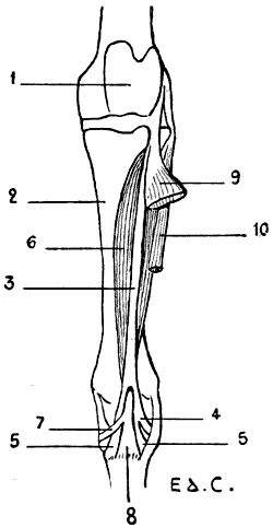
Fig. 83.—Myology of the Horse: the Anterior Tibial Muscle (Flexor of the Metatarsus), Left Leg, Anterior View.
1, Femoral trochlea; 2, tibia; 3, tendinous portion of the tibialis anticus; 4, cuboid branch of same; 5, 5, its metatarsal branch; 6, fleshy portion; 7, cuneiform branch of its tendon; 8, metatarsal branch of the same tendon; 9, extensor longus digitorum (anterior extensor of the phalanges turned outwards); 10, peroneus brevis (lateral extensor of the phalanges).
Tibialis Anticus (Fig. 83; Fig. 84, 6; Fig. 85, 4; Fig. 87, 10; Fig. 88, 10, 11).—It is further named by veterinarians the flexor of the metatarsus.
In the dog and the cat this muscle, which is rather large, arises from the external tuberosity of the tibia and from the crest of this bone. In its superior part it is flat, but lower down it is thick and produces a prominence in front of the tibia. Finally, it becomes tendinous, and passes towards the tarsus; thence it is directed towards the inner side of the metatarsus, and is inserted into the great-toe, this latter being sometimes well developed, but also often merely represented by a small bony nodule on which the muscle is then fixed.
In the other animals with which we here occupy ourselves, the tibialis anticus presents a complexity which would be incomprehensible unless this muscle be first studied in the horse.
In this latter the tibialis anticus consists of two distinct portions, placed one in front of the other: a fleshy portion, and a tendinous portion running parallel to it.
The muscle is covered, except on its internal part and inferiorly, by a muscle with which we will occupy ourselves later on—that is, the common extensor of the toes.
The tendinous portion of the tibialis anticus (Fig. 83), especially covered by the extensor of the toes, arises from the inferior extremity of the femur, from the fossa situated between the trochlea and the external condyle; thence it descends towards a groove which is hollowed out on the external tuberosity of the tibia, and is directed towards the tarsus, where it divides into two branches, which are inserted into the cuboid bone and the superior extremity of the principal metatarsal. These two parts form a ring through which the terminal tendon of the fleshy portion of the same muscle passes.
This fleshy portion, situated behind the preceding, arises from the superior extremity of the tibia, on the borders of the groove in which the tendinous portion lies; thence it passes downwards for a short distance on the inner side of the common extensor of the toes, which covers it in the rest of its extent. It ends in a tendon which, after passing through the tendinous ring above noticed, divides into two branches. One of these branches is inserted into the anterior surface of the superior extremity of the principal metatarsal, the other into the second cuneiform bone.
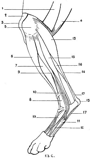
Fig. 84.—Myology of the Dog: Left Hind-limb, External Aspect.
1, Biceps cruris and fascia lata, divided in order to expose the upper part of the muscles of the leg; 2, inferior portion of the triceps cruris; 3, patella; 4, semi-tendinosus; 5, inferior extremity of the femur; 6, tibialis anticus (flexor of the metatarsus); 7, extensor longus digitorum (anterior extensor of the phalanges); 8, tibia; 9, peroneus longus; 10, peroneus brevis; 11, fifth metatarsal; 12, fasciculus detached from the peroneus brevis and passing towards the fifth toe; 13, external head of gastrocnemius; 14, tendo-Achillis; 15, calcaneum; 16, flexor digitorum sublimis; 17, 17, tendon of the flexor digitorum sublimis; 18, flexor longus pollicis (portion of the deep flexor of the toes); 19, dorsal muscle of the foot (short extensor of the toes).
In the ox the same two portions of the tibialis anticus exist, but with this capital difference—that the anterior portion is fleshy, superficial, and blended for a great part of its length with the common extensor of the toes.
The portion which corresponds to that which is fleshy in the horse arises from the tibia; below, it ends on the inner surface of the superior extremity of the metatarsus and the cuneiform bones. That which represents the tendinous part, which is also fleshy, as we have just pointed out, arises above with the common extensor of the toes, from the femur, in the fossa situated between the trochlea and the external condyle; whilst below, after having given passage to the tendon of the preceding portion, as in the horse, it is inserted into the metatarsus and the cuneiform bones.
In the pig, the tibialis anticus presents an arrangement nearly similar to that which we have just described.
It seems to us of interest to add that it has been sought to ascertain to what muscle of the human leg the tendinous part of the tibialis of the horse corresponds—a part which has become fleshy in the pig and the ox.
According to some authors, it represents the peroneus tertius; but that muscle is situated on the outer side of the common extensor of the toes; and here the portion with which it has been compared is placed on the inside. It has also been likened to a portion of the common extensor of the toes, but it does not pass to the latter. Lastly, it has been considered as being the homologue of the proper extensor of the great-toe; but why, then, in the ox, which has no great-toe, is it so highly developed? Nevertheless, its position and its relations sufficiently warrant this method of comprehending it. The tibialis anticus is a flexor of the foot. It is also able, in animals in which the tarsal articulations allow of the movement, to rotate the foot inwards.
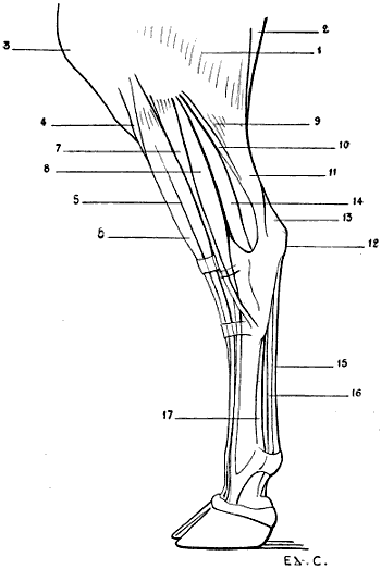
Fig. 85.—Myology of the Ox: Left Leg, External Aspect.
1, Gluteus maximus and biceps cruris; 2, semi-tendinosus; 3, patella; 4, tibialis anticus (flexor of the metatarsus); 5, extensor longus digitorum (anterior extensor of the phalanges); 6, fasciculus of the extensor longus digitorum, which is considered as the representative of the tendinous portion of the tibialis anticus in the horse; 7, peroneus longus; 8, peroneus brevis (proper extensor of the external toe); 9, external head of gastrocnemius; 10, soleus; 11, tendo-Achillis; 12, calcaneum; 13, tendon of the extensor longus digitorum (superficial flexor of the phalanges); 14, flexor longus pollicis and tibialis posticus (deep flexor of the phalanges); 15, tendon of the superficial flexor of the toes; 16, tendon of the deep flexor of the toes; 17, suspensory ligament of the fetlock.
With regard to the tendinous part, called by veterinarians the cord of the flexor of the metatarsus, it serves, in the horse, to produce the flexion of the metatarsus when the knee is already flexed; it thus acts in a passive fashion, which is explained by its resistance and the position which it occupies in relation to these two articulations.
Extensor Proprius Pollicis.—This muscle exists only in the dog and the cat, and there in a rudimentary condition.
It is covered by the common extensor of the toes and the tibialis anticus, and passes, accompanied by the tendon of this latter muscle, to terminate on the second metatarsal, or the phalanx, which articulates with it. When the first toe exists in the dog, it is inserted into this by a very slender tendon.
Extensor Longus Digitorum (Fig. 83, 9; Fig. 84, 7; Fig. 85, 5, 6; Fig. 86, 4; Fig. 87, 12; Fig. 88, 7).—It is also called by veterinarians the anterior extensor of the phalanges.
In the dog and the cat this muscle is to be seen in the space limited behind by the peroneus longus and in front by the tibialis anticus. Above it is covered by this latter. In the lower half of the leg, it is also in relation, on the inner side, with the tibialis anticus; but behind it is separated from the peroneus longus by the external surface of the shaft and inferior extremity of the tibia. This arrangement, besides, recalls that which is found in man, the peroneus longus of the latter diverging in the same way, at this level, from the common extensor, and leaving exposed the corresponding portion of the skeleton of the leg.
This muscle, fusiform in shape, arises at its upper part from the external surface of the inferior extremity of the femur, then its tendon passes into a groove hollowed on the external tuberosity of the tibia. The fleshy body which succeeds is directed towards the tarsus, but before reaching it is replaced by a tendon. This tendon, at the level of the metatarsal bones, divides into four slips, which pass towards the toes, and are inserted into the second and third phalanges of the latter.
In the horse it covers, to a great extent, the tibialis anticus, so that it is the latter which forms the large fusiform prominence especially noticeable in the middle region, to which the contour of the anterior surface of the leg is due.
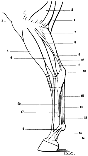
Fig. 86.—Myology of the Horse: Left Hind-limb, External Aspect.
1, Semi-tendinosus; 2, biceps cruris; 3, patella; 4, extensor longus digitorum (anterior extensor of the phalanges); 5, reinforcing band arising from the ligament of the fetlock; 6, peroneus brevis (lateral extensor of the phalanges); 7, external head of gastrocnemius; 8, soleus; 9, tendo-Achillis; 10, calcaneum; 11, tendon of the superficial flexor of the toes (superficial flexor of the phalanges); 12, flexor longus pollicis and tibialis posticus (deep flexor of the phalanges); 13, 13, tendon of the superficial flexor of the phalanges; 14, 14, tendon of the deep flexor of the phalanges; 15, suspensory ligament of the fetlock; 16, principal metatarsal: 17, external rudimentary metatarsal.
It arises above from the inferior extremity of the femur, from the fossa situated between the trochlea and the external condyle; therefore, it has a common origin with the tendinous portion of the tibialis anticus, or flexor of the metatarsus.
The tendon, which at the level of the inferior part of the leg succeeds to the fleshy body, passes in front of the tarsus, the principal metatarsal, and receives the tendon of the peroneus brevis which we will describe later on. It then reaches the anterior surface of the fetlock. There it presents an arrangement analogous to that which we have pointed out in connection with the anterior extensor of the phalanges—a muscle which, in the fore-limbs, corresponds to the common extensor of the digits; that is to say, it is inserted, in form of an expansion, into the pyramidal prominence of the third phalanx, after having formed attachments to the first and second, and having received on each side a strengthening band from the suspensory ligament of the fetlock.
In the ox the long extensor of the toes is united above, and for a great part of its length, with the portion of the tibialis anticus, which represents, albeit in the fleshy state, the tendinous cord of the latter in the horse.
In common with this portion, it arises from the inferior extremity of the femur. Thence it passes towards the tarsus and divides into two fasciculi, internal and external, which are continued by tendons. These pass towards the phalanges, and, in case of the common extensor of the digits belonging to the fore-limbs, the internal is destined for the internal toe, and the external is common to the two toes.
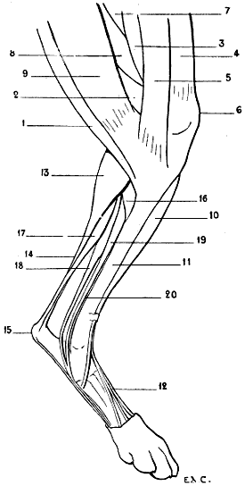
Fig. 87.—Myology of the Dog: Left Hind-limb, Internal Aspect.
1, Semi-tendinosus; 2, semi-membranosus; 3, triceps cruris (vastus internus); 4, sartorius (patellar); 5, sartorius (tibial); 6, patella; 7, first or middle adductor; 8, small and great adductor united; 9, gracilis; 10, tibialis anticus (flexor of the metatarsus); 11, tibia; 12, tendon of extensor longus digitorum (anterior extensor of the phalanges): 13, gastrocnemius, inner head; 14, tendo-Achillis; 15, calcaneum; 16, popliteus; 17, superficial flexor of the toes; 18, flexor longus pollicis (portion of the deep flexor of the toes); 19, flexor longus digitorum (portion of the deep flexor of the toes); 20, tendon of the tibialis posticus.
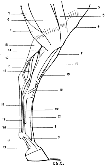
Fig. 88.—Myology of the Horse: Left Hind-leg, Internal Aspect.
1, Semi-tendinosus; 2, semi-membranosus; 3, triceps cruris (vastus internus); 4, patella; 5, sartorius; 6, gracilis; 7, extensor longus digitorum common extensor of the toes (anterior extensor of the phalanges); 8, tendon of the preceding muscle; 9, reinforcing band given off by the suspensory ligament of the fetlock; 10, tibialis anticus (flexor of the metatarsus), its tendinous portion; 11, tibialis anticus (flexor of the metatarsus), its fleshy portion; 12, cuneiform branch of the tendon of this fleshy portion; 13, internal head of gastrocnemius; 14, popliteus; 15, tendon of the flexor brevis digitorum (superficial flexor of the phalanges); 16, flexor longus pollicis and tibialis posticus (deep flexor of the phalanges); 17, flexor longus digitorum (oblique flexor of the phalanges); 18, 18, tendon of the superficial flexor of the phalanges; 19, 19, tendon of the deep flexor of the phalanges; 20, suspensory ligament of the fetlock; 21, principal metatarsal; 22, internal rudimentary metatarsal.
In the pig the general arrangement of the muscle is similar, but the tendons end in a manner which is a little more complicated. Apart from the fasciculi which correspond to the tendinous portion of the tibialis anticus (fleshy here, as in the ox), the long extensor of the toes at the level of the tarsus divides into three tendons: the internal goes to the great internal toe; the middle bifurcates in the upper part of the digital portion of the foot, and each of its branches goes towards one of the great-toes; the external divides to pass towards each of the two small toes, and towards the great ones; but this latter disposition is not constant.
By its contraction the muscle which we have just studied extends the phalanges and flexes the foot.
Peroneus Tertius.—This muscle is not found in domestic quadrupeds.
We should remember, nevertheless, that certain authors consider as representing it the tendinous portion of the anterior tibial of the horse, or the corresponding portion now fleshy, of the same muscle in the pig and the ox. It is by reason of this fact that it is called the third peroneal, notwithstanding that in the numerical order of the peroneals it is rather the first.
But that which still further complicates this question of nomenclature is that some authors give this name of third to a peroneal which, in the carnivora, is situated more definitely in the group of external muscles (see below, Short Lateral Peroneal).
Muscles of the External Region
In man, two muscles constitute this region; they are the peroneus longus and peroneus brevis.
Peroneus Longus (Fig. 84, 9; Fig. 85, 7).—This muscle does not exist in the domestic animals; only in the flesh-eaters, the pig and the ox excepted.
It is in relation superiorly with the tibialis anticus, and inferiorly with the common extensor of the toes; in the ox, it is in contact with this latter muscle throughout its whole length.
The peroneus longus arises from the external tuberosity of the tibia; towards the middle of the leg it is replaced by a tendon. This proceeds towards the tarsus, but previously it passes between the tibia and fibula. In the ox it is situated in front of the coronoid tarsal bone; we recollect that this bone is regarded as representing the inferior extremity of the fibula (see p. 97). Then it passes into a groove belonging to the cuboid bone or to the cuboido-scaphoid bone in the ox, traverses obliquely the posterior aspect of the tarsus, and is inserted into the rudimentary bone which represents the first toe; or, if this does not exist, into the innermost of the metatarsal bones.
This muscle is an extensor of the foot. It also rotates it outwards in the animals in which the articulation permits this latter movement.
Peroneus Brevis (Fig. 83, 8; Fig. 83, 10; Fig. 84, 10; Fig. 86, 6).—In the dog and the cat, this muscle is covered in part by the peroneus longus, and arises from the inferior half of the tibia and the fibula; at the level of the tarsus it becomes tendinous, passes into a groove hollowed out on the external surface of the inferior extremity of the fibula, and terminates on the external aspect of the superior extremity of the fifth metatarsal. A little before this insertion it crosses the tendon of the long peroneal in passing to the outer side of the latter.
To the short peroneal muscle is found annexed a very thin fasciculus which lies upon it. This fasciculus arises from beneath the head of the fibula, and is soon replaced by a thin tendon, which, accompanying that of the short peroneal, proceeds towards the foot, after having traversed the groove in the inferior extremity of the fibula; passes along by the fifth metatarsal (Fig. 84, 12); blends at the level of the first phalanx of the fifth toe with the corresponding tendon of the long extensor of the toes, and partakes of the insertions of this tendon.
This fasciculus is designated by some authors under the name of the peroneal of the fifth toe, or the proper extensor of the same toe. But what makes still further complications is that other authors regard it as an anterior, or third, peroneal. Now, these names are those which other anatomists have applied to the fasciculus of the anterior tibial, which, in the pig and the ox, is fused in part with the long extensor of the toes. Hence there results a confusion which is truly regrettable.
In brief, we can, without inconvenience, consider it as a fasciculus of the short peroneal muscle.
We sometimes find in man, but abnormally, an arrangement which partly recalls that which we have just indicated. It consists in a duplication of the tendon of the short peroneal, one of the branches of which goes to the fifth metatarsal, and the other to the fifth toe; it is sometimes a single fasciculus which goes to the phalanges of this latter. We have met with examples of these anomalies.[31] In the pig, the short peroneal is situated on the same plane as the long. It consists of two clearly distinct fasciculi, which arise from the fibula. The tendon of the anterior fasciculus proceeds to the great external toe—that is to say, the fourth, of which it is the proper extensor. The posterior fasciculus terminates on the small external toe, the fifth, of which it is in like manner the extensor.
In the ox, the fleshy fibres of the short peroneal arise from a fibrous band which replaces the fibula, and from the external tuberosity of the tibia. Situated behind the long peroneal and on the same plane, it terminates in a tendon which appears at the level of the inferior part of the leg; it passes in front of the canon, and is inserted into the external toe, of which it is the proper extensor.
In the horse, it is the sole representative of the peroneal muscles, and veterinary anatomists have given it the name of the lateral extensor of the phalanges.
Its fleshy body arises above from the external lateral ligament of the knee-joint, and from the whole length of the fibula. In the middle third of the leg it is narrowed; lower down it is replaced by a tendon. This is lodged in a groove hollowed on the external surface of the inferior extremity of the tibia; then after passing along the external surface of the tarsus, it is directed forward, and proceeds to blend towards the middle of the canon-bone with the tendon of the long extensor of the toes, or anterior extensor of the phalanges, of which it shares the insertions. It extends the phalanges into which it is inserted. It also flexes the foot.
Muscles of the Posterior Region
It will not be unprofitable to recall to mind that, in man, the muscles of this region are arranged in two layers: a superficial layer consisting of the gastrocnemius and soleus, to which is added a muscle of little importance, the plantaris, and a deep layer formed by four muscles—the popliteus, flexor longus digitorum, tibialis posticus, and flexor longus pollicis.
The gastrocnemius and soleus, independent in their upper portion, unite below in a common tendon; they thus form also a triceps muscle, which we designate under the name of the triceps of the leg, or triceps suralis, because it forms the elevation of the calf of the leg (from sura, calf).
Gastrocnemius (Fig. 83, 9, 11; Fig. 84, 13, 14; Fig. 86, 7, 9; Fig. 88, 13).—The external and internal heads of the gastrocnemius, distinct from one another only in their upper portion, arise from the shaft of the femur, above the condyles, on the borders of the popliteal surface, to a relatively considerable extent in the great quadrupeds.
At this level they are situated in the popliteal region—that is to say, in the space limited externally by the biceps, and internally by the semi-tendinosus. But as they descend to a rather low level on the leg in quadrupeds, and especially in carnivora, they do not, properly speaking, determine a projection of the calf of the leg. However, they pass from this region but to be soon continued by a tendon—the tendo-Achillis, which is inserted into the calcaneum.
Now, the region of the tarsus is called by veterinarians the ham, the posterior surface of which is angular, because of the oblique direction of the leg with regard to the vertical direction of the metatarsus and the presence of the calcaneum; the prominence which this surface presents has received the name of the point of the ham, and the tendon which ends there that of the cord of the ham.
But the tendo-Achillis does not alone form this cord. Indeed, as we will soon see, the tendon of the superficial flexor of the toes takes part in its formation.
We may add, with regard to the tendo-Achillis, that it is more clearly perceived as an external feature, because the skin sinks in front of it, as it does in man, over the lateral parts of the region which it occupies.
The gastrocnemius, when it contracts, extends the foot on the leg.
It serves to maintain the tibio-tarsal angle in the standing position, and during walking, to determine the steadying of the hind-limbs, which then, after the fashion of a spring, project the body forward.
By an analogous movement they take part in the posterior projection of the hind-limbs in the act of kicking; but they are not the only ones to act in this case, the muscles of the buttock and thigh also being brought into play.
Soleus (Fig. 83, 10; Fig. 86, 8).—This muscle, much less developed in quadrupeds than in man, does not exist in the dog.
With regard to the soleus in the pig, Professor Lesbre says: ‘Meckel denied its existence; we, however, believe that it is united to the external head of the gastrocnemius, its origin being transferred to the femur.’[32]
But in animals in which it exists, this muscle, of but little importance, occupies the outer side of the leg. It arises above from the external tuberosity of the tibia, and terminates below in a tendon which is united with that of the gastrocnemius.
The soleus has the same action as these latter.
Plantaris.—In quadrupeds this muscle is blended with the superficial flexor of the toes, which we will study afterwards.
Popliteus (Fig. 87, 16; Fig. 88, 14).—In man, this muscle, which occupies the posterior surface of the tibia, above the oblique line, is completely covered by the gastrocnemius.
In quadrupeds, where it is more voluminous, it projects internally beyond the gastrocnemius, so that it is seen in the internal and superior part of the region of the superficial layer of muscles, immediately behind the internal surface of the tibia, which, as we know, is subcutaneous.
The popliteus arises from the external surface of the external condyle of the femur. Thence its fibres which diverge pass to be inserted into the superior part of the posterior surface and of the internal border of the tibia. It is in this latter region that it projects beyond the gastrocnemius, but we may add that there it is more or less covered by the semi-tendinosus.
It flexes the leg, and rotates it forwards.
Superficial Flexor of the Toes (Fig. 83, 13, 15; Fig. 84, 17; Fig. 86, 11, 13, 13; Fig. 87, 17; Fig. 88, 15, 18, 18).—In man, the homologue of this muscle is found in the sole of the foot. It is called the short flexor of the toes. It arises from the calcaneum, and passes to the four outer toes. In quadrupeds, it rises as high as the back of the knee, and is found blended with the plantaris.
Further designated by the name of the superficial flexor of the phalanges, covered in part by the gastrocnemius, with which it is in relation for a great part of the course which it traverses, this muscle arises from the posterior surface of the femur, on the external branch of the inferior bifurcation of the linea aspera. In the horse, this origin takes place in a depression situated above the external condyle, in the supracondyloid fossa. Then it accompanies the gastrocnemius, and becomes tendinous where the tendo-Achillis commences. It then winds round the latter in placing itself on its inner side, then on its posterior surface, and reaches the calcaneum. It accordingly contributes, as we have already pointed out, to form the cord of the ham. After having become expanded, and having covered as with a sort of fibrous cap the bone of the heel, it descends behind the metatarsus, and presents there an arrangement analogous to that which we pointed out in connection with the superficial flexor of the digits—that is, it contributes to form the tendon. This prominence, in the form of a cord, we see behind the canon-bone in solipeds and ruminants. It finally terminates in the same way as the muscle with which we have compared it (see p. 197).
In the horse, its fleshy body is but slightly developed, so that its tendon alone is specially visible in the superficial muscular layer, but in the dog and the cat it is large. Hence it results that its fleshy body appears on each side of the inferior half of the gastrocnemius, and produces an elevation which recalls that which the soleus produces on each side of the same muscles in the human species.
The muscles which follow form, with the popliteus, which we have already studied, the deep layer of the posterior region of the leg.
Flexor Longus Digitorum (Fig. 87, 19; Fig. 88, 17).—This muscle, in man, is the only common flexor of the toes belonging to the muscles of the leg.
In comparison with the preceding muscle, it is a deep flexor. Veterinarians have given it the name of the oblique flexor of the phalanges.
Visible on the internal aspect of the superficial layer of the muscles of the leg, this muscle arises above from the posterior surface of the external tuberosity of the tibia, becomes tendinous, passes towards the metatarsus, and blends with the tendons of the posterior tibial and the long proper flexor of the great-toe. In the dog and the cat it is blended with this latter only.
Tibialis Posticus (Fig. 85, 14; Fig. 86, 12; Fig. 87, 20; Fig. 88, 16).—This muscle arises from the external tuberosity of the tibia, and from the head of the fibula. Thence it passes to the tarsus, and terminates in different fashion in carnivora and other quadrupeds.
In the dog and the cat, it is inserted into the ligamentous apparatus of the tarsus, or into the base of the second metatarsal.
In the other quadrupeds with which we are here occupied it is blended with the long proper flexor of the great-toe.
It is accordingly in the carnivora that the mode of termination of the tibialis posticus most nearly resembles that of this same muscle in the human species. From this independence there results a special action.
It is an adductor and internal rotator of the foot.
Flexor Longus Pollicis (Fig. 84, 18; Fig. 85, 14; Fig. 86, 12; Fig. 87, 18; Fig. 88, 16).—This muscle, as that in man, is the most external of the deep layer of the leg. It is on the external aspect of the latter we perceive it, between the peroneals and the gastrocnemius or tendo-Achillis.
It arises from the fibula and tibia, and is thence directed towards the tarsus. It unites with the long common flexor of the toes to form with it the deep flexor of the phalanges, of which it is the principal fasciculus. We may add that in the dog and the cat the posterior tibial remains independent of this latter, but that in the pig, ox, and horse the posterior tibial is united to the preceding to form with them the deep flexor muscle.
Thus constituted, the deep flexor goes towards the phalanges, where it terminates as the deep flexor of the digits of the fore-limbs (see p. 197). In animals possessed of a canon it contributes to form the tendon (Fig. 85, 16; Fig. 86, 14, 14; Fig. 88, 19, 19).
Muscles of the Foot
We must remember that on the dorsal surface of the foot in man we find but a single muscle—the dorsalis pedis. The remaining subcutaneous structures of this region consist of the tendons of the anterior muscles of the leg which occupy this dorsal aspect.
Dorsalis Pedis (Fig. 84, 19).—Also called the extensor brevis digitorum, the dorsalis pedis muscle is found in all domestic quadrupeds; but its development is so much the less as the number of digits is more reduced.
In the dog and the cat it arises from the calcaneum, and is inserted into the three internal toes (the first toe excepted) by uniting with the corresponding tendons of the common extensor.
In the pig its disposition is analogous.
As for the dorsalis pedis of the ox and the horse, it is extremely rudimentary, and occupies the superior part of the canon. It is a small, fleshy body, situated on the anterior surface of the metatarsus, which arises from the calcaneum, whence it passes to unite at its inferior extremity with the tendon of the extensor of the phalanges.
As regards the muscles of the sole of the foot, we think it unnecessary to occupy ourselves at any length with them because of their slight importance with regard to external form.
We will only recall that in the median portion of this plantar surface we find in man the short flexor of the toes, which in quadrupeds arises higher up, from the posterior surface of the femur; that it belongs to the muscles of the leg; and that it forms the superficial flexor of the toes, which we have already studied.
We may further add that the suspensory ligament of the fetlock in ruminants and solipeds represents, as in the fore-limbs, the interosseous muscles.
MUSCLES OF THE HEAD
We will divide these muscles into two categories: masticatory and cutaneous.
Masticatory Muscles
The muscles of this group which specially interest us are the masseter and the temporal. As regards the pterygoids, since they are situated within the borders of the inferior maxillary bone, and consequently do not reach the surface, we shall not require to occupy ourselves with them here.
Masseter (Fig. 89, 2; Fig. 90, 1; Figs. 91, 92).—For those who have studied the masseter of man, it is not difficult to recognise that of quadrupeds. Nevertheless, the particular aspect which it presents in different species gives to its study a certain interest.
Arising from the zygomatic arch, and passing downwards and backwards, it is inserted into the external surface of the ramus of the mandible and into its angle.
Its posterior border is in relation with the parotid gland (Fig. 90, 14; Figs. 91, 92), this gland being situated between the corresponding border of the lower jaw bone and the transverse process of the atlas. Such are the general characters; the following are the particular ones:
In the carnivora it is thick and convex. In the horse it is flat, but more expanded; it forms the flat of the cheek. In the ox it is flat, as in the latter; but, while being less thick, it is more prolonged in the vertical direction.
The form of the osseous parts which give it origin is, besides, in relation with these differences, and explains the peculiar characters which the masseter presents.