Chapter I · (continued 3)
Reading edition
It is true that, in the position we have adopted, the face is directed obliquely downwards and forwards, and that there may result a certain confusion in describing the position of its different parts. On this account, with the object of not making complications, we purpose, for the present, to substitute, for example, for the term ‘antero-superior’—which when speaking of the position of the forehead and nose would be more exact—the term ‘anterior,’ which is sufficiently comprehensible. The mouth will be, for the same reason, referred to as being situated at the inferior part of the face, and not the antero-inferior.
The Skull.—The elevation of the cranial region becomes especially appreciable when we examine the occipital bone. Before verifying this fact, it is not superfluous to recall the general arrangement which this bone presents in the human skull. A portion of the occipital bone occupies the base of the skull; but this base in man is horizontal; to this region succeeds the shell-shaped portion of the occipital bone, which, passing vertically upwards, forms with the preceding portion an angle situated at the level of the external occipital protuberance, and of the curved line which starts from it on each side. In animals a portion of the occipital bone is horizontal, it is true; but this bone being sharply bent at the level of the occipital foramen and condyles, the result is that the portion which surmounts these latter looks backwards, and is limited above by the external occipital protuberance, which forms the culminating point of the skull; this point is situated between the ears.
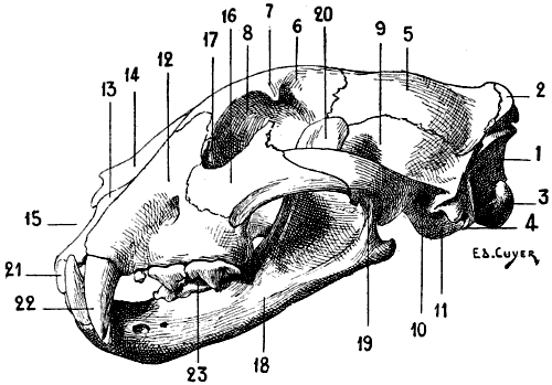
Fig. 58.—Skull of one of the Felidæ (Jaguar): Left Lateral Aspect.
1, Posterior surface of the occipital bone; 2, external occipital protuberance; 3, condyle of the occipital bone; 4, jugular process; 5, parietal bone; 6, frontal bone; 7, orbital process; 8, orbital cavity; 9, squamous portion of the temporal bone; 10, external auditory canal, in front of which is situated the zygomatic process; 11, tympanic bulla; 12, superior maxillary bone; 13, intermaxillary or incisor bone; 14, nasal bone; 15, anterior orifice of the nasal cavity; 16, malar bone; 17, ungual or lachrymal bone; 18, inferior maxillary bone; 19, condyle of the inferior maxillary bone; 20, coronoid process; 21, incisor teeth; 22, canine teeth; 23, molar teeth.
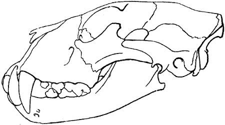
Fig. 59.—Skull of the Lion: Left Lateral Aspect.
This figure is intended to show that in the lion the contour of the face between the nasal bones and the cranial region is more flattened than in other felidæ, such as the tiger, jaguar, panther, and domestic cat. This difference is shown by comparison of this figure with the preceding one (Fig. 58). We are indebted to M. Tramond, the well-known naturalist, for the indication of this differential character which, from the artistic plastic point of view, is one of real interest.
This protuberance, prolonged on each side by the superior curved line of the occipital bone, is so much the more prominent as this bone bends sharply a second time, so as to form a third portion, which, looking forwards, forms part of the anterior aspect of the skull, and proceeds to articulate with the parietals. On this third portion is found a crest which, proceeding from the occipital protuberance, is continuous in front with the parietal crests, to which we will again refer in speaking of the parietal bones.
On the inferior surface of the human occipital bone are found, at the level of, and external to, the condyles two bony elevations which bear the name of jugular eminences. They are long in quadrupeds, and constitute what are designated by some authors the styloid processes, but they must not be confounded with the processes of the same name which in the case of man form part of the temporal bone. These processes are very highly developed in the pig, horse, ox, and sheep.
In the ox, the occipital bone is deprived of the protuberance, and is not bent on itself in the anterior portion, neither does it form the most salient part of the skull; this latter, which is situated at the level of the horns, belongs to the frontal bone. In the pig, also, the occipital bone is not bent upon itself in its anterior portion, but forms the summit of the head. The occipital protuberance, hollowed on its posterior surface, rises vertically, and rests upon the parietal bone, with which it forms an acute angle.
The parietals, two separate bones in the dog and the cat, but fused in the median line in the ox, sheep, and horse, are of special interest in regard to the two crests which, in the carnivora, and also in the pig and the horse, occupy their external surface, and, after diverging from one another, are continued by a crest which crosses the frontal bone and ends at the external orbital process of the latter bone.
These crests, known as the parietal or temporal crests, recall both in position and relations the temporal curved line of the parietal bone of man. They contribute, as in the case of the latter, to the formation of the boundaries of the temporal fossa.
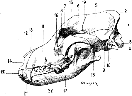
Fig. 60.—Skull of the Dog: Left Lateral Aspect.
1, Posterior surface of the occipital bone; 2, external occipital protuberance; 3, occipital condyle; 4, jugular process; 5, parietal bone; 6, frontal bone; 7, orbital process; 8, orbital cavity; 9, external auditory canal, in front of which is found the zygomatic process; 10, tympanic bulla; 11, superior maxillary bone; 12, intermaxillary or incisor bone; 13, nasal bone; 14, anterior opening of the cavity of the nasal fossæ; 15, malar bone; 16, lachrymal bone; 17, inferior maxillary bone; 18, condyle of the inferior maxillary bone; 19, coronoid process; 20, incisor teeth; 21, canine teeth; 22, molar teeth.
In the carnivora, these crests are situated, throughout their whole length, in the median line, the temporal fossæ being, accordingly, as extended as they possibly can be. In certain species, the development of these crests is such that they form by their union a vertical plate, which, in separating the two temporal fossæ, gives them a greater depth. In the pig, the parietal crests, analogous in this respect to the temporal curved lines of the parietal bones of man, are separated by an interval, proportionately less extended, however, than that of the human skull. The parietal bone in the ox and the sheep does not enter into the formation of the anterior surface of the skull; it is formed by an osseous plate, narrow and elongated transversely, which, with the occipital bone, constitutes the base of the region of the nape of the neck. It is bent upon itself at the level of its lateral portions so as to occupy the temporal fossa.
The anterior surface of the frontal bone, which is depressed in the median line in the dog, but plane in the horse, is limited by two crests, which, situated on the prolongation of the parietal crests, diverge more and more from one another in proportion as they occupy a lower position. This surface terminates externally in two processes, which are the homologues of the external orbital processes of the human frontal bone.
The superior border of these orbital processes, situated on the prolongation of the corresponding parietal crests, contributes to limit the temporal fossa. Each of these orbital processes terminates in the following manner: In the bear, dog, cat, and pig, in which the orbital cavities are incompletely bounded by bone, this process, slightly developed, is not in connection, by its inferior extremity, with any other part of the skeleton of the region. In the ox and the sheep, it articulates with a process of the malar bone. In the horse, it articulates with the zygomatic process of the temporal bone. The inferior margin of this process forms a part of the boundary of the anterior opening of the orbital cavity.
The supra-orbital foramen, which does not exist in carnivora, occupies in the horse the base of the orbital process. In the ox, it is situated a little nearer the middle line; and its anterior orifice opens into an osseous gutter which is directed upwards towards the base of the horn, while inferiorly it meets the inferior border of the frontal bone; in the sheep this groove is but slightly developed. In this latter, as in the ox, it is the frontal bone which forms the most elevated portion of the skull. In fact, being bent upon itself at a certain level, its external surface is formed of two planes: one, posterior, which is inclined downwards and directed backwards; the other, anterior, is also inclined downwards, but with a forward obliquity. At the union of these planes the bone forms an elbow, on either side of which are found the osseous processes on which the horns are mounted.
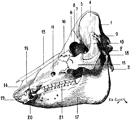
Fig. 61.—Skull of the Pig: Left Lateral Aspect.
1, Occipital bone; 2, condyle of the occipital; 3, jugular process; 4, parietal bone; 5, parietal crests; 6, frontal bone; 7, orbital process; 8, orbital cavity; 9, external auditory canal; 10, zygomatic process; 11, superior maxillary bone; 12, intermaxillary or incisor bone; 13, nasal bone; 14, anterior orifice of the cavity of the nasal fossæ; 15, malar bone; 16, lachrymal bone; 17, inferior maxillary bone; 18, condyle of the inferior maxillary bone; 19, incisor teeth; 20, canine teeth; 21, molar teeth.
In the bear, the anterior margin of the frontal bone is prolonged by two small tongues of bone, which, descending on the lateral borders of the nasal bones, articulate with the superior half of the latter.
The temporal bone is, as in man, furnished with a squamous portion, from which springs the zygomatic process, which is directed towards the face, to terminate in the following manner: in the carnivora, the pig, and ruminants, it articulates with the malar bone by its inferior border; in the horse, it insinuates itself as a sort of wedge between the malar bone and the orbital process of the frontal bone, with which it articulates, as we have already pointed out, and contributes, by a portion situated in front of this articulation, to form the boundary of the anterior opening of the corresponding orbital cavity. As in man, the zygomatic process arises by two roots: one, transverse, behind which is situated the glenoid cavity of the temporal bone; the other, antero-posterior, which proceeds to join above with the superior curved line of the occipital bone.
Behind the glenoid cavity is found the external auditory canal, and, further back still, the mastoid process. This latter, but slightly developed in the carnivora, a little more so in the ruminants, and still more in the horse, has its external surface traversed by a crest, the mastoid crest, which, after becoming blended with the antero-posterior root of the zygomatic process, proceeds with this latter to join the superior occipital curved line.
Below the auditory canal is situated a round prominence, highly developed in carnivora; this is the tympanic bulla, also called the mastoid protuberance; it is an appendage of the tympanum.
The Face
The bone of this region, around which all the others come to be grouped, is, as in man, the superior maxillary. The relations of this maxillary with the neighbouring bones is not exactly the same in all animals; for example, in the ox, sheep, and horse, in which the bones of the nose are wide in their upper part, and in which the lachrymal bone, which is very highly developed, encroaches on the face, the superior maxillary does not meet the frontal bone; it is separated from it by the above-named bones. It unites with it, on the other hand, in the dog and the cat. In the bear, it is separated from the bones of the nose by a small tongue of bone which springs from the anterior border of the frontal—a process which we have noticed in connection with this latter.
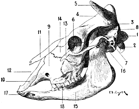
Fig. 62.—The Skull of the Ox: Left Lateral Aspect.
1, Occipital condyle; 2, jugular process; 3, parietal bone; 4, frontal bone; 5, osseous process, which serves to support the horn (horn-core); 6, orbital cavity; 7, external auditory canal, in front of which is found the zygomatic process; 8, temporal fossa; 9, superior maxillary bone; 10, intermaxillary or incisor bone; 11, nasal bone; 12, anterior orifice of the cavity of the nasal fossæ; 13, malar bone; 14, lachrymal bone; 15, inferior maxillary bone; 16, condyle of the inferior maxillary bone; 17, incisor teeth; 18, molar teeth.
In the pig, ox, sheep, and horse, the external surface is traversed, to a greater or less extent, by a crest which is situated on the prolongation of the inferior border of the malar bone. This crest, which is straight in the horse, but curved with its convexity upwards in the ox and the sheep, is known as the maxillary spine or the malar tuberosity: it gives attachment to the masseter muscle, and, in the horse, is distinctly visible under the skin. It does not exist in the carnivora. On the same surface is situated the sub-orbital foramen.
The inferior border is hollowed out into alveoli, in which are implanted the superior molar and canine teeth. This border is prolonged forwards from the alveolus, which corresponds to the first molar tooth, to terminate, after a course more or less prolonged, at the alveolus of the canine. This space, more or less considerably expanded, which thus separates these teeth is called the interdental space; but this denomination is not applicable to ruminants, because these latter possess neither canine nor incisor teeth in the upper jaw (see p. 125, dentition of the ox and sheep). The superior maxillary bone of one side and that of the opposite side do not meet in the median line in the region which corresponds to the incisor teeth; they are separated by a bone which, in the human species, is present only at the commencement of life, and afterwards coalesces with the maxilla; this is the intermaxillary or incisor bone. This bone, which is paired, is formed of a central part, which bears the superior incisor teeth; it is prolonged upwards and backwards by two processes: one, external, which insinuates itself between the superior maxillary and the nasal bone, except in the sheep, in which it remains widely separated from the latter; the other, internal, which is united to that which belongs to the bone of the opposite side to form part of the floor of the cavity of the nasal fossæ; the external border of this process, which is separated from the body of the bone by a notch, forms the internal boundary of the corresponding incisor opening or the incisor slit. Owing to the absence of superior incisors in ruminants, the intermaxillary bone presents no alveoli.
The malar bone, and the os unguis or lachrymal, are more or less developed according to the species considered. With regard to the malar bone, it is most important to notice the part which it takes in the formation of the zygomatic arch, and that its inferior border contributes to form the crest to which is attached the masseter muscle.
As for the nasal bones, they present differential characters which, as they affect the form of the region which they occupy, are worthy of notice.
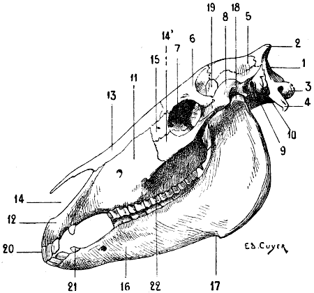
Fig. 63.—Skull of the Horse: Left Lateral Aspect.
1, Posterior surface of the occipital bone; 2, external occipital protuberance; 3, occipital condyle; 4, jugular process; 5, parietal bone; 6, frontal bone; 7, orbital cavity; 8, zygomatic process of the temporal bone; 9, external auditory canal; 10, mastoid process; 11, superior maxillary bone or maxilla; 12, intermaxillary or incisor bone; 13, nasal bone; 14, malar bone; 15, lachrymal bone; 16, inferior maxillary bone or mandible; 17, inferior maxillary fissure; 18, condyle of the inferior maxillary bone; 19, coronoid process of the inferior maxillary bone; 20, incisor teeth; 21, canine teeth; 22, molar teeth.
Their dimensions in length are proportional to those of the face. Very small in man, they are more developed in carnivora. We recognise in the latter the two curves which characterize them in the human species, and which we clearly notice when we view them on one of their lateral aspects: a concavity above, and a convexity below. These curves are more or less accentuated—very strongly marked in the bulldog, and scarcely at all in the greyhound. Moreover, in the carnivora also the nasal bones are wider below than above, and form, by their junction, a semicircular notch which limits, in its superior portion, the anterior opening of the cavity of the nasal fossæ. In the horse they present an opposite arrangement with regard to their dimensions in width; broad above, each terminates below by forming a pointed process which, separated from the intermaxillary bones, is prolonged in front of the nasal orifice.
The inferior maxillary bone is, as in man, formed of a body and two branches. But among the many special characteristics of form and size which sharply differentiate it from the human bone, one detail must be indicated; this is the absence of a mental prominence. Hence it results that the anterior border of the body of the lower jaw, instead of being directed obliquely downwards and forwards, is, on the contrary, oblique downwards and backwards, and that in certain animals this border is actually found almost exactly on the prolongation of the inferior border of the body of the bone.
On the external surface of the body are found the three mental foramina. The superior border is hollowed out by alveoli.
With regard to the branches (rami), they terminate in two processes: one, the posterior, is the condyle; the other, situated more forwards, is the coronoid process, which gives insertion to the temporal muscle. These two processes are separated by the sigmoid notch.
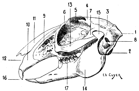
Fig. 64.—Skull of the Hare: Left Lateral Aspect.
1, External occipital protuberance; 2, occipital condyle; 3, parietal bone; 4, frontal bone; 5, orbital process; 6, orbital cavity; 7, zygomatic process; 8, external auditory canal; 9, superior maxillary bone; 10, intermaxillary or incisor bone; 11, nasal bone; 12, anterior opening of the nasal fossa; 13, malar bone; 14, inferior maxillary bone; 15, condyle of the inferior maxillary bone; 16, incisor teeth; 17, molar teeth.
For reasons which we will explain further on (see p. 127, movements of the lower jaw), the condyle presents differences of form. In the carnivora, it is strongly convex from before backwards, expanded transversely, and firmly mortised in the glenoid cavity of the temporal bone; in the ruminants, it is less convex from before backwards, it is more slightly concave in the transverse direction; in the rodents—we give as an example the hare (Fig. 64)—the condyle is still convex from before backwards, but it is flattened from without inwards.
In the animals in which the muscles of mastication are very highly developed, and especially in the carnivora, the osseous regions occupied by these muscles are more extensive and more deep than in the human species. The length of the coronoid process, the depth of the temporal fossa, the extent of the zygomatic arch, the appearance of the external surface of each of the rami of the lower jaw, deeply hollowed out for accommodation of the masseter, and to provide extensive surfaces of insertion for this muscle, are sure proofs furnished by the skeleton of the occasionally enormous development of the muscles of mastication.
In the carnivora, a rather strong process, which is directed backwards, occupies the angle of the inferior maxilla; it is, accordingly, situated below the region of the condyle.
The teeth which the jaws carry vary in number, and even in appearance, according to species; it is useful to note their differences. In order to establish the nature of these latter more effectively, we will first recall the fact that in man the teeth, thirty-two in number, are equally distributed between the jaws, and are divided into incisors, canines, and molars, of which the arrangement is thus formulated:
5m. 1c. 2i. 2i. 1c. 5m. = 32.[19]
5m. 1c. 2i. 2i. 1c. 5m.
We also note that the incisors are edged, the canines are pointed, and that the molars, cubical in shape, have their surface of contact provided with tubercles.
The teeth of the cat are thirty in number; they are thus arranged:
4m. 1c. 3i. 3i. 1c. 4m. = 30.
3m. 1c. 3i. 3i. 1c. 3m.
Those of the dog number forty-two:
6m. 1c. 3i. 3i. 1c. 6m. = 42.
7m. 1c. 3i. 3i. 1c. 7m.
In these animals, the incisors, such as are not damaged by use, are furnished, on the free border of their crown, with three tubercles, of which one, the median, is more developed than those which are situated laterally. We denote these teeth, commencing with those nearest the median line, by the names central incisors or nippers, intermediate and corner incisors. The canines, or fangs, are long and conical; they are curved backwards and outwards. The upper canines, which are larger than those of the lower jaw, are separated from the most external of the incisors (corner) by an interval in which the canines of the lower jaw are received. The lower canines, on the other hand, are in contact with the neighbouring incisors, and are each separated from the first molar which succeeds them by a wider interval than that which is situated between the corresponding teeth in the upper jaw.
The molars differ essentially from the teeth of the same class in the human species. Their crown terminates in a cutting border bristling with sharp-pointed projections; this formation indicates that these teeth are principally designed for tearing. During the movement of raising the lower jaw, which is so energetic in the carnivora, they act, indeed, in the same manner as the two blades of a pair of scissors. The largest molars are: in the dog, the fourth of the upper jaw, and the fifth in the opposite one; in the cat, the third both above and below.
The pig has forty-four teeth disposed in the following manner:
7m. 1c. 3i. 3i. 1c. 7m. = 44.
7m. 1c. 3i. 3i. 1c. 7m.
Of the incisors, the nippers and the intermediate ones of the upper jaw have their analogues in those of the horse; in the lower jaw, the corresponding teeth, straight, and directed forward, rather resemble the same incisors in rodents. The corner incisor teeth are much smaller, and are separated from the neighbouring teeth. The canine teeth, also called tusks or tushes, are greatly developed, especially in the male. The molars increase in size from the first to the last; they are not cutting, as in the carnivora, but they are not flattened and provided with tubercles on their surfaces of contact as in the herbivora.
In the ox and the sheep the teeth are thirty-two in number:
6m. 0c. 0i. 0i. 0c. 6m. = 32.
6m. 0c. 4i. 4i. 0c. 6m.
As we see from this dental formula, the incisors are found only in the lower jaw; they are replaced in the upper jaw by a thick cartilaginous pad on which the inferior incisors find a surface of resistance.
These have their crowns flattened from above downwards, and gradually become thinner from the root to the anterior border, which is edged and slightly convex. These teeth gradually wear away. In proportion to the progress of this wear, on account of the fact that it involves the anterior borders and upper surfaces of the incisor teeth, and that these teeth are narrower towards the root than at the opposite extremity, the intervals which separate them tend to become wider and wider; and when the roots become exposed by the retraction of the gums, they are then separated from one another by a considerable interval. The molars have their grinding surface comparable to that of the horse; they increase in size from the first to the sixth.
The teeth of the horse are forty in number; they are thus distributed:
6m. 1c. 3i. 3i. 1c. 6m. = 40.
6m. 1c. 3i. 3i. 1c. 6m.
As they become worn, these teeth continue to grow, and as, on the one hand, this phenomenon takes place throughout the whole life of the animal, and, on the other hand, the process of wear brings out and makes visible at the surface of friction parts formerly deeper and deeper, and of which the configuration varies at different levels, there result special features which permit the determination of the age of the animal by an examination of its jaws. The incisors are called, commencing with those situated nearest the middle line, central incisors or nippers, intermediate and corner incisors. The canines, also designated as the fangs, exist only in the male. It is exceptional to find them in the mare, and when they exist in this latter they are less developed than those of the horse. The molars have cuboid crowns; the surface of friction is almost square in the case of the upper molars, and is inclined so as to look inwards; in the case of the inferior ones, it is a little narrowed, and is inclined so as to look outwards. In the upper jaw the external surface of the crown is hollowed by two longitudinal furrows; in the lower jaw the same surface has only one furrow, which at times is but slightly marked.
In the hare the teeth are twenty-eight in number:
6m. 0c. 2i. 2i. 0c. 6m. = 28.
5m. 0c. 1i. 1i. 0c. 5m.
The four incisors of the upper jaw are divided into two groups; one of these is formed by the two principal teeth, the other by two very small incisors which are placed behind the preceding.
Having studied the jaws and examined the arrangement of the teeth, we should say a few words on the movements which the lower jaw is able to execute. In man, these movements are varied in character: the jaw is lowered and raised; it can also be projected forwards and drawn backwards, or carried to the right or left side by lateral movements. Owing to the different modes of nutrition of animals, with which the shape of the teeth is clearly correlated, being more specialized than in the human species, the lower jaw is moved in a fashion less varied and in the direction most suitable for the mastication of the foods which form the aliment of the species considered. Moreover, this is plainly shown in the skeleton by the shape of the condyle of the lower jaw (see p. 122, different forms of this condyle). In the carnivora, whose teeth, as we have seen, are all cutting ones, the jaw rises and falls; the food then is, if we consider the two jaws, cut as by the blades of a pair of scissors. In the ruminants, the incisors exist only in the lower jaw, but the molars are thick and well developed; the food is ground by these latter as by millstones, and the movements which favour this action are, above all, the lateral. As for the rodents, in which the incisors are formed for filing down and cutting through hard resisting bodies, their lower jaw moves in the antero-posterior direction, in such a way that the inferior incisors alternately advance and recede beneath those of the upper jaw. The free cutting border of these teeth effectively fulfils the function to which they are destined; their constant wear preserves and revivifies the chisel edge which characterizes them, without leading to their destruction, for the incisors in rodents are of continuous growth.
THE SKULL OF BIRDS
The Skull of Birds (Fig. 65).—If, because it is less important from the artistic point of view, we do not consider it necessary to describe in detail the skull of birds, we yet think it useful to indicate, in their general lines, the peculiarities it presents.
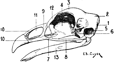
Fig. 65.—Skull of the Cock: Left Lateral Surface.
1, Occipital bone; 2, parietal bone; 3, frontal bone; 4, ethmoid bone; 5, cavity of the tympanum; 6, quadrate bone; 7, superior maxillary bone; 8, malar bone; 9, nasal bone; 10, 10, intermaxillary bone; 11, nasal orifice; 12, os unguis or lachrymal bone; 13, inferior maxillary bone.
In this group the skull is generally pear-shaped; to the cranium, of which the bones are arranged in such a way as to give it a form more or less spherical, succeeds a face more or less elongated, according as the bill is more or less developed.
In general, the bones of the skull coalesce very early, with the result that it is only in very young individuals that we can determine their presence.
We find the skull to consist of an occipital bone, two parietals, a frontal, etc.; we will indicate but one detail in connection with these bones: it is the presence of a single condyle for the articulation of the occipital bone with the atlas. We also note the quadrate bone, which is situated on the lateral part of the cranium, is movable on this latter, and acts as an intermediary between it, the bones of the face, and the lower jaw. The quadrate bone is regarded as a detached portion of the temporal; on the signification of this we do not now propose to dwell.
On the anterior portion of the face we find the nasal bones, which, articulating with the frontal on one side, circumscribe, on the other, the posterior border of the nares. The nasal bone of the one side is separated from that of the opposite by the intermaxillary or premaxillary bone, which forms the skeleton of the superior mandible.
The superior maxillaries, which are rudimentary, are situated on the lateral parts, and prolonged backward by an osseous style which articulates with the quadrate bone; this styloid bone, the homologue of the malar, is designated by certain authors as the jugal or quadrato-jugal bone.
It is with the quadrate bone also that the inferior maxillary articulates.