Chapter II · (continued 3)
Reading edition
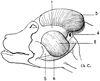
Fig. 89.—Myology of the Dog: Masticatory Muscles (a Deeper Dissection than that shown in Fig. 90).
1, Zygomatic arch; 2, masseter; 3, temporal exposed by the suppression of the auricular and occipital muscles and the pinna of the ear; 4, auditory canal; 5, inferior maxillary bone; 6, digastric.
Indeed, in the dog and the cat the zygomatic arch, strongly convex, springs up in a marked manner from the plane of the lateral aspects of the skull.
In the horse the same arch, less prominent externally, is prolonged by a rectilinear crest on the superior maxillary bone, where it is continued in forming the zygomatic or maxillary spine.
In the ox the same crest ascends a good way towards the inferior margin of the orbit in a curved direction with the concavity inferior, to redescend afterwards on the external surface of the superior maxilla.
The masseter is an elevator of the lower jaw. It acts, above all, as in the human species, in the process of mastication.
Temporal Muscle (Fig. 89, 3).—The development of the temporal is in proportion to the energy of the movements of elevation which the lower jaw has to execute.
It arises from the temporal fossa, and is inserted into the coronoid process of the inferior maxilla.
Its development, enormous in the carnivora, is such that the muscle projects beyond its fossa. It is less voluminous in the horse, and still less so in the ox. In the latter, indeed, the temporal fossa, although deep, is of small extent (see Fig. 62, p. 119); the frontal bone being large, it is found to be thrown back on the lateral walls of the cranium, below the osseous processes which support the horns and overhang the fossa in question, as well as the muscle which it contains.
It is covered by the auricular muscles, and by the base of the pinna of the ear.
Like the masseter, the temporal is an elevator of the lower jaw.
Cutaneous Muscles of the Head
Occipito-Frontalis.—The epicranial aponeurosis is extremely thin. In the dog the occipital muscle occupies the superior part of the head; it overlies the temporal muscle.
With regard to the frontal muscle, which is of great extent in the ox (Fig. 91, F), it is represented in the horse and the carnivora by a small fleshy fasciculus only, the fronto-palpebral muscle, similar to the superciliary muscle. This, occupying the superior and internal part of the border of the orbit, ends by blending its fibres with those of the orbicular muscle of the eyelids at the region of the eyebrow.
Orbicularis Palpebrarum (Fig. 90, 2; Figs. 91, 92).—This annular muscle surrounds the palpebral orifice, and takes its origin on the internal part of the orbital region. In the horse it arises, by a small tendon, from a tubercle which occupies the external surface of the os unguis, or lachrymal bone.
This muscle determines the narrowing and closure of the palpebral orifice.
Pyramidalis Nasi.—The pyramidal muscle is not found in the domestic animals. It appears to be blended with the internal elevator of the upper lip and wing of the nose; this is easy of comprehension if we bear in mind the relative position of these two muscles in the human species.
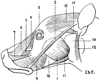
Fig. 90.—Myology of the Dog: Muscles of the Head.
1, Masseter; 2, orbicularis palpebrarum; 3, zygomaticus major; 4, lachrymal (this muscle is sometimes described under the name of the small zygomatic); 5, levator labii superioris proprius; 6, levator labii superioris alæque nasi; 7, caninus; 9, buccinator; 11, zygomatico-auricularis; 12, external temporo-auricularis; 14, parotid gland; 15, parotido-auricularis; 16, inferior maxillary bone; 17, digastric.
Corrugator Supercilii.—This muscle is represented by the fronto-palpebral muscle noticed above, which is by some regarded as a vestige of the frontal.
Zygomaticus Major (Fig. 90, 3; Figs. 91, 92).—This is the zygomatic-labial of veterinarians. This muscle is of an elongated form, and has a ribbon-like aspect.
In the dog and the cat it arises from the base of the pinna of the ear, from the portion of this base which bears the name of scutiform cartilage. (With regard to this cartilage, see p. 242, Zygomatico-auricularis.) From this it is directed downwards and forwards, to terminate, after having crossed the masseter, on the deep surface of the skin of the corresponding labial commissure.
This mode of termination is the same in the ox and the horse; but where the muscle differs is at the level of its upper extremity. There it ascends less than in the carnivora. In the ox it arises from the zygomatic arch in the neighbourhood of the temporo-maxillary articulation; in the pig and the horse its origin is still lower, on the surface of the masseter, close to the maxillary spine.
When it contracts, it draws upwards the labial commissure.
Now, in man, we remember, it is the great zygomatic that, by an action of the same kind, determines the essential characters of the expression of laughing.
There is, accordingly, a connection to be established between those displacements which are similar and the analogy of facial expression which necessarily results from them.[33]
Zygomaticus Minor (Fig. 90, 4; Figs. 91, 92).—The existence of this muscle has not been clearly demonstrated. Nevertheless, Straus-Durckheim noted its presence in the horse, and described it as ‘a muscle arising by two heads, of which one, the superior, arises from the malar bone below the orbit, and passes downwards and forwards over the fibro-adipose layer which supports the moustache. The second, the inferior, arises from the alveolar border in front of the second molar tooth, and passes forward to be inserted into the same fibro-adipose layer.’[34]
In connection with other quadrupeds, it is described by certain authors as a very thin muscle, arising below the cavity of the orbit, where it is blended with the fibres of the internal elevator of the upper lip and the ala of the nose; thence it proceeds to terminate below by uniting with the subcutaneous muscle. But this muscle is regarded by other authors as the lachrymal muscle, which does not exist in this state in man, but of which the development is particularly remarkable, as to extent, in the ox, in which it descends as far as the buccinator.
According to other authors, some of the fibres of this muscle constitute the small zygomatic.
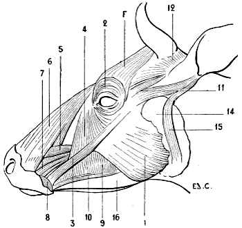
Fig. 91.—Myology of the Ox: Muscles of the Head.
1, Masseter; 2, orbicularis palpebrarum; F, frontalis; 3, zygomaticus major; 4, lachrymal (this muscle is sometimes described under the name of small zygomatic); 5, levator labii superioris proprius; 6, levator labii superioris alæque nasi; 7, levator anguli oris or caninus; 8, orbicularis oris; 9, buccinator; 10, maxillo-labial; 11, zygomatico-auricularis; 12, external temporo-auricularis; 14, parotid gland; 15, parotido-auricularis; 16, inferior maxillary bone.
Levator Labii Superioris Proprius (Fig. 90, 5; Figs. 91, 92).—Also named by veterinarians the supramaxillo-labial, or again, the proper elevator of the upper lip, this muscle arises from the external surface of the superior maxillary bone, passes under the superficial elevator, which we shall study in the succeeding paragraph, and goes to be inserted into the thickness of the lip, to which its name indicates that it belongs.
The peculiarities of this muscle in different animals are the following:
In the dog and the cat it arises behind the infra-orbital foramen.
In the pig it arises from a depression below the orbital cavity, and its fleshy body is terminated in front by a strong tendon in the upper part of the snout, in which it divides into fasciculi.
In the ox it arises from the maxillary spine.
In the horse it arises below the orbital cavity; then, after having crossed the superficial elevator, it ends in a tendinous expansion, situated in the median line between the nasal fossæ. This expansion divides into fasciculi, which end in the thickness of the upper lip.
By the contraction of this muscle, the lip is raised, on one side only, if a single muscle contracts, or in its whole extent, if the two muscles act simultaneously.
Internal Elevator (or Superficial) of the Upper Lip and the Wing of the Nose (levator labii superioris alæque nasi) (Fig. 90, 6; Figs. 91, 92).—This is the muscle veterinarians designate the supranaso-labial.
Arising from the frontal and nasal bones, it thence passes towards the upper lip, where it is inserted as well as into the wing of the nose.
In the ox it is united above with the frontal muscle, and below is divided into two fasciculi, between which pass the elevator described above and the canine muscle.
In the horse it is also divided into two fasciculi; but the arrangement is the opposite as regards, their relations with neighbouring muscles, in this animal and in the preceding.
In the ox the external fasciculus is covered by the external elevator and the canine, which pass under the internal fasciculus; in the horse the deep elevator passes under the two fasciculi, and the canine passes under the external bundle, and afterwards covers the internal.
In the pig, the internal elevator is wanting.
As its name indicates, it raises the upper lip and the wing of the nose.
Transversus Nasi.—In the horse this muscle, which is very thin, is situated on the dorsum of the nose, and proceeds to be inserted into the cartilaginous skeleton of the nostrils. In the pig, it occupies an analogous situation. It does not exist in the ox or in carnivora. The transversus nasi is a dilator of the nostrils.
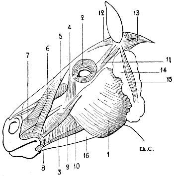
Fig. 92.—Myology of the Horse: Muscles of the Head.
1, Masseter; 2, orbicularis palpebrarum; 3, zygomaticus major; 4, lachrymal (this muscle is sometimes described under the name of the small zygomatic); 5, external elevator (or deep) of the upper lip and ala of the nose; 6, internal elevator (or superficial) of the upper lip and of the ala of the nose; 7, levator anguli oris or caninus; 8, orbicularis oris; 9, buccinator; 10, maxillo-labialis; 11, zygomatico-auricularis; 12, temporo-auricularis externus; 13, cervico-auricularis; 14, parotid gland; 15, parotido-auricularis; 16, inferior maxillary bone.
Caninus (Fig. 90; Figs. 7, 91, 92).—This is the muscle called by veterinarians the great supramaxillo-nasal.
In the dog and the cat it is situated below the inferior border of the external elevator of the upper lip, of which it follows the direction. It arises, as does this latter, from the external surface of the maxilla, and goes also to terminate in the upper lip by blending with the internal elevator of this lip and of the alæ of the nose. It raises the upper lip.
In the ox, it arises from the maxillary spine, and then divides into three parts; the superior passes under the internal portion of the internal elevator of the upper lip and the alæ of the nose, and goes into the nostril; whilst the two others, situated lower down, terminate in the upper lip.
In the pig, it is formed of two superimposed fasciculi, which arise from the spine of the maxilla and the impressions in front of it. These two fasciculi terminate in the snout, which they move laterally.
In the horse, it is situated at a certain distance from the external elevator; in the preceding animals it is in contact with the latter. Arising behind from the external surface of the maxilla, in front of the maxillary spine, it is directed towards the anterior part of the face, passes under the external portion of the internal elevator (it is the opposite of this in the ox), and proceeds, on expanding, to terminate in the skin of the nostril. By its contraction it dilates the latter.
Orbicularis Oris (Fig. 91, 8; Fig. 92).—This muscle, very fleshy in the solipeds and the ruminants, is arranged as a ring round the buccal orifice, in the thickness of the lips, where it is blended with the other muscles of this region.
Having for its function the narrowing of the orifice it surrounds, it acts during suction and in the prehension of food.
Triangularis Oris.—This muscle does not exist in domestic quadrupeds.
Quadratus Menti.—In the pig and the carnivora, it arises from the anterior part of the body of the inferior maxillary bone, and passes at the other end to terminate in the corresponding portion of the lower lip, which it depresses by its contraction.
In the ox and the horse this muscle does not exist; it is replaced for the depression of the lower lip, which it affects in other animals, by supplemental fibres of the buccinator.
The Prominence of the Chin.—Below the lower lip in the horse is situated the so-called prominence of the chin, limited posteriorly by the beard, a depressed region which gives point to the curb of the bridle.
The prominence, which also exists in the ox, is a fibro-muscular pad which blends with the orbicular muscle of the lips, and receives on its superior aspect the insertion of the two muscles (levator menti) by which it is suspended. These arise, above, on each side of the symphysis of the inferior maxillary bone. They raise the lower lip with force, and they are the agents which, as we can sometimes observe in the horse, make it click against the upper lip, suddenly projecting it upwards. This action sometimes becomes a habit, and its continuance constitutes a vice.
A corresponding structure is found in the pig and in the carnivora, but in them it does not produce an external prominence such as we have described.
Buccinator (Fig. 90, 9; Figs. 91, 92).—Further designated by the name of alveolo-labial, this muscle is situated on the lateral portions of the face, in the thickness of the cheeks. It consists of two layers, one superficial and the other deep.
The deep portion arises from the portion of the alveolar border of the superior maxillary bone which corresponds to the molar teeth, and from the anterior border of the ramus of the mandible. Thence it is directed forwards, passes under the superficial layer, and blends with the fibres of the orbicular muscle of the lips. To this part of the buccinator some authors give the name of molar muscle.
The superficial portion is formed by fibres which pass from the alveolar border of the superior maxillary bone to the corresponding border of the opposite bone. It is very highly developed in the herbivora.
This muscle acts especially during mastication; it serves to press back again under the molar teeth the portions of food which fall outside the dental arch.
In the pig, the ox, and the horse, a muscle which is considered as supplemental to the buccinator is placed along the inferior border of the latter.
This muscle, which we describe separately under the names of maxillo-labialis (Fig. 91, 10; Fig. 92) and depressor of the lower lip, is clearly distinct from the buccinator, especially in the horse. It arises, behind, with the deep layer of the muscle to which it is annexed, from the anterior border of the ramus of the lower jaw; in front it terminates in the thickness of the lower lip.
In the ox, it is more intimately united with the buccinator.
It depresses the lip to which it is attached, and displaces it laterally when it acts on one side only.
In the human species, the pinna of the ear being generally immobile, the muscles which belong to it are, very naturally, considerably atrophied. Accordingly, the auricular muscles, anterior, superior, and posterior, are reduced to pale and thin fleshy lamellæ, whose action is revealed in certain individuals, only in a way which may be said to be abnormal.
It is not the same in quadrupeds. The pinna of the ear is extremely mobile, and its displacements have a real value from the point of view of physiognomical expression. It is therefore necessary to review the muscles which move this pinna without giving them, at the same time, more importance than they merit, since in themselves they do not determine the formation of surface reliefs, which are sufficiently apparent.
Notwithstanding that for certain of these muscles it is possible to trace their analogy with those of the auricular region of man, it is very difficult, because of their complexity, to trace this analogy for all. This is why we shall not be able here, as we have done for the other muscles of the subcutaneous layer, to give at the head of each paragraph the name of a human muscle, and then to group in the same paragraph the muscles which correspond to it in different quadrupeds. Therefore the nomenclature and the divisions adopted for these latter must serve us as a base or starting-point.
Because the pinna of the horse’s ear is so very mobile, we will first begin with a study of its auricular muscles.
Zygomatico-auricularis (Fig. 92, 11).—This muscle, which is formed of two small bands of fleshy fibres, arises from the zygomatic arch in blending with the orbicular muscle of the eyelids; thence it is directed towards the base of the pinna of the ear, and is inserted into this base, and also into the cartilaginous plate situated in front of and internal to this, and resting on the surface of the temporal muscle; this is the scutiform cartilage.
The zygomatico-auricularis, which we look on as the homologue of the anterior auricular of man, draws the pinna of the ear forwards.
Temporo-auricularis Externus (Fig. 92, 12).—This, which is thin and very broad, covers the temporal muscle.
It arises from the whole extent of the parietal crest, blending in this plane, in its posterior half, with the muscle of the opposite side. Thence it is directed outwards towards the pinna of the ear, and is inserted into the internal border of the scutiform cartilage and on the inner side of the concha—that is to say, of the conchinian cartilage—which forms the principal part of the pinna. We are supposing, in the description of the muscles which move it, that this pinna has its opening directed outwards.
The external temporo-auricular, which recalls, from its situation, the superior auricular of man, is an adductor of the ear; besides, it causes it to describe a movement of rotation from without inwards, so as to direct its opening forwards.
Scuto-auricularis Externus.—This muscle may be considered as supplementary to the external temporo-auricular; the concha fasciculus of this latter partly covers it.
Extending from the scutiform cartilage to the inner side of the concha, it contributes to the movement of rotation by which the opening of the pinna of the ear is directed forwards.
Cervico-auricular Muscles (Fig. 92, 13).—These muscles, three in number, are situated behind the pinna of the ear; they are called, from their mode of superposition, the superficial, middle, and deep.
These arise, all three, from the superior cervical ligament, and pass from there towards the cartilage of the concha. They recall, as regards situation, the posterior auricular muscle of man.
Superficial Cervico-auricular (Cervico-auricularis superioris).—This muscle, inserted into the posterior surface of the concha, draws this cartilage backwards and downwards.
Middle Cervico-auricular (Cervico-auricularis medius).—Situated between the two other muscles of the same group, it proceeds, after having covered the superior extremity of the parotid gland, to be inserted into the external part of the base of the concha. It determines the rotation of this concha in such a way as to direct the opening of the ear backwards.
Deep Cervico-auricular (Cervico-auricularis inferioris).—Covered by the preceding muscle and the superior portion of the parotid, it is inserted into the base of the pinna of the ear, and has the same action as the middle cervico-auricular.
Parotido-auricularis (Fig. 92, 15).—This is a long and thin fleshy band which arises from the external surface of the parotid gland, and tapering as it passes upwards towards the pinna of the ear, is inserted into the external surface of the base of the concha, below the inferior part of the angle of reunion of the two borders which limit its opening.
It inclines the pinna outwards; it is, accordingly, an abductor of the pinna.
Temporo-auricularis Internus.—This muscle is covered by the external temporo-auricular and the superior cervico-auricular. It arises from the parietal crest, and is inserted into the internal surface of the concha. It is an adductor of the pinna of the ear.
There are, finally, an internal scuto-auricular muscle and a tympano-auricular; but they do not present any interest for us; we can simply confine ourselves to making mention of them.
In the ox, because of the situation of the temporal fossa and the fact that the external temporo-auricular muscle is applied, as in the horse, over the muscle which this fossa contains, this temporo-auricular muscle does not reach the middle line (Fig. 91, 12).
But in the cat and the dog this muscle covers all the upper part of the head (Fig. 90, 12). It is divided into two parts: the interscutellar and the fronto-scutellar.
The interscutellar is a single muscle, thin and broad, covering the temporal muscle and a portion of the occipital, extending from the scutiform cartilage of the pinna of one side to the same cartilage of the pinna belonging to the side opposite. It approximates the two pinnæ to one another by bringing them each into the position of adduction.
The fronto-scutellar arises from the orbital process of the frontal bone, and from the orbital ligament, which at this level completes the interrupted osseous boundary of the orbital cavity. Thence it is directed, widening as it proceeds, towards the scutiform cartilage, and is there inserted by blending with the corresponding part of the great zygomatic. Its action is analogous to that of the preceding muscle; but, further, it directs the opening of the pinna forwards.
These are the muscles which act, for example, when the dog, having his attention strongly attracted by any cause, pricks up his ears and turns the openings forward, in order the better to understand every sound which proceeds, or may possibly proceed, from that which he observes. From this, which may be extremely well seen in some individuals, results the appearance of vertical wrinkles of the skin in the interval between the pinnæ of the ears, these being caused by the folding of the integument, whilst the pinnæ approach one another. These movements, with which are associated fixation of look and a widening of the palpebral fissure, produce a peculiarly expressive look; this is why they merit our attention.
Zygomatico-auricularis (Fig. 90, 11).—Arises from the internal surface of the great zygomatic, passes towards the pinna of the ear, and goes to be inserted into the external part of the base of the pinna, below its opening, to a prominence which corresponds to the antitragus of the human ear. It is to this antitragus, but proceeding from another direction, that the parotido-auricular muscle is inserted (Fig. 90, 15).
With regard to the cervico-auriculars, they are all three present. The superior, or superficial, situated behind the interscutellar portion of the external temporo-auricular, has its origin on the median line of the neck; thence it passes towards the pinna of the ear, blending its fibres with those of the interscutellar muscle, and is inserted into the scutiform cartilage and the internal surface of the pinna.
Such are the principal muscles of the ear in the carnivora; it would seem to us superfluous to dwell on the others of this region, so that we will here conclude the study of the muscles in general, and that of the myology of the head in particular.