Chapter I · (continued 2)
Reading edition
The forearm is directed obliquely downwards and inwards, so as to form, with the hand, an angle of which the apex is internal; this angular outline of the knee (wrist) is so characteristic of ruminants that the corresponding region of the horse, when salient inwards, receives the name of ox-knee. The radius bears the coronoid process, and the larger part of the articular surface which comes in contact with the inferior extremity of the humerus; the condyle and the trochlea articulate with the radius in front; while behind, the trochlea articulates with that part of the sigmoid cavity which belongs to the ulna. The posterior surface of the shaft of the radius is flattened; its anterior surface is slightly convex. The inferior extremity articulates with the carpus by a surface which is directed obliquely downwards and inwards. The shaft of the ulna is very slender, and fused in its middle third with the body of the radius; it terminates below, at the level of the external part of the inferior extremity of the radius, by a slightly expanded portion which, fused with this latter, forms the articular surface for the carpal bones.
In the ox the forearm is short; in the sheep it is proportionally longer.
The bones of the carpus are six in number—four in the upper row, and two in the lower; they form an irregular cuboid mass which contributes to the formation of the region known as the knee in ruminants, as in the horse; we have already remarked that the name ‘wrist’ would be more accurate. The anterior surface in its foremost part is vertical, and is slightly convex from side to side. At its posterior and external part the pisiform bone forms a prominence.
The metacarpus consists of two bones only—one, well developed, which is known as the principal metacarpal, or the canon bone (this is the name given to the region in the hoofed animals); and a rudimentary one, which is situated at the superior and external aspect of the preceding metacarpal. Sometimes there is found a third metacarpal at the internal aspect; but, when present, it is but very slightly developed.
The principal metacarpal consists of two metacarpals fused together; on this account the bone is longitudinally marked in the median line by a slight depression which marks the junction of the two bones of which it is formed. In some ruminants (certain species of chevrotains) the coalescence does not take place, and the two metacarpals remain separate.
The anterior surface of the principal metacarpal is convex transversely; its posterior surface is flattened. The superior extremity of this bone articulates by two facets with the two bones of the inferior row of the carpus; on the internal part of the anterior surface of this extremity is found a tubercle. The inferior extremity is divided into two parts by a fissure or notch; each part is articular, and consists of two separate condyles, which are separated from each other by an antero-posterior crest; on each side of this crest, and behind, are found two sesamoid bones. As for the external rudimentary metacarpal bone, it is nothing more than a small, short tongue of bone; which, in goats and sheep, is often absent.
The division of the inferior extremity of the principal metacarpal into two parts is correlated with the two perfect digits which give the foot of the ruminant its forked appearance. Each digit consists of three phalanges, which are directed obliquely downwards and forwards; further, these phalanges are inclined a little outwards from the axis of the limb, so that the two digits diverge from each other as they descend.
The first phalanx, which is the longest, articulates superiorly with the principal metacarpal; its inferior extremity terminates in a trochlea, and the lip of this, which is situated towards the axis of the limb, descends lower than that of the opposite side; this arrangement is correlated with the divergent direction of the digits. The second phalanx has its superior extremity moulded on the trochlea which terminates the extremity of the first; its inferior extremity is articular, and elongated from before backwards. On the posterior surface of this extremity is found a sesamoid bone.
With regard to the third phalanx, it presents the form of a triangular pyramid, and displays a postero-superior concave surface with which the second phalanx articulates; an anterior, convex surface, which terminates in a point on its anterior part; and an internal surface, which is flattened. The third phalanx of each digit is contained in a hoof (onglon).
There is also found in ruminants two imperfect rudimentary digits, which are represented by two small bones situated behind the articulation of the metacarpal and the digits which we have just been studying. These rudimentary digits are each enveloped in a layer of horn; they constitute the spurs. The two digits of the ruminants represent the third and fourth fingers of the human hand; the two lateral digits, greatly atrophied, are the homologues of the second and fifth fingers; the thumb is not present.
It is the same as regards the metacarpal bones, which form, by their union, the principal metacarpal; the external represents the fourth metacarpal, and the internal the third. It is to the latter that the tubercle, of which we have already made mention, belongs; and with the signification of which, because it gives attachment to a muscle, we shall concern ourselves in the section on myology (see Radial Muscles).
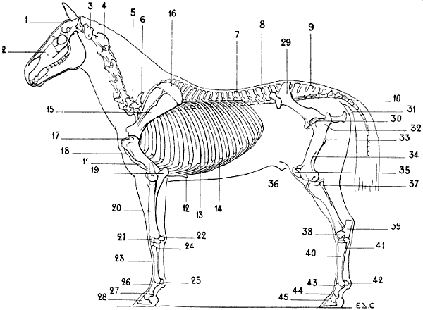
Fig. 40.—Skeleton of the Horse: Left Lateral Surface.
1, Cranium; 2, face; 3, atlas; 4, axis; 5, seventh cervical vertebra; 6, first dorsal vertebra; 7, eighteenth and last dorsal vertebra; 8, lumbar vertebræ; 9, sacrum; 10, coccygeal vertebræ; 11, sternum; 12, xiphoid appendix; 13, eighteenth and last sternal rib; 14, costal cartilage; 15, scapula; 16, cartilage of extension; 17, great tuberosity of the humerus; 18, deltoid crest; 19, olecranon process; 20, radius; 21, carpus; 22, pisiform; 23, principal metacarpal; 24, metacarpal, external rudimentary; 25, large sesamoids; 26, first phalanx; 27, second phalanx; 28, third phalanx; 29, ilium, showing external iliac fossa; 30, pubis; 31, tuberosity of the ischium; 32, great trochanter; 33, infratrochanteric crest, or third trochanter; 34, supracondyloid fossa of the femur; 35, knee-cap; 36, anterior tuberosity of the tibia; 37, the fibula; 38, tarsus astragalus; 39, calcaneum; 40, principal metatarsal; 41, rudimentary external metatarsal; 42, large sesamoids; 43, first phalanx; 44, second phalanx; 45, third phalanx.
To face p. 64.
Unguligrades: Horse (Fig. 40).—The scapula is narrow, compared with that of the animals we have just been considering. The anterior border is convex in its superior portion, and concave in its inferior; the posterior border is slightly hollowed out. The supraspinous fossa is less in extent than the infraspinous; but the difference is less than that between the same fossæ in the ox and the sheep; in the ox, as we have already indicated, the proportion is one-third; in the horse, one-half. The spine, which disappears at the extremities, is rough and thick in its middle third, there forming a kind of tuberosity—tuberosity of the spine. Above and in front of the glenoid cavity is found a strong process consisting of a rugous base, and a summit which is directed inwards. This forms a kind of hook curved towards the inside; it represents the coracoid process. The scapula is surmounted by the cartilage of prolongation, of which the superior border, which is thin and curved, is parallel to the superior border of the prominence of the withers; the cartilage forms, consequently, the lateral surface of this region. The cartilage of prolongation undergoes ossification in old horses. The humerus is short; the bicipital groove, situated on the anterior surface of the superior extremity, separates the greater tuberosity from the lesser, and is divided into two parts by a median ridge; it is this portion of the humerus which forms the prominence known as the point of the shoulder, or point of the arm. The deltoid impression well deserves the name of tuberosity which has been given to it, for it is very prominent; the musculo-spiral groove is very deep.
At the inferior extremity, the trochlea is large; the portion corresponding to the condyle of the humerus in man is, in proportion to the latter, of small extent. The olecranon fossa is deep. The epicondyle and the epitrochlea are somewhat different from those of the human bone. In the latter, the epitrochlea is salient towards the inner side, causing an increased transverse diameter of the inferior extremity of the humerus. In the horse—it is the same in ruminants—this tuberosity projects backwards, folds on itself in forming the internal boundary of the olecranon cavity, and exceeds in diameter, in the antero-posterior direction, the prominence of the epicondyle, which presents a nearly similar arrangement. This latter has, however, a part which, projecting externally, is situated at the inferior part of a crest, that forms the posterior boundary of the musculo-spiral groove. The result is that, contrary to the condition found in the human being, the epicondyle is more prominent transversely than the epitrochlea, but this latter is more salient on the posterior aspect. The epitrochlea and the epicondyle offer a larger surface for the origin of muscles of the forearm than the same prominences in the human bone do for the analogous muscles of the same region.
Some veterinary anatomists have given to the inferior and external articular surface of the humerus the name of trochlea; and to the internal one, that of condyle. On this account they designate the external prominence as the epitrochlea, and the internal one as the epicondyle. In addition to the fact that this point of view is not legitimate, it produces inevitable confusion when comparing the parts with those of the human humerus, and this confusion exists, not alone in describing the bone, but also in the description of the muscular attachments, and in the comparison of the muscles of the forearm of quadrupeds with the corresponding muscles in the human species.
The radius is placed in front of the ulna; its body, slightly convex forwards, has the anterior surface convex transversely, and the posterior surface plane in the same direction. It is to the external part of this latter that the ulna is applied, which is completely fused with the radius.
The superior extremity of the radius is a little larger than the inferior. Its superior aspect, concavo-convex, moulded on the inferior articular surface of the humerus, presents internally two cavities, which receive the lips of the trochlea, and, externally, another, smaller, cavity, which receives the condyle. The radius articulates with the trochlea and the condyle, having appropriated a portion of the ulna, as is proved by the presence of the coronoid process, which belongs to the former. This superior extremity presents, internally, a tuberosity into which the biceps is inserted; this is the bicipital tuberosity; and on the other side is another tuberosity, which is a little more prominent than the preceding.
The inferior extremity, which is flattened from before backwards, is furrowed on its anterior surface by grooves for the passage of muscles (the names of the muscles whose tendons pass in these grooves have already been given on p. 43). It articulates at the lower end with the superior row of the carpus, and it terminates laterally in tuberosities: one, external, on which is found a groove for the tendon of the lateral extensor of the phalanges, the homologue of the special extensor of the little finger; the other, internal, is a little more prominent than the one we have just described. These tuberosities are visible under the skin which covers the superior and lateral parts of the region known as the knee; but which, we again repeat, is no other than the wrist.
The ulna has a triangular shaft, situated at the posterior surface of the radius, with which it is fused. It disappears completely at the level of the inferior third of the forearm. Occasionally, in some horses, the ulna is abnormally long, in the form of a slender tongue of bone; and extends to the neighbourhood of the external tuberosity of the inferior extremity of the radius (see Fig. 79, p. 196). Its superior extremity is chiefly represented by the olecranon process, which is voluminous in bulk, and forms the projection known as the point of the elbow. This process is flattened laterally; its internal surface is excavated; the anterior surface, which is concave, forms a part of the great sigmoid cavity; the remainder of the cavity is formed by the radius.
In the ass, the ulna is a little longer than in the horse—that is to say, it descends lower; and the radius is a little more convex anteriorly.
The carpal bones are seven in number—four in the superior row, and three in the inferior. The trapezium is wanting in the latter. Sometimes, however, in certain varieties of horses the trapezium is developed, but then it is no more than a very small osseous nodule. The pisiform bone, situated at the external part of the first row of bone, is prominent posteriorly. It is of rounder form and flattened from without inwards. It articulates with the trapezium and the radius. It presents, on its external surface, a groove for the passage of the tendon of the posterior ulnar muscle, which is named by veterinary anatomists the external flexor of the metacarpus.
The carpus, as a whole, is of an irregularly cuboid shape; its anterior surface, slightly convex from side to side, forms the skeleton of the region of the knee (wrist). The metacarpus is formed of three bones: the principal metacarpal and the two rudimentary ones.
The principal metacarpal, which forms the region of the canon, is directed vertically; its anterior surface is slightly convex transversely. This surface is covered by a number of tendons, which slightly alter its appearance; so that it is the principal base of this part of the fore-limb. Its posterior surface is flattened. The superior extremity of this metacarpal presents plane surfaces, variously inclined, with which the bones of the inferior row of the carpus articulate. On the anterior surface, and a little to the inner side, is found a tuberosity, which is destined for the insertion of the anterior extensor of the metacarpus, the homologue of the radial muscles. The inferior extremity is formed by two condyles, an internal and an external; between which is found a median crest.
This extremity, the superior extremity of the first phalanx, which articulates with it, together with two sesamoid bones—the great sesamoids—which are situated on its posterior surface, collectively form the region which from its rounded outlines is called the ball.
With regard to the rudimentary metacarpals, external and internal, to which some authors give the name of fibulæ, they are applied to the sides of the posterior surface of the principal metacarpal. They are elongated bones, of which the superior extremity, which is a little thickened, is called the head; the lateral bones of the second row of the carpus partly rest on the heads of these. They become more slender as they descend, and terminate opposite the inferior fourth of the principal metacarpal. Each ends in a slight swelling, to which the name button has been given. The internal one is the better developed.
The rudimentary metacarpals are vestiges of atrophied digits, as will be explained further on.
The single finger of the horse consists of three phalanges. The first phalanx, which is directed obliquely downwards and forwards, corresponds to the constricted region situated below the ‘ball,’ and known as the pastern. It is flattened from before backwards; its anterior surface is convex transversely, while the posterior surface is plane. Its superior extremity is moulded on the inferior extremity of the principal metacarpal, and its inferior extremity, which is smaller, presents a trochlea with which the second phalanx articulates. This is also directed downwards and forwards, and is shorter. It corresponds to the region which, situated between the pastern and the hoof, is known as the cornet.
The third phalanx, situated entirely within the hoof, has the same direction as the first and second. It is large and broad, and presents three surfaces separated by well-marked angular borders (see Fig. 96). The anterior surface is oblique downwards and forwards; it is convex transversely. The inferior surface is slightly hollowed, and is in relation with the sole, or plantar surface of the hoof.
The superior surface, which is articular, is divided by a median ridge into two lateral cavities, which correspond to the trochlea on the inferior surface of the lower extremity of the second phalanx. The inferior border corresponds in shape with the hoof. The superior border presents in its median part a projection, the pyramidal eminence, which prolongs at this level the anterior surface of the bone. Finally, the posterior border, which is concave, is in contact with a sesamoid bone, the lesser sesamoid, which increases the superior articular surface behind, and is also in contact with the second phalanx.
As we have just seen, the horse possesses but one digit. In the ancestors of the animal—that is, in the prehistoric species which are now extinct (orohippus, miohippus, protohippus, or hipparion)—the number of digits was larger; this fact conclusively proves that the rudimentary metacarpals of the existing horse are vestiges of digits which have disappeared through want of use. In the first of those ancestors—orohippus—there were four digits; all save the first, the thumb, being then developed. In the others of the series there existed but three digits. It must, however, be noted that in those animals it is always the digit which corresponds to the middle finger of the pentedactyl hand that is longest. In other less ancient species the lateral fingers are reduced to the condition of mere splints of bone. It follows from what has been said that the digit which persists in the equine species should be considered as the third finger, and that the rudimentary metacarpals represent lateral digits considerably atrophied.
This disappearance of the lateral digits cannot excite surprise when we consider the functions of the organs. Becoming useless, they must undergo gradual atrophy from want of use.
There undoubtedly is, in this former existence of supplementary digits in the horse, something analogous to what we still find in the pig; where the two principal digits are accompanied by two shorter ones, which very probably, from their infrequent use, are destined to disappear in a more or less distant future.
Proportions of the Arm, the Forearm, and the Metacarpus
As a supplement to the study of the anterior limbs which we have just finished, it appears necessary to give some indications of the relative proportions of certain of the segments which form these limbs in the plantigrades, the digitigrades, and the ungulates.
First, we would remark that, in following this order of classification, the scapula becomes less and less narrow, and assumes a form more and more elongated. In order to convince ourselves of this, it will be sufficient to study the bone first in man, then in the bear, the cat, dog, ox, and finally in the horse.
As to the proportions of length, which are those we should chiefly study, we shall commence with the comparison of the forearm and arm—that is to say, the radius and the humerus. The radius is found to be longer in proportion to the humerus, as the number of digits is smaller, and the hand loses more and more the functions of an organ of prehension. In man, the radius is shorter than the humerus; in the horse, on the contrary, it is longer.
To give an idea of this proportion, we shall employ what is known as the antibrachial index. This index gives the relation which exists between the length of the forearm and that of the humerus; the length of this latter, whatever may be the actual measurement, is represented by a fixed figure, the number 100. A very simple arithmetical operation gives the proportion—
forearm × 100 , the quotient obtained furnishes the index.
humerus
The index is less than 100 if the forearm is shorter than the bone of the arm. The index is more than 100 if, on the contrary, the forearm is longer.
In man, the radius is shorter than the humerus; indeed, in adult individuals of the white race the average index is 74.
In the bear, the length of the radius approaches closely to that of the humerus; the index is about 90. In the skeleton of a bear in the anatomical museum of the École des Beaux-Arts, the humerus is 33 centimetres in length, and the radius 30 centimetres.
In the cat, the radius is very little shorter than the humerus. In the dog they are equal. The antibrachial index of the latter is, accordingly, 100.
In the horse, the radius is longer than the humerus; the index is therefore above 100. Thus, in the skeleton of the horse which we have in the museum of the École des Beaux-Arts, the index is 113—length of humerus, 29 centimetres; length of radius, 33 centimetres. In other skeletons which we have measured we found: in one, 108—humerus, 34 centimetres; radius, 37 centimetres; in another, 116—humerus, 25 centimetres; radius, 29 centimetres.
The metacarpal bone undergoes, relatively to the humerus, a proportional elongation, analogous to that of the forearm.
In man, the length of the metacarpus is contained about 51⁄2 times in that of the humerus; in the bear, it is contained 4 times; in the dog, 21⁄2 times; in the horse, 11⁄3 times only.
It is well known that the proportions vary according to race, and that what we have here given are but the general indications.
The Articulations of the Anterior Limbs
The knowledge of human arthrology which we presume the reader to have previously acquired makes it unnecessary for us to enter into numerous details regarding the configuration of the articular osseous surfaces and the disposition of the fibrous bands that retain them in position. Accordingly, in the description which follows, and also in that of the articulations of the posterior limbs, we shall occupy ourselves but very briefly with the details above referred to, so as to devote ourselves especially to the indication of the movements—that is to say, of that which, while easily comprehended on recollection of former studies, presents the greatest interest from the artistic standpoint in these studies in comparative anatomy.
The Scapulo-Humeral Articulation.—The head of the humerus and the glenoid cavity of the scapula being in contact, the two bones are bound together by a rather loose articular capsule, which is strengthened by the muscles of this region which fulfil the function of active ligaments.
This articulation, so movable in every direction in the human species, is not so much so in quadrupeds; the arm in the latter, as also the shoulder, being kept in contact with the lateral region of the thorax by the numerous muscles which surround it.
Of the movements performed by the humerus, flexion and extension are the most extensive; those of abduction and adduction are much less so.
It is necessary, before proceeding further, to determine what the two principal movements which we have just mentioned really are, viz., flexion and extension.
We know that in man the displacements of the humerus which take place in the antero-posterior direction are known as movement or projection forwards, and movement or projection backwards, respectively. We do not say that the humerus is flexed or extended, because, in reality, on account of the position which the skeleton of the shoulder occupies, it is not able to flex or place itself on the line of prolongation of the scapula with which it articulates.
In quadrupeds it is not so. The humerus and the scapula are contained in almost the same vertical plane; and the bone of the arm can take, in relation to the latter, the positions characteristic of flexion and extension—that is, of approach to the scapula and removal from it.
What makes the meanings of these terms a little confusing is that, in human anatomy, some authors consider the backward movement of the humerus as extension, and the forward movement as flexion; in order to be able to compare these movements to those that the femur executes in relation to the pelvis.
Now, in our opinion, the indication of this correspondence is not absolutely necessary; since it ceases to be exact if we wished, from the point of view of the direction given to other segments of the skeleton, to establish the same relation between the elbow and the articulation of the knee.
It is therefore indispensable, when discussing quadrupeds, to discontinue these terms, in order the more readily to recognise that: in flexion the inferior extremity of the humerus is directed backwards; in extension, on the contrary, it is directed forwards. In the first case the humerus approaches the scapula; in the second, on the contrary, it moves away from it.
These movements, which take place during walking, are executed in the following manner: When one of the anterior limbs is at the end of that stage of progression which is called support (see p. 289, Displacements of the Limbs)—that is to say, during the time that the foot remains in contact with the ground, whilst the trunk is moving forward—the direction of this limb becomes more and more oblique downwards and backwards. At a certain moment the limb is raised from the ground, to be carried forwards, in order to be again pressed on the ground, and recommence a new resting stage. In these different phases the humerus is flexed. But at the moment that the limb, when carried forwards, is about to resume its contact with the ground it becomes directed obliquely downwards and forwards; then the humerus is in the position of extension.
During these movements of the humerus, there exists an essential factor—that is, the scapular balance. (It is the same as what occurs in man when he balances his arm in the antero-posterior plane.) When the humerus is flexed, the scapula moves in such a way that the superior portion projects forwards; when it is extended, the scapula, on the other hand, is inclined more backwards. But it is necessary to add that, during these displacements, the scapulo-humeral angle varies; it tends to close during the flexion of the humerus, and becomes more open during extension.
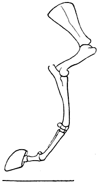
Fig. 41.—Flexion of the Humerus: Right Anterior Limb of the Horse, External Surface (after a Chromophotographic Study by Professor Marey).
The movements and the relations of the humerus and the scapula are clearly represented in Figs. 41 and 42, reproduced from the chromophotographic studies of Professor Marey—studies relative to the analyses of the movements of the horse.[13] They show clearly the movements of flexion and extension of the humerus, also the balancing of the scapula which accompanies the movements.
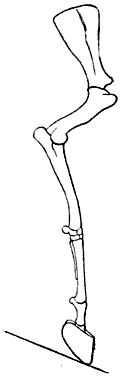
Fig. 42.—Extension of the Humerus: Right Anterior Limb of the Horse, External Surface (after a Chromophotographic Study by Professor Marey).
The Articulation of the Elbow, or the Humero-ulnar Articulation.—In this articulation, which is constructed in the form of a true hinge, the movements of flexion and extension alone are possible. In flexion, the forearm, directed forwards, is folded on the arm, with which, in certain circumstances, it comes in contact. For example, in a horse of mettle which leaps over an elevated obstacle, the animal forcibly raises his fore-limbs by flexing them. Flexion is produced to the same extent, and even more so, and for a longer period, in felides which crouch.
In extension, on the contrary, the forearm is carried backward. This movement being limited only by the contact of the tip of the olecranon with the bottom of the olecranon fossa of the humerus, the forearm is enabled, in this case, to move until it is in line with the arm. For example, during walking, when one of the anterior limbs, having reached the end of its resting stage, is considerably inclined downwards and backwards.
The apex of the olecranon process—that is to say, the point of the elbow—forms a marked prominence, more salient in flexion than in extension, as in the corresponding region of the human elbow.
The Radio-ulnar Articulation.—It is in the dog and the cat, in which the two bones of the forearm articulate by their extremities only, and remain separate in the rest of their extent, that the articulations call for special notice.
In the upper part, the radius rotates on itself; while below, it rotates around the ulna. It follows that the forearm, which in all quadrupeds is in a state of permanent pronation, can, in carnivora, take the position of supination, or rather, of demi-supination. In fact, whatever be the mobility of the two bones of the forearm, the movement is not able to bring the palmar surface to the front, but only to direct it towards the median line.
The Articulation of the Wrist.—Here are found, as in man, three superimposed articulations: the radio-carpal, intercarpal, and carpo-metacarpal.
If we remember the movements which take place at the plane of these articulations in man, and take account of the fact that the mobility of the limbs is reduced just in proportion as they are simplified in structure so as to become organs of support only, we can easily comprehend that, in the horse and the ox, and, in a word, animals that have a canon bone, the movements of the wrist are little varied in character, while in carnivoræ, on the other hand, they are relatively more so.
We will remember that in the ox and the horse the region of the wrist is called the knee.
In flexion, the hand is bent backwards; in extension it is carried forwards. These two movements take place especially in the radiocarpal and intercarpal articulations. In the first of these articulations, it is the superior row of the carpus which glides backwards and forwards on the corresponding articular surface of the forearm. In the second articulation, it is the second row which moves; gliding on the inferior articular surfaces of the row above it. This inferior row carries the metacarpus with it; for the carpo-metacarpal articulation is much less mobile than either of the other two.
In flexion, the articular surfaces are separated from one another in front; and the changes of form which result from this are noticeable on the anterior surface of the ‘knee.’ Moreover, at that moment this region contrasts markedly in its outlines with the parts above it and below it—that is to say, with the corresponding surfaces of the forearm and of the canon bone.
As for the lateral movements, by which the hand is inclined outwards and inwards in its movements at the wrist, they exist to an appreciable extent in the cat and the dog only; in order to understand this, it is enough to compare the shape of the articular surfaces of this region in carnivora and the horse, for example. In the latter, those surfaces are almost plane; in the cat, on the contrary, they are curved (inferior surface of the forearm, concave; superior border of the carpus, convex). These latter, then, are, in form, similar to those which exist at the same level in the human being; this explains the possibility of analogous movements of the whole hand—that is to say, of the movements of abduction and adduction.
The Metacarpo-phalangeal Articulations.—With regard to the mobility, it is in these articulations, as in those of the wrist—that is to say, although in all quadrupeds the first phalanges can be flexed and extended on the metacarpus, it is only in the cat and dog that lateral movement is possible. Indeed, in the horse, in which the principal metacarpal terminates inferiorly in two convex surfaces, which are separated by a crest; and where the whole articulates with a cavity on the superior extremity of the first phalanx; because of the hinging of these surfaces, there can only be movements of opening and closing of this articulation. The first phalanx is directed backwards during flexion and forwards during extension. In the dog and the cat the digits can be separated from each other, and also drawn together—that is to say, abducted and adducted; but, as in man, these movements can be made only when the first phalanges are in the state of extension. During flexion they are impossible, because of the tension of the lateral ligaments, which increases as the flexion is more pronounced. This can be demonstrated, for example, in the cat, which, in order to separate the digits, opens the hand widely—that is to say, forcibly raises the first phalanges.
The Interphalangeal Articulations.—The phalanges are in contact with one another by surfaces, which, on one side, are of trochlear form, and, on the other, are moulded on these trochleæ; accordingly, at the level of these articulations, the movements of flexion and extension only can take place.
In the felidæ, the claws which the third phalanges bear cannot be utilized when the latter are in a state of extension, at which time, being forcibly raised, they are, in fact, placed on the outer sides of the phalanges, which are grooved to receive them. But when the animal wishes to use them, it flexes those third phalanges, of which the terminal extremity is then projected forward, and the claws are ready to fulfil their function. But at the same time it extends the first phalanges, to produce a certain tension of the flexors of the digits, and thus enable the latter to act with greater efficacy, with a minimum of contraction. We can demonstrate this action experimentally on ourselves. It is enough to carry the first phalanges forcibly into a state of extension; the third phalanges then become flexed, quite spontaneously, by the tension of the tendons of the flexors which are inserted into them.
At the same time, if we examine the felidæ which we have taken as examples, when the first phalanges are in the state of extension, the digits will be found to be separable, as we have already indicated in connection with the metacarpo-phalangeal articulations, with the result that the claws are then able to lacerate a wider surface.
The extension of the ungual phalanx, which determines the retraction of the claw and stops its action, is the mechanical result of an elastic, fibrous apparatus which is attached to each of the third phalanges, and has its origin of the second.
THE POSTERIOR LIMBS[14]
The posterior limbs are divided, as are the inferior limbs of the human being, of which they are the homologues, into four parts: pelvis, thigh, leg, and foot.
The Pelvis
The pelvis, which incompletely limits the abdominal cavity, inferiorly in the vertical position of the body and posteriorly in the normal attitude of quadrupeds, is formed by the iliac bones and sacrum—the coccyx forming a prolongation of the latter. We have already described the two latter (pp. 10 and 11) in connection with the vertebral column, of which they form the inferior or posterior portion or segment, according to the attitude of the individual.
The Iliac Bone.—The iliac or coxal bone, is a paired or non-symmetrical bone, united below to its fellow of the opposite side, while it is separated from it above by the sacrum.
In all animals, as well as in man, the iliac bone, at the beginning of life, consists of three parts, which afterwards unite and fuse together and join at the middle of the bottom of a deep cavity which is situated on the outer aspect of the bone—the cotyloid cavity.
Of those three portions when examined in the human iliac bone, that above the cavity is the ilium; that on the inside is the pubis; and the last, the lower one, is the ischium. In quadrupeds, the iliac bone being, in its entirety, directed much more obliquely downwards and backwards, the relative position of these constituent parts is a little modified: the ilium is in front, the pubis is still internal, but in a more inferior position, and the ischium is behind the cotyloid cavity. We notice this peculiarity of the development of the iliac bone because it is customary to continue to apply to the osseous regions which correspond to these parts the names by which they were known when independent bones.
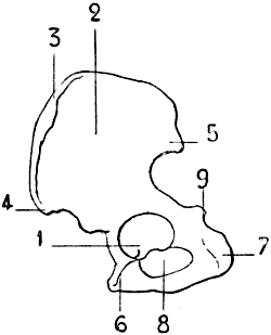
Fig. 43.—The Left Iliac Bone of the Human Being: External Surface, placed in the Position which it would occupy in the Skeleton Of a Quadruped.
1, Cotyloid cavity; 2, ilium; 3, iliac crest; 4, anterior iliac crest; 5, posterior iliac spine; 6, pubis; 7, tuberosity of the ischium; 8, obturator foramen; 9, ischiadic spine.
The bones which form the skeleton of the pelvis of quadrupeds are proportionally more elongated and less massive than those of the human pelvis (Figs. 43 and 44).
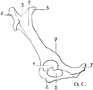
Fig. 44.—Left Iliac Bone of A Quadruped (Horse): External Surface.
1, Cotyloid cavity; 2, ilium, external iliac fossa (directed upward in the horse); 3, iliac crest; 4, anterior iliac spine (directed inwards in the horse, it is the angle of the haunch); 5, posterior iliac spine (directed inwards in the horse; it is the angle of the haunch); 6, pubis; 7, tuberosity of the ischium; 8, obturator foramen; 9, ischiadic spine, or subcotyloid foramen.
We find, on the external surface of the iliac bone, the cotyloid cavity, whose border is interrupted by the cotyloid notch; a deep notch which looks downwards.
In front of this cavity is the ilium. This portion, narrow in the part which is next the cavity, is directed forwards and upwards, expanding more and more as it passes upwards. It presents an external or superior surface (external in some animals, superior in others), which recalls the external iliac fossa; and an internal or inferior surface, at the superior part of which is found the auricular surface for articulation with the sacrum.
The anterior border of the ilium is rough; this is the iliac crest, at the extremities of which we find, below or outside, a prominence which corresponds to the anterior superior iliac spine of man; and internally another projection which corresponds to the posterior iliac spine.
Immediately above the cotyloid cavity is a rough crest, which is known as the supracotyloid crest, which is, however, no other than the homologue of the sciatic spine. In front of this prominence, the border of the ilium, which is notched, forms the great sciatic notch.
If, still taking the cotyloid cavity as the point of departure, we proceed inwards—that is, towards the median line of the body—we find the pubis; if in a posterior direction, the ischium. These two portions, pubis and ischium, limit an oval orifice, the subpubic foramen.
In the human skeleton, the pubis of one side is united to that of the opposite side, to form the pubic symphysis. In the animals which we are now studying a portion of the ischium enters into the formation of the symphysis; in other words, it is formed, not only by the body of the pubis, but also by the descending branch or ramus of the pubis and a portion of the ascending branch or ramus of the ischium, which are fused with those of the opposite side. It results that, though in the human being the symphysis is short and the ischio-pubic arch large, in quadrupeds it is the opposite. In them the arch is a mere slot, and being formed by the ischium alone, merits the name of the ischial arch. The ischio-pubic symphysis is very large, and forms a horizontal surface relatively extensive, a sort of floor, on which rest certain organs which occupy the cavity of the pelvis.
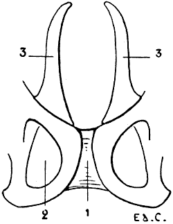
Fig. 45.—Pubic Region of the Pelvis of a Marsupial (Phalanger, Fox).
1, Symphysis pubis; 2, obturator foramen; 3, marsupial bones.
The posterior and external angle of the ischium is rough and prominent; it is the tuberosity of the ischium. This forms a projection under the skin; it also does in man when the trunk is strongly inclined forwards, while the thighs are maintained in the vertical position. In marsupials—opossum, kangaroo, and phalanger—the pelvis at its pubic region is surmounted by two bones, situated one on each side of the median line, and arranged in the form of a fork of two prongs (Fig. 45). These, which are called marsupial bones, support the pouch which, in animals of this genus, lodges their young, which, at the time of birth, are incapable of supporting a separate existence, their development being absolutely incomplete.
In the cetaceans—for example, the dolphin—because of the absence of posterior limbs, the pelvis is represented by two separate bones only, which have no connection with the vertebral column. In birds, the pelvis is remarkable for its elongated form (see for its form Fig. 21, and for details Fig. 46). The cotyloid cavity is pierced by an opening, and presents on its posterior border, which is here a little prominent, a surface with which the great trochanter is in contact.
The ilium is very highly developed, and is fused in the median line with the ilium of the opposite side, the last dorsal vertebræ, the lumbar vertebræ, and the sacrum. Because of these relations with the dorsal vertebræ, it is in contact anteriorly with the last ribs, which consequently emerge from each side of the iliac region of the pelvis.
The ischium forms a plate of bone which, in part, closes the external portion of the cavity of the pelvis. Its superior border is separate for a certain distance from the external border of the ilium; there is thus left an opening of more or less considerable size, which represents or takes the place of the great sciatic notch.
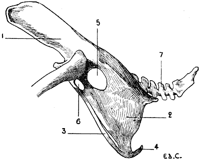
Fig. 46.—Pelvis of a Bird (the Cock): External Surface, Left Side.
1, Ilium; 2, ischium; 3, pubis; 4, inferior extremity of the pubis; 5, sciatic foramen; 6, oval foramen, homologous to the obturator; 7, coccygeal vertebræ.
The pubis, long and slender, is in connection with the inferior border of the ischium, of which it follows the general direction; and circumscribes with this latter, below the cotyloid cavity, an oval orifice, which is the homologue of the obturator foramen. Its inferior extremity reaches beyond the corresponding part of the ischium, bending towards the middle line, but without joining the pubis of the opposite side. On this account there is no symphysis pubis in birds. Nevertheless, an exception must be noted in the case of the ostrich, the pubic bones of which meet in the middle line, and are articulated in form of a symphysis.
The Thigh
A single bone, the femur, forms the skeleton of this portion of the lower limb.
The Femur.—The bone of the thigh is, in man, directed downwards and inwards; this obliquity, we may remind the reader, is due to the difference in length of the two condyles which form its inferior extremity; the internal is the more prominent, the result of which is that when the femur is held vertically, the internal condyle descends lower than the external. Now, as those two articular expansions rest on the horizontal plane formed by the upper extremity of the tibia, it follows that the superior part of the femur inclines towards the side of the shorter condyle—that is to say, outwards—and that, the leg being vertical, it and the bone of the thigh unite in forming an angle, of which the apex is directed towards the inner side of the knee.
In many mammals the two condyles are equally prominent, the result of which is that the femur inclines neither inwards nor outwards, but is contained in a plane parallel to the axis of the trunk; while the leg is included in the same plane. Nevertheless, although contained in the plane which we have just indicated, the femur is obliquely placed, and directed downwards and forwards; it accordingly forms, with the pelvis, an angle, of which the opening is directed to the anterior aspect of the body.
In reptiles and in birds the femur and leg are both placed in the same plane, but this plane is not parallel to the axis of the trunk. This is the result, on the one hand, of the thorax being wide, and, on the other hand, of the femur, which is directed forwards, being in contact by its anterior extremity with the lateral aspect of the costal region, it is thus necessarily placed in a direction forwards and outwards, and the knee is further removed from the axis of the trunk than is the articulation which unites the thigh with the pelvis.
The femur, like the humerus, is almost completely enveloped by muscular masses, which bind it to the lateral walls of the abdomen. Its inferior extremity alone is free, and is always the more so in proportion to its elongation—that is to say, as it belongs to an animal whose foot is more divided. The femur in this respect conforms to the law which we have indicated in connection with the bone of the arm, in which the development, as to length, is in proportion to the division of the hand.
If we compare the femur of certain animals with that of man, we see that the corresponding details of form are readily recognisable, but they are slightly modified. Thus, on examining the superior extremity, we find there a head, a neck, a great trochanter, and a lesser; but the neck is usually short and thick, and the great trochanter does not occupy the same level with regard to the articular head of the bone. In man, the great trochanter does not rise to the level of the head of the femur; in the dog and the cat it approaches that level; in the horse and in ruminants it rises above it.
With regard to the inferior extremity, its surfaces undergo modifications which are further accentuated as we pass from the digitigrades to the ungulates, or unguligrades. We know that in man the femoral trochlea is continuous behind, without interruption, with the condyles—that is to say, that each of the condyles is the continuation of one of the lips of the trochlea. We have just said that the trochlea is continuous without interruption with the condyles; this is accurate. Nevertheless, we must remark that, at the level of the junction of these surfaces, the bone presents a slight constriction, which is more marked on the external than on the internal aspect. This constriction, which is but slightly marked in man, is accentuated in the dog and the cat; in the ruminants and the solipeds it is still more pronounced so that we may say that in these latter the trochlea and the condyles are almost completely separated.
There is another modification in regard to the prominence and extent of the two lips of the trochlea. In man, the external lip of the trochlea reaches higher than the internal, and it is more prominent in front. In the dog, these lips are equal with regard to thickness, but the external still reaches higher than the internal; in the cat, they are equal in every respect; in ruminants and solipeds the internal lip is wider, thicker, and rises higher than the external.
In animals the trochlea is, as a general rule, narrower than in man, and the condyles are more prominent posteriorly; so that, when viewed from one of the lateral aspects, the inferior extremity of the femur is, in them, better developed in the antero-posterior direction.
In birds, the femur is shorter than the bones of the leg; its great trochanter is in contact with a prominence which occupies the posterior part of the border of the cotyloid cavity. Instead of articulating at the level of the knee, with the knee-cap and tibia only, as in man, it articulates, in addition, with the superior extremity of the fibula. A similar arrangement is found in marsupials and reptiles.
The Knee-cap.—This bone, developed in the thickness of the tendon of the triceps muscle of the thigh, is in contact, by its posterior surface, with the femoral trochlea. The two articular surfaces which are applied to the lips of the trochlea present, with regard to their extent, an inequality which is in proportion to the arrangement which we have above indicated—that is, while in man it is the external surface which is the larger, in the horse it is the internal. We shall see what the general form of the knee-cap is when we come, later on, to study more particularly the posterior limbs of some animals.
The Leg
The skeleton of the leg consists of two bones: the tibia and the fibula. The tibia is the more internal and the larger of the two; the fibula is slender, and situated on the outer side, and a little posterior to, the preceding. The fibula is more or less developed according to the species; in some it is complete, in others it is very much atrophied.
This peculiarity may be compared with that which we have drawn attention to regarding the development of the ulna; but here the seriation is less distinct. Not only in the different species, but even in the individuals of the same species, the development of the fibula presents little regularity. In quadrupeds, the bones of the leg are directed obliquely downwards and backwards, so that they form, with the femur, which is directed obliquely downwards and forwards, an angle, the apex of which is placed at the anterior surface of the knee.
Tibia.—The tibia of quadrupeds is readily comparable with that of man; as in the case of the latter, its shaft has three surfaces—an external, which is hollowed out in its upper portion, and becomes anterior below; an internal, slightly convex and subcutaneous; the posterior, which presents, in its superior part, a crest, the oblique line of the tibia, and some rugosities. The borders separate the surfaces. The anterior border, or crest of the tibia, is prominent in its superior part; below it gradually disappears in passing towards the internal aspect of the inferior extremity. The external and internal borders separate the corresponding surfaces from the posterior one.
The superior extremity is thick, and expands in forming three tuberosities: two lateral and an anterior. The anterior tuberosity, situated at the superior part of the crest of the tibia, is very prominent; for this reason the superior extremity is very much expanded in the antero-posterior direction—hence it results that this diameter is equal to the transverse, and sometimes even greater. In man, it is the latter which is the larger. The anterior tuberosity is visible under the skin.
The inferior extremity, less thick, is prolonged internally by a prominence which corresponds to the internal malleolus of man. In animals whose fibula is but slightly developed the tibia presents, on the external part of its inferior extremity, a small prominence, which replaces the fibular malleolus. The ruminants must, however, be excepted, in which we find in this region a special bone, which certain authors look on as the inferior part of the fibula (see p. 97). The inferior surface of this extremity of the tibia is articular; and is in contact with one of the tarsal bones, the astragalus. Because the superior surface of this latter has the form of a pulley, a pulley much more marked than that on the human astragalus, the corresponding surface of the tibia, which has the opposite form, presents two lateral cavities, separated by a median ridge, which is directed forwards and slightly outwards; this ridge projects into the groove of the pulley.
The Fibula.—This bone, situated at the back of the external surface of the tibia, is, as we have said, more or less developed. Its superior extremity, or head, articulates with the external tuberosity of the tibia. Its inferior extremity, when it exists—it is this which disappears in animals which have the fibula incompletely developed—forms a prominence which, placed on the external surface of the inferior extremity of the tibia, articulates with the astragalus, and recalls the external malleolus of man.
We have stated above that it is the inferior extremity of the fibula which disappears when the bone is incompletely developed; it is necessary to except the bat, in which the fibula, fairly well developed at its inferior extremity, by which it articulates with the tibia, thins off in its superior portion, and does not reach the corresponding extremity of the latter. Further, as in this animal the surface of the knee, which corresponds to the anterior surface of the same region in other animals, is turned backwards, the result is that the fibula is situated on the inner side of the tibia, instead of being placed on the outer.
The Foot
The foot, in animals, as well as in man, is formed of three portions, which, as we pass from the part which articulates with the leg towards the terminal extremity, are: the tarsus, the metatarsus, and the toes. These three portions are the homologues of the carpus, the metacarpus, and the fingers, which, as we have already seen in the case of the hand, are the osseous groups which form its skeleton. The tarsus is formed of short bones, as the carpus is; these are, in man, seven in number. The bones are arranged in two rows: one, the posterior, formed of two bones superimposed—the astragalus, by which the tarsus articulates with the leg, and the calcaneum, which forms the prominence of the heel; and an anterior row formed of five juxtaposed ones—the cuboid, situated externally, and the scaphoid internally, in front of which are found the three cuneiforms. To the tarsus succeeds the metatarsus, whose form reminds us very much of that of the metacarpals.
With regard to the toes, which we enumerate in proceeding from the most internal to the most external, they are formed of phalanges, which are three in number for the four outer toes; but the number is reduced to two in the case of the first—that is, the so-called great-toe.
The bones of the tarsus are not seven in all animals; they are fewer in ruminants and solipeds. We already know that, in the latter, the metacarpals and the digits are equally reduced in number; the same is the case for the metatarsals and the toes. We will analyze these differences when dealing with the species individually.
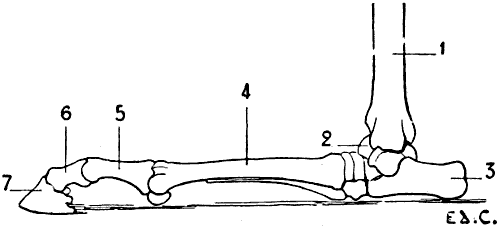
Fig. 47.—Posterior Limb of the Horse placed in the Position which it should occupy if the Animal were a Plantigrade: Left Limb, External Surface.
1, Tibia; 2, astragalus; 3, calcaneum; 4, metatarsus; 5, first phalanx; 6, second phalanx; 7, third phalanx.
When we studied the anterior limbs, we saw in passing from the plantigrades to the digitigrades, and finally the ungulates, or unguligrades, as the hand became hyperextended, the carpus was raised and more and more removed from the ground. We shall establish the existence of the same condition in the posterior limbs; in the plantigrades the tarsus rests on the ground; in the digitigrades it is removed from it; while in the unguligrades the distance which separates it from the point of support is still more considerable; and it is, indeed, necessary to imagine that if these latter were plantigrades, would occupy the position on the ground which is indicated by Fig. 47.
In veterinary anatomy the tarsus is called the ham; a name we adopt in conformity with usage, but which we cannot but regret, as in human anatomy the ham is the region of the posterior surface of the knee.
The general arrangement of the region of the digits of the posterior limbs in birds, presents some points of interest.
We shall merely say with regard to the metatarsus, that it is formed by a single bone, which in the cock is furnished towards its inferior third with a pointed process, the spur. At the inferior part, there is, however, found another, which is but very slightly developed, and with which the first phalanx of the innermost toe articulates.
The toes are, in the majority of species, four in number:[15] an internal, which is directed backwards, and corresponds to the great-toe; the others are directed forwards. This arrangement is constant in grallatores (wading birds), gallinaceæ[16] (domestic fowls), and raptores (birds of prey).
In climbing birds (parrots, woodpeckers, and toucans), the innermost toe is not only directed backward, but the external toe accompanies it in that direction; consequently, there are two posterior and two anterior toes. Sometimes they are all directed forwards; this disposition is found in the martins. In some birds, the number of toes is reduced to three: the cassowary shows this reduction; in others, the number is still further diminished—the ostrich, for example, has but two.
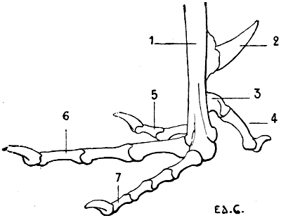
Fig. 48.—Skeleton of the Foot of a Bird (the Cock): Left Side, External Surface.
1, Metatarsus; 2, spur; 3, rudimentary metatarsal; 4, first toe; 5, second toe; 6, third toe; 7, fourth toe.
Further, we find that, in general, the number of the phalanges increases, when we examine the toes in commencing with the most internal (Fig. 48): this has two; then the following one three; that which comes next in order has four; and the most external toe has five. The phalanges of this last are short; so that, although it is formed by a larger number of bones, it is not the longest of the toes.
THE POSTERIOR LIMBS IN SOME ANIMALS.
Plantigrades: Bear (Fig. 33, p. 50).—The external iliac fossa is very deep. The femur is longer than the bones of the leg; the great trochanter does not reach the level of the head of the femur. The fibula is well developed; it is united to the tibia at its superior and inferior extremities only.
The foot, which, as in the case of the hand, rests on the ground by the whole extent of its plantar surface, presents five toes; the shortest of these is the internal—that is, the toe which corresponds to the great-toe in man; the third and fourth are the longest, and they are almost equal; there is a very slight difference in favour of the fourth, which is slightly superior in dimensions to the third.
Digitigrades: Cat, Dog (Fig. 34, p. 52).—The external iliac fossa, which looks outwards, is deep; the iliac crest is convex anteriorly, the convexity is continued from one iliac spine to the other.
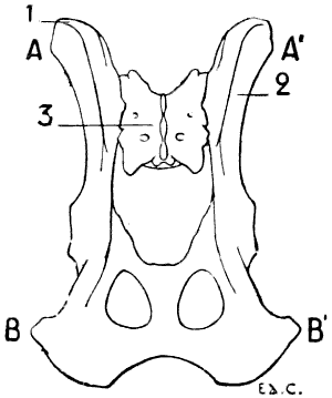
Fig. 49.—Pelvis of the Dog, seen from Above.
1, Iliac crest; 2, external iliac fossa; 3, sacrum; AA′, bi-iliac diameter; BB′, bi-ischial diameter.
In the dog, the distance which separates the anterior iliac spines is less than that which separates the ischia (Fig. 49). On a skeleton which we measured, the transverse diameter, the distance from the anterior iliac spine of one side to that of the opposite side, was 8 centimetres, whilst the distance which separated the ischia was 105 millimetres; on another skeleton, the first measurement was 127 metres, and the second was 146 millimetres. It seems to us unnecessary to multiply examples.
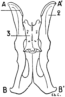
Fig. 50.—Pelvis of a Felide (Lion), viewed from Above.
1, Iliac crest; 2, external iliac fossa; 3, sacrum; AA′, bi-iliac diameter; BB′, bi-ischial diameter.
In the cat, the iliac spines are but slightly marked; the result is that the iliac crest is almost confounded with the inferior and superior borders of the ilium. The two diameters referred to above are almost equal (Fig. 50).
We draw particular attention to what we have just noted in regard to the transverse proportions of the iliac and ischiatic regions of the dog and the cat. These relations are evidently of importance with regard to shape, since the iliac crests and the ischia are noticeable beneath the skin.
In the dog, the shaft of the femur is slightly convex in front; but in the cat it is straight. The borders of the shaft are slightly marked, so that it is almost cylindrical. The linea aspera, less prominent than in man, gains in width what it loses in elevation; it constitutes what may almost be called a rough surface. This surface is narrower in its middle portion than at its extremities, where it bifurcates to go upwards to the two trochanters, and downwards to the two condyles. At the superior extremity, the neck is short, the great trochanter reaching almost to the level of the head of the femur; the digital cavity, which is situated on the internal surface of the great trochanter, is very deep. At its inferior extremity it projects strongly backward. The trochlea is narrow; in the cat its two lips are equally prominent, while in the dog the external is a little more elevated than the internal, which on its part is a little thicker. The trochlea is still more independent of the condyles than in the human femur; it is separated from these latter by a slight constriction.
The knee-cap is long and narrow.
The tibia of the dog is slightly curved from before backward: it has the form of an elongated S; this conformation is in great part due to the very marked projection of the anterior tuberosity and of the superior portion of the crest, which, a little below that tuberosity, turns abruptly backwards, and thus describes a curve the concavity of which is directed forward. The superior part of the external surface is very much hollowed out.
The superior extremity is much thicker than the inferior one. It is not only wide in the transverse direction, but is more especially extended from before backwards; the prominence of the anterior tuberosity is the cause of the elongation of this antero-posterior diameter. On the posterior part of the external tuberosity is found a surface to which the superior extremity of the fibula is applied.
The inferior extremity presents an articular surface, which is formed of two lateral cavities, separated by a crest, which is directed obliquely forwards and outwards. The internal part is prominent, and forms the internal malleolus.
With regard to the fibula, it is united to the tibia by its extremities and by the inferior half of its shaft. This latter is more expanded below than in its upper part. The superior extremity is flattened from without inwards. The inferior extremity projects beyond the articular surface of the tibia, and forms the external malleolus, which, instead of, as in man, descending further than the tibial malleolus, stops at the same level, and even descends a little less than does the latter.
In the cat, the curve of the tibia is less pronounced; this is due to the fact that the crest, instead of being concave in its middle portion, is slightly convex anteriorly. The fibula, less flattened than that of the dog, is united to the tibia by its extremities only, and is separate in the rest of its extent.
The bones of the tarsus are seven in number, and arranged as in man, with this difference (which is easily comprehended), that their general relations are changed on account of the vertical direction of the tarsus. For example, the astragalus, instead of being above the calcaneum, is situated in front of it; the cuneiform bones, instead of being situated in front of the scaphoid, are found below it, etc.
These animals have but four well-developed metatarsals; that which corresponds to the great-toe is represented merely by a small style-shaped bone, situate at the internal part of the region.
Nevertheless, we find this toe fully developed in some dogs. Notwithstanding this, the bones which form it are, however, but rudimentary, and much smaller than those of the innermost digit of the fore-limb.
Sometimes it is double; this condition is demonstrable in individuals belonging to breeds of large size. The median metatarsals are more fully developed than the other bones of the same region which are next them. Viewed as a whole, the metatarsal bones are a little longer than the metacarpals; the result is that the distance which separates the tarsus from the ground is a little greater than that which separates the carpus from the plane on which the anterior limbs rest. The length of the calcaneum still further exaggerates this difference, and, as in the animals with which we shall occupy ourselves later on, the projection which this bone forms is distinctly higher than that which is produced by the pisiform.
The metatarsus, as a whole, is a little narrower than the metacarpus; not only on account of the presence of a thumb in the anterior limb, but, further, because the bones of this latter region are wider than those of the corresponding part of the posterior limb.
The phalanges closely resemble those of the anterior limbs.
Unguligrades: Pig (Fig. 38, p. 58).—The pelvis in this animal presents a few of the characters which we shall again meet with in the ruminants and the solipeds; however, the posterior (or internal) iliac spines are relatively more widely separated from one another than in the latter. This arrangement reminds us of that found in the carnivora.
The femur presents nothing very special. The knee-cap is thick, and ovoid in outline.
The fibula is completely developed, as in the carnivora; and is connected with the tibia at both its extremities.
The tarsus consists of seven bones. The astragalus and the calcaneum differ slightly from those of ruminants.
The foot, like the hand, has two median digits which rest on the ground by their third phalanges; and an internal and an external digit, which are removed from it. The metatarsals are a little longer than the metacarpals.
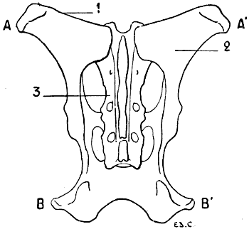
Fig. 51.—Pelvis of the Ox: Superior Surface.
1, Iliac crest; 2, external iliac fossa; 3, sacrum; AA′, bi-iliac diameter; BB′, bi-ischiadic diameter.
Unguligrades: Sheep, Ox (Fig. 39, p. 61).—The pelvis of ruminants of this group closely resembles that of the horse, which we will study later on (see p. 99). That which we must at once point out is that, with regard to the ratio formed by a comparison of the bi-iliac and bi-ischiatic diameters, it may be placed between the ratio obtained in comparing those diameters in the pelvis of the carnivora and that of the solipeds. Indeed, in the ruminants, the distance which separates the ischia exceeds the width of one iliac only, and does not equal, as in the felide, the total width of the anterior part of the pelvis (Fig. 51). In the skeleton of the ox, which forms part of the anatomical museum of the École des Beaux-Arts, the bi-ischiadic diameter is 39 centimetres, whilst the width of one iliac crest is 29 centimetres, so that, in contrast to that which we find in the dog, the width of the ischiadic region is less than that formed in front by the addition of the iliac crests.
The great trochanter is large, and extends beyond the level of the plane in which the head of the femur is found.
In the ox, the linea aspera, instead of being a narrow crest, is spread out, and forms in reality a surface; the posterior surface of the femur. At the inferior and external part of this surface is situated a cavity which surmounts the corresponding condyle, and is known as the supracondyloid fossa. On the internal part of the same region there are a series of tubercles, which, because of their position in relation to the corresponding condyle, constitute the supracondyloid crest.
The internal lip of the trochlea is much thicker and much more prominent than the external.
The details which we have just now examined in connection with the ox are less marked in the sheep.
The trochlea, narrow as a whole, is clearly separate from the condyles by a very marked constriction.
The patella, which is thickened in the antero-posterior direction, has the shape of a triangular pyramid with the base upwards. Its posterior surface, which articulates with the trochlea, presents an arrangement which is adapted to the disposition of this latter—that is to say, the surface which is in contact with the internal lip is larger than that which articulates with the lip of the opposite side.
The tibia of the ox is proportionately shorter than that of the sheep. The shaft of this bone is flattened from before backwards, in its inferior half. The median crest of the articular surface of the inferior extremity is the most prominent part of that region.
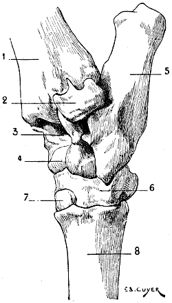
Fig. 52.—Tarsus of the Ox: Posterior Left Limb, Antero-external Surface.
1, Tibia; 2, coronoid bone of the tarsus; 3, superior articular surface of the astragalus; 4, inferior articular surface of the astragalus; 5, calcaneum; 6, cuboido-scaphoid bone; 7, great cuneiform bone—the small cuneiform bone is situated at the back of the latter; 8, principal metatarsal—the small, or rudimentary, metatarsal bone is very small; it is situated at the back of the preceding, and is not to be seen in the sketch. It would be visible if the view were directly lateral, but then the superior and inferior articular surfaces of the astragalus would be less apparent.
The fibula is extremely atrophied. The shaft and superior extremity of this bone are represented merely by a simple ligamentous cord, which is sometimes ossified. There remains of the fibula, as a portion well and distinctly developed, the inferior extremity only. This presents itself under the form of a small bone situated in the region ordinarily occupied by the inferior extremity of the outer bone of the leg—that is to say, the external part of the inferior extremity of the tibia; this little bone articulates with the astragalus and the calcaneum. Some authors consider it to be a tarsal bone, and describe it under the name of the coronoid bone of the tarsus (Fig. 52, 2). It is not, perhaps, quite legitimate to describe it as a bone of this region, for it has not a homologue in the tarsus of other animals. Its external surface is rough; its superior border is furnished with a small pointed process occupying a depression which is provided for it by the tibia. It reaches lower down than the latter, and forms in this way a sort of external malleolus, which frames, on the outer aspect, the mortise in which the astragalus is maintained.
The tarsus, as a whole, has an elongated form; it is formed of five bones: the astragalus, calcaneum, cuboid and scaphoid, which coalesce, to form a single bone, and two cuneiform bones, which correspond to the second and third cuneiform bones of the human foot. These cuneiforms are called, from their size, commencing internally, by the names small and great cuneiform.
The calcaneum is long and narrow; it is longer than that of the horse; it is on the anterior and external part that the bone (coronoid tarsal bone) which represents the inferior extremity of the fibula is situated. It forms the prominence known as the point of the ham, a prominence which is no other than the heel, which, in the unguligrades, is, as we have already said, very far removed from the ground.
The astragalus, which is elongated in the vertical direction, has three articular surfaces disposed in the form of trochleæ: a superior trochlea, which is in contact with the skeleton of the leg, and which is present in all animals; an inferior, which replaces the articular head found on the anterior aspect of the astragalus in man; this articulates with the portion of the scaphoido-cuboid that corresponds to the scaphoid; and, lastly, a posterior trochlea with which the calcaneum articulates. Of these three trochleæ, the superior is the most strongly marked. Between this latter and the inferior is found, on the anterior surface of the astragalus, a deep depression, which, during flexion of the foot on the leg, receives a prominence which the inferior extremity of the tibia presents in its median portion.
We can easily recognise the trochleæ which we have been discussing, in the little bones which children use ‘to play at bones’; these bones are no other than the astragali of sheep.
We have already mentioned that the scaphoid and the cuboid are ankylosed; they form by their union an irregular bone, on which the astragalus and calcaneum are supported.
The cuneiforms articulate with the internal half of the superior extremity of the principal metatarsal; the external half of this metatarsal articulates with the portion of bone which represents the cuboid.
The metatarsus is represented by a principal metatarsal, formed by the coalescence of two metatarsals; we also find in this region a very small rudimentary metatarsal.
The metatarsus is a little longer than the metacarpus; its transverse measurement is a little less; on the other hand, it is a little thicker in antero-posterior direction; from these two differences it results that the body of the metatarsus is quadrilateral, whereas the metacarpus presents only an anterior and a posterior surface.
The rudimentary metatarsal is a very small roundish bone, situated at the back of the superior extremity of the principal metatarsal.
The phalanges closely resemble those of the anterior limbs; nevertheless, the first and second phalanges differ from the latter in the fact that they are a little longer and narrower.
At the back of the metatarso-phalangeal articulations, as in the corresponding region of the anterior limbs, are found the sesamoid bones. Such also exist at the articulations of the second and third phalanges.
Unguligrades: Horse (Fig. 40, p. 64).—The pelvis of the horse presents a general form which sharply differentiates it from that of the carnivora; in fact, the ilium is twisted in such a way that the external iliac fossa does not look outwards, but upwards. It results from this twist that the anterior iliac spine, which we have seen to be directed downwards in the carnivora, has become external; and this prominence is much farther removed from the vertebral column than in the dog or cat. On the other hand, the posterior iliac spine, which is directed upwards in the carnivora, has become internal; it is also placed nearer to the vertebral column, with the result that the distance which separates this spine from that of the bone of the opposite side is proportionately less.
The internal iliac spine, which is conical in shape, and curved upwards, forms a prominence known as the angle of the crupper; the external iliac spine, thick and provided with tuberosities, forms a clearly-defined prominence; this is the angle of the haunch.
The iliac crest, extending directly from one spine to the other, is curved, its concavity being turned upwards. The external iliac fossa, which looks upward, is limited anteriorly by this crest, and is, like the latter, slightly hollowed. The portion of the bone which connects the ilium to the region occupied by the cotyloid cavity is extremely narrow; posteriorly, the bone enlarges again to form the ischial and pubic portions.
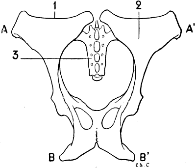
Fig 53.—Pelvis of the Horse: Superior Surface.
1, Iliac crest; 2, external iliac fossa; 3, sacrum; AA′, bi-iliac diameter; BB′, bi-ischiatic diameter.
The tuberosity of the ischium, thick and curved upwards, but less so than in the ox, forms the most prominent part of the posterior border of the region of the thigh; this projecting portion, so sharply defined in spare subjects, is known as the point or angle of the buttock. Contrary to what we have indicated in the case of the dog, the distance which separates the ischiatic tuberosities is inconsiderable in proportion to that which we find between the external iliac spine of one side and that of the opposite. The bi-ischiatic diameter does not even equal the width of one iliac bone measured at the level of its crest (Fig. 53). On the skeleton of the horse in the École des Beaux-Arts, the distance which separates the tuberosities of the ischia is 225 millimetres; that between the two spines of each iliac bone is 25 centimetres.
The anterior region of the crupper is thus much broader than that occupied by the ischia.
The femur is relatively short. Its shaft is rectilinear, and does not present the anterior convexity which is found on the human femur, and which we indicated when discussing that of the dog. The shaft of the bone, instead of being prismatic and triangular, presents four surfaces; the anterior, internal, and external, almost pass into each other, being separated one from the other merely by rounded and slightly marked borders; the posterior surface, which is plane, replaces the linea aspera, which in the horse, instead of presenting the appearance of a crest, is considerably widened. The numerous irregularities which this surface presents give insertion to the muscles which correspond to those attached to the linea aspera.
Between this posterior surface and the external is found a rough prominence which curves forward; this was designated by Cuvier the third trochanter; it replaces the external branch of the superior line of bifurcation of the linea aspera; other authors call it the infratrochanteric crest, because it is situated below the great trochanter. At the inferior part of the same region is found a deep fossa, the borders of which are rough; this is the supracondyloid fossa.
Between the posterior surface and the internal are found: above, the lesser trochanter, which is long and rough; below, at the level of the supracondyloid fossa, an equally rough surface known by the name of the supracondyloid crest.
The superior extremity is flattened from before backwards. The neck is not well marked. The great trochanter is very prominent, and projects beyond the level of the head of the femur. We divide the great trochanter into three parts: the summit, which is the most elevated portion; the convexity, which is situated in front; and the crest, formed by muscular impressions, situated outside and below the convexity. The digital fossa is situated behind and below the summit of the great trochanter. With regard to the lesser trochanter, it is placed so far down that it really forms part of the shaft of the bone, with which, besides, we have described it.
On the inferior extremity of the femur are two condyles and a trochlea; the condyles are clearly separated from this latter by a marked constriction.
The trochlea is directed with a slight obliquity downwards and inwards; its internal lip is much thicker and more prominent than the external; this is, accordingly, a condition exactly the opposite of that which characterizes the corresponding region of the human femur.
The knee-cap is lozenge-shaped; its superior angle projects upward, and produces a prominence at the part which corresponds to the base of the human patella, the part which is here the thickest portion of the bone. Its anterior surface is convex and rough. Its posterior surface presents two lateral articular facets, separated by a crest; this surface is in contact with the trochlea of the femur, and, as it is the internal lip of the latter which is the more developed, it results therefrom that the internal articular surface of the knee-cap is larger than the external.
The knee-cap contributes to the formation of the region of the posterior limb which is called the stifle.
The tibia is large in its upper portion; in its inferior part it is flattened from before backwards. The posterior surface of the shaft presents an oblique line, below which are found vertical rough lines for the insertion of muscles. The external surface is hollowed out in its upper part. The anterior tuberosity of the tibia rises just to the level of the flat articular surface; it is hollowed in its median portion by a vertical groove of elongated form, which receives the ligament that binds the knee-cap to the tibia. The external tuberosity is more prominent than the internal; in it is found a groove for the passage of the anterior tibial muscle.
The inferior extremity, flattened from before backwards, presents a surface which is moulded on the trochlea of the astragalus; the median crest of this surface is thick, and descends lower posteriorly than the tuberosities which are situated on the external and internal aspects of this extremity.
Of the two tuberosities, that which is internal is comparable to the internal malleolus of man, the one on the outer side forms a sort of external malleolus; but this latter here belongs to the tibia, and not to the fibula.
The fibula, in fact, does not reach the inferior extremity of the tibia; it is a poorly developed bone, elongated and terminating inferiorly in a point, at the middle of the shaft of the tibia or at its lower third. Its superior extremity, which is slightly expanded, articulates with the tuberosity which occupies the outer aspect of the corresponding extremity of the tibia.
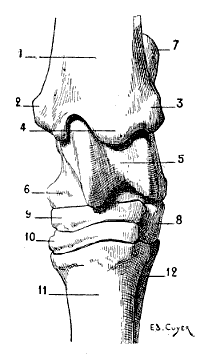
Fig. 54.—Tarsus of the Horse: Left Posterior Limb, Anterior Surface.
1, Tibia; 2, internal tuberosity of the inferior extremity of the tibia (homologue of the internal malleolus of man); 3, external tuberosity of the inferior extremity of the tibia (homologue of the external malleolus); 4, median crest lodged in the groove of the pulley of the astragalus; 5, pulley of the astragalus; 6, internal tuberosity of the astragalus; 7, calcaneum; 8, cuboid; 9, scaphoid; 10, great cuneiform, the small cuneiform is placed behind this latter; 11, principal metatarsal; 12, external rudimentary metatarsal. The internal rudimentary metatarsal, being more slender than the external, does not appear in the figure.
The bones of the tarsus are six in number: the calcaneum and astragalus form the upper row; the cuboid, scaphoid, and two cuneiforms form the lower (Fig. 54).
The astragalus has not, as in ruminants, an inferior trochlea for articulation with the scaphoid; this portion of the bone presents a surface which is slightly convex. It articulates with the tibia by a trochlea that occupies not only the superior surface, but also the anterior. This trochlea, which is directed slightly obliquely downwards and outwards, has a very pronounced form; its lips, which are extremely prominent, determine by their anterior part one of the features which we recognise on the anterior aspect of the ham—a feature which is still more accentuated when the metatarsus (canon) is extended on the leg. On the internal surface of the astragalus is found a tubercle, which forms a projection in the corresponding region of the ham.
The calcaneum, which is not quite so long as that of the ox, forms by its summit a prominence which is called the point of the ham.
The cuboid is small; the scaphoid is large, and flattened from above downwards. Of the two cuneiforms, the more external is the larger; it closely resembles the scaphoid; it is flattened from above downwards as is the latter; but it is a little smaller in size. The small cuneiform, which occupies the inner side of the tarsus, is the smallest bone in this region; it is sometimes divided into two parts; this raises the number of the cuneiforms to three, and that of the bones of the tarsus to seven.
The bones of the metatarsus and the phalanges are equal in number to the corresponding bones in the anterior limbs; they are formed on a type analogous to that of these latter. Accordingly, we shall merely indicate the differences which characterize them.
The principal metatarsal is longer than the metacarpal of the same class; its shaft is more cylindrical; its inferior extremity is somewhat thicker. The external rudimentary metatarsal is better developed than the internal; in the metacarpus the reverse is the case.
The phalanges so far resemble those of the anterior limb that, as differential characters, we need point out only the following: the first phalanx of the hind-foot is a little shorter than that of the fore-foot; its inferior extremity is a little narrower, and its superior extremity a little thicker. The second phalanx is a little less expanded laterally.
The difference in appearance which the three phalanges, anterior and posterior, respectively present are to be borne in mind; for they are correlated to the general form of the fore and hind feet. We will establish this point when we come to study the hoof (see Figs. 101 and 102, p. 257). In the fore-foot the ungual phalanx has its inferior surface limited externally by a circular border, while the same bone of the hind-foot has this surface a little narrower, more concave, and limited by two curved borders which unite anteriorly to form an angle—an arrangement which gives to the general outline of this region the form of the letter V.
Articulation of the Posterior Limbs
The Coxo-femoral Articulation.—The head of the femur is received in the cotyloid cavity; these are the osseous surfaces in contact in this articulation. They are maintained in position by a fibrous capsule and a round ligament. To this latter is found attached, in the horse, a fasciculus which, commencing, as does the round ligament, at the depression on the head of the femur, emerges from the cotyloid cavity by the notch which is present in its circumference, and is attached to the anterior border of the pubes, to blend with the tendon of the rectus muscle of the abdomen. This is the pubio-femoral ligament.
The movements which this joint permits are the same in the quadrupeds as in man, but less extensive. They are: flexion and extension, abduction and adduction, the two latter being much more limited than the former. There is also rotation.
By flexion, the inferior extremity of the femur is directed forwards; the bone of the thigh then takes a more oblique direction than the normal. This movement takes place, for example, when the animal carries forward one of its hinder limbs. Extension, which takes place in an inverse sense, is produced when the foot is fixed on the ground, while the body is projected forward. It is also produced in the action of kicking.
As for the lateral movements—viz., abduction and adduction—they are less extensive than the preceding movements. The absence of the pubio-femoral ligament in other quadrupeds than the horse explains why in them abduction is less limited than in the latter. Indeed, it is the tension of this ligament, occasioned by the abduction of the thighs, which arrests more quickly the movement in question.
Articulation of the Knee.—This articulation, as in man, is formed by the femur, the patella, and the tibia.
In the horse the ligament of the patella is not single, but consists of three parts, designated, on account of their position, by the respective names of external, internal, and median patellar ligaments. The two former come from the angles on the corresponding borders of the knee-cap; the median springs from the anterior surface and inferior angle of the same bone. They all three pass to their termination on the anterior tubercle of the tibia. The external ligament is the strongest, and the internal ligament the least developed.
In the dog, the cat, the pig, and the sheep, the patellar ligament consists of a single band. The articulation is further strengthened on the sides by lateral ligaments—an internal and an external.
With regard to the principal movements, these are flexion and extension, to which may be added movements of rotation of limited extent. In flexion, the leg bends on the thigh; its inferior extremity is directed upwards and backwards; the angle which the tibia naturally forms with the femur becomes less obtuse.
But it should be understood that one part of this description—that which has relation to the leg—holds good only when the femur is in its normal condition, or in flexion. Indeed, at the close of the movement in which, during a step, the foot is in contact with the ground—that is, at the termination of the resting stage—the inferior extremity of the tibia is directed backwards. But the femur is then in a state of extension, and in regard to this latter the attitude of the leg is unchanged.
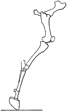
Fig. 55.—Extension of the Leg: Right Posterior Limb of the Horse, External Surface. (After a Chronographic Study by Professor Marey.)
At this moment, notwithstanding the direction, which recalls that which it has at the time of flexion, the leg is not bent on the thigh; on the contrary, it is almost in the line of its continuation (Fig. 55). As we have done in connection with the articulations of the anterior limbs, we borrow this figure from the interesting chronophotographic studies of Professor Marey.[17]
The Tibio-tarsal Articulations and of the Bones of the Tarsus.—In the region which veterinary anatomists call the ham, the articulations of the leg and foot alone call for special study in the case of the horse. The articulations of the bones of the tarsus, and of these with the metatarsus, do not offer any interest with regard to mobility, this being almost wholly absent at that level.
The leg and the astragalus, in a general way, are placed in contact by such articular surfaces that the resulting joint, which is a true hinge, permits movements of flexion and extension only. Indeed, as we have indicated above, the tibia is furnished, on the inferior surface, with a crest that fits into the deep groove which is situated on the corresponding surface of the astragalus.
During flexion, the anterior surface of the foot tends to approach the anterior surface of the leg, the angle formed by these two segments becoming more and more narrowed. The displacement in the opposite direction characterizes extension.
In other quadrupeds, the articulations which bind together the bones of the tarsus possess a little more freedom of movement. The shape of these bones, and particularly the shape of the surfaces of the astragalus, which are in contact with them, allow movements in this region, in the case of the dog and cat, which, without being so extensive as those of the human foot, in the subastragaloid articulation, nevertheless, recall the mobility which we find in the human species at this level—that is to say, rotation, abduction, and adduction of the foot.
As for the articulations of the metatarsus with the phalanges, and of the phalanges with one another, they resemble those of the anterior limb too closely that it should be necessary to study them here. Such a study would be, in this case, but a repetition (see p. 76, a description of the articulations in question).
THE HEAD IN GENERAL, AND IN SOME ANIMALS IN PARTICULAR.
When we compare, by the examination of one of their lateral aspects, the skull of man and the same region in other mammals, it is easy to observe that the relative development of the cranium and face is entirely different. In the case of man the cranium is large, and the face relatively small; in animals the face is proportionally much more highly developed. The measure of the facial angle permits us to note these differences, and the figures relative to the value of this angle are sufficiently demonstrative to induce us to indicate those which are, in a general way, connected with some of the forms in individuals which here occupy our attention. In the first place, we must remember that the angle in question is more acute, as the cranium is less developed in proportion to the facial region (Figs. 56 and 57). It is especially to this character that we wish to draw attention.
Man 70° -80°
Cat 41°
Dog 28° -41°
Sheep 20° -25°
Ox 18° -20°
Ass 12° -16°
Horse 11° -13°
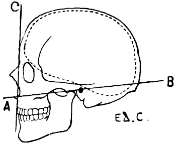
Fig. 56.—Human Skull: Measure of the Facial Angle by the Method of Camper. Angle BAC = 80°.
The internal wall of the cranial cavity is marked by the dotted line.
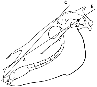
Fig. 57.—Skull of the Horse: Measure of the Facial Angle by the Method of Camper.[18] Angle BAC = 13°.
The internal wall of the cranial cavity is shown by the dotted line.
Besides, in animals the cranium is very prominent superiorly, and the face, more or less elongated, is sharply projected downwards and forwards; in man the cranial region occupies not only the superior, but also the posterior part; the face is short and of a compact form. The human head, in its general aspect, may be compared to a sphere, while the skull of the quadrupeds presents the aspect of a quadrangular pyramid, with the base turned upwards and the summit at the incisor teeth.
Direction of the Head.—Before entering on the study of the bones of the head, it is necessary, in our opinion, to agree as to the position in which we shall suppose it to be placed.
The question may seem to be one of little importance; nevertheless, it cannot be regarded as indifferent, since authors are not all agreed on this subject.
Some suppose it to be placed vertically—that is, with the incisor teeth turned directly downwards. Others, on the contrary, suppose it to be placed horizontally, resting on the whole length of the lower jaw, the face being then turned upwards. These two extreme methods of arrangement appear to us to possess inconveniences—at least, for comparison with the human head.
Indeed, if, when the head is vertical, the same regions of the face (forehead, nose) are, in the case of animals as well as man, turned forward, the lower jaw ceases to merit its appellation, as it is then situated, not below, but behind the upper. Furthermore, if this position is chosen, for example, for modelling or drawing, it cannot be obtained without difficulty when we have to deal with an isolated piece of the skeleton, on account of the absence of equilibrium, which it is necessary to obviate. It is true that the question of convenience should not take precedence of all others, and it suffices for us in this connection to recall, in regard to the human pelvis, that, although the older anatomists used to represent it as resting commodiously on the three angles which terminate it at its lower part (ischial tuberosities and coccyx), this attitude being false, it is customary now to incline the superior aspect forwards, inasmuch as this arrangement more nearly conforms to reality, in spite of the fact that it is a little more difficult so to dispose an isolated pelvis. Further, to return to the head; if its vertical direction can be demonstrated, for example, in many horses, it is not sufficiently general to be adopted as the classic position.
In regard to the facility of placing in position, the horizontal direction is certainly to be preferred; but this is also far removed from the natural position in the animal while in the state of repose. On the other hand, the mind is not satisfied with the idea that certain regions of the face, such as the nose and the forehead, are then directed upwards. And yet it is necessary to come to a decision, seeing that what we are now investigating applies also to the position to which it is necessary to give the preference in placing the skeleton of the head when we wish to draw it in profile. That which we adopt is a compromise, but to us it seems more rational.
The position of the head of the horse, to be normal, should be such as to give it an inclination of 45°. In this case the lower jaw is still posterior; and, for this reason, we see in adopting this position some inconveniences from a didactic point of view. Accordingly, we will suppose the head brought a little nearer to the horizontal, and this, from the imaginative point of view, has certainly an advantage which we cannot afford to neglect when addressing artists.
Indeed, let us suppose that to a clay model of a human head we wish to give the aspect of the head of a quadruped. We should elevate the occiput; and then, taking hold of the lower part of the face, we should lengthen it, not in a direction precisely antero-posterior, but downwards and forwards. It is obviously this latter procedure which, on the other hand, is carried out when a person wishes to give to his own face some resemblance to the muzzle of a quadruped.