Chapter I
Reading edition
OSTEOLOGY AND ARTHROLOGY
THE TRUNK
The Vertebral Column
We commence the study of the skeleton with a description of the trunk.
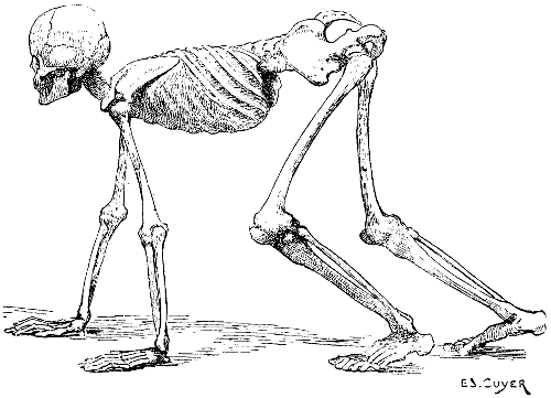
Fig. 1.—A Human Skeleton in the Attitude of a Quadruped. To give a general Idea of the position of the Bones in other Vertebrates.
The trunk being, in quadrupeds, horizontal in direction (Fig. 1), the two regions of which it consists occupy, for this reason, the following positions: the thorax occupies the anterior part, the abdomen is placed behind it; the vertebral column is horizontal, and is situated at the superior aspect of the trunk; it projects beyond the latter: anteriorly, to articulate with the skull; and, posteriorly, to form the skeleton of the tail, or caudal appendix.
The number of the vertebræ is not the same in all mammalia. Of the several regions of the vertebral column, the cervical shows the greatest uniformity in the number of the vertebræ of which it consists, with but two exceptions (eight or nine in the three-toed sloth, and six in the manatee); we always find seven cervical vertebræ, whatever the length of the neck of the animal. There are no more than seven vertebræ in the long neck of the giraffe, but they are very long ones; and not less than seven in the very short neck of the dolphin, in which they are reduced to mere plates of bone not thicker than sheets of cardboard. If the cervical region presents uniformity in the number of its bones, it is not so with the other regions of the column.
The following table shows their classification in some animals:
Vertebræ.
Cervical. Dorsal. Lumbar.
Bear 7 14 6
Dog 7 13 7
Cat 7 13 7
Rabbit 7 12 7
Pig 7 14 6 or 7
Horse 7 18 6 or 5
Ass 7 18 5
Camel 7 12 7
Giraffe 7 14 5
Ox 7 13 6
Sheep 7 13 6
It is worthy of notice that in birds the number of the cervical vertebræ is not constant, as in mammals; they are more numerous than the dorsal. These latter are almost always joined to one another by a fusion of their spinous processes; the two or three last vertebræ are similarly united to the iliac bones, between which they are fixed. The dorsal vertebræ thus form one piece, which gives solidity to the trunk, and provides a base of support to the wings, for the movements of flying. There are, so to speak, no lumbar vertebræ, the bones of that region, which cannot be differentiated from the sacrum, having coalesced with the bones of the pelvis.
Vertebræ.
Cervical. Dorsal.
Vulture 15 7
Eagle 13 9
Cock 14 7
Ostrich 18 9
Swan 23 10
Goose 18 9
Duck 15 9
In reptiles, the relation between the number of the cervical vertebræ and that of the dorsal is very variable; some serpents are devoid of cervical vertebræ, having only dorsal ones—that is, vertebræ carrying well-developed ribs.
Vertebræ.
Cervical. Dorsal. Lumbar.
Crocodile 7 14 3
Caiman 7 12 5
Boa 3 248 0
Python 0 320 0
Viper 2 145 0
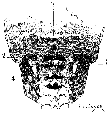
Fig. 2.—Size of the Atlas compared with the Transverse Dimensions of the Corresponding Parts of the Skull in Man.
1, Atlas; 2, mastoid process; 3, external occipital protuberance; 4, inferior maxilla.
Regarding the direction of the vertebral column in animals, in which the trunk is not vertical, it is evident that the spinous processes point upward, and that in comparing them with those of man they must be arranged so that the superior surface of the human vertebra will correspond to the anterior surface of that of the quadruped. Of the cervical vertebræ, the atlas and axis call for special notice. Apropos of the atlas, we find that it, in the human being, is narrower than the corresponding parts of the skull, and is therefore hidden under the base of the cranium (Fig. 2); in quadrupeds its width is equal to that of the skull, and sometimes exceeds, because of the great development of its wing-shaped transverse processes, that of the neighbouring parts of the head (Fig. 3). On this account those transverse processes often project under the skin of the lateral surfaces of the upper part of the neck.
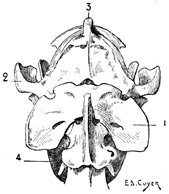
Fig. 3.—Size of the Atlas compared with the Transverse Dimensions of the corresponding Regions of the Skull in a Dog.
1, Atlas; 2, zygomatic arch; 3, external occipital protuberance; 4, inferior maxilla.
The axis is furnished on its anterior surface with the odontoid process, which articulates with the anterior (or inferior) arch of the atlas, according to the direction of the neck. The spinous process, flattened from without inwards, is more or less pointed; it is elongated from before backwards, so as partly to overlap the atlas and the third cervical vertebra.
We find that this process overlaps less and less the neighbouring vertebræ when we examine in succession the bear, the cat, the dog, the ox, and the horse. With regard to the other vertebræ of this region, they diminish in width from the second to the seventh; and, in some animals, the anterior surface of the body presents a tubercle which articulates with a cavity hollowed in the posterior surface of that of the vertebra before it; this feature dwindles away in the dorsal and lumbar regions.
The spinous process, slightly developed in the third cervical vertebra, gradually increases in size to the seventh, the spinous process of which, long and pointed, well deserves the name of the prominent which is bestowed on it; but it should not be forgotten that the spinous process of the axis is equally developed.
On the inferior surface of the body of each of the vertebræ is found a prominent crest, especially well marked at the posterior part; this crest is but slightly developed in the bear and in the cat tribe, and is not found in swine.
The transverse processes of the cervical vertebræ, from their relation to the trachea, are known as the tracheal processes.
The most marked characteristic of the dorsal vertebræ is furnished by the spinous processes. They are long and narrow. As a rule, the spinous processes of the foremost dorsal vertebræ are the most developed and are directed obliquely upwards and backwards. As we approach the last vertebræ of this region, the processes become shorter and tend to become vertical, and the last ones are even, in some cases, directed upwards and forwards; this disposition is well marked in the dog and the cat. In the cetaceans, on the contrary, the length of the spinous processes increases from the first to the last.
In the horse the spinous processes of the first dorsal vertebræ produce the prominence at the anterior limit of the trunk, where the mane ends, which is known as the withers.
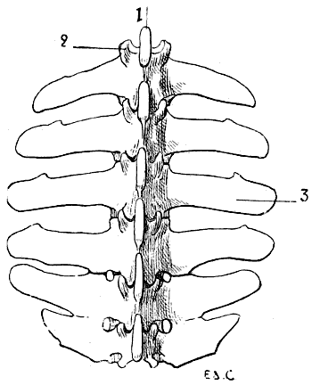
Fig. 4.—Lumbar Vertebræ of a Quadruped (the Horse): Superior Surface.
1, Spinous process; 2, anterior articular process and transverse process of the first lumbar vertebra of the left side; 3, costiform process.
The lumbar vertebræ are thicker than the preceding; they are known by their short and latterly-flattened spinous processes, and still more readily by their transverse processes, which, as they are evidently atrophied ribs, it is more accurate to denominate costiform processes (Fig. 4). These are long, flattened from above downwards, and directed outwards and forwards.
The true transverse processes are represented by tubercles situated on the superior borders of the articular processes of each of the vertebræ of the lumbar region. Apropos of these different osseous processes, we are reminded that they are also present in the human skeleton.
In the horse the costiform processes of the fifth and sixth lumbar vertebræ articulate, and are sometimes ankylosed, one with the other; the terminal ones articulate with the base of the sacrum. Sometimes the processes of the fourth and fifth are thus related; this is the case in the figure (4) given; here the costiform processes of the fourth and fifth vertebræ articulate, and the two terminal ones have coalesced.
In the ox, the same processes are more developed than in the horse; their summits elevating the skin, produce, especially in animals which have not much flesh, prominences which limit the flanks in the superior aspect. The costiform processes of the last lumbar vertebræ are separate from each other; those of the latter are not in contact with the sacrum.
The Sacrum.[3]—This bone, single and median, is formed by the mutual coalescence of several vertebræ, which vary in number according to the species observed.
Vertebræ Constituting the Sacrum.—Bears, 5; dogs, 3; cats, 3; rabbits, 4; swine, 4; horses, 5; camels, 4; oxen, 5; sheep, 4.
The sacrum is situated between the two iliac bones; with which it articulates, and contributes to the formation of the pelvis. It is obliquely placed, from before backwards, and from below upwards; immediately behind the lumbar section of the vertebral column; and is continued by the coccygeal vertebræ, which form the skeleton of the tail.
It is triangular in outline, and is generally more narrow in proportion than in the human being. All things considered, it is more large and massive, and of greater density, in species which sometimes assume the upright posture, rather than in those which cannot assume that attitude; for example, the sacrum of the ape, of the bear, of the dog, and of the opossum are proportionately larger than those of the horse.[4]
Its superior surface presents a crest, formed by the fusion of the spinous processes of the vertebræ which form it. In certain species these processes are attached only by their bases, and are separated from each other superiorly. In the pig they are wholly wanting.
The Coccygeal Vertebræ.—These vertebræ, few in number (and sometimes ankylosed) in the human being, form in the latter a small series, the coccyx; which is inclined forwards, that is to say, towards the interior of the pelvis. In quadrupeds, on the contrary, their number is large; they are not ankylosed, and they form the skeleton of the caudal appendix.
The first coccygeal vertebræ—that is, those which are next the sacrum—present characters which are common to those of other regions: they have a body, a foramen, and processes. As we trace them backwards, these characters become gradually effaced; and they become little more than small osseous cylinders simply expanded at their extremities.
Direction and Form of the Spinal Column
The curves of the vertebral column are, in quadrupeds, slightly different from those which characterize the human spine. First, instead of their being, as in the latter, curves in the antero-posterior aspect, because of the general attitude of the body, they are turned in the supero-inferior direction.
The cervical region is not a single curve, as in the human being. It presents two: one superior, with its convexity looking upwards; the other inferior, the convexity of which is turned downwards. This arrangement reminds one of that of a console.
The dorsal and lumbar regions are placed in a single curved line, more or less concave downwards; so that in the lumbar region there is no curve analogous to that which exists in man; a form which, in the latter, is due to the biped attitude—that is to say, the vertical position of the trunk. Briefly, there is in quadrupeds one dorso-lumbar curve; and not both a dorsal and a lumbar, with convexities in opposite directions.
At the extremity of the dorso-lumbar region is the sacrum and the caudal appendix, which describe a curve of which the concavity is directed downwards and forwards.
It is necessary to point out that it is not the curves of the three anterior portions of the spinal column which determine the form of the superior border of the neck and shoulders, and of the same part of the trunk. For the first portion, there is a ligament which surmounts the cervical region, and substitutes its modelling influence for that of the vertebræ. It is the superior cervical ligament, which arises from the spinous process of the first cervical vertebræ, and is inserted into the external occipital protuberance on the upper part of the posterior surface of the skull. The summits of the spinous processes of the vertebræ alone give form to the superior median border of the trunk. In this connection we here repeat that it is not the general curvature of the vertebral column which produces the withers, but the great length of the spinous process of the first vertebræ of the dorsal region.
The Thorax
The dorsal vertebræ form the posterior limit in man, and superior in quadrupeds, of the region of the trunk known as the thorax. A single bone, the sternum, is situated at the aspect opposite; the ribs bound the thorax on its sides.
In its general outlines the thorax in quadrupeds resembles that of man—that is to say, that, as in the latter, the anterior portion—superior in the human being—is narrower than the part opposite. But the progressive widening takes place in a more regular and continuous fashion, so that it presents a more definitely conical outline. This purely conical form is nevertheless found in the human species, but only during infancy; the inferior portion of the thoracic cage being then widely expanded, because of the development of the abdominal viscera, which at that period are relatively large.
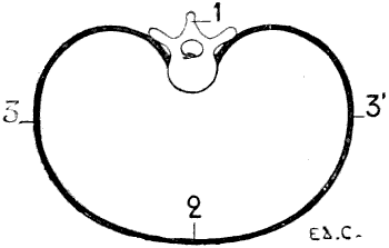
Fig. 5.—A Transverse Section of the Thorax of a Man placed Vertically—that is to say, in the Direction which it would assume in a Man placed in the Attitude of a Quadruped (a Diagrammatic Figure).
1, Dorsal vertebra; 2, sternal region; 3, costal region of one side; 3′, costal region of the other side.
But the proportionate measurements of the thorax are different. Indeed, we may recall that in man the thorax is flattened from before backwards, so that the distance between the sternum and the vertebral column is shorter than the distance from the rib of one side to the corresponding one of the opposite side (Fig. 5). In animals, on the contrary, it is flattened laterally. Its vertical diameter—measured from the sternum to the vertebral column—is greater than the transverse measurement (Fig. 6).
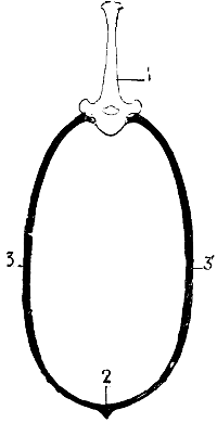
Fig. 6.—A Vertical Section of the Thorax of a Quadruped (Diagrammatic).
1, Fifth dorsal vertebra; 2, sternal region; 3, costal region of one side; 3′, costal region of the opposite side.
From this results a peculiar arrangement of the muscles that we are able to bring directly into prominence, which presents points of interest from the point of view of the contraction of the subcutaneous layer. Indeed, in man the region occupied by the pectorals is very broad; it is a wide surface turned directly forward. In quadrupeds, this region of the pectorals is narrowed. It is not spread out, as in the preceding instances; and the appearance it presents is explained by the fact that the thorax is compressed laterally. If we examine the thorax on one of its lateral surfaces, the muscles, on the contrary, are more extended. We see the contour of the vertebral column, and the median part of the abdomen; and, especially in the horse, between the great dorsal and the great oblique of the abdomen, we find a large space, in which the ribs, with the intercostals which join them, are uncovered; the muscles in question separate the one from the other, under the influence, it would seem, of the great dimensions of the lateral wall of the thorax.
The Sternum.—The sternum is, in quadrupeds, directed obliquely downwards and backwards; its form varies in different species. In the carnivora, it consists of eight bones, irregularly cylindrical in form, being slightly flattened from within outwards, and thickened at their extremities. They remain separate, and this contributes elasticity and flexibility to the thorax. The first nine costal cartilages articulate directly with the sternum. The first of these cartilages articulates with a nodule situated a little above the middle of the first bone of the sternum.
In the horse the sternum is flattened laterally in its anterior portion, and from above downwards in its posterior half. The six bones which form the sternum are connected by cartilage. The keel-shaped piece, situated in front of the sternum, is also cartilaginous. This process, but slightly marked posteriorly, becomes more and more prominent in front, and terminates at its anterior extremity by a prolongation, slightly curved backwards, which projects for some centimetres beyond the cavity in which the first costal cartilage is received. This process is known as the tracheal process, or rostral cartilage. The posterior extremity of the sternum, flattened from above downwards, ends in a cartilaginous plate; concave superiorly, and convex inferiorly: this is the abdominal prolongation, or xiphoid appendix.
In the ox, the sternum is formed of two distinct bones, which are united by an articulation. One, the anterior, is short, and forms the first portion of the sternum; it is slightly flattened from side to side, and vertical in direction. The other, the posterior, is longer, and is formed by the fusion of several small bones; it is placed horizontally, and is flattened from above downwards. At the level of articulation of these two portions, and because of their different directions, the bone is bent. This bend occurs at the point of articulation of the second costal cartilage. On the superior border of the anterior segment the cartilage of the first rib is articulated. The xiphoid appendix, which is cartilaginous, is attached to the extremity of a long process of the last bone of the sternum.
The shape of the anterior extremity of the sternum is influenced by the presence or absence of clavicles. We have seen that in some quadrupeds the clavicles are wanting. In the first case, this extremity is large, and approaches in shape to the corresponding part of the human sternum, which is so clearly designed to give a point of support to the anterior bone of the shoulder. In the second, on the contrary, this extremity is narrow.
The sternum in birds is very different from that in mammalia, which we have been studying. It varies greatly in extent and shape, under the influence of certain conditions. To understand the cause of these variations it is necessary to remember that in man (as, indeed, in other animals; but the example of man, for that which follows, will be more striking, on account of the mobility of his upper limbs) the sternum gives origin to the pectoral muscles, and that these muscles are inserted into other parts of the thoracic limbs, designed by their contraction to draw the arms downwards, forwards, and inwards—that is, when these are in a state of abduction and in a horizontal direction, they draw them towards the anterior surface of the thorax and downwards. Now, this movement is similar to that made by birds during flight. It is necessary to add that, in the latter case, the more the displacement of the upper limbs has of force and extent, the more the pectoral muscles are developed.
For these reasons, birds, in which, during flight, the movements of the thoracic limbs—the wings—are necessarily energetic, present a great development of the pectoral muscles; having consequently, because an extent of surface for the origin of the muscles commensurate with their development is necessary, a very large and peculiarly shaped sternum (Figs. 18, 6; and 21, 6). Indeed, not only is the sternum large, but, further, in order to form a deeper surface, proportionately adapted to the muscles which arise from and cover it, its anterior surface presents, in the median line, a prominent crest known as the keel. This prominence forms two lateral fossæ. We cite as examples, the sternum of the eagle, the vulture, the falcon, and the hawk.
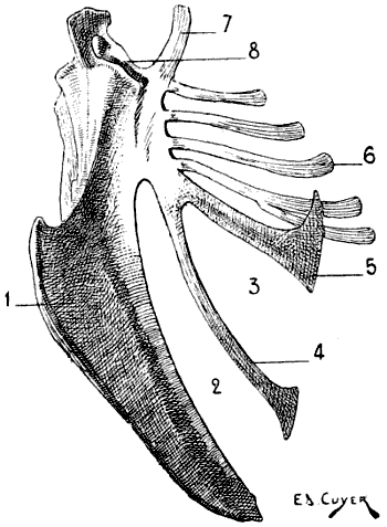
Fig. 7.—Sternum of a Bird (the Cock): Left Side, External Surface.
1, Keel; 2, internal slot; 3, external slot; 4, internal process; 5, external process; 6, inferior ribs; 7, costal process; 8, surface for articulation with the coracoid bone.
All birds are not, however, equally adapted to flight, for in the domestic cock, which flies but a short distance, and badly, the sternum is less developed (Fig. 7); it is also diminished by slots, which diminish its surface. These slots, two on each side, are called from their position the internal and external slots. They are bounded by narrow, elongated, bony processes, an internal and an external; the expanded lower extremity of the latter overlaps the last inferior ribs (see p. 19). The part of the external border which surmounts this external process is hollowed out into grooves, which receive the inferior ribs, and terminates superiorly in an osseous projection known as the costal prominence.
In the ostrich, the cassowary, and the apteryx, which run, but do not fly, the sternum has the form of a plate of bone slightly convex, but without a keel.
The shape of the sternum, correlated to the faculty of flight (or of swimming; apropos of which we may cite the penguin, of which the rudimentary wings resemble fins, and perform their functions only), or the absence of this faculty, has furnished the division of birds into two groups. In one are included, under the name Carinates (carina, keel), those in which the sternum is provided with a keel; in the other division are those in which the sternum is not furnished with one. These latter, on account of their unique mode of progression, are more nearly allied to the mammals.
The keel is developed in flying mammals (bats).
Ribs and Costal Cartilages.—There are on each side of the thorax as many ribs as there are dorsal vertebræ. In animals, as in man, the ribs which articulate with the sternum by their cartilages are called true, or sternal ribs; those whose cartilages do not articulate with the sternum are called false, or asternal. The longer ribs are those situated in the middle region of the thorax.
The ribs are directed obliquely downwards and backwards, and this obliquity is more marked in the posterior ones than in the anterior. They are, however, less oblique than in the human being; what proves this is that the first rib in man is oblique, while in quadrupeds it is vertical.
The curvature of the ribs is less pronounced in quadrupeds than in the human being, but this is not equal in all animals. The ribs of the bear are more curved than those of the dog; the latter has ribs more curved than those of the horse.
Each rib, at its vertebral extremity, presents, from within outwards, a wedge-shaped head for articulation with two dorsal vertebræ, a neck, and a tuberosity. External to the tubercle are found some rough impressions, for muscular attachments, which correspond to the angle of the human rib.
In the following table, we give the number and classification of the ribs of some animals:
Number of the Ribs on Each Side of the Thorax.
Sternal. Asternal.
Bear 14 divided into 9 and 5
Dog 13 „ „ 9 „ 4
Cat 13 „ „ 9 „ 4
Rabbit 12 „ „ 7 „ 5
Pig 14 „ „ 7 „ 7
Horse 18 „ „ 8 „ 10
Camel 12 „ „ 8 „ 4
Ox 13 „ „ 8 „ 5
Sheep 13 „ „ 8 „ 5
The costal cartilages, by which the first ribs are united to the sternum (sternal ribs), whilst the latter are united one to the other without being directly connected with the sternum (asternal ribs), are, as a rule, in quadrupeds, directed obliquely downwards, forwards, and inwards; each forms, with the rib to which it belongs, an obtuse angle more or less open anteriorly. Their length is proportionate to that of the ribs. The cartilages, which are continued from the asternal ribs, unite and form the borders, directed obliquely downwards and forwards, of the fossa which is found at the inferior and posterior part of the thorax, and which forms the lateral limits of the epigastric region. In the dog and cat the ribs are thick and almost cylindrical; the costal cartilages are thicker at the margin of the sternum than at their costal extremity. In the ox, the ribs are flattened laterally and are very broad, the more so as we examine a portion further from the vertebral column. From the second to the twelfth they are quadrangular in the superior fourth, and thicker than in the rest of their extent. The first costal cartilage is vertical; the following ones are progressively more oblique in a direction downwards and forwards. The four or five cartilages which succeed the first unite with slight obliquity to the sternum; their union with that bone gives the impression of a very strong, well-knit apparatus. The costal cartilages which unite with the sternum are flattened laterally in the portions next the ribs, and flattened from front to back in the rest of their extent.
In the horse the ribs increase in length from the first to the ninth; they are flattened from without inwards, and increase in width from the first to the sixth or seventh, and the following ones become narrower. The costal cartilages, from the second to the eighth, are, as in the ox, at first flattened laterally, near the ribs; while near the sternum they are flattened from front to back.
In birds, the ribs are each furnished with a flat process (Fig. 18, 10), which springs from the posterior border, is directed backwards, and overlaps the external surface of the succeeding rib. These processes are not found, as a rule, on the first or last ribs.
As for the costal cartilages, they are, as a rule, ossified, and receive the name of inferior ribs (Fig. 18, 11), united to the preceding (superior ribs; Fig. 18, 9) by articulation; by the other extremity they are joined to the sternum; the first superior ribs generally want them. Sometimes the last inferior rib becomes connected with the one that precedes it, not articulating with the sternum; and thus recalls the relations of the asternal ribs which we have noticed in our study of the mammals.
In the bat, as in birds, the costal cartilages are ossified.
THE ANTERIOR LIMBS[5]
The anterior limbs, homologous to the upper limbs in man, are formed, as in the latter, of four segments: the shoulder, the arm, the forearm, and the hand. These limbs, considered in the vertebral series, present themselves under very different aspects, which are determined by the functions they are called upon to perform.
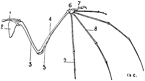
Fig. 8.—Anterior Limb of the Bat: Left Side, Anterior Surface.
1, Clavicle; 2, scapula; 3, humerus; 4, radius; 5, cubitus; 6, carpus; 7, thumb; 8, metacarpus; 9, phalanges.
They constitute the forepaw in terrestrial mammals; in aerial vertebrates they form wings; in aqueous mammals they act as paddles. In whatever series we study them, we can readily find the relationship of the different parts; it is very easy to recognise the same bones in the upper limbs of the human being, the wings of the bat (Fig. 8) and of birds (Fig. 21), and in the anterior paddles of the seal (Fig. 9) and of the dolphin.
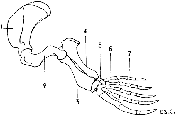
Fig. 9.—Anterior Limb of the Seal: Left Side, External Surface.
1, Scapula; 2, humerus; 3, radius; 4, ulna; 5, carpus; 6, metacarpus; 7, phalanges of the fingers.
In quadrupeds, the shoulder and arm are hidden, the latter more or less completely, in the muscular mass which binds it to the lateral wall of the trunk; so that the anterior limbs only present; free from the trunk: the elbow, forearm, and hand.
The Shoulder
In some vertebrates, the shoulder is formed of two bones—the scapula and clavicle; in others of only one bone—the scapula; the clavicle in this case does not exist.
The Scapula or Omoplate.—The scapula is situated on the lateral surface of the thorax, and is directed obliquely, from above downwards and from behind forwards.
We must first recall, so as to be able to make a comparison, that in man this bone is placed at the posterior surface of the thoracic cage; so that if we look at the human thorax on one of its lateral aspects we see chiefly the external border of the scapula; it is the external surface (homologous to the posterior surface of the human scapula) which we see in its full extent when we look on the same surface of the thorax in quadrupeds.
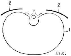
Fig. 10.—Situation and Direction of the Scapula in the Human Being, the Trunk being Horizontal, as in Quadrupeds. Vertical and Transverse Section of the Thorax (Diagrammatic Figure).
1, Contour of the thorax; 2, 2, the scapula.
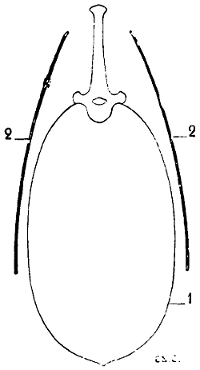
Fig. 11.—Position and Direction of the Scapula in Quadrupeds. Vertical and Transverse Section of the Thorax (Diagrammatic Figure).
1, Contour of the thorax; 2, 2, the scapula.
To sum up, if we fancy the human being in the position of the quadruped, the scapula will have its surfaces almost parallel to the ground (Fig. 10); while in quadrupeds, the surfaces are situated in a plane which is almost perpendicular to the ground (Fig. 11). This position of the scapula in an almost vertical plane is designed to give the necessary point of support to the osseous columns that form the skeleton of the other portions of the anterior limbs.
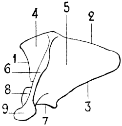
Fig. 12.—Left Scapula of the Human Being, Posterior Surface, placed in the Position which it would Occupy in the Skeleton of a Quadruped.
1, Cervical border; 2, spinal border—the scapula here represented, being from a hoofed animal, has a cartilage of extension attached to its spinal border; 3, axillary border; 4, supraspinous fossa; 5, subspinous fossa; 6, spine of the scapula; 7, glenoid cavity; 8, coracoid process. The scapula of the horse has no acromion process, but it is easy, if we compare the human scapula, to judge of the position which this process would occupy if it were present.
Because of this position of the scapula (Figs. 12 and 13), the spinal border is superior, the cervical, anterior, and the axillary, posterior. In direct contrast to what obtains in the human scapula, the spinal border is the shortest of the three; except in the bat, and the majority of the cetaceans.
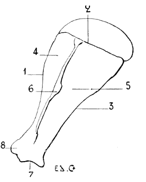
Fig. 13.—Left Scapula of a Horse: External Surface.
1, Cervical border; 2, spinal border; 3, axillary border; 4, supraspinous fossa; 5, subspinous fossa; 6, scapular spine; 7, glenoid cavity; 8, coracoid process; 9, acromion process.
In certain animals (in the ungulates [hoofed[6]]—pigs, oxen, sheep, horses) the superior, or spinal, border of the scapula is surmounted by a cartilage called the cartilage of prolongation.
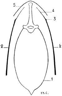
Fig. 14.—Vertical and Transverse Section, at the Site of the Shoulders, of the Thorax of the Horse (Diagrammatic Figure).
1, Outline of the thorax at the level of the third dorsal vertebra; 2, 2, scapula; 3, spinal border of the scapula; 4, cartilage of prolongation; 5, contour of the skin.
This is the cause why the border to which it is fixed is so slightly noticeable under the skin in these animals; indeed, in the upper part, the bone and cartilage are not distinguishable in the contour of the corresponding region of the back; being applied to the lateral surfaces of the spinous processes, the prominence formed by the extremities of which is directly continuous with the plane of the scapula (Fig. 16).
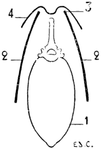
Fig. 15.—Vertical and Transverse Section, at the Plane of the Shoulders, of the Thorax of a Dog (Diagrammatic Figure).
1, Outline of the thorax at the level of the third dorsal vertebra; 2, 2, scapula; 3, spinal border of the scapula; 4, contour of the skin.
In quadrupeds whose scapula, on the contrary, is wanting in the cartilage of prolongation (in the clawed,[7] such as the cat and dog), the superior border of the scapula is visible, especially when the animal is resting on its fore-limbs, particularly when it crouches; at such a time the skin is markedly raised by that border; and the spinous processes of the vertebræ, beyond which it projects, occupy the bottom of a fossa (Fig. 15). The internal surface of the scapula is turned towards the ribs; it is known, as in man (in whom this surface is anterior), as the subscapular fossa.
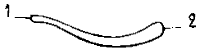
Fig. 16.—Left Clavicle of the Cat: Superior Surface (Natural Size).
1, Internal extremity; 2, external extremity.

Fig. 17.—Clavicle of the Dog (Natural Size).
Its external surface is divided into two parts by the spine of the scapula; which, in some animals, terminates inferiorly in a flat and clearly distinct process, the homologue of the acromion process of the human scapula. The two regions separated by the spine are known as the supraspinous fossa and the infraspinous fossa. The supraspinous fossa is anterior to the spine, and the infraspinous is posterior to it. The surfaces of the scapula are, in quadrupeds, flatter than in the human being, and in particular the subscapular fossa, which is also less concave. Some authors attribute this to the lesser curvature of the ribs in quadrupeds. A few words will suffice to prove that there must be another reason. The scapula is not in immediate contact with the ribs; the subscapular fossa is not moulded on them. Besides, the form of the scapula is, as in other parts of the skeleton, dependent on the disposition of muscles, and the development of these latter is correlated to the extent and energy of the movements which the individual is able or required to execute. But the movements which those muscles produce (more especially the rotation of the humerus) are, in quadrupeds, less extensive than in the human being; and, consequently, the muscles which produce them are, proportionally, less strongly developed. The inferior angle (superior and external in man), situated at the junction of the cervical and axillary borders, presents the glenoid cavity, which, looking downwards, receives the articular surface of the superior extremity of the bone of the arm—that is to say, the head of the humerus. Above this cavity, on the lower part of the cervical border, is situated a tubercle which reminds us of the coracoid process of the human scapula. The region occupied by the glenoid cavity is separated from the body of the bone by a constriction—the neck of the scapula.
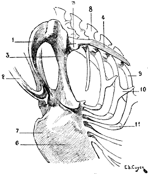
Fig. 18.—Skeleton of the Shoulder of a Bird (Vulture): Antero-External View of the Left Side.
1, Left clavicle; 2, inferior portion of the right clavicle, forming by its ankylosis with that of the other side the fourchette; 3, coracoid bone; 4, scapula; 5, articular surface for humerus; 6, superior half of the sternum; 7, keel of sternum; 8, spinous process of the dorsal vertebræ; 9, superior ribs; 10, process of one of these ribs; 11, inferior ribs.
In birds the scapula is elongated in a direction parallel to the vertebral column, and very narrow in the opposite (Fig. 18): it is also flat, and has no spine. Its coracoid process is represented by a peculiar bone—the coracoidean or coracoid bone—which we shall describe later on when we come to the study of the clavicle and of the anterior region of the shoulder (see p. 26).
The Clavicle.—The clavicle is found only in the human being, and in animals whose anterior limbs, possessing great freedom of movement in all directions, require that the scapula should possess a point of support which, while affording this, can be displaced with it, or draw it in certain directions. Now, this point of support is furnished by the clavicle.
In animals possessed of hoofs (ungulates), such as the sheep, ox, and horse, the clavicle does not exist. Indeed, in them the freedom of movement of the anterior limbs is limited; they move by projection in the forward and backward directions only; they merely fulfil the functions of giving support to and carrying about the body. The clavicle is rudimentary in the cat and the dog; in the cat it is a small, elongated bone (Fig. 16), 2 centimetres in length, thin and curved, connected with the sternum and the scapula by ligamentous bundles. In the dog it is represented by a small osseous plate only (Fig. 17), which is not connected with any of the neighbouring bones.
It is on the deep surface of a muscle which passes from the head and neck to the humerus (mastoido-humeral, a muscle common to the arm, neck, and head) in which this rudimentary bone is found to be developed.
The clavicle exists in perfect state in mammals which use their limbs for digging, grasping, or flying; the insectivora (hedgehog, mole) and some rodents (squirrel, woodchuck) are provided with it.
The cheiroptera (bats) possess an extremely well-developed clavicle, on account of the varied movements which their thoracic limbs execute.
This formation of the shoulder which favours flight in the bat is even more remarkable in birds. In these latter (Fig. 18) the clavicles, fused together by their lower extremities, form one bone, having the shape of the letter V or U, which is known as the fourchette; this bone, acting as a true spring, keeps the shoulders apart, and prevents their approximation during the energetic movements which flight necessitates.
In birds whose power of flight is strong, the two limbs of this bone are widely separated and thick, and the fourchette is U-shaped. Those whose flight is awkward and but slightly energetic have the limbs of the fourchette slender; they unite at a more acute angle, and the bone is V shaped.
Furthermore, a bone named the coracoid joins the scapula to the sternum; this bone, often fused with the scapula, where it contributes to the formation of the glenoid cavity, represents in birds the coracoid process of the human scapula. If we fancy this process directed inwards, and sufficiently lengthened to join the sternum, we shall have an idea of the disposition of the bone we are now discussing, and the reasons for which the name has been chosen by which it is designated. The coracoid bone, like the fourchette which it reinforces, offers to the wings a degree of support proportionate to the efforts developed by those limbs; for this reason it is thick and solid in birds of powerful flight.
The superior extremity of each branch of the fourchette, at the level of its junction with the coracoid and the scapula, bounds, with these latter, a foramen which gives passage to the tendon of the elevator muscle of the wing, or small pectoral. The importance of the fourchette being, as we have seen, in proportion to the movements of flying, it is easy to understand that the bone is not found in the ostrich.
The Arm
A single bone, the humerus, forms the skeleton of this portion of the thoracic limb.
The Humerus.—The bone of the arm is, in quadrupeds, inclined from above downwards and from before backwards.
It is, with relation to other regions, short in proportion as the metacarpus is elongated, and as the number of digits is lessened. In the horse, for example, whose metacarpus is long, and in which but one digit is apparent, the humerus is very short. The slight development in length of the humerus explains its close application to the side of the animal as far as the elbow.
In animals in which the humerus is longer, the bone is slightly free, as well as the elbow, at its inferior extremity. Later on we will return to the consideration of this peculiarity and of the proportions of the humerus, after we have studied the other parts of the fore-limbs.
The humerus in quadrupeds is inflected like the letter S; in man this general form is less accentuated, the humerus being almost straight. On its body, which appears twisted on its own axis, we find the musculo-spiral groove,[8] which crosses the external surface, and is very deep in some animals. Above this groove, and on the external surface, there exists a rough surface which is the impression of the deltoid. In some species this rugosity is very prominent, and is called the tuberosity of the deltoid; it is prolonged downwards by a border which forms the anterior crest of the musculo-spiral groove and limits this latter in front. The external border of the bone, or posterior crest of the groove, limits it behind.
The superior extremity is enlarged, and remarkable in three portions which it presents; these are: an articular surface and two tuberosities.
The articular surface, or head of the humerus, smooth and round, is in contact with the glenoid cavity of the scapula. This head in the human skeleton is directed upwards and inwards; in quadrupeds its direction is upwards and backwards. The inferior extremity, having in both one and the other its long axis directed transversely, and the point of the elbow looking backwards in all, the result is that the head of the humerus is not situated vertically above the same regions; in the first, it is almost directly above the internal part of this extremity; in the latter, it is situated above its posterior surface, or the point of the elbow in the complete skeleton. This difference of direction is correlated with the position of the scapula, the glenoid cavity of which, as we have already seen, is in man turned outwards, whereas in quadrupeds it looks downwards. In the latter case the scapula consequently rests on the head of the humerus; and this position is most favourable for the performance of the functions which the anterior limbs have to fulfil in these latter.
Of the tuberosities of the head of the humerus, one is situated on the external aspect—it is the great tuberosity, or trochiter; the other is placed internally—it is the small tuberosity, or trochin. The great tuberosity is divided into three parts—summit, convexity, and crest; these different parts give insertion to the muscles of the shoulder. We recollect that the facets (anterior, middle, and posterior) of the great tuberosity of the humerus in man give attachment to the muscles of the same region. The head of the humerus in the human body projects above the tuberosities. We shall see afterwards, when dealing with some special quadrupeds, that in some of these, on the other hand, the tuberosities are on a higher level than the articular head of the bone. Between the two tuberosities is the bicipital groove.
In man, the superior extremity of the humerus, although covered by the deltoid, reveals its presence by elevating the corresponding portion of the latter. In quadrupeds, the anterior part of this extremity, although similarly covered by muscular bundles, produces a prominence under the skin. This prominence is situated at the summit of the angle formed by the opposing directions of the scapula and the bone of the arm, and constitutes what is known by the name of the point of the shoulder, or of the point of the arm.
The inferior extremity, transversely enlarged, presents an undulating articular surface, which reminds us of the trochlea and the condyle of the human humerus; on which, however, the condyle is more sharply defined from the trochlea.
In the human skeleton, the internal lip of the trochlea descends lower than the external; and also lower than the condyle. In the bear, the cat, and the dog, it is the same. In the ox and the sheep, the condyle is lower than the trochlea, but only very little lower. In the horse the arrangement is still the same, but a little more accentuated.
On the lateral parts of this extremity we find: internally, a prominence, the epitrochlea; and, externally, another, the epicondyle. It is from this latter that the crest arises, which, passing upwards, forms the posterior limit of the groove of torsion.
The two prominences, which we have just described from a general point of view, present special arrangements which it is necessary to point out. When we examine the form of the outline of the inferior extremity of the humerus in man, the bear, the cat, the dog, the ox, and the horse, we find in following this order that the extremity tends to become narrow transversely, and that the epicondyle and the epitrochlea are less and less prominent on the external and internal aspects respectively. These two processes, indeed, project backwards; the epitrochlea always remaining more developed than the epicondyle. Because of this projection backwards, the cavity situated on the posterior surface of the inferior extremity, the olecranon fossa, is very deep, more so than in the humerus of man. Its borders being thus formed by the two processes, are very prominent. In front we find the coronoid fossa, which is less deep than that of which we have just spoken.
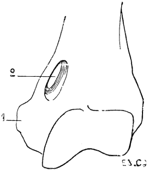
Fig. 19.—Inferior Extremity of the Left Humerus of a Felide (Lion).
1, Epitrochlea; 2, supra-epitrochlear foramen.
There exists in some mammals an osseous canal, situated above the epitrochlea, and known as the supratrochlear canal (Fig. 19). It is bounded by a plate of bone which at its middle portion is detached from the shaft of the humerus, and blends with the latter at both its extremities. The brachial artery and median nerve pass through the foramen.
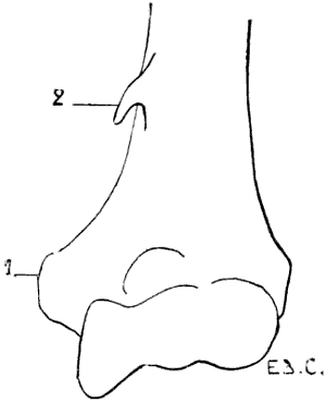
Fig. 20.—Inferior Extremity of the Left Human Humerus, showing the Presence of a Supratrochlear Process.
1, Epitrochlea; 2, supra-epitrochlear process.
A similar condition is sometimes found, as an abnormality, in man, which presents itself under the following aspect (Fig. 20): an osseous prominence more or less long, in the shape of a crochet-needle—supra-epitrochlear process—situated 5 or 6 centimetres above the epitrochlea; the summit of this process gives attachment to a fibrous band, which is inserted by its other end into the epitrochlea and the internal intermuscular aponeurosis. The fibro-osseous ring thus formed gives passage to the brachial artery and the median nerve, or in case of a premature division of this artery to the ulnar branch of the same.[9]
There is also found in some mammals a perforation of the thin plate of bone which, in others, separates the olecranon fossa from the coronoid. This perforation is sometimes found as an abnormality in the human humerus.
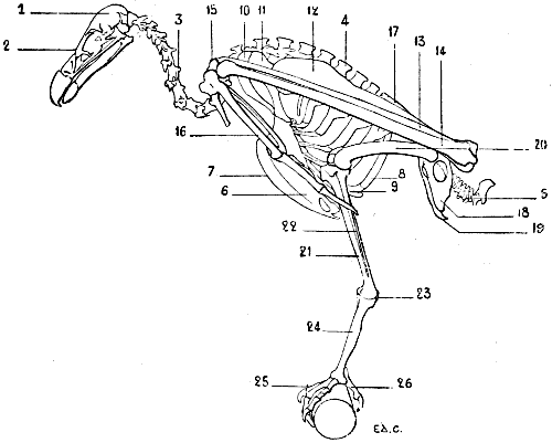
Fig. 21.—Skeleton of a Bird (Vulture): Left Surface.
1, Cranium; 2, face; 3, cervical vertebræ; 4, spinous processes of the dorsal vertebræ; 5, coccygeal vertebræ; 6, sternum; 7, keel; 8, superior ribs; 9, inferior ribs; 10, clavicle; 11, coracoid bone (for the details of the skeleton of the shoulder, see Fig. 18); 12, humerus; 13, radius; 14, ulna; 15, carpus; 16, hand (for details of the skeleton of this region, see Fig. 31); 17, ilium; 18, ischium; 19, pubis (for the details of the pelvis, see Fig. 46); 20, femur; 21, tibia; 22, fibula; 23, osseous nodule, which some anatomists think represents the calcaneum; it is the sole vestige of the tarsus; 24, metatarsus; 25, foot; 26, first toe (for the details of the skeleton of the foot, see Fig. 48).
As does the sternum and the skeleton of the shoulder, the humerus of birds presents differences correlated to the functions which the thoracic limbs are destined to fulfil. Lying on the side of the thorax, directed obliquely downwards and backwards (Fig. 21), it is proportionately longer in individuals of powerful flight than in those which fly less or not at all. In the vulture it projects beyond the posterior part of the pelvis; in the cock it does not even reach the anterior border of the same. To these differences in length are added differences in volume and in the development of the processes which serve for muscular attachment, which are more considerable in birds of powerful flight.
The humerus is so placed that the radial border, external in man and quadrupeds, looks upwards, with the result that the surface of the bone of the arm, which in these latter is anterior, in the former looks outwards. The humeral head, which is turned forwards and a little inwards, is convex and elongated in the vertical direction. Behind and above this head is found a crest for the insertion of muscles. It is the same for the region below, where there is a tuberosity whose inferior surface presents a pretty large opening which looks inwards to a fossa from the floor of which a number of minute openings communicate with the interior of the bone. This is the pneumatic foramen of the humerus.
It is of interest to remember in connection with this subject that in birds, in keeping with the conditions of flight, every system of organs is adapted to diminish the weight of the body. We particularly draw attention to the osseous framework, the structure of which is such that the weight of the animal is greatly lessened. This condition is secured by the pneumaticity. The bone consists of a cover of compact tissue, which, instead of enclosing marrow, is hollowed out by cavities which contain air, and communicate with special pouches, the air-sacs, which are appendages of the lungs.[10]
The antibrachial extremity of the humerus is flattened from without inwards. It terminates in two articular surfaces, which articulate with the radius and ulna.
The olecranon process of the ulna being slightly developed, it follows that the olecranon fossa is not large; neither is the coronoid.
General View of the Form of the Forearm and Hand
We now proceed to the study of the two regions of the fore-limbs which present the greatest variety in regard to the number of bones and also in regard to form and proportions. These two regions are the forearm and the hand.
It is first of all necessary to say that in man, when the fore-limb hangs beside the body, and the dorsum of the hand looks backwards, the two bones of the forearm are parallel, and that this position is known by the name of supination. It is also necessary to remember that there is another attitude, in which the radius, crossing the ulna, and carrying the hand with it, displaces the latter in such a way that the palmar surface looks backwards. This second position is known as pronation.
Let us now suppose that a man wishes to walk in the attitude of a quadruped. It will be necessary, in order that his upper limbs, being for the moment anterior ones, may act as members of support, to place the forearm in pronation, in order that, as is more normal, the hands may rest on the ground by their palmar surfaces. In this position the radius, being rotated on its own axis at its upper extremity and around the ulna in the rest of its extent, shall have its inferior extremity situated on the inner side of the corresponding extremity of the latter.
Such is the situation of the bones of the forearm and the attitude of the hand in quadrupeds. In short, quadrupeds have their anterior members in the position of pronation.
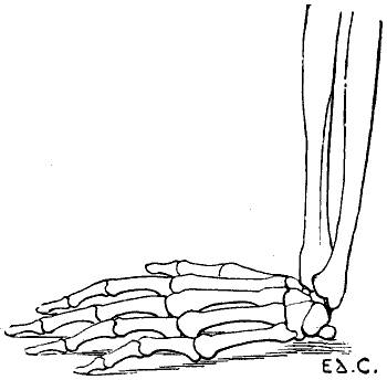
Fig. 22.—The Human Hand resting for its Whole Extent on its Palmar Surface: Left Side, External Surface.
The individual whom we have just supposed placed in the attitude of a quadruped would be able to maintain this position by pressing on the ground more or less extensive portions of his hands; the whole palm of the hand may be applied to the ground (Fig. 22); or the fingers only—that is to say, the phalanges (Fig. 23); or the extremities of the fingers only—that is to say, the third phalanges (Fig. 24). This last position, which is certainly difficult to maintain, should here be regarded rather as theoretical.
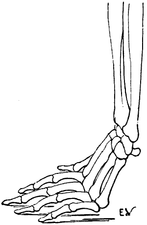
Fig. 23.—The Human Hand resting on its Phalanges: Left Side, External Surface.
We shall meet with each of these modes of support in certain groups of animals. Thus, the bear, badger, and the majority of rodents, have the paws applied to the ground by the whole extent of the palmar surface of the hand, from the wrist to the tips of the fingers. They are therefore called plantigrade, from the analogy, in this case, of the palm of the hand to the plantar surface, or sole of the foot.
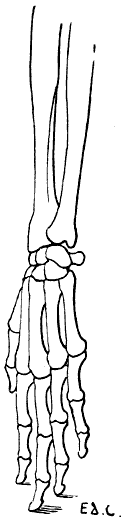
Fig. 24.—The Human Hand resting on the Tips of some of its Third Phalanges: Left Side, External View.
In others, such as the lion, tiger, panther, cat, wolf, and fox, the support is made no longer on the whole extent of the palmar surface, but on the corresponding surface of the fingers only—the metacarpus is turned back, and, consequently, the wrist—that is to say, the carpus—is removed from the ground. These are the digitigrades.
Lastly, the ruminants (sheep, oxen, deer, etc.), and also the pig, ass, and horse, rest on the third phalanx only. In them not only is the metacarpus turned back, but also the two first phalanges. The wrist is very far removed from the ground. In these animals, the third phalanx is enclosed in a case of horn, a nail (the hoof), and because the support of the limb is on that nail, the name of unguligrades has been given them. Nevertheless, as the point of support is on the third phalanx, which is also known by the name of phalangette, we are of opinion that, in order to specify definitely, although they walk on their fingers, as do the digitigrades, the support is provided not by the whole extent of those appendages, they might receive the name of phalangettigrades.
It is necessary among the ruminants to make an exception of the camel and the llama, which are digitigrades.
Just in proportion as the hand is raised from the ground, as we have just seen in passing from the plantigrades to the digitigrades and unguligrades, the number of bones of that region diminishes, the bones of the forearm coalesce, and the ulna tends to disappear; the hand becomes less and less suitable for grasping, climbing, or digging, so as to form an organ exclusively adapted for walking and supporting the body.
Thus, the bear (plantigrade) has five digits, and the power of performing the movements of supination and pronation. Indeed, we know with what facility this animal is able to move his paws in every direction, and climb a tree by grasping it with his fore-limbs. It is well known, however, that no animal except the ape can perform the movements of rotation of the radius around the ulna with the same facility as man; and that none possesses the same degree of suppleness, extent, and variety of movements of the forearm and hand.
In the digitigrades there is one finger which is but slightly developed, and which is always removed from the ground—that is, the thumb: there is also a little less mobility of the radius around the ulna.
In the ungulates the limbs are simply required to perform the movements of walking, and form veritable columns of support, which become the more solid as they are less divided. The bones of the forearm are fused together; there is therefore no possibility of rotation of the radius around the ulna. The metacarpus is reduced to a single piece, which in the horse constitutes what is known as the canon. The number of digits becomes diminished, so that in ruminants there are not more than two, and in the horse but one. We should, however, add that, up to the present, we have taken into account only perfect digits, those that rest on the ground. We shall see further on that there exist supplementary digits, but that they are only slightly developed, and are represented in some cases by mere osseous spurs; it is this fact that has permitted us to ignore them in the general study which we have just made.
Because, as we have already said, the unguligrades have the inferior extremity of the digit encased in a horny sheath, which forms the hoof of the horse and the corresponding structures (onglons) in the ox, those animals have been placed in a special group, which is based on that peculiarity—that is, the group of ungulate mammals.
The plantigrades and digitigrades, of which the paws have their surfaces of support strengthened by an epidermic sole and fatty pads, have the free extremities of the third phalanges covered on their dorsal surface by nails or claws; hence they are named unguiculate mammals.
The bat and birds have the bones of the forearm so arranged that the radius cannot rotate around the ulna. This is necessary in order that during flight, when the wing is being lowered, the radius and hand shall not be able to turn; for, if such rotation took place, each stroke of the wing would place it in a vertical position, which would occasion a loss of resistance incompatible with the effect to be obtained.
The Forearm
The skeleton of the forearm in quadrupeds is vertical in direction; consequently, it forms with the arm an angle open anteriorly; this is well seen on examining the lateral surface. If we examine it on its anterior surface, we find a slight obliquity directed downwards and inwards. In animals in which the bones of the forearm are separate—that is to say, susceptible of supination and pronation—we find a more close resemblance to those of the human skeleton. The ulna, the superior extremity of which always projects beyond that of the radius, has a shaft which gradually narrows from above downwards. Its inferior extremity is terminated by a round head in those animals in which the ulna is fully developed; in others, as it is atrophied, it ends in a thin, long process.
The ulna presents at its superior extremity a posterior process, the olecranon, which forms the point of the elbow. We find on the anterior surface of the same, another process, the coronoid.
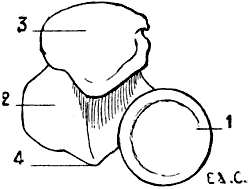
Fig. 25.—Superior Extremity of the Bones of the Human Forearm: Left Side, Superior Surface.
1, Radius; 2, ulna; 3, olecranon process; 4, coronoid process.
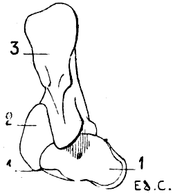
Fig. 26.—Superior Extremity of the Bones of the Forearm of the Dog: Left Limb, Superior Surface.
1, Radius; 2, ulna; 3, olecranon process; 4, coronoid process.
It is necessary to dwell on the relations of these parts. In man the head of the radius is situated at the anterior part of the external surface of the superior extremity of the ulna (Fig. 25); indeed, the small sigmoid cavity with which the head articulates is situated on the outer side of the coronoid process, and this apophysis is placed in front. In the plantigrades and digitigrades the head of the radius is placed still more forward, so much so that it is situated almost in front of the superior extremity of the ulna (Fig. 26). In the unguligrades it is placed directly in front of this latter (Fig. 27).
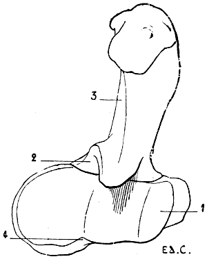
Fig. 27.—Superior Extremity of the Bones of the Forearm of the Horse: Left Limb, Superior Surface.
1, Radius; 2, ulna; 3, olecranon process; 4, coronoid process.
Further, the displacement of the radius is made at the expense of the superior extremity of the neighbouring bone; the radius appears to appropriate more and more the parts which in man belong exclusively to the ulna—for example, the coronoid process. In the plantigrades and the digitigrades half of the process still belongs to the ulna and the remainder to the radius. In the ungulates—the horse, for example—the coronoid process belongs to the radius; the ulna, situated behind the latter, is correspondingly diminished in size.
In brief, when we study this region of the skeleton in plantigrades, then in digitigrades, and finally in unguligrades, we find a kind of progressive absorption of one of the two bones (ulna) by the other (radius), which thus becomes the more developed.
It is easy to explain this partial disappearance of the ulna. When the forearm is capable of performing the movements of pronation and supination, the ulna is completely developed, for it is in its small sigmoid cavity that the head of the radius revolves, and it is around its inferior extremity, the head, that the corresponding extremity of the radius turns. But when the movements of rotation of the forearm do not exist, the inferior extremity of the ulna becomes functionally useless and disappears. As to its rôle in the movements of the region of the wrist, that is nil, for we may remember—we will observe it again when we come to treat of the articulations—that the hand articulates with the radius alone (radio-carpal articulation); this is the reason that, when the forearm possesses the fullest mobility, the hand follows the movements which that bone makes around the ulna.
It is not so with the articulation at the elbow-joint; there it is the ulna, which, with the humerus, forms the essential parts (humero-ulnar articulation); its olecranon process limits the movement of extension of the forearm. It is for this reason that, even in those quadrupeds in which the ulna is atrophied, the olecranon process presents a relatively considerable degree of development.
We know that on the posterior surface of the inferior extremity of the bones of the human forearm are grooves in which pass the tendons of the posterior and external muscles which, belonging to this region, are directed for insertion towards the hand.
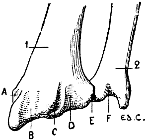
Fig. 28.—Inferior Extremity of the Bones of the Forearm of a Man: Left Side, Posterior Surface, Position of Supination.
1, Radius; 2, ulna; A, groove for the long abductor and short extensor muscles of the thumb; B, groove for the radial muscles; C, groove for the long extensor of the thumb; D, groove for the special extensor of the index finger and of the common extensor of the fingers; E, groove for the proper extensor of the little finger; F, groove for the posterior ulna.
In animals, because of the movement of rotation of the radius, the surface of this bone, which is anterior, corresponds to the posterior surface of the same in man. (To possess a clear conception of this, it is necessary to remember that, in this latter, the bones of the forearm are always described as in the position of supination; they are thus represented in Fig. 28. The direction of the surfaces of the radius is the reverse of that in animals, since the latter have the radius always in a state of pronation.)
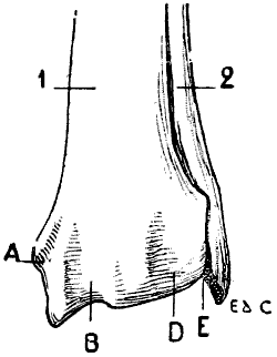
Fig. 29.—Inferior Extremity of the Bones of the Forearm of the Dog: Left Side, Anterior Surface, Normal Position—that is, the Position of Pronation.
1, Radius; 2, ulna; A, groove for the long abductor and for the short extensor of the thumb; B, groove for the radials; D, groove for the long extensor of the thumb, the special extensor of the index-finger, and the common extensor of the fingers; E, groove for the special extensor of the little finger.
Consequently it is on the anterior surface of the bone that we find the grooves concerning which it is necessary to give some details. Regarding them in passing from the radius towards the ulna, those grooves give passage to the tendons of the muscles whose names occupy the columns on p. 43. The letters which are referred to each serve to define their order, and to facilitate reference to Figs. 28, 29, and 30.
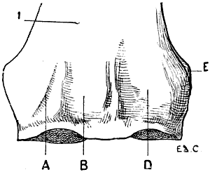
Fig. 30.—Inferior Extremity of the Bone of the Forearm of the Horse: Left Side, Anterior Surface.
1, Radius; A, groove for the long abductor and the short extensor of the thumb; B, groove for the radials; D, groove for the common extensor of the digits; E, groove for the special extensor of the little finger.
We should mention that the groove E is situated, both in man and in the dog, at the level of the inferior radio-ulnar articulation; but that in the horse, as the ulna does not exist at that level, the groove is situated on the external surface of the inferior extremity of the radius. It is necessary to add that, in some horses, the ulna is, nevertheless, represented in this region by a tongue-like process of bone; and in such cases the groove is situated in front of this process, at the level of the line of coalescence, which there represents the articulation.
Man. Dog. Horse.
A. Long abductor of the thumb, and short extensor of the thumb. A. Long abductor of the thumb and short extensor of the thumb united as one muscle, the oblique extensor of the metacarpus.[11] A. Oblique extensor of the metacarpus, the homologue of the long abductor of the thumb and the short extensor of the thumb, united as one muscle.
B. First and second external radials (extensor carpi radialis longior and brevior). B. The two radials blended superiorly, distinct inferiorly; this is the anterior extensor of the metacarpus. B. The radials represented by a single muscle, the anterior extensor of the metacarpus.
C. Long extensor of the thumb. C. Long extensor of the thumb and special extensor of the index finger united superiorly. These muscles pass in the following groove. C. The long extensor of the thumb and the special extensor of the index are absent.
D. Special extensor of the index finger and the common extensor of the fingers. D. Common extensor of the digits and the two preceding muscles. D. Anterior extensor of the phalanges, the homologue of the common extensor of the digits.
E. Special extensor of the little finger. E. Extensor of the third, fourth, and fifth digits, or the lateral extensor of the digits, the homologue of the special extensor of the little finger. E. Lateral extensor of the phalanges, the homologue of the special extensor of the little finger.
F. The posterior ulnar (extensor carpi ulnaris). There does not exist on the forearm a groove for the posterior ulnar muscle, or external flexor of the metacarpus.
It is also useful to note, with reference to the groove F, in which passes, in man, the tendon of the posterior ulnar muscle, that, when the forearm is in pronation, the radius alone being displaced, we can only see this groove on the surface which looks backwards; and that it is then separated from the groove which contains the tendon of the special extensor of the little finger by an interval equal to the thickness of the head of the ulna.[12] When the forearm is supinated, the two grooves are found, on the other hand, one beside the other: and the tendons which they contain are very naturally in contact.
In birds the forearm is flexed on the arm, and the latter being directed downwards and backwards, the former is, consequently, directed upwards and forwards. Further, because of the position of the humerus, which, as we mentioned on p. 32, has its inferior extremity so turned that the surface which is anterior in man becomes external, the radius, instead of being outside the ulna, is placed above it. This latter is larger than the radius, but its olecranon process is very slightly developed.
The Hand
The hand in animals, as in man, is formed of three parts—the carpus, metacarpus, and fingers. In man, the forearm and the hand being described in the position of supination; the bones of the carpus are named in passing from the most external to the most internal—that is to say, from that which corresponds to the radial side of the forearm to that which corresponds to the ulnar side. In animals in which, as we know, but it is not unprofitable to repeat, the hand is in pronation, the radial side of the forearm being placed inside, we enumerate the carpal bones in counting the most internal as the first; this is the only method which permits us, in taking our point of departure from the human skeleton as our standard, to recognise the homologies of the bones of the carpal region.
These bones, eight in number, are arranged in two transverse rows, of which one, the first, is superior or anti-brachial; the other, the second, is inferior or metacarpal. Each of these rows contains four bones. Considered in the order we have indicated above—that is to say, proceeding from the radial to the ulnar side—they are thus named: scaphoid, semilunar, cuneiform, and pisiform, in the first row; trapezium, trapezoid, os magnum, and unciform, in the second. The number of these bones is not the same in all animals on account of the coalescence or absence of some. In each row the bones are placed side by side, with the exception of the pisiform, which being placed on the palmar surface of the cuneiform, produces a small projection in man, but a very pronounced one in quadrupeds.
The pisiform is called the hooked bone in some veterinary anatomies. If we consider the hook which it forms, we may recognise that the name is appropriate; but from the point of view of comparison with the human carpus, the name is unfortunate, for it creates confusion between the true pisiform (the fourth bone in the upper row), and the last bone in the lower row, which is the veritable unciform bone. We do not here seek for similarity of form, but homology of regions; and it is only by using the same names to denote the same things that we can succeed in determining such homology.
Taken as a whole, the bones of the carpus form a mass which, by its superior border, articulates with the bones of the forearm, and by its inferior border is in relation with the metacarpal region. Its dorsal surface (anterior in quadrupeds) is slightly convex; its palmar surface (posterior in quadrupeds) is excavated, and forms a groove in which pass the tendons of the flexors of the fingers. This last, in man, has the appearance of a gutter, because of the prominences caused by the projection of the internal and external bones beyond their fellows.
In quadrupeds the palmar groove is especially determined by the pisiform bone, of which we have just mentioned the great development.
The region occupied by the carpus, in the unguligrades, is known as the knee; it would have been more appropriately named had it been called the wrist.
The number of the metacarpal bones in mammals never exceeds five, but it often falls below it; the same is true for the digits. The first are generally equal in number to the latter; an exception is met with in ruminants, whose two metacarpals coalescing soon after birth, form but one bone; this, the canon bone, articulates with two digits.
The number of metacarpals and digits diminishes in proportion as the limbs cease to be organs of prehension, and become more exclusively organs of support and locomotion.
The number of phalanges is two for the thumb and three for each of the other digits; except in the cetaceans, in which they are more numerous.
In the bat, the metacarpals and phalanges are very long, and form the skeleton of the wing; these phalanges are not furnished with nails; the thumb, which is very short, is alone provided with one (Fig. 8).
With regard to the relative dimensions of the bones of the metacarpus, it is necessary to remember that, in the human being, the second metacarpal is the longest; then, in the order of decrease, come the third, fourth, fifth, and first. In quadrupeds we shall also find differences in length (see the chapter relating to the anterior limbs in certain animals), but the order of decrease is not always that which we have just mentioned.
In man the articular surface, situated at the inferior extremity of each of the metacarpals, is rounded, and is called the head. This allows the first phalanx, which is in relation with that surface, to be displaced in every direction; indeed, this phalanx can not only be flexed and extended, but it can also be moved laterally; this latter movement allows of the fingers being separated and drawn together.
In quadrupeds which can only perform the movements of flexion and extension of the digits—for example, the horse—the inferior extremity of the metacarpal has not a rounded head of a regular outline; it is marked by a prominent median crest, directed from before backwards, so that the articular surfaces, which fit more exactly, form a sort of hinge which allows of backward and forward movements only, and permits no lateral displacement. In man, at the level of the inferior extremity of the first metacarpal, in the vicinity of the articulation of this bone with the first phalanx of the thumb, we find two sesamoid bones—small bones developed in the fibrous tissue which surrounds the articulation. We also meet with such structures, but more rarely, at the level of the corresponding articulation of the index and auricular digits; and, more rarely still, at those of the middle and ring fingers. In quadrupeds, these bones are normally developed, and we shall see afterwards that in some animals, as they reach a considerable size, they are able to influence the external outlines; we shall see this, for example, in the horse.
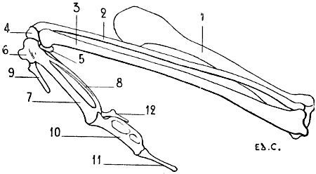
Fig. 31.—Skeleton of the Superior Limb of a Bird (Vulture): Left Side, External Surface.
1, Humerus; 2, radius; 3, ulna; 4, radial bone of the carpus; 5, ulnar bone of the carpus; 6, first metacarpal; 7, second metacarpal; 8, third metacarpal; 9, first digit, the homologue of the thumb; 10, first phalanx of the second digit; 11, second phalanx of the second digit; 12, third digit.
The hand, in birds, is directed obliquely downwards and backwards (Fig. 31). For the better understanding of its position in relation to the forearm, we should remember that this latter, as we have described (p. 44), directed obliquely upwards and forwards, has the radius placed above the ulna; the hand being oblique in the opposite direction and placed under the forearm is, by this arrangement, inclined towards the ulnar border of the latter.
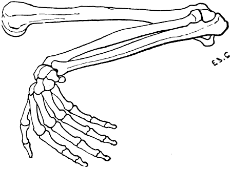
Fig. 32.—Superior Limb of the Human Being, the Different Segments being placed in the Attitude which the Corresponding Parts occupy in Birds: Left Side, External Surface.
For the rest, in order to be able to distinguish readily the corresponding parts in the hand of a bird and that of a man, we merely have to place the human forearm obliquely, in a direction upwards and forwards (Fig. 32), the radius being above; this position we can obtain by semi-pronation; then, to incline strongly the hand downwards and backwards, moving the ulnar border of the hand towards the ulna; the thumb is then anterior, the little finger posterior, and the palm of the hand is turned towards the trunk.
The carpus in birds is formed by two bones only, with which the skeleton of the forearm articulates. That which is in contact with the radius is called the radial bone of the carpus; and that with which the ulna articulates is named the ulnar bone.
The metacarpus is formed of three bones; the first, which is very short, is fused at its superior extremity with the adjoining one; this latter and the third, both longer than the first, but of unequal size, are fused at their extremities. The metacarpal, which articulates with the radial bone of the carpus, is larger than the one which is in line with the ulna. To the metacarpus succeed three digits, of which the central is the longest, and is formed of two phalanges; the other two are formed each by a small, stylet-shaped bone. The middle finger, situated on the prolongation of the metacarpal, which articulates with the radial bone of the carpus, has its first phalanx large and flattened transversely; this phalanx seems to have been formed by the union of two bones of unequal development; the second phalanx is styloid in form. As to the other two fingers, they are placed, one in front and the other behind; the first, which articulates with the short metacarpal, fused at its upper end with the principal bone of the metacarpus, in position represents the thumb. The other, which is the third finger, articulates with the inferior extremity of the thinnest bone of the metacarpus; it is sometimes closely united to the corresponding border of the first phalanx of the large—that is to say, of the median—digit.
The Anterior Limbs in Certain Animals
Plantigrades: Bear (Fig. 33).—The scapula of the bear approaches in shape to a trapezium, of which the angles have been rounded off. The anterior border (cervical) is strongly convex in the part next the glenoid cavity. The junction of the superior (spinal) and the cervical border forms almost a right angle, the summit of which corresponds to the origin of the spine. At its posterior angle there is a prominence, directed downwards, the surface of which is hollowed and is separated from the infraspinous fossa by a crest, so that at this level a third fossa is added to the infraspinous one. The neck of the scapula is but slightly marked. The acromion is prominent, and projects a little beyond the glenoid cavity.
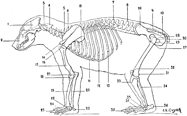
Fig. 33.—Skeleton of the Bear: Left Lateral Surface.
1. Cranium; 2, face; 3, atlas; 4, axis; 5, seventh cervical vertebra; 6, first dorsal vertebra; 7, fourteenth and last dorsal vertebra; 8, lumbar vertebræ; 9, sacrum; 10, coccygeal vertebræ; 11, sternum; 12, ninth and last sternal rib; 13, costal cartilages; 14, acromion process; 15, third fossa on the external surface of the scapula; 16, great tuberosity of the humerus; 17, musculo-spiral groove; 18, epicondyle; 19, radius; 20, ulna; 21, olecranon process; 22, carpus; 23, pisiform; 24, metacarpus; 25, phalanges; 26, ilium, external fossa; 27, pubis; 28, tuberosity of the ischium; 29, obturator foramen; 30, great trochanter of the femur; 31, condyles of the femur; 32, patella, or knee-cap; 33, anterior tuberosity of the tibia; 34, fibula; 35, tarsus; 36, calcaneum, or heel-bone; 37, metatarsus; 38, phalanges.
The clavicle is rudimentary, but, as an example of the complete development of this bone in plantigrade quadrupeds, we may cite the marmoset.
The humerus is furnished at its superior extremity with a large tuberosity, wide, and situated in front of the head of the bone; the effect of this is that the bicipital groove is internal. As in man, the great tuberosity does not reach so high as the humeral head, but it approaches more nearly to that level. The deltoid impression is very extensive, and descends pretty far down on the body of the bone. The epitrochlea is prominent; the epicondyle is surmounted by a well-marked crest, curved and flexuous in outline.
The articular surface, which is in contact with the radius, is not a regularly formed condyle; it is a little flattened on its anterior surface, and presents at this level a slight depression which corresponds to a small eminence on the anterior aspect of the superior extremity of the radius. The surface which articulates with the ulna, viewed on its anterior aspect, has the shape of a slightly-marked trochlea; except at the level of the internal lip, which, as in man, descends lower than the surface for articulation with the radius (condyle). Behind, the trochlea is more clearly defined.
The bear possesses a considerable power of rotation of the radius; the bones of the forearm are joined only at their extremities, while in the remainder of their extent they are widely separated. The ulna terminates below in a head and a styloid process; these articulate with the two last bones of the first row of the carpus—viz., the cuneiform and pisiform. The bones of the carpus are seven in number, the scaphoid and the semilunar being fused together.
The metacarpals, five in number, differ very little from one another in regard to length, though they increase in size from the first to the fifth; this may be demonstrated by looking at the palmar surface of the hand. It is the reverse of that which we find in man, for the fifth metacarpal is the thickest of all, and the first is the most slender.
At the level of each metacarpo-phalangeal articulation are two sesamoid bones.
The third digit is the longest. The terminal phalanges present two very different portions: one, the anterior, is curved and pointed; it serves to support the nail, whose shape it assumes; the other, posterior, forms a sort of sheath into which the base of the nail is received.
The inferior portion of the posterior surface of this latter part articulates with the second phalanx in the case of each of the last four digits, but with the first phalanx in the case of the thumb.
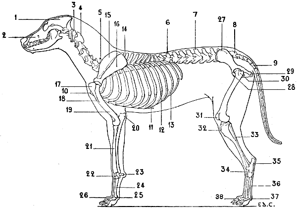
Fig. 34.—Skeleton of the Dog: Left Lateral Surface.
1, Cranium; 2, face; 3, atlas; 4, axis; 5, seventh cervical vertebra; 6, thirteenth and last dorsal vertebra; 7, lumbar vertebræ; 8, sacrum; 9, coccygeal vertebræ; 10, anterior extremity of the sternum; 11, xiphoid appendix; 12, ninth and last sternal rib; 13, costal cartilages; 14, spinal border of the scapula; 15, supraspinous fossa of the scapula; 16, infraspinous fossa of the scapula; 17, great tuberosity of the humerus; 18, deltoid impression; 19, musculo-spiral groove; 20, olecranon process; 21, radius; 22, carpus; 23, pisiform; 24, metacarpus; 25, sesamoid bones; 26, phalanges; 27, ilium, iliac crest; 28, pubis; 29, tuberosity of the ischium; 30, great trochanter of the femur; 31, patella, or knee-cap; 32, anterior tuberosity of the tibia; 33, fibula; 34, tarsus; 35, calcaneum, or heel-bone; 36, metatarsus; 37, sesamoid bones; 38, phalanges.
Digitigrades: Cat, Dog (Fig. 34).—In these animals the anterior (cervical) border of the scapula is convex; the posterior (axillary) border is straight or slightly concave. The supraspinous and infraspinous fossæ are of equal extent (Figs. 35 and 36). The neck is short. The spine of the scapula becomes more and more prominent towards its inferior extremity, where it ends in a twisted and inflexed portion, which represents the acromion process; this process terminates at the level of the glenoid cavity. The coracoid process is represented by a small tubercle, slightly curved inwards; this tubercle is situated above the glenoid cavity, at the inferior part of the cervical border.
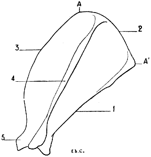
Fig. 35.—Scapula of the Dog: Left Side, External Surface.
1, Posterior or axillary border; 2, superior or spinal border; 3, anterior or cervical border; 4, spine of scapula; 5, coracoid process; AA′, length of spinal border.
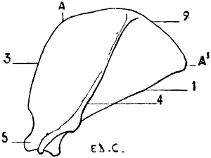
Fig. 36.—Left Scapula of the Cat: External Surface.
1, Posterior or axillary border; 2, superior or spinal border; 3, anterior or cervical border; 4, spine of the scapula; 5, coracoid process; AA′, length of the spinal border.
In the dog, the posterior angle, formed by the junction of the axillary and the superior (spinal) borders, is obtuse; the spine rises perpendicularly from the surface of the bone. The width of the scapula, measured at the level of the spinal border (from A to A′, Fig. 35), equals about half the length of the spine. We must, however, make an exception for the turnspit dog, in which the superior border equals three-fourths of that length. The scapula is, in this case, of a more compact type; it is broader, but shorter. In the cat, the anterior outline of the scapula, formed by the union of the cervical border and the corresponding half of the spinal, is more convex; the posterior angle is not obtuse, as in the dog. The spine is bent slightly downwards and backwards; before terminating in the acromion process it presents a triangular projection, the apex of which is directed downwards. The tubercle which represents the coracoid process is curved inwards more strongly than that of the dog, thus resembling more closely the appearance of this process in the human being.
All proportions considered, the scapula of the cat is broader than that of the dog; its width, measured along the length of its spinal border (from A to A′, Fig. 36), equals three-fourths of the length of the spine.
The clavicle is rudimentary; it is, however, better developed in the cat than in the dog. The clavicle of the cat is represented by a small, elongated bone, curved in outline, the convexity being turned forward; it is united to the acromion and the sternum by ligamentous fibres; that of the dog is merely a scale-like osseous plate situated on the posterior surface of a muscle of this region (see Figs. 16 and 17).
The humerus is long and twisted in the shape of an S. The inferior articular surface has the form of a simple pulley, for the condyle is very slightly marked. The internal part of this articular surface descends lower than the external; this condition resembles that found in the human being, where the inner lip of the trochlea is lower than the condyle.
In the dog, the olecranon fossa communicates with the coronoid by an opening.
In the cat, there is a supra-epitrochlear canal (see Fig. 19), but no olecranon perforation.
The bones of the forearm articulate at their extremities. The body of the radius is united to the body of the ulna by a short, thick, interosseous ligament; the fibres of this ligament, though short, do not prevent the production of some movements at the articulations of the bones.
The radius so crosses the ulna that above, it is in front and external to the latter, while below, it is internal. This bone is flattened from front to back, and slightly convex anteriorly. Its superior extremity is formed, externally, of a portion which represents the head of the radius in man; internally, by another portion which represents half of the coronoid process of the ulna, which, in the human being, belongs exclusively to the latter (see p. 39, the encroachment of the radius on the ulna). This extremity is surrounded with a vertical articular surface which is placed in contact with a small cavity which is hollowed out on the ulna (the lesser sigmoid cavity); and presents at its superior aspect a surface which articulates with the inferior extremity of the humerus. The shaft of the bone has on its internal border rugosities analogous to the imprint of the pronator radii teres of the human skeleton; these rugosities, indeed, give insertion to a muscle of the same function, and bearing the same name. The inferior extremity, broader than the superior, is hollowed on its external aspect by a small cavity which receives the inferior extremity of the ulna; its inferior surface (concave) articulates with the carpus; its anterior surface (the homologue of the posterior surface of the corresponding extremity of the human radius) presents grooves which serve for the passage of the tendons of the muscles which pass from the forearm to the back of the hand. (For the names of the muscles whose tendons pass in these grooves, see Fig. 29.)
The ulna is furnished at its superior extremity with an olecranon process, which is more prominent than that of the human ulna; this process is compressed laterally, and its internal surface is hollowed; there we also find a great sigmoid cavity, and a coronoid process situated at the internal part of the anterior surface, a process which, as we have previously shown, it shares with the radius.
The shaft of the bone, prismatic and triangular, diminishes in thickness as it approaches the lower extremity, which articulates with the corresponding extremity of the radius. In the dog, the ulna terminates inferiorly in a blunt point, without enlargement, analogous to the head of the human ulna; in the cat, by a head which is prolonged into a styloid process, by which it articulates with a portion of the carpus.
The carpus consists of seven bones—three in the superior row and four in the inferior. In the superior row the scaphoid and semilunar bones are fused together. The pisiform is elongated and expanded at its two extremities; it forms a prominence which, directed backwards, projects beyond the level of the other bones of this region.
The metacarpal bones are five in number; they are enumerated from within outwards; they articulate with the carpus and with each other. The inferior extremity of each metacarpal bone presents the form of a condyle in front; and is divided behind so as to form two lateral condyles, which are separated by a median crest; on these posterior condyles are applied two small sesamoid bones. The metacarpal bone of the thumb is very short; the third and fourth are the longest. The metacarpus, as a whole, is directed vertically.
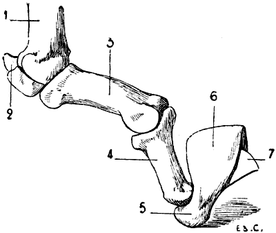
Fig. 37.—Skeleton of the Finger of a Felide (Lion): Left Side, Internal Surface.
1, Metacarpus; 2, sesamoid bones; 3, first phalanx; 4, second phalanx; 5, third phalanx; 6, gutter for the reception of the base of the nail; 7, prominent osseous crest formed to lodge in the concavity of the nail.
The phalanges are three in number for each finger, except the thumb, which has but two. The first phalanx, directed almost horizontally forwards, is the longest; the second is directed downwards and forwards; the third consists of two portions: a posterior part, which forms a sort of sheath into which the base of the nail is received; and an anterior, conical in form, and curved in crochet shape, which forms a support for the nail (Fig. 37).
The third and fourth digits are the longest; the second and fifth are of equal length; the thumb is the shortest; it does not touch the ground, and does not even reach the articulation of the metacarpal bone and first phalanx of the second finger.
In the cat, the metacarpal bone of the thumb, although shorter than any of the others, is quite as thick. The third digit is a little longer than either the second or fourth. In animals of this genus, the claws, in the condition of repose, are retracted, and removed from the ground; this prevents their being worn, and thus preserves their sharpness. At such times the third phalanx is received into a groove which is found on the external surface of the second phalanx. In the dog, the claws are not tractile.
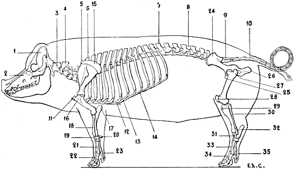
Fig. 38.—Skeleton of the Pig: Left Lateral Surface.
1, Cranium; 2, face; 3, atlas; 4, axis; 5, seventh cervical vertebra; 6, first dorsal vertebra; 7, fourteenth and last dorsal vertebra; 8, lumbar vertebræ; 9, sacrum; 10, coccygeal vertebræ; 11, anterior extremity of the sternum; 12, xiphoid appendix; 13, seventh and last sternal rib; 14, costal cartilage; 15, cartilage of prolongation of the scapula; 16, great tuberosity of the humerus; 17, olecranon process; 18, radius; 19, ulna; 20, pisiform; 21, metacarpus; 22, phalanges of the two great toes; 23, phalanges of the external toe; 24, ilium; 25, pubis; 26, tuberosity of the ischium; 27, great trochanter; 28, knee-cap; 29, anterior tuberosity of the tibia; 30, fibula; 31, tarsus; 32, calcaneum; 33, metatarsus; 34, phalanges of the two great toes; 35, phalanges of the external toe.
Unguligrades: Pig (Fig. 38).—The scapula is markedly narrowed in the region above the glenoid cavity. The spine is atrophied at both its extremities, so that at its inferior part we do not find the acromion process. In its middle portion the spine is prominent, and presents a triangular process which turns backwards, overlapping a part of the infraspinous fossa; this latter is much larger than the supraspinous. The spinal border is surmounted by the cartilage of prolongation, the superior margin of which is convex; this cartilage extends posteriorly beyond the posterior (axillary) border of the bone.
The small tuberosity of the superior extremity of the humerus is but slightly developed; the great tuberosity, on the contrary, is very large. The bicipital groove is situated internal to this. The deltoid impression is scarcely marked.
The forearm is short, directed obliquely downwards and inwards, thus forming with the hand an angle, of which the apex is directed inwards. The two bones of the forearm are strongly bound to one another by an interosseous ligament, which is formed of very short fibres. The radius appropriates, at its superior extremity, the coronoid process of the ulna. The latter is, notwithstanding, well developed in the rest of its extent; it has a flattened shaft which almost completely overlaps the posterior surface of the radius; its inferior extremity reaches to the carpus.
The carpus is formed of eight bones—four in the superior row, and four in the inferior. The third bone of the superior row (cuneiform) is more in contact with the ulna than with the radius.
There are but four metacarpal bones; there is no metacarpal of the thumb. The two median metacarpal bones are the longest; they are those which correspond to the digits which alone touch the ground. The internal digit and the external one are thin and short; they are functionless, as a rule, taking no part in supporting the limbs on the ground. Notwithstanding this, they are formed, as the other digits, of a number of phalanges, which give them the semblance of perfect digits. (We shall soon see that in certain animals there exist digits which, being incomplete with regard to the numbers of their constituent bones, more accurately merit the name of imperfect digits.)
The third phalanges are each enclosed in a horny hoof, to which the name of onglon has been given.
We have already drawn attention to the smaller lateral digits, and noted the general fact that they do not come in contact with the ground. It is necessary to modify this statement by adding that under certain conditions they give a slight amount of support; for example, when the individual is the subject of excessive obesity, the limbs yield under the weight, and the nails of the lateral digits may touch the ground.
A similar fact may be noticed in pigs of ordinary bulk at the moment when, during walking, each of the fore-limbs commences to bear the weight—that is to say, when it is directed obliquely downwards and forwards; then all the digits are in contact with the ground.
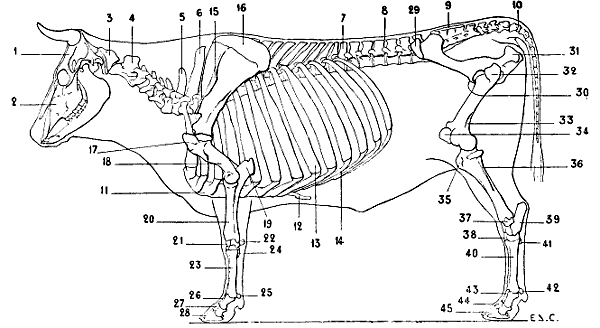
Fig. 39.—Skeleton of the Ox: Left Lateral Surface.
1, Cranium; 2, face; 3, atlas; 4, axis; 5, seventh cervical vertebra; 6, first dorsal vertebra; 7, thirteenth and last dorsal vertebra; 8, lumbar vertebræ; 9, sacrum; 10, coccygeal vertebræ; 11, sternum; 12, xiphoid appendix; 13, eighth and last sternal rib; 14, costal cartilages; 15, spine of scapula; 16, cartilage of prolongation of the scapula; 17, great tuberosity of the humerus; 18, musculo-spiral groove; 19, olecranon process; 20, radius; 21, carpus; 22, pisiform; 23, metacarpus; 24, rudimentary metacarpal; 25, sesamoid bones; 26, first phalanges; 27, second phalanges; 28, third phalanges; 29, anterior iliac spine; 30, pubis; 31, tuberosity of the ischium; 32, great trochanter; 33, supracondyloid fossa of the femur; 34, patella, or knee-cap; 35, anterior tuberosity of the tibia; 36, fibula; 37, coronoid tarsal bone; 38, tarsus; 39, calcaneum; 40, metatarsus; 41, rudimentary metatarsus; 42, sesamoid bones; 43, first phalanges; 44, second phalanges; 45, third phalanges.
Unguligrades (Ungulates): Sheep, Ox (Fig. 39).—The scapula, which is of elongated form, is very narrow in the vicinity of the glenoid cavity. The spine, which becomes more and more salient towards its inferior part, terminates abruptly in a border, which, forming an acute angle with the crest, produces a projection which represents the acromion process—a very rudimentary acromion, for it does not reach the level of the glenoid cavity. The supraspinous fossa is much smaller than the infraspinous; it hardly equals one-third the extent of the latter. The anterior border, thin and convex in its superior portion, is concave in the rest of its extent; the posterior border is thick and slightly concave; the spinal border is surmounted by the cartilage of prolongation. In the ox the spine of the scapula, in its middle portion, is flexed a little backwards on the infraspinous fossa.
The great tuberosity of the humerus is highly developed; its summit, very prominent, is flexed over the bicipital groove; a prominence of the small tuberosity also bends over the groove, with the result that at this level the latter is converted into a sort of canal. At the inferior extremity the condyle, although not large, is recognisable; for it is separated from the trochlea by a depression in form of a groove. In contrast to the condition found in man, the condyle descends to a level a little below that of the internal lip of the trochlea. (For the arrangement of the epicondyle and the epitrochlea, see p. 30.) In the sheep, the deltoid impression is but slightly marked; in the ox, it is more evident.