Volume 3 — DISEASES OF THE RESPIRATORY SYSTEM. (continued 5)
Reading edition
Topical treatment of a more decided character being required, the physician usually chooses between powder and solution. Powders are usually propelled by a puff of air through a properly curved tube, whether from a rubber ball, a reservoir of compressed air, or the mouth. The mouth allows the most delicate and accurate application, but the mouthpiece should be protected by a valve from receiving a return current when the patient coughs. Solutions may be applied by means of pipette, syringe, brush, cotton wad, or sponge, according to indications. A fragment of sponge securely fastened to a properly-bent rod or pair of forceps is the safest and most effectual material for positive contact against a limited surface, and a brush the best for painting larger surfaces. The use of the cotton wad involves a slight risk of leaving a detached shred of fibre in the larynx, but renders the manipulation less unpleasant to the patient than the use of the sponge, and is less irritating to the mucous membrane. Spasm of the larynx is usually excited the first time that a medicinal application is made within it, and even death by suffocation has followed the incautious use of powerful agents. Hence strong solutions should not be used until the tolerance of the parts has been sufficiently tested by weak or innocuous ones. The remedies which have been employed topically for intra-laryngeal medication seem to include every available medicinal agent that could be mentioned, from rose-water to the incandescent cautery. The list of really useful ones is not very long. Those upon which the most reliance is placed by the writer comprise tannic acid (a saturated glycerite), zinc sulphate (thirty grains to the ounce of rose-water), and silver nitrate (forty to sixty grains to the ounce) in obstinate and protracted cases of simple chronic laryngitis; iodine and carbolic acid, singly or in combination (one grain or more to the ounce of glycerin), and chinoline tartrate or salicylate (five or more grains to the ounce), in cases attended with infiltration; iodoform (finely pulverized or in recent saturation in sulphuric ether) in ulcerative or proliferative tuberculosis; and iodoform and acid solution of mercuric nitrate (one part to ten or twelve of water) in progressive ulcerative syphilis resisting appropriate constitutional treatment. Other astringents in the simple varieties; resorcin in the glandular, hypertrophic, polypoid, and tuberculous varieties; chromic acid and incandescent metal in the circumscribed hypertrophic and in the polypoid varieties; and zinc chloride and copper sulphate in the syphilitic varieties,—proffer additional resources. These applications are to be made at intervals of one day or more, according to results. Hyperæsthesia and pain, whether of the larynx or of parts adjacent, can usually be subdued by the local anæsthetic effect of solutions of erythroxyline hydrochloride (2 per cent. or stronger) applied at intervals of a few hours, or even by the fluid extract or a strong aqueous infusion of the erythroxylon-leaves. Before the anæsthetic effect of this drug was known, morphine powder (one-eighth to one-fourth of a grain, alone or associated with tannin or with iodoform) or aqueous solutions of morphine salts and of aconite were employed to relieve pain and obtund sensitiveness. The oleate of morphine (2 to 4 per cent. solution) and the oleate of aconitine (2 per cent. solution) are similarly useful. Morphine, by its constitutional influence, is preferable to erythroxyline in some instances, though less prompt in its effects. Where ulcerative processes at the top of the larynx or thereabouts entail odynphagia, these preparations should be used before administering nourishment. The use of erythroxylon products may be entrusted to the nurse or to the patient with comparative safety. Morphine and aconite should be applied only by a medical attendant or an exceptionally skilled nurse. Before any medicinal curative or reparative agent is applied the parts should be thoroughly cleansed of suppurative and secretory products. This may be done with sprays of alkaline solutions—five or more grains of sodium borate or bicarbonate, for example—dissolved in pure water, in tar-water, or in an emulsion of coal tar. An excellent agent, especially in the presence of pus, is hydrogen dioxide, usually furnished in a 10-volume solution which should be diluted with two or more parts of distilled water. It is likewise disinfectant and gently stimulant to mucous membrane. The manipulations by the physician preparatory and medicatory should be performed laryngoscopically, otherwise the entire procedure must be haphazard.
Neoplasmata and fungous growths may require removal should they interfere with respiration. In the presence of stricture, surgical interference by tracheotomy may become requisite. Elsberg, according to the testimony of his assistant, Schweig, seems to have been particularly favorable to the performance of this operation in obstinate cases of ulcerative laryngitis of whatever character, and even in protracted non-ulcerative cases, for the purpose of securing physiological rest to the parts, although the procedure might not be indicated to relieve any embarrassment in respiration. The writer's experience in tracheotomy as a factor in producing rest has not been favorable, such a result being usually defeated by the cough so frequently following a tracheotomy, no matter how well-adjusted a tube may have been inserted. His recommendation, therefore, is limited to cases of embarrassment to respiration due to stricture or constriction unamenable to intra-laryngeal interference.
Morbid Growths of the Larynx.
DEFINITION.—Neoplastic formations, benign and malign, in the interior of the larynx, in its cartilaginous framework, in its investment-tissues, or upon the exterior of the organ.
ETIOLOGY.—Inflammation of the mucous membrane, local irritation or injury, ulceration, cell-proliferation, and excessive granulation seem to be the exciting causes of benign neoplasms. They follow on laryngitis, whether catarrhal, syphilitic, tuberculous, exanthematic, toxic, or traumatic. They are quite common, so to speak, several thousands of cases being on record, and as many or more probably being unrecorded. Heredity does not seem to play any special part in their production. They are occasionally congenital, and may be developed at any age; but they are encountered the most frequently in subjects between the ages of thirty and sixty years, probably because of the greater exposure to laryngitis attending the activity incidental to the prime of life. Males are affected far more frequently than females, probably on account of greater exposure to sources of laryngitis. Benign growths are sometimes followed by malign growths in recurrence, and are sometimes converted into malignity by irritation, whether physiological, mechanical, or instrumental. Malign growths are attributed to cold, chronic laryngitis, and traumatism as the initial exciting causes. Butlin suggests a cryptogamic origin. They are far more common in males than in females, and occur chiefly between the ages of twenty-five and seventy, but they have been noted as occurring exceptionally much later, and even as early as the first year.
PATHOLOGY AND MORBID ANATOMY.—By far the greater number of laryngeal morbid growths belong histologically to the category of benign neoplasms, but the important location they occupy often renders them clinically malign. By far the greater number of benign growths are papillomas, perhaps fully two-thirds, although Elsberg has reported that but 163 instances were papillomas out of 310 seen in his own practice.7 This has been an exceptional experience. Then we have fibromas, myxomas, adenomas, lymphomas, angeiomas, cystomas, ecchondromas, lipomas, and composite neoplasms. Laryngeal morbid growths, too, occasionally undergo the fatty, colloid, or amyloid degenerations. Papillomas are frequently multiple, and most frequently sessile, but the other benign neoplasms are most frequently single and are more often pedunculated. All this class of morbid growths affect the anterior half of the larynx more than the posterior. They are most frequent on the vocal bands or very near to them, although they may occupy any portion of the larynx. They vary in size from the smallest protuberance to a bulk sufficient to block up the cavity of the larynx and even project above it. The dimensions of the greater number of papillomas vary from the size of a pea to that of a small mulberry. Other benign neoplasms rarely reach the bulk attained by papillomas.
7 Archives of Laryngology, p. 1, New York, 1880.
Malign growths are far less common than benign ones. They comprise both sarcomas and carcinomas. Sarcomas occur in the varieties of spindle-celled, round-celled, giant-celled, mixed-celled, fibrosarcoma, lymphosarcoma, and myxosarcoma. Some attain only the size of small beans, and few exceed the size of a pigeon's egg. The majority of them are primary growths. Most of them originate in the interior of the larynx, whence they may extend by contiguous infiltration, even penetrating the laryngeal walls. The vocal band and the ventricular band are the most frequent seat. The epiglottis is a common seat. These growths appear either in irregular, smooth, spheroid masses, or nodulated, mamillated, and dendritic. They are much the more common in males, and occur chiefly in subjects between the ages of twenty-five and fifty. Their growth is slow for a year or more, and then becomes more rapid.
Carcinoma is much more common than sarcoma. It is most frequently primary, and primarily limited to the larynx, but occurs likewise in extension of carcinoma of the tongue, palate, pharynx, oesophagus, or thyroid gland. It rarely extends to the oesophagus or penetrates the laryngeal walls.
Squamous-celled carcinoma or epithelioma is the commonest variety, large spheroidal-celled or encephaloid being much less frequent, and small spheroidal-celled and cylindrical-celled occurring still more rarely. Intrinsic laryngeal carcinoma is usually unilateral at first, and most frequently in the left side. Its most frequent seat is at the vocal band. It rarely occurs below this point, and when it does, as in the five cases analyzed by Butlin,8 it seems to be at some point just beneath. Extrinsic laryngeal carcinoma usually begins in the epiglottis, and sometimes occupies that structure only. It may begin in a cicatrix in the skin.9 Carcinoma is the more common in males, chiefly in subjects between the ages of fifty and seventy. It has occurred within the first year, at three years, and as late as at eighty-three years. Carcinoma is liable to extend by infiltration of tissue and destroy all the contiguous and overlying tissues, so that it may extend into the pharynx or even externally; the large spheroidal-celled variety presenting the most frequently progressive ulceration into contiguous tissue, and the squamous-celled, intrinsic ulceration. Hemorrhage is frequent. Perichondritis, abscess, necrosis, and fistula take place in old cases.
8 On Malignant Disease of the Larynx, p. 36, London, 1883.
9 Cohen, Transactions American Laryngological Association, p. 113, 1883.
SYMPTOMATOLOGY.—Small growths in localities where they neither provoke cough nor interfere with voice or respiration may run their course for a long time without giving rise to any symptoms at all. Growths of larger size, pedunculated growths, and growths located upon important structures give rise to interference with voice, respiration, or deglutition as may be—to cough, and even to pain. Dysphonia is due to mechanical interference with vibrations of the edges of the vocal bands; aphonia, to mechanical interference with their approximation; diphthonia, to mechanical interference at an acoustic node. These manifestations may be permanent or intermittent. Dysphonia is one of the earliest symptoms of carcinoma, and is usually continuous for a number of months before any other indication. Aphonia in carcinoma is often due to nerve-lesion. Dyspnoea is due to some considerable mechanical occlusion of the respiratory tract, whether by the growth itself or in consequence of oedema or of intercurrent tumefaction. It is inspiratory rather than expiratory, and subject to aggravation at night. As with the dysphonia, it varies with the size, location, and mobility of the growth and the position of the head and neck. It may be intermittent or permanent; be slight or severe; or it may terminate in apnoea by spasm, by mechanical occlusion of the calibre of the larynx, or by impaction of the growth at the chink of the glottis. Marked encroachment on the breathing-space is not accompanied with as marked dyspnoea as in acute processes, the parts seeming to acquire tolerance during the slow growth of neoplasms.
Dysphagia is due to a growth at the top of the larynx or on some portion of its pharyngeal surface. It is quite frequent in carcinoma, preceding dysphonia in the extrinsic varieties. It may be associated with regurgitation of food, drink, or saliva into the larynx, provocative of paroxysms of suffocation. Cough is due to growths which project from the vocal bands or press upon them, or to hemorrhage or accumulation of secretory or suppurative products. Hemorrhage, cough, and expectoration of bloody and fetid masses are indicative of carcinoma. Pain is usually due to intercurrent conditions. Aches in the part and sensations of the presence of a foreign substance are more frequent. Intense pain is exceptional in benign neoplasmata; it is often an early symptom in carcinoma, in which it is apt to radiate toward the ears and along the neck. Epileptic seizures and vertigo are sometimes occasioned by reflex influence. Exceptionally, large growths may produce change in the external configuration of the larynx. The general health is not much involved in benign growths, unless they interfere seriously with important physiological functions. Impaired health is far less manifest in sarcoma than in carcinoma. Emaciation, pyresis, and marasmus eventually occur as constitutional manifestations of malign growths.
DIAGNOSIS.—Laryngoscopic inspection usually reveals the growth and furnishes the best means of diagnosis. Intra-ventricular and subglottic growths may elude detection. Palpation is sometimes available, especially with children. Palpation with probes under laryngoscopic inspection is sometimes requisite to determine the mobility of a growth, its form and seat of attachment, and even its size. It seems, too, to discriminate a neoplasm from an eversion of a ventricle. While the histological character of a growth cannot be definitively decided by laryngoscopic inspection, the varieties present a series of characteristics sufficiently pronounced for approximative discrimination. Papillomata are often multiple, usually sessile, and usually racemose or dendritic. Some are white, but the majority are red, and the tinge varies from one extreme of the tint to the other. Some are as small as the smallest seeds; most of them have a bulk varying from that of a pea to that of a berry; some of them are so extensive as to appear to fill the larynx or even project above its borders. They are far the most frequent in the anterior portion of the larynx, and are often located upon a vocal band. Fibromata are most frequently single, smooth and pedunculated, and red. Some are white or gray. Some are vascular. When fully developed they vary in size from small peas to large nuts. They are more frequent upon a vocal band. Their development is slower than that of papillomata. Myxomata are usually single, smooth, pyriform, and pedunculated. They are usually red or reddish. Their ultimate size varies from that of grains of rice to that of Lima beans. They are most frequent at the commissure of the vocal bands. Angeiomata are usually single, reddish or bluish, vary in size from that of small peas to that of berries, and are most frequent on the vocal bands. Cystomata are usually globular, sessile, translucent, and white or red. They are most frequent in a ventricle or on the epiglottis. Their size varies from that of hempseed to that of peas. Ecchondromata are usually developed in the posterior portion of the larynx. Other benign growths are very rare, and do not seem to present special features for recognition by laryngoscopic inspection. Sarcomata are usually present as sessile, hard, well-circumscribed growths, smooth or lobulated. Some are dendritic on the surface, but not to the extent noticed in papillomata, and their location at the posterior portion of the larynx would suggest their true character, for papillomata rarely occupy this position except in tuberculosis. Superficial ulceration occurs in some cases, but is not extensive. There is no peculiarity in the color of the mucous membrane, which may be paler or redder than is normal. The lymphatic glands are not involved.10 Carcinomata present first as diffuse tumefactions in circumscribed localities, gradually undergoing transformation into well-formed growths, then nodulation, and then ulceration. Meanwhile, especially in extrinsic varieties, the submaxillary and the cervical lymphatic glands become successively involved and tumefied. Squamous-celled carcinoma becomes pale, wrinkled, and nodulated, and sometimes dendritic. Large spheroidal-celled carcinoma becomes nodulated, dark, and irregularly vascular, and finally ulcerated, perhaps at a number of points. In the ulcerative stage of carcinoma of the epiglottis and of the interior of the larynx discrimination is requisite from syphilis and from tuberculosis. In all cases of doubt as to malignancy, laryngoscopic inspection should be supplemented by microscopic examination of fragments detached for the purpose. The early detection of sarcoma may lead to surgical measures competent to save life—a remark applicable, perhaps, in a far more limited degree to intrinsic carcinoma.
10 Butlin, op. cit., p. 14.
PROGNOSIS.—The prognosis is usually good in benign growths submitted to proper surgical treatment. Left to themselves or treated medicinally, the prognosis is bad both as to function and to life. Such growths are occasionally expectorated after detachment during cough or emesis. Some occasionally undergo spontaneous absorption. Some remain without change for years. Most of them enlarge and compromise life as well as function. Recurrence occasionally follows thorough removal, and this recurrence is occasionally malign in character. Repullulation frequently follows incomplete removal. The prognosis is favorable in sarcomata, provided thorough eradication can be accomplished by surgical procedure. Incomplete removal is followed by repullulation or recurrence. Unsubmitted to operation, sarcoma will destroy life either mechanically by apnoea or physiologically by asthenia.
The prognosis is unfavorable in carcinoma. Recurrence takes place as the rule despite the best devised resources of surgery. Intrinsic carcinoma offers some hope of success to the surgeon; extrinsic carcinoma, little if any. Life is shortest in the large spheroidal-celled, and longest in the small spheroidal-celled variety, other conditions being equal. Death may take place by apnoea or asthenia, as in sarcoma, or by hemorrhage, collapse, or pyæmia. Submitted to tracheotomy at the proper moment in cases in which death is threatened by occlusive dyspnoea, life is prolonged and suffering mitigated. The fresh lease of life is longest in the squamous-celled variety.
TREATMENT.—The essential treatment is surgical, and to surgical works the reader must be referred for details. Suffice it to say that when a benign growth is small and does not embarrass respiration, it need not be attacked at all, unless its interference with the voice deprives the patient of his means of livelihood. The majority of benign growths are accessible to instruments passed through the mouth. Some require external incision into the larynx, whether partial or complete. The intra-laryngeal procedures in vogue include cauterization, both chemical and by incandescence, incision, abscission, crushing, brushing, scraping, and evulsion. According to the character and location of the growth, direct access from the exterior is practised by infra-hyoid pharyngotomy, by partial or complete thyroid laryngotomy, mesochondric laryngotomy, cricoid laryngotomy, complete laryngotomy, laryngo-tracheotomy, or tracheotomy, as may be indicated.
The thorough eradication of sarcomata usually requires a direct access by section of the thyroid cartilage or even of the entire larynx. This procedure failing or appearing insufficient, partial or even complete laryngectomy may be necessary. Temporizing is of no avail.
The treatment of carcinoma is palliative, unless it be decided advisable to attempt eradication, which may offer some chance of success in intrinsic carcinoma still confined to the larynx. Laryngectomy may be unilateral in some instances, and must be bilateral in others. Unilateral laryngectomy is the more hopeful. Eradication proffers no hope in cases of extrinsic carcinoma in which the growth has passed the boundaries of the larynx. After recovery from the laryngectomy an artificial appliance may be adjusted to the parts for the purpose of supplying a mechanical method of producing sound in the larynx for speaking purposes. Should no radical procedures be instituted, treatment is relegated to general principles, with prophylactic performance of tracheotomy in the presence of dangerous occlusion of the larynx. The voice should be used but little. All sources of laryngitis should be avoided. Ergot or hamamelis may be given to restrain hemorrhage, and morphine to relieve pain and secure sleep. Sprays can be used to keep the parts free from morbid products. Erythroxyline may be applied to produce local anæsthesia as required. Semi-detached portions of growth may be removed from time to time. Nourishment may be given by the bowel when necessary, and so on as in other diseases of the larynx in which the functions of respiration and deglutition are seriously impaired. Medicinally, arsenic may be given in the early stages, as that drug is conceded to possess some slight retarding influence on the growth of carcinoma.
Lupus of the Larynx.
Lupus is rare in the larynx. It usually occupies the structures above the vocal bands. It is most frequent in females, and usually associated with cutaneous lupus.
ETIOLOGY.—Scrofulosis and syphilis seem to be the predisposing causes. Climate may have some influence. The reason of the special proclivity of the female is undetermined. Of 9 reported cases, records of which are before the writer, 8 were in females.
PATHOLOGY AND MORBID ANATOMY.—Laryngeal lupus is usually an extension of the disease from the upper lip or the nose, extending along the nasal passages, pharynx, and palate. Destructive ulceration takes place, with irregular cicatrization and the formation of hard nodules of hyperplastic tissue of irregular conformation, varying from the size of hempseeds to that of small peas, similar to the cutaneous buccal and pharyngeal nodules.
SYMPTOMS.—These include dysphonia, dyspnoea, dysphagia, and cough. Pain is exceptional.
DIAGNOSIS.—Laryngoscopic inspection reveals the characteristic nodulation, the nature of which is inferred from the coexistence of external lupus. The disease may be confounded with lepra, syphilis, tuberculosis, or carcinoma. Discrimination from syphilis is the most difficult, and is predicated chiefly on its slow progress and on the absence of constitutional manifestations.
PROGNOSIS.—This is unfavorable. The reported cures seem to have occurred only under the influence of antisyphilitic treatment.
TREATMENT.—The prolonged use of cod-liver oil and of potassium iodide seems to be more beneficial than any other systemic treatment. Destruction of the nodules and ulcerated tissues is indicated when the diseased structures are sufficiently circumscribed and accessible. This may be done with the sharp spoon or with the electric cautery. Silver nitrate and iodine have been lauded as topical remedies.
Lepra of the Larynx.
Lepra is rare in the larynx.
ETIOLOGY.—Its cause seems to be climatic. In Europe it is most frequent in Norway and Sweden, and in America in Cuba and the West Indies.
PATHOLOGY AND MORBID ANATOMY.—It is always associated with cutaneous lepra, and usually with lepra of the nasal passages and the pharynx. According to Schroetter's observations, laryngeal lepra occurs as small connective-tissue nodules on the epiglottis or in the interior of the larynx, or as uniform thickenings, general or circumscribed. These may lead to stricture. Extensive ulceration may ensue.
SYMPTOMS.—Dysphonia, aphonia, dyspnoea, cough, and local anæsthesia are the main symptoms. Pain is infrequent.
DIAGNOSIS.—This depends upon the external manifestations of lepra and the laryngoscopic detection of the characteristic thickenings and nodulations.
PROGNOSIS.—This is unfavorable.
TREATMENT.—This must be conducted on general principles. Elsberg commended iodoform topically and gurgun oil internally.
DISEASES OF THE TRACHEA.
BY LOUIS ELSBERG, A.M., M.D.
Disease originating in or confined to the trachea is rare. It hardly ever follows tracheotomy unless the shape of the canula or its relation to the windpipe be improper; the normal tracheal mucous membrane probably resists cadaveric disintegration longer than any other mucous membrane of the body. But morbid processes of the larynx often extend downward, and those of the bronchial tubes still more frequently upward, so that the trachea is found affected in connection with both. Indeed, in what is ordinarily simply called bronchitis (see article on BRONCHITIS) the windpipe is seldom free from the inflammatory condition.
We shall here consider Inflammation, Ulceration, Morbid Growths, Stenosis, and Dilatation (hernia, fistula). Tracheotomy may have to be performed in any of these diseases to prevent impending suffocation, and in some to gain access to the part for further treatment. (See article on TRACHEOTOMY.)
INFLAMMATION.
Tracheitis is either simple or complicated, and acute or chronic.
Simple Tracheitis.
DEFINITION.—Inflammation of the windpipe limited to the mucous membrane.
SYNONYMS.—Catarrhal tracheitis, Tracheal catarrh.
Its ETIOLOGY may be gathered from the corresponding sections on Catarrhal Laryngitis and Bronchitis.
SYMPTOMATOLOGY.—In acute catarrhal tracheitis local irritation is complained of, varying according to the severity of the case from a mere tickling sensation to soreness and pain. This morbid sensation is increased by pressure on the part, and with it there is cough and expectoration—the former either brassy and hacking, or paroxysmal and violent; the latter at first scanty, but very soon more copious than when the larynx alone is affected, although much less so than when the inflammation involves the bronchial tubes at the same time. The sero-mucous secretion gradually becomes muco-purulent or even purulent. When inflammation is confined to the trachea there is no alteration of the voice, and, except in children, in whom the calibre of the windpipe is proportionately small, usually no or only very slight dyspnoea. In mild cases there are no constitutional disturbances. Severe cases are accompanied by the febrile symptoms of a bad cold. The disease runs its course in from a few days to a week or two.
Uncured or too frequently repeated attacks of acute catarrh of the windpipe lead to chronic tracheitis, occasionally with considerable hypertrophy of the mucous membrane. In mild cases the cough and expectoration are less than in the acute disease, but persist, with exacerbations in cold, damp weather; in other cases the cough is more frequent, and the expectoration either thick, glutinous, and scanty, or else thin, frothy, or glairy, semi-transparent, and abundant. The separation by forcible paroxysmal coughing of accumulated adherent tough secretion from the tracheal mucous membrane has been observed to cause not only slight dyspnoea, but even the dangerous suffocating attacks of foreign bodies in the larynx. In color the sputa vary from gray to green and yellow; occasionally they are streaked with blood; sometimes they are without taste or odor; sometimes they are nauseous and fetid. Frequently patients with chronic tracheitis complain of "a sort of tightness at the root of the neck." In some cases a sense of dryness in the region of the trachea is the principal or the only symptom complained of, and this may alternate with, or even actually coexist with, occasional hypersecretion of tracheal or bronchial mucus.
In chronic bronchitis and senile pulmonary emphysema mucorrhrea and cough usually depend to some extent upon the chronic tracheitis that is present.
PATHOLOGY AND MORBID ANATOMY.—The pathological characteristics of simple tracheitis are hyperæmia, active or passive, swelling, and increased secretion of mucus. There is no fibrinous exudation.
Acute inflammation causes the mucous membrane to become softened, swollen and red, either uniformly or in points or patches, frequently with ecchymoses and catarrhal erosions, more perceptible in the lower than in the upper portions of the trachea. Scanty secretion sometimes lies upon the surface in pearl-like drops, which might be mistaken for solid elevations only that they can be wiped off.
In chronic inflammation the redness is more dull, reddish-blue or grayish; the secretion, sometimes more scanty and sometimes more abundant, is puriform and usually spread out over larger portions of the surface; and the glands are enlarged and prominent, with their ducts so dilated that their mouths are readily visible, sometimes, to the naked eye, and always with a low-power lens, and the rest of the tissue is hypertrophied, especially at the back wall of the trachea. Catarrhal tracheal ulcers are exceedingly rare, superficial, and of but slight extent, but they do occur, and are usually situated on the intercartilaginous membrane.
DIAGNOSIS.—Tracheoscopy, a modification of laryngoscopy, can alone determine with certainty whether, and to what extent, the trachea is inflamed. Unfortunately, very few practitioners have as yet mastered this method of examination, which, though really not more difficult than laryngoscopy, requires greater illumination (necessitating under some circumstances a mirror of longer focal distance) and different relative position of patient and operator. (See article by Seiler.) Figs. 25 and 26 show the tracheoscopical images of a case in which there was intense acute tracheitis. The anterior wall is seen in Fig. 25, and the posterior in Fig. 26; on both, but especially the latter, clumps of phlegm and ramifying injected blood-vessels are distinctly seen. In many cases, by means of the stethoscope, either dry sonorous or mucous râles may be heard over the windpipe; at other times we may be aided in coming to a conclusion by the presence of dysphagia—increased when the chin is raised and diminished when the chin is pressed on the chest, as pointed out by Hyde Salter—and by the morbid sensations, increased by pressure, in the region of the windpipe when there is cough and expectoration.
FIG. 25.
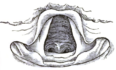
Acute Tracheitis: anterior wall.
FIG. 26.
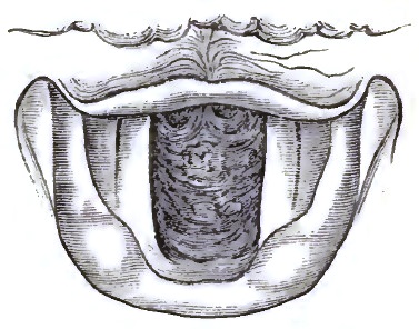
Same case as Fig. 25: posterior wall.
PROGNOSIS.—Simple tracheitis, though occasionally not without danger in extremely young and very old patients, rarely if ever destroys life. Under good hygienic circumstances it frequently gets well of itself, and it does not usually produce sufficient swelling or hypertrophy to cause stenosis. It is, however, when severe, an annoying disease, apt to recur, and, unless properly managed, difficult to eradicate.
TREATMENT.—Tracheitis is treated very much like bronchitis confined to the larger tubes, only that local measures are more prominently applicable, especially in chronic cases. Frequently, when acute, the disease may be arrested by a Dover's powder, a warm bath, and a diaphoretic drink at night, with hygienic attention, regulation of systemic functions, and soothing applications, such as inhaling simply vapor of water or medicated water, or using warm-water poultices externally. Expectorant mixtures, containing ipecacuanha, sanguinaria, squills, or senega, may be given, according to the age and condition of the patient, with matico and the like, when the secretion is abundant, and with ammonium acetate or sodium bromide (potassium carbonate or ammonium carbonate where there is depression) or tincture of aconite (especially when fever is present), or a very minute quantity of tincture of veratrum viride, when there is much dryness. Inhaling the steam arising from a pint of hot water (160–170° F.) containing 10 grs. of extract of conium, 1 drachm of compound tincture of benzoin, and half a drachm of ammonium sesquicarbonate, or inhaling nebulized solution of potassium bromide, 10 to 20 grains to the ounce, or fumes of evolving ammonium chloride or of nitre-paper, is very serviceable, as well as placing a mustard plaster or a hot poultice on the upper part of the chest (not directly over the windpipe) and on the back of the neck or between the shoulders. Some patients require for several days to take daily from 8 to 10 grains of quinia sulphate, then a smaller quantity, care being taken not to discontinue the remedy suddenly. Smoking eucalyptus-leaves, with much inhalation of the smoke, is useful in protracted cases. In chronic as well as acute tracheitis not only balsamic, anodyne, and astringent inhalations either of vapors, or of liquids nebulized by the various spray-producers are in vogue, but also insufflations of powders, injections of liquids, and touchings with the sponge or cotton-wad probang or tracheal applicator. Powders should never or only rarely (as, e.g., morphia, 1/16–1/8 of a grain, when the cough is troublesome, etc.) be blown into the trachea; injections and touchings should be made use of only after the operator has acquired the necessary skill to apply them by means of the mirror. A few drops of a solution of silver nitrate, varying in strength inversely as the chronicity of the case from 5 grains to 60 to the ounce of water, thus accurately applied at proper intervals of time, have proved successful in otherwise intractable cases. In chronic tracheitis general tonic treatment must be combined with the local, and attention be paid to possible coexistent cardiac and broncho-pulmonary affections or other morbid conditions. In some cases it is advisable to administer potassium iodide; in rheumatism, sodium salicylate; in gout, colchicum. The utility of producing alkalinity of the blood (as by giving alkaline mineral waters to drink, etc.) has received a new and direct support by Rossbach's recent observations of diminution of the blood-supply and of the secretion in the tracheal mucous membrane of cats whose blood was made alkaline by injecting sodium carbonate into the femoral vein.
Patients subject to tracheitis should observe all the precautionary measures of so-called bronchitics as to sponging, bathing, and friction of the body, wearing a respirator, clothing, exercise, habits, etc.
Complicated Tracheitis.
Under this heading are here classed together all inflammatory conditions of the windpipe differing from simple or catarrhal tracheitis. In these, other tissues may be affected as well as the mucous membrane. In exanthematous, erysipelatous, and exudative tracheitis the mucous membrane is prominently involved; in oedematous and phlegmonous tracheitis, the submucous connective tissue; and in perichondritic and chondritic tracheitis, the cartilages and their investing membrane. The latter forms are connected with suppurative and ulcerative processes, and, unless traumatic, almost never occur, except in phthisical and syphilitic tracheitis. I shall speak of them under the head of Ulceration.
The tracheitis of measles and scarlatina consists in an acute catarrh, with sometimes considerable desquamation of epithelium, erosion, and capillary hemorrhage. In cases of small-pox in which the larynx is affected, the same disease may extend into the trachea, varying in severity from a congestion of the mucous membrane to an intense pustular process. Erysipelas of the larynx may also involve the windpipe, and when it does is exceedingly dangerous. More than half a century ago Gibson observed in an epidemic of erysipelas that when it spread to the trachea it generally proved fatal.1 Tracheal oedema is extremely rare even when the larynx is oedematous. Phlegmonous inflammation and abscess have been observed in a few instances. Tracheal diphtheria is usually an extension of diphtherial disease of the larynx. Without entering into a discussion of the nature and cause of diphtheria, as either a local or general disease, it is here sufficient to refer to the fact that while in simple inflammation of mucous membrane no fibrinous exudation takes place, certain poisonous irritations lead to the exudation of lymph which infiltrates the tissue and may form a pseudo-membranous deposit upon it: experiments have proved that ammonia, chlorine, and, certainly, bacteria, are able to produce this. In laryngo-tracheal diphtheria or croup the disease most frequently commences in the pharynx, occasionally in the larynx, and much more rarely in the trachea.
1 Transactions of the Edinburgh Medico-Chirurgical Society, vol. iii., 1828.
The treatment of each of these forms of complicated tracheitis is the same as the treatment of the corresponding form of laryngitis.
ULCERATION.
Tracheal ulcers are just as multiform as laryngeal ulcers, but far more rare. Like inflammation, they may occur by extension from above or below, and only those following localized morbid conditions are certain to have arisen in the trachea. Under the head of Inflammation it has been stated that simple catarrhal ulceration does occasionally occur; of this there is really no doubt, but some writers have denied it and thrown the whole subject into great confusion. It is true, however, that a tracheal ulcer has usually a so-called dyscratic base, and either is diphtherial or phthisical (tuberculous) or syphilitic or lupoid or leprous or carcinomatous, or else comes from extraneous causes; as, for instance, from traumatic ulceration or extension or perforation from neighboring abscess, etc. There are two kinds of ulcers—viz. one in which the molecular death of tissue proceeds from the surface inward, and another in which it proceeds from within to the surface. Catarrhal ulcers, as well as ulcers from decubitus after tracheotomy, from pressure of the canula, belong to the first kind; when involving only the epithelium or the epithelium and the layer immediately underneath it the name erosions is given them; and if it were true that catarrhal erosions never penetrate to the deeper structures, it would be justifiable to say that there are no catarrhal ulcers, but only erosions: they do, however, penetrate, and sometimes to great depths. In the second kind of ulcers the epithelium is at first normal or intact, and the loss of substance of underlying tissue in consequence of inflammatory processes in the mucosa, submucosa, or perichondrium affects the epithelium secondarily. This occurs whenever, from any cause, there is primarily caries of cartilage or suppuration of submucous tissue, especially in typhoid conditions, in phthisis, and in syphilis.
FIG. 27.
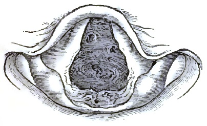
Tuberculous Ulceration of the Trachea, as seen during life.
FIG. 28.
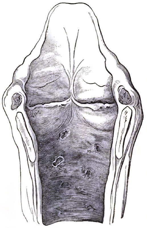
Same case as Fig. 27: post-mortem appearance.
FIG. 29.
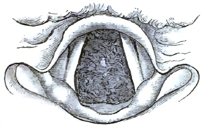
Syphilitic Ulceration of Trachea, as seen during life.
FIG. 30.
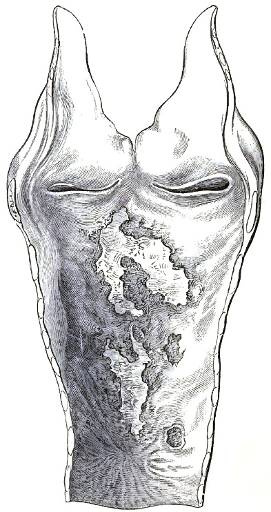
Same case as Fig. 29: post-mortem appearance.
The seat of tracheal ulcers is usually the posterior wall and the lower portion, unless the upper portion is affected by extension from the larynx or by pressure from a tracheotomy-tube. They are found also in other portions, and sometimes are so numerous that they give to the membrane a sieve-like appearance. Occasionally they denude some of the tracheal rings. In shape they vary, being mostly irregularly circular or oval, and excavated or scooped out; in size they vary from that of a pin's head to that of a marble. In tuberculosis they are generally small and numerous, have a pale background, and are occasionally confluent, while in syphilis they are usually isolated and large, very destructive, and apt to cause contractions or other deformities by partial or extensive cicatrization. Such contracting ray-like cicatrices have more than once produced fatal stenosis.
The SYMPTOMS are frequently obscure, but local pain and irritation are usually, purulent or muco-purulent sputa are sometimes, present. The diagnosis is difficult unless tracheoscopic examination reveals the condition. Fig. 27 shows the tracheoscopical image, and Fig. 28 the post-mortem appearance, of a case of tuberculous tracheal ulceration on the upper portion of the front wall, while Figs. 29 and 30 show the image during life and the appearance after death of a case of syphilitic ulceration. In Fig. 30 the posterior wall is seen with the ulcers, and below them a star-shaped cicatrix.
The PROGNOSIS generally depends upon the underlying disease, and is grave because the latter is. Perforation may take place, as well as cicatrization and hypertrophy, and either process may lead to a fatal issue. In a number of instances post-mortem examination has shown that tracheal ulceration may produce surprisingly great ravages before destroying life.
TREATMENT, like the prognosis, depends somewhat upon the disease underlying the ulceration. Pain is relieved by anodyne, and cicatrization promoted by alterative inhalations, as of nebulized glycerated solutions of morphine, ethereal solution of iodoform, iodinic preparations, oil of solidago, citronella oil, etc. Catarrhal ulcers heal without special treatment with the subsidence of the catarrhal inflammation. In syphilitic ulceration, stenosis from cicatrization is to be dreaded, and specific constitutional treatment is the main reliance. The internal administration of cod-liver oil has been found of service in nearly all cases of tracheal ulceration, especially in phthisis, lupus, etc. Appropriate general treatment must be combined with the local.
MORBID GROWTHS.
DEFINITION.—Tumors, benign or malignant, growing from the wall and projecting into the interior of the windpipe. Inversion of the mucous membrane forming a protrusion into the interior will be spoken of under the head of Stenosis; and tumors of other organs extending into the trachea, such as cancer of the oesophagus, lymphatic glands, thyroid body, etc., are excluded from consideration under the present head.
FREQUENCY OF OCCURRENCE.—Aside from post-tracheotomic granulation-tumors, which with careless tracheotomy or after-treatment occur often, the disproportion in the frequency of laryngeal and tracheal morbid growths is even greater than that of other laryngeal and tracheal affections. I have met with only eight instances of tracheal morbid growths, strictly so called, in a special practice during more than twenty-five years. This is exclusive of post-tracheotomic vegetations and tumors from contiguity.
ETIOLOGY.—Local irritations and chronic inflammatory conditions seem often, if not always, to be the forerunners of tracheal tumors, but the real cause of the latter is unknown. Recently it has been suggested (see the article on LARYNGEAL TUMORS) that the ever-present bacilli play a rôle in the production of morbid growths as well as in that of other diseases. As it is known that some parasitical organisms on plants use up their nidus very slowly, with the formation of peculiar excrescences, while others very rapidly destroy the tissue of their host, it would be easy to suppose that some such difference in the micro-organism causing the tumor determines its benign or malignant character.
Post-tracheotomic vegetations may arise from the irritating pressure of a tracheotomy-tube, especially from the use of a fenestrated tube or a tube ill fitted to the patient. Some observers are of opinion that such tumors existed before the performance of the operation, and, indeed, led to it, even though the supposed reason may have been laryngeal or some other tracheal disease. While it cannot be denied that such may have been the case sometimes, there is no doubt that in other instances—and not only in those in which the vegetations "always grow from the cicatrix" (Petel)—they are truly caused by the operation, or by the wearing of the tube, especially if it be in any way unsuitable as to size, form, etc.
SYMPTOMATOLOGY.—The symptoms of tracheal tumors are local irritation; tickling or other morbid sensation, sometimes inducing and sometimes not inducing cough; and encroachment upon the breathing-space—dyspnoea—depending on their precise seat, size, and rapidity of growth. It is usually difficult for the patient to specify the beginning of his trouble, because, on account of the large size of the windpipe, dyspnoea generally comes on very gradually. An accidental catarrhal condition of the tracheal mucous membrane from a cold usually first arrests the patient's attention. The very great diminution of the calibre of the tube that the patient can bear when the tumor enlarges slowly is sometimes astonishing. Unless the tumor is pedunculated (so that expiratory efforts can throw it up into the larynx), which is generally not the case, expiration and inspiration are equally affected, both becoming gradually more and more labored and noisy. Sometimes the act of swallowing large morsels brings on an increased dyspnoea; sometimes respiration is accompanied by a sort of valvular sound. Cough is frequently, but not always, present, and depends, together with expectoration, upon either coincidental catarrhal condition or irritation from the tumor: in the latter case it is essential, dry, and persistent, and may vary with the position of the patient. Sputum may be bloody and even contain shreds of the tumor, as in similar cases of laryngeal growth. With increase of the tumor the voice becomes weak and suffers in extent of range, as in other cases of tracheal stenosis; the same is true of the diminished rising and falling of the larynx. The course and duration of the disease vary considerably with its nature. I have observed a tracheal fibroma to remain stationary for eight years, when the patient died from other causes and the diagnosis was confirmed post-mortem; and, on the other hand, a cancer to grow so rapidly that the patient died from suffocation within five months of its first causing the slightest symptom. If not relieved, suffocatory paroxysms, with or without consequent bronchitis and pneumonia, lead to a fatal termination.
PATHOLOGY.—As in the larynx, so in the trachea, the pathological character of neoplasmata is generally that of papilloma. Of my eight cases, all observed during life, four were papillomatous (two examined microscopically after successful extirpation, one post-mortem, and one in situ macroscopically only), one was a fibroma, microscopically examined, one an osteo-chondroma, one a sarcoma, and one a carcinoma, the three last having been examined post-mortem.
Of non-malignant tracheal tumors observed by others, the large majority were papillomata; next in number come fibromata. Aside from these two kinds of tracheal tumor, the cases recorded in literature are the following: Rokitansky more than thirty years ago described tracheal enchondromata found after death; and Cohen discovered in the corpse of a phthisical patient a number of small enchondromata on the central portions of the tracheal cartilages. Steudener, Demme, Wilks, Chiara, and Eppinger have observed, post-mortem, tracheal osteomata. Gibbs has described a tracheal cystic tumor2 seen with the laryngoscope; Müller, under the guidance of Gerhardt, a myxo-adenoma observed tracheoscopically and carefully studied during life and after death; and Eppinger has recorded a case of post-mortem tracheal adenomata and cysts, Simon having previously found three similar tumors on dissecting a new-born tigress. Virchow speaks of the occurrence of retro-tracheal retention-cysts, and Gruber has observed several; but there can be no doubt that at least some of the tumors thus described are nothing but circumscribed dilatations of the tracheal mucous membrane—practically, dilated mucous glands. As to malignant tumors, in addition to my two cases Schrötter has reported two cases of sarcoma, and Labus one of fibro-sarcoma, while Rokitansky, Klebs, Koch, Schrötter, Langhans, and Mackenzie have described cases of carcinoma.
2 Cohen questions whether this was a cyst or an abscess. It burst spontaneously.
Cases of cancer of the oesophagus, which involve the trachea—excluded, as before stated, from present consideration—are, comparatively speaking, by no means rare, and are apt to establish a fistulous communication between the two tubes.
DIAGNOSIS.—The symptoms mentioned are those common to nearly all cases of tracheal stenosis, and will be referred to again under that head. Tracheoscopy alone makes the diagnosis certain; unless when the seat of the disease is ascertainable without, its nature is shown by the expectoration of portions of the tumor. The first case of tracheal tumor ever diagnosed during the patient's life was observed by means of the mirror by Tuerck in 1861; but it is very difficult in the mirror to estimate distances as to depth, and unless the number of tracheal rings above a tumor can distinctly be counted, a growth in the lower cavity of the larynx may readily be mistaken for one in the trachea, and vice versâ. Catheterism of the trachea shows the distance at which the tumor is situated, sometimes very accurately, but it is dangerous unless performed under the guidance of the mirror, and even then requires great care. The introduction without the mirror of a probe or sound for the same purpose is still more dangerous and unjustifiable, while with the mirror it is perfectly safe in proper hands. Localized protrusion of the mucous membrane into the interior is the condition which most simulates tracheal tumor. (Compare Fig. 32.)
FIG. 31.
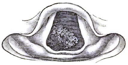
Papilloma of Trachea.
The pathological nature of a tracheal tumor can sometimes be determined in situ with more or less probability. Without microscopical examination it is not always possible to say whether a growth is benign or malignant unless the mass has advanced to ulceration, and then specific disease must be excluded by the history and concomitant symptoms. Papillomata have a peculiarly uneven surface; fibromata are usually more smooth. With equally good illumination, tumors of the trachea resemble tumors of the larynx, and may be similarly differentiated. The former are almost always non-pedunculated, or at least none of those hitherto observed have had a long pedicle. Their seat is generally the posterior wall, or the cicatrix of the anterior wall after tracheotomy. In Fig. 31 is seen the tracheoscopic appearance of one of my cases of tracheal papilloma.
PROGNOSIS.—The prognosis is always unfavorable in malignant cases, and also in non-malignant when the tumor grows rapidly or has already attained a large size. The introduction of the laryngoscope has bettered the prognosis, inasmuch as in many cases early recognition enables us, by performing tracheotomy, to prevent sudden death from suffocation, and also because by the aid of the mirror removal has been accomplished through the natural passages.
TREATMENT.—Removal of a tracheal tumor through the natural passages by means of either cutting or cautery instruments requires so much special ability on the part of the operator that it need not be described in detail in a work designed for general medical practitioners. When the tumor is situated above a point at which tracheotomy can be judiciously performed, no physician worthy of the name should hesitate to lay open the trachea in any case in which suffocation is impending. Removal of the tumor by surgical operation after opening the windpipe may be attempted or not according to circumstances, but in all cases palliative measures by sedative inhalation and otherwise may be resorted to, and the patient's general health, especially in malignant cases, must be kept up as much and as long as possible.
STENOSIS.
DEFINITION AND PROXIMATE ETIOLOGY.—Stenosis is narrowing or more or less occlusion of the windpipe. It is either stricture or constriction from within, or compression from without, or both combined. Constriction within the trachea is due to swelling or thickening or cicatricial displacement of the mucous membrane or other tissue, inversion of its walls, or morbid growth or foreign body in its interior. Compression from without is due to goitre (which has in some cases prevented viability) or other disease of the thyroid body; aneurism; abscess; enlarged bronchial glands or cervical lymphatics; disease of the sternum, clavicle, or vertebræ; mediastinal tumor; cystic, emphysematous, or other tumor of neighboring tissue; or foreign body. According to Rose's observations of goitre,3 compression of the trachea leads to fatty degeneration of the cartilages and their subsequent softening and absorption; after which, the windpipe having become membranous throughout and no longer patulous, death can easily—in some positions or flexion of the body, etc.—take place.
3 Der Kropftod und die Radicalcur der Kröpfe, Berlin, 1878.
In acute tracheitis, though there is swelling of the mucous membrane, the large size of the tube usually obviates stenotic symptoms, while chronic tracheitis does occasionally lead to sufficient contraction to interfere with respiration; but generally stenosis is the result of syphilis, and frequently follows ulceration and cicatrization. In a case recorded in the Bullétin des Sciences médicales for January, 1829, the lumen of the trachea was reduced to two lines.
FIG. 32.
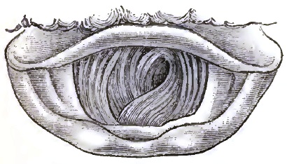
Involution of Trachea, due to aneurism.
SYMPTOMS AND DIAGNOSIS.—The main symptom is the peculiar, gradually increasing dyspnoea; once observed, it is recognized without much difficulty. There may also be mucous râles; cough rough and sibilant; attempts at clearing the throat without expectoration, or occasionally with some expectoration, which is at first light-colored, then streaked with blood, and at last purulent, but never abundant (unless accidentally complicated by catarrh), and always difficult to eject; perhaps occasional pain, but constant disagreeable sensation (tightness) in the trachea just above the sternum. Tracheoscopy settles the diagnosis. The tracheal rings are seen either as diminished circles or arcs—sometimes concentrically placed, sometimes in two different directions, as shown in a case of tracheal stenosis from compression causing protrusion of the mucous membrane into the interior, represented in Fig. 32, or else constricting bands are visible.
As to the dyspnoea, both inspiration and expiration are affected—frequently, however, the former more than the latter, as is shown by pneumatometry. The head is thrown forward and the chin up; the larynx moves up and down less energetically than in health (while the respiratory movements of the larynx are abnormally increased in laryngeal dyspnoea); the thorax is less expanded than normally, especially its upper portions.
As to catheterization and probing, see the remarks under the head of Morbid Growths.
PATHOLOGY.—The pathological changes in cases of stenosis vary with its cause. In the great majority of cases of stricture from within, syphilis—antecedent ulceration followed by cicatrization—has produced the stenosis; in compression thyroid disease, and next often aneurism, is the cause. The stenosis is most frequently situated in the lower, next in the upper, and least in the middle, portion; more often than the latter alone the whole tube is affected.
PROGNOSIS.—This is rather favorable with timely and proper treatment unless a continuing active cause be irremovable; without treatment, however, the cases almost invariably terminate fatally from pneumonia, tracheal spasm, apnoea as before explained, etc.
TREATMENT.—When the symptoms are urgent and the stenosis is not too low down, tracheotomy must be performed. Sometimes a very long and flexible tube may be introduced with success in case of very low stenosis, but more often tracheotomy is disappointing on account of the stenosis extending too low down even when its beginning is higher up.
Stricture, especially when the symptoms are not very urgent, may be relieved by dilatation through the natural passages, with, or if possible without, previous tracheotomy. The cure of compression implies removal of the compressing tumor or disease. Soothing inhalations, such as of hops, benzoin, etc., diminish irritation and give temporary relief.
DILATATION (HERNIA, FISTULE).
Dilatation of the trachea is either confined to the tube (when the synonym tracheaectasy is applied to it) or is diverticular. In the former case it may involve only a part or else the whole extent of the windpipe. Whenever free respiration, especially expiration, is chronically impeded, some portion of the air-tract below the obstruction is apt to become dilated; thus, a bottle-shaped dilatation is sometimes found immediately below an annular contraction. On the other hand, tracheaectasy may extend upward from bronchiectasy. It has been observed post-mortem to a slight extent in public criers, trumpeters, etc., and in old coughers from laryngeal disease, chronic bronchitis, pulmonary emphysema, etc., but without giving rise to distinct symptoms during life.
Diverticular dilatation forms an air-containing tumor which either looks into the oesophagus or is discernible on the outside of the neck. Though rarely met with, it ought to be thought of in all appropriate cases, and when pointing externally ought always to be recognized by the careful practitioner. It is either hernial, glandular, or fistular—three pathological conditions which have hitherto been confounded. On account of the construction and position of the trachea there can be but little protrusion outward without previous dilatation. Unless there be a deficiency of the cartilaginous rings, only the posterior wall, which is always unsupported, and to a slight extent also the intercartilaginous membranous portions, are liable to tracheal hernia. This is properly called tracheocele; but the various terms aërial goitre, aërial bronchocele, pneumatocele, tracheal air-cyst, tracheal retention-cyst, internal tracheal fistule, subcutaneous or incomplete fistule of the trachea, have been indiscriminately used as synonyms of tracheocele, and have added all the more to the confusion, as some of them originated, no doubt, as correct appellations of the particular cases to which they were applied. Aside from the occasional occurrence, both congenital and acquired, of tracheo-cutaneous fistule, complete and incomplete, and the still more rare occurrence of hernia of entire portions of the mucous membrane, the cases of diverticular dilatation of the trachea—or saccular tracheaectasy, as it may be called—are glandular, as found by Rokitansky more than fifty years ago. Virchow seems to regard all such glandular dilatations as retention-cysts (see Morbid Growths), but although retro-tracheal retention-cysts doubtless do occur (Gruber has reported two unquestionable instances), and although the tumors now under consideration do in fact sometimes contain a little mucus in addition to air, they do not constitute cysts or adenomatous new growths, but are simply distended portions of the tracheal mucous membrane, respiratory glands, whether the dilatation be caused, as Rokitansky thought, by traction (Zerrung) and hypertrophy of the mucous glands, or, as Eppinger suggests—and which is more likely—mainly by increased intra-tracheal air-pressure. There must, however, I think, coexist some deficiency or weakness of the cartilaginous or other tissue, either congenital or acquired.
When the dilatation is retro-tracheal only, the symptoms are very obscure, and diagnosis during life is at best uncertain. In one such case under my care, confirmed (death having occurred from another cause) by post-mortem examination, there was some dysphagia and slight alteration of the voice. In all other cases the characteristic and unmistakable sign of the disease is the peculiar intermittent, or, at all events variable, aërial cervical tumor. It increases and diminishes with forcible expiration and inspiration, and attains its largest size during violent coughing, hawking, blowing of the nose, or other expiratory effort. Occasionally the voice is considerably affected. The tumor, especially by the manner in which it can be made to temporarily disappear and reappear, can usually be easily differentiated from subcutaneous emphysema and goitre, the only two conditions with which it might be confounded. In the fistular variety the opening into the trachea can sometimes be seen by means of tracheoscopy.
Aside from the deformity which the tumor may cause, it sometimes induces laryngeal spasm and dyspnoea; otherwise it is of no gravity.
As to TREATMENT, methodical and continued compression by applications of astringent collodion or by mechanical means is the only palliative measure applicable; when suffocatory attacks call for it, tracheotomy must be performed.
TRACHEOTOMY.
BY GEORGE M. LEFFERTS, A.M., M.D.
The operation of tracheotomy, or the artificial opening of the air-passage—using the term in its modern acceptation as including all of the five incisions that are both anatomically and surgically possible, either singly or in combination, between the lower border of the thyroid cartilage and the upper edge of the sternum (incisura jugularis sterni), and reserving the term laryngotomy to denote the division of the thyroid cartilage alone—fulfils two important and usually urgent indications: First, in allowing the respiratory current free access to the lungs in cases where the laryngeal obstruction is of such a sudden or of so progressive a character as to either immediately or remotely threaten the life of the patient; and, secondly, in affording a ready means of direct access to those portions of the air-tract which lie below the level of the glottis, and thus permit not only of the direct extraction of such foreign bodies as may accidentally have found their way within the air-passage, but of neoplasms here located and of occluding diphtheritic membranes. Catheterization and aspiration of the trachea are likewise both rendered not only possible, but easy of execution. Both general indications mentioned often coexist, and are met by the operation in a large class of cases; the first alone plays its important life-saving rôle in many.
The disease or accident which renders the operation necessary varies greatly, and upon this variation depends not only the surgeon's decision as to the precise time at which the opening into the air-tube must be made, but also the precise point at which the operation should be performed. These general questions I treat of in detail. The special indications may conveniently, but somewhat arbitrarily, be arranged as follows, in groups, which I have attempted to make complete, although some of the conditions, being purely surgical, do not strictly come within the compass of this essay:
A. Acute inflammatory diseases of the larynx and trachea:
1. Acute oedema of the larynx.
2. Erysipelatous and exanthematous laryngitis.
3. Acute perichondritis, with abscess.
4. Diphtheritic croup.
B. Chronic affections of the larynx and trachea:
1. Syphilitic laryngitis.
2. Phthisical laryngitis.
3. Chorditis vocalis inferior hypertrophica.
4. Carcinoma of the larynx or trachea.
5. Non-malignant growths of the larynx or trachea.
6. Tumors overlying the superior aperture of the larynx.
7. External compression of the trachea by tumors of the neck or chest.
8. Strictures of the larynx or trachea.
C. Neurotic diseases:
1. Paralysis of the abductors of the vocal cords.
2. Spasm of the adductors of the vocal cords.
D. Traumatic conditions:
1. Foreign bodies in the larynx or trachea.
2. Impaction of foreign bodies in the pharynx or oesophagus.
3. Fracture of the larynx. Rupture of the trachea.
4. Scalds and burns of the larynx.
5. Incised and gunshot wounds of the throat.
6. Poisonous bites inflicted by certain insects about the mouth or neck.
7. Suffocation from the passage of blood, fluids, etc. into the air-passages (tracheotomy, with aspiration of the windpipe and artificial respiration).
8. Suffocation from the acute collection of either mucus or serum in the bronchia (ditto).
9. Suffocation from the inhalation or development of poisonous gases (tracheotomy, with artificial respiration).
Finally, although it pertains alone to the province of the surgeon, I may allude to the temporary tracheotomy and "tamponing of the trachea" which has been recommended—and certainly found efficient—in preventing the entrance of blood to a dangerous degree into the lower trachea and lungs during the performance of certain operations in the neighborhood of or upon the air-passages, such as resection of the upper jaw, the extirpation of large nasal and naso-pharyngeal polypi, removal of the tongue, subhyoidean pharyngotomy, laryngotomy, and extirpation of the larynx.1
1 For the details of this procedure consult Schüller, Die Tracheotomie, etc., Stuttgart, 1880.
All-important as a preliminary to the operation itself is a thorough knowledge of the surgical anatomy of the region upon which it is proposed to operate; and this not alone in the adult, but especially in the child, where essential differences often exist. Possible anomalies also are not to be forgotten.2 The assurance of the surgeon depends upon this knowledge: mere, manual skill will not compensate for its want; the success, both immediate and remote, of the operation is in great measure the reward of its possession.
2 See Pilcher, "The Anatomy of the Anterior Median Region of the Neck," Ann. of Anat. and Surgery, Brooklyn, April, 1881.
It will be remembered that the trachea commences at the inferior border of the cricoid cartilage, directly opposite to the lower edge of the fifth cervical vertebra, and reaches thence downward, in the median line of the neck, until it bifurcates opposite to the third dorsal vertebra. In its upper part it is nearly subcutaneous, and is surmounted by the prominent ring of the cricoid cartilage (easily identified, even in the young child), above which, in turn, lies a slight depression (the crico-thyroid space) between the cricoid and thyroid cartilages. As the trachea descends in the neck it recedes gradually, lying at the episternal notch about one and three-eighths of an inch from the surface. Throughout the whole of this course it is in relation with important structures. In its cervical portion it is covered by the sterno-hyoid and sterno-thyroid muscles, and in the median space, which is usually distinct between them, by layers of the deep cervical fascia. It is also crossed by the isthmus of the thyroid gland, which lies between the second and fourth tracheal rings; by the arteria-thyroidea ima, when present, and below by the plexus formed of inferior thyroid veins with their tributary and communicating branches. In the latter region, but more superficially, are some communicating branches between the anterior jugular veins. The innominate and left carotid arteries are also anterior to it in the episternal notch as they diverge from their origin. Laterally, the trachea is in relation with the common carotid artery, the lateral lobes of the thyroid body, the inferior thyroid veins, and the recurrent laryngeal nerves. The thoracic portion of the trachea is covered by the manubrium sterni, with the origins of the sterno-hyoid and sterno-thyroid muscles, by the left innominate vein, and by the commencement of the innominate and left carotid arteries. Still lower, the transverse portion of the arch of the aorta crosses, and the deep cardiac plexus of nerves lies in front of it. Posteriorly, throughout its length, it rests upon the oesophagus.
In performing, then, either the superior or inferior operation of tracheotomy, after cutting through the skin and superficial cervical fascia—which is really loose areolar tissue containing fat—the superficial layer of the deep cervical fascia is reached, and immediately below it more or less adipose tissue and the two anterior jugular veins lying in an inferior tracheotomy to either side of the wound, which is always made in the median line. As a matter of fact, these various layers are rarely demonstrable, and the surgeon proceeds irrespective of them until he reaches this point in his operation—viz. the muscles which overlie the trachea. These may overlap in the median line, and have to be retracted after having been separated; or, again, a thin line of connective tissue marks a slight interval between their inner edges, and is readily seen and dissected through if the operator has kept his incision vertical and strictly in the median line of the neck—a matter so important to the success of his operation that I do not hesitate to again allude to it. The muscles separated and gently retracted, together with the overlying tissues, toward the sides of the wound, the upper edge of the isthmus of the thyroid gland overlying the second and third, perhaps fourth, rings of the trachea, is always seen in a superior tracheotomy—its lower edge very frequently in the inferior operation. The isthmus is adherent to the trachea and to the larynx through the deep layer of the deep cervical fascia, but is capable of being slightly displaced or pushed upward or downward as the case may be, and thus kept from obscuring the operative field. This being done, the deep layer of the deep cervical fascia is seen covering and strongly adherent to the tracheal wall together with the thyroid veins. A few touches of the knife, carefully avoiding the blood-vessels, serve to clear it away, and the tracheal rings are clearly exposed.
In carrying out this dissection, which has been described as occurring in an ordinary and uncomplicated adult case, several matters must be borne in mind; and especially is this true if the operation concerns infants. In them, for instance, the thymus gland rises half an inch above the level of the sternum, and is frequently to be found as late as the sixth or seventh year. In both adults and children the innominate artery occasionally comes into view in an inferior tracheotomy, obliquely crossing the lower portion of the right half of the trachea. It is relatively higher in the child than in the adult. The left innominate vein is also often observed when the trachea is opened low down.
Certain abnormalities of the blood-vessels have been alluded to above. The commonest consists in the existence of a thyroidea ima artery, which when present usually arises from the innominate trunk, but sometimes from the right common carotid or the aorta: it passes to the thyroid body directly in the median line of the neck and close to the trachea; again, the place of the anterior jugular veins may be taken by a single central vessel, almost sure to be wounded during the operation if it exist (Mackenzie).
In performing the operation through the thyro-cricoid membrane (thyro-cricotomy) or through the cricoid cartilage alone (cricotomy), the same tissues are met with, and the same dissection is necessary in the earlier stage of the operation, as have been described in the operation of superior or inferior tracheotomy; but the parts are more superficial, adipose and cellular tissue less abundant, blood-vessels much less numerous, and the operation very much simpler. The thyroid gland of course does not come into view, and the crico-thyroid artery, a very small vessel, needs no attention in the dissection.
I have here and elsewhere included under the general term tracheotomy five distinct operations, having for their object the opening of the air-passages, which are surgically possible between the lower border of the thyroid cartilage and the upper edge of the sternum. In this classification I have followed that of Schüller, and its simplicity, but exactness, and the avoidance of the old confusion of different terms which results from the use of one intelligently employed, seem to me to commend it. These five operations are—1. Thyro-cricotomy, or the opening made through the crico-thyroid membrane alone. 2. Cricotomy, or the division of the cricoid cartilage alone. 3. Superior tracheotomy, the incision being made above the point where the isthmus of the thyroid gland crosses the trachea and below the cricoid cartilage. 4. Median tracheotomy, when, the isthmus being displaced or torn through, the trachea is opened immediately below its site. And 5. Inferior tracheotomy, the incision being made below the point of crossing of the isthmus of the thyroid gland, and at varying distances, dependent mainly upon the age of the patient and size of the parts, above the sternal notch.
Rarely, I am bound to admit, is the field of all of these operations as distinctly limited in practice as is here indicated, and one, perhaps two, are rarely selected. Thyro-cricotomy (old term laryngotomy) is often indicated, and cricotomy and median tracheotomy are sometimes performed as here described. Superior tracheotomy is commonly a combination of at least two of the methods—viz. the division of the upper rings of the trachea and the cricoid cartilage as well. It may even, probably frequently does, trench also upon the thyro-cricoid membrane (thyro-cricotomy) and upon the field of a median tracheotomy, the isthmus being pushed downward or even cut or torn through. The latter operation and cricotomy are, I believe, rarely if ever done from choice. Finally, inferior tracheotomy is a common method. As here described, it meets a large number of indications, and, despite its superior difficulties over the higher operations, is therefore necessarily often chosen; not infrequently, however, does it invade the median region, the isthmus of the thyroid being pushed upward.
Which of these operations shall be selected in a given case depends upon the particular conditions which render it necessary, and likewise, to some extent, upon the age of the patient. Durham summarizes the question very fairly. Thyro-cricotomy (old term laryngotomy) is by far the easiest operation to perform, and its execution is attended by least risk; therefore it is the operation to be preferred in any sudden emergency when suffocation threatens, and especially where the surgeon is alone with the patient. Generally, it is not as applicable as the others, especially in early childhood, on account of the limited dimensions of the thyro-cricoid space. It cannot be recommended in cases of acute or extensive diseases or injuries of the larynx, nor is it likely to be of much service if a foreign body is in the trachea or bronchus. On the other hand, it is probably the best operation to adopt in cases in which foreign bodies are impacted in the larynx, in cases of limited chronic disease or contractions of the superior laryngeal parts—usually the result of syphilitic ulceration—and in cases in which respiration is impeded by intra-laryngeal growths which cannot be removed by the natural passages.
Cricotomy, combined with superior tracheotomy (old term laryngo-tracheotomy), is not a difficult operation, and may be advantageously practised, especially in children; in the adult it meets many indications. Holmes recommends it the more urgently, in preference to an inferior tracheotomy, the earlier the age of the subject may be.
Inferior tracheotomy is comparatively difficult to perform, and during its performance dangers may have to be encountered greater and more numerous than those met with in either of the other operations. This is true certainly of children. As regards young children, Holmes states that after the age of five or thereabouts the surgeon can, if he prefer it, open the trachea below the isthmus of the thyroid gland. He himself does not recommend the operation before puberty. In the case, however, of a foreign body loose in the windpipe of a child, where a large opening is required, it can hardly be obtained above the thyroid body and below the cricoid. To cut through the isthmus of the thyroid (median tracheotomy) is, in early life at least, a doubtful proceeding when it is of large size, on account of its vascularity, and the incision must be made below it—in other words, an inferior tracheotomy.
When the operation of tracheotomy shall be performed is a question which the experience and individual views of the surgeon, based on experience, must decide in each case. The doubt always arises in the mind of the inexperienced operator whether the symptoms are sufficiently urgent to render the operation necessary. To him these general rules may be given: The immediate indication for the operation is to be looked for in the thorax. It is the recession of the lower part of the sternum and contiguous ribs and the retraction of the intercostal spaces and clavicular fossæ at each act of inspiration. He must not wait until lividity of the lips and blueness of the fingernails prove that the blood is being imperfectly oxygenated (Mackenzie). Let him remember also that, aside from the immediate and imminent danger of sudden suffocation, a remote one exists and increases the longer he postpones his operation and allows the struggle for air to continue—viz. vascular engorgement and oedema of the lungs, especially in young children; the production of all those conditions which allow, and even predispose, the lung after the operation to fall an easy prey to the inflammatory processes.
The instruments necessary for the performance of the operation of tracheotomy are few and simple, and are such as may ordinarily be found in any small operating-case. A scalpel, a probe and sharp-pointed bistoury, dissecting and artery forceps, a tenaculum, a grooved director, two small retractors, scissors, and a dilator for the tracheal wound, are necessary. To these may be added the needles and thread, waxed ligatures, sponges, and tape. The tracheal tube is elsewhere described. A faradic battery, good suction syringe, and a large flexible catheter may render good and timely service if at hand.
It is true that many other and more or less complicated instruments have been devised for the purpose of facilitating the operation; and other methods, aside from that of the knife, have come of recent years into vogue; but, still, simplest means, as above given, have in the experience of most surgeons been proven to be the best. This statement, undeniably true for all surgical measures, is especially so for the operation under consideration, which is often necessarily undertaken without opportunity for elaborate preparation and under the most adverse and inconvenient circumstances. The more familiar, therefore, the surgeon is with his instruments, the better and more certain will be his work.
Holding this view, it is unnecessary for me to more than briefly mention such instrumental aids as the grooved tenaculum of Chassaignac, the groove serving to guide the operator's knife into the trachea; the sharp double hooks of Langenbeck, which, after being caught in the tracheal walls to either side of the site of the intended incision, are sprung apart after the latter is made, thus dilating the wound and rendering the introduction of the tube easy; the tracheotome of Thompson, a pair of curved cutting forceps, the blades of which are caused to open by a screw after they have been plunged through the tracheal walls; that of Garin, a forceps with curved blades—one, the longest and sharpest-pointed, being made to penetrate the trachea, the instrument then opened, and both blades cut their way to the desired extent of incision; finally, the tracheotome of Maisonneuve, a curved dilating hook with cutting inner edges. Its point is entered between the first and second rings of the trachea and brought out again between the fourth and fifth; the handle is then carried under the chin, so that the blades are made to cut through the trachea and the skin between the points of insertion and exit, after which, upon pushing a spring, the two halves of the hook separate, and the canula is introduced between them (Thornton). And the trachea-stretcher of Marshall Hall, by means of which a portion of the trachea is cut out and the opening kept patent.
None of these instruments have been proven to possess any practical worth; on the contrary, their use, especially that of the latter forms, has in more than one instance been attended with disastrous results.
To obviate the danger of serious hemorrhage during the performance of tracheotomy, both the galvano-cautery knife and the thermo-cautery instrument of Paquelin have been recommended within the past few years, and a number of operations placed upon record. The procedure is the same whichever means be used. The skin and soft parts overlying the trachea are usually alone cut through by means of the cautery-knife, the cartilaginous rings of the tube, when reached, being divided with the ordinary knife. This fact alone speaks against the thoroughness attainable by means of these methods; but, still more important, neither has been found reliable in checking hemorrhage, and in several instances the operator has been obliged in haste to lay aside his cautery apparatus and turn to the ordinary and better-known means to complete his operation. The healing of the tracheal wound made by the cautery is slow: erysipelatous inflammation may attack the wound as the result of the burn, and extensive sloughing of the edges is not unknown, while the resulting cicatrix is large, strong, and contractile, and has caused, in one case at least, a stenosis of the trachea. In the face of these facts he must indeed be an enthusiastic advocate who would recommend the procedure. Mackenzie justly remarks that the use of the thermo-cautery for opening the air-passage merely introduces an unnecessary complication into the operation.
The choice of a proper tube, one suited to meet the special indications in a given case and specially adapted to the age of the patient and the calibre and position of his trachea, is no unimportant matter, and may do much not only to facilitate the immediate success of the operation, but likewise prevent the occurrence of those possible unfortunate results, ulceration, fatal hemorrhage, abscess, pneumonia, and pyæmia, no lack of which are recorded in our literature.
Although the number and variety of mechanical devices and forms of tracheal tubes that have from time to time been devised by the inventive ingenuity of operators is large, the choice practically centres upon one of two forms. The first, and the one most commonly used, is but the original canula of Trousseau, modified by Roger, in that the tracheal portion of the tube is detached from the collar or neck-piece, and moves freely with the movements of the patient; and by Obré, by the important device of an inner tube to prevent clogging of the outer or original tube by mucus. Starting upon this essential basis, the instrument-maker has perfected the instrument of to-day. It is a silver tube, double throughout, the inner tube projecting at the lower or tracheal end beyond the outer—an important point, as it prevents any possible permanent occlusion by mucus or blood-crusts, membranes, and the like at this point, removal of the inner tube at once clearing the end of the outer one. The curve of both tubes should correspond to the arc of a quadrant, and the outer is fastened to a transverse collar or shield by means of two small projections or pins upon its sides which lie under small wire bridges upon the shield after it has passed through an opening in the transverse neck-collar large enough to permit of its free movement during the respiratory movements of the trachea, as well as during the forcible action caused by cough. The ends of this collar or shield curve slightly backward to correspond with the curve of the neck, and are perforated by, preferably, large oval openings, instead of the usual small, inconvenient slit, through which the tapes are passed which hold the tube in position by encircling the neck. To this same shield is fastened, by means of a small turn-screw or a revolving collar, the end of the inner tube, which is thus prevented from being forced out of the outer tube by coughing or any motion of the patient. Upon the upper or convex surface of the outer tube a small ovoid opening is usually made for the purpose of permitting the expiratory current to pass upward (the inner tube being removed) into the larynx and render phonation possible; also, the free opening of the outer tube being closed, to allow of respiration being carried on through the larynx and natural passages—often an important matter, as the case progresses toward recovery, in instances where the operation of tracheotomy has been performed on account of laryngeal obstruction.
A set of these tubes, which can now be readily obtained, should consist of four, with the following diameters: No. 1, one centimeter; No. 2, nine millimeters; No. 3, seven millimeters; No. 4, five millimeters: their length is of course in relative and fixed proportion to these measurements. A tube should always be selected less in diameter than the trachea operated upon: to seek to introduce one of the same calibre is not only unnecessary, but cannot fail to be dangerous. Tubes constructed upon the same principles as that just described (Lüer's) are made of hard rubber instead of silver (Leiter): their lessened cost is their principal recommendation, added to the one that they are more easily kept clean and sweet than the silver tubes. The fact that they are necessarily made much heavier and thicker than the latter is a disadvantage, the lumen of a hard-rubber tube being smaller than that of a silver tube of corresponding external diameter. The objection urged against them, of their great danger of breakage, I have not found borne out by experience. Tracheal tubes are also constructed of platinum, and recommend themselves on the score of lightness.
The main objection to any of the forms of tube just described exists in the nature and shape of their curve, which not infrequently causes the lower or tracheal end to lie in contact with the anterior tracheal wall, or its convexity with the posterior, and irritate, even ulcerate, them. This misfortune is entirely obviated by the canula of Durham, the second of the two forms to which I have called special attention, and which is essentially a right-angled tube, made of four sizes, with a long horizontal portion, varying from 7 to 4 centimeters, and short vertical portion, of from ½ to ¾ of an inch in length and slanting slightly backward. The former portion is capable of being lengthened or shortened in any sized tube by means of a screw arrangement attached to it as it passes through the usual neck-collar or shield; and the vertical tube can thus be correctly adapted to the particular depth at which the trachea naturally lies in a given case from the surface; and not alone this, but also to the condition of the overlying parts, whether thin or fat, swollen or otherwise. Once in position, the vertical portion of the tube remains in the long axis of the trachea, and does not touch its walls to any injurious degree. Owing to its right-angled shape, the angular and descending portions of the inner tube of this canula are necessarily made upon the lobster-tail principle, with joints—a possible disadvantage, as they can become clogged with mucus and may become detached. Other modifications and improvements exist in this Durham canula over the older one first described, which add to its utility, but need not here be dwelt upon. Suffice it to say that the tube is an excellent one for its purpose, and is deservedly highly spoken of and recommended by those who have had experience in its use. Its cost is an objection.
The other forms of tracheal tube need but passing mention. The bivalve canula of Fuller is made in two lateral segments, fastened to a collar and tapering when closed to a point, so that introduction of the apparatus through the tracheal wound is made easy. Once introduced, an inner complete canula is slid into its place, thus separating the two outer halves and rendering the whole round and compact. It has been criticised unfavorably on account of the danger of hemorrhage that it is likely to cause through pressure on the tracheal walls by the sharp edges of the outer canula. In Gendron's canula the same lateral blades are separated after introduction by means of a screw fastened on a transverse bar.
Soft-rubber canulas were introduced to the profession not long since by Morrant Baker for subsequent use after the operation of tracheotomy, the usual tube having been worn meanwhile for a few days. Being soft and flexible, they are certainly safe and comfortable for the patient, but their thickness and the absence of any inner tube are, especially the latter, serious disadvantages. They are not, I believe, generally used. Finally, the long, flexible tracheal tube of König was devised by its author to meet the indications in cases where the trachea is compressed from without by tumors, and where a long canula that is flexible, but at the same time rigid enough to resist pressure, becomes a necessity. It is made in the form of the ordinary tracheal canula, only larger, some three or more inches of the centre of the descending portion of the tube being constructed of spirally-twisted silver wire.
It may not be out of place to remind at this point that a tracheotomy is not infrequently performed, of necessity, very hastily, and in the absence not only of a tracheal tube, but likewise of other and even more essential instruments. The lack of the former need never be a barrier to the prompt performance of the operation, for the ready wit of the true surgeon will show him various ways out of his temporary difficulty. A thick goosequill fastened by threads passed through its outer end makes an efficient improvised canula. A bit of elastic catheter answers the same purpose. Retractors for the edges of the tracheal wound, made of wire—silver if it be at hand, a couple of hairpins if it be not—and connected together by an elastic tape which passes around the neck, will not only answer a good temporary purpose in holding the tracheal wound dilated, but have been recommended by Martin—in a more elegant form, it is true—as a proper method of treatment after opening the trachea. Finally, one or more stitches passed through the cartilaginous edges of the wound, and attached to the soft parts beyond it, will serve to secure its patency, at least temporarily.
If a patient be doomed to wear a tube constantly in his trachea, the instrument described above can be removed at a suitable interval after the operation and its place supplied by a single tube of the same size and form as has been found adapted in the case. In the convexity of this permanent tube an ovoid opening should be made to allow of the passage to the larynx of the respiratory current, in part at least, and to its mouth a pea-valve may be fitted which shall admit air on inspiration, and not allow it to escape on expiration, thus doing away with the necessity of the patient's closing the opening of his tube with his finger each time that he requires to speak. Several forms of these valves have been devised, but practically they are of little use, are annoying to the patients, and, as a rule, not tolerated by them.
How shall the operation of tracheotomy be performed? An answer to this question necessitates a short description of the operative steps of the different procedures that is given in the order in which, I believe, the operations are, as a matter of experience, found to occur in practice—viz. 1st, superior tracheotomy, combined or not with cricotomy; 2d, thyro-cricotomy and, 3d, inferior tracheotomy. Certain preliminaries are common to all.
The patient should be extended upon a table covered with one or two thicknesses of blanket and of suitable height, which has been placed sideways in front of a window if the operation is done by daylight. (At night several candles tied together afford a better and safer light than a kerosene or oil lamp.) The surgeon stands at the right side of his patient and facing the window. Of his two assistants—and the value of trained assistance in this operation is inestimable—one faces him, without obscuring the light, and is prepared to use the sponges, hand the instruments, manipulate the retractors, and render such direct assistance as may be required. The second sits at the head of the table and holds the head of the patient steadily, the neck being well extended and thrown backward over a small round pillow (or, better, a wine-bottle wrapped in a towel) which has been placed beneath it. The head must be held directly in the median line of the patient's body, and even in that of the operating-table. The assistant's attention must never waver from this important duty. In certain cases too great inclination of the head backward serves to increase the urgent dyspnoea, or even to check respiratory efforts. This effect he must watch for, and be prepared to relieve instantly by raising the head. His duties also include the preliminary administration of an anæsthetic, and its use during the operation if required. That such use is safe in this class of operations is now generally admitted, but it is not always necessary. The operation is not an exceedingly painful one, and I have often performed it, with the adult patient's consent, without using any anæsthetic (sometimes freezing the skin over the site of the incision before making it), he submitting rather than undergo any addition to the sense of urgent dyspnoea from which he is already suffering. In children anæsthetics—ether being more commonly employed, although chloroform is often used—are much more necessary, often indispensable. Their effects are speedily manifested when asphyxia is present in any marked degree, and but little of the vapor need be inhaled. The administration, always to be carefully watched and profound anæsthesia avoided, renders breathing easier in many instances, certainly lessens laryngeal spasm, and may be discontinued early in the operation when the air-tube is or has been nearly reached by dissection. Any slight risk attending their use is more than outweighed by the safety and precision which they ensure in the more difficult and delicate steps of the operation (Sands). If the patient be already insensible or if death be imminent, their use, of course, is contraindicated.
The operator having previously decided which operation he will perform, and after carefully identifying the position of the various parts, the larynx especially, marking them with ink upon the skin if he chooses, now steadies the loose skin over the site of his intended incision, and then makes it, freely, firmly, cleanly, and exactly in the median line. If it be for a superior tracheotomy, combined or not with cricotomy, the operation I shall first describe, it must extend from just at the notch of the thyroid cartilage downward for about four inches. A free external incision is very desirable in all cases. The subcutaneous tissue now rapidly dissected through by the careful use of the knife, the veins as met with either being pushed to one side or, if they cross the line of incision, cut if small, then twisted or immediately ligated, or if large doubly ligated and then cut between the ligatures, the interval between the sterno-hyoid muscles is sought for and found, then separated by the blade or handle of the knife and held apart by retractors at the side of the wound. It is important that the faint whitish line of connective tissue which marks the interval between the muscles be recognized, otherwise it happens that the operator passes through the body of one of them, deviates at once from the median line, and approaches the side of the trachea instead of the front. The ring of the cricoid cartilage above and the upper edge of the isthmus of the thyroid gland below can now be either seen or felt by the finger in the wound between them; and about the latter lies more or less connective tissue and numerous small veins. As a rule, careful touches of the point of the knife, or, as some operators prefer at this stage, its handle or the use of a blunt director, serves to dissect up piecemeal or tear through and clear this away, the veins again being pushed out of the way, or if necessary cut and tied, and all parts held aside by removing and replacing freshly the retractors from time to time as the dissection proceeds, until the ring of the cricoid and the upper rings of the trachea come plainly into view; that is, are seen, not alone felt. During this dissection, especially if the handle of the scalpel be used, too much pressure must not be made upon the trachea. More than once I have known it to cause sudden suspension of the respiration, probably by exciting reflex spasm of the larynx. If the isthmus of the thyroid gland extend far upward, it must be pressed downward, its facial attachments to the cricoid and trachea cut or torn through, and may require to be held downward in the lower angle of the wound by an additional retractor. The upper rings of the trachea having been thus well cleared of their overlying parts, the next step of the operation follows. I am in the habit of now removing the retractors and allowing the trachea, which may have become displaced by them, to resume its normal position, the head of the patient being meanwhile readjusted. All this takes but a few seconds. A tenaculum is then implanted in the median line, either just below the edge of the thyroid or the cricoid cartilage, if the latter is not to be severed, and held firmly by the assistant at the head of the table, thus steadying and elevating slightly the trachea and rendering the incision into it certain. The retractors are now reintroduced at the sides of the wound, and the operative field is clear and steady. A glance having shown that all bleeding has ceased, another that the tracheal dilator and tracheotomy-tube lie ready at hand, the operator plunges a straight-pointed bistoury through the tracheal wall at the level of the third or fourth ring in the median line, and cuts quickly upward until the cricoid cartilage is reached, if he proposes, as in the adult can usually be done, to limit his operation to a superior tracheotomy. If not, as in the child, and the cricoid cartilage must be cut through to gain sufficient space for the introduction of the tube, it also is severed by prolonging the incision upward to the thyro-cricoid membrane. A hissing of escaping air, with the bubbling of a little blood and paroxysms of cough, follows the incision and shows that the trachea has been fairly opened. The tracheal dilator is now introduced, the lips of the tracheal wound separated, and the canula slipped neatly into the windpipe (unless in the case of a foreign body), and secured a moment or two later, when respiration is fairly established, by tapes passing around the neck. The tenaculum and retractors are removed at the same moment that the tube is slipped into place.
Many different methods have been recommended for the dilatation of the tracheal wound and to assist the introduction of the canula. The dilator (Trousseau) which has been mentioned surely answers all purposes, and is simple and easily used. An ordinary dressing forceps will likewise do the work if introduced closed and afterward opened. More complicated procedures are unnecessary.
Thyro-cricotomy requires that the superficial incision be so made over the larynx that the thyro-cricoid space shall lie in the centre of one, about two inches long, made in the median line. Following now the dissection just described, the thyro-cricoid membrane is easily reached and quickly seen as soon as the sterno-hyoid muscles are retracted. It should then be divided transversely close below the lower edge of the thyroid cartilage, the wound dilated, and the tracheotomy-tube slipped into place.
Inferior tracheotomy demands that the external incision be free. In children, and in adults with a short neck, it should extend from the cricoid cartilage to just above the sternum. The subsequent steps of the operation are as for superior tracheotomy, with but slight differences. The anterior jugular veins may come into view, but can generally be avoided. If they are joined by a transverse branch, this is necessarily cut through after being doubly ligated. After the thyro-hyoid muscles are separated, the rings of the trachea are much less distinctly felt at first than in superior tracheotomy, being covered by more connective tissue and numerous veins. These inferior thyroid veins, especially if large, are the great obstacle in the way of this operation, and much care is necessary in order to avoid them, which should be done if possible. The lower edge of the isthmus of the thyroid gland, which presents to a variable extent above in the wound, does not, as a rule, offer any obstruction. The thymus gland present in infants is easily pulled downward and out of the way. The trachea at length fairly exposed and all bleeding controlled, the left fore finger of the operator is placed in the lower angle of the wound to securely protect the large blood-vessels here located, and the incision made through some three tracheal rings from below upward.