Volume 3 — DISEASES OF THE BLOOD, AND OF THE HÆMATOPOIETIC SYSTEM. (continued 13)
Reading edition
Leukæmic growths in the skin have been described.
The leukæmic tumors demand further consideration. They are not common. In 10 of the 11 Montreal cases careful post-mortem examinations were made, and in not one were there definite new growths. In Case I. there was diffuse leukæmic infiltration of the liver, the histological characters of which were carefully studied. In the 157 cases collected by Gowers140 there were only 13 instances of leukæmic nodules in the liver, and 10 in the kidneys. They are still more uncommon in the lungs. In the spleen—unlike this organ in Hodgkin's disease—they are very rarely seen. The nodules consist of leucocytes in a meshwork of delicate reticular tissue. Their mode of origin has been much discussed. There can be no doubt, I think, that they are new growths of lymphoid tissue of local origin. Possibly they start from accumulations of colorless corpuscles which pass out of the capillaries. In the infiltration of the liver one sees diffuse collections which resemble new growths, but which have evidently resulted from the aggregation in and outside of the capillaries of enormous numbers of leucocytes, which cause the atrophy of the cells of the organ. Doubtless, they multiply in loco by a process of fission, and these aggregations may themselves be foci for the origin and development of colorless cells which pass into the blood-current and augment the number.141 Quite recently Bizzozero has studied the development of these leukæmic new growths, and has shown clearly that the cells which compose them are in process of active fission.
140 Loc. cit.
141 "On the Histology of Leucocythæmia," Canada Medical and Surgical Journal, 1876.
The COURSE OF THE DISEASE is slow and chronic, a matter of months and years. There are exceptional instances in which the disease has proved fatal in a few weeks; this occurs sometimes in children,142 but acute leukæmia is very rare. In a table of 63 cases collected by Gowers, in which the date of the first symptoms was fixed with tolerable accuracy, the duration was less than one year in 13 cases; from one to two years in 16; from two to three years in 19; from three to four years in 9; from 4 to 5 years in 3; and five years and upward in 3 cases. The course is rarely uniform, but periods of improvement occur in which the fever subsides, the painful sensations in the abdomen diminish, the appetite improves, and the spleen reduces in volume. Such intervals, corresponding to the administration of certain drugs, are apt to lead to therapeutic errors. A patient may sometimes get about for months, and even attend to a light business, with an enormous spleen and a ratio of white to red corpuscles of 1 to 6 (Case VIII.). Hemorrhages, high fever, profuse diarrhoea, and the occurrence of dropsy shorten the course. Toward the close there is great muscular debility, and usually a wandering delirium.
142 Golitzinsky, Jahrb. f. Kinderheilkunde, 1860–61.
In the majority of cases death is by asthenia—a gradually progressive weakness and ultimate failure of the heart. Diarrhoea and hemorrhage hasten the fatal result. A profuse hemorrhage may cut off a patient early or after the disease is well established. Cerebral hemorrhage was noticed in 6 of 60 cases in which Gowers was able to ascertain accurately the cause of death. A few are carried off by pleurisy or pneumonia or peritonitis after tapping.
Pyæmia and rupture of the spleen are mentioned as causes of death in some cases.
The DIAGNOSIS of leukæmia rests upon the determination of a great and persistent increase in the colorless elements of the blood. Cases of Hodgkin's disease and of splenic anæmia, almost identical in general features, can only be distinguished by an examination of the blood. I should say that in any case we can speak of the blood as leukæmia when the ratio of white to red cells falls below 1 to 50. Some writers hold that to determine leukæmia the ratio should be at least 1 to 20, but when the study of the variations in the proportion of the corpuscles in any case extends over weeks or months, we not uncommonly find that the ratio, which, at one observation may be 1 to 8, or 1 to 10, a week later may be 1 to 60 or 80, or even 150. Indeed, the state of the blood is a variable factor, and too close attention to it has diverted our minds from the broad features which this disease has in common with others. For practical clinical purposes we have to distinguish ordinary lieno-lymphatic leukæmia from—(1st) chronic malarial infection with splenic hypertrophy; (2d) from cases of non-malarial splenic enlargements with anæmia; (3d) from general lymphadenoma or Hodgkin's disease. The history in malarial cachexia, the absence of lymphatic enlargement, and the blood-condition will usually be sufficient for purposes of a diagnosis. Great increase in the white blood-corpuscles is not often seen in the chronic splenic tumor of malaria; indeed they may be much diminished in number. Toward the end in very chronic cases the clinical picture may be very similar: the large abdomen, possibly ascites, dropsy of the feet, and irregular fever may resemble closely splenic leukæmia, and the absence of an increase in the colorless corpuscles may be the only marked difference. From anæmia splenica there is still greater difficulty, and I have seen instances in which the absence of an excess of the colorless corpuscles in the blood formed the sole criterion: the hemorrhages, the dropsy of feet and abdomen, retinal extravasations, the general cachexia, and the fever were identical with those of leukæmia. Still greater may be the difficulty of separating certain cases of lymphatic leukæmia from general lymphadenoma or Hodgkin's disease; but in the latter affection the glandular enlargement is usually greater and altogether a more prominent feature, and the spleen is not so often increased in size. There may, however, be a considerable increase in the number of the white corpuscles, 1 to 150 or 1 to 100 red, and cases do occur which appear intermediate or transitional in character, and upon which judgment must be reserved until the progress of the case decides the question.
Pure cases of myelogenous leukæmia are almost unknown; if the osseous symptoms are not marked the course is very like that of pernicious anæmia. Indeed, there are two interesting cases on record in which the progressive anæmia seemed to pass into leukæmia. In Litten's case143 the patient presented the symptoms of profound anæmia, and five days before death the blood became markedly leukæmic. There was no enlargement of spleen or lymph-glands, but the bone-marrow was intensely leukæmic—i.e. of the pyoid form. In the case reported by Leube and Fleischer144 the patient, aged thirty, four months after her confinement became anæmic and the left leg was swollen. Though at first only anæmic, subsequently the ratio of white to red corpuscles rose to 1 in 10. Gangrene of the leg supervened, necessitating amputation, from the effects of which she died. There was no affection of spleen or lymph-glands, but the marrow was of the red lymphoid variety. A gastric ulcer was also present. This was no doubt a case of post-partum anæmia aggravated by the presence of ulcer of the stomach, and the great interest of the case lies in the transition of the anæmia into leukæmia.
143 Berliner klin. Wochenschrift, 1877.
144 Virchow's Archiv, lxxxiii.
There are certain general conditions, accompanied by an increase in the colorless corpuscles, which must be distinguished from leukæmia. In suppuration there may be marked leucocytosis; so also in cancer and protracted cachectic states, as phthisis. In cases with large cancerous masses about the stomach and omentum, or where, as occasionally happens in chronic phthisis, there is a greatly enlarged amyloid spleen, if the white blood-corpuscles are much increased, care may be necessary to escape a mistake in diagnosis. In diphtheria the colorless elements may be much increased. Bouchat says that in some instances there may be an acute leukæmia.145 In puerperal fever also the condition of leucocytosis is not uncommon.
145 Gazette des Hôpitaux, 1877.
The PROGNOSIS is in the highest degree unfavorable, and in those cases, few, indeed, in number, in which there were symptoms like leukæmia and which disappeared under treatment, the doubt remains whether they were true examples of the disease. When once established, the spleen and glands enlarged, the hemorrhages and dropsies present, and the blood condition marked, death is the only termination to be expected. Specially unfavorable signs are a tendency to hemorrhage, persistent diarrhoea, early dropsy, rapid increase in the splenic tumor, great excess of colorless corpuscles, and high fever. Temporary improvement may occur for weeks or even months, and the white blood-corpuscles reduce in number, but such breaks are usually transient.
TREATMENT.—If, as some writers hold, chronic malarial poisoning is an important factor in the induction of leukæmia, we should take special pains with patients so affected, and endeavor by the use of quinine and arsenic to free the system and reduce the volume of the spleen. There certainly may be danger of the development of leukæmia in any case of chronic splenic tumor, though my own experience has been that in these cases the production of anæmia of high grade, without increase in the white blood-corpuscles, is more common. It is a mistake to suppose that anæmia always accompanies chronic splenic enlargement: it may persist for years with a percentage of red corpuscles little if at all below normal, but grave anæmia or leukæmia are probabilities to be dreaded.
In an early stage, when the spleen is moderately enlarged, the lymph-glands scarcely swollen, and the leucocytosis not intense, there is a hope that by the persistent use of quinine, iron, and arsenic a cure may be effected; but when the disease is fully established and the leukæmia marked, a recovery is rarely if ever witnessed, and the treatment is largely palliative and symptomatic. To reduce the volume of the spleen various remedies are recommended, and so long as the organ is only moderately enlarged and hardened some of them may be beneficial. Quinine should be given a full and prolonged trial, as undoubtedly under its use the organ may reduce in size. As anæmia is almost always present, iron may be administered at the same time. That the quinine has any special influence over the production of the white corpuscles, as some think, I have not been able to satisfy myself. To be of use, it must be employed early and in large doses. Ergotin internally and by injection into the spleen has been recommended. I have not seen any permanent benefit from its use. Local measures, such as inunction of biniodide of mercury ointment over the spleen, the interrupted voltaic current, the application of cold, either ice or the cold douche, may be employed. Moderate reduction in the volume may be effected by these means—most effectually by the electricity and mercurial inunction.
Arsenic should always be given a trial, and pushed for several months in increasing doses. Several cases are reported in which the improvement lasted for many months. Direct injections into the spleen are also of service. Phosphorus, from which much was expected after the favorable reports of Broadbent and Wilson Fox, has not proved of much value. There are very curious remissions in the course of the disease which render therapeutical deductions somewhat fallacious. I have seen the most marked improvement occur without any special treatment: ascites and dyspnoea disappear, the white corpuscles decrease in number, and the patient from a bed-ridden, wretched condition get up, attend to light duties, and walk half a mile to hospital (Case IX.). In Case VIII. there were also during eighteen months remarkable variations, depending more on the state of the gastro-intestinal canal than the blood condition.
Transfusion has proved useless. Leukæmic blood to the amount of several ounces has been withdrawn and other healthy blood substituted.
Excision of the spleen has been frequently practised in leukæmia. Collier146 gives a résumé of 16 cases, and concludes that it is a useless and unjustifiable operation, as all of them proved fatal. A successful case, however, is reported from Italy. If performed early, there is a possibility of success, but when the organ is enormously enlarged and the blood intensely leukæmic, the conditions are most unfavorable.
146 Lancet, 1882, i.
Gastric symptoms and diarrhoea call for careful treatment, as the comfort of the patient depends largely on the condition of the primæ viæ. Hemorrhage is frequent, and is a dangerous symptom, particularly when it depends upon engorgement of the portal system, and calls for appropriate remedies. Purgatives are to be employed with caution. The dragging pain in the left hypochondrium, and the sense of weight and distension after eating, are very distressing, and the splenic pain may require sedatives. Inhalations of oxygen relieve the dyspnoea and have been found to check the progress of the disease.
HODGKIN'S DISEASE.
DEFINITION.—A disease characterized by progressive hyperplasia of the lymph-glands, sometimes also of the spleen, with anæmia and the development of secondary lymphatic growths in various parts of the body.
SYNONYMS.—Pseudo-leukæmia; General lymphadenoma; Malignant lymphoma (Billroth); Lympho-sarcoma (Virchow); Adénie (Trousseau); Desmoid carcinoma (Wagner); Anæmia lymphatica (Wilks); Lymphatic cachexia (Mursick); Adenoid disease (Southey).
HISTORY.—Morgagni and other writers mention cases of enlargement of the lymph-glands proving fatal, but Hodgkin of Guy's Hospital first called special attention to the subject in a paper before the Medico-chirurgical Society of London,147 entitled "On Some Morbid Appearances of the Absorbent Glands and Spleen." Some of the cases then described were undoubtedly examples of scrofulous glands, but four at least were instances of the disease which now bears his name; and at the meeting of the London Pathological Society in 1878, when a discussion on lymphatic disease took place, Wilks exhibited the original specimens collected by Hodgkin. Other cases were recorded in England by several observers, and in 1856, Wilks148 reported several examples of enlarged lymph-glands with growths in the spleen associated with anæmia, but without any leukæmia; and again in 1865 this observer published additional cases,149 and gave the name of Hodgkin's disease to the affection characterized by enlargement of the lymph-glands, growths in the spleen and other organs, and anæmia. The cases and discussions contained in the Transactions of the Pathological Society of London and Gowers' exhaustive article in Reynolds's System of Medicine embrace the most valuable of the English contributions. In Germany, Virchow described the cases under the term lympho-sarcoma, and in his work on tumors gave a full account of the histology. Billroth gave the term malignant lymphoma to these growths to distinguish them from local non-infective lympho-sarcomas. Cohnheim and Wunderlich used the term pseudo-leukæmia to express the distinction between these cases and leukæmic enlargements.
147 Transactions, vol. xvii., 1832.
148 Guy's Hospital Reports, 3d Series, vol. ii.
149 Ibid., vol. ix.
In France, Trousseau described it under the term adénie, and Ranvier used the term lymph-adénie. In America many cases have been described, and one of the first and fullest analyses of recorded observations is by J. H. Hutchinson in the Transactions of the College of Physicians of Philadelphia, Series 3, vol. i.
ETIOLOGY.—No satisfactory etiological relations have been determined in the disease.
Age has an important predisposing influence. The majority of the cases are young persons. In Gowers' table of 100 cases, 30 were under twenty years, 34 between twenty and forty, and 36 above forty. Most of the cases I have seen have been in young adults.
Sex has a still more marked influence; at least three-fourths of all cases are in males, the proportion being considerably higher than in leukæmia—75 per cent. in Gowers' tables, and 40 out of 58 in Hutchinson's tables.150
150 Loc. cit.
Heredity has in a few instances been adduced as a possible cause, but not, I think, on very reliable grounds. In two cases (II. and III.151) the patients were each a twin. It might be supposed that members of tuberculous families, or those who had suffered from scrofulous enlargements when young, would be more liable to the disease, but the cases in which such connection can be traced are very few in number.
151 These figures refer to cases of which I have notes.
Antecedent syphilis has been noted in a few instances.
Exposure, intemperance, bad food, etc. are possible predisposing causes.
Local irritation, which so often produces lymphatic swellings, appears to stand occasionally in causal connection with the development of general lymphadenoma. Trousseau lays particular stress upon this, and gives instances in which chronic irritation of the skin, otorrhoea, chronic nasal or pharyngeal catarrh, irritation of a decayed tooth, gave rise to local gland swelling which preceded the general development of the disease. But this is a comparatively rare affection, and think of the hundreds of instances met with of local lymphatic irritation!
SYMPTOMS.—Enlargement of the lymphatic glands in the neck, axillæ, or groins is the earliest symptom noticeable in the great majority of cases. This may be quite painless at first, and the patient seeks advice on account of the disfigurement or the inconvenience felt in adjusting the collar. Occasionally the anæmic and constitutional symptoms first attract attention. When the trouble begins in the deeper groups—bronchial, mesenteric, or retro-peritoneal—pressure effects are the first complaint, and there may be great obscurity and uncertainty about the nature of the case. Thus, the first symptom may be dyspnoea, with pain in the chest, or pain in the abdomen with swelling of the legs and shooting pains in the course of the nerves; or in rare cases symptoms of a totally different nature may be among the first to attract attention. Thus in J. H. Hutchinson's case there was paraplegia from pressure of a secondary growth, and the same was observed in a case which I dissected at the Montreal General Hospital (Case VI.). But such are very exceptional, and in the great majority swelling of the superficial glands is the earliest phenomenon. In rare instances the tonsils and pharyngeal adenoid tissue have been first affected.
Hemorrhage is not an early symptom. Epistaxis has been noted, but not with the frequency with which it occurs in leukæmia.
With the progressive enlargement of the glands the patient becomes anæmic, and finally cachexia is developed.
The Lymphatic System.—In an early stage it is difficult or impossible to distinguish the affection from syphilitic or scrofulous adenitis. The gradual increase in the size and the involvement of other groups, and the oncoming anæmia, will alone in certain cases render a decision possible. In the cervical group, in which the trouble usually begins, the chain of glands on one side becomes enlarged—perhaps only those just above the clavicle, or in some instances the posterior ones are also affected. They are isolated, movable, and not, as a rule, tender. Months, or even years (three years, Case VII.), may elapse before the enlargement becomes general or affects the other side. With their increase in size and number the separation between the glands, at first evident, disappears, and they form distinct groups or bunches. Thus the submaxillary set, those of the anterior triangle, and those of the posterior may form irregular aggregations of various sizes. Ultimately, huge tumors may develop which obliterate the neck, extending upon the shoulders and over the clavicles and sternum. When these grow inward, toward the trachea, great dyspnoea may be produced, and the pressure may be so extreme that tracheotomy must be performed.
The skin becomes involved, and ulcerates. Usually it is freely movable over the masses. The pharynx and oesophagus may be compressed, and occasionally the carotids. The submaxillary tumors may limit the movement of the jaws.
Next to the cervical, the axillary glands are most frequently involved. If small, no inconvenience is felt, but when large bunches occur there is great pain in moving the arms, and pressure upon the brachial or axillary veins may cause swelling of the limbs. The tumors may pass far out, almost to the nipple.
The inguinal glands are not so often involved. In only one of the ten cases which I have seen were they affected, but they may form large and even pendulous tumors, as well shown in the cases of Surgeon-Major Porter.152
152 Figured in Path. Soc. Trans., xxix.
Of the internal glands, those of the thoracic cavity are most often attacked. The chain in the posterior mediastinum may be involved and surround the aorta or compress the gullet; or they may pass up the trachea to the neck, and involve the thyroid (Case V.). When the bronchial group is enlarged there are signs of pressure on the tubes, dyspnoeal attacks, and serious implication of the lung (Case VI.) In the mediastinum there may be large masses covering the aorta, extending over the pericardium, and producing bulging of the sternum and ribs, perhaps pulsation, and ultimately erosion of the bones and outward projection of the tumors (Cases II. and III.). There may be considerable pressure upon the veins and obstruction to the flow in the superior cava and jugulars.
In the abdomen the mesenteric glands are often affected, and if the belly-walls are thin can be readily felt. The continuous chain of retro-peritoneal glands may be greatly enlarged, and extend from the diaphragm into the pelvis, surrounding the aorta, cava, and nerves. When the patient is thin there may be no difficulty in detecting these, but when there is an enormously thick panniculus the diagnosis may be impossible, as in Case I., in which intense lumbar and sacral pain and swelling of the legs were the only symptoms. The matting of organs in the pelvis caused by these growths may be a source of great difficulty in the diagnosis, as in a case in which I saw an eminent and careful surgeon open the abdomen to extirpate a uterus for fibroids, and found general lympho-sarcoma of the retro-peritoneal and pelvic glands.
It is probably in connection with affection of the abdominal glands that the bronzing of the skin occurs which is mentioned in a few instances. It was well marked in Case IV. of my series.
The glands present great variations in the rate of growth and there may be fluctuations from month to month. They may diminish rapidly, and almost disappear from a region to develop again in a few weeks. The enlargements may diminish very much before death.
The spleen does not present the almost constant enlargement of leukæmia, and in the majority of cases cannot be felt below the ribs. Moderate hyperplasia is common, but I have never seen the large splenic tumor. In some instances it has been found extending into the umbilical region, and if there are secondary lymphoid growths the surface may be very irregular.
The thyroid may be enlarged; it was so in Cases II. and IV., and in Case V. the growth in the glands of the neck involved the right lobe.
The thymus has also been found affected; indeed, the disease may, according to Virchow, sometimes begin in the gland.
Blood and Circulation.—The blood presents the characters of anæmia, and as a rule the more advanced the glandular trouble the greater the impoverishment. The red corpuscles are reduced in numbers one-half or even three-fourths, but never, in my experience, to the extent in pernicious anæmia. The lowest number per cubic millimeter which I have counted was in Case II., when on one occasion the numbers sank to 2,100,000 per c.m. There may be most advanced disease without great anæmia. In one case (IV.) with enormous enlargement of the cervical and axillary gland there were 4,250,000 to the c.m., and during his three weeks' stay in the hospital the numbers were never much reduced. So also in Case III. there was not profound anæmia to within two months of the patient's death.
The red corpuscles are usually uniform in size. I have never seen extreme poikilocytosis, though occasionally the microcytes have been numerous. The colorless corpuscles are not greatly increased, although there may be moderate leucocytosis, as in Case IV., in which the ratio of white to red kept about 1:150. A condition of actual leukæmia may be induced. The corpuscles may be smaller than usual, and present the characters of the blood in lymphatic leukæmia. I have not met with nucleated red corpuscles in any of the cases which I have examined.
The granule-masses of Schultze are in variable numbers.
Cardiac weakness and palpitation are common, due chiefly to the anæmia. The mediastinal growths in some cases cause great embarrassment from pressure. Fatty heart-muscle is an almost constant sequence of the anæmia. The pulse is quickened—80–110, or, if much fever, 120–130. Hæmic murmurs may be heard at the base of the heart, and the venous hum at the root of the neck is often very distinct. Pressure of the tumors upon the nerves may influence the heart's action, and in one case in which sudden death took place it may have been due to interference with the innervation of the heart by pressure on the nerve-trunks.
Respiratory System.—Shortness of breath from the anæmia is common, particularly on exertion. When the tracheal and bronchial glands are affected urgent attacks of dyspnoea may occur and suffocation be induced. Pressure on the pneumogastric or recurrent laryngeal may cause hoarseness or aphonia. The gland-tumors may invade the lung, or there may be secondary growths. These are not usually large enough to induce symptoms. The shortness of breath may be caused by pleuritic effusion, which may be an early symptom and the one for which the patient is sent to hospital (Case X.). It is due to pressure on the azygos and intercostal veins.
Fever is observed in nearly all cases; even in the early stages slight elevation of temperature may be noted. When the disease is firmly established the fever is a marked feature. It may be of an irregular hectic type, with morning remissions—this is, I think, the most common—or it may be continuous, with an evening exacerbation. More rarely there are ague-like paroxysms, with rigor, hot and sweating stage (Case I.), and during these the fever may rise to 104° and glands may become more swollen. The range is never very great, rarely exceeding 103°.
Digestive System.—Difficulty in swallowing may result from the enlargement of the lymph-follicles at the base of the lungs and of the tonsils and pharyngeal adenoid tissue. This may be so great as to necessitate feeding with a tube. There may be early gastric trouble when the mesenteric and abdominal glands are first affected—dyspepsia, nausea, and vomiting. Secondary tumors of the stomach are not common. The loss of appetite and feeble digestion, prominent symptoms in so many cases, are largely due to the anæmia.
Diarrhoea is not met with so frequently as in leukæmia; it may come on toward the close and carry off the patient. New growths in the intestine may produce severe attacks and sometimes hemorrhage. Obstinate constipation may be the result of pressure.
The liver is rarely enlarged, and there are not often hepatic symptoms. The new growths do not produce irregularity in the enlargement. Pressure of enlarged glands at the hilus may cause jaundice and ascites.
Genito-urinary System.—The urine is usually clear and presents no striking changes. Reaction acid; albumen may be present. The testicles may be the seat of secondary growths.
Nervous System.—Headache, giddiness, and noises in the ear are common, and are dependent upon the anæmic state. Southey153 has noticed delirium and coma in some cases.
153 Barth. Hospital Reports, vol. ix.
Special Senses.—Deafness is not uncommon, caused by pressure of the large glands in the neck or by the growth of adenoid tissue about the pharynx, closing the Eustachian tube. Inequality of the pupils has been noted, from pressure of a gland on the sympathetic. Retinal hemorrhages are uncommon.
Skin.—There may be definite secondary lymphatic tumors apart from direct infiltration by continuity.154 Bronzing may occur (Case IV.). Papular rashes may be very troublesome. Subcutaneous oedema of feet and eyelids may occur when the anæmia is very profound.
154 Greenfield, Path. Soc., xxvii.
MORBID ANATOMY.—The Lymph-glands.—Virchow made the division into the hard and soft varieties, the difference depending on the proportion between the cells and the adenoid reticulum. Where the cells predominate the growth is soft—may be semi-fluctuating—but when the stroma is much hypertrophied the glands are hard, firm, and feel like organs in a state of chronic induration. The great majority of the cases are of the soft variety. When first affected the glands may be hard, and as the development proceeds become less consistent; but there are cases in which they maintain their firmness and solidity throughout.
When examined in the early stage the individual glands are more or less isolated, perhaps not larger than almonds or walnuts, adherent by their capsules, but readily separated and movable. Even when death has been caused, some groups may generally be found in this state, as it is rare for all to be equally developed. When advanced, the glands fuse together, distinction is lost between them, and the bunch may form a large tumor the size of an orange or even a cocoanut. When of moderate size the section may show normal-looking gland-substance, and the distinction between cortical and medullary portions may be well preserved. When much enlarged the section has usually a grayish-white appearance, smooth, and of variable consistence, either firm and dry or soft and juicy. The vascularity is not often marked, and extravasation and areas of congestion are not seen so frequently as in some actively-growing neoplasms of the lymph-glands.
The capsules are thinned, and may disappear in the fusion of contiguous glands, traces being seen on the section as strands of connective tissue. About large groups the capsular tissues may be much condensed, forming a very firm investment. The growth may perforate the capsule and invade contiguous parts—muscle, skin, or the solid organs.
The chief changes which the tumors may undergo are fibroid induration, suppuration, and caseation. The gradual increase of the stroma may give a high degree of density, and the gland on section may present a smooth, glistening appearance. Suppuration is most frequently seen when the growth reaches the skin; it may point and an abscess discharge. In the deep glands the formation of pus is not often met with. Caseation is extremely rare. Hemorrhages may take place from rupture of the thin-walled vessels.
The chief characters of the lesions in the different groups have been dealt with in the section on Symptoms. The superficial glands are most often attacked, and the cervical or axillary may form huge masses before there are any signs of internal trouble. The superficial and deep cervical groups may be uniformly affected, the muscles lifted and wasted, and vessels and trachea surrounded by a solid mass. Sometimes all distinction between the tissues is lost, and the carotids run in the midst of the new growth, which may extend far out beneath the trapezius and down into the chest or over the clavicle on to the outside. When the neck is not primarily affected the groups are more isolated, and can be traced as chains of enlarged glands along the trachea and the carotids continuous with those of the axillæ and mediastinum.
The axillary group is next involved in the order of frequency, and the masses when large grow out under the pectorals and back beneath the scapulæ and high into the fossa, compressing the axillary vessels and causing great swelling of the arm. In Case VII. the growth infiltrated the neighboring muscles and eroded the humerus and neck of the scapula, perforated the blade, and exuded on its outer surface. Though an enormous mass, the vessels were not infiltrated, and only moderately compressed. The inguinal glands when very large may obstruct the femoral artery and vein, and seriously interfere with the circulation in the legs.
Of the internal groups, those of the thorax are most often affected, and we may have the chain in the posterior mediastinum along the aorta and the sides of the trachea and gullet, and along them pass into the neck (Case V.), or the bronchial group may be primarily attacked, with the formation of a great bunch at the fork and numerous small masses along each bronchus at the root of the lung, which may be extensively involved (Case VI.); or those of the anterior mediastinum beneath the sternum may be affected, with the production of large masses extending over the pericardium and passing even to the diaphragm. In these cases bulging of the sternum and ribs, with erosion and perforation, may occur. In Case II. the sternum was completely destroyed to a level with the fourth rib. The heart may be pushed aside and the aorta and its branches completely surrounded by growths (Cases II. and VI.). It is remarkable in these cases that great vessels do not suffer more from compression. When the abdominal glands are involved, the retro-peritoneal are most frequently enlarged, and form a continuous chain from the diaphragm to the internal rings on either side of the aorta and its branches, extending into the pelvis. Pressure effects are not common, but they may compress the ureter, causing hydronephrosis, the sacral and lumbar nerves, the iliac veins, and, as in the case I mentioned, may adhere to the broad ligaments and uterus in such a way as to deceive the most skilled gynæcologist. The mesenteric glands may present slight enlargement, but in my experience they are but little affected, even when the retro-peritoneal are of large size. When the glands at the portal fissure are involved they may compress the vein and duct. Phelps of Chateaugay, N.Y., sent me a specimen in which the glands of this region formed two huge masses the size of cocoanuts, and, so far as I could ascertain, they were primary lympho-adenomatous growths. The possibility of ovarian disease had been discussed by several consultants.
The chief change is an increase of the cells with or without thickening of the reticulum. The cells correspond to ordinary lymph-corpuscles; some may be a little larger, with darker granules and more pronounced nuclei. Giant cells are frequently met with, more often in the small glands. I have not seen them in the large soft tumors. In the early stage there may be simple hyperplasia and the relations of the lymph-paths are maintained, but when the glands are much developed the normal arrangement is disturbed and they cannot be injected. The reticulum varies much; in the very soft form it is expanded and can scarcely be found; the substance may be semi-diffluent. The firmer the structure the more evident is it, and in the hard forms the network of fibres in whose meshes the cells are enclosed can be distinctly seen and by pencilling very clearly brought out. It is not merely a thickening of pre-existing fibres, but probably there is a new development of adenoid tissue. In some cases of advanced fibroid change very few cells can be seen. The vessels passing to the glands are sometimes dilated.
Spleen.—In about 75 per cent. this organ is hypertrophied or presents lymphoid growths (Gowers). The enlargement is not often great, rarely approximating the colossal size of the leukæmic organ. It is due to either simple hyperplasia or to the presence of the new growths, sometimes to both. In the 75 cases of enlarged spleen new growths occurred in 56 (Gowers). Of the 38 cases in Hutchinson's table, 27 presented the splenic tumors. These are grayish-white bodies, ranging in size from a small pea to a walnut or larger, scattered irregularly through the substance, usually rounded in outline, but in some instances irregularly shaped. They contrast by color strongly with the red spleen-pulp. The numbers may vary from one or two to many dozens, the spleen-substance being a mere remnant between them. These masses often resemble the lymph-glands in appearance and consistence. They are not encapsulated, but in immediate contact with the spleen-tissue. They originate from the Malpighian corpuscles, and may be regarded as the enlarged and developed lymph-elements in the spleen. The larger ones probably arise from the fusion of several small ones. When uniform in size and scattered throughout the organ, they may resemble coarse tubercles, but the absence of any caseation may serve to distinguish them. Their histological characters are those of the glands, lymph-corpuscles in a fibrous reticulum; the consistence depends on the preponderating element.
Amyloid degeneration was found by Gowers in two cases in the growths.
The thymus has been found involved in the mediastinal growths, and is occasionally affected primarily. The thyroid may be attacked by the cervical tumors.
The suprarenals may contain secondary growths. In Case VII. both were extensively involved.
The medulla of the long bones has been found converted into red lymphoid marrow, and in a few instances into the pyoid variety met with in leukæmia. It has been found normal in other cases.
Digestive System.—In the mouth and pharynx the lymphatic elements are very commonly affected when the cervical glands are enlarged, sometimes independently. The tonsils may form large masses, and with the follicles at the root of the tongue and at the pharynx produce great obstruction. Sloughing may occur. In the gullet and stomach secondary tumors have occasionally been seen. In Case VII. there was a flat elevated mass at the cardia beginning to ulcerate.
The small intestines may be extensively involved; the glands of Peyer enlarged and even ulcerated. In Case VII. there were over twenty ulcers in the jejunum and ileum, ranging in size from a split pea to a bean, edges elevated and indurated and the bases sloughing. The large intestines may be secondarily affected, the intertubular adenoid tissue be greatly developed and compress the crypts of Lieberkühn, and lead to thickening of the mucosa.
The liver is often enlarged, and presents scattered lymphoid tumors, rarely larger than a pea, of a white or yellow-white color, and may be readily mistaken for tubercles. They are most common beneath the capsule and in the interlobular tissue. A diffuse interacinous growth may also occur. Cirrhosis has been observed in the vicinity of the growths, and fatty degeneration.
The pancreas may be the seat of secondary masses.
Genito-urinary System.—The kidneys are very often the seat of new growths, usually small and of a character similar to those in the spleen and liver. When the disease is very rapid the tumors may be large and very vascular. The texture of the kidney is usually soft, and parenchymatous change is common. The testicles may also be the seat of adenoid growths; this was the case in one of Hodgkin's patients.
The Respiratory System.—Growths in the trachea are rare. The lungs are frequently affected, either by the direct invasion at the root from the bronchial glands (Case V.), or by numerous scattered nodules through the substance. They develop about the bronchi, and may reach the size of marbles. Intense bronchitis, oedema, and congestion may be secondary changes induced by pressure on the bronchi or trachea.
The serous membranes occasionally present lymphoid growths. Pleural effusion is not uncommon.
The heart presents no very constant changes. When the anæmia is profound it may be very fatty. It may be compressed by mediastinal growths, and has been found much atrophied. Lymphoid growths may occur in it.
The Nervous System.—The brain itself is rarely affected, but growths have been found in the dura mater. In Case VI. a secondary mass compressed the spinal cord, as in Hutchinson's case, producing paraplegia.
The skin may be the seat of adenoid growths, as in Greenfield's case.155 The growing tumors may involve it (Case IV.), and ulceration may occur.
155 Loc. cit.
COURSE, DURATION, AND TERMINATION.—Trousseau and other French writers have divided the disease into different stages—the latent and period of early development, the period of generalization, and the cachectic state; but the course of the disease is very variable, and depends much upon the position of the glandular enlargements, the rapidity of development of secondary growths, and also the constitutional peculiarities of the patient. Early and rapid growth in the mediastinal groups may produce pressure effects, and cause death before any marked anæmia—much less cachexia or the development of secondary masses in important organs, as the cord, may prove quickly fatal. In some cases the glandular enlargement rapidly spreads, and group after group is involved in the space of a few months; in others there may be hyperplasia of a single set, as the cervical on one side, for months, or even years, before the glands on the other side or in other regions become involved. The most acute cases may run a course in three or four months, the most chronic in as many years. Periods of quiescence are not uncommon, and the tumors may not only cease to grow, but actually diminish, or even disappear in a region, and this without any special treatment.
The mode of death is commonly by asthenia; cachexia is gradually developed, the anæmia becomes more profound, and finally, with local or even general dropsy, the end comes from heart failure. Very frequently the patient is cut off before grave constitutional disturbance is established, particularly by asphyxia from the pressure of enlarged glands on the trachea and bronchi or occlusion of the pharynx. Hemorrhage and diarrhoea, such common symptoms in leukæmia, are rarely seen. Coma has been the cause of death in a few cases. Oedema of the lungs, pneumonia, extensive pleuritic effusions, may hasten, and in some instances cause, the fatal result.
The DIAGNOSIS is in most cases easy; in others time alone will decide the true nature of the glandular enlargement. Of the chronic forms of adenitis which are liable to be confounded, the scrofulous is the most common. The points to be attended to in the diagnosis are—the age; scrofulous glands affecting chiefly the young and individuals presenting other signs of the so-called scrofulous habit, or there may be a well-marked family history of phthisis. In the question of age, however, it is to be remembered that there is a condition known as adult or senile scrofula, in which there may be general enlargement of the glands. Of all groups the cervical are most frequently involved in scrofula, and the submaxillary set more often than those of the anterior and posterior triangles, while in Hodgkin's disease the latter are usually affected first. The enlargement in scrofula is rapid at first, and may last for years in a group without extending; the bunches are often, even when small, welded together, and, most important of all, they tend to suppurate—a feature scarcely ever seen in true lymphadenosis. Size is an uncertain criterion. I have seen masses of scrofulous glands in the neck as large as two fists and without suppuration. A single large bunch in the neck, particularly if submaxillary, persisting for over a year or eighteen months without involvement, however slight, of the glands in the same or the opposite side or in the axillæ, is almost certainly not malignant lymphoma. On the other hand, a group of slowly-enlarging glands in the anterior cervical triangle, with gradual affection of those of the opposite side of the axillæ, particularly if in a person between twenty and thirty and becoming anæmic, would render the suspicion of Hodgkin's disease strongly probable.
In connection with this it may be mentioned that occasionally in acute phthisis there may be great swelling of the glands, from a growth of miliary tubercles in them. A case of the kind was admitted into my wards in the General Hospital, Montreal: a man aged twenty-four, with great swelling of the cervical glands in both sides, tonsillitis, and sloughing pharyngitis, irregular fever, and diarrhoea, and for a time the case was believed to be one of Hodgkin's disease.
PATHOLOGY.—Local benign lymphomata occur, identical in histological characters with the tumors of Hodgkin's disease, and differing only in the absence of any tendency to extend in the neighborhood or to generalize. They are not uncommon about the neck, may grow slowly, and last for years.
The lymphatic growths of leukæmia are not in any essential particular different from those of Hodgkin's disease, and the diagnosis rests upon the examination of the blood. There are, however, certain broad differences when any considerable number of cases of the two diseases are compared. Thus the lymphatic element in leukæmia is less pronounced, the splenic and medullary forms predominate; in Hodgkin's disease exactly the reverse prevails. It is rare in leukæmia for the internal glands to be much involved, and patients do not often die from the pressure effects of the tumors. The hemorrhages so common in leukæmia, and the diarrhoea, are rare symptoms. The bone-marrow is more generally affected, and, lastly, the tendency to generalize seems greater in the growths of Hodgkin's disease.
From other forms of malignant growths in the lymph-glands there may be difficulty in the diagnosis, and even a microscopical examination may not serve to make the distinction.
Thus there is a true lympho-sarcoma, a small-celled growth of the lymph-glands, which must be distinguished, though it is hard in some cases, from the general lymphadenoma. The distinctions laid down by some writers, such as a special tendency to attack contiguous parts, and a more general distribution of the metastatic growths, will not hold, as we have seen that cases of lymphadenosis or Hodgkin's disease may attack neighboring structures, and the secondary tumors, though preferably in lymphatic textures, may occur in every organ. In the retro-peritoneum, for example, true lympho-sarcoma is not uncommon, forming large tumors which may press forward the viscera and produce a very prominent mass in the abdomen. They are not uncommon in children, and with renal sarcomas make up three-fourths of the abdominal growths of early life. But they may occur in adults and attain large size, involving adjacent organs, such as the kidneys, or, as in a case I saw a short time since, grow into the colon and cause death by gradual hemorrhage. These are local growths as regards the lymphatic system, not involving distant glands, and not often, indeed, producing metastasis.
We may recognize in the lymphatic glands—1st, the local benign growth which seems nothing more than hypertrophy, lymphadenoma, and which may persist for years; 2d, a local malignant growth, lympho-sarcoma, which invades contiguous structures and may be followed by metastasis, but there is not general involvement of the lymphatic tissues; and 3d, there is a generalized lymphoma involving groups of glands in succession, and the adenoid tissue throughout the body, usually accompanied by anæmia alone, in which case we term it Hodgkin's disease—sometimes by an excess of colorless corpuscles as well, when we call the affection lymphatic leukæmia.
PROGNOSIS.—When established sufficiently to make a sure diagnosis, the prognosis is in the great majority of cases bad; true examples of the disease rarely if ever recover. A hopeful prognosis may be given in those cases in which only a few glands are involved, and where there is any suspicion of a scrofulous habit or where the enlargement has persisted for years without extending. The presence of profound anæmia, the existence of swelling in distant groups and in internal glands, are grave indications. High, irregular fever, rapid growth, and the development of cachexia are symptoms of the full establishment of the disease. The physician must not be deceived by intervals of improvement, with perhaps subsidence of the glandular swelling in places. Such breaks in the onward progress are not uncommon.
TREATMENT.—When small and localized, the question of the removal of the glands may be raised. If they persist after appropriate remedies, and if there is not grave anæmia, and other groups and the spleen are not affected, excision should certainly be performed. Circumscribed lymphadenoma, particularly of the neck, may exist for years before the glands in other regions become involved; and in such cases removal affords the best guarantee that the disease will not extend.
Local applications are of doubtful benefit. I have never seen any permanent improvement follow the persistent use of iodine, biniodide of mercury ointment, or friction with oil. Galvano-puncture has not been successful, and the same may be said of the various substances injected into the glands—iodine, arsenic, chromic acid, etc.
Internally, iodine and iodide of potassium have been extensively used, but without much benefit. Quinine, iron, and cod-liver oil are useful as tonics, but have no influence on the size of the tumors. Arsenic is the only medicine which has seemed to me of positive value, and under its use I have seen the gland-tumors decrease greatly in size. It should be given in increasing doses until some of the unpleasant effects of the drug are manifested, when a return should be made to a small dose, and again gradually increase. When well borne, large doses, 20 or 25 minims, of the liquor arsenicalis should be taken three times a day for many weeks. In two cases with moderate enlargement of the cervical and axillary glands the progress of the disease seemed arrested, and the glands certainly became smaller and softer. In the history of these cases the patients will often speak of changes in the volume of the gland quite uninfluenced by any treatment; and these fluctuations must be taken into account in estimating the value of a drug; but, making due allowance for this, the beneficial effects of the arsenic are unquestionable when given early in large doses and the administration kept up for months. Many recent writers have borne testimony to this, among them Karewski,156 who reports three recoveries.
156 Berl. klin. Wochenschrift, 1884, 17 and 18.
Phosphorus has been of service in the hands of Gowers and Broadbent, and when arsenic is not well borne it should be tried.
Change of air and scene has benefited some cases. The patient's strength must be supported by every possible means; fortunately, gastro-intestinal disturbance is not so marked as in leukæmia, and even with most extensive and progressive enlargement of many groups of glands the appetite may be good and the digestion excellent.
When the glands of the neck compress the trachea, or when the lymphoid elements of the tonsils and pharynx obstruct the orifice of the glottis, tracheotomy may be necessary.
HÆMOPHILIA.
DEFINITION.—An hereditary or congenital fault of constitution, characterized by a tendency to bleeding, spontaneous or traumatic, and often associated with swelling of the joints.
SYNONYMS.—Hæmatophilia; Hereditary hæmorrhage; Hæmorrhagic diathesis; Idiosyncrasia hæmorrhagica. Ger. Bluterkrankheit, Blutsucht; Fr. Hémophilie. The term bleeder is applied to a patient.
CLASSIFICATION.—In this article the congenital or hereditary disease will alone be considered, to the exclusion of cases of transient hemorrhagic diathesis, the hemorrhages of scurvy, fevers, anæmia, purpura simplex, and purpura hæmorrhagica.
HISTORY.—So far as is known, the classical writers make no mention of the disease, though in the Pharsalia of Lucan there is a passage, quoted by Legg,157 which well describes the hemorrhagic diathesis. The first positive reference is in the writings of Alzaharvi, a physician of Cordova who died in 1107 A.D. A doubtful case is mentioned by Benedictus in 1539, who relates the history of a barber who bled to death from slight wounds of the nose caused by clipping the hairs. Hochstetter described a case in 1674 to which Virchow has called attention.158 Legg159 found a well-recorded case by Banyer in the Philosophical Transactions (1743). Fordyce in 1784 described a Northamptonshire family the members of which suffered from hemorrhages.160 With brief references to the disease by two German writers in 1793 and 1798, these scanty materials comprise the facts known at the beginning of this century.
157 Hæmophilia, London, 1872.
158 Virchow's Archiv, Bd. xxviii.
159 Loc. cit.
160 Fragmenta Chirurgica et Medica, London, 1784.
To American physicians belongs the credit of the full recognition and description of the disease and the discovery of its remarkable hereditary nature.
Otto161 gave an account of a New England family members of which had been bleeders for several generations. He also referred to a Maryland family observed by Rush. Otto appears to have been the first to note the immunity of females in bleeder families, and their tendency to transmit the disposition. In the Philadelphia Medical Museum, vol. i., 1805, a letter of E. H. Smith is published, written in 1794, in which he gives an account of a boy affected with the disease. Hay162 reported the Appleton-Swain families of Reading—one of the most remarkable histories ever published of the disease. In 1817 the Buel Brothers described the Collins family,163 and Coates164 a family in Delaware county, Pa. Hughes165 and Gould166 also described notable examples. Holton, Harris, and Dunn have studied other American bleeder families, and a brief record of the local literature of the subject will be found at the end of this article.
161 Medical Repository, New York, 1803, vol. vi.
162 New England Medical Journal, 1813, vol. ii.
163 Transactions of the Medical and Physical Society of New York, 1817.
164 North American Medical and Surgical Journal, Philada., vol. vi., 1828.
165 Transylvania Journal, 1831, vol. iv., and American Journal Med. Sciences, 1833, vol. xxi.
166 Boston Medical and Surgical Journal, 1857.
In Germany, Nasse (1820), Rieken (1829), Schönlein, Canstatt, Wachsmuth, Lange, Virchow, and others added greatly to our knowledge of the disease. Grandidier published a monograph in 1855, a new edition of which in 1877167 contains a most exhaustive account of the disease and a statistical résumé of all cases to date. In England the disease has not attracted much attention. Legg published an important monograph in 1872, and many papers of value are scattered through the Transactions and journals.
167 Die Hämophilie, Leipzig, Zweite Auflage, 1877.
In France the articles in the encyclopedias and a few theses—of which Gavoy's (1861) and Simon's (1874) are the most important—comprise the chief literature.
ETIOLOGY.—The disposition is, in the majority of cases, hereditary, but there may be a spontaneous origin, the disease appearing in the child of a family in which no previous cases had occurred. Nothing is known of the conditions under which the disease may thus arise in a healthy stock. Many of such cases die early, but others live and may become the starting-points of new bleeder families. In the history of sixty families Grandidier168 found statements of this mode of origin of the affection.
168 Op. cit., p. 136.
The two most interesting features in the etiology relate to sex and heredity. The disease is much more common in males than females, the proportion being variously estimated at 11 to 1, or even 13 to 1. In 64 bleeder families, in 5 were sons and daughters alike affected; in 27 all the sons were bleeders; and in 6 of these there were no daughters.
There is no disease with so marked a tendency to transmission, and it may appear in four or five generations in succession. In the Appleton-Swain family of Reading, Mass., there have been cases since the early part of the last century, and F. F. Brown of that town writes me that cases still occur in the descendants.169 Legg gives a chart of the Clitherow family, in which it has existed for the past two hundred years.170
169 The last case Brown has been able to ascertain was in a lad, Warren Coburn, aged seventeen, who died about twelve years ago. His mother's brother was a bleeder, and died of hemorrhage from a slight scalp wound after having been brought to death's door on three or four other occasions by trivial wounds. Mrs. Coburn was a daughter of Daniel Hart, whose wife was a Norton. Her mother was a Bacheller and a granddaughter of Oliver Appleton's daughter. This lad is an instance of the transmission of the disposition to the seventh generation within a period of two hundred years. Brown further states that there do not appear to be in the vicinity of Reading any Appleton or Swain families in which bleeders exist. As the tendency is chiefly transmitted through the female members of a family, who lose the patronymic by marriage, it is often difficult to trace the relationship. I think if we had fuller genealogical details we should find that several of the bleeder families now thought to be distinct belonged to the same stock.
170 St. Barth. Hospital Reports, 1881.
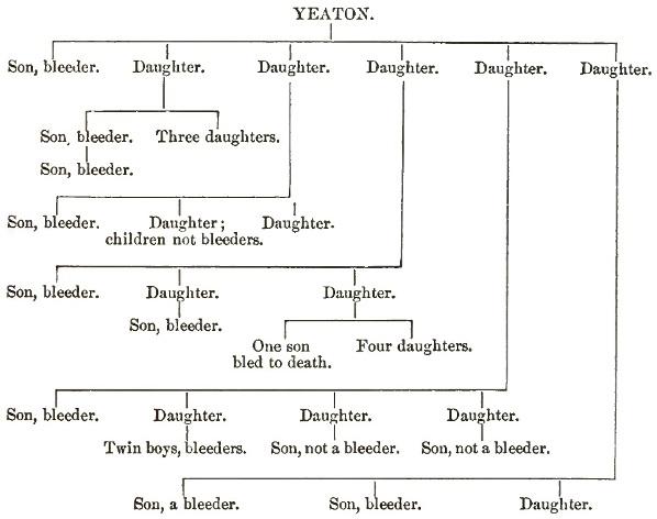
In the celebrated bleeder families of Tenna, Switzerland, five generations have been affected. The modes of transmission are as follows: (1) Father to son, grandson, etc. This is rare, but instances are on record. (2) Father not a bleeder, but of bleeder stock, transmits the tendency to son—very uncommon. (3) Father to daughter, granddaughter, etc.—not common. The daughters of a bleeder are usually free, though their brothers may be affected. (4) Mother a bleeder, transmits to sons and daughters. (5) Mother not a bleeder, but daughter of one, transmits to her sons, the daughters remaining free, but their sons affected. This is the most common mode of inheritance. Atavism by transmission through the female line is almost the rule, and the daughters of a bleeder, though healthy and free from any tendency, are almost certain to transmit the disposition to their male offspring. The 657 cases analyzed by Grandidier occurred in two hundred families. The chief facts of heredity are well illustrated by the preceding chart of the Yeaton family, given by Gould in the Boston Medical and Surgical Journal, 1857.
The Anglo-Germanic nations appear especially prone to the disease. Of 194 families in Grandidier's table, 154 were of the Teutonic stock. Records of the disease among the Latin races are rare. Jews are probably not more liable than other people, but the rite of circumcision gives an unusual opportunity for its manifestation at an early age.
The age at which the bleeding tendency first appears was determined by Grandidier in 113 cases as follows: in 63 during the first year; in 17 during the second, and up to the end of the second year in 93. It is rare for the first manifestation to occur after the twelfth year, and there was only one case in which the first bleeding appeared after the fifteenth year.
The constitution and temperament of bleeders, about which the older writers had much to say, probably present no peculiar characteristics. Some persons claim to be able to recognize bleeders even before they have manifested any tendency to hemorrhage. They are usually fresh, healthy-looking persons, with fine, soft skins, through which the superficial veins may show with more than usual distinctness. A division of cases into erethetic and atonic forms has been made by Wachsmuth and Grandidier. The mental activity of bleeders has been noted to be above the average, due, doubtless, to the fact that the liability to bleed from slight blows and cuts has made sedentary and studious habits preferred to out-of-door employments and amusements.
Families in all conditions of life are affected. Much interest was excited in the disease in England from the fact that the late Prince Leopold was a sufferer.
Climate appears to have an influence in determining attacks. Cold, damp, changeable weather is favorable, while a residence in a warm, equable climate diminishes the tendency in a very marked manner. Some patients have an extraordinary susceptibility to changes in the weather.
All observers have noted the great fertility of bleeder families. Those first born seem less liable to bleed than subsequent ones.
SYMPTOMS.—The existence of the defect of constitution may not be suspected until an uncontrollable hemorrhage follows some trivial injury or operation, or a spontaneous bleeding may occur and present great or insuperable difficulties in its arrest. The symptoms usually occur in the first years of life, and in the great majority of cases, as mentioned above, the first bleeding occurs before the fifth year. The symptoms may be grouped under three divisions (Legg, Grandidier): external bleedings, spontaneous and traumatic; interstitial bleedings, petechiæ, and ecchymoses; and the joint affections. Legg recognizes three grades of the disease. The first and most severe is characterized by bleedings of every kind, external and internal, and by troublesome joint affections: this form is most often seen in men. The second grade is less severe; there are spontaneous hemorrhages from the mucous surfaces, but no traumatic bleeding or ecchymoses and no joint troubles: this form is most often seen in women. The third and lowest degree is when there is a tendency simply to ecchymoses; no dangerous bleedings occur: this form is often seen in members of bleeder families, and if in women the menstruation may be early and profuse.
External Bleedings.—The spontaneous bleedings may occur from the skin, the mucous, and in rare instances the serous, membranes. There are frequently preliminary symptoms—prodromata—such as flushing, fulness of the head, and throbbing of the arteries—signs of so-called plethora; often there is irritability of temper, but sometimes, in children, extra cheerfulness has been observed. The localities affected and the frequency are shown by the following analysis of 334 cases by Grandidier: Epistaxis, 169 times; from the mouth, 43; stomach, 15; bowels, 36; urethra, 16; lungs, 17; cerebral hemorrhage, 2; skin of head, 4; tongue and finger-tips, 4 each; tear-papilla, 3; eyelids, 2; external ear, 5; female generative organs, 10; ulcer of skin, 2; navel (long healed), 2. An odd situation for spontaneous bleeding is mentioned by Townsend,171 in which a child bled to death from the scrotum. In many cases these spontaneous hemorrhages prove fatal—most frequently the epistaxis. The traumatic bleeding may result from blows, cuts, scratches, etc., and the blood may be effused into the tissues or discharged externally. Fatal hemorrhages have occurred from the following wounds: blow on head, 11 times; slight scratches on skin or abrasion of dermis; laceration of the frænum of the lip, slight cut (two lines deep) in a duel wound; bite of the tongue (7 cases); fall on the mouth; blow on the nose; blow of a stone on the finger; cut in paring the nail; fall on the head with meningeal hemorrhage (2 cases, brothers); and rupture of the hymen on the wedding-night.
171 Boston Med. and Surg. Journal, lv.
After operations, trivial and severe, many fatal cases have occurred, and the statistics of the same author give the following: cutting of the frænum linguæ, 1; leeching, 5; venesection, 4; blister, 2; extraction of tooth, 12; circumcision, 8; cutting umbilical cord, 4; vaccination, 2; fistula, stone, ligation of carotid, of radial, of ulnar, of femoral arteries, amputation of arm and of thigh, 1 each; phimosis, 2. Leeching, extraction of teeth, and circumcision are most dangerous operations in bleeders.
The bleeding is always a capillary oozing, and the vessels are not seen. It may last for hours, or even many days and weeks, and the amount of blood lost may be enormous. Epistaxis may be fatal in twenty-four hours. In Coates' case a medical student lost half a gallon of blood in twenty-four hours, and in the ten days which the bleeding continued it was estimated that he lost about three gallons.
The healing of a wound in a bleeder may take place rapidly, either with or without suppuration. When the hemorrhage is large or prolonged, severe anæmia follows, from which, as a rule, the patients recover with remarkable rapidity.
The interstitial hemorrhages—petechiæ, ecchymoses, hæmatoma—may be spontaneous or the result of injuries. The petechiæ occur most frequently in the skin, particularly of parts distant from the heart—the legs and arms, less often the face. On the mucous and serous surfaces they are less common. They resemble ordinary purpuric spots, and crops may come out with symptoms of swelling and pain in the joints. Large extravasations—hæmatoma—are most frequently of traumatic origin and may follow the slightest blow, as in a case of Sir Wm. Jenner's, in which from the fall of a rubber ball on the thigh an enormous extravasation took place between the knee and trochanter.172 They are blue, black, or reddish-black at first, and in their absorption go through the various changes in color which we notice in a bruise. These blood-tumors may occasionally arise spontaneously.
172 Legg, etc., p. 68.
The arthritic affections in hæmophilia are very remarkable, and so common as to form prominent features in the disease. There may be simple pain in and about the joints, or swelling with redness and signs of intense inflammation. The attacks may come on suddenly with fever, resembling closely acute rheumatism. The large joints are usually affected, the knees most often, then the elbows, ankles, and shoulders. There may be repeated attacks, and at last great crippling and deformity. The small joints are rarely affected. In cold, damp weather the attacks are most common; occasionally they follow traumatism. In addition to the joint troubles, bleeders suffer much with irregular pains in the limbs, particularly during change of weather, or these pains with arthritis may usher in an attack of hemorrhage.
Many other irregular symptoms are described in the monographs, some of which have no intimate relation with the disease. The anæmia has, of course, all the features of the traumatic form. Digestive troubles, after the bleeding, are common, and are due to the anæmia. The Buel Brothers173 mention that in two of their cases the patients showed a marked inclination to eat sand and earth. Children with the hemorrhagic tendency pass through the ordinary diseases of infancy like others. Whooping cough is very liable to cause epistaxis. Rheumatism and scrofula are said to be common in bleeder families.
173 Op. cit.
The blood in bleeder cases is, as a rule, normal, so far as our present means of investigation enable us to decide. When a hemorrhage has continued for some time, it is thin and watery, but at the beginning of the bleeding the blood is usually rich in corpuscles and fibrin and coagulates firmly. The salts have been found increased in quantity. No change has been noted in the corpuscles, the number of which is stated by several observers to be increased. Prior to a hemorrhage there may be, according to some writers, a state of plethora or increase in the total quantity of blood, and the tolerance of the loss, so much greater in bleeders than in ordinary persons, is adduced in support of this view.
MORBID ANATOMY.—Not many changes other than those of profound anæmia have been found in the bodies of bleeders. An unusual thinness of the walls of the vessels, first noted by Bladgen in 1817,174 has been met with in a number of cases; in a few instances hypertrophy of the heart; in others a rounded foetal shape of the organ. Within the past few years careful microscopical examination has been made of the tissues and blood-vessels of bleeders. Kidd175 found degeneration of the muscle-fibres of the middle coat of the arteries, and the endothelium of the small arteries, veins, and capillaries was swollen, proliferated, and some of the small veins were blocked with the products. Legg176 reports a case in which Klein made a most careful examination with negative results, and he stated that of six such examinations which had heretofore been made, in only one case (Kidd's) were important changes found. At the same meeting of the London Pathological Society, Theodore Ackland also reported a case with negative results as regards histological changes.
174 Medico-Chirur. Transactions.
175 Medico-Chirurgical Society's Transactions, vol. lxi.
176 Lancet, Oct. 27, 1884.
The joint changes have been studied in a number of cases. Hemorrhage has been found in and about the capsule, and the acute swelling may be due largely to it, as was shown in Hutchinson's case,177 in which he aspirated the joint. When it lasts any time, there is great staining of the cartilages and discoloration. There may be inflammation of the synovial fringes and erosion and destruction of the articular surfaces (Legg).
177 Trans. State Med. Soc. N.Y., 1877.
The PATHOLOGY of the disease is unknown. No doubt two circumstances combine in hæmophilia—congenital fragibility of the vessels and a defect in coagulability of the blood—but whereon these depend we are as yet entirely ignorant. There is no evidence of the nature of the anatomical changes in the vessels which permits of their ready laceration, and none on the nature of the alteration of the blood which prevents the normal thrombus formation in a wound; and in the absence of information on these points theories must necessarily be unsatisfactory, and their discussion, in a work of this practical nature, profitless.
The DIAGNOSIS presents no difficulty in members of a bleeder family, in whom slight joint trouble and petechiæ are as much manifestations of the disease as the more severe hemorrhages. In a large majority of cases the tendency becomes manifest at an early date. The spontaneous umbilical hemorrhages of infants are, as a rule, to be excluded, being dependent upon, or associated with, jaundice or syphilis or a mycosis (Weigert178). The hemorrhagic diathesis may develop in children or members of a healthy family and prove fatal, and the question in such cases always comes up, Are they instances of hæmophilia? There seems to be a desire to limit this term to cases of an hereditary nature only; but when a child shows a marked tendency to multiple hemorrhages, spontaneous or traumatic, which tendency persists and is not merely transitory, and particularly if there are joint troubles, I think that under these circumstances we have a genuine case of hæmophilia; and such a child, if he—it is more likely to be a male—survives and marries, may be the founder of a bleeder family. These are the congenital in contradistinction to the hereditary cases. In the histories of the bleeder families we frequently come back to the origin in a person born of a healthy stock in which there have been no hemorrhagic tendencies. On the other hand, single severe uncontrollable hemorrhages in children or adults are not to be ranked as hæmophilia unless there have been other features pointing to the existence of the diathesis. The literature abounds in cases of this kind, many of which are described as hæmophilia. In doubtful cases it is very difficult to decide, as in a case of Forscheimer brought before the Academy of Medicine of Cincinnati.179 In the review of American literature we have excluded all cases in which the hereditary or congenital characters were not well marked.
178 Cohnheim's Pathologie, i. 382.
179 Cincinnati Lancet and Clinic, 1884.
It may be useful to put down here for the guidance of the practitioner the varieties of bleeding commonly met with, and which must not be confounded with hæmophilia:
(1) The umbilical hemorrhages of infants, due to jaundice or to syphilis hæmorrhagica neonatorum, etc.
(2) Purpura simplex, seen often in debilitated, rarely in healthy, children, usually confined to the legs, and in some cases I have seen it associated with rheumatic pains or swellings in the knees and ankles.
(3) Peliosis rheumatica, an affection which in the large interstitial hemorrhages and the joint swellings touches hæmophilia in a curious way. It too may show itself in several members of the same family.
(4) Purpura hæmorrhagica, Morbus maculosus Werlhöfii, a grave disease, characterized by extensive cutaneous ecchymoses, mucous hemorrhages, but not dependent on any local disease, or, so far as is known, on any specific poison. The bleedings in scurvy may be mentioned here, but there could be little difficulty in determining their nature.
(5) Infective purpura, due to the action of some specific poison—small-pox, measles, scarlet fever, cerebro-spinal fever, etc. The hemorrhages may be cutaneous and trivial, or may be in the most aggravated form of interstitial and mucous bleedings, as seen, for example, in black small-pox.
(6) Toxic purpura, as in snake-bites and many poisons, such as phosphorus.
(7) Simple hemorrhagic diathesis, under which may be included those cases in which, without any hereditary disposition or previous hemorrhagic history, there is a tendency to uncontrollable hemorrhage from a slight wound.
(8) Hæmatidrosis, bloody sweats, which occur usually in hysterical or epileptic females, and are in rare instances accompanied with mucous hemorrhages.
In considering the PROGNOSIS it is well to remember that the patients rarely die in the first bleeding. The younger the individual the worse is the outlook. As above stated, the attacks are most frequent under five years of age, and of 152 boys the subject of the disease, 81 died before the termination of the seventh year (Grandidier). Legg, however, states that it is rarely fatal in the first year. The longer a bleeder lives, the greater the chance of his outlasting the tendency; but that it may persist to the end of a long life, and then prove fatal, is shown by the case of old Oliver Appleton, the first recorded American bleeder, who died at an advanced age of hemorrhage from a bedsore and from the urethra. A bleeder may have years of existence, in which the tendency seems lessened or even absent. The prognosis is always worse in a boy than in a girl. In the latter menstruation is sometimes early and excessive, but, happily, in the female members of hæmophilic families neither this function nor the act of parturition bring with them special dangers.
TREATMENT.—The prophylaxis is all-important. The members of a bleeder family, particularly the boys, must be guarded from injury as much as possible, and operations of all kinds must be avoided, except when life itself is in jeopardy. The extraction of a tooth should be absolutely prohibited. Occupations must be sought which will give the least possible risk of injury. Daughters of bleeder families should not be permitted to marry, as it is through them that the tendency is chiefly propagated, and, even if not bleeders themselves, some of their male children are certain to be affected. The question of the marriage of male bleeders is more difficult to decide, but in any case where the tendency is marked it should be prohibited.
When an injury or wound has occurred, absolute rest, cleansing the wound, and compression should first be tried. If in a favorable locality pressure on the artery may be employed, failing in this, the various styptics may be used. In epistaxis, ice, tannin, and gallic acid may be tried before plugging.
Internally, ergot seems to have been of use in several cases. Otto180 speaks of the value of sulphate of soda in purgative doses. The perchloride of iron, 30–40 minim doses, every two hours, is advised by Legg, with a purge of sulphate of soda if there is no bleeding from the bowels. Venesection has been resorted to in several instances. Transfusion has been employed, but without benefit. The diet should be light and supporting. After the attacks the patients should take iron and cod-liver oil until the health seems restored. When possible, a residence in the South during the winter is advisable, as most cases are aggravated by the cold weather, and in any case care must be taken to protect patients against cold and wet.
180 Loc. cit.
The joint troubles must be treated on general principles.
AMERICAN LITERATURE OF HÆMOPHILIA.
Buel: Transact. of the Med.-Physic. Society of New York, vol. i., 1817; 1 F., 4 cases.
Coates: North Am. Med. and Surgical Journal, Philada., 1828, vi. p. 37; 1 F., 5 cases.
Dunn: Am. Journal Med. Sciences, 1883, vol. lxxxv. p. 68; 4 F.
Felt: History of Ipswich, 1834—Appleton family referred to.
Gould: Boston Med. and Surgical Journal, 1857, p. 500; 1 F., 11 cases.
Harris: Philada. Med. Times, 1872; 2 F., 7 cases.
Hay: New England Journal of Med. and Surgery, 1813, vol. ii. p. 221; 1 F., 15 or 16 cases (accurate number rather difficult to make out).
Holton: Am. Journal of Med. Sciences, April, 1874; 1 F., 7 cases.
Hughes: Transylvania Med. Journal, 1831, vol. iv. p. 518, and Am. Journal Med. Sciences, 1833, vol. xxi. p. 543; 1 F., many cases.
Hutchinson: Trans. State Med. Society of New York, 1877, p. 208.
Otto: Medical Repository, 1803, vol. vi.; 3 F., 8 cases.
Pepper: Philada. Med. Times, 1881, vol. xii. p. 109; 1 case—Lancaster county family described by Dunn.
Sewell: Med. Chronicle, Montreal, 1857, vol. iv.; 2 F., 4 or 5 cases.
Smith: Philadelphia Medical Museum, 1801, vol. i. p. 284.
Traneus: St. Louis Med. and Surgical Journal, 1870, p. 535; 1 F., 4 cases.
Townsend: Boston Med. and Surgical Journal, vol. lv. p. 447; 1 F., 3 cases.
ADDISON'S DISEASE.
DEFINITION.—A constitutional affection characterized by asthenia without emaciation, a depressed circulation, gastric irritability, and usually pigmentation of the skin. In the majority of cases it is associated with a fibro-caseous degeneration of the suprarenal capsules, and in many there are changes in the abdominal sympathetic system.
SYNONYMS.—Morbus Addisonii; Bronzed-skin disease. Fr. Maladie d'Addison, Maladie bronzée; Ger. Addisonische Krankheit.
HISTORY.—In the Halle Hospital Reports for 1823 Schötte describes a case, and one is also given by Bright in vol. ii. of his Medical Reports, 1831. A few other instances are also on record before 1855, when Addison published his monograph On the Constitutional and Local Effects of Disease of the Suprarenal Capsules, from which we may date our knowledge of the affection. Following close upon the work of Addison numerous observations were made in England, where the disease appears to be more common than elsewhere. Wilks of Guy's Hospital,181 and Greenhow of the Middlesex Hospital, may be mentioned among those who in England have specially studied the disease, and the latter published an important monograph in 1875.182 In France, besides numerous minor contributions, the exhaustive articles in the encyclopædias have been the most important publications. In Germany the monograph of Averbeck (1869) and the abstracts of Meissner in Schmidt's Jahrbücher may be specially mentioned. Virchow, Griesinger, Oppolzer, Bamberger, and others have made valuable contributions. Recently Burger183 has published a small monograph. In other European centres contributions have been made, among which may be mentioned that of Schmidt of Amsterdam, who brought forward cases in support of the view that the disease was an affection of the sympathetic ganglion. In America the first cases were reported by Ranking184 and Taylor.185
181 In numerous communications in Guy's Hospital Reports and Trans. Path. Society.
182 "On Addison's Disease," Croonian Lectures.
183 Die Nebeunieren und der Morbus Addison, Berlin, 1883.
184 Am. Journ. Med. Sci., 1856.
185 New York Med. Journal, 1856.
ETIOLOGY.—The causation of the disease is unknown. Cases are more frequent in hospital than in private practice. Males are more often attacked than females; the proportion in Jaccoud's table186 is 79 to 48, and in Greenhow's187 analysis of 183 good cases, 119 were males and 64 females. Under ten and over fifty years of age the disease is very uncommon; the majority of the cases occur between the twentieth and the fortieth year. Greenhow has called attention to the fact that in a number of instances the disease appears to have followed an injury, such as a blow upon the abdomen or back, and in several cases caries of the spine has preceded the attack. He refers also to the greater frequency of the disease in the laboring classes and those exposed to injury from over-exertion. The disease does not seem to be more prevalent among members of phthisical families, although the morbid process in the glands has been regarded as of a tuberculous nature, and it is common for other tuberculous lesions to occur in the course of the disease.
186 Dictionnaire de Médecine.
187 Op. cit.
The disease is rare in America—apparently much more so than in England.
SYMPTOMS.—In the words of Addison, the leading and characteristic symptoms are: "Anæmia, general languor and debility, remarkable feebleness of the heart's action, irritability of the stomach, and a peculiar change of color in the skin occurring in connection with a diseased condition of the suprarenal capsules."
Although, perhaps, not the most essential, the symptoms pertaining to the skin are in the majority of cases the most prominent, and have given rise to the names bronzed skin, melasma suprarenale, etc. which have been applied to the disease. A gradual increase in the pigment of the rete mucosum, either patchy or diffuse, causes a gradual discoloration, which may ultimately reach such a degree that a previously blonde individual may have the aspect of a Malay or a mulatto. The grades of coloration may range from a light yellow to a deep brown, or even black. In some instances there is a greenish-brown tinge, to which the term bronzed is peculiarly applicable. In typical cases it is diffuse over the whole surface, but as a rule deeper on exposed parts, face, neck, and hands, and also in those regions where the normal pigmentation is most intense, nipples, scrotum, and penis, or in the vicinity of cicatrices or regions of chronic irritation. It is usually first noticed on the face, either diffuse or in spots, and the extension may be rapid or gradual, in many instances not reaching a high grade and not becoming universal. It may be absent, and is not to be regarded—as was formerly the case—as an essential feature of the disease. Patches of leucoderma may occur in connection with the pigmentation, as beautifully delineated in Pl. xi. of Addison's monograph. The pigmentation is not confined to the skin, but may extend to the mucous surfaces—mouth, conjunctivæ, vagina. In the mouth the patches may be as dark as in the dog; they are usually scattered, often on the margins of the lips and on the edges and under surface of the tongue and on the cheeks. The conjunctivæ are less often affected. The vagina may be very deeply pigmented. An intensification of the choroidal pigment has been observed. In some cases a patchy pigmentation of the serous membranes has been found, and is figured in one of Addison's original plates, and pigmentation of the nails, hair, and teeth may also occur. A variation has been observed in the intensity of the coloration with the general health of the patient. The discoloration rarely precedes the general symptoms, but more usually follows the asthenia.
Some observers have noted a peculiar odor of the breath and from the skin, particularly during the last few days of life.
Anæmia of a moderate degree may exist, but it is not, as often stated, a constant symptom. Greenhow states that "there is no real anæmia, the blood being often rich in red corpuscles, even in excess, and there is no increase in the white." No special alterations in the appearance of the corpuscles have been noted. In some instances free pigment has been found.188 In a case recently at the hospital of the University of Pennsylvania, Hughes found the number of red corpuscles over five millions per cubic millimeter, and there were free pigment-granules in the blood.
188 Corput, Gazette hébdomadaire, 1863.
Hemorrhages are rare; extravasations into the retinæ have not, so far as I can ascertain, been observed, nor are there often the other common features of anæmia.
The pulse is frequent and small, the heart's action weak—sounds clear; a venous murmur may sometimes be heard. In some cases there appears to be a special enfeeblement of the heart and a liability to fainting attacks, and without any warning a fatal syncope may occur. Cold feet and hands result from the weak circulation, and may be a most annoying symptom.
Symptoms in connection with the respiratory system are not common. There may be dyspnoea, and the complication of phthisis may give rise to all the features of that disease. There may, however, be extensive lung trouble with but few symptoms. The temperature is rarely elevated, more often it is subnormal.
Gastric disturbances are very common—anorexia, nausea, vomiting—particularly toward the close, but they may be early and prominent features, persisting in spite of all remedies and proving the most formidable symptoms of the malady. They appear to be of nervous origin, and not referable to changes in the organ itself. It is doubtful if the case reported by Gilliam,189 in which there was degeneration of the gastric mucosa, was Addison's disease. The state of the bowels is variable; constipation is more frequent than the normal condition. Diarrhoea is common, and may come on suddenly without obvious cause, and is a not infrequent cause of death.
189 Phil. Med. and Surg. Reporter, xxiv.
The urine is usually pale, free from albumen, not often increased in amount. It is interesting to note, in connection with the involvement of the abdominal sympathetic, that in a few cases there has been polyuria. The nitrogenous elements may be greatly reduced, the urea to 13–20 grammes daily, and the amount of indican may be increased as much as 64–75 milligrammes in 1000 c.c. (Samuel). In one case Thudichum found the urinary pigments greatly reduced in amount, the uromelanin not amounting to more than one-twelfth the normal quantity. A recent observation of Nothnagel is of interest.190 In a patient aged twenty, who had the typical symptoms of the disease for two years, death took place by coma and the condition of acetonuria was determined.
190 Zeitschrift für klin. Med., Bd. ix.
The symptoms connected with the nervous system are the most prominent in the disease, and are more constant than the anæmia or the bronzing. The most marked is a depression and enfeeblement of the nervous forces, a profound asthenia out of all proportion to the general condition. The patients complain of a lack of energy, mental and bodily; the least exertion is an effort, and there is a feeling of tire and weariness with which the facial expression is quite in keeping. The fainting fits, giddiness, noises in the ears, may also be due to faulty innervation, as they occur in cases in which the anæmia is by no means advanced. Headache, lumbar and abdominal pains are frequent, and in a considerable proportion of cases there is tenderness on pressure in the lumbar region. With the advance of the disease the prostration becomes more marked, the patient remains in the recumbent posture, the voice gets weak and small, the intelligence dulled, and occasionally there is delirium. Head symptoms may suddenly supervene, and death by coma or convulsions cut short the progress even early in the disease (Pye-Smith). In Jaccoud's series convulsions were noted in 19 cases.
The disease may be said to be invariably fatal, but the course presents many variations. The majority of cases die within eighteen months of the first onset of the symptoms. B. Fenwick, in an analysis of 30 recent cases,191 calls attention to the fact that when bronzing does not occur the course is more rapid. Thus the average duration of the non-bronzed cases was only 4.8 months, while for bronzed ones it was 23.6 months. There are acute cases in which, with great weakness, vomiting, and diarrhoea, the fatal end may occur in a few weeks. Some of these rapid cases resemble typhus. Syncopal attacks, coma, or convulsions cut short not a few cases. In a few instances it is much prolonged—six years (Niemeyer) or even ten years (Greenhow). Whether recovery ever takes place is doubtful. Finney192 has reported an apparently genuine case which got well. Some French observers (Potain) think that recovery takes place more often than is supposed. Sir Wm. Gull mentioned a case of recovery.193 Periods of improvement lasting many months may occur.
191 Path. Soc. Trans., vol. xxxiii., London.
192 Dublin Med. Journ., April, 1882.
193 Int. Med. Congress (London) Transactions, vol. ii.
MORBID ANATOMY.—The panniculus adiposus and subperitoneal fat may be in normal quantity. There is rarely great emaciation, nor are the organs blanched and bloodless. The most constant lesion is in the suprarenal organs, which present a caseo-fibrous change, more rarely simple atrophy or other alterations. So frequent is the caseo-fibrous condition that some writers (Wilks) hold that it is the specific lesion of the disease. The organs are enlarged—may weigh several ounces each. The capsules are thickened, and may present caseous or even calcareous masses. The normal shape of the gland is lost, and it forms an irregular nodular mass closely adherent to contiguous parts—liver, kidney, and cava on the right side, kidney, spleen, and often pancreas, on the left. There is usually a good deal of fibrous thickening and matting in the vicinity, and the adhesions to adjacent structures may be very strong. The peritoneum often shows patches of fibroid induration. On section the diseased organ cuts with great resistance, and to the touch has an almost cartilaginous hardness. The exposed surface shows caseous masses of a yellow or grayish-white color, varying in size from a pea to a walnut, imbedded in a grayish semi-translucent fibrous tissue, pale when first cut, becoming reddish on exposure. These caseous masses may undergo softening or calcification, and pockets of pus are not uncommon. Definite small miliary granulations are not often seen, though nodular grayish-yellow bodies the size of small peas may occur. The strands of fibrous tissue which separate and enclose the caseous masses have often a very peculiar translucent, infiltrated appearance. When the cheesy lumps are small, the amount of this tissue is considerable and gives a remarkable character to the section. Wilks has described a case in which this tissue made up the entire mass. The substance of the gland is usually destroyed. The vessels and nerves can be traced to the organs where they become imbedded in the fibrous tissue.
Histologically, the soft translucent tissue consists chiefly of spindle-shaped fibre-cells, and in firmer older parts of ordinary fibrous stroma. In the immediate neighborhood of the cheesy masses there are round corpuscles—about the size of or a little larger than white blood-cells—imbedded in a fine reticulum of fibres. Giant-cells are sometimes found, but they have not been common elements in the specimens which I have examined. The caseous substance consists of granular débris in which the remains of cells and fibres can be seen. In coarse and microscopical features the lesion resembles closely local tubercular affections. The extension is by a small-celled growth, which gradually invades the adjacent parts, extending peripherally as the central portions caseate. Distinct miliary granulations are not often met with. The relation of this local growth to tuberculosis is a very interesting question. It is usually regarded as a scrofulous or tuberculous process, to which in its general features it quite conforms. I have been interested in ascertaining whether the bacillus tuberculosis existed or not in the local lesion. In Cohnheim's laboratory Karl Hüber kindly gave me an opportunity of examining the adrenals in two cases, in only one of which were the bacilli evident. Since then I have re-examined the fibro-caseous tissue in Ross's case,194 which was a most typical one, the suprarenals alone involved, and in the recent case reported by Pepper,195 and in neither have I been able to demonstrate bacilli. Future examinations must decide whether the local affection is inflammatory or whether it belongs to the infective granulomata.
194 Can. Med. Assoc. Trans., vol. i., 1877.
195 Phila. Med. Times, 1885.
That other alterations may occur in cases of Addison's disease appears well established, though some still regard the caseo-fibrous change essential and specific. Atrophy of one or both glands has been frequently seen. Jaccoud gives 7 cases in his tables. Good recent cases have been described by W. B. Hadden,196 Hebb,197 and Goodhart.198 The atrophy is due to a chronic interstitial process similar to cirrhosis of the liver. Hadden199 states that the lesion is identical with that in the thyroid gland in myxoedema.
196 British Medical Journal, 1885.
197 Lancet, 1883, i.
198 Path. Soc. Trans., 1882.
199 Loc. cit.
Absence of one or both the capsules has been noted by Legg, Spender, Borland200 and Hubbard.201
200 Boston Med. and Surg. Journal, 1867.
201 Proceedings of Conn. Med. Society, 1868–71.
Cancer of the adrenals, by no means uncommon as a secondary process, rarely produces any special symptoms; but there are cases which are difficult to exclude from the category of Addison's disease. Jaccoud gives several, and in the case of Edes, often quoted,202 the asthenia and discoloration may have been due to the capsular affection, but there was also extensive peritoneal cancer.
202 Boston Med. and Surg. Journal, 1878.
By far the most constant morbid change after that in the adrenals is a more or less widely distributed tuberculosis, particularly of the lungs. A very considerable proportion of the cases are complicated with chronic phthisis. Regarding the disease of the suprarenals as primary, the general tuberculous affection may be secondary; and it is just in these organs, as Weigert has shown, that the veins are apt to be perforated by tubercles and systemic infection induced. The retro-peritoneal and mesenteric lymph-glands may also be tuberculous. Ulcers of the ileum may occur, and swelling of Peyer's glands and the solitary elements in the bowels is very common. In Ross's case there were numerous lymphoid infiltrations of the mucosa of the stomach, chiefly about the pylorus and cardia. The changes in the skin are confined to an increase of the pigment in the cells of the rete mucosum, most pronounced in the deeper layers and in the deeper parts in the connective-tissue cells of the papillæ and subcutaneous tissues. The condition is not to be distinguished from a deeply-pigmented scrotum or from the dark skin of the negro. The pigment resembles the ordinary coloring matter of the skin, but is possibly different from it in containing no iron.203 Nothnagel has made204 an exceedingly interesting study of the pigmentation in Addison's disease, and concludes that it is identical in distribution with that in the skin of the dark races; that it does not originate in the cells of the rete mucosum, but is elaborated in deeper cells, about the vessels of the cornium, and transported by them to the more superficial layers—a mode which recent observations seem to show is the normal one; and, lastly, that it is a process induced through the nervous system in some way as yet unknown.
203 Arnold, Virchow's Archiv, xxxv.
204 Loc. cit., Bd. ix.
The spleen has been found enlarged. The thymus gland may also persist or be much larger than normal. In Ross's case it weighed six ounces. The heart and blood-vessels do not present any constant changes: the heart has been found small in some cases. Venous engorgement of the abdominal viscera has been noted in a few cases, but it is not a common feature. It was not present in two typical cases which I have examined.
In the nervous system the condition of the abdominal sympathetic has received special attention, and in a number of cases definite changes have been met with, chiefly of a sclerotic or chronic nature and intimately associated with the fibroid induration about the capsules. The nerve-cells of the semilunar ganglia are described as degenerated, deeply pigmented, and often present a new growth of connective tissue about and between the cells. There are at least thirty or more cases in which such alterations have been found. In some instances the medulla of the nerves passing from the ganglia has been found wasted and the fibres in a state of fatty degeneration. In some cases these parts have been found normal (Foa,205 Huber,206 Hebb,207 and Hadden208). In a most typical case under Ross at the Montreal General Hospital, I could find no differences in the cells and nerves, comparing them with those of a woman dead on the same day of heart disease. More recently, I have examined a case for Pepper in which the right semilunar ganglion was imbedded in the sclerotic tissue of the right adrenal; the nerve-cells were undergoing atrophy from compression; and there were fatty changes and degenerations in the nerves connected with this ganglion. The left was uninvolved, and the cells and fibres appeared normal.
205 Virchow-Hirsch, 1879.
206 Virchow's Archiv, 86.
207 Lancet, 1883, i.
208 Loc. cit.
Jacquet has described pigmentary changes in the ganglia of the cord as well as in those of the abdomen, and Guermonprez209 alterations in the brain similar to those of senile dementia. At the Congress in 1881 at London, Semmola of Naples showed a figure illustrative of degeneration of the ganglia of the abdomen, and also an infiltration of leucocytes in the neighborhood of the central canal of the cord, from a case of Addison's disease without affection of the adrenals.