Volume 3 — DISEASES OF THE BLOOD, AND OF THE HÆMATOPOIETIC SYSTEM. (continued 3)
Reading edition
Valvular diseases which cause murmurs result either in a condition of the valves that allows regurgitation, or one that obstructs the onward blood-current. Valvular insufficiency arises when extensive retraction, perforation, or partial detachment of the valves prevents them from completely closing their respective orifices. And when the chordæ tendineæ have been ruptured, or when calcareous degeneration has made the valves or the parts in the immediate vicinity abnormally rigid, the regurgitant current through the aperture thus left gives rise to a regurgitant murmur.
When the valves are thickened, retracted, adherent, hypertrophied, or degenerated, so that their edges are prevented from being accurately applied to the walls of the ventricles or vessels, they obstruct the current of blood, and the impinging of the blood-current against the obstruction gives rise to obstructive murmurs. These conditions—stenosis and insufficiency—are often found coexisting, but rarely in equal degree, one usually predominating sufficiently over the other as to give a dominant character to the murmur.
The lesions which produce these conditions may be temporary or permanent—temporary when they occur during the course of acute endocarditis, and permanent when they consist of a new growth either of connective, fibroid, calcareous, or atheromatous tissue, which alters the form of the valves and impairs their function. Acute and chronic valvular disease may produce the same murmurs. The effect of the valvular deformity depends entirely upon its seat.
In the study of the relations of valvular lesions to cardiac murmurs physical signs are the important factors in their diagnosis, and it is necessary always to bear in mind the normal physiological conditions which constitute a complete cardiac pulsation.
The apex of the normal heart is felt between the fifth and sixth ribs on the left side, about two inches below the nipple and one inch to its sternal side. The base of the heart is on a level with the third costal cartilages. The tricuspid valve lies behind the middle of the sternum, on a line with the articulations of the cartilages of the fourth ribs with the sternum. The mitral valve lies behind the cartilage of the fourth left rib, near the edge of the sternum. The aortic valves lie behind the sternum, a little below the junction of the cartilages of the third ribs with the sternum, near its left edge. The pulmonary valves lie behind the junction of the third left rib with the sternum.
Let it be remembered that the tricuspid orifice is the most superficial, then the pulmonary, next the aortic, and, deepest of all, the mitral orifice. Ranged from above downward, the pulmonary orifice comes first, then the aortic, then the mitral, and lastly the tricuspid.
Aortic Obstruction, or Stenosis.
Stenosis at the aortic orifice is a common cardiac lesion, and one that is always accompanied by more or less hypertrophy of the left ventricular walls.
MORBID ANATOMY.—In aortic obstruction the cardiac valves will be found to present some or all of the changes which have been described as taking place in the course of acute and interstitial endocarditis, together with degenerative changes due to atheromatous, calcareous, fibroid, fatty, or connective-tissue metamorphosis.
Sometimes the valves may be covered with thick, warty, irregular excrescences that cause loud murmurs, and yet do not seriously interfere with the outgoing blood-current. At other times stenosis of the aortic orifice may be so extensive as to almost obliterate it. When such is the case, the extent of the lesion will be measured much more by the consequent hypertrophy and its effects on the systemic circulation than by the loudness or harshness of the murmur which it produces.
Very frequently the valves are so rigid that they cannot be pressed back against the wall of the aorta, and these unyielding prominences are greater obstacles to the outgoing current of blood than vegetations on the surface of the valves.
In a few rare cases the outlet may be diminished by constriction of the aorta at the point of insertion of the valves. Adhesion of the aortic valves begins at their bases and extends along their free edges to their tips; sometimes they become fused together into a mass, so that they project into the blood-stream in the form of a funnel irregular in shape and studded with calcareous nodules. The line of attachment of the valves to the aorta frequently becomes entirely obliterated.
In some instances the contraction of the valves between their points of attachment causes them to form a deep pocket or pouch, and their points of attachment may be a quarter of an inch apart.
Obstructions at the aortic orifice are frequently accompanied by atheromatous changes in the aorta, the result of chronic inflammation of its tunics—arteritis deformans.
As a result of aortic stenosis the wall of the left ventricle becomes hypertrophied. This change is a gradual one, and is called compensatory hypertrophy: it is due to the increased force required to propel the blood through the constricted orifice.
After a time insufficiency of the mitral valves is apt to occur, caused either by the extension of endocardial inflammation from the aortic valves or by the forcible pressure of blood upon the ventricular surface of the valves.
A slight thickening or roughening of the aortic valves may cause slight obstruction to the outgoing blood-current, which will interfere but little with the emptying of the ventricular cavity, and which rarely leads to hypertrophy of their walls.
ETIOLOGY.—Aortic obstruction is most frequently met with in early and advanced life, the mean age being forty-seven years. It is not uncommon in children; valvular lesions have been found in children under two years of age. It may be induced where the aorta is defectively developed, and some think that imperfect development of the trachea may lead to imperfect expansion of the chest, and thus induce disease of the aortic valve.6
6 Barlow in Guy's Hospital Reports, S. 1, vol. vi. p. 235.
Its most frequent cause is acute exudative and interstitial rheumatic endocarditis. The origin of nearly all valvular disease may be traced back to an attack of rheumatic fever. Next to acute rheumatism, chorea is its most frequent cause. Bright's disease and pyæmia may cause it, and atheroma or arteritis deformans extending to the valves will give rise to valvular lesions which cause obstruction.
Any of the conditions that cause acute exudative and interstitial endocarditis may effect changes in the valves, and the tissue thus developed, undergoing atheromatous, fatty, fibroid, calcareous, or connective-tissue change, will cause obstruction.
Increased tension of the aorta may be the result of chronic spinal deformity, and may be regarded as the indirect cause of aortic stenosis.
The connection between cancer and cardiac valvular disease is to be noticed, if not as cause and effect, at least as a remarkable and noteworthy coincidence.
Women are far less subject to aortic obstruction after rheumatism than men. In men the aortic valves are subject to more pressure and strain than in women, and hence non-rheumatic disease of these valves is very common, while in women it is very rare.
Aortic disease especially occurs in men whose occupations involve repeated, sudden, and great muscular effort.
In old age the walls of the aorta are weakened, and when aortic disease is met with in young subjects it must be regarded as the result of a premature senile condition of the vessels. Allbutt says that in Leeds quite young men have aortic valvular disease, and Peacock mentions several cases where the disease has occurred in young girls who have been placed at service before they were fully developed.
Sometimes the valves are found to be studded with vegetations, apparently of syphilitic origin. Corvisart and Virchow both admit the possibility of such an origin for valvular disease of the heart, but no unquestionable case has as yet been advanced in proof of it. It has been claimed that this is the reason why soldiers so frequently suffer from heart disease; but sailors are notoriously more subject to syphilis than soldiers, and heart disease is rare among them.
The reason is evidently to be found in their mode of dress: sailors wear loose clothes, soldiers have the tightest possible fitting garments. More force is required to pump the blood through the constricted vessels, hence arises more strain on the aorta and more strain on the valves.
Single, sudden muscular efforts have in a limited number of cases produced disease at the aortic orifice.
Aortic valvular disease more frequently than mitral is of non-rheumatic origin; it is slower in its development, and is more commonly met with in advanced life.
SYMPTOMS.—The subjective symptoms of obstruction at the aortic orifice are not usually well marked. Extensive aortic stenosis is not incompatible with a state of comparative good health. As the obstruction to the outflow of blood from the ventricle increases, compensatory ventricular hypertrophy enables the heart to fill the arterial system and relieve the pulmonary pressure. As soon as the ventricular hypertrophy no longer compensates for the obstruction, the arteries are inadequately filled; the left auricle cannot empty itself into the left ventricle, and hence the pulmonary vessels are abnormally full, as is also the entire venous system. The scanty arterial supply gives the pallor to the face which so frequently accompanies this condition, and syncope is liable to occur as a result of partial cerebral anæmia.
These are late effects, and in many cases do not make their appearance until the mitral valve is secondarily involved. The pulse in aortic stenosis is normal in frequency, diminished in volume and power, usually regular in rhythm, though it may be intermittent, and is compressible and jerky in character.
As a general rule, in aortic stenosis signs of arterial anæmia precede evidences of venous engorgement. The obstruction to the exit of blood is shown in the sphygmographic tracing by a slanting or oblique up-stroke, as seen in the accompanying tracing, or, as Mahomed says, "the influence of percussion is lost." Tracings of the pulse in aortic stenosis sometimes show considerable separation between the percussion and the tidal waves. In some rare instances the pulse is slowed. There may be slight palpitation, and pain in the chest may sometimes occur in paroxysms; but pain in the chest is far more common in regurgitation than in obstruction. Aortic obstruction is more frequently connected with cerebral embolism than any other valvular lesion.
FIG. 41.

Aortic Obstruction (after Foster).
The left middle cerebral artery is the most common seat of aortic cardiac emboli. The left lower limb is more subject to embolism from aortic valvular disease than the right. The splenic and renal vessels are also the frequent seat of such emboli. Sometimes embolism is due to small auricular or ventricular clots that form behind the obstruction. Such clots have occluded the aortic orifice and caused sudden death.7
7 Pathological Transactions, vol. ix. p. 91.
PHYSICAL SIGNS.—The physical signs of aortic obstruction are generally distinctive and easily appreciated.
Inspection.—The visible area of the cardiac impulse is abnormally increased. Very extensive increase in the area of impulse is frequently accompanied by a lifting of the chest-wall over the heart.
Palpation.—The impulse is felt to be forcible, and is sometimes accompanied by a heaving or lifting motion. The apex is displaced toward the left and slightly downward. A sensation will sometimes be imparted to the hand during systole similar to that produced on the sense of hearing by the whizzing of a missile by the ear. This is often nothing more than an intensified endocardial thrill. This systolic frémissement radiates to the ensiform process of the sternum, being most intense in the second right intercostal space.
Percussion.—The area of cardiac dulness will be increased in proportion to the displacement of the apex-beat to the left. The increase in dulness measures the amount of left ventricular hypertrophy.
Auscultation.—Aortic stenosis produces a systolic murmur which more frequently accompanies than replaces the first sound of the heart. The maximum intensity of this murmur is usually at the second sterno-costal articulation of the right side, but it may be heard with equal intensity over the whole upper part of the sternum, and followed up the aorta and along the carotids; again, it may be loudest at the xiphoid cartilage, or it may be heard with greatest intensity at the junction of the left third rib with the sternum. In most cases the first sound is heard with the murmur, but the murmur may entirely replace or obscure it. This murmur is usually loud and harsh in character, and is loudest at the beginning of the systole. Harshness is one of its distinguishing characteristics.
In pure aortic stenosis the aortic second sound may be inaudible, and is always feeble, but the pulmonic second sound will always be audible. The area of diffusion of this murmur follows the law that a murmur is propagated in the direction of the blood-current. It is conveyed along the aorta into the carotids, and one of its characteristics is that it is heard in the great vessels of the neck. It may sometimes be heard in the thoracic and abdominal aorta.
When an aortic obstructive murmur is heard at the apex its intensity is diminished, and when heard behind it is most distinct at the left of the third and fourth vertebræ near their spines, and frequently extends downward along the spine in the course of the aorta, but with diminished intensity. It is to be noted here that a systolic murmur, audible at the base, and traceable along the ascending arch toward the end of the right clavicle, is by no means limited to cases of aortic stenosis, although aortic stenosis always produces a murmur with these characteristics.
Arterial murmurs, synchronous with the cardiac systole, are far more frequent than diastolic murmurs. When the mitral or tricuspid valves are thickened or incompetent, or when the myocardium is the seat of extensive fatty degeneration, the murmur of aortic obstruction will entirely replace the first sound of the heart.
DIFFERENTIAL DIAGNOSIS.—Aortic obstruction may be mistaken for mitral regurgitation, tricuspid regurgitation; an anæmic bruit, for the murmur of a thoracic aneurism and for a murmur produced by a scabrous condition of the ascending arch of the aorta.
1. Both mitral and tricuspid regurgitation, as well as aortic stenosis, are recognized by a systolic murmur. The murmur of aortic obstruction is heard with its maximum intensity at the second right sterno-costal articulation, and diminishes in intensity toward the apex. The murmur of mitral regurgitation is heard with greatest intensity at the apex-beat. The murmur of aortic obstruction is conveyed into the vessels of the neck; that of mitral regurgitation to the left, in the direction of the apex-beat, and is heard behind, between the fifth and eighth dorsal vertebræ, at the left of the spine, with very nearly the same intensity as at the apex. The pulse in aortic stenosis is hard, firm, and wiry in character, but regular, while in mitral regurgitation the pulse is irregular in rhythm as well as in force, is never incompressible, and is easily increased in frequency. Gastric, intestinal, renal, hepatic, and bronchial symptoms are present in mitral regurgitation, while the subjective symptoms of aortic obstruction are cerebral in character. The pulmonic second sound is generally feeble in aortic stenosis, while in mitral regurgitation it is intensified. The murmur of aortic stenosis is harsh; the murmur of mitral regurgitation is soft, and frequently musical in character.
2. Tricuspid regurgitation is also accompanied by a systolic murmur. But while the murmur of aortic stenosis has its maximum intensity at the right second sterno-costal articulation, the murmur of tricuspid regurgitation is very rarely heard above the third rib: this is an important diagnostic sign. Tricuspid regurgitation is accompanied by jugular pulsation, while the murmur of aortic obstruction is heard in the arterial trunks of the neck. To distinguish between intrinsic pulsation of the jugular vein and throbbing of the carotid arteries press lightly on the vessel above the clavicle; this arrests pulsation when due to tricuspid disease, while if due to aortic stenosis the result is negative. Moreover, respiration influences jugular pulsation, while it has no influence over carotid throbbing. The area of transmission of tricuspid regurgitant murmurs is not more than two inches from the point of their maximum intensity; whereas the aortic obstructive murmurs are conveyed along the sternum into the vessels of the neck. There is nothing peculiar or abnormal about the pulse of tricuspid regurgitation, while the hard and wiry pulse of aortic obstruction is quite characteristic.
3. An anæmic bruit may be mistaken for aortic stenosis, since the rhythm and seat of the bruit are often identical with those of the stenosis. Anæmia, however, produces a murmur that is heard loudest in the carotids, and is accompanied by a venous hum, the bruit du diable, which is continuous, and heard best on the right side of the neck. Thus in anæmia there are three murmurs, arterial, cardiac, and venous. In aortic disease the murmur has its maximum intensity at the second sterno-costal articulation of the right side, and is not accompanied by a venous hum. There is always more or less cardiac hypertrophy in stenosis, and an increase in the force of the apex-beat, while anæmia is attended by a feeble cardiac impulse. The murmur is soft and blowing in anæmia and harsh and rasping in aortic obstruction. The pulse is characteristic in aortic stenosis; in anæmia it may have a thrill, but is never hard and wiry. Lastly, the subjective signs of anæmia will render the diagnosis comparatively easy, especially when the hum in the veins coexists.
Aortic disease usually occurs in those who have passed middle life as a rule, and in men, while young females are the chief subjects of anæmic murmurs.
4. Thoracic aneurism may produce murmurs resembling those of aortic stenosis. The dilating impulse on palpation, the normal force of the heart-beat, the single or double bruit, the pain,—all these symptoms of thoracic aneurism are absent when aortic stenosis alone is present. Moreover, the history of the case will greatly aid in the diagnosis; and, lastly, aneurismal murmurs have their maximum intensity at the seat of the tumor, and not at the base of the heart.
5. A murmur from a scabrous state of the arch of the aorta is exceedingly rare. It is located higher up than that of aortic stenosis, is not transmitted into the cervical vessels, and has its maximum intensity over the transverse portion of the arch.
Aortic Insufficiency, or Regurgitation.
Aortic insufficiency is an abnormal condition of the aortic valves which prevents their complete closure and allows a backward current of blood to flow from the aorta into the left ventricle during the diastole. This lesion is rarely found unassociated with aortic stenosis, and together they constitute one of the most important and frequent valvular lesions. It is sometimes called aortic incompetence, aortic inadequacy, and aortic reflux.
MORBID ANATOMY.—In a normal heart at diastole the aortic semi-lunar valves are firmly closed, so as to completely fill the orifice between the left ventricle and the aorta. In aortic insufficiency the valves are prevented from performing their normal function, on account of the following anatomical changes. As a result of interstitial endocarditis the valves may have been thickened, puckered, and shortened, so that they do not meet when brought into the plane of the orifice.
When the central portion of the segment is indurated, the whole valve subsequently curls up, either toward the orifice or back against the wall of the aorta, and in either case there is insufficiency of the valves. In the first case there is insufficiency with great obstruction; in the second, with but very slight obstruction.
These processes of thickening and shortening are usually the result of the train of changes which attend and follow endocardial inflammation, but they may also come as the result of an atheromatous process extending from the aorta to the valves; and it may be mentioned here that the atheromatous changes, by impairing the elasticity of the aortic walls, become a source of imperfect coronary circulation, and hence prepare the heart for that dilatation whose other causes will subsequently be described.
Regurgitation may result not so much from shortening as from adhesion of the valve-tips to the walls of the aorta. There may be depression of the valves which comes from over-extension, and then extreme insufficiency will be the result. When this pathological lesion occurs, usually only one segment is involved. Complete retroversion of the valves is a questionable lesion; still, it may occur. Again, one or more segments may be more or less detached from their points of insertion, or from the same causes a valvular aneurism or a diseased valve may be torn or ruptured, and then perforation allows a free opening for the regurgitant passage of the blood.
After extensive obstruction has existed for a long time little tunnels may form by the side of the valves and permit a regurgitant current from the aorta to the ventricle. The aortic valves are more liable to laceration than any other valves. Not infrequently the ragged edge of a lacerated or displaced aortic valve is found covered with fibrinous efflorescences of larger or smaller size.
During a cardiac diastole, normally, the blood is passing from the auricle into an empty ventricle; when, however, regurgitation has persisted for a considerable time, there will be added to the primary stream (which of itself is capable of filling the cavity of the ventricle) a regurgitant stream from the aorta, and by this combination of two streams the left ventricle becomes over-distended and permanently dilated. This dilatation occurs all the more readily since during the diastole the ventricular walls are relaxed and less capable of resisting the increased blood-pressure. Thus, permanent dilatation of the left ventricle occurs in a comparatively short time; and to overcome the dilatation and the obstruction to the cardiac circulation the left ventricular walls hypertrophy. The hypertrophy goes on increasing until it compensates for the dilatation; but before this point is reached the ventricular cavity sometimes becomes very much dilated and the left heart reaches an immense size.
This dilatation and hypertrophy may be so extensive that the organ often weighs twenty or thirty ounces, a case being recorded where the enormous weight of forty-eight ounces was reached.8 The heart is then frequently called the cor bovinum. In such cases the organ has a peculiar pointed form, the right ventricle appearing like a mere appendix. The left ventricle is thus capable of containing so much blood, and such an abnormally large amount is thrown into the aorta at each cardiac cycle, that the arterial system is largely over-filled. Hence the arteries are elongated during their pulsations more than in health, and often become distinctly flexuous with each cardiac pulsation.
8 See Hilton Fagge, Diseases of the Valves of the Heart.
The increase in the ventricular power and in the amount of blood contained in the ventricles and thrown against the aortic walls leads to endarteritis and subsequent atheromatous degeneration of the arterial walls, and the arteries become so brittle that during excitement they may suddenly rupture and cerebral apoplexy result; aneurism is also liable to be developed under such conditions.
In the normal heart the aortic recoil is the force which propels the blood into the coronary arteries. When the aortic valves are insufficient, and furnish little or no resistance to the return blood-current, the coronary blood-supply is consequently diminished. When perfect compensation has existed for some time, it begins to fail, and dilatation again commences at the expense of the walls of the heart. This dilatation is aided, first, by the condition of the coronary arteries above referred to, and, secondly, by the fact that aortic recoil is now expended as much in driving a regurgitant current into the ventricle as in forcing blood through the coronary vessels.
In some cases atrophy of the papillary muscles allows the mitral flaps to swing back into the left auricle when increased pressure is exerted upon them. When from any one of these causes mitral incompetence becomes secondary to, and coexistent with, aortic insufficiency, all the signs of impeded venous circulation will be present. These changes will be considered under the head of Mitral Disease.
When over-distension of the left ventricle causes incomplete emptying of the left auricle, a greater or less amount of passive hyperæmia of the lungs may be present without mitral insufficiency.
ETIOLOGY.—The etiology of aortic insufficiency is similar to that of aortic stenosis. Rheumatic endocarditis is undoubtedly its most frequent cause, but it may also have its origin in sudden and violent muscular effort, atheroma of the aorta, endarteritis, congenital malformation, and enlargement of the aortic orifice. Congenital malformation or congenital non-development is, according to Virchow, a frequent cause in chlorotic females.
In many cases the atheroma that causes the incompetence is of gouty origin, especially when gouty kidneys coexist or when alcoholismus is associated with a gouty diathesis.
Sometimes aortic incompetence is the result of imperfect development of the aortic valves. A rare case is recorded in the Pathological Transactions (vol. xvi. p. 77), where a young man fell from a height upon his side and tore off an entire flap of the semi-lunar valve: there was no external mark of injury, and the rupture was plainly due to the transmission of rapid vibrations from the jarred surface. Valvular inadequacy sometimes results from dilatation of the aorta at its origin.
There can be little doubt but that the interstitial inflammation which gives rise to the valvular changes which allow aortic regurgitation is often excited by the violence with which the aortic valves are closed by the backward rush of blood on the aortic recoil during prolonged and violent physical exertion.
Thus, although rheumatism plays a very important part in its development, it is so far from being its sole cause that C. Hilton Fagge says that in at least half the cases of this form of valvular disease met with in London hospitals one fails to elicit a rheumatic history.
SYMPTOMS.—Rational Signs.—So long as hypertrophy of the left ventricle compensates for its dilatation, the individual will suffer little or no inconvenience, even though the regurgitation is extensive. When the regurgitant stream is a very small one there will also be little or no disturbance of the general health.
But the compensation is only maintained for a short time. When the equilibrium is lost the eccentric hypertrophy induces excessive heart-action during mental excitement or violent muscular effort. The action of the heart then becomes labored, and the patient becomes anxious, nervous, and fretful. Sufferers from aortic regurgitation are generally aware that exercise will augment all their uncomfortable symptoms. Their respirations are accelerated by moderate exercise, and are accompanied by cardiac palpitation. As the insufficiency increases attacks of headache and vertigo become more and more prolonged and severe; the patient complains of muscæ volitantes, dyspnoea, giddiness, and is compelled to sleep with his head elevated. Palpitation is now a constant symptom, and a visible carotid impulse is persistently present.
A comparatively frequent symptom of aortic regurgitation is a distinctly paroxysmal shooting or stabbing pain over the heart, in the left shoulder, or extending down the left arm. Sometimes this pain is accompanied by numbness and a peculiar whiteness of the skin along the line of the pain. In other cases the pain passes from the middle of the sternum to the right arm. This pain is increased by mental excitement and muscular exertion, and sometimes by over-distension of the stomach. In a few cases patients will complain of a sickening fluttering of the heart.
When the nutrition of the hypertrophied ventricular walls becomes markedly interfered with, or when insufficiency of the mitral valves occurs, the veins of the systemic circulation become overloaded, as is evidenced by cyanosis and dropsy; the dropsy appears first as oedema of the feet, and gradually extends upward until a condition of general anasarca is reached. The cyanosis is increased after slight exertion, and is accompanied by dyspnoea, carotid pulsation, and puffiness of the face.
In the advanced stages of the disease there is orthopnoea, sudden starting in sleep, angina pectoris, and in some cases albuminuria and enlargement and tenderness of the liver. Attacks of syncope at first occur only after active muscular exercise, but later they occur independently of it, and are extremely distressing. These patients are in danger of death at any moment, either during a state of the utmost calm or the most intense excitement; the danger is greater, however, during exertion.
The pulse is the most characteristic subjective symptom of this form of valvular lesion. It was first accurately described by Sir Dominick Corrigan,9 and it is frequently called Corrigan's pulse. He especially said that the disease was indicated by visible pulsation of the vessels of the head, neck, and upper extremities. On account of the elongation of the arteries during their pulsation, and their becoming distinctly flexuous, the pulse is frequently called the locomotive pulse. It is large and distinct, rapidly projected against the finger, and just as quickly the arterial tension sinks to its minimum and the impulse vanishes. It is sometimes accompanied by a vibrating jar, on account of which it is called the water-hammer, jerking, splashing, or collapsing pulse. Its characteristics are more apparent when the arm is raised above the head. Although slightly infrequent, quick, and jerking, it is always regular in rhythm; the radial impulse is felt a little after the apex-beat. Thus the pulse-wave of aortic regurgitation travels slowly along the arteries. This delay in the pulse is constant.
9 Edin. Med. and Surq. Journ., April, 1832.
As soon as the systemic circulation is overloaded from insufficiency of the heart or from secondary mitral insufficiency, the pulse becomes feeble and irregular upon the slightest exertion, and may intermit, but it is still of the same peculiar jerking character. The sphygmographic tracings of this pulse show a high upstroke and absence of the dicrotic wave.
FIG. 42.

Aortic Regurgitation.
FIG. 43.

Aortic Obstruction and Regurgitation (from a Patient in Bellevue Hospital).
This vibrating pulse or pulse of unfilled arteries is usually possessed of fulness of volume, but when obstruction coexists it may be small and flickering unless the arteries are calcified or atheromatous. The pulse of aortic insufficiency taken by the sphygmograph resembles strongly the pouls des vieillards, but the senile pulse gives a rounded instead of a pointed summit. Still, in old age the two tracings may be indistinguishable.10 The peculiar crochet or beak is noticeable in graphic tracings of the pulse of aortic inadequacy.
10 Marey, Phys. Méd. de Circ. du Sang, Paris, 1863.
Stokes has described, under the designation of steel-hammer pulse, a peculiar and characteristic pulsation of the arteries which occurs in cases of acute rheumatic arthritis supervening upon chronic inadequacy of the aortic valves. The pulse is abrupt and energetic, as the rebound of a smith's hammer from the anvil; it is exhibited, however, only in the arteries adjacent to the affected joints.11
11 Continued Fever, 1874, p. 244.
PHYSICAL SIGNS.—Inspection.—There is an increase in the area of the apex-beat, which is plainly more forcible and is visible over a wider area than in aortic obstruction. After compensation has ceased to balance the forces in the heart the apex-beat becomes more and more feeble and diffused. One of the most important points obtained by inspection is pulsation of the carotids and the vessels of the upper extremities. Becker and Quincke have observed pulsation of the retinal vessels in cases of extensive aortic regurgitation.12
12 London Ophth. Hosp. Rep., Feb., 1873.
Palpation.—On placing the hand over the præcordial region a heaving, lifting impulse will be perceived, which is transmitted over a large portion of the thoracic walls. The apex-beat is displaced downward and toward the left, sometimes as far as the eighth rib and two and a half inches to the left of the left nipple. Occasionally a continuous diastolic thrill, equally intense during the whole of the diastole, is felt over the sternum, most distinctly at the site of the aortic valves. In some cases there is a slight pulsation in the scrobiculus cordis.
Percussion.—The area of percussion dulness corresponds to the extent of the cardiac enlargement. Deep dulness is elicited below and to the left of the normal area, and its outline has more of an oval contour than in health. So soon as the cardiac dilatation exceeds the hypertrophy, the area of dulness will extend horizontally rather than vertically, and it may be carried slightly upward, the apex beating in the axillary space. The area of dulness may extend six and a half inches from right to left, and from the upper edge of the third rib to the line of the liver dulness. The superficial area of dulness is likewise increased horizontally and toward the left.
Auscultation.—Aortic regurgitation is characterized by a diastolic murmur, which may take the place of, or immediately follow, the second sound of the heart. It is very distinct at any point over the base of the heart, but usually has its maximum intensity either at the sternal end of the second right costal cartilage, in the second right intercostal space, or at the sternal junction of the third rib on the left side. It is transmitted over the sternum, and sometimes will be loudest at the xiphoid cartilage, and is thence transmitted in the direction of the apex. Its area of diffusion is greater than that of any other cardiac murmur: it is not only conducted down the sternum to the xiphoid cartilage and to the apex, but it may be heard at the sides of the chest along the spinal column, and sometimes faintly in the ascending and transverse portions of the arch, in the carotids, and in rare instances as far as the radial arteries. The murmur of aortic reflux is accompanying rather than substitutive, for the pulmonic second sound is audible at the right base.
Foster13 regards incompetency of the posterior segment of the valve as producing a murmur which is conducted to the apex, whereas inadequacy of either or both of the anterior segments is accompanied by a murmur which is conducted to the ensiform cartilage. This point has a practical bearing on account of the relationship of the anterior segments of the valve to the coronary arteries. If the murmur indicates a lesion of the posterior flap of the valve, the prognosis will be better. When the second sound of the heart is distinct the murmur immediately follows it. Many English writers call the murmur a post-diastolic aortic murmur.
13 Med. Times and Gaz., 1873, vol. ii. p. 658 et seq.
Although having the greatest area of diffusion, aortic reflux has not the loudest murmur; it is soft, blowing, sometimes rough, and frequently musical. It is loudest at the beginning of diastole, gradually decreasing in intensity, although it may preserve its rushing, blowing character during all the diastole.
An aortic regurgitant murmur may temporarily disappear if a plug of fibrin closes the orifice, or if the walls of the left ventricle are the seat of extensive fatty degeneration, the aorta being rigid and inelastic.14 When aortic stenosis coexists there will be a double murmur, audible over a very large space, having its maximum intensity at the right edge of the sternum in the second interspace.
14 Brit. Med. Journ., 30th March, 1882.
Systolic and diastolic murmurs, though sometimes separated by a well-defined pause, may run into each other. If mitral regurgitation occurs with aortic regurgitation, each murmur retains its own location of maximum intensity. In rare instances, when two segments of the valve are healthy, a clear aortic second sound is heard, which is preceded by a faint reflux murmur. Such a murmur is said to be prediastolic in rhythm. Aortic reflux murmurs are often very indistinct, and can only be heard when the patient is in the recumbent posture. There is no necessary connection between the amount of reflux and the loudness of the murmur.
A diastolic murmur heard at or below the level of the aortic valves, chiefly audible in the line of the sternum, indicates considerable aortic incompetence. If a diastolic murmur is inaudible in the carotids, it is usually preceded by a systolic murmur, which has its maximum intensity at the aortic valves or in the so-called aortic area: such a murmur indicates comparatively trifling incompetence with considerable obstruction, probably produced by calcified semi-lunar valves.
If a diastolic murmur is distinctly audible in the carotid arteries, it is invariably preceded by a loud systolic murmur in them, the systolic portion of the murmur not being very plainly audible in the aortic nor in any part of the cardiac area: this indicates very considerable incompetence with comparatively trifling obstruction.
DIFFERENTIAL DIAGNOSIS.—The diagnosis of aortic regurgitation is generally not difficult, as it rests almost exclusively upon the existence or nonexistence of a diastolic murmur. It may, however, be mistaken for aortic stenosis, for mitral obstruction, for pericarditis localized over the aorta, for aneurism of the aorta, for aneurism of the aorta immediately above the valves, patency of the ductus arteriosus, for insufficiency of the pulmonic semi-lunar valves, and, occasionally, for a rough and inelastic condition of the ascending aorta.
1st. Mitral obstruction gives a presystolic murmur, while aortic reflux produces a diastolic murmur. Mitral stenosis is accompanied by no hypertrophy or dilatation of the left ventricle, whereas these conditions are always present with aortic reflux. The quality of a presystolic mitral murmur is harsh and rough, and it has a churning, blubbering, or grinding character, while aortic reflux has a murmur of low pitch and of a soft, blowing, or musical character. Mitral stenosis is accompanied by a purring thrill which is absent in aortic regurgitation. The murmur of mitral stenosis is the longest of all the cardiac murmurs. The murmur of mitral stenosis is never heard behind, whereas that of aortic regurgitation is often heard at the sides of the chest and along the spinal column. Finally, mitral stenosis is attended by well-marked pulmonary symptoms during active physical exertion, which are rarely present in aortic insufficiency.
2d. A pericardial friction sound over the aorta has its maximum intensity over the seat of its production, and is usually audible during both the cardiac systole and diastole. In aortic regurgitation the character of the pulse, the existence of hypertrophy and dilatation of the left ventricle, and the carotid pulsation will establish the diagnosis.
3d. An aneurism at the sinuses of Valsalva is diagnosticated by the history of the case, the presence of the murmur over the pulmonary artery, the evidences of arterial degeneration, the absence of left ventricular dilatation and hypertrophy, and the peculiar jerking pulse. An aneurismal murmur is circumscribed, has a booming quality, and is usually systolic in rhythm and never transmitted to the apex of the heart.
4th. Patency of the ductus arteriosus is a rare condition: in a case where this was diagnosticated15 the murmur was audible at the left of the sternum, was not everywhere continuous with the second sound, was only transmitted very feebly to the left, and had a wavy character, sufficient of itself to distinguish it from an aortic regurgitant murmur.
15 Guy's Hosp. Rep., Ser. 3, vol. xviii., 1872–73.
5th. Insufficiency of the pulmonic semi-lunar valves is the rarest of all valvular lesions: the murmur should be diastolic, having its maximum intensity in the second intercostal space of the left side; it should be transmitted only downward and toward the right apex; and should not be attended by arterial pulsation, a jerking pulse, or by left ventricular hypertrophy and dilatation.
6th. A diastolic murmur in the ascending arch, due to roughening, rigidity, and dilatation of the artery, is also rare, while the condition which some say can produce it is very common.
Two cases are recorded in which the diagnosis rested upon the character of the pulse, throbbing of the arteries, and the absence of hypertrophy and dilatation of the left ventricle.16
16 Bellingham Dis. of Heart, 1857, p. 152; also Trans. Path. Society, vol. iii., March, 1868, p. 3, article by Prof. Law.
Mitral Stenosis.
Stenosis, or obstruction of the auriculo-ventricular opening of the left heart, is due partially to constriction at the base of the mitral valves, and partially to adhesions of the valve-tips or chordæ tendineæ. It usually occurs as a consequence of rheumatic endocarditis, rarely of atheromatous degeneration, and is most likely to occur in endocarditis affecting young persons.
Mitral disease is present in one-half the cases of valvular diseases of the heart. Usually, insufficiency and stenosis of the mitral orifice occur together, and stenosis probably never occurs without some insufficiency.
MORBID ANATOMY.—As a result of acute exudative or interstitial endocarditis, the valves are rendered shorter and narrower, as well as thicker and more cartilaginous, than normal. These rigid valvular projections not only obstruct the flow of blood from the auricle to the ventricle, but allow of its regurgitation from the ventricle into the auricle. In mitral stenosis there is not only thickening and contraction of the valves, but the valve-tips or the chordæ tendineæ become adherent, and sometimes each papillary muscle gives rise to a corrugated, cylindrical mass pierced with one or more slits, indicating the chordæ of which it was originally made up. The wall of the valve, especially toward its free edge, is greatly thickened, and these thickened portions are so dense that they have a distinctly cartilaginous feel. On the valvular flaps that have undergone this sclerotic change calcareous masses are very frequently developed, and are especially liable to form when a gouty diathesis exists.
When the chordæ tendineæ and papillary muscles have become adherent, the edges of the valves are drawn down toward the apex of the heart; and since the flaps are adherent for a greater or less distance upward from their bases, the valve presents a funnel-shaped appearance with its base looking toward the auricle and its apex toward the ventricle, whose smaller opening, rarely circular, usually resembles a slit with its axis in the line which unites the original segments of the valve. This button-hole slit may scarcely admit the tip of the little finger, while the normal mitral orifice permits the easy introduction of three fingers.
Annular (ring-like) stenosis is far more common at the mitral than at the aortic orifice. Hard, wart-like vegetations frequently develop on the puckered and seamed flaps, which increase the already existing obstruction. Sometimes the funnel-shaped appearance is wanting, and the flaps are stretched horizontally across, with a small opening in the centre, like a diaphragm: looked at it from the auricle, this slit is often crescentic in shape.
In cases of long standing the vegetations may become calcified. If the new tissue in the diseased valves undergoes fatty change and softens, ulcerative processes are set up and the chordæ tendineæ may rupture. On the floor of such ulcers calcareous masses and débris are frequently found.
Hayden thinks that "all funnel-shaped mitral stenosis is the result of primary acute inflammation and thickening of the valve-segments, with cohesion of their adjacent edges." Out of 62 cases of mitral stenosis, 59 assumed the button-hole form, and 3 only the funnel-shape (Fagge and Hayden).
In rare instances the tendons will adhere to the wall of the heart as well as become matted together. Adjacent to the valves the endocardium will usually be found slightly thickened.
The following changes are developed in the heart and vessels as the result of mitral stenosis: The left ventricle becomes smaller, and sometimes its walls are thinner than normal. The aorta is also small and thin-walled. An almost necessary result of mitral stenosis is dilatation, with subsequent hypertrophy of the left auricle. Sometimes the auricular cavity is enormously dilated—so much so that fifty years ago Thurman described it as true aneurism of the left auricle.17 Not infrequently the left auricular walls are from one-eighth to one-fourth of an inch in thickness. Its appendix is elongated, assuming a peculiar curved form, the aperture between it and the auricle becoming wider than normal. Moxon records a case of extensive mitral stenosis where the appendix was two and three-quarter inches long.
17 Med.-Chir. Trans., vol. iii., Ser. 2, p. 244.
As soon as the auricular hypertrophy ceases to be compensatory and dilatation begins, the pulmonary circulation becomes obstructed, causing increased tension in and distension of the pulmonary vessels. The walls of the pulmonary vessels, especially those of the main trunk, are thickened and hypertrophied; in rare cases they have been found twice the thickness of those of the aorta.
Although mitral stenosis is a disease of youth, and atheroma one of old age, yet it not infrequently happens that even before the age of puberty atheromatous degeneration occurs in the pulmonary vessels, especially in the small branches, as a result of the increased blood-tension in the pulmonary system.18
18 Trans. Path. Society, vol. xvii. p. 90.
The passive pulmonary hyperæmia which results from the obstructed pulmonary circulation may lead to those changes which collectively constitute brown induration of the lung. Another occasional occurrence, directly due to extensive mitral stenosis, is nodular hemorrhagic infarction. Hemorrhagic infarction of the lungs is in nearly every case preceded by thrombosis of the right side of the heart.
In some instances the enormously dilated left auricle may, by pressing on a bronchus, reduce its calibre one-half, and thus interfere with the functional activity of the left lung. When the pulmonary hyperæmia is extensive violent physical exertion or violent coughing may cause a rupture of one of the larger pulmonary vessels, and true pulmonary apoplexy result.
Bronchorrhoeal expectoration of large quantities of glairy mucus is a very frequent result of the intense hyperæmia of the mucous membrane of the bronchial tubes which sometimes occurs in mitral stenosis. The secretion is increased with every increase in the passive hyperæmia. The lungs are at all times so liable to congestion and oedema that any sudden or violent exercise may lead to a rapidly fatal result. Again, when the conditions enumerated have existed for some time, mitral stenosis may lead to hypertrophy of the right heart. In some rare cases the tricuspid orifice has become slightly insufficient.
ETIOLOGY.—Mitral disease is especially met with in the young, and in the child it is almost invariably a stenosis. The average age is about thirty-one; it is very rare to find it occurring after the fiftieth year of life. It seems from statistics that it is nearly twice as frequent in females as in males.
It is not infrequently of congenital origin. Acute rheumatic endocarditis is its most frequent cause. The mitral valves are more frequently affected in chorea than the aortic. In some few instances stenosis results from extension of the inflammatory process from the aortic semi-lunar valves, or prolonged aortic regurgitation and stenosis may lead mechanically to mitral disease, but not to stenosis. Niemeyer regards atheroma as an exceptional cause of mitral stenosis. No other authority regards it as a possible cause.
It is a question whether scarlatina or diphtheria tends to produce in children a valvular endocarditis which is followed by mitral stenosis. It seems plausible, since in many young children it is certain that mitral stenosis has not resulted from either rheumatism or chorea. Finally, with the exception of atheroma, all the causes enumerated in the etiology of aortic stenosis may be the cause of mitral stenosis.
SYMPTOMS.—Rational Signs.—The subjective cardiac symptoms of mitral stenosis are few. There may be no such symptoms. Usually, after violent exercise there is more or less cardiac palpitation, but this will cease as soon as the auricle can relieve itself, which is readily accomplished by the patient's assuming a recumbent position on the right side with the head slightly elevated. This class of patients as a rule are pale and anæmic. There is a sharp pain frequently felt in the region of the apex, which is always suggestive of mitral stenosis. The pulse is regular and normal in character so long as the auricular hypertrophy compensates for the auricular dilatation.
When the ventricle is unable to receive and discharge its normal quantity of blood with normal regularity, the pulse becomes small in volume, feeble in force, rapid and irregular in rhythm. The sphygmograph exhibits a tracing, frequently called the mitral pulse; the sphygmograph tracing is the same as when the ventricle throws a greatly diminished blood-current into the aorta (Fig. 44). This is asystolism, and the pulse is a clear indication of the condition.
FIG. 44.

Mitral Obstruction (from Patient in Bellevue Hospital).
Balfour differs from other authorities in the statement that among the most remarkable subsidiary phenomena of mitral stenosis is irregularity of cardiac rhythm, which, always present in a greater or less degree, is sometimes a diagnostic phenomenon. The auricular systole commences earlier than normal on account of its hypertrophy. This premature contraction of the auricle, stimulating ventricular contraction, is indicated by a second ventricular systole which is much less forcible than the first.
The passive pulmonary hyperæmia attending the advanced stages of this form of cardiac disease causes habitual dyspnoea, which is exaggerated by physical exertion and is attended by a dry, hacking, teasing cough which resembles the so-called nervous cough.
After violent or prolonged exertion there may be bronchorrhoea, a pint of glairy, watery mucus often being expectorated in a few moments. Not infrequently severe exercise induces attacks of profuse, watery, blood-stained expectoration, indicative of pulmonary congestion and oedema. Sometimes the exertion of walking rapidly against a strong wind will induce such intense congestion and oedema of the lungs in one with extensive mitral stenosis as to cause sudden death.
Hæmoptysis is not infrequent, small quantities of pure florid blood being expectorated.
Orthopnoea is not a frequent symptom of mitral stenosis, for even in extensive and long-standing cases the pulmonary congestion is not constant, as the auricle is able ordinarily to empty itself, and only becomes engorged during active physical exertion or great mental excitement.
It should be mentioned here that the old idea, that "mitral stenosis sometimes produces hypertrophy of the left ventricle," is fallacious. In no instance can it be attributable to mitral stenosis.
Physical Signs.—Inspection.—As the left ventricle does not receive its normal quantity of blood, the cardiac impulse is feeble. Sometimes it has a visible undulating movement.
Palpation.—On palpation, although the apex-beat is less forcible than normal, a distinct purring thrill will be communicated to the hand: this thrill is a constant attendant of mitral stenosis, and may be regarded as its diagnostic sign. It should be remembered, however, that a purring thrill does not always indicate mitral stenosis. It is most distinct at the apex-beat, although it may be diffused over the whole præcordial space. It either continues through the entire diastole or is only present just before the systole. It is sometimes called a presystolic thrill. It ceases with the apex-beat. The only conditions besides mitral stenosis which will cause a purring thrill at the cardiac apex are mitral regurgitation, with extensive dilatation of the left ventricle, and left ventricular aneurism; in both instances the thrill will not be presystolic, but systolic.
Percussion.—The increased size of the left auricle may cause an increase in the area of cardiac dulness upward and to the left at the inner part of the second left interspace. This increased area of dulness will only be recognized on careful percussion during expiration.
Auscultation.—Mitral stenosis is characterized by a loud churning, grinding, or blubbering presystolic murmur; this murmur is of longer duration than any other cardiac murmur, on account of the time required for the blood to pass through the narrowed and obstructed orifice. It ends with the commencement of the first sound and the apex-beat, being synchronous with the purring thrill. The murmur is heard with its maximum intensity a little above the apex-beat.
Cryan records a case where the murmur was absent, but the diagnosis of mitral stenosis was made from the other symptoms. At the autopsy the orifice would barely admit the tip of the little finger, and the absence of the murmur was accounted for by the smallness of the aperture.19
19 Trans. Path. Society, Dublin, Part 2, vol. iv., 1870.
As a rule, mitral stenosis is accompanied by the loudest as well as the longest cardiac murmur. The murmur is always louder when the patient is erect than when in the recumbent posture. For a few days before death, and at any time when there is great constitutional debility, the murmur may be held in abeyance. A presystolic murmur is never present when auriculo-ventricular narrowing does not exist. When this lesion does exist it is never permanently, and very seldom temporarily, absent. A prolonged murmur and a sharp first sound indicate a funnel-shaped stenosis. A murmur immediately following the second sound, and running through the apex-beat, indicates great contraction of the orifice—diaphragmatic contraction. The murmur of mitral stenosis is very rarely, if ever, conveyed to the left of the apex-beat, and it is rarely heard more than two inches to the right of the apex. The second sound of the heart is intensified over the pulmonary valves. When mitral reflux and mitral obstruction coexist, the two murmurs run into each other, constituting a single murmur that may be mistaken for a systolic murmur. The harsh character of the presystolic element of the murmur can always be recognized.
A mitral obstructive murmur is never soft or musical, but there is a rare form of presystolic mitral which is so short as to resemble a tone. A mitral stenotic murmur does not often merge into the first sound of the heart, but is usually separated from it by a short interval. Sometimes a stenotic murmur only becomes audible when the patient sits up. In about one-third of all cases of stenosis of the mitral orifice the second sound is reduplicated. It is best heard at the apex and when the heart's action is slow. The reduplication may be temporarily absent. Pulmonary congestion efficiently accounts for this reduplication. Geigel ascribes it to "non-coincidence in the closure of the valves." Guttman regards it as originating at the stenotic orifice itself. Balfour thinks that thrill and reduplication of the second sound are sufficient to make a diagnosis in the absence of murmur. Some regard the length of the pause between the murmur and the first sound as a measure of the stenosis—the shorter the pause, the greater the stenosis.
DIFFERENTIAL DIAGNOSIS.—The diagnosis of mitral stenosis is not difficult; it mainly depends upon the existence of two physical signs—the purring thrill and a loud, long, blubbering presystolic murmur.
Mitral obstruction may be mistaken for the murmur of aortic regurgitation (see page 657), for a pericardial friction located over the apex, for a prolonged systolic murmur replacing the first sound at the apex, and for a prediastolic basic murmur transmitted to the apex.
1. To diagnosticate between local pericarditis and mitral stenosis, the same methods are employed and the same rules are to be observed as were mentioned in the diagnosis between aortic reflux and local pericarditis (p. 664).
2. A prolonged systolic apexial murmur, enduring as it does for the period of the first sound, that of the short pause, and reaching the second sound, is often accompanied by a muffled second sound readily mistaken for the first. The diagnosis of this murmur rests upon its soft and blowing character, the synchronism of the murmur with the systolic impulse and carotid pulsation, and the fact that there is no murmur with the second sound at the base.
FIG. 45.
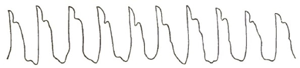
Mitral and Aortic Obstruction and Regurgitation (from a Patient in Bellevue Hospital).
A prediastolic murmur is distinguished from a mitral stenotic murmur by its progressively diminishing intensity from the base to the apex, by its being accompanied by hypertrophy of the left ventricle, and by a jerking, irregular pulse. The preceding tracings explain themselves.
Mitral Regurgitation.
Regurgitation at the mitral orifice is due to a condition of the mitral valves which allows the blood to flow back from the left ventricle into the left auricle. The backward effects of mitral reflux are more varied than those of any other valvular lesion.
It is a common form of valvular disease, and in the majority of cases is the result of acute exudative or interstitial endocarditis.
MORBID ANATOMY.—The most common lesions which give rise to mitral regurgitation are thickening, induration, and shortening of the mitral valves. In rare instances it may occur independent of valvular disease from displacement of one or more of the segments of the valve, the result of changes in the papillary muscles, chordæ tendineæ, or the ventricular walls. It may also occur in extreme anæmia, or from relaxation of the papillary muscles and dilatation of the left ventricle, without a corresponding elongation of the papillary muscles, and from rupture of the chordæ tendineæ. In most instances, however, the valves are shortened, thickened, and indurated.
In some instances lime salts and large masses of chalky matter are found imbedded in the indurated valves. In such cases the surface and edges of the valves are so rough and jagged that more or less obstruction accompanies the regurgitation.
All these changes, except calcification, may also occur in the chordæ tendineæ and columnæ carneæ. The valves may also become adherent to the walls of the ventricles, or as a result of the shrinking and shortening of the chordæ tendineæ the valve-flaps may not pass back to the plane of the orifice.
Again, the valves or the chordæ tendineæ may be ruptured, so that the valves are pressed during the cardiac systole back into the auricle. If the chordæ tendineæ which are inserted nearest the centre of the valve become lengthened, that part of the flap will be bent upon itself, having evidently yielded to the blood-pressure, and this allows of regurgitation. Sometimes, when the valves appear perfectly healthy, by the application of the water test they will be found to be insufficient.
The first effect of mitral regurgitation is dilatation of the left auricle, due to the pressure of the two blood-currents during its diastole—one from the lungs, and the other from the left ventricle. This dilatation leads to thickening and hypertrophy of the left auricular walls. Following this, the pulmonary circulation is impeded, the pulmonary vessels enlarge, and they may undergo degeneration as a result of the continued regurgitant pressure.
Passive congestion of the lungs with brown or pigment induration is an early pathological sequel of mitral regurgitation. The constant interference with the return circulation from the lungs obstructs more or less the outward current of blood to the lungs from the right ventricle. As the obstruction is a gradual one, the right ventricle becomes so hypertrophied as to overcome it. Consequently, the hypertrophied right ventricle compensates at first for the mitral regurgitation, and as long as the right ventricle is able to fully overcome the abnormal pressure of the blood in the lungs from the mitral regurgitation, so long the patients are comfortable. Sooner or later, however, the compensatory hypertrophy of the right ventricle ceases, and a secondary dilatation occurs which admits of no compensation.
This final dilatation of the right ventricle is favored by the myocardial degeneration, which occurs as a result of defective nutrition of the heart-walls; when this condition is reached the veins throughout the body are placed in a similar condition to those in the lungs.
This general venous congestion is indicated by passive hyperæmia of the abdominal viscera and by cyanosis of the surface during active physical exercise.
The liver is the organ first affected, on account of its great vascularity and from the fact that the hepatic veins do not collapse readily and possess no valves. Thus the liver becomes enlarged and stony (the nutmeg liver) as a result of the obstruction to the emptying of the hepatic vein, and when there is coexistent obstruction of the bile-ducts jaundice will be present.
This portal obstruction induces passive hyperæmia of the intestines and stomach, enlargement of the spleen, and large and painful hemorrhoidal tumors. The impediment to the return of blood from the brain causes cerebral congestion; from the kidney, renal congestion; and, finally, the obstruction to the systemic venous return leads to the accumulation of fluid in the areolar tissue and in the cavities. This dropsy generally begins in the feet and extends upward. In females the obstruction in the vena cava inferior induces derangements of the menstrual functions. Ascites, hydrothorax, hydro-pericardium, and pulmonary oedema may subsequently develop.
In addition to these changes, the dilated and hypertrophied left auricle throws an abnormal quantity of blood with abnormal force into the left ventricle during its diastole, which leads to dilatation of its cavity and necessitates a compensatory hypertrophy of the left ventricular walls. This hypertrophy of the left ventricle increases the force of the reflux current, so that during excitement and active physical exertion pulmonary congestion, oedema, and cerebral apoplexy are liable to occur. In many cases of mitral regurgitation, when the venous engorgement is excessive, general dropsy is favored by the anæmia produced by the obstruction of the thoracic duct.
Friedreich maintains that the augmented tension in the venous system causes an increased resistance in the systemic arteries, which leads to left ventricular hypertrophy.
ETIOLOGY.—Mitral regurgitation may occur at any age; it is especially liable to follow rheumatic endocarditis in the young.
Acute exudative and interstitial endocarditis of rheumatic origin is the primary cause of most of the changes which lead to mitral insufficiency. These changes cause the extensive retractions and thickenings which are present in most cases.
It may occur in conditions of extreme anæmia or where there is degeneration of the walls of the left ventricle.
It is not infrequently secondary to changes at the aortic orifice, produced either by an extension of endocarditis from the aortic to the mitral valves and their appendages, or by the secondary mitral valvulitis excited by regurgitant blood-currents from the aorta.
Mitral insufficiency may also be the result of the enlargement of the left auriculo-ventricular orifice which accompanies excessive dilatation of the left ventricle.
Disease of the columnæ carneæ and chordæ tendineæ, when their structures are so weakened as to allow the flaps of the valve to pass back of the plane of the orifice, will also cause mitral insufficiency.
Ulcerative endocarditis may cause it, either by perforation and rupture of the valves or by rupture of the chordæ tendineæ.
SYMPTOMS.—During the early stage of mitral insufficiency, when the hypertrophy of the right ventricle compensates for the regurgitation, there are no rational symptoms which would lead one to suspect its existence; but when the right ventricle is unable to overcome the obstruction to the pulmonary circulation caused by the regurgitant blood-current, there will be more or less dyspnoea, accompanied by a short, hacking cough, with an abundant expectoration of frothy serum. Sometimes the watery expectoration is blood-stained.
Frequently, the blood-stained expectoration is accompanied by free hæmoptysis, although it should be remembered that profuse hæmoptysis is far more frequent with stenosis than with regurgitation at the mitral orifice. But a cough and watery expectoration with occasional dark blood-stains are usually present as an advanced symptom of mitral regurgitation. Active physical exertion increases the dyspnoea and causes cardiac palpitation.
In advanced cases the extremities, face, and lips become blue, the result of the interference with the capillary circulation, and the liver becomes enlarged and hardened—conditions easily recognized by palpation and percussion.
The patient will complain of a sense of weight and fulness in the right hypochondrium, and there will be anorexia, nausea, and a sense of oppression in the epigastrium. Sometimes the hepatic circulation becomes so obstructed that the biliary secretion is interfered with, and jaundice will be added to the cyanotic discoloration, which gives to the surface a peculiar greenish hue.
Following the hepatic derangement are frequent attacks of gastric and intestinal catarrh and evidences of embarrassed renal circulation.
The urine is diminished in quantity, high-colored, and loaded with lithates. Sometimes albumen and fibrinous or blood casts are found in it.
Headache, dizziness, vertigo, stupor, somnolence, and sometimes a peculiar form of delirium of short duration, result from the passive cerebral hyperæmia induced by obstruction in the superior vena cava.
A late symptom of mitral regurgitation is dropsy, which results both from impaired general nutrition and the abnormal blood-pressure in the venous system, both together causing an exudation of the watery portion of the blood through the walls of the vessels. Dropsy, from mechanical causes having their seat in the heart, first appears in the lower extremities, the ankles becoming oedematous, and thence may extend over the whole body. For this condition to be reached it may require several years or only a few months, depending upon the general condition of the patient and the amount of the reflux. With the general anasarca the dyspnoea becomes extreme; the serous cavities of the body as well as the lungs become oedematous; erythema may occur in the region of the groins, the skin exhibiting a tendency to diffuse gangrene.
Late in the disease pulmonary hemorrhagic infarction may occur as a result of metastasis, and this, in the vast majority of cases, lights up a rapidly fatal pneumonia.
All these changes, however grave and urgent they may be, are gradual in their development, so that the condition of the patient is not so insufferable as its description would lead one to suppose.
The pulse of mitral regurgitation is at first in no respect characteristic. It remains regular in force and rhythm, but later it becomes somewhat diminished in force and volume, irregular in its rhythm, and increased in frequency, but never jerking in character. This tracing illustrates my meaning. While it remains full it is feeble and always compressible. When the heart's action is excited, it has a certain tremulousness: these last-named characteristics are to be regarded more as the result of the failure of the left ventricle than of changes in the valvular insufficiency. If a mitral regurgitant pulse has any distinctive peculiarity, it is its diminution in volume.
FIG. 46.

Mitral Regurgitation (from a Patient in Bellevue Hospital).
Coincident mitral or aortic stenosis may render the pulse regular even in extensive mitral regurgitation.
FIG. 47.

Mitral and Aortic Regurgitation (from a Patient in Bellevue Hospital).
Physical Signs.—Inspection.—The area of visible cardiac impulse extends over an abnormal space, and is more or less distinct according as the right ventricular hypertrophy is moderate or extensive. Sometimes the thoracic wall is seen to rise and fall with each cardiac cycle, and not infrequently the epigastrium exhibits slight pulsation corresponding in rhythm with the heart-beats.
The epigastric pulsation is due to the right ventricular hypertrophy always found with extensive mitral regurgitation.
Skoda, Bamberger, and Leyden record a few instances in which inspection revealed a double impulse accompanying, with more or less regularity, each cardiac systole. This double impulse only occurs in aggravated cases of mitral insufficiency, and arises from non-coincidence of contraction of the two ventricles.
The jugular veins appear swollen, and this is always most conspicuous when the patient is lying down.
Palpation.—The apex-beat is displaced to the left. When hypertrophy predominates over dilatation, the apex-beat is felt lower than normal. When the dilatation exceeds the hypertrophy, the apex-beat is carried outward and often slightly upward. The impulse is diffused and more or less forcible according as the right or left ventricular hypertrophy predominates. This systolic frémissement is most noticeable when the base of the heart lies close to the chest-wall from retraction of the margin of the left lung.
Purring tremor, systolic in rhythm, felt most intensely at the apex and becoming feebler the farther the hand is removed from that part, either to the right or upward, is invariably due to mitral reflux.
Hayden says that it is exceptional to have a purring thrill with simple mitral reflux. I have never found it except in those cases where left ventricular dilatation greatly exceeded the hypertrophy.
Percussion.—Percussion reveals an increase in the area of cardiac dulness, especially laterally; it extends both to the left and right of the normal line, as well as downward. The area of superficial as well as deep-seated dulness will be increased laterally and downward.
Auscultation.—Mitral insufficiency is attended by a systolic murmur which either completely or partially replaces the first sound of the heart. The quality of the murmur is variable, and not in itself as distinctive as that of mitral stenosis. It is usually a soft and blowing bellows murmur; sometimes, toward its end, the murmur will assume a distinctly musical character.
While the first sound of the heart may be heard distinctly in the early stages of mitral reflux, later the murmur in nearly all cases takes the place of the heart-sounds. Hence many English writers rightly denominate this murmur as post-systolic rather than systolic in its nascent stages. It is heard with its maximum intensity at the apex-beat. Its area of diffusion is to the left on a line corresponding to the apex-beat. It is audible at or near the inferior angle of the left scapula. It can be heard between the lower border of the fifth and the upper border of the eighth vertebra, at the left of the spine, with nearly the same intensity as at the apex. The murmur may be absent from the latter situation until cardiac hypertrophy is developed.
The second sound of the heart over the pulmonary valves is accentuated, while below the junction of the third rib with the sternum on the left side both heart-sounds are feeble. Skoda first drew attention to exaggeration of the second pulmonary arterial sound as a positive and unerring indication of mitral regurgitation.
An intensified pulmonary second sound requires a strong right ventricle and an intact tricuspid valve, and is not always present. In general terms, the area of diffusion of a mitral regurgitant murmur is toward the left of the apex-beat. Whatever may be its character, the murmur is generally loudest at its commencement. A loud systolic murmur at the apex, and not heard at the back, is probably not produced by mitral reflux.
As at the aortic orifice, so at the mitral, stenosis and regurgitation are apt to occur in the same individual, giving rise to a combined presystolic and systolic murmur, which is a continuous murmur that begins shortly after the second sound of the heart and often continues until the second sound commences. The two sounds, although mingling to form one murmur, can, in the majority of cases, be readily distinguished from each other, for the point of maximum intensity and the very limited area of diffusion of a presystolic murmur readily distinguish it from a mitral systolic which is audible in the left scapular region. It is important to recognize the existence of both these murmurs in estimating the prognosis in any case. Guttman mentions a case where five distinct murmurs were combined and yet clearly distinguishable.
DIFFERENTIAL DIAGNOSIS.—It is usually not difficult to recognize mitral regurgitation. The seat and rhythm of the murmur and its area of diffusion are sufficient to distinguish it from other cardiac murmurs. The character of the pulse, the symptoms referable to the right heart, and the pulmonary complications will also assist in its diagnosis.
It may, however, be mistaken for aortic obstruction, since both give rise to a systolic murmur, for tricuspid regurgitation, for fibroid disease of the heart, and for roughening of the ventricular surface of the mitral valve or of the ventricular wall near the aortic orifice.
The diagnosis between mitral regurgitation and aortic stenosis has already been given (see page 657).
Mitral and tricuspid insufficiency both produce a systolic murmur, but a mitral regurgitant murmur has its maximum of intensity at the apex, and is conveyed toward the left axillary and scapular regions, while the maximum intensity of a tricuspid regurgitant murmur is to the right of the base of the xiphoid cartilage, and it is transmitted upward and to the right: the area of transmission establishes the diagnosis.
Pulmonary symptoms are prominent in mitral reflux, and absent in tricuspid regurgitation. The pulmonary second sound is markedly enfeebled in tricuspid regurgitation, and markedly intensified in mitral regurgitation.
Fibroid disease of the heart may produce a systolic apex murmur, but it is an exceedingly rare disease, a pathological curiosity.20
20 In the Pathological Transactions (1874, vol. xxv. p. 64) Fagge records a few cases, and mentions that perhaps one positive indication of fibroid disease of the heart, rather than of a valvular lesion, may be found in its resisting treatment with greater obstinacy.
Roughening of the ventricular wall gives rise to a murmur which has its maximum intensity at the base of the heart, and is transmitted along the aortic arch and into the vessels which spring from it in the thorax.
The vibration of an irregular chordæ tendineæ stretched across the aortic orifice, its extremities being inserted into opposite walls of the ventricle, may produce a systolic musical murmur, but the line of its transmission will correspond to that of an aortic obstruction. A systolic mitral murmur due to the sudden rupture of one or a number of the valve-flaps, of the papillary muscles or tendons, is accompanied by a loud systolic blowing murmur, which is immediately accompanied by all the urgent symptoms of acute pulmonary congestion.
Pulmonary Obstruction.
On account of the infrequency of disease of the pulmonic valves very little is known of the phenomena to which such diseases may give rise. In fact, they are so rare that there is no written history of their subjective symptoms; their diagnosis is only arrived at by exclusion, and they cannot be recognized except by the physical signs which attend them.
As has been already stated, endocarditis in the right heart is rare, except in intra-uterine life, and the various conditions of the aorta, atheroma, aortitis, etc., which I have mentioned in the etiology of aortic valvular disease have no analogues in the pulmonary vessels.
Usually, valvular disease of the right heart is the sequela of lesions in the left. It must be remembered, however, that the pulmonary artery may become atheromatous. I have already shown (see p. 666) how certain valvular diseases of the left heart may induce such a pathological condition. But even under such conditions disease of the pulmonary valves is rare. Balfour believes that constriction of the pulmonary artery may occur at various periods of intra-uterine life. As a rule, the pulmonary valves are subject to no lesions except congenital malformation.
MORBID ANATOMY.—Bertin records an instance of pulmonary obstruction where the valves, distorted and adherent, formed a horizontal septum across the orifice, it being barely one-fourth of an inch.
A rigid tricuspid valve has been found to be the cause of obstruction at the pulmonary orifice, the pulmonary valves themselves being normal. A few autopsies have revealed obstructions at the pulmonary artery, caused not so much by valvular defect as by aneurisms, tumors of the pericardium or of the anterior mediastinum, enlarged bronchial glands, or pressure of a solidified lung.
The pulmonary artery may be occluded just beyond the valves by a cancerous tumor, and there are examples where a phthisical process in the left lung has induced it.
A murmur indicative of pulmonary obstruction may be produced by a cardiac thrombosis.
I have placed these statements under the head of its morbid anatomy for the reason that they cannot be appreciated and their pathological significance realized during life.
Reasoning from analogy, obstruction at the pulmonary orifice ought to be followed by compensatory hypertrophy of the right ventricle and accompanied by tricuspid regurgitation and dilatation of the right auricle.
Ormerod records 3 cases21 where pulmonary obstruction was diagnosticated during life, and where the post-mortem proved the accuracy of the diagnosis: 2 of these cases occurred in men under twenty-eight, and the other in a woman of twenty-one. In 2 of these cases all the other cardiac valves were healthy. The pulmonic orifice would barely admit the introduction of a goosequill. Warburton Bigbie mentions a case (man æt. eighteen) where reflux and stenosis at the pulmonary orifice coexisted. There were four valves, and these were incompetent. All the other valves were normal.
21 Edin. Med. and Surg. Journ.
Congenital stenosis of the infundibulum of the right ventricle is the probable result of foetal myocarditis or of syphilis.
I have never met but two pulmonic obstructive murmurs where subsequent autopsies were obtained. In both cases it was found that the murmur had been produced by mediastinal tumors pressing on the pulmonic artery so as to diminish the calibre.
ETIOLOGY.—Pulmonary stenosis is rarely the result of endocarditis or of degenerative changes in the pulmonary artery. Bertin states that when abnormal communication between the two sides of the heart has existed, the arterial blood has excited endocarditis in the right heart.
Syphilis has been advanced as a possible cause of degenerations at the pulmonic orifice.
SYMPTOMS.—The only rational symptoms that have been noted in the few recorded cases of pulmonic disease admit of manifold explanations, and no one is either constant or diagnostic. In some cases anæmia existed, in others there were cardiac palpitation, dyspnoea, cyanosis, and dropsy; but none of these belong exclusively to a pulmonic lesion nor do they necessarily depend upon it.
Physical Signs.—Inspection, palpation, and percussion give negative rather than positive results. In a few instances palpation may give a systolic thrill confined to the second left intercostal articulation. Such a frémissement results both from roughness and contraction of the pulmonic orifice.
Auscultation.—A systolic murmur is heard with its maximum intensity directly over the pulmonic valves; it is very superficial, and consequently very distinct, and it is limited in its diffusion. It is never heard at the xiphoid cartilage nor along the course of the aorta. If it has an area of diffusion, it is toward the left shoulder. The murmur is loud and soft in character, sometimes bellows. It is not audible in the vessels of the neck nor is it attended by arterial pulsation.
When phthisical consolidation partially occludes the pulmonary artery, a loud but soft systolic murmur is heard, which is sometimes high-pitched and musical, and often entirely suspended during a full inspiration. In some few instances there is a bruit de diable in the jugular veins.
DIFFERENTIAL DIAGNOSIS.—It is possible to confound a pulmonic obstructive murmur with a mitral regurgitation which is propagated upward into the left auricular appendix. But the area of a mitral regurgitant is also backward, and by this it could be distinguished from a pulmonic obstruction. Besides, in mitral disease the pulse is very different from the pulse of pulmonary stenosis.
Aortic stenosis can hardly be mistaken for pulmonary obstruction, for the arterial pulsation, the peculiar pulse, and the transmission of the murmur into the arteries of the neck will suffice to discriminate between them.
An aneurism at the sinus of Valsalva may produce a systolic pulmonary murmur by the pressure which it produces upon the pulmonary artery. It would be impossible to distinguish it from a pulmonic stenosis.
The diagnosis of pulmonary obstruction is usually reached only by exclusion.
Pulmonary Regurgitation.
This form of valvular lesion is exceedingly rare; indeed, many doubt its occurrence. The lesion seldom occurs except as the result of injury or congenital defect, and there are but few well-authenticated cases in medical literature.22
22 Path. Trans., vol. xvi. p. 74.
The statement23 that the pulmonary valves exhibit a cribriform condition nearly as often as the aortic is not sustained in this country by the results of post-mortems. In one of the cases to which I have referred (p. 675) as an example of pulmonary stenosis the valves were likewise found insufficient. In Bigbie's case (referred to on p. 675), where there were four flaps to the valve (producing obstruction), there was marked insufficiency coexisting.
23 Dis. of the Heart, Bellingham.
The morbid anatomy, etiology, and rational symptoms do not require a separate consideration. The anatomical appearances are the same as those found in similar conditions of the aortic valves, and the etiology and rational symptoms are the same as those of pulmonic stenosis.
Physical Signs.—Theoretically, pulmonic regurgitation should be accompanied by a diastolic murmur having its maximum intensity over the pulmonic valves, and its area of diffusion should be downward and toward the xiphoid cartilage. It should be soft and blowing in character. This murmur is rarely heard alone: it is usually associated with obstruction at the same orifice or with some murmur whose origin is on the left side of the heart.
Niemeyer states that dyspnoea, hemorrhagic infarction, and consumption of the lungs have followed insufficiency at the pulmonary orifice. No other authority mentions any such symptoms, while the assignment of valvular disease as a cause of phthisis is not based upon clinical facts.
With a pulmonic regurgitant murmur there should be on palpation and percussion physical evidences of hypertrophy and dilatation of the right heart, the rationale of whose production would be identical with that which was considered in aortic regurgitation. I have never heard a regurgitant pulmonic murmur.
DIFFERENTIAL DIAGNOSIS.—The murmur of pulmonary regurgitation may be mistaken for that of aortic regurgitation. The points in connection with their differentiation are fully discussed on p. 664.
The PROGNOSIS and TREATMENT are identical with those of the former lesion.
Tricuspid Stenosis.
This valvular lesion is so rare that there are no established rules for its diagnosis.
Its MORBID APPEARANCES and ETIOLOGY are the same as those of pulmonic stenosis.
The SYMPTOMS of tricuspid stenosis would be those due to obstruction to the entire venous circulation. The right auricle would be dilated, and there would be visceral enlargements in the abdomen, cyanosis of the face and extremities, scanty and albuminous urine, hemorrhoidal tumors, headache, dizziness and vertigo due to passive cerebral hyperæmia, and finally general anasarca. The few recorded cases were associated with mitral stenosis with one exception, a case of Bertin's.24
24 Traité des Maladies du Coeur, Obs. 17.
In a case exhibited by Quain the tricuspid flaps, thick and opaque, were united for one-third of their extent. In the other cases the valve-flaps formed a diaphragm whose central opening varied in size, admitting only the point of one finger. In every condition of tricuspid stenosis the heart was enlarged.
Tricuspid stenosis (as in pulmonic stenosis) may be the result of pressure of tumors.
In all well-authenticated cases the chief symptoms seem to be extreme lividity, palpitation, and dyspnoea.
Physical Signs.—Inspection reveals general cyanosis. The jugulars are turgescent and exhibit presystolic pulsation. This pulsation is sometimes the only inconvenience the patient suffers.
Palpation may discover a venous thrill at the base of the neck.
Percussion may show the right auricle to be greatly enlarged, and cardiac dulness will be increased laterally and toward the right.
Auscultation.—Tricuspid stenosis should be attended by a presystolic murmur whose maximum intensity would be at the lower portion of the sternum just above the xiphoid cartilage. This murmur may be propagated faintly toward the base, but never toward the apex of the heart. It is sometimes accompanied by fremitus.
Hayden offers the following diagnostic point: The murmur of mitral stenosis (without which tricuspid stenosis never occurs) is limited to the apex region; a murmur of the same rhythm is produced at the sternum by tricuspid stenosis, "and between these two localities there is a point where no murmur can be heard."
It is unnecessary to consider its differential diagnosis.
The lesion would be diagnosticated (if at all) by exclusion, and prognosis and treatment would depend on the gravity and sequelæ of the accompanying condition—viz. Mitral Stenosis (q. v.), for the rule is, that stenosis of the tricuspid never occurs unless there is extensive mitral obstruction, and the latter condition is always the predominant one.
Tricuspid Regurgitation.
Regurgitation at the tricuspid orifice is generally secondary to mitral stenosis or regurgitation; primary disease of the tricuspid valves, however, is not infrequent.
MORBID ANATOMY.—The valvular lesions which lead to tricuspid insufficiency are similar to those which produce mitral insufficiency. The valves are thickened, shrunken, and opaque, the papillary muscles are shortened, thickened, and the chordæ tendineæ undergo similar changes and are sometimes adherent.
The valves or the chordæ tendineæ and columnæ carneæ may rupture; in either case acute and extensive insufficiency results, as has been stated. Acute endocarditis of the right heart is rare in adult life, but when it occurs the tricuspid valves are its primary and principal seat.
The reason for this is found in their anatomical structure and in the tension to which they are subject in diseases of the mitral valves. They are rarely the seat of rheumatic endocarditis or calcareous degenerations.
Ulcerative endocarditis is seldom met with in the right heart. In a case recorded by Charcot and Vulpian one of the tricuspid valves was softened and perforated, presenting numerous vegetations. Scattered abscesses in the lungs were found in this case.
Any infection through emboli from the tricuspid flaps will produce secondary effects within the thoracic cavity. The first effect of tricuspid regurgitation is dilatation of the right auricle; following this there will be more or less hypertrophy of its walls. As soon as the valves in the subclavian and jugular veins are no longer able to resist the regurgitant current jugular pulsation follows. But before this occurs the tributaries of the inferior cava and the organs to which they are distributed will become greatly engorged, for they have no valves to resist the regurgitant current, as are found in the veins coming from the upper part of the body. The inferior cava and the hepatic veins sometimes become enormously distended under these circumstances, and the liver will show the peculiar section that has gained for it the name of nutmeg liver.
Following the hepatic changes, the skin assumes a dingy yellow hue. When this is combined with cyanosis it produces a peculiar greenish tint which is only met with in heart disease. The spleen enlarges and hardens; the mucous membrane of the stomach is congested, ecchymotic, and often presents numerous hemorrhagic erosions. Intestinal catarrh is subsequently developed, and the general venous congestion within the abdominal cavity is exhibited by hemorrhoids and ascites. The kidneys become congested and stony, and thrombi may form in the femoral vein and induce subsequent pulmonary infarctions.
The stasis in the veins below the diaphragm is accompanied by transudation of serum—first in the ankles, and thence the dropsy progresses upward until the patient may finally reach a condition of general anasarca. The obstruction to the general systemic circulation which results may subject the left ventricle to so much extra labor that it hypertrophies, and then we have the infrequent occurrence of disease of the left heart following that of the right.
Since tricuspid reflux has mitral disease for its principal cause in abnormal cases, the heart becomes greatly enlarged and a condition of extreme cardiac dilatation and hypertrophy is reached.
ETIOLOGY.—As has been stated, the most frequent cause of tricuspid regurgitation is mitral disease, either stenosis or regurgitation. Any condition of the lungs which will produce hypertrophy and dilatation of the right ventricle will lead to it; it is met with in extensive pulmonary emphysema, in cirrhosis of the lung, and in extensive chronic bronchitis. Balfour regards chronic bronchitis as its most frequent cause after mitral stenosis.
It is possible for any valvular disease in the left heart, when of long duration, to lead to tricuspid regurgitation. From all these causes the rationale is the same: the abnormal amount of blood in the right ventricle presses with undue force against a valve, which physiologists regard as normally slightly insufficient; the stress upon the valve-flaps and the valvular attachments is such that endocardial inflammation is excited at the part subject to the greatest strain, and valvular insufficiency is the result.
It is possible for disease of the tricuspid valves to result from any of the causes which have been enumerated on p. 666 as etiological factors in valvular diseases.
SYMPTOMS.—Tricuspid regurgitation being in the majority of cases secondary to some other valvular disease or some chronic pulmonary affection, its symptoms during the early stages are vague and masked by those of the primary disease. But as soon as the valves become so insufficient that the venous return is markedly impeded, a train of symptoms is developed which has its origin in the visceral derangements already referred to.
In addition to these symptoms there may be, with extensive tricuspid regurgitation, cardiac palpitation, cardiac dyspnoea, and marked irregularity in the force and rhythm of the heart. The liver and spleen are enlarged, the skin becomes dingy, and there is obstinate constipation with hemorrhoids. The liver is likewise rendered very liable under such circumstances to attacks of interstitial hepatitis. Venous stasis is evinced by dyspepsia, nausea, vomiting, and hæmatemesis. The secretion of the kidneys is scanty, dark-colored, of high specific gravity, often containing albumen and casts.
Passive cerebral hyperæmia is marked by headache, dizziness, vertigo, and muscæ volitantes, and there is a peculiar mental disturbance which is not met with in any other form of heart disease.
Late in the disease, if the patient is placed in a horizontal position, the face becomes turgid and blue, and if he remain long in the recumbent position stupor and coma may supervene. Jugular and epigastric pulsation are characteristic physical signs.
A very late symptom is dropsy, which begins at the ankles and extends upward until there is general anasarca. It is a point to be noticed that in the dropsy from tricuspid reflux the genital organs suffer slightly if at all.
FIG. 48.

Tricuspid Regurgitation (after Galabin): a, a, anadicrotic wave synchronous with the auricular systole, and caused by reflux into the large veins.
Physical Signs.—Inspection.—In extensive tricuspid disease the area of the cardiac impulse is increased more than in any other valvular lesion. This area sometimes extends from the nipple to the xiphoid cartilage, and it may reach as high as the second right intercostal space. There is a visible impulse in the jugular veins, more apparent in the right than in the left. Sometimes the veins in the face, arms, and hands, or even the thyroid and mammary veins, are seen to pulsate.
Palpation.—The apex-beat is indistinct, except in cases where there is marked hypertrophy of the left ventricle. Pulsation occurs in the epigastrium, which may be due to reflux into the enlarged hepatic veins or to the fact that the dilated and hypertrophied right ventricle so presses on the liver that the impulse is conveyed through the diaphragm with each cardiac pulsation. Guttman thinks epigastric pulsation is due wholly to reflux into the veins of the liver, and not to right ventricular pulsation.
Early in the disease the impulse in the jugulars is confined to the lower part of the vessels, particularly to the sinus. Beyond this point the vein merely undulates. Later, a systolic pulsation is felt as high up as the angle of the jaw, and may be accompanied by distinct though feeble presystolic pulsation.
The liver may first simply undergo systolic depression, chiefly at the left lobe; secondly, the whole liver may have an impulse coming from an enormously dilated vena cava; and thirdly, the systolic pulsation of the veins within the organ may give to it a palpable expanso-pulsatory movement. The hepatic pulsation is rhythmical with the cardiac impulse. In rare cases it precedes jugular pulsation. Sometimes pulsation is felt in the femoral veins.
Sphygmographic tracings of the jugular pulse show it to be dicrotic.
Percussion shows an increase in the area of cardiac dulness to the right and upward, sometimes as far as the second intercostal space.
Auscultation.—The murmur of tricuspid insufficiency is heard with, or takes the place of, the first sound of the heart; it is superficial, of low pitch, blowing, soft, and faint, and is heard with the greatest intensity over the lower part of the sternum, at its left border, between the fourth and sixth ribs. It is rarely audible above the third rib or to the left of the apex-beat. This murmur is transmitted from the region at the base of the xiphoid cartilage upward and to the right from one to two inches. Sometimes it is heard only over a very limited area, and then it may be overlooked.
DIFFERENTIAL DIAGNOSIS.—A tricuspid regurgitant murmur may be confounded with that due to aortic obstruction, pulmonic obstruction, and mitral regurgitation. A tricuspid regurgitant murmur is never audible above the third rib; is not accompanied by an accentuation of the second sound over the pulmonary artery, but by jugular and epigastric pulsation; and is heard with maximum intensity near the base of the ensiform cartilage. These points are sufficient to differentiate it from an aortic or pulmonary obstructive murmur. The differential diagnosis between it and a mitral regurgitant murmur has been given.
PROGNOSIS IN VALVULAR DISEASES OF THE HEART.—Any statements as to the duration of life in valvular diseases of the heart, and their relative frequency as a cause of death (especially of sudden death), must be based upon personal observation, and necessarily will differ with different observers.
In order to establish, if possible, a basis of comparison for the different valvular lesions, I give a résumé which I have made of 81 cases, in all of which autopsies were made and the diagnosis of valvular disease verified.25
25 Med. Rec. N.Y., April 1, 1870, p. 66 et seq.
In 14 cases of various valvular lesions, each of which was accompanied by cardiac hypertrophy and dilatation, 50 per cent. of the deaths were due directly to the valvular lesion. In 1 of these, where there was stenosis at both auriculo-ventricular orifices, death was sudden.
In 15 cases of valvular disease, in which there was only cardiac hypertrophy, there were 11 deaths from the heart lesion. In 5 of these death occurred suddenly, and these 5 sudden deaths were all directly due to the heart lesion.
In 6 cases of valvular disease accompanied by dilatation alone, 4 deaths resulted directly from the heart lesion, and 2 of these were sudden.
In 15 cases where the aortic valves were involved (either calcified, rigid, or atheromatous) the heart lesion was not the cause of death in any case. Of these 15 cases, sudden death occurred but in 2; in 1 there were firm and long-standing pericardial adhesions, and in the other cerebral apoplexy.
In 12 cases of calcification of the mitral valve, no death occurred as the direct result of the valvular lesion, and there were only 2 sudden deaths, both from cerebral apoplexy.
The aortic and mitral valves were diseased in 14 cases; in 2 of these only did death result from the heart lesion, and the only three sudden deaths in this class were from uræmia, apoplexy, and croupous laryngitis.
The aortic and pulmonic valves were both diseased in 3 cases which died suddenly, and in no instance was death due directly to the heart lesions.
In 2 cases there was disease at the aortic, mitral, and tricuspid orifices, and no sudden death.
Thus it will be seen that of these 81 cases, in 24 only was death due directly to the heart lesion. There were only 8 sudden deaths due directly to the heart lesion.
The results of personal, clinical, and pathological observation lead me to the opinion that the loudness, harshness, and the area of diffusion of any cardiac murmur have little to do with its prognosis.
I deduce from the above-mentioned cases that cardiac murmurs rarely necessitate a bad prognosis unless hypertrophy and dilatation coexist; but so soon as the signs of considerable dilatation and hypertrophy are present a great variety of complications are liable to occur.
In 1870, I had a patient sixty years of age with extensive aortic reflux, who had been under my observation eight years, during which time he had three attacks of pneumonia. There were no appreciable signs of cardiac dilatation in his case.