Volume 3 — DISEASES OF THE BLOOD, AND OF THE HÆMATOPOIETIC SYSTEM. (continued 11)
Reading edition
8 Reigel, Virchow's Arch., vol. xlix.
9 Langenbeck, Klinic, p. 157.
PATHOLOGY AND MORBID ANATOMY.—Pulmonary processes associated with bronchial catarrh frequently lead to enlargement of the bronchial glands, because, owing to the impervious character of the basement membrane of the bronchial passages, the mucous and epithelial portion of the exudation is expectorated, and that portion of the exudate which occurs from the bronchial blood-vessels is absorbed and carried by means of the pulmonary lymphatics to the bronchial glands. Tubercular deposits frequently occur in the glands of the posterior, and much less frequently in those of the anterior, mediastinum.
Independently of the above conditions, caseating bronchial glands have been found as complications of scarlatina with nephritis or tubercular meningitis. An interesting case of this condition has been reported as following an abscess in the glands at the root of the neck as a sequel to measles nine months before.10 Riegel also mentions an instance in which some of the mediastinal glands were enlarged to the size of hen's eggs. The trachea was compressed at the point of bifurcation, so that its calibre was reduced to one-third its natural size. This case was free from other glandular enlargements. Coupland has described a case in a boy four years of age, in whom the cervical glands were enlarged and idiopathic hyperplasia of the bronchial glands was suspected. Autopsy: On raising the sternum a collection of indurated glands was found in the anterior mediastinum, and over the root of the right lung one of these glands had broken down into a cheesy mass. A chain of enlarged lymphatics accompanied the right bronchus. The largest caseous mass had ulcerated through the trachea just above the origin of the right bronchus by an aperture measuring half an inch along the axis of the tube, while for half an inch above its lumen was compressed. In this case the right lung was solidified and contained cheesy matter, with a cavity at the apex. The father of the child had also suffered from increase in the glandular tissues.
10 See Path. Soc. London, 1884.
The historical literature of intra-thoracic morbid growths has been exhaustively reviewed in a monograph by Cockle, but until within the last fifteen years little attempt was made to separate mediastinal tumors into definite groups.
Our present knowledge on this subject was first shaped by Virchow,11 since which period numerous cases have been recorded.
11 Virchow's Archiv, Bd. xciii. Heft 3.
Sarcoma of the Mediastinum.—Primary sarcomatous growths are relatively uncommon. In 7566 cadavers examined at the Marine Hospital at Kronstadt there were found 158 malignant tumors, 127 being carcinomatous, the other 31 being sarcomatous. In 24 cases reported by Kahnlich, 13 occurred in the anterior mediastinal region, and a similar location was found in a case reported by the writer,12 also in one instance reported by West.13
12 Philada. Med. News, March 15, 1884.
13 Path. Soc. London, 1883.
The anterior mediastinal space is a favorite location for the origin of the purely sarcomatous form of tumor. Sarcoma may arise from a persistent thymus (as in cases reported by Gee, Church, and Powell), from the parietal or visceral layers of the pericardium or pleura, from the periosteum of the sternum, or from the mediastinal connective tissue.14
14 Kahnlich, loc. cit., describes 13 as originating in the connective tissue of the anterior mediastinum, 5 in the periosteum of the sternum, and 1 in the pericardial substance.
FIG. 53.
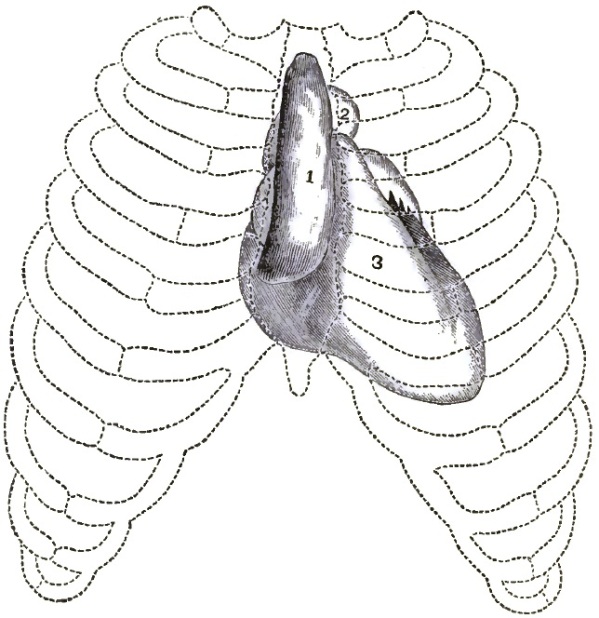
1, tumor; 2, aorta; 3, right ventricle of heart.
In a disease of this rare nature we can best formulate an idea of the character of the growths by the recital of a few typical cases. In an autopsy made by the writer, on removing the sternum and cartilages they were found to be adherent on the right side to a mass which occupied the anterior mediastinum (see Fig. 53). The growth was seven inches long, measuring from the sternal notch, and terminated in a somewhat diffused thickening of the visceral pleura, which covered the anterior margin of the upper and middle lobe of the right lung. The growth was two and a half inches broad. It overlaid the aorta, pulmonary artery, and the vessels of the neck. The calibre of the trachea was slightly diminished. The glands of the neck were unaffected on either side. The posterior mediastinal glands were very slightly enlarged along the sides of the trachea and upper bronchi. Laterally, at the lower portion of the growth, the pulmonary pleura was thickened at the line of contact with the tumor, but the lungs were free from any traces of disease. The new formation was of fibrous consistence, of a gray-white color, and through its centre a softened tissue was found. Microscopic examination showed the growth to be composed of medium-sized lymphoid cells mixed with spindle-shaped cells, and imbedded in a homogeneous stroma or a stroma which consisted of reticulated fibres and wavy fibrous tissue. Other portions of the body were normal.
In West's case the tumor also occupied the anterior mediastinum, extending toward the second left intercostal space. The mass was about the size of a boy's head, soft, cellular, and adherent to the upper lobe of the left lung; it also rose into the episternal notch and left supra-clavicular fossa. The brachial plexus and vessels of the left side, subclavian and carotid arteries, the jugular and innominate veins, were imbedded in the tumor. The left bronchus and a portion of the trachea were flattened. The left phrenic and left pneumogastric nerves passed through the mass, and on dissection were found much thickened as they ran through the tumor. The tenth nerve measured three times its normal diameter, and was pushed out of its course nearly an inch from the carotid. The recurrent laryngeal was also thickened; the right pneumogastric and phrenic nerves were not involved. The heart lay beneath the tumor; nodules of the new growth were found upon the anterior surface of the heart and along the vessels issuing from it. No secondary deposit was found in the lungs except at the margin of the left upper lobe, into which the tumor spread directly. The spleen, liver, kidneys, and lumbar glands were normal.
Microscopic examination determined the growth to be a round-celled sarcoma, the thickening of the nerves being due to infiltration by similar small-celled growth.
In primary sarcoma of the mediastinum—and the same is true of lymphadenoma—the invasion of the various intra-thoracic organs is chiefly by continuity or direct spreading of the growth. The lymphatics of the neck are very rarely implicated in this form of malignant disease; and while in lympho-sarcoma the glands may be involved, they are not so frequently as in cancerous processes. Sarcomata of the mediastinum with implication of the lungs and pleura are more frequently secondary processes; indeed, the lungs would seem never to be the seat of primary sarcoma. The pleural tissues, however, may be primarily involved. Lepine, Birch-Hirschfeld, Böhme, Eppinger, Schultz, Greenish, and others have reported cases in which the growths were abundantly distributed in the pleural tissues as primary formations. The point of origin is believed to be either directly from the ordinary connective-tissue cells or from the endothelium of the lymphatics.
Secondary sarcomata may form in the mediastinum or in the lungs within a month or so long as a year after the removal of tumors from other parts of the body, probably by metastasis prior to the removal. In some of these cases the seat of original growth and the neighboring glands may be entirely healthy.
In a typical case of multiple osteoid sarcoma of the lung reported by West fleshy vegetations were found on the visceral pleura: upon the parietal pleura, over the seventh rib, two inches from the spine and growing from it, was a lobular spongy mass as large as an orange, but perfectly disconnected with the parts beneath. The right lung was irregular in shape, owing to the presence of masses of new growth in its different parts. The middle lobe seemed almost completely converted into the new growth. Between the lower lobe and the diaphragm, but attached to the lung, was a mass the size of a cricket-ball, covered with a dark, laminated, but easily separated coagulum. The tumor occupied the upper lobe of the left lung, forming an irregular oval mass six by four and a half inches. It was white in color, and adherent to its upper border was compressed lung-tissue. There were also four or five independent nodules situated near the surface, and of a white color. The lower lobe contained one medium-sized growth and four or five small ones. The bronchial glands were not involved. The tumors appeared soft and spongy, but on incision they were found so hard that a knife could scarcely divide them.
Frequently, the lungs are found infiltrated with sarcomatous nodules of a soft consistency, varying in size from a walnut to an orange. To sum up: primary sarcomata may be the round- or spindle-celled variety; but myeloid sarcomata also occur, chiefly as secondary growths. (See Fig. 54.)
FIG. 54.
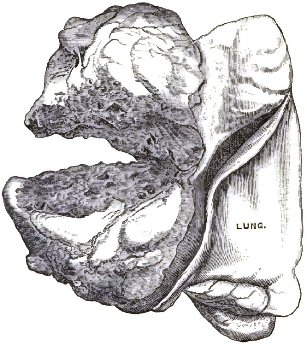
From photograph of a case of Professor Osler's, showing secondary myeloid sarcoma of mediastinum—appearing six months after removal of myeloid sarcoma of radius. The tumor figured in the plate occupied the front of the thorax lying beneath the sternum and the cartilages and ribs of the left side, pushing back and completely covering the pericardium. It was loosely adherent to the ribs and sternum, and appeared to grow from the pulmonary pleura, to which it was attached in a large part of its extent, and only had penetrated the lung at one spot on the anterior border of upper lobe. Right lung contained secondary masses, chiefly in the pleura.
Lympho-sarcoma of the Mediastinum.—Lympho-sarcoma, lymphoma, or lymphadenoma is the form of malignant process which probably includes the majority of cases of primary mediastinal growth. It is sometimes, however, a part of a more general disease, affecting more or less the whole glandular system.
Murchison15 classified the first case of this disease involving the intestines, liver, mesentery, and heart. The same observer the following year described a case in which the glands of the neck, mediastinum, axillæ, and spleen were involved. Wunderlich has recorded a case of malignant mediastinal disease which commenced in the glands of the neck; but the cervical glands may be enormously enlarged without implication of the bronchial.
15 Path. Soc. Trans., London, vols. xx. and xxi., together with a summary of the literature of the subject.
The general disease dates back to the time of Hodgkin, Bright, and Wilkes, and was then known as anæmia lymphatica. It has been specifically described by Virchow,16 Cornil, and Ranvier as independent of leukæmia, and was designated lymphadénie. It was noticed by Trousseau under the title of adénie, and Ogle and numerous clinical observers since have also recorded cases.
16 Die Krankhaften Geschwülste, Band ii. p. 376.
As a mediastinal growth the characteristics of lympho-sarcoma can be made more vivid by the reproduction of one of the first cases recorded of this disease. On removing the sternum and cartilages they were found adherent to a mass occupying the anterior mediastinum. The morbid growth reached backward to the trachea, surrounding it with a thickness posteriorly of a quarter of an inch; it extended downward to the bifurcation of the trachea, and, involving the superior prolongation of the pericardium, invaded and greatly thickened the parietal part of that membrane, covering the heart at its upper half. The diseased structure reached upward to the root of the neck, involving the anterior mediastinal glands, and surrounded the trachea by a thin layer as high as the thyroid cartilage. Some of the glands on each side of the neck were affected as high as the angle of the jaw. Laterally, the morbid growth extended on each side to the line of junction of the cartilages with their ribs, displacing the anterior margin of the lung. The pulmonary pleura was involved and thickened at the line of contact, and the right lung at the upper part of its anterior margin was invaded from the pleura by white, fibrous-looking branched bands. At the lower part of the anterior lobe the lung was also invaded from the pericardium. Some of the glands at the root of the lung were involved by extension, but they were not generally affected, nor was the lung invaded except to the limited extent above mentioned. The heart and pericardium were free from disease.17
17 Powell, Path. Trans., vol. xxi., London.
The malignant growths of the mediastinal region implicate the surrounding structures so rapidly that it is, as a rule, quite impossible to determine, even after death, the starting-point of the disease; and while lymphadenoma can originate in the same tissues as the other forms of sarcoma already alluded to, yet it in most cases probably originates in the lymphatics of the anterior or posterior mediastinum.
In reference to the location of this form of morbid growth, we find, on consulting a series of cases reported by Fenwick, Eve, Payne, Peacock, Powell, Murchison, Bennett, Dickinson, that the region for principal development seems to be the anterior mediastinal space, although in several instances the posterior mediastinal region was also involved.
The characteristics of the growth of lymphadenoma are the involvement by continuity of all adjacent tissues, thus affording a contrast to secondary sarcomata. The glands of the neck are sometimes invaded, but are unaffected in a considerable proportion of cases. The lungs may be involved slowly, the growth following the lymphatic paths along the bronchial or vascular sheaths. The malignancy of lympho-sarcoma is unquestionable, but as a local growth it is less so than when the process is general; it is less malignant than cancer or certain forms of sarcoma.
Carcinoma of the Mediastinum.—Primary carcinoma of the mediastinum, as separated from the foregoing groups, is relatively rare; even as a secondary growth the same is true, unless it directly penetrates the chest-wall from a cancerous breast. The cancerous growths present a special peculiarity in the fact that they incorporate all the tissues with which they come in contact, and are followed by contraction. Carcinoma often originates in the lymph-tissue at the root of the lung, and may form a mass which may involve the bronchial glands, lower part of the trachea, the right and left bronchi, and surround the aorta and oesophagus. Scirrhous cancer frequently originates in the tissues at the root of the lung surrounding the bronchi and vessels, compressing them, and extending by branching rays through the lung-substance toward the periphery, following the course of the large bronchi, the lymph or arterial vessels. Carcinomatous formation may also originate in the follicles of the mucous glands of the bronchial tubes, and the mucous membrane of the same is frequently ulcerated by extension of the morbid process. The mucous membrane of the bronchi may be covered with villous-like formations springing from the surrounding growth.18 Obstruction of the bronchial lumen by carcinomatous growth may prevent the expectoration of the bronchial secretions, and dilatation of the bronchial tubes may be consecutive. These dilated tubes may become filled with pus from associated bronchitis or forms of catarrhal pneumonia.
18 See cases by Bennett and Williams, Lond. Path. Trans., vols. xix. and xxiv.; also Burrows, Med.-Chir. Trans., vol. xxvii.
The special pathological characteristics of cancerous growths are that they exist most frequently in the posterior mediastinum, and therefore exert special pressure on the respiratory passages. Again, they are subject to contraction, by which the various pulmonary structures are fused together. Hard, nodulated, cervical glands usually appear in the supra-clavicular spaces, affording special contrast in this respect with the pure sarcomata. Since, in general, the same tissues may be affected as in lympho-sarcoma or other processes affecting the bronchial glands, a positive diagnosis can usually only be made by a microscopic study of the growth. Only one lung is usually implicated, while the sarcomata spread by extension in all directions and may involve both lungs.
The effect upon the lungs of mediastinal pressure on the bronchial tubes may be very serious. Collapse of the bronchial tubes and oedema of the lungs may ensue, or subacute catarrhal inflammation with consolidation—a process which has been described by Fuchs as a form of pneumonia under the title of apneumatosis. The affected tissues not uncommonly break down by necrotic disintegration, which may lead to the formation of cavities sometimes erroneously described as resulting from softening of cancerous nodules.
Pleural effusions are prominent in the clinical history of malignant intra-thoracic disease, and especially in mediastinal processes. These effusions are consequent on pressure on the intra-thoracic circulation, or may be traceable to inflammation, either developed by irritation of the contiguous morbid process or extension of the same upon the serous membrane. Purulent pleural collections have been noted in certain cases, and they may be hemorrhagic. In 31 cases in which the character of the effusion was mentioned, 6 only were tinged with blood. This characteristic is therefore simply of relative importance. Pericardial effusion is also possible from causes similar to those operating upon the pleural tissues. Pressure may occasion dilatation or thrombosis in the vena cava. The vessels of the neck suffer, either directly from pressure inducing dilatation, or by being converted into rigid tubes, allowing of no adaptation to the amount of blood passing through them. There may be corresponding collateral swelling of the azygos or hemi-azygos veins, and at the same time collateral circulation is established between the jugular and the subclavian on the one side and the azygos and hemi-azygos on the other through the superior intercostal veins. The external thoracic veins may, in some cases, become enlarged, and infrequently compression of the inferior cava may occasion effusion into the abdominal cavity and cause oedema of the lower extremities. Morbid growths have occasionally invaded the spinal canal and excited sufficient pressure to occasion paralysis.19
19 Bennett, loc. cit.
There are certain forms of mediastinal and pulmonary tumors very seldom met with; for example, fibromata and osteomata,20 the latter sometimes occurring as an exostoses springing from the posterior surface of the sternum. Dermoid cysts of this region, as in the lungs, are also most unique. Mohr records the case of a woman æt. twenty-eight who had spat up hair since her sixteenth year. In the left lung was found a cyst which communicated with the bronchus. Inside of it was found several rounded knobs, here and there pedunculated, varying in size from a nut to a hen's egg, consisting of fibrous tissue provided with sebaceous and sweat-glands, and from which sprang numerous long hairs. The remaining contents consisted of fat and balls of hair. Teeth, bone, and cartilage can sometimes be recognized in these cysts.21
20 Die Krankhaften Geschwülste, ii. p. 102; Förster, loc. cit., p. 105; Wagner, Arch. für Physiol. Heilk., 1859, p. 411; Luschka, Virchow's Arch., Bd. x. p. 500; Förster, Ibid., Bd. xiii. p. 105; Didardier, L'Union méd., 1867, No. 83.
21 Nederland Weekblat. vor Geneesk., 1851, p. 44.
Enchondroma may occur in the mediastinum or lungs; it is rare as a primary process, but is more often found as secondary to enchondromata of the bones.22
22 Lebert, Physiol. Pathol., ii. p. 213; also, Förster, Virchow's Arch., xiii. p. 106.
ETIOLOGY.—Predisposing and Exciting Causes.—The etiology of morbid growths in the mediastinum, as elsewhere, is subject to debate and conjecture. The most practical query relates to location—viz. that sarcomatous growths originate in the anterior mediastinum, and carcinoma more frequently in the posterior.
The trade of shoemaker was followed by several subjects of sarcoma observed by the writer. These men were accustomed to press the last against the sternum. With a pure family history free from taint of malignant disease the etiology of sarcomata may be more readily linked with some cause of irritation than is the etiology of cancerous tumors. This irritation may be a blow or other direct injury or some local irritation, as antecedent inflammatory process in the lungs, bronchial mucous membrane, or pleura. Intemperance, insufficient food, and over-exercise have been noticed in rare instances as antecedents. In reference to lympho-sarcoma, preceding causal irritation may have existed, but in two-thirds of the cases the etiology is obscure. Hereditary transmission has not been distinctly proven in regard to any of the forms of sarcomata. The etiology of cancerous tumors is still more vague, though possibly the previously-named conditions may have preceded the growth. Louis, speaking generally upon intra-thoracic cancer, places it fourth in the scale of comparative frequency of organs affected—viz. uterus, stomach, liver, and lungs. The history of the removal of a morbid growth may attest the secondary character of some growths apparently primary.
In the question of age and sex the autopsies at Kronstadt already referred to show that in 158 malignant growths 127 were carcinomatous; 81 occurred in men of an average age of fifty-three, and 46 in women of an average age of fifty-six. So we may conclude that carcinomatous growths occur after the middle period of life. In 31 cases of sarcomatous tumors, 20 occurred in men of an average age of thirty-eight, and 11 in women of an average age of forty-eight. Powell gives 24.8 as the mean age for the occurrence of mediastinal growths in general. As a rule, a mediastinal tumor recognized at a relatively early period of life, before the thirtieth year, is most likely to be one of the forms of sarcomata. Sarcomatous tumors, however, sometimes occur in the aged; for example, in a woman æt. seventy-six (Laboriou23) and in a woman over sixty reported by Wilson.24 The question of liability through sex is somewhat uncertain, but while these growths may occur in either, a slight preponderance exists in favor of the male sex, especially if the growth be a lympho-sarcoma.
23 Virchow's Arch., loc. cit.
24 Trans. Path. Soc. Philada., Jan., 1884.
SYMPTOMS.—In studying the semeiotic characteristics of mediastinal growths an accurate history of the case is a prerequisite of paramount importance to a correct understanding of the essential features of the malady. It should be borne in mind that no single fact determined by the methods of physical diagnosis has special pathological significance, but simply indicates certain definite physical conditions in the region under examination. The purely objective physical signs are so closely intertwined with the general symptoms of morbid process that any study is partial which does not recognize this combination. Both physical signs and general symptoms must be in turn considered in connection with a thoughtful analysis of the processes of morbid anatomy, because symptomatology is the study of the expression of pathological changes. The general nutrition of patients suffering from primary sarcoma or lympho-sarcoma is often good in the early stages of the disease unless the oesophagus is pressed upon or implicated, and at the last patients may even die in a well-nourished condition. Indeed, the special import of the peculiar respiratory disturbance with pain seems set at naught by the appearance of fair health. In some cases of sarcoma or lympho-sarcoma, however, emaciation is progressive, though slower than in cancerous growths.
When cancer itself is primary, the ordinary characteristic train of disturbances of nutrition, with cachexia, follows, and then emaciation is rapid and decided. The loss of nutrition with anæmia is more marked in secondary sarcoma, and in cases of secondary cancer cachexia is the rule.
In reviewing the clinical history attention is specially directed to the development of the mediastinal growth by the gradual increment of subjective sensations of shortness of breath, with a sense of discomfort or tightness in the chest, with or without radiating pains. The respiratory phenomena present great diversity, yet the neurotic character of the dyspnoea is characteristic. Rest or change of posture may remove all oppression, or on the least exertion dyspnoea may be at once manifested. With limited physical signs there may be great distress of breathing or orthopnoea, while in many cases with unquestioned evidence of tumor there may be only a little quickening of respiration. As a rule, tumors of the anterior mediastinum are less characterized by dyspnoea than those involving the posterior space. The dyspnoea depends upon the size and seat of the tumor, and increases day by day with its growth; but in certain cases the tumor is so placed that pressure on the trachea, bronchus, or direct pneumogastric irritation may induce severe paroxysmal attacks of dyspnoea, with laryngeal symptoms resembling the condition so common in aneurismal tumors. Pressure symptoms, traceable to irritation of the pneumogastrics, are, however, as a rule, less marked than in aneurism. Pressure on the trachea without implication of the laryngeal nerves can occasion many of the symptoms usually assigned to the latter cause.25 Even when the tenth nerves have been surrounded or involved by the growth, special symptoms may be absent, although in other cases serious phenomena follow, such as vomiting or other gastric disturbance, or even inability to swallow; sometimes palpitation, angina, irregular action, or tendency to faintness may follow implication of the cardiac plexus.
25 Bristow, St. Thomas's Hosp. Rep., vol. lxxi.: "Influence of Pressure on Trachea without Implication of the Recurrent Laryngeal Nerves."
The symptom of pain is usually far less than in cases of aneurism, since it is only in rare instances that the chest-walls become eroded by the outward pressure of the tumor, as so frequently occurs in aneurism. Moreover, the growth more readily adapts itself to the contour of the chest, and tends to envelop rather than compress organs or nerves. From the time that pressure commences, either on the trachea, bronchi, or intra-thoracic nerves, cough is more or less constant. It may, however, be due to pulmonary changes occasioned by the pressure or actual involvement of the lung by the growth. Cough is an earlier symptom when the growth is situated in the posterior mediastinum than when it is located anteriorly. It is usually laryngeal and ringing in timbre, and may occur paroxysmally, as in pertussis. It is ineffectual, dry, or attended with only scanty mucous or frothy expectoration. The sputa may be tinged with blood, or profuse hæmoptysis is a possible symptom. A microscopic examination of the sputa in a case of intra-thoracic tumor is always important, because portions of the morbid growth may be found, or by perforation of the trachea or oesophagus the pus from a mediastinal abscess may be mixed with the sputa. Mediastinal tumors are not, as a rule, characterized by febrile symptoms. Inflammatory complications of the lungs or pleura may account for the exceptional thermometric variations. Cases have been reported by Bennett and Church in which there was persistent elevation of temperature, with daily fluctuations and rapidity of pulse and respiration. In one instance of lympho-sarcoma the paroxysms of fever corresponded with the periods of growth in the enlarged glands, but in this case the lymphatics of the general system were also implicated. From the fact that the growths are strictly mesial, dysphagia is a far more common and persistent symptom than in aneurism, especially in growths of the posterior mediastinum. When the growth is situated in the anterior mediastinum the dysphagia is less frequent; but it must be borne in mind that prolongations of the tumor may occasion lateral oesophageal pressure, or narrowing of the lumen of the oesophagus can occur from pressure upon the trachea by the growth. Exceptionally, dysphagia may be due to implication of the oesophagus in the new growth. (See Cancer of the Lung.) Neural influences may increase the dysphagia, in which case it is doubtless a reflex phenomenon and is associated with hiccough or vomiting. The passage of a bougie can be readily effected in such cases.
In reference to the foregoing pressure symptoms one fact deserves recognition—viz. that in aneurismal tumors the pressure symptoms are subject to variations in intensity due to changes in the intra-aneurismal tension, while in morbid growths in the mediastinum the pressure symptoms exhibit a progressive tendency, advance upon the same lines, and are more constant than in aneurism. With this principle in mind, the additional pressure symptoms in doubtful cases of mediastinal growth must all be considered; for instance, in some histories recorded by Rossbach the pupils could be dilated by firm pressure on the tumor above the clavicle. The pulses in the brachials or radials may be unequal, and variations of rhythm, volume, and rate may be noted as evidences of pressure, which may occasion thrombosis by retarding the circulation in the innominate, subclavian, or azygos vein. The blood may reach the heart by the collateral circulation elsewhere described or by the dilated mammary superior and inferior epigastrics and the inferior vena cava. Pressure may therefore give rise to cyanosis, oedema of the upper or lower portions of the body, with enlargement of the superficial veins, or dropsy may be traceable to hydræmia.
PHYSICAL SIGNS.—When mediastinal tumors are of small size, physical signs may afford no help in making a diagnosis, and they will always vary according to the location of the growth.
Inspection may reveal venous repletion of the veins of the face and neck, with distension of the superficial veins of the chest; the latter symptom is more frequently obvious than in aneurism. In the case of sarcoma represented by Fig. 53 the foreign growth was so limited to the mesial line as not to involve the vessels or create pressure symptoms upon them. If the anterior mediastinum is implicated, there may be circumscribed alterations in the contour of the chest. Prominence of the upper piece of the sternum and of the sternal attachment of one or more ribs may be recognized. The sternum itself may appear thickened upon palpation of the notch. One side of the chest may be larger than the other above the nipple-line; the affected side, however, may be smaller, since vicarious respiratory function may create distension. The usual changes in the contour of the chest-walls will indicate pleural effusions. (See Cancer of the Lungs.) Since tumors of the anterior mediastinum overlie the aorta, transmitted pulsation may be detected in rare instances; this pulsation can be differentiated from aneurismal vibrations by the absence of the sense of expansile pulsation characteristic of dilated aorta or aneurism, but it sometimes closely resembles that yielded by an aneurismal sac thickly lined by coagulum.
Lympho-sarcoma and cancer are often accompanied by painless, movable glandular enlargements, recognizable by palpation in the supra-clavicular spaces; but the absence of the glandular implication in sarcomata is conspicuous; swelling occasionally manifests itself in the suprasternal notch. Tumors of the posterior mediastinum must attain considerable size before they can be recognizable by the foregoing methods. Growths in the latter space are those especially liable to complication by pleural effusion in one or both sides. Mediastinal growths may occasion collapse of the lung, or cirrhotic processes or pleural adhesions may diminish the circumferential measurements of the chest. The heart may be displaced backward, downward, to the left or to the right side; and since in aneurism, uncombined with valvular disease, little cardiac displacement occurs, this sign is of importance. Rarely, as in Cotton's case in Brompton Hospital, the heart may be fixed in situ by the extension of the growth on both sides of it.
Percussion.—It has been observed that a very small tumor may, from its particular site, at a very early stage give rise to symptoms both of pressure and disordered innervation of great severity, whilst another may attain considerable magnitude before the patient experiences any distress or any decided evidence of pressure is manifested. It is equally true that percussion and auscultation may be most valuable, or, on the other hand, indefinite. The degree of dulness occasioned by a morbid growth in the anterior mediastinum is dependent on its size, large growths yielding flatness; but when the tumors are small the osteal resonance of the sternum is simply hardened. Respiratory percussion is available if the growth lies anteriorly. The full, clear resonance of full-held inspiration contrasts with the increased dulness developed when the lungs are stripped from the mediastinal space by forced expiration. The boundaries of the growth on either side of the sternum may be defined by percussion, and it is possible that the greater part of one side may be occupied by the new formation. The adjacent tissues are involved by direct invasion, or indirectly by extension along the bronchus from behind forward, thus involving the middle tier of the lung. Mediastinal tumors therefore in their mode of growth yield a contrast with pleural effusions, because the latter usually advance steadily from below upward. When the growth is located in the posterior mediastinum, percussion should be practised after the manner recommended by Mussey to facilitate recognition of enlarged bronchial glands. (See Pulmonary Syphilis.) The apices, humeral, scapular, basic, or marginal regions often yield a tympanitic type of resonance, since they are often in a condition of vesicular emphysema. The pericardial sac may be distended with effusion or implicated in the growth, and an area of pyramidal dulness with the base above may be recognizable.
Auscultation.—In growths situated anteriorly, in the mesial line, one of the most forcible lessons may be impressed by the distance and obscurity of the second sounds of the heart over the aortic and pulmonary artery, cartilages, or the upper piece of the sternum. The cardiac sounds may be transmitted downward, and can be heard distinctly in some abnormal position. Even a murmur can occur due to compression of the aorta, or pericardial friction. The respiratory murmur will probably seem feeble and distant over one or both apices, and whistling near the trachea.
If the posterior mediastinal space be involved, the respiratory murmur may represent some type of bronchial breathing, or if the lumen of the trachea or one of the bronchi be decidedly lessened, the respiratory murmur may be whistling, feeble, or suppressed over the affected side. Over the other bronchus the respiratory murmur may be more high-pitched than in health, and slightly exaggerated. The rhythm is often jerky and paroxysmal; the paroxysms are more or less constant, but are liable at times to increase. Auscultation should be especially practised over the roots of the lungs or in the neighborhood of the second dorsal vertebra. Frequently it can be demonstrated, both by auscultation and percussion, that there is diminished air-supply to one or other of the lungs, while the respiratory murmur is not sufficiently changed for classification. The respiration may acquire a stridulous or sibilant character, most marked on expiration, but less often than in aneurism, because there is a greater tendency to occlusion of the bronchi. It should always be remembered that the lung undergoes very various and opposite changes as the result of pressure on the bronchi, interrupting the entrance and egress of air from the lobules, and the physical signs of emphysema, infarction, congestion, or consolidation may exist in one or the other side. The ordinary methods of physical examination indicate the existence of pleural effusions, but large growths extending from the mediastinum or originating in the lung may so closely simulate such effusions that a positive diagnosis can be arrived at only by paracentesis.
When tumors exist in the form of very small nodules as diffused sarcomata, no changes in the character of the respiration may be noted. Friction râles and pleuro-pericardial frictions may be heard in some cases. Distension of the bronchial tubes from pressure may occasion the dilatation of the distal bronchial passages and pulmonary lobules with retained muco-purulent secretions. The cross-sections of the bronchi have been described as multiple abscesses. Areas of collapse or slow inflammation with softening of the secondary inflammatory product can follow. The bronchial pressure may prevent the sufficient transit of air through the bronchi to create râles, or moist râles indicative of tracheo-bronchitis or oedema may abound.
The study of the vocal resonance and fremitus presents nothing novel, but corresponds with the generally-understood principles.
DURATION.—It is very difficult to determine accurately the duration of malignant diseases of the mediastinum, since for a long time the patient may be quite free from any local subjective symptom, even though a growth may have attained to a considerable size. Moreover, intra-thoracic malignant disease, especially in the non-cancerous varieties and if the digestive tract be normal, may be unattended by any of those symptoms commonly associated with malignant process, such as a peculiar tint of skin, progressive and great emaciation, or the aspect of suffering. Sarcomatous tumors usually grow rapidly, as in a case related by Jaccoud, in which death occurred within eight days after admission to the hospital. Prior to this time the patient had suffered from no objective symptoms whatever, although when admitted there was physical evidence of a large growth extending from the clavicle to the nipple.26 West records a fatal case at two and a half months; Horstman, one in which the disease originated on the right of the sternum, as evidenced by a very small area of dulness; the entire right side of the thorax was invaded within five weeks.27 Berevidge reports a case of sudden death from hæmoptysis in a man aged sixty-four years, who up to that time had appeared healthy, and only a few days before had complained of a slight cough and a feeling of oppression in the chest. At the autopsy two cancerous masses the size of a hazelnut were found, one of which overlaid a bronchus which was ulcerated to a considerable extent. The bronchi were filled with blood. Virchow mentions a case the duration of which was only two months. Walsh, speaking of malignant growths in general, assigns three and a half months as the minimum duration of these cases.
26 Leçon de Clin. méd., p. 636, Paris, 1867.
27 Trans. Path. Soc. London, 1883.
Undoubtedly, the duration will depend on the freedom from pressure upon the oesophagus, or from interference with digestion due to pneumogastric irritation, or from malignant processes in the stomach or intestines. Pain, and consequent loss of sleep, will also accelerate the termination of any case. Lebert assumes an average duration of thirteen months, and Walsh states the maximum duration in intra-thoracic malignant processes at twenty-seven months. The soft secondary malignant sarcomata or carcinomata grow more quickly, and have a relatively shorter course, than the harder forms of the same species. Lymphadenoma may persist a long time, and appear for a while to be stationary and unattended by any serious impairment of the general health, but the cases are exceptional. The persistence of fibrous, fatty, or cystic tumors depends chiefly on the mechanical inconvenience occasioned by them. All forms of malignant intra-thoracic disease, however, are steadily progressive to a fatal termination. Death commonly arises from the gradual increase in seriousness of the pressure symptoms. Inability to lie down, harassing cough, want of sleep, all tend to induce fatigue which may prove fatal. Deficient aëration of the blood may occasion stupor, or sudden simultaneous pleural and pericardial effusion or general pulmonary oedema may terminate the scene. In exceptional instances death has resulted from laryngeal spasm or from acute hypertrophy of the thyroid gland with tracheal occlusion. In a remarkable case reported by Bennett paroxysmal dyspnoea had been the only symptom of intra-thoracic disease for a few months, when suddenly a severe seizure occurred which persisted uninterruptedly for three days, till weakness and exhaustion terminated in death by asphyxia. In this case the thyroid gland was found enlarged to the size of a double fist, but the enlargement was mainly below the sternum and along the sides of the trachea, which was literally surrounded by the greatly-enlarged and firm lateral lobes of the thyroid, so as to be completely flattened laterally. The structure of the thyroid appeared healthy, but very firm, and the enlargement was due solely to hypertrophy, and not to cystic or other disease, nor was there any exophthalmos.28 Death is possible from sudden asthmatic attack, or, more rarely of all, by hæmoptysis.
28 See "Cancerous and Other Intra-thoracic Growths," Bennett, The Lumleian Lect., 1872, p. 169.
PROGNOSIS.—The prognosis is invariably unfavorable, and must continue so unless the more recent attempts for removal of primary growths in the anterior thoracic regions yield grounds for a more hopeful outlook. We may also hope that some remedy may influence or control the development of lymphoma. Considerable relief may be obtained by rest, suitable feeding, careful regulation of the digestive system, and such hygienic measures as may seem most available.
DIAGNOSIS.—From Aneurism.—When we consider that in the diagnosis of aneurism of the aorta every sign and symptom has in turn been found fallacious in the ever-varying conditions under which aneurisms appear, and that one is forced to say that aneurism has no pathognomonic signs or symptoms, the difficulties in the way of the diagnosis of intra-thoracic morbid growths may be recognized. Moreover, the diversity in the peculiarities of each case, the multifarious character of the pressure symptoms and physical signs, and the absence of a precise order of phenomena peculiar to tumors in this situation, may render a positive diagnosis in the early stages very difficult.
Aneurism in the absence of unequivocal signs of its existence may be excluded on the following grounds: the absence of conditions which predispose to disease of the coats of the arteries—i.e. syphilis, alcoholism, Bright's disease, rheumatism, laborious avocations, violent exercise. Aneurism may occur at any age, but it is rare before the age of thirty years, and most prevalent between the ages of forty and fifty years. Aneurism is also less frequent in the female sex. The distal pressure symptoms of aneurism are more variable than in other morbid growths of the mediastinum, and especially dysphagia is less constant. Great emaciation without intense pain is adverse to the diagnosis of aneurism, while severe pain with occasional exacerbations is favorable to this diagnosis. However, instances of morbid growths are recorded in which intercosto-humeral neuralgia was an initial symptom.
"An extensive area of dulness must in aneurism mean a large sac, and with such a large tumor we should almost invariably get marked expansive pulsation. Again, aneurismal sacs, before they produce extensive dulness in any portion of the parietes of the chest, point, as it were, in some particular direction, becoming distinctly prominent and producing an eccentric motion around them in consequence of the thoracic parietes being absorbed or yielding at the point of greatest pressure" (Graves). Hæmoptysis may occur not only from aneurismal leakage, but from the effects of pressure of morbid growths upon a bronchus or the invasion of the same by the malignant process. Blood-spitting cannot therefore be regarded as an important differential symptom. Unless valvular disease be associated with aneurism, the displacement of the heart is less frequent in aneurism than in morbid growths.
From Abscess.—The etiological relation in this process is traumatic, or mediastinal abscess occurs in connection with caries or fracture or after an operation in the neighborhood of the throat or neck, or of suppurative disease elsewhere in the thorax, as abscess of the lung or empyema. The pain in cases of abscess is deep-seated, constant, slowly increasing, rather than the paroxysmal pain of aneurism or solid tumor. The febrile movement may afford decided aid in the diagnosis, but it is also true that high temperature may mark the progress of lymphadenomata, as in Bennett and Sutton's case, in which from Jan. 11th to Feb. 28th the thermometrical wave vibrated between 103.5° maximum, with a pulse of 148 per minute, to 100.5° minimum, with a pulse of 108. In this remarkable case sweating was also a prominent feature; and a somewhat similar example has been recorded by Murchison. In corresponding circumstances the existence of secondary processes in the lungs or elsewhere, with enlarged glands in the neck, may prevent error. In mediastinal abscess there will probably be a tendency to point, with the appearance of a fluctuating, circumscribed, superficial tumor at the sternal border or adjacent to this bone. There may also be tenderness on pressure associated with the pain, and an oedematous condition of the tissues of that portion of the sternal region covering the tumor, although this symptom sometimes attends malignant new formations. Pulsation may accompany abscess, but will be of the transmitted variety. In suspicious cases the sternal bone can be drilled and an exploratory needle introduced into the tumor.
The general diagnosis of mediastinal tumor can be more easily made upon the basis of regional invasion. But in any suspicious case an elaborate and thorough clinical history is an essential prerequisite. In proportion as one completes the natural history of a case of obscure intra-thoracic disease the more likely one is to approach by exclusion a correct interpretation of the existing physical signs and symptoms.
Growths in the Anterior Mediastinum.—Tumors located in the anterior mediastinal space overlie the heart and aorta, and consequently the heart-sounds, especially the second, may be indistinct or muffled; or the second sounds may be audible in some new situation, owing to displacement of the heart. The sternal region may be distinctly prominent or bulged, and at the notch the bone may appear thickened. The resonance in the interscapular regions remains unimpaired, but over the sternum percussion should yield a very dull sound if the growth be large, but when a comparatively small tumor exists the sternal resonance will be hardened and high-pitched. An additional explanation of this modification exists in instances where the growth is not adherent to the sternum and the bone is arched over the tumor.
The respiration may be whistling or stridulous if the stethoscope is placed over the trachea, and over one or other apex anteriorly the respiratory murmur may be feeble or blowing, in proportion to the volume of air which is permitted to enter the chest. Posteriorly, the respiratory murmur may be unaffected at first, although as the growth advances evidence of pressure on the bronchial tubes may be detected over the interscapular region. The superficial veins of the chest may be enlarged, especially those below the level of the upper segment of the sternum. Dysphagia is usually slight in proportion to the other pressure symptoms or entirely absent. It may be simply a symptom of irritation of the intra-thoracic nerves or due to enlargement of the glands of the mediastinum.
Mediastinal growths usually develop in the middle line; they spread in all directions, especially laterally, but avoid at first the roots of the lungs. Pressure is rather exercised upon the parts in the mesial line. They reach a large size and grow with great rapidity, producing symptoms rather as a consequence of their size than by virtue of contractile properties.
From Pericarditis.—A possible pericarditis may be mistaken for a tumor of the anterior mediastinum. The diagnosis of pericarditis must be sustained by evidence showing the dependence of this process upon rheumatism, syphilis, nephritis, or propagated inflammation. The distension of the pericardial sac due to pericarditis exhibits a definite outline. The dulness of a tumor is irregular, with a tier of dulness upon a higher level than in effusion. The absence of various pressure signs is marked in pericarditis, while disturbance of the heart's rhythm is more frequent. Kussmaul states that there are two signs characteristic of chronic pericardial inflammation with thickening and adhesion—viz.: a "complete or almost complete failure of the radial pulse during inspiration, and simultaneously visible swelling of the great veins of the neck, instead of the collapse that usually takes place during this portion of the expiratory act. Adhesion of the great vessels to the sternum, either directly or through the medium of the pericardium, is supposed to account for these phenomena."
Febrile movement is usually present in pericarditis, and, while a possible temporary feature in new growths, is not persistent unless complicated by inflammation in the pulmonary tissues. Finally, the progress of the case will often decide the question.
Growths in the Posterior Mediastinum.—In growths located in the posterior mediastinum one or the other bronchus is one of the earliest structures implicated by the pressure, because in these cases the chief mass of tumor is found at the root of the lung. Secondary lesions in the lungs directly traceable to pressure are frequent, but unilateral, although secondary cancer from malignant lesions elsewhere than in the lungs may be bilateral. Pressure symptoms as a class occur early, are grave, constant, and progressive. Percussion according to directions of Guéneau de Mussy may be made available. Abolition or great impairment of breath and voice sounds over one or other posterior aspect of the chest is the rule, since these tumors are prone to contraction. Sometimes the respiratory murmur is whistling or blowing if the bronchial pressure is less decided. Progressive emaciation and cachexia are commonly present, not only from the inherent tendencies of the disease, but also depending upon the disturbance of the functions of many important organs which have been encroached upon by the tumor. The exclusion of a malignant disease of the oesophagus is very difficult. The passage of a bougie might determine the seat of obstruction, and thus assist in the diagnosis, but great caution must be observed lest penetration of the softened tissues occur. (See Cancer of Oesophagus.)
From Pleural Effusion.—The greatest difficulty may be experienced in deciding between uncomplicated pleurisy and effusion complicated by morbid growth.
Aside from the history of the case and state of nutrition, paracentesis may aid the diagnosis, since, if the fluid is turbid, highly albuminous, with a large proportion of coagulable fibrin, it is an evidence of inflammatory origin; but if it is clear, limpid, and on standing gives but a delicate veil of pseudo-fibrin, it indicates a passive or mechanical cause. Hemorrhagic exudation is only of relative importance. The recognition of pleural friction râles over parts flat on percussion will be an evidence of tumor. Hæmoptysis in this association would negative the idea of simple effusion. The presence of signs of pressure on central parts is indicative of tumor (Walsh), but Powell has recorded an instance of simple pleural effusion accompanied by husky voice and laryngeal cough; and also an instance in which, from a similar cause, there was increased size, tortuosity, and throbbing of the radial and brachial arteries on the affected side without oedema of the limb, yet probably attributable to obstruction of the return circulation.
Enlarged glands in the neck, or enlarged veins with evidence of thrombosis of the descending vena cava, would indicate tumor. Dulness from a tumor itself might resemble sacculated effusion, yet there might be retraction in place of distension of the chest, and particularly characteristic dulness in the mediastinal region as compared with the circumferential regions, or peripheral patches of resonance may be suggestive and lead to critical revision of the symptoms.
From Chronic Pneumonia.—Mediastinal growth invading the lung from its root has been mistaken for chronic pneumonia. Walsh lays stress on the following signs as distinguishing tumor: 1. A tendency to increase instead of diminution of bulk of the affected side. 2. Implication of the mediastinum, with dyspnoea out of proportion to the extent of consolidation. 3. Different characters of respiration in the two diseases. To these may be added pressure symptoms in general in cases of tumor, with displacement of the heart toward the side unaffected by the pulmonary process. Hæmoptysis is very often a concomitant of bronchial pressure, but occurs so frequently in basic pneumonia, especially in the syphilitic, that it is devoid of importance except from the standpoint of relative investigation. With reference to symptoms of bronchial irritation without assignable cause, we should always do well to remember the observation of Stokes, that they may be characteristic of disseminated morbid process.
Differentiation of Malignant Growths.—The younger the patient the more probable the existence of lymphoma or sarcoma. The majority of primary tumors of the mediastinum are lymphomatous, and when the growths originate in the anterior space they are almost certainly lympho-sarcoma or sarcoma. Widespread enlargement of the lymphatic glands, with or without enlargement of the spleen, indicates a lymphadenoma.
Finally, primary lympho-sarcoma or sarcoma tends to spread by extension of the process by continuity of structure, although secondary forms of the process present lesions distributed through the lungs.
The evidence in favor of sarcoma may be drawn from exclusion of the other forms of morbid process, from the rapidity of the growth, and from the history of previous operative interference for the removal of foreign growth, especially if the previous disease were sarcomatous.
Carcinomata may be suspected in cases in which there has been an hereditary predisposition to carcinomatous disease or the previous or concomitant existence of cancerous disease in the mammæ or elsewhere, particularly if the period of life is relatively advanced. The development of the tumor may be more slow than other forms of growth, and is associated with tendency to progressive emaciation in the absence of evidences of direct pressure on the oesophagus and the existence of cachexia. Carcinomatous disease is more commonly coincident with the presence of hard, nodular, immovable masses in the neck.
Cystic tumors present signs of fluctuation. Syphilitic gummata must be diagnosticated by exclusion and the existence of the syphilitic history. The possibility of substernal thickening due to syphilis, with reflex disturbances, particularly oesophageal spasm, must be borne in mind.
Those rare forms of disease due to hyperplasia or caseous deposit in the thoracic glands, independent of pulmonary disease, must be recognized by exclusion. The fact must be remembered that with great enlargement of glands in the neck and elsewhere the bronchial glands may remain constantly unaffected.
TREATMENT.—From the inaccessible location of these growths but little assistance can be rendered by surgery. The progress of this branch of science has of recent years included resection or excision of the sternum or some of the ribs for the removal of growths involving the mediastinum or pleura. Küster29 has successfully made partial resections of the sternum for the removal of mediastinal tumors, and the entire bone has been excised by König30 in a case of sarcoma. The pericardial and both pleural cavities were opened in the course of the dissection; the wound became gangrenous, and the heart was afterward surrounded with pus: notwithstanding this, the wound slowly healed and the patient ultimately recovered. In cases treated by this method pleural adhesions usually prevent double pneumothorax; portions of the ribs have been resected with the sternum, and have been succeeded by unilateral pneumothorax, and recovery has ensued. (See Fig. 55.)
29 Berliner klinische Wochenschrift, No. 20, 1883, pp. 127, 136, 274.
30 Centralblatt f. Chir., No. 42, 1882.
FIG. 55.
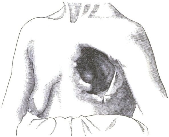
From a case of Kolaczek's, in which the resection of the third to the sixth ribs, with a portion of the sternum, was practised for the removal of an enchondroma. Diagram exhibits the aperture in the thoracic wall which permitted the exposure of the pericardium. Pneumothorax occurred, but patient recovered (Deutsches Archiv für klinische Medicin, Bd. xxx. 1882).
Paracentesis must sometimes be practised to relieve accumulation of fluid in the pleural sacs in instances in which dyspnoea is serious, and life may be prolonged by repeatedly practising this operation. Reflex laryngeal irritation, or paroxysmal dyspnoea with stridulous breathing, requires the use of inhalations or atomization of antispasmodics, and among the most useful of these are ether and chloroform. This group of neural symptoms can sometimes be markedly palliated by hypodermic use of morphia with atropia. But too often the symptoms are caused by actual pressure, and not by nerve-irritation, and this mode of treatment is futile, and therefore these measures should be employed with caution.
Sleeplessness, cough, bronchial or other pulmonary complications, must be managed upon general principles. The local pains may be met by local treatment, such as mustard sinapisms or soothing lotions; even blisters may secure temporary relief. The digestive system should be carefully studied, and assimilable and appropriate food should be selected. In lymphadenoma combinations of iodine with arsenic, as in Donovan's solution, may be tried, but, unfortunately, the utmost aid from present resources consists in a palliative and expectant policy.
DISEASES OF THE BLOOD AND BLOOD-GLANDULAR SYSTEM.
BY WILLIAM OSLER, M.D.
INTRODUCTION.
The blood is a fluid tissue composed of cells floating in an albuminous plasma, and it differs from other tissues not less in the arrangement of its elements than in the activity of the changes which go on in it. It is the mart into which is poured from the alimentary canal the commodities needed in nutrition, and the elements of the body select from it the various materials which they require, giving in exchange those chemical combinations which result from the metabolism of the tissues. In spite of ceaseless changes, a uniformity of composition is one of the most striking features of the blood in health. This is maintained, as regards the constituents of the plasma, by the activity of the organs which regulate income and expenditure—the alimentary canal and liver on the one hand, and the kidneys, lungs, and skin on the other; while histological uniformity is maintained by the adenoid or cytogenous tissue throughout the body, the function of which is to replace the wornout blood-corpuscles.
The corpuscles form rather less than one-half by weight of the blood. The plasma contains about 90 per cent. of water, which holds in solution proteids in the form of serum, albumen, and the fibrin-forming factors; sugar in traces; creatin, hypoxanthin, and urea; various fatty bodies in small amount; salts, chiefly sodium; and gases. The corpuscles (red) consist of hæmoglobin (90 per cent.), proteid bodies, and traces of lecithin and cholesterin.
So far as we know at present of the function of these two portions of the blood, the plasma ministers to the general nutrition of the tissues, while the corpuscles (red) are chiefly concerned with respiratory processes, acting as the carriers of oxygen and carbonic oxide.
We shall first give a brief account of the histological characters of the blood, and of the relation of the groups of adenoid or cytogenous tissue to the corpuscles.
Two forms of corpuscles are usually described, but we can recognize four varieties of blood-corpuscles in the body: (1) red, (2) white, (3) nucleated red, and (4) the hæmatoblasts (Hayem), or blood-plates of Bizzozero.
(1) Red Corpuscles.—In each cubic millimeter of plasma there are about 5,000,000 red cells. The percentage may vary within health limits from 90 to 110. The corpuscles are circular, non-nucleated, biconcave disks, homogeneous, to ordinary inspection structureless, and consist of a colorless stroma which is possibly reticulated, and a red coloring matter, the hæmoglobin. In health they are tolerably uniform in size, about 7.9 µ1 in diameter, or in English measurement 1/3200 of an inch (Gulliver). Even in normal blood there may be slight variations in size between 6.5 µ and 8.5 µ, the average, according to Hayem, being 7.5 µ.
1 µ is used to signify a micro-millimeter or 1/1000 part of a millimeter.
(2) Colorless or white corpuscles, nucleated masses of protoplasm, with an average diameter of 10 µ, or about 1/2500 of an inch. The majority have a finely granular protoplasm, but in a few the granules are coarse and do not completely fill the clear protoplasm. The ultimate structure is reticular (Heitzman). Erhlich2 has shown by their varying reaction to eosin that there are chemical differences among the colorless cells quite unrecognizable by other means. In healthy blood they display active amoeboid changes at ordinary temperatures. Their protoplasm does not, as is commonly stated, rapidly disintegrate, but if kept at a medium temperature retains its vitality, as shown by movements, for hours. The number per cubic millimeter is from 8 to 15 millions, and the ratio to the red is variously computed as 1 to 300 or 1 to 500.
2 Frerichs find Leyden's Archiv, Bd. i.
(3) Nucleated red corpuscles, which occur in the blood of the foetus and the infant, gradually diminishing until at the third or fourth year they disappear. In the adult they do not occur in the blood in health, but are normal constituents of the red marrow of the short bones. They measure from 1/1500 to 1/2000 of an inch, and are of somewhat variable intensity of color, often quite as deep as the ordinary red forms. There may be two or even three nuclei, not colored, grouped together, often eccentric, and in some instances protruding from the cell.
(4) The hæmatoblasts of Hayem, the blood-plates of Bizzozero, the elementary or intermediate corpuscles, are small discoid colorless corpuscles about 3 µ in diameter, and are normal constituents of healthy blood. When the blood is withdrawn, they aggregate together into irregular clumps or masses, which have long been known as Schultze's granule-masses. It can be readily demonstrated in new-born rats or kittens, in which these masses abound, that the corpuscles composing them are isolated in the vessels, and only run together when the blood is drawn. The statement is commonly made that the granule-masses of Schultze result from the disintegration of the white corpuscle (of the red, Erhlich), but half an hour's study of the question in a new-born rat will convince any competent histologist that we have here to do with a separate blood-element.3 It appears to have important relations with the production of fibrin.
3 Consult Proceedings Royal Society, 1874; Centralblatt f. d. Med. Wissenschaften; Medical News, 1882, 2; Bizzozero, Virchow's Archiv, Bd. xi.; Hayem, Recherches sur l'Anatomie normal et pathologique du Sang, Paris, 1878.
Of the origin and life-history of the red corpuscles during post-embryonic life we have still much to learn. They are stated to develop—
(1) From colorless corpuscles, the lymph-cells or leucocytes. In the lymph-glands, the Malpighian bodies of the spleen, in the thymus, or the adenoid tissue of the tonsil, of the lymph-elements in the intestines and other regions, colorless cells are constantly being manufactured, and the general belief has been since Hewson's time that the red corpuscles develop in some way or other from these leucocytes. How or where has not yet been settled. It does not apparently go on in the blood, or we should surely catch, in the many observations and with the excellent powers now in use, a glimpse of the birth of one of them. Some observers (Johnstone4) maintain that they develop from the granular protoplasm of the adenoid reticulum by a process of budding. This may be so, but we should expect to find the lymph in the efferent vessels and of the thoracic duct much more rich in red cells than is usually the case, and in specimens of healthy glands we should find young-looking elements such as he describes.
4 Seguin's Archiv, vol. vi.
(2) From the nucleated red corpuscles. In the embryo this undoubtedly takes place, and as the weeks of development proceed, the ordinary red forms gradually predominate. In the child the red nucleated cells disappear early, and are then found only in the red marrow. So far as my observations go,5 they apparently originate from colorless marrow-cells, which gradually become more homogeneous, and hæmoglobin develops in the protoplasm. The nucleus degenerates and disappears, when the cell has the appearance of an ordinary red disk. Rindfleisch thinks that the nucleus of the nucleated red is extruded in the development. It is possible that from these nucleated red corpuscles cells may originate in another way—viz. by budding. This I have seen and sketched in the marrow-cells,6 and Malassez has studied the same process.7 The gemmæ are small, and sprout from the protoplasm, not the nuclei, and when they break off they resemble the microcytes which occur so abundantly in certain anæmic states. Bizzozero8 holds that these nucleated red corpuscles are independent elements which do not develop from the colorless marrow-cells. They multiply by fission, and develop into the ordinary red forms with the disappearance of the nuclei. Several recent investigations support this view.9
5 Centralblatt f. d. Med. Wissensch., 1878.
6 Trans. Am. Ass. Ad. Science, 1882.
7 Archives de Physiologie, 1882.
8 Centralblatt f. d. Med. Wissenschaften, Bd. xix.
9 Fortschritte der Medicin, 1885, No. 1.
(3) Hayem believes that the red corpuscles develop from the small hæmatoblasts, but, so far as I know, his observations have never been confirmed. He states that in normal blood they occur in the proportion of about 1 to 20 red. In all states of blood reparation they increase greatly. He describes a hæmatoblastic crisis as occurring after hemorrhage, fevers, etc., when the number of these elements rapidly augments, and is succeeded by the addition of many small pale-red corpuscles, which he looks upon as intermediate between the hæmatoblasts and the ordinary red forms.
The colorless corpuscles are regarded as the direct offspring of the cells of the follicular cords in the lymph-glands and adenoid tissue, but whether by process of division of existing leucocytes or by sprouting from the endothelial places, or from the protoplasm in the fibres of the reticulum, remains to be settled.
The nucleated red corpuscles are in the healthy adult confined to red marrow, in which they probably develop from colorless cells, and may be regarded, as Neumann originally suggested, as transitional or intermediate forms between white and red cells. In anæmic states they may occur in the spleen and in the lymph-glands.
Of the origin of the hæmatoblasts or blood-plates we know absolutely nothing. They occur most abundantly under two most opposite conditions—in the young growing animal just entering upon life, and in the diseased, cachectic, wornout animal just preparing to abandon it.
Our knowledge of the relation of the cytogenetic organs to blood-formation may be thus briefly stated: The spleen certainly takes part in the development of colorless corpuscles, but its participation in red blood-formation is more doubtful. The nucleated red or embryonal forms do not occur, at least in any numbers, in health, though some observers have noted that after a repeated bleeding the organ was swollen and contained many such cells, as if it was the seat of an active development. Though the opinion prevails widely that the spleen is one of the important organs in the formation of red corpuscles, the evidence for this belief is of an exceedingly scanty nature.
The lymphatic glands and the adenoid tissue in other regions are the seats of constant production of colorless corpuscles, but of their relation to the red corpuscles there is the same lack of information as in the spleen. I do not know of any corroboration of the observation of Johnstone above mentioned, and in any case the number of red cells in the efferent vessels of a lymph-gland is so small—and indeed in the thoracic duct itself—that we cannot believe they are produced as red corpuscles in large numbers within the lymphatic system.
The red bone-marrow, as pointed out by Neumann10 and Bizzozero,11 appears to be the seat of blood-formation, and in the adult body is the only region in which the embryonic or nucleated red cells are found. It is a tissue similar in many respects to the spleen, and, though confined to the short and flat bones, the total amount in the body is very considerable. In the young it also fills the long bones. The evidence of the development of red corpuscles in the marrow rests upon the constant presence of nucleated cells infiltrated with hæmoglobin, and of their fission. Forms undergoing the process of karyokinesis can be seen without difficulty. In excessive hemorrhage, natural or induced, it appears to undergo an active proliferation, and in the long bones a red marrow may replace the fatty tissue.
10 Centralblatt f. d. Med. Wissenschaften, 1868.
11 Ibid., 1868.
The liver is doubtless the seat of blood-destruction, for the bile-pigments and leucocytes with red corpuscles in their interior have been found in its tissue. Nicolaides12 has shown that in the blood of the hepatic vein there may be a reduction of from one million to one million and a half of red corpuscles per c.m. In the embryo Neumann13 has shown that it may be the seat of the production of corpuscles, but there is no satisfactory evidence that in the adult this ever takes place.
12 Archives de Physiologie, 1882.
13 Archiv der Heilkunde, xv.
The remarkable rapidity with which, after a profuse bleeding, the normal proportion of red corpuscles is reached shows with what activity the development may proceed, and how favorable the conditions must be for their production. After the loss of a large quantity of blood the manufacture of new corpuscles may proceed at the rate of 30,000, 40,000, or even 50,000 a day.
What becomes of the red corpuscles? Here, again, is a question not satisfactorily settled. We do not know the average length of life of corpuscles. They are supposed to be short-lived—three weeks, according to Quincke. The need for their dissolution is assumed to provide pigment for the various secretions and tissues, and we occasionally see a few cells in the blood with a pallor which may be regarded as an indication of senility.
Positive evidence, however, of their destruction is afforded by the occurrence of the so-called corpuscles containing red corpuscles, which occur normally in red marrow and in the spleen, and under some circumstances in the lymphatic glands and liver. The red cells undergo gradual transformation into a yellow granular, and finally black, pigment. In normal spleen and marrow the numbers found are very variable; in fevers and cachectic states they may be in extraordinary numbers. Quincke and his pupil Peters14 have studied with great care this process of transformation of the red corpuscles and accumulation of the pigment in the cells of the marrow, spleen, liver, and lymph-glands, to which the term siderosis is applied. These pigment-granules are in the form of an iron albuminate, and are used in the development of new corpuscles. Thus, after repeated bleedings in animals, they may disappear completely in the restoration of the blood, while in animals into whose vessels blood has been transfused or injected subcutaneously the iron-containing cells in the various organs are very numerous, and even the cortical cells of the kidney contain numerous granules.
14 Deutsches Archiv f. klin. Med., Bds. xxv., xxvii., xxxii., xxxiii.
The amount of hæmoglobin in 100 grammes of healthy blood is 13.45 grm. (Preyer). Malassez estimated the quantity in a cubic millimeter of blood at between 0.125 and 0.134 milligramme, and, taking the corpuscular richness at from 4,000,000 to 4,600,000, he has estimated approximately the amount of hæmoglobin in each corpuscle.
PLETHORA.
General and persistent polyæmia or plethora has scarcely a place in recent pathology. Formerly it was thought that either from over-production or lowered expenditure the total amount of blood accumulated and filled the blood-vessels to an abnormal extent. The amount of blood undergoes, within limits, constant daily alterations, and after a full meal the vessels are in a state of plethora compared with their condition at the end of a ten hours' fast. If a plethysmograph could be devised to record graphically the variations in the total quantity of blood, each ingestion of food or drink into the vessels would be followed by a rise, and each interval by a gradual decline. So long as the organs of secretion and excretion are active the quantitative and qualitative condition of the blood is maintained at a tolerably uniform standard in each individual. At different periods of life the relation of blood-weight to body-weight varies. In the new-born the blood amounts to one-eighteenth part by weight of the body, while in the adult the average is from one-twelfth to one-fourteenth; so that in the infant there is a condition of comparative plethora. There are no reliable observations on the proportion of blood- to body-weight at respective ages, but there appears to be a reduction in old age. In women it is stated that just before each menstrual period there is a state of polyæmia.15
15 Mary Putnam Jacobi, The Question of Rest for Women, New York.
Worm-Müller16 and Cohnheim17 have made some very interesting experiments on this question of plethora. By transfusion in dogs a state of artificial plethora is readily established, and the animals stand the injection of as much as 10 or 12 per cent. of the body-weight of blood, above which quantity a fatal result ensues. After an injection of 20 or 30 per cent. of the total amount of blood, the superfluous plasma and corpuscles are got rid of in a few days, while with a larger injection of 60 to 80 per cent. it takes two or three weeks before the normal state is again reached. The albuminous and nitrogenous materials are largely got rid of by the urine, which increases rapidly in quantity and also in the amount of urea. The excess of corpuscles gradually disappears, and the hæmoglobin becomes deposited, as Quincke has shown, in the form of small granules in the cells of the liver, spleen, and bone-marrow.
16 Transfusion and Plethora, Christiania, 1875.
17 Allgemeine Pathologie, 2te Auflage.
In a similar way, it is reasonable to think that the body is quite capable of disposing of surplus albuminous materials in over-fed, lazy individuals with active digestion, whose red faces, full vessels, and bounding pulses give the impression of a distended circulatory system, and whom we term plethoric. Their appearance is the result rather of blood-distribution than of actual increase in the total volume, and there is no evidence that under any circumstances a rich and abundant diet without much exercise can permanently increase the amount of blood. It was formerly held that the healing of an old sore or the cessation of an accustomed discharge or loss of blood, by diminishing expenditure while the blood-making power was maintained, could induce plethora if no local disorder was excited "before the vessels in general reached a state of plethoric tension." Of such a condition, and of the plethora apocoptica that was thought to occur after the amputation of a limb, we do not hear much now, and the prevalent opinion of pathologists is expressed by Cohnheim, when he says "that, except as a transitory state, polyæmia does not occur under any circumstances." What, then, is the meaning of the full-blooded, rubicund condition which we see in some men, not necessarily large feeders, but often with vigorous active constitutions and perfect types of health? The appearance of plethora is caused chiefly by the distension of the superficial vessels; the circulation of the skin is remarkably active, particularly in the face, and it is probable that we have here to deal with local peculiarities of the vessels or of their innervation, and not with any general augmentation of the total blood-mass. It may be, however, that in such persons there is a plethora of certain of the constituents of the blood—viz. the red corpuscles—and there may be a state of polycythæmia rubra, as it has been called, in which the percentage of red cells is increased. In several such cases I have found, as has been previously noted, the number of red corpuscles considerably over the average. A relative increase in the number of red corpuscles also occurs in those sudden and excessive losses of fluid, as in cholera, in which the blood may become thick and sticky from the great reduction in the plasma, particularly of the water and salts—anhydræmia—or in cases in which the income of fluid is greatly restricted. Henry (F. P.) has recorded a case18 of stenosis of the cardia, in which, with great emaciation, the corpuscles per c.m. were 5,525,000.
18 Archives of Medicine, New York, vol. vii.
The condition known as hydræmic plethora develops whenever there is a great reduction in the number of corpuscles, as after a hemorrhage, or when the blood has been impoverished by long-standing suppuration, albuminuria, or in the growth of large tumors. So also when the secretion of urine is diminished, as in some cases of Bright's disease, and at the same time charged with albumen, the blood may become very watery; but in these states there is not an absolute increase in the entire blood, but only a relative excess of the water. Occasionally, this great excess can be noticed in the blood-drop as it comes from the finger-tip; the corpuscles do not fill the entire drop, and consequently leave irregular areas unoccupied by the red disks.
ANÆMIA.
A reduction in the amount of the blood or of its corpuscles occurs under a great variety of circumstances. Broadly speaking, we can recognize two great clinical and pathological groups of cases: I. Those induced by causes acting upon the blood itself; and, II. Those induced by disturbance in the functions of the blood-making organs.
I. Of causes acting directly upon the blood, we shall consider—
1st. Hemorrhage, traumatic or spontaneous. A high grade of anæmia may be quickly produced by loss of a large quantity of blood, and the reduction is in all the constituents; there is a true oligæmia. If the amount lost be excessive, death results from the diminution in the total volume of blood and general lowering of the arterial pressure. If the hemorrhage is sudden and profuse, as from a large vessel, the loss of four or five pounds of blood, or even less, may be sufficient to induce fatal syncope. In hemorrhage into the pleura or peritoneum from rupture of aneurisms, etc., it is rare to meet with more than three or four pounds of clot and serum; seven and a half pounds is the largest amount I know of shed into one of the cavities (pleura) by rupture of an aneurism. When the bleeding extends over several days, the amount lost may be very much greater. In cases of hæmophilia extraordinary accounts are given of the amount collected in the course of a few days. In a case of hæmoptysis a patient lost over ten pounds by measurement in one week, and then recovered from the immediate effects. After the most severe hemorrhages the reduction in the number of red corpuscles is not nearly so great as in forms of idiopathic anæmia. Thus in the case just mentioned at the termination of the week of bleeding there were 1,390,000 red corpuscles to the cubic millimeter. In any single bleeding a fatal result follows the loss of one-third or one-half of the total blood-volume. The process of regeneration of the blood goes on with astonishing rapidity, and in some bleeders a week or ten days will suffice to re-establish the normal amount. The restitution begins even during a hemorrhage by the absorption of lymph from the tissues under the lowered pressure in the vessels. The dryness and stickiness of the serous membranes after death from a profuse hemorrhage is usually very marked. The water and saline constituents of the blood are readily restored by absorption from the gastro-intestinal tract. The albuminous elements are also quickly renewed, but it may take weeks or months before the number of corpuscles reaches the normal standard. Indeed, this condition of oligocythæmia, as it is called, may persist, grow worse, and ultimately prove fatal. The microscopical characters of the blood after severe hemorrhage are not much changed, except as regards the white corpuscles, which are relatively increased, and the fibrin network, which is much less marked than in health. The white corpuscles may be very slightly reduced in number per cubic millimeter—a fact to be accounted for either by a relatively diminished loss during the bleeding, owing to their adhesiveness and wall-loving properties, or to a quick restitution from the lymph which is poured into the blood-stream. It has been observed both in dogs and men by Lyon19 that after a severe hemorrhage the number of red per cubic millimeter diminished for several days after the bleeding had been checked. How and where does the regeneration of red corpuscles take place after a severe hemorrhage? One would think that under these circumstances, if any, we should be able to get information which might be of service in determining the problem of blood-development; but, in spite of the numerous experiments on the subject, we are still far from a knowledge of full details. The observations of Neumann,20 Litten and Orth,21 Bizzozero,22 Lepine,23 and others appear to prove conclusively that the bone-marrow plays an important part in the formation of the new red disks, becoming lymphoid, losing its fat, and the nucleated red cells increase enormously. The same process has been observed in many cases in man. In a case of profuse metrorrhagia with profound anæmia Neumann24 found the marrow in all the bones of a rich raspberry red, full of the nucleated forms, which were also very abundant in the blood, and more in the vena azygos than in the aorta. The evidence in favor of the active participation of the spleen is not so conclusive. Neumann25 concludes that the spleen takes no share in the process, and holds that the nucleated red cells found in it are probably derived from the bone-marrow. Bizzozero, on the other hand, has found the spleen swollen and showing signs of lively blood-formation. He states that after removal of the spleen the restitution of the red corpuscles takes place much more slowly. Pouchet, on the contrary, says the regeneration goes on just as rapidly without the spleen. Of the action of the lymph-glands there is even less evidence. They have been found swollen, but in traumatic anæmia I do not know of any observations on their swelling and conversion into a red spleen-like tissue, such as have been found in some cases of idiopathic anæmia.
19 Of Norwich, Conn.: Virchow's Archiv, lxxxiv.
20 Archiv der Heilkunde, Bd. x.; Frerichs and Leyden's Archiv, Bd. iii.
21 Berliner klin. Wochenschrift, 1877, li.
22 Centralblatt f. d. Med. Wissenschaften, 1879, xvi.
23 Revue Mensuelle de Méd. et de Chirurg., 1877.
24 Loc. cit.
25 Loc. cit.
The microcytes which occur in numbers in blood in some cases of traumatic anæmia have been regarded as young developmental forms, but there is a great diversity of opinion as to their real nature, and their connection with productive blood-processes is somewhat doubtful.
Cohnheim suggests26 that after a profuse hemorrhage the rapid consumption of red corpuscles may be reduced, in which case we need not suppose such an active development; but the fact noted by Lyon27 and others of the increased reduction after a bleeding is against the view. In any case, if Quincke is right in assuming that the average life of a red corpuscle is only three or four weeks, what is the restitution of a couple of millions of corpuscles per cubic millimeter in comparison with the monthly renovation of the entire mass?
26 Loc. cit.
27 Loc. cit.
In the regeneration of the blood the development of the hæmoglobin does not keep pace with that of the corpuscles, so that they may, even when normal in amount, have a lowered hæmoglobin percentage, indicated under the microscope by a paleness in the cells.
2d. There is a large group of cases in which the anæmia is induced by a long-continued drain on the albuminous material of the blood—pus in a chronic suppuration, albumen in Bright's disease, prolonged lactation, etc. Rapidly-growing tumors act in the same way.
3d. The anæmia of inanition, brought about by defective food-supply or by conditions of the digestive organs which interfere with the proper reception and preparation of nourishment, as cancer of the gullet, chronic dyspepsia, etc. The reduction in the blood-mass may be extreme, but the plasma suffers proportionately more than the corpuscles, which even in the extreme wasting of cancer of the oesophagus may not be reduced more than one-half or three-fourths.
4th. Toxic anæmia, induced by the action of certain poisons in the blood, such as lead, mercury, and arsenic among inorganic substances, and the virus of syphilis and malaria among organic poisons. They act by increasing the rate of consumption of the red corpuscles, and the reduction may be considerable. The gradual impoverishment of the blood in pyrexia may be in part due to the toxic action of the fever-producing agent on the blood itself; but in this there is probably also disturbance of function in the blood-making organs.
The last three groups comprise what are known as secondary anæmias, and the condition of the blood is characterized by an increase in the water and diminution in the albuminous elements; the fibrin is often increased, and the network which separates, as seen under the microscope, is unusually dense; the white corpuscles are not much increased; there is rarely microcytosis or poikilocytosis; the reduction in the number of red corpuscles is not so great; hemorrhages do not often occur; when fever is present it is due to the disease or some complication, and is not the pyrexia of anæmia; and, lastly, they are more or less amenable to the action of iron and other remedies.
II. A consideration of the anæmias induced by disturbance in the blood-making organs themselves presents difficulties proportionate to our ignorance of the details of hæmatogenesis. We may regard, as above stated, the spleen, the general lymphatic tissue, and the marrow as the sites of production of corpuscles which are passed into the circulation fully formed. Certain of these organs—the spleen and marrow particularly—are also concerned with blood-destruction as well as blood-elaboration; but there is evidence to show28 that they, to use an ordinary simile, consume their own smoke, using the waste products for the purpose of further manufacture. Looking now upon the hæmatogenetic tissues as a single organ scattered through the body, let us consider what general disturbances of function it may suffer comparable to those met with in other structures. We can evidently suppose the physiological activity to be diminished or increased, and we should expect to find corresponding to these changes equivalent alterations in the character of the blood. Unfortunately, our knowledge of the normal processes as they go on in these tissues is so scanty that it amounts to a discussion upon the disturbances of a function itself imperfectly understood.
28 Quincke, quoted above.
With diminished functional activity in an organ we commonly meet with reduction in volume, the one depending on the other: now, the only instance in the blood-making organs in which a decrease in size and diminished functional activity go together is in the senile atrophy in which the spleen becomes small, the marrow more fatty, and the lymph-glands sclerotic, and in consequence the blood also is reduced in amount; but this is only a part of the general failure of nutrition in old age. Pathologically, there is no such well-recognized condition of uniform atrophy of spleen, lymph-gland, and bone-marrow, with a corresponding general reduction in the elements of the blood. Certain cases of idiopathic anæmia come close to it, in which these parts are wasted, but there are other differences which make the two conditions scarcely comparable. In fact, as we shall see, diminished activity in blood-making is usually associated with an increase in what we call hæmatogenetic tissues. Of increased functional activity in these parts we know very little, apart from the changes met with in cases of traumatic anæmia, in which the hyperplasia of the spleen and bone-marrow may be regarded as intimately connected with the rapid development of red corpuscles.
One fact is evident: that a progressive increase in the cytogenic tissues, local or general, is associated with disturbance in the process of blood-formation, and sooner or later induces anæmia. Thus, progressive enlargement of the spleen or of the lymph-glands or marked hyperplasia of the marrow, either singly or combined, is invariably accompanied with alteration in the characters of the blood. Even in those rare instances in which the lymphoid elements of the tonsils and fauces or of the gastro-intestinal canal are chiefly involved the same change may take place.
The nature of the process in the organs is of a hyperplastic character. In the spleen the pulp at first increases and the Malpighian bodies enlarge, but ultimately there is such a development of the fibrous reticulum that the consistence is greatly augmented and the organ becomes indurated. Histologically, there is very little distinction to be made between forms of chronic enlargement of this organ. In the lymph-glands there is increase in the cells; the tissue becomes more succulent, and is in a state of hyperplasia which may terminate in a great development of the fibrous elements, with induration. So also with the bone-marrow: in the short and flat bones, where in the adult a reddish or slightly fatty tissue exists, the fat disappears entirely, and the long bones, normally filled with yellow marrow, become occupied with a red-gray or greenish-gray cytogenous tissue not unlike spleen-pulp, and in many instances more consistent than the red marrow of early life.
A reduction in the number of red corpuscles is the chief and most constant change in the blood; anæmia seems to be the invariable result, whether the spleen, marrow, or lymph-glands are affected singly or together, and is the central feature in the entire group of cases. This diminution in the red cells may or may not be accompanied by an increase in the white corpuscles, which in some cases may be so striking as to be regarded as the special blood-change, and is, as a rule, permanent, though it may be a variable or even a transitory state.
The general and histological differences between forms of hypertrophy of these blood-making organs are exceedingly slight, and in their clinical features they present a large number of symptoms in common; indeed, we may say that all the important symptoms are present, whether the spleen is affected alone or with the lymph-glands and bone-marrow, or whether these parts are independently involved, and whether there is simple reduction in the red or with it an increase in the white corpuscles. Such common features are—the progressive anæmia with its group of circulatory symptoms; the irregular febrile reaction, essential fever of anæmia; the absence of marked emaciation; the tendency to effusions of serum; the progressive debility; the occurrence of hemorrhages; gastric and intestinal disturbances; and resistance to treatment.
The affections characterized pathologically and clinically by so many similar features are known and recognized as distinct diseases under the names leukæmia, Hodgkin's disease or pseudo-leukæmia, splenic anæmia, and idiopathic anæmia (some cases); and we shall now consider these a little more closely.
First, of the hyperplasias of the cytogenic tissues associated with simple anæmia. The various groups, spleen, lymph-glands, and marrow, may be involved singly or together; usually one is first affected, and the others, if at all, subsequently. Progressive enlargement of the spleen induces sooner or later anæmia, the anæmia splenica of Griesinger. These cases are by no means rare: certain of them represent the final stage of a malarial intoxication, but there are others in which the enlargement seems causeless. There may also be hyperplasia of the bone-marrow, less often of the lymph-glands. The anæmia may be profound, and the clinical picture is that mentioned above. Two cases of it under my care died of hæmatemesis. The diagnosis of this affection from splenic leukæmia rests solely on the microscopical examination of the blood. It is also classed as the splenic form of Hodgkin's disease or pseudo-leukæmia.
Primary enlargement of the lymph-glands with anæmia constitutes Hodgkin's disease or pseudo-leukæmia, in which there may be general hyperplasia of the lymphatic elements throughout the body, with nodular growths of adenoid tissue in other organs. The spleen and marrow are not often affected. Here, too, the diagnosis from lymphatic leukæmia rests with the microscope.
Is there a form of anæmia dependent upon hyperplasia of the bone-marrow—an anæmia medullaris? In 1875, Pepper and Tyson29 found affection of the marrow in idiopathic anæmia, and Pepper suggested that this might be the starting-point of the disease, which could thus be regarded as a medullary form of pseudo-leukæmia. Cohnheim in 187630 described the same condition, and I had an opportunity of examining several cases.31 Granting that the marrow is a tissue which shares in the blood-making functions, it seemed reasonable to suppose that a general hyperplasia of its elements might disturb the processes of hæmatosis and produce anæmia, just as in hyperplasia of the spleen and lymph-glands. Two facts soon came to light which seem opposed to this explanation of the pathology of idiopathic anæmia. A hyperplasia of the marrow was found in cases of chronic disease with wasting, and cases of idiopathic anæmia were described in which the marrow was normal. The numerous observations of the past five or six years have not brought us nearer to a solution of the problem. The observations of Neumann,32 and those of Litten and Orth,33 on the changes in the marrow in chronic diseases have been abundantly confirmed, and a red lymphoid marrow may be met with in various cachectic states. This, too, I have frequently seen, yet it is in my experience rare to find such marked, rich hyperplasia of the marrow, such an entire absence of fat, as in some cases of idiopathic anæmia. In 9 autopsies in typical cases at Montreal, not parturition cases, the marrow of the long bones was lymphoid and red in 6; in 1 it was not examined; in 1, which I did not see, the marrow was stated to be normal; and in 1, an old woman over sixty years of age, the marrow of the short bones was rich in lymphoid cells and nucleated red corpuscles, and the long bones contained a grayish gelatinoid—atrophic—marrow. It does not appear possible with our present knowledge to arrive at a satisfactory conclusion on this question. Some regard the marrow-change as the consequence, others as the cause, of the anæmia. Both Cohnheim34 and Pye-Smith35 regard those cases of idiopathic anæmia in which the marrow-changes are pronounced as cases of anæmia medullaris.
29 American Journal Med. Sciences, 1875, ii.
30 Virchow's Archiv, Bd. lxviii.
31 Centralblatt f. d. Med. Wissenschaften, 1877, Nos. 15 and 28; 1878, No. 26.
32 Berl. klin. Wochenschrift, 1877, xlvii.
33 Ibid., 1877, li.
34 Loc. cit., Bd. i. S. 467.
35 Loc cit.
Next of the parallel series of hyperplasias of the blood-forming organs with anæmia, plus an increase of the colorless corpuscles—leukæmia. Here, too, we have the three forms—splenic, lymphatic, and medullary.
The splenic leukæmia is the most common, and in its general features is identical with splenic anæmia, the excess of white corpuscles being the only distinguishing feature. It is almost invariably associated with changes in the marrow.
The lymphatic leukæmia may arise in connection with hyperplasia of the lymph-glands or of the adenoid elements in the alimentary tract—tonsils and Peyer's glands. It is much less common than lymphatic anæmia or Hodgkin's disease, and there are not many uncomplicated cases on record. Apparently, a very limited bunch of glands—cervical—may induce the change in the blood.36 Medullary changes are almost invariably associated with a great increase of colorless corpuscles in the blood, and a myelogenous form of leukæmia is now, owing chiefly to the investigations of Neumann, well established. Indeed, he would regard the change in this tissue as the primary and important, and those in the lymph-glands and spleen as secondary.
36 Gowers, Reynolds's System of Medicine, art. "Leucocythæmia."
The hyperplasia, either lymphadenoid in character or pyoid, may result in the expansion and softening of the bones, with the production of irregular tumor-like masses.
We have, then, the following group of anæmias induced by a primary disturbance of function in the blood-making organs:
PRIMARY OR CYTOGENIC ANÆMIA. Leucocytic Splenic, Lymphatic, Medullary, Leukæmia.
Non-leucocytic Splenic, Anæmia splenica. Lymphatic, Hodgkin's disease. Medullary, Idiopathic anæmia (certain cases).
There remain for consideration the relation of the tissue-change to the anæmia and the nature of the leucocytosis; but until the chief facts in the development of the corpuscles are thoroughly known we cannot expect a satisfactory solution of these problems.
The anæmia may be explained on the view of diminished production (anæmatosis) or increased consumption of the red corpuscles (hæmophthisis). We know nothing of the intimate processes connected with lessened production, but as anæmia so constantly accompanies the hyperplasia, we assume they are intimately connected with each other, and the diminution in the number of corpuscles in some way the result of disturbed functional activity in the blood-making organs. An increased consumption of corpuscles in anæmia is indicated by the presence in large numbers of cells containing red blood-corpuscles in the spleen and marrow, and occasionally in the lymph-glands; by the increased amount of iron which has been found in the liver; and in some cases by the deep color of the muscles and an intensification of the color of the urine. Either a failing production with normal rate of consumption, or a normal output with heightened destruction, would produce anæmia. Possibly, in some instances, both factors may prevail. Quincke's interesting observations37 may enable us to determine the cases in which one or other has been dominant. Where there is great destruction we shall expect to find the granules of iron albuminate in the spleen, bone marrow, and liver-cells, possibly in the cells of the cortex of the kidneys, and the iron reaction should be present.