Volume 3 — DISEASES OF THE BLOOD, AND OF THE HÆMATOPOIETIC SYSTEM. (continued 9)
Reading edition
2. Diminution in Volume.—The pulse in one radial may be much smaller than in the other or altogether absent.
3. Diminution in Force.—The pulse of one side may convey a less sudden and less forcible blow to the finger. This diminished suddenness of the sensation imparted to the finger corresponds to the sloping up-stroke of the sphygmographic tracing.
4. Thrill.—Under certain rare and not very clearly defined circumstances the pulse imparts a sensation of thrill to the finger. Mahomed says this probably occurs when the entrance to the aneurismal sac is very narrow and the aneurism is directly in the course of the vessel. It may also be occasionally produced by the rigidity of the wall of the vessel or by a partially-dilated clot vibrating in the blood-stream.
Under the enthusiastic and elaborate study of Mahomed the sphygmograph has attained a certain degree of usefulness. Though difficult in its application and limited in its results, yet many of the points demonstrated by it are of sufficient importance to justify their consideration. The sphygmographic tracing of the normal pulse is shown in Fig. 50.
FIG. 50.
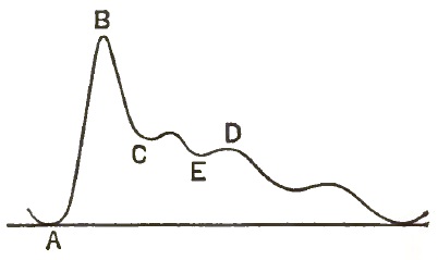
AB. The Up-stroke. ABC. Percussion Wave. E. Aortic Notch. D. Dicrotic Wave.
Now, the points which distinguish an aneurismal tracing from the normal are—1, a sloping up-stroke; 2, impairment or loss of the percussion wave; 3, obliteration of the secondary waves; 4, diminished volume of the curve; 5, vibratile waves; 6, a different blood-tension.
FIG. 51.
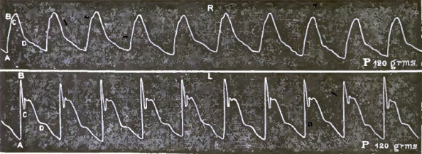
Right and Left Radial Pulse in Aneurism of Aorta.
In comparing the curves shown in Fig. 51, taken from Powell's article upon aneurism, it will be noticed that the up-stroke AB is more sloping in the curve of the right wrist than in that of the left. The percussion and dicrotic waves are entirely smoothed out into an almost uniform wavy slope. As one writer has expressed it, an aneurism acts like an air-chamber in an engine, and tends to break up the intermittent pulse into a steady stream. The relative difference of the blood-tension of two arteries is determined by the relative amount of pressure required of the instrument to develop the tracing. This amount of pressure is sometimes greater and sometimes less on the affected side.
In comparing the tracings from the radials the following points are to be noted: 1. Is there any difference in the percussion waves?—i.e. is the up-stroke more sloping or the apex less pointed in the one than in the other? 2. Is the tidal wave equally high and sustained in both? 3. Is the dicrotic wave equally developed?
If a difference exist in the tidal wave alone, it need not, and probably will not, be due to aneurism. It is the loss of the percussion wave and of the dicrotism which characterizes aneurism.
It must be conceded here that the use of the sphygmograph and the interpretation of its tracings are beset by the greatest difficulties. Mahomed, to whom I am chiefly indebted for these sphygmographic details, declares that the use of the instrument requires great care and skill, and it may easily lead to error. "No one should attempt to use it who cannot readily obtain similar tracings from the two radials of a healthy person." Great care in the application of the instrument should be exerted, and we must guard against all causes of transient excitement. It is well to let the patient see the instrument applied to others before attempting it on him, in order that he may not fear it. The patient must be placed in a comfortable position, with both arms alike, and the points of application of the instrument must be alike on the two sides. The amount of pressure on the two sides must be equal, or the difference carefully noted. Moreover, one should never be contented with one tracing, but a number on each wrist should be taken. If, then, the two radials appear to differ, the precautions must be redoubled, and the pulse tested again on another day. Inequalities of the tracings may be produced by abnormal distribution of the radials, and an old fracture or other injury of one arm may affect the flow of blood in the arm.
Paralysis of the arm, by interfering with the vaso-motor nerves, and thereby with the venous return of the blood, may alter the character of the pulse. A tumor external to the artery, either intra-thoracic or extra-thoracic, will produce aneurismal pulse and endarteritis, or congenital contraction of the aorta may so block the artery as to produce diminished pulse-waves. It may be said that the sphygmograph is incapable of distinguishing between an endarteritis and an aneurism.
On the other hand, the instrument is very useful in distinguishing between an aneurism and a tumor compressing an artery, because in the latter case the up-stroke and percussion wave remain normal, whereas in the former they are strongly modified, as described above. With aneurism of the ascending aorta both radials must be similarly affected, if at all, and in these cases the sphygmograph teaches very little. If the right radial is alone or mainly affected, then the aneurism involves the innominate and arch together. When an aneurism of the innominate includes the aorta, then the whole sac forms virtually a dilated aorta, and no difference in the radials will appear. Hence it follows as a corollary: Given an innominate aneurism, if the radials remain equal the aorta is certainly involved. When the left radial pulse is alone affected, the aneurism lies beyond the brachio-cephalic branch, and may or may not involve the left subclavian.
The sphygmograph is of less avail in aneurisms of the descending portion of the thoracic aorta or of the abdominal aorta. It may be of service in affording information regarding the condition of the aorta itself with reference to an operation, and it may also be of service in determining the upper limits of an aneurism under the following conditions: A case is reported which presented all the physical signs of aneurism of the descending aorta, but the sphygmograph showed that the left radial was affected, and thereby proved that the aneurism extended as high as the left subclavian at least.
While the foregoing facts prove that the sphygmograph by itself affords very inconclusive and untrustworthy evidence, yet when the presence of a tumor and other physical signs prove the existence of an aneurism, the written pulse-record will often be the guide to the accurate placement of the tumor, and thereby will often furnish decisive indications in the selection of the method of treatment.
Auscultation.—The typical aneurismal bruit is not an ordinary souffle, but it is an accentuated booming sound of a very peculiar character. Many writers describe it as a systolic jog or shock. Occasionally this bruit de battement is double—i.e. one hears two shocks, so to speak, just as one feels a double impulse. No satisfactory explanation for this reduplication of murmur has yet been given. The aneurismal murmur is almost invariably systolic. Balfour reports two cases of a diastolic murmur heard with abdominal aneurism. One of these cases was observed by himself and the other by Wickham Legg.
When this peculiar booming sound is heard over a circumscribed dull patch, it is very distinctive of aneurism, but its absence possesses no eliminative value. Many aneurismal tumors are absolutely quiet, and some of them give only a soft murmur like an ordinary cardiac souffle.
Associated with the aneurismal sound one also hears the normal heart sounds much intensified. This is peculiarly noticeable of the second cardiac sound, which acquires a ringing, booming, accentuated character when heard over an aneurism. Johnson thinks that this intensification of the heart sounds is due to the sudden tension of the walls of the sac. Balfour in referring to the same phenomenon considers it of greatest diagnostic value, and thinks that proper emphasis is not ordinarily given it.
A fundamental rule in the examination of a suspected case of aneurism is to auscult over every inch of the thorax, front and back. Not only the intrinsic signs of the tumor itself are important, but all testimony from the neighboring organs must be collected and weighed. The modification of the respiratory sounds have already been mentioned. Stokes attaches great importance to this fact, that "over one lung, more rarely over both, the breath sound has often communicated to it a peculiar sonorous vibrating quality, probably by conduction from the laryngeal stridor present."
Valvular complications of the heart are not necessarily associated with aneurism. Cases are reported, however, where a tumor is situated so near the aortic orifice as to interfere with its closure, and thus induce the ordinary phenomena of aortic insufficiency.
Of course when valvular disease is coincident with aneurism the customary signs will be added to those of the tumor, and must be carefully distinguished.
Drummond of England has recently contributed a new sign of aneurism. It is a familiar fact that after sudden exertion, and with the heart acting violently, one can hear in the mouth during expiration a well-marked whiff proceeding from the glottis. Under normal conditions of the chest this whiff is only heard after exertion, and never during perfect repose. Now, Drummond has noticed that this oral whiff, as he terms it, occurs regularly in many cases of aneurism of the aorta. When the sign is well marked the whiff is audible in the trachea with the mouth shut, but disappears on compressing the nostrils with the fingers. The whiff may be double, synchronous with both the expansion and contraction of the tumor. The sign does not exist in cases of valvular lesions of the heart without aneurism. As indicated above, this sign possesses a diagnostic value only when it is observed under conditions of absolute bodily and cardiac composure. One should make a patient lie quietly for a while before examining him for this sign.
Percussion.—Circumscribed dulness is always present when the tumor reaches the chest-wall. Owing to the globular shape of the tumor, its size is usually larger than the area of dulness would seem to indicate. There is no abrupt line of demarcation, but the dulness shades off gradually into the surrounding pulmonary resonance. The dull patch is most frequently situated to the right of the sternum and on a level with the second and third ribs. More rarely it may be found on the sternum or to the left of the same. If the neighboring lungs are solidified from any cause, the percussion signs of the aneurism will of course be obscured.
Localization.—When the signs of aortic aneurism are all conclusive, the next point in the diagnosis is to determine the probable seat and extent of the tumor.
In a general way, it may be stated that the physical signs of an aneurism of the ascending aorta are grouped about the upper two right intercostal spaces. Tumor of the transverse portion presents itself at the manubrium, and aneurism of the descending aorta may be detected in the upper interscapular region to the left of the spinal column. Balfour says that the aneurism is probably about the middle of the transverse portion when the point of greatest pulsation is situated at the middle of the manubrium or from that to the fourchette above, and the veins of the root of the neck are congested.
An aneurism of the left extremity of the transverse portion usually points below the left clavicle. There are many startling exceptions to these rules. One case is reported where an aneurism of the ascending aorta pointed at the left of the sternum and pressed upon the left bronchus. Another case of aneurism of the descending aorta passed behind the oesophagus and compressed the right bronchus. An innominate aneurism occupies the episternal notch, and usually appears first along the tracheal edge of the sterno-mastoid muscle. As it increases in size it will extend across the episternal notch and push out the inner end of the right clavicle. It may appear first under the end of the clavicle, but then it is at the cardiac end of the vessel and involves the aorta.
An innominate aneurism must be distinguished from a low carotid aneurism. The latter usually appears between the sternal and clavicular portion of the sterno-mastoid muscle, and its pulsations can be felt by pushing the finger into this space when the muscle is relaxed. Cockle said that he knew of no instance of a carotid aneurism distending the episternal notch. Barwell also mentions the fact that the ear on the affected side will gain color more slowly than its mate after pinching when the aneurism is situated upon the carotid.
It is always serviceable, and often essential, to determine whether an aneurism of the innominate also involves the aorta. If the tumor appears first under the sterno-costal articulation, the aneurism probably extends on to the aorta. Again, if the radials are both equal, the tumor undoubtedly includes the aorta, for reasons already explained in connection with the pulse-curves. If the right pulse alone is affected, we can eliminate aortic complication. Barwell also states that innominate aneurism involving the aorta presents the following symptoms: The pulsation, dulness, and abnormally loud heart sounds are on and to the right of the middle line. The various congestions are on the left side, and do not encroach upon the right side until later. This venous symptom is especially marked on the left pectoral.
A subclavian aneurism may cause confusion when it occupies the first third of the vessel. Such an aneurism, however, is an elongated oval in shape, and is partly covered by the clavicle, and this bone will move up and down in front of it with movements of the shoulders.
I have emphasized the unreliable character of the pulse as a diagnostic sign of aneurism, but when other signs of this lesion are well marked the pulse furnishes some evidence regarding the locality of the tumor. The following summary of the pulse-signs serves as a useful guide, therefore, in examining the pulse.
1. Both radials affected alike, the aneurism is limited to the ascending aorta.
2. Right pulse more altered than the left, the aneurism involves both the aorta and the innominate artery.
3. Right pulse alone affected, the left remaining normal, the aneurism is confined to the innominate artery.
4. Left pulse not affected, the aneurism is situated beyond the innominate.
5. Both pulses aneurismal. This occurs sometimes with aneurisms of the arch which involve the large vessels.
Varicose aneurism can only be suspected by exclusion. Thurman emphasizes one symptom which is significant when heard, but it is rare. This sign is an intense superficial souffle, accompanied by a frémissement cataire, and situated over the opening of the aneurism. It is continuous in time, though louder during systole; and this element of continuity serves to distinguish it from the ordinary bruits of aortic aneurism or valvular lesions. When there is a varicose communication between the aorta and the vena cava superior or the right auricle, the souffle will be extended along the right border of the sternum, with its maximum at the level of the second intercostal space. If the aneurism opens into the pulmonary artery or the upper part of the right ventricle, the souffle will be heard along the left border of the sternum. When the signs are manifested as the result of some excessive effort, and are accompanied by præcordial pain, Thurman thinks them almost conclusive of varicose aneurism of the ascending aorta. He adds a few other symptoms likely to be present, but less characteristic of this particular lesion. These are anasarca, venous congestion, dilatation of cutaneous veins, dyspnoea even to orthopnoea, cough with sanguinolent sputa, a bounding pulse, and less frequently general feebleness, with diminution of the animal heat. These signs have a general significance, however, except when the vena cava superior is involved, and there the venous congestion and oedema occupy the upper half of the body. We have, however, previously seen such phenomena limited to the upper part of the body, resulting from pressure upon the vena cava.
T. Gallard has related a very interesting case of an arterio-venous aneurism of the arch of the aorta communicating with the vena cava superior. This case furnished all the ordinary signs of a tumor of the mediastinum with compression of the vena cava superior. It emitted a souffle which began with the first cardiac sound and persisted through the short interval of silence and to the end of the second sound. This souffle was especially pronounced at the base of the heart, and Gallard diagnosed a communication with the vein above mentioned. The autopsy revealed the accuracy of the diagnosis.
Hayden says that aneurisms opening into the heart, the pulmonary artery, or the vena cava have, so far as he knows, without exception, arisen from the ascending aorta. The simple projection of an aneurism into one or more of the chambers of the heart is attended only by symptoms of obstruction to the blood-current, and he knows of no symptom characteristic of a communication between an aneurism and the heart. When the sac opens into the pulmonary artery there occur sudden and most urgent dyspnoea and blood-expectoration, without spasm or stridor. If aneurism of the ascending aorta has been primarily determined, then the sudden eruption of such symptoms would be almost pathognomonic of this accident.
DIFFERENTIAL DIAGNOSIS.—We have enumerated a large number of symptoms, direct and indirect, which are grouped about aortic aneurism. It is an unknown thing, however, for any one aneurism to present the entire group in one tableau. A few only appear in a given case, and the possible kaleidoscopic combinations of the whole number are almost infinite. There are also numerous other conditions of the thoracic organs which produce groups of phenomena closely resembling those of aneurism, and requiring critical analysis.
An aneurism is a tumor, and the majority of its symptoms are simply signs of a tumor. It is necessary, therefore, to determine whether the tumor at hand is a solid growth or an expanded vessel. This is always difficult when the tumor is beyond reach. It may be pulsatile from lying upon the aorta. The following points, therefore, should be carefully noted and tested:
1. A solid tumor may be pulsatile, but it is never distensile.
2. The shock of a solid tumor is not markedly stronger than that of the heart (Balfour).
3. There is no accentuation of the second heart sound (Walshe), nor bruit of a booming character (Hayden).
4. In the sphygmographic tracing of a tumor-pulse the up-stroke is never sloping, and the percussion wave remains well marked.
5. Variations in the position and size of a tumor, and also in the pressure phenomena, are important. An aneurism varies constantly in its size and in its mural tension; hence all its signs vary correspondingly; whereas with a solid tumor in the mediastinum the phenomena are more constantly progressive. An aneurism which is visible and palpable upon the external chest-walls will sometimes recede within the thorax, whereas solid or cancerous tumors never act thus.
Abscess of a gland in the episternal notch may closely simulate aneurism of the innominate. Mahomed and Golding-Bird report such a case. The imitation was so close in this case as to balk a number of very careful observers, and no absolute diagnosis was reached until the sudden rapid increase of the growth and of acute superficial inflammatory symptoms revealed the probability of pus. The abscess was supposed to result from the pressure of a collar-button. A companion case was reported by the same author where an actual aneurism of the innominate presented such neutral signs that no diagnosis was reached until the patient was etherized and an exploratory incision was made down to the sac. It is well to remember that an aneurism may rise and fall with deglutition and with coughing and straining when it is adherent to the trachea. A case is reported of a very vascular sarcoma attached to the manubrium sterni and projecting into the episternal notch, which presented the double murmur, pulsation, and pressure symptoms of an aneurism, and was diagnosed as such, the mistake being discovered only at the autopsy. In such very obscure cases I know of no reliable or distinctive signs on which a diagnosis may be established: the only resort seems to be to await developments. In process of time the appearance of cancerous growth in other parts of the body will often throw light upon a thoracic tumor. Occasionally aneurism of the aorta may simulate insufficiency of the aortic valves. Guttmann reports a case which presented all the classical symptoms of aortic regurgitation and none of aneurism. The autopsy revealed a large aneurism of the ascending aorta and the aortic valves intact. The aorta itself was notably dilated throughout, and it is probable that the change in the arterial walls affected the proper systolic contraction of the aortic orifice, so that insufficiency resulted. Chronic endarteritis of the aorta may produce aneurismal signs. Dujardin-Beaumetz reports a case where there were contraction of the left pupil, sudden reddening of the left side of the face, transient aphonia, intermittent dyspnoea, suppression of the left radial pulse, and a double souffle along the track of the aorta; and yet the autopsy revealed simply endarteritis of the transverse portion of that vessel, without the least dilatation. Many of the symptoms of this case could be explained by the extension of the inflammation to the sympathetic nerves.
Many aneurismal signs connected with the voice, eye, and vascular supply of the heart may be produced by the implication of either vagus in neighboring inflammation. Chronic empyema of the left side will sometimes pulsate synchronously with the heart and simulate aneurism. The following points are important:
1. Such pulsations occur only on the left side.
2. There is always a disproportion between the pulsations, which are feeble, and the extent of dulness, which is large.
3. There is absence of expansile pulsations.
4. There is usually ample evidence of the presence of a pleuritic effusion, displacement of the heart, etc.
5. Aneurism may be coexistent, however, and therefore it may sometimes be advisable to make an exploratory capillary puncture before opening the chest freely.
Berard reports a case of empyema which formed a tumor on the left side of the sternum, which pulsated and looked like an aneurism. Finally, the tumor burst and discharged pus.
DURATION.—The progress of aneurism of the aorta is very rapid, and in the majority of cases the fatal termination is not delayed many months. In 40 cases where the duration of the disease was well defined, I found that 20 of them died within one year, 9 lived for two years, and 3 lingered five years. About 75 per cent., therefore, died within two years.
TERMINATION.—Rupture of the sac is a frequent cause of death. In 106 cases analyzed by me, 39 terminated in this manner. The seat of the rupture and the organs into which the blood escapes vary according to the location of the sac.
Aneurisms of the ascending aorta burst most frequently into the pericardium, right auricle, right ventricle, right pulmonary artery, and rarely externally. Tumors of the transverse portion burst into the trachea, left lung and left bronchi, left pleural cavity, oesophagus, and externally. Those of the descending aorta empty into the oesophagus, left pleural cavity, and spinal cord.
The most frequent point of rupture appears to be into the pericardium, as 13 out of 39 ruptures emptied into that cavity. It will also be noticed that the right side of the heart and the left pleura and lung are the chosen seats of hemorrhage. I found no case of rupture into the left side of the heart.
The bursting of an aneurism is not always an immediately fatal accident. The so-called weeping aneurism may pour forth small amounts of blood for weeks and months. Neligan reports a case of external rupture near the second rib on the right side which discharged blood at intervals for more than a year. At times the bleeding was with difficulty arrested, and yet the aneurism finally solidified and the patient left the hospital calling himself well. Another man with an external aneurism thought it was a blood-boil, and squeezed it with his chin to favor the flow until he fainted. The bleeding then ceased, and never occurred again. He died one year later of typhoid fever. Such cases, however, are very rare, and usually when an aneurism bursts externally the death is sudden and tragic.
Rupture of a sac into the pericardium or pleural cavity may not prove fatal for several hours, and the patient will exhibit the ordinary symptoms of internal hemorrhage. Rupture into the heart or pulmonary artery causes great dyspnoea and distress, and death follows rapidly.
Aneurism may cause death indirectly by starvation from pressure on the oesophagus, or by suffocation from occlusion of the trachea. The pain and distress occasioned by the tumor may cause death from exhaustion. Pain at times is so great that the sufferers can neither lie down nor stand, and, deprived of rest and food, they wear out. A few patients die from intercurrent accidental diseases or complications, but it may justly be said that the death of a patient with aneurism is usually directly referable to the tumor itself.
TREATMENT.—Aneurisms of the aorta occasionally solidify by the formation of a clot, and thus a spontaneous cure is established. Unfortunately, however, such a result is a rare exception to the rule of steady progress to death.
A number of methods of treatment have been advocated, and some of them present here and there gleams of hope for some cases. The aim of all these methods is to produce coagulation of the blood in the sac, either by mechanical means or by the chemical action of drugs.
The introduction of fine wire has been attempted. A canula is plunged into the aneurism, and then either short pieces of wire are dropped into the sac or one long wire is pushed in. Murchison introduced twenty-six yards of steel spring into an aneurism of the ascending aorta. This method is attended with great danger, and has not been successful, and is therefore abandoned at present.
The hypodermic injection of ergotin into the sac was also recommended by Langenbeck, but it has not met with success.
Pressure upon the aorta can only be applied to cases of abdominal aneurism, and here it has been successful. The pressure must be applied under ether, and great care must be exercised not to injure the other abdominal organs.
The starvation method was first advocated by Hippocrates, and was espoused later by Valsalva. The idea of this treatment was to render the blood more coagulable by making it less watery and richer in fibrin. Valsalva commenced by bleeding a patient freely, and then reduced his meat and drink until only half a pound of pudding was allowed morning and evening. The bleedings were repeated at intervals until the patient was too weak to lift his hand from the bed on which he lay. The vital objection to this treatment is that starving renders the blood less coagulable, though it may lower tension. Copland has seen aneurisms previously quiet begin to grow and end fatally on the starving and bleeding method.
A few years ago Valsalva's method was resurrected by Tufnell, but was modified somewhat in detail. The bleeding was omitted and the starving was less vigorous. Tufnell's three rules are—rest, restricted diet, and medicine. The rest must be absolute repose in bed, and must continue two months or ten weeks at least, without the patient sitting once erect. By this means Tufnell reduces the frequency and force of the heart-beats, and thereby lessens the number of distending blows upon the interior of the aneurism. This is of course a very tedious treatment, and many patients will be unwilling to submit to it. Others who are unable to appreciate the gravity of their disease, and seek merely relief from their subjective suffering, will refuse to continue the treatment as soon as they obtain such relief. Hence the ingenuity of the physician will often be taxed to the utmost in devising means and measures for controlling refractory patients and lessening the tedium as much as possible for all.
The room of confinement should be light, cheerful, and airy, and should command a view of outdoor life if possible. Tufnell urges the choice of a south room, because the presence of sunlight is very restful to the spirits, while absence of the same is depressing. The bed should be made as comfortable as possible, and with mechanical contrivances to obviate the necessity of raising the patient. It should not be too narrow, and should be of a height most convenient for the nurse attending. Tufnell recommends a large water-cushion, not over full, under the hips. The sheets and protectives should be drawn taut and pinned to the sides of the bed to prevent wrinkling. No movement should be allowed the patient except to turn upon his side now and then, and occasionally upon his face in case such movement relieves dorsal pain. A urinal and bed-pan should be at hand, and a pleasant, agreeable nurse who will be willing to read to, converse with, and amuse the patient as desired.
The diet recommended by Tufnell is as follows: Breakfast: Two ounces of white bread and butter; two ounces of cocoa or milk. Dinner: Three ounces of boiled or broiled meat; three ounces of potatoes or bread; four ounces of water or light claret. Supper: Two ounces of bread and butter; two ounces of milk or tea. This makes an aggregate of ten ounces of solid and eight ounces of fluid food in the twenty-four hours, and no more. Thirst is liable to be present at first, especially in the summer months; and this may be relieved by holding a pebble in the mouth or by occasionally sucking a piece of ice. Tufnell thinks that the diminished amount of fluids reduces the duty of the heart and renders the blood thicker and more fitted for deposit. If the patients are very intolerant and restless, it is better oftentimes to indulge them in a little more liberal diet, but only enough to appease them and keep them in control.
Medicinal Agents.—As rest is the great refrain of his method, Tufnell recommends anodynes and soperfacients at night. For mere restlessness he prescribes the following combination: Lactucarium, 20 grains; extract of hyoscyamus, 10 grams—made into six pills, two to be taken at bedtime. The bowels will naturally be constipated, owing to rest in bed, and for this he recommends compound jalap powder. Too much purgation should be avoided, as irritation of the bowels will hasten the circulation. Obstinate constipation, however, must not be allowed, or anything which can produce straining. The instant such a condition manifests itself, enemata by tepid water should be administered.
The principal symptom to contend with is pain, and for this purpose opiates should be used freely according to the exigencies of the case. In one case it was found that smoking twenty grains of stramonium at bedtime would produce a quiet night. This was discovered accidentally by the patient, who began to smoke the stramonium under the false impression that he was suffering from asthma.
Maclean recommends the use of eucalyptus globulus for the relief of the distress due to irritation of the pneumogastric nerve.
Issues and blisters upon the back are not advisable, as they interfere with the recumbent position. Relief to dorsal pain will often be obtained by change of position, by turning upon the side or upon the face. Sometimes the application of a heated flat-iron, with the protection of brown paper, over the tender portion of the spine will relieve the boring pain. Iron may be used in anæmic cases.
We have been explicit in giving the details of the Tufnell method for two reasons. In the first place, the Tufnell method means to many people simply putting a man to bed, but it also means keeping him there for a prolonged interval of time; and this is a difficult task, and one that requires great ingenuity and patience in its execution. In the second place, when any method is attempted it should be carried out conscientiously and literally in every detail, and then the results obtained can be legitimately scored to the credit or discredit of the method. But it is neither fair nor honorable to pretend to follow a method, and, neglecting important details, accredit the method with the failures which follow. Tufnell claims to have cured many cases, and he declares that absolute recumbency is the price paid. With regard to the prospects in individual cases, he says that with a strong pulse at the wrist and an excessively strong action of the heart, and a healthy state of the cardiac valves and of the aorta in general, the aneurism is difficult to cure. On the contrary, when the aorta in some part of its course is dilated into a cavity, with its walls so plated with atheroma as to be passively recipient of the blood, and not capable of transmitting it with force, the cure is comparatively easy. If this be true, it would appear that the Tufnell method is best adapted to just these cases which are least amenable to the surgical methods of treatment.
The use of iodide of potash for aortic aneurism was first advised by Nélaton and Bouillaud in 1859, and this treatment has found its warmest advocate in Balfour. The points in favor of this treatment are its simplicity, the ease with which it can be carried out, and the frequent happy results which have followed its employment. The drug may be given with an infusion of cinchona in doses of 20 grains three times daily. It almost invariably lessens the amount of pulsation in an aneurism, and rapidly diminishes the subjective discomforts of the patient.
Balfour rejects entirely the starvation diet, and even bodily repose. He allows his patients to keep about their ordinary employments while under treatment. Kämmerer has shown that iodide of potash destroys the albuminates in the blood, and therefore Balfour is inclined to feed more freely than he formerly did. He avoids any unnecessary amount of fluids in the food, but as the iodide of potash produces free diuresis, this point does not require special attention. Balfour's theory is that iodide of potash lowers the blood-tension of the artery, and also brings about a thickening and contraction of the aneurismal sac. He says: "Post-mortem examinations teach us that under the influence of iodide of potassium coagula are only occasional and concomitant, and that the essential relief is obtained by thickening and contraction of the wall of the sac."
Barwell's Operation.—During the latter part of the last century a French surgeon named Brasdor conceived the idea of placing a ligature beyond an aneurism in cases where it is impossible to tie between the tumor and the heart. A few years later Wardrop carried this idea one step farther, and suggested tying the branches of an aneurismal artery when the main vessel cannot be reached, and Cockle recommended tying the left carotid for aneurism of the aorta. In this way the idea of distal ligature for aortic aneurism was worked up. The operation was attempted a number of times, but was not attended with great success at first. Recently, Barwell of England has revived the operation and elaborated its details, so that now it is attended by encouraging success. Barwell says that one should try the milder measures first, but when a case has resisted the effects of rest, diet, and medicine, then it is time to consider the practicability of surgical interference.
Barwell's operation consists in ligating the carotid and subclavian arteries, and he performs it for aneurisms of the innominate and of the aorta also. Contrary to the ordinary teaching that the inner coat of a vessel must be ruptured in order to ensure the coagulation of the blood after a ligature, Barwell declares that such a rupture of the inner coat is a positive detriment to the operation, and more likely to lead to secondary hemorrhage. He simply endeavors in his tying to bring the inner surface of the artery into contact, and hold it thus; and in order to accomplish this without cutting the arterial tunics, he discards the round ligature in favor of a flat one. Catgut is unsafe, because it is liable to decompose, even in a preservative fluid, and it is also too readily absorbable in a wound. After considerable experimenting, Barwell has adopted the aorta of an ox as the best material for a ligature. The aorta should be obtained perfectly fresh from the butcher. Peel away the outer cellular coat, and then with a pair of scissors cut the middle and inner coats spirally round and round, taking care to keep the breadth equable. The ribbon thus obtained is very elastic, and must be suspended with weights (two to four pounds) attached to it. In this way, the ribbon dries in about six hours into a horny or vellum-like substance. Any irregularities of surface can be easily scraped off, and the cord stored in antiseptic gauze. About fifteen or twenty minutes before it is needed a piece of ribbon can be picked out and soaked in a 3 per cent. solution of carbolic acid, when it will be ready for use. Care should be taken not to bend these ribbons when in the dry state or fibres in them will crack and render them fragile. In view of such chances a piece should be soaked and tested by pulling. (For details regarding the surgical work of this operation one should consult the ordinary authorities upon surgery.)
The manner of the action of the distal ligature is not clear. Brasdor and Wardrop supposed that it reduces the force and velocity of the blood in the aneurism. But the tension and blood-momentum are still transmitted to the sac. Holmes thinks that a clot forms on the proximal side of the ligature and extends down the artery into the sac.
Bennet May, in a recent discussion of this operation, says that 35 cases of double distal ligature for aneurism at the root of the neck have been recorded up to the present time. In 29 operations the two vessels were tied simultaneously. In 6 cases the subclavian artery was tied at varying intervals after the carotid. 23 of these cases died outright or were hastened to a fatal termination by the operation. In 6 cases the progress of the disease was apparently not affected by the operation. A practical cure is claimed for the remaining 6 cases. One patient lived four and a half years, another three and a half years, and the remainder are living from two years downward.
It is a noticeable fact that all the recoveries except one follow operations performed since 1877, and the betterment in result is due to improvements in the method of operating. Barwell acknowledges, however, that "success in great measure depends upon a judicious selection of cases, while want of judgment or insufficient care in examination will most certainly bring a valuable operation into disrepute." He submits the following conclusions from his own experience—
I. An aneurism commencing suddenly, especially if traceable to some traumatism or over-exertion, is more likely to be benefited by operation than one arising gradually and without assignable mechanical cause.
II. Distinct sacculation is a most desirable condition; fusiform dilatation of the innominate indicates almost certainly a similar condition of the aorta and widespread arterial disease.
III. The absence of other aneurisms of the aorta should be determined if possible.
IV. Absence of rasp-sound along the aorta or any other indication of extensive atheroma should be verified.
V. Aortic incompetence (obstruction, regurgitation, or both), unless very slight, is a decided objection, as is also mitral disease or considerable hypertrophy of the heart.
VI. Patency of the vessels leading to the brain should be investigated by making a few seconds' pressure on the carotids alternately and then simultaneously.
VII. Absence of visceral disease must be ascertained.
Electrolysis.—Like all other methods of treating aneurism, electrolysis has had enthusiastic advocates and bitter opponents. Cuisselli began employing it in 1846, and was able to report 4 successful cases in 1869. He says that success may be looked for when one can diagnosticate that the aneurism is slightly developed, is lateral, and communicates with the artery by a limited opening. The heart and vessels otherwise must be in good condition. Balfour recommends electrolysis as a dernier ressort in cases where an external rupture is imminent. He says that four cells of a Bunsen's battery are sufficient, as more than four cells cause pain and require the use of chloroform. Balfour inserts both electrodes. Robin, however, strongly insists that the use of both poles produces greater pain, is more destructive to the neighboring tissues, and gives unsatisfactory results in the aneurism. He advises one to place the negative electrode upon the skin outside, and introduce the positive needle. This invariably determines the formation of a coagulum which is more firm and more resistant to the finger than the ordinary clot of stagnant blood. This clot is always small, whatever the strength of the electric current, but it forms a nucleus for further coagulation in the sac. The negative pole should not be introduced into the sac, according to Robin, because it forms only a soft diffluent clot which readily breaks up and floats away. The negative pole also is much more destructive to the surrounding tissues than the positive pole, and its withdrawal is almost invariably followed by hemorrhage. The coagulation is more rapid and more energetic when the needles are oxidizable, as iron or steel.
Robin lays down the following rules for operating: The patient should lie comfortably in bed, with his shoulders elevated by pillows, and he should be cautioned not to jump or move during the operation. Three or four needles should be inserted about one centimeter and a half from each other, and about thirty millimeters in depth. One will recognize that the needles are well in the aneurism when they exhibit movements synchronous with the sac itself. One of the needles is then attached to the positive pole of the battery, while the negative pole is attached to a sponge and pressed upon the outside of the chest. The galvanic current is allowed to pass for ten or twenty minutes, when it is gradually reduced to nothing. Then the positive pole is transferred to the second needle, which is similarly treated, and so on until the three or four needles have each been used in turn. After stopping the current leave the needles quiet for some moments; then withdraw them gently, so as not to disturb the clots, cover the punctures with charpie in collodion, and apply ice or cold-water compresses if any inflammation occurs. Sometimes morphine may be required on account of pain, but the crises of pain, dyspnoea, and other painful phenomena of the aneurism are calmed almost immediately.
The cure of an aneurism by electrolysis must not be expected from one session. More often several sessions are required, but the repetitions should be separated by four to five weeks, so that time may be allowed to develop the full benefit of the preceding operation, and to heal any secondary inflammation which may have been produced.
Acupuncture.—Constantine Paul conceived the idea of applying simple acupuncture to aneurism. He treated one case as follows: Four needles were introduced into the sac, and allowed to remain there fifteen minutes. Little or no pain was experienced. In three days there was a notable diminution of anxiety and dysphagia. A second introduction was made four days later, which was followed by still greater improvement. The patient felt so much better that he insisted on leaving the hospital. Paul thinks that electrolysis and acupuncture produce an endarteritis which thickens and strengthens the pouch-wall.
Abdominal Aneurism.
This lesion is much more rare than aneurism of the thoracic aorta. Among 551 cases of aortic aneurism accumulated by Crisp, only 59 were abdominal. I find no one particular point of the abdominal aorta which is especially liable to aneurism, but in general terms the upper part is more often affected than the lower. Of 103 cases noted by Lebert, only 3 occurred at or near the bifurcation. Abdominal aneurisms are twelve times more frequent in men than in women, and they are more common between the ages of twenty to forty than after that period.
They form adhesions with all the neighboring organs and tissues, and thus develop a certain number of pressure symptoms. These symptoms, however, are by no means so diversified or numerous as in the cases of thoracic aneurism.
Abdominal aneurism is invariably false after it has attained cognizable size, and it causes death in various ways. Oftentimes it kills from exhaustion by reason of intense pain, which prevents sleeping or eating. Again, by blocking up the arterial supply to neighboring organs, as in the lower aorta itself, it will cause secondary diseases which produce death. The most common termination, however, is by rupture. The sac may rupture into the peritoneum, retro-peritoneal tissue, bowels, bladder, pleural cavity, vena cava, or into the spinal column. Lebert says he has never found a case of external rupture through the skin, but Bramwell reports a case of rupture into the retro-peritoneal tissues and subsequent escape of blood through a bedsore.
SYMPTOMS.—In a large majority of cases pain in the back is the first symptom which heralds abdominal aneurism. This pain may precede the appearance of a tumor for weeks and months. At first the pain is usually due to a stretching of the nerve-plexus which surrounds the dilating vessels, and hence it is of a neuralgic character. It is intensely severe and shooting. Beginning in the lumbar region, it shoots down into the hips and knees, or through the abdomen to the epigastric and umbilical region. It is usually more or less continuous, but subject to great exacerbations. Motion, change from reclining to upright posture, acts of coughing and sneezing, increase it. One peculiarity of this pain is that it is increased by eating and drinking. This is explained by the fact that the taking of food and drink increases the amount of blood and thereby stretches still more the sensitive wall of the aneurism. The pain often obliges patients to keep in bed, and even there the relief is very slight, so that death may result from the exhaustion of sleepless days and nights.
When the aneurism encroaches upon the vertebræ there is added a gnawing, grinding pain which is constant, and is relieved but little by change of posture. Pressure upon the stomach and bowels and upon the nerve-plexuses which supply these organs produces dyspepsia, vomiting, constipation, and a tendency to accumulation of gas in the bowels. This interference with the nutrition of the body invariably causes marked cachexia, so that a patient who has suffered some time from abdominal aneurism will look as if he were affected with cancer.
Pressure upon the renal vessels causes atrophy of the kidneys and hemorrhagic impactions. Patients may die with uræmic symptoms, such as convulsions, dropsy, and stertor.
Pressure on the bladder causes painful micturition, which is a not uncommon symptom of this complaint. Pressure upon the aorta itself below the seat of the tumor will produce symptoms of obliteration of that artery, and will be treated of under that head. Rupture of an abdominal aneurism into the vena cava produces orthopnoea, pallor, and dropsy. Smith reports such a case in which gangrene of the right leg followed a puncture to relieve the dropsical tension.
Physical Signs.—The aneurismal tumor often appears suddenly after a preceding interval of pain or after some sudden strain. It may show itself in the epigastrium, iliac regions, or about the umbilicus. It presents the classical symptoms of expansile pulsation and souffle. But these are often wanting. Every case should be auscultated both front and back, because the murmurs are sometimes more audible behind than in front. François Frank calls attention to the fact that manual pressure upon an abdominal aneurism will produce an increase of tension in the vessels of the lower extremities. This rise of tension is caused by the forcing of the blood in the aneurism out into the lower vessels.
If the pressure be now suddenly removed, the general pulse will almost entirely disappear for one to two pulsations. This is due to the aspiration of the elastic wall of the tumor, which goes back to its original size. The reverse of these phenomena is true in case the tumor is solid and lies across the artery.
Scheele of Dantzig draws attention to a new diagnostic sign, which he considers pathognomonic. This is a suddenly-heightened pressure in the region of the aneurism when both femorals are compressed. This test is not without danger, however, as Sandsby found in one case which he compressed for ten to fifteen seconds. There was a momentary retardation, and then increase of impulse in the tumor, with an increased loudness of the systolic murmur. Directly after, the patient complained of a sharp attack of pain which continued during the day, and that night death followed from rupture of the tumor.
DIFFERENTIAL DIAGNOSIS.—A few diseases of the chest and abdomen may simulate this affection, and require to be eliminated in the diagnosis. A gravitating empyema may present symptoms of abdominal aneurism. The distinguishing points are the signs of an effusion in the left chest, the reducibility of the tumor by pressure, and the absence of a thrill or bruit.
A case is reported of a vast aneurism of the thoracic aorta which grew downward until it pointed in the right iliac fossa. It was considered an abscess with pulsations from the iliac arteries. It would seem as if the only safeguards against mistake in such cases were great skill in examining the whole breadth and depth of every doubtful case and a knowledge of the fact that eccentric developments may occur. Aneurism of the abdominal aorta may be simulated by excessive pulsation of that vessel. This condition appears usually in nervous, weak people, and is often the occasion of great alarm. It occurs frequently in anæmia, and may follow hæmatemesis from gastric ulcer, and thus lead to a fear of a ruptured aneurism.
The diagnosis is easy if the abdominal wall is thin enough, so that the aorta can be reached and felt. If the abdomen is distended by gas, the diagnosis may be more difficult. Duckworth reports a case where it was necessary to give ether and entirely relax the muscles of the abdomen before a satisfactory examination could be made.
Finally, in examining the abdominal aorta by auscultation, one should be careful about any murmur which may be heard. It may be due simply to pressure of the stethoscope upon the vessel. Constriction at a low point of the oesophagus, which causes an accumulation of food above and a dilatation of the tube, may closely resemble aneurism. Hayden refers to a case which exhibited dysphagia, epigastric pulsation with tenderness and percussion dulness, pain in the back and shoulder, and a tearing or raking sensation at the epigastrium on attempting to swallow.
No opinion regarding an abdominal aneurism should be formed until it is certain that the bowels are not loaded with fecal accumulations. Evacuation of the bowels, therefore, is a proper preliminary to an examination for abdominal aneurism. The condition of the bladder and uterus must also be carefully noted, and the bladder should be emptied.
TREATMENT.—Excellent results have been obtained by the Tufnell method. Compression of the aorta above the tumor has been recommended, and has been followed by good results. One case is reported in which the tourniquet was applied four inches above the umbilicus on three occasions, the patient being under an anæsthetic. The first session lasted half an hour, the second three-quarters of an hour, and the third for one and a half hours. The tumor was as large as a cricket-ball, and it became solid in forty-eight hours after the last application. Three weeks later there was no evidence of an aneurism to be found. Another case is reported of one compression of five hours, and another of ten and a half hours. One case in England required fifty-two hours of pressure under chloroform.
These results encourage one to persevere in repeated sessions in case of failure at first. But a word of caution must be given to avoid injury to the abdominal organs during pressure.
Rupture of the Aorta.
Although very frequent in connection with aneurism, rupture of the aorta is otherwise relatively rare. It almost never happens in a normal aorta, but a few cases are reported where the arterial wall is described as merely thin. Usually the rupture occurs at a spot weakened by atheromatous disease, and is produced by sudden strains, falls, or blows upon the chest, or by rapid exercise of the arms. Congenital narrowing of any part of the aorta will produce so much strain behind the obstruction as to cause rupture. Fernand reports such a case in a boy fifteen years old. The ascending and transverse portions were dilated, and the inner surface was covered with small red vascular plaques. The remainder of the aorta was contracted to the size of the iliac vessels.
Men and women are both liable to rupture, but the former more than the latter. One would suppose that women during the terrible strain of childbirth would be especially liable to such an accident, but I have found only one such case reported. This woman, thirty-eight years of age, died suddenly during the first stage of labor, and a living child was extracted five minutes later by forceps. The rupture was seated one and a half centimeters above the aortic valves, and reached nearly round the entire circumference of the artery. Heinricius reports the case,1 and says that he has been unable to find any similar case recorded. I have found one case of rupture of the aorta during the sixth month of pregnancy, but not associated with any sign of labor.
1 Cent. f. Gynäkol, No. 1, 1883.
The majority of the ruptures occur in the immediate neighborhood of the valves or within two inches of the same. It is a very rare thing to find a rupture of the transverse or descending portion of the arch. One case is reported of a girl twelve years of age who was trampled upon by a pony and never rallied. The descending aorta was found ruptured, and the tear was apparently produced by the nipping of the vessel between the vertebral column and the heads of three left ribs, which projected forward and could be protruded still farther by pressure upon the sternum.
When the inner coat of the aorta ruptures and the blood escapes, it immediately forms a pocket between the arterial tissues, and then one of two things may occur: the escaped blood may coagulate solid, and so fill up the opening and prevent further leakage. This occasionally happens; more often, however, the escaped blood pushes along, dissecting apart the tissues of the artery, and advancing until it finds some point of escape. Sometimes the blood bursts back into the aorta and rejoins the main current. In such cases the separation of the tissues continues transversely until the entire circumference of the aorta is included, and then the vessel forms a double tube. When the blood does not re-enter the aorta, it may push ahead until it reaches the iliac arteries, which is not at all uncommon. While advancing in this direction the blood also dissects backward toward the heart, and finally bursts into the pericardium. Almost invariably in these cases the pericardium is found more or less full, and the pressure of a large amount of blood in the pericardium upon the heart no doubt contributes largely to the fatal result by obstructing the action of that organ.
There may be two pints of blood in the pericardium. Death by rupture is by no means instantaneous. As a rule, the victims continue to live several hours, and even days, after the initial accident.
If the escaped blood coagulates and plugs, several months may elapse before death, as in a case examined by myself. A washwoman while shaking out a heavy piece of wet cloth in November was suddenly seized with severe pain in the chest. This pain continued with other distressing symptoms which disabled her for work, but she did not die until the latter part of the following January. The autopsy revealed a rupture, plugged by a clot, two inches above the aortic valves.
Rupture is usually announced by sharp pain coming on during exertion. There may also be a sense of choking, but this is not invariable.
Generally, the head is clear, and there is no paralysis, but occasionally the patient will swoon and appear collapsed. This of course depends upon the size of the rent and the freedom of the escape of blood. The heart is excited and rapid. The pain is located in the front of the chest or in the epigastrium, and the victims are a prey to great anxiety. Excessive trembling and inability to restrain muscular movements have been noticed. Profuse sweating, together with vomiting and evacuations of the bowels, may occur. Often the only record is, "Obscure symptoms, referable to the heart." There are no characteristics or pathognomonic symptoms of rupture of the aorta. Death is the invariable result, sooner or later, and no treatment has yet been devised to remedy the evil.
Perforation of the Aorta.
This accident causes death very rapidly, but not always instantly. Instances are reported where patients, after the piercing of all the arterial coats, have lived from one hour to three days. A case is reported of a boy sixteen years old who swallowed a needle. It passed through the wall of the oesophagus into the descending aorta, where it remained impacted. Blood poured out into the connective tissue and acted as a plug. Food escaped from the oesophagus, and putrefaction, hemorrhage, and death occurred in ten days.
Occlusion of the Aorta.
Occlusion of the aorta is produced by the formation of a clot. Such clot may occur in any part of the aorta. It may extend out from the heart or from the ductus Botalli. Such localization of the clot, however, is comparatively rare, and the most common seat of occlusion is in the abdominal aorta. The clot is usually associated with an aneurism, but it may sometimes be occasioned by an atheromatous patch. The attack is always abrupt and unheralded by any prodromata. The effect of the clot is to cut off the blood-supply to all organs below the obstruction and disturb the nutrition and function of the same.
SYMPTOMS.—The attack is sudden, and begins with a shooting pain in the abdomen or sometimes under the sternum. Almost immediately the patient loses power over his legs and falls completely paraplegic. At the same time there is an intense desire to stool, which rapidly increases to involuntary evacuations. This lesion may be accompanied by intense pain at the anus. The abdomen may be very tender to pressure. The head is always clear, and the inability to stand is not associated with giddiness. There is no anxiety of the face, and often no sign of distress there.
In a few moments the legs become cold and numb, and patients complain of a sense of deadness in them. The reflexes are entirely abolished. If the renal arteries are occluded the urine is suppressed at first, but reappears as soon as collateral circulation is established through the capsule. The urine rapidly becomes albuminous and foul smelling from the cystitis which develops. In the course of forty-eight hours bullæ appear upon the legs and thighs, bedsores appear over the sacrum; violent cystitis and inflammation of the rectum follow. Some patients live long enough for gangrene of the lower extremities to form.
Great thirst is present, and vomiting with hiccough may aggravate the suffering. The bodily temperature rises above 100° F., while the temperature of the legs falls. It may reach 94° F. There is usually no pulsation perceptible in the abdomen or legs, except in rare cases, when the occlusion is incomplete.
DURATION.—Death results from exhaustion, and occurs in a few days. Two weeks is a long time for life to continue under such circumstances. One case is reported, however, where the occlusion was evidently imperfect and the man survived seven months. Collateral circulation was developed, and the epigastric was mentioned as very much enlarged.
TREATMENT.—The treatment is wholly symptomatic. Pack the extremities for warmth and protect from bedsores if possible.
Stenosis of the Aorta.
PATHOLOGY.—In 1789 attention was first called to a peculiar constriction of the thoracic aorta at the insertion of the ductus arteriosus Botalli. Careful search for this lesion since that date has discovered a series of cases, so that in 1878, Kriegk was able to report 55 instances of it. This constriction is a definite, locally circumscribed lesion, always limited to the same region, and is entirely independent of all other affections of the aorta, although it may itself be the cause of atheroma and aneurism. Beyond the locality specified stenosis of the aorta is an extremely rare affection, except as the result of outside pressure or of local arteritis. Kriegk says he found only two cases of stenosis of other parts of the aorta, although he searched through forty years of medical literature. A few instances of complete obliteration of the aorta have been recorded, and some instances of universal narrowing of the aorta from congenital obstruction in the heart are given.
The constriction at the ductus Botalli is a congenital lesion, and consists of a sinking in of the superior wall of the aorta just at the insertion of the ductus arteriosus or a little above or a little below the same. This sinking may extend to and involve the origin of the left subclavian artery, but this is not usual. The lower wall of the aorta rarely exhibits any depression.
The ascending and transverse portions of the aorta, together with the main branches, become very much enlarged. As the aorta approaches the constriction, its dilatation does not terminate abruptly, but the vessel tapers down to the stenosed section in a funnel shape. Beyond the stricture the descending aorta may recover its normal size or may remain smaller than natural.
In many cases the aorta, barring the stenosis, is perfectly healthy, but the increased pressure behind the obstruction tends to develop atheroma, aneurism, hypertrophy of the heart, and rupture.
Naturally, the lower part of the body must be deprived of a portion of its quota of blood except for the compensatory circulation which develops. This collateral supply may be so complete that the person affected is unconscious of any circulatory deficiency, and may live an active life to old age. An Austrian officer born with this lesion was able to serve in all the campaigns from 1790 to 1815, and then died one day sitting at a card-table. Another man lived ninety-two years with his aorta constricted. The collateral communication between the upper and lower segments of the aorta is established by means of the deep arteries of the neck, the transversus colli, the dorsalis scapulæ, the subscapularis, the intercostals, and the lumbar arteries. The internal mammary also communicates directly with the epigastric artery. These vessels become enormously dilated, so that the superior intercostal, for instance, may equal the femoral in size.
FIG. 52.
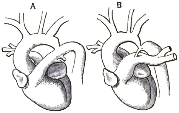
A, Appearance of Aortic Arch in Early Foetal Life.—B, Stenosis of the Aorta.
ETIOLOGY.—The lesion is a congenital one, and results from a defective development of the aorta. In early foetal life the descending aorta is a continuation of the ductus Botalli, and the aortic arch looks like an independent communicating vessel. (See fig. 52, A.) As the arch develops, however, it gradually forms a more direct union with the descending portion, until finally the longitudinal axes of the two parts form one uniform curve and the ductus Botalli becomes a side branch. At birth there is physiologically a slight nicking of the upper wall of the aorta at the point where the two sections are joined, and the stricture we are studying seems to be merely an exaggeration of this physiological mark. Just how the depression becomes established is not clear and the explanations given are not satisfactory.
SYMPTOMS.—Indications of this lesion are usually very obscure or absent, and it is only discovered at the autopsy. Severe headache is sometimes complained of, and dyspnoea, cough, hæmoptysis, and vertigo may occur if the stenosis is excessive.
Physical Signs.—One of the most marked signs is the conspicuous beating of the dilated arteries around the shoulders and ribs. These arteries may be seen and felt. If the patient is very fleshy, however, they may be concealed. There is usually a marked contrast between the arteries of the upper and lower extremities. The former are full and strong, while the latter are weak and barely perceptible. In many cases it is almost impossible to feel any pulse in the abdominal aorta or in the crural arteries. A loud murmur is also described as occurring over the aorta. This murmur is post-systolic, and does not correspond to any of the ordinary aortic murmurs.
DIAGNOSIS.—This lesion has rarely been suspected, much less diagnosed, during life, but a better knowledge of its peculiarities may lead to more frequent recognition of it hereafter. When the collateral circulation is fully established, stenosis of the aorta could hardly be mistaken for anything else. The resulting excessive dilatation of the great vessels at the root of the neck may simulate aneurism, and it should be borne in mind that aneurism is liable to follow stenosis.
PROGNOSIS.—The death of most of the victims of stenosis of the aorta is directly referable to the lesion itself, although the existence of the trouble is compatible with long life and active occupation. The duration of life and the amount of suffering caused by stenosis both depend upon the amount of obstruction in the aorta and the efficiency of the collateral circulation.
In 49 cases death occurred in the following manner:
Rupture of the aorta 10 times.
Rupture of the heart 3 "
Sudden pulmonary oedema 4 "
Cardiac failure 8 "
Apoplexy 4 "
Pneumonia 8 "
Capillary bronchitis 4 "
Paralysis 2 "
Pleurisy 1 time.
No cause assigned 5 times.
49 times.
TREATMENT.—Obviously, no treatment for the lesion itself is possible. If recognized, the existence of the sufferer may be prolonged by adopting moderation in all things as the maxim of his life. Subjective symptoms of discomfort must be combated on general principles as they arise.
DISEASES OF THE CORONARY, PULMONARY, SUPERIOR MESENTERIC, INFERIOR MESENTERIC, AND HEPATIC ARTERIES, AND OF THE COELIAC AXIS.
BY E. G. CUTLER, M.D.
DISEASES OF THE CORONARY ARTERY.
Chronic Endarteritis (Arterio-sclerosis; Atheroma).
This is the most important inflammatory disease of the coronary artery which has been observed. It resembles chronic endarteritis elsewhere, and frequently accompanies the same affection of the aorta, though it may occur alone. The disease may be general, affecting both coronary arteries equally, or one may be more involved than the other, or the disease may be confined to one vessel or to even a small branch.
ETIOLOGY.—Chronic endarteritis of the coronary arteries is especially a disease of middle and advanced life. It occurs most frequently in the male sex. The coronary artery stands fifth in the order of frequency in which the vessels are attacked. The disease is attributed to the misuse of alcoholic drinks, syphilis, chronic lead-poisoning, gout, and chronic kidney disease, by encouraging an early senescence of the tissues, and hence favoring the occurrence of the arterial change.
SYMPTOMS.—There are no symptoms which are peculiar to the disease, those which exist being due to the consecutive changes in the substance of the heart. We may divide cases for convenience of description into those with an acute course and rapid death; those pursuing a subacute course; and, finally, those having a chronic one. In the first instance, sudden death either occurs in a person apparently in perfect health after the manner of a syncope, as in one getting out of bed or standing on the street, while straining at stool, or under sudden emotional excitement. Death may not follow on the instant, but occurs in the course of a longer or shorter time. The attack begins with pressure in the cardiac region, anxiety, restlessness, streaming pain. The complaints and anxiety increase; the breath becomes short and troublesome, the pulse small, frequent, and intermittent; finally, collapse occurs, with oedema of the lung. Death takes place with either a clear mind or slight delirium. Such a fatal ending may cover a day or two or only a few hours. Almost always careful subsequent inquiry elicits the fact that for some time past respiratory or cardiac difficulties have existed, which appeared and disappeared and were not regarded as serious or suspicious. Sudden death may also occur in cases of protracted chronic heart disease following arterio-sclerosis, with an old history of the symptoms of angina pectoris, under the appearance of a fainting fit or of a severe attack of angina or oedema of the lung lasting several days. In such a case rupture of the heart may be found, with bloody infiltration of the cardiac muscle and effusion of blood into the pericardium. In other cases there may be small hemorrhages, often with pronounced infarct formation and softening. In still other cases neither hemorrhage nor infarction is found, but fatty degeneration of the muscle or beginning softening. The sclerosis in such cases is usually very distinct, and affects the trunk and anterior descending branches of the left coronary artery. Sometimes it is hard to find the diseased spot, as it may be circumscribed or on a side branch. In the last-mentioned cases, where sudden death occurs in a chronic process, no post-mortem signs of acute disease are usually found. A chronic fibroid process, with atrophy, exists, which has run a tolerably latent course and leads to death under the appearance of sudden cardiac weakness.
PATHOLOGY.—There are two stages of chronic endarteritis: 1. The stage of simple thickening of the intima; 2. The stage of ulceration and the accompanying further changes.
At first, the normal smooth, shining inner surface of the intima is interrupted here and there or in long stretches by flat rounded elevations, which gradually merge into the healthy surrounding tissues, and are characterized by a paler, more transparent character, and at the same time softer but elastic consistence. The surface of these thickenings, which are frequently located at the point where branches are given off, is either perfectly smooth or slightly wrinkled. Besides these translucent spots there are similar ones which are opaque, whitish or yellowish in color, and have a somewhat rougher surface. Lastly, there are very pronounced thickenings with a yellow color. In the slighter degrees these spots occur singly. In the more pronounced cases they may take up the greater part of the surface; the wall of the vessel is thickened, the inner surface is uneven, and the vessel itself more or less dilated. In the beginning the intima retains its shining surface: after the disease has lasted a long time this is changed, and the second stage appears. Roughnesses, erosions, and ulcerations appear, or more commonly calcification of the wall. This latter appears at first as little thin layers, and finally in large shield-like plates of lime salts, which may occupy the whole circumference of the artery and change it into a stiff, bony tube. It is found where ulceration has occurred, and often without the appearance of the latter. Together with the rigidity of the wall there occurs a slight tortuousness of the vessel. At first the superficial layers of the intima are soft; next they become more sclerosed, and their tissue denser and finally striated; or disintegration, commencing deep in, may reach as far as the surface and lead to an atheromatous ulcer. A more or less abundant deposit of lime salts follows in the sclerosed layers of the intima, leading to the formation of homogeneous plates as hard as bone.
The result of the process at first is diminution of the calibre of the vessel, next diminution of the elasticity and contractility of the artery: it loses its resistance and suffers dilatation in consequence of the blood-pressure, and may attain aneurism. Or if calcification occurs early the diminution of the lumen remains, or perhaps even increases, and may reach an almost complete occlusion of the vessel.
The effects on the heart which follow this form of disease of the coronary artery, though described in another place, had best be enumerated here: 1. The flow of blood not being sufficiently interfered with to cause disease, the heart may remain unchanged. 2. Hemorrhagic infarction may result, accompanied by simple fatty degeneration or softening, which is the most frequent cause of rupture of the heart. 3. Fibrous degeneration or myocarditis may occur, leading perhaps to aneurism of the heart. 4. There may be a combination of these two—a greater or less marked fibrous degeneration, to which a fresh hemorrhagic softening is added.
DIAGNOSIS.—There are no pathognomonic symptoms of this disease, and it is doubtful if a diagnosis can be arrived at. When the conditions spoken of under Etiology pertain, and certain of the symptoms mentioned in connection with the disease are present, a suspicion of chronic endarteritis of the coronary artery may be entertained with some degree of probability.
PROGNOSIS.—This must necessarily be unfavorable where the suspicion of the disease is entertained.
TREATMENT.—Little is to be expected in the way of treatment beyond mere palliation. In the rapid cases death occurs so soon that the medical attendant barely has time to reach the patient. In those cases which last longer the treatment must bear special reference to the symptoms. Pain and spasm may be allayed by opiates or by the inhalation of some anæsthetic cautiously administered, as ether or nitrate of amyl, or by the cautious use of nitro-glycerin and the application of counter-irritants, as mustard, over the cardiac region. Digitalis is to be used with the greatest caution, if at all, as its action may be positively harmful. The same is true of the bromides.
Obliterating Endarteritis.
Besides the preceding, another form of endarteritis has been met with in the coronary artery—namely, the obliterating endarteritis, more especially found in cases of syphilis and occurring in the smallest branches. It is characterized by a gradually increasing thickening of the intima through the formation of a connective tissue rich in cells, and which leads to a narrowing, or even complete closure, of the lumen of the artery. This thickening may involve one side of the artery or its whole circumference. The inner surface of the intima on microscopic examination is found to be covered by a layer of intact endothelium where occlusion is not complete. There is deposit of neither fat nor lime salts in the thickened intima. The outer coats of the artery show little change.
The disease is accompanied by indurating myocarditis. Its symptoms are those seen in this disease—namely, weakened cardiac activity, cardiac dilatation and irregularity, possibly cardiac murmurs, an accentuated pulmonary second sound, a pulse of moderate frequency, weak and non-rhythmical, dyspnoea, cough.
DIAGNOSIS.—Impossible.
TREATMENT.—Purely symptomatic.
Aneurism.
Aneurism of the coronary artery is of rare occurrence. There is no place of election for the disease, all parts and each artery being alike liable to be affected.
ETIOLOGY.—The most common cause of the affection is chronic endarteritis, where, through disease of the intima, the resistance to the blood-pressure is diminished. Embolism is another though far less frequent cause of the disease, several such cases having been reported; and other highly suggestive cases are on record in which embolism of the artery had occurred, with the production of considerable dilatation for a short distance above the obstruction.
PATHOLOGY.—This does not differ from aneurism in other vessels. The number may be from one to many, usually not more than two or three. The size is generally that of a pea, often it is smaller, and sometimes it is as large as a large nut. The termination is usually rupture with fatal hemorrhage, and in far the majority of cases this occurs into the pericardium.
SYMPTOMS.—In most all of the cases I have found recorded there were no symptoms till rupture of the sac occurred, giving rise to death from hemorrhage. Then those symptoms which might be expected occurred—namely, great præcordial pain, dyspnoea, suffocation, tumultuous heart, irregular and intermittent pulse, and sudden death.
DIAGNOSIS, PROGNOSIS, and TREATMENT need not be considered, as the disease is not recognizable.
Occlusion of the Coronary Artery.
Occlusion, more or less complete, of one or both of the orifices of the coronary artery has been met with in connection with chronic endarteritis of the root of the aorta. The accompanying sclerosis may draw the orifices up like the strings of a purse, or a calcific plate may extend from one side, or perhaps, detached, may lie simply applied to the orifice. In rare cases the chief disease may be in the artery itself, one of the main trunks or a branch being affected.
The PATHOLOGY is the same as that already described under Endarteritis, stenosis being an early consequence of the process, and persisting, or even increasing, to the last.
The SYMPTOMS observed in such cases are neither peculiar nor diagnostic. They consist of those depending on the concurrent affections, as of the cardiac valves, muscular tissue, or aortic arterio-sclerosis. Prominent among them are dyspnoea, palpitation, sudden cardiac distress, painful pressure in the region of the heart, great anxiety; at last pallor of the skin, feeble cardiac impulse, indistinctness of the cardiac sounds, the right ventricle continuing to contract forcibly till the end. There is oedema of the lungs at last, and on post-mortem examination fatty degeneration of the heart-walls is found as a secondary consequence of the occlusion.
Embolism and Thrombosis.
Although these conditions are rarely found, yet a sufficient number of cases is already on record to enable us to form a tolerably good idea of the symptoms which accompany them. These latter in embolism remarkably resemble those observed in the lower animals on ligation of the coronary arteries. In the animals experimented on a rapid enfeeblement of the heart's action ensued. The phenomena occurred in the following order: First, there was retardation of the rhythmical cardiac contractions, the left ventricle being primarily affected. At first, the right ventricle beat faster, and then gradually became slow. The beats became slower and slower till they ceased, the left ventricle ceasing to contract a little before the right. The second result was a gradual loss of power of the cardiac contraction. The third result was the gradual distension of the left auricle when the left coronary artery was compressed. The auricle swelled up more and more, became bright red, and the rhythmical contractions changed to oscillatory movements, which ultimately ceased entirely. The right ventricle and auricle continued to contract powerfully, and the left ventricle feebly.
EMBOLISM.
ETIOLOGY.—Rheumatism with its attendant complications—that is, disease of the valves, and especially of the aortic valve, atheroma of the coronary artery and possibly cardiac or other thrombosis—forms the chief cause of embolism, a small fragment of tissue being borne away by the current of blood.
SYMPTOMS.—These are acute paralysis of the heart's movements, pain, feeling of impending annihilation, retained consciousness, and regular respiration. Nausea and vomiting have been observed. The lips are livid, extremities cold and covered with a clammy sweat. In one case there was inability to lie down. No pulse could be felt in any of the accessible arteries, and neither apex-beat nor heart-sounds could be detected. The ear applied to the cardiac region could hear only a kind of cardiac tremor, which was very like the sound of a vibrating steel plate. There was no loss of consciousness. The respiration was regular and rhythmical, not exceeding eighteen or twenty in the minute. The patient died twenty hours after the first symptoms.
DIAGNOSIS.—Although a positive diagnosis is impossible, the negative pulmonary physical signs, the regular and rhythmic character of the respiration, and the enfeeblement of the heart's action may lead to a very strong suspicion of embolism of the coronary artery.
PATHOLOGY.—A small coagulum may stop up the main branch, usually the left anterior, of one artery, or both arteries may be occluded by a larger coagulum. In one instance an atheromatous softened patch ruptured into the anterior portion of the left coronary artery, and filled up the lumen with a soft putty-like mass (the sculptor Thorwaldsen). A fatal issue is likely to occur in a very short time, as the anastomosis cannot be sufficient for the sudden demand.
PROGNOSIS and TREATMENT need hardly be considered, as the affection is necessarily fatal in cases which can be made out.
THROMBOSIS.
The same causes which give rise to thrombosis elsewhere are operative in this case. They are chiefly arterio-sclerosis and rheumatism.
SYMPTOMS.—There have been observed slight tightness in the cardiac region, lasting a few days, or a sense of oppression or constraint at the back of the sternum. The pulse has been quickened, but is usually very much slowed and very feeble; it has been observed as low as eight beats in the minute. There is a sense of great lassitude and feebleness of all the limbs. The respiration is normal in rhythm and frequency. Auscultation reveals nothing but ordinary respiration till near a fatal issue, when moist râles indicative of oedema of the lungs are heard. Percussion gives at all times a normal resonance. There is no dyspnoea. The heart-tones are clear, though weak, if occlusion is not complete or anastomosis is perfect. (West was able to inject the arterial system of the heart completely from one coronary artery, the other having been tied.) If there is complete obstruction, we may expect to hear a fremitus such as is produced by muscular spasm instead of normal heart-sounds (observed in two cases). The skin of the body and face is cool, pallid, and covered with sweat. The visible mucous membranes are anæmic and pale. The mind is clear.
DIAGNOSIS.—The cardiac feebleness and progressive slowness, together with the absence of symptoms connected with the lungs, might lead one to suspect the presence of thrombus.
PROGNOSIS.—If a large branch of the artery is affected a fatal termination is probable. If, on the other hand, the affection occurs in a small branch, there is reason to believe that the circulation is sometimes re-established through anastomosis.
TREATMENT must be purely symptomatic.
Rupture of the Coronary Artery.
This may occur independent of aneurism. There are no premonitory symptoms in some cases, death taking place suddenly. In other cases vague and irregular symptoms lead the patient to understand that he is not in perfect health. The symptoms of the disease are not characteristic. Those which have been recorded are a difficulty of breathing, a sense of constriction across the chest, or a pain and feeling of anxiety in the præcordia; a frequent, feeble, and perhaps very irregular pulse; epigastric pain and tenderness. The extremities are cold. The mind remains clear. The physical signs are increased area of flatness in the cardiac region, due to the escape of blood into the pericardium, and scarcely audible cardiac sounds. The symptoms may extend over a period varying from a few moments to several days. Usually, some of the changes indicative of arterio-sclerosis are found in the artery.
DISEASES OF THE PULMONARY ARTERY.
Acute inflammation of the coats of the pulmonary artery has only been found associated with the pyæmic process as circumscribed abscesses of the wall.
Chronic Endarteritis (Atheroma; Arterio-sclerosis).
Endarteritis of the pulmonary artery, though quite rare, is occasionally met with in persons the subject of rheumatism, gout, syphilis, or alcoholism. It is seen only when the pressure is abnormally increased in the pulmonary vessels, especially in diseases of the mitral valve. It is usually accompanied by a more pronounced disease of the aorta, but is occasionally seen alone. The extent of disease is hardly ever so great as that found in the other large vessels, and at most amounts to the presence of prominent hard yellow or gray patches in the intima, with perhaps ulcerated surfaces, and rarely containing a deposit of lime salts. Complete rigidity has been observed extending far into the lung. The wall of the vessel may be irregularly dilated and its elasticity diminished. Usually, the disease is in a much milder form, presenting perhaps a small amount of fatty degeneration of the intima, and is not infrequently associated with mitral stenosis or insufficiency (notably the former), pulmonary fibrosis or emphysema, with accompanying hypertrophy of the right ventricle. No symptoms have thus far been found to be distinctly referable to atheroma of this artery.
Dilatation and Aneurism.
Dilatation of the pulmonary artery from primary disease of its walls is of so rare occurrence that it may be merely mentioned. It depends on chronic endarteritis, just spoken of. Where, on the other hand, there is great pressure in the pulmonary circulation, as in marked mitral stenosis, or insufficiency, collapse, or emphysema of the lung, with great hypertrophy of the right ventricle, general dilatation of the pulmonary artery may take place. The artery has been found to be six and a half inches in circumference in a case of emphysema, the normal average being three and a half inches; the semilunar valves were insufficient, and the walls of the artery very much diminished in thickness. From this as a maximum all degrees of dilatation have been recorded, with sometimes thickening and degeneration of the coats, at others thinning with or without degeneration.
A systolic murmur has been observed over the artery when the dilatation was considerable. The second pulmonary sound is usually strengthened (unless the elasticity of the pulmonary artery is very much diminished or the blood-pressure lowered in the right ventricle by changes of its walls, or the pulsation is very quick and irregular). A circumscribed dulness on percussion has been found in a few cases at the left edge of the sternum, when the position of the heart was normal, between the second and third cartilages. Sometimes there is a double impulse, a systolic thrill, or more often a systolic pulsation, felt in this position without any perceptible dulness, the edge of the lung being retracted and the dilated artery taking its place. It is to be borne in mind, however, that this sign (impulse, thrill, or pulsation) may be present without any dilatation of the pulmonary artery or hypertrophy of the ventricle, when inflammatory contraction of the lung has occurred or the respiration is superficial, as may happen in phthisical subjects, women, feeble and anæmic individuals, pregnant women, convalescents, and persons afflicted with acute rheumatism. In such persons the pulmonary second sound frequently seems to be unusually loud when compared with the aortic second sound, without any evidence of hypertrophy of the right ventricle being present. The determination of the position of the lung establishes the diagnosis in such cases. On the other hand, a lung dilated by emphysema may interpose and completely cover the heart and pulmonary artery, which, though dilated, may thus be masked.
Aneurism of the trunk or primary branches of the pulmonary artery, on the other hand, is an exceedingly rare disease. But few cases are on record. Aneurisms may be spindle-shaped or sacculated, of moderate size, and are usually situated on the trunk. Lividity of the face, dyspnoea, cough, dysphagia, headache, pain in the chest and epigastrium, are the principal symptoms; and a systolic pulsation (sometimes also diastolic) between the second and third left ribs near the sternum, more or less prominence here, a superficial rough systolic murmur propagated to the left and upward, a purring thrill, and flatness on percussion in the same region and a little above it, are the principal physical signs which have been recorded.
But the physical signs and symptoms above enumerated are not all present in each case, nor are they when present distinctive of pulmonary aneurism. Dysphagia is mentioned in but a single case, dyspnoea is not constant, and cyanosis was at times absent. Also, the physical signs were not constant. Even if all were present they might be produced, as has been the case, by aneurism of the left wall of the aorta, infiltrated lung-tissue, or by a solid tumor lying over the vessels. The locality of the cardiac hypertrophy and dilatation aids in establishing the diagnosis. If it is on the left side of the heart, aneurism of the aorta is indicated; if it is on the right side, pulmonary aneurism. These aneurisms tend to rupture into the pericardium sooner or later.
Dissecting aneurism of the pulmonary artery has been observed once. It was of small extent.1
1 Bul. de la Soc. Anat. de Paris, 1881, pp. 589–591.
Stenosis of the Trunk or Main Branches of the Pulmonary Artery.
Narrowing of the trunk or of one of the main branches of the pulmonary artery is of very rare occurrence. It may follow compression by an aneurism of the ascending or transverse portion of the aorta, compression by tumors in the mediastinum, as from new growths or enlarged glands; it may be caused by cicatricial contraction following mediastinitis, inflammation of a portion of lung or of the bronchial glands, or it may follow disease of the coats of the artery (endarteritis).
The phenomena produced by stenosis of the trunk of the pulmonary artery are similar to those found in stenosis at the orifice, which are treated of in another place. They are anæmia of both lungs, accompanied by persistent dyspnoea with occasional exacerbations (the patient assumes a horizontal position either habitually or during the paroxysm—a fact of true diagnostic importance [Chevers], as in all other forms of disease of the heart and great vessels the patient breathes easier when the shoulders are raised. But in this the dyspnoea results from insufficiency of the supply of blood to the lungs and system generally, and hence the recumbent posture affords relief by removing the impediment of gravity, and thus promotes the supply of blood to the brain), congestion, dilatation and hypertrophy of the right side of the heart, cardiac palpitation, and finally general venous congestion. Hypertrophy of the right ventricle is shown by increase in the transverse measurement of the cardiac area of flatness and increase in the force of the cardiac impulse. The artery up to the point of constriction is dilated; the second sound is abnormally loud and accentuated. Pulsation may be felt and a systolic murmur heard in the second left intercostal space (observed in the right once), propagated upward to the neck at the left of the sternum, or heard in the interscapular space close to the spinal column.
PROGNOSIS is unfavorable.
There is nothing to be gained by treatment.
Rupture of the Pulmonary Artery.
Violent effort and great excitement have been followed by rupture of the trunk or a main branch of the pulmonary artery. In the majority of cases the coats were degenerated, though this was not always the case (Chevers). Death is often instantaneous, but sometimes is delayed some hours. In one case observed by Ollivier the duration was twenty-seven hours.
Thrombosis and Embolism.
The pulmonary artery, from its position, is especially prone to become plugged, either by substances coming from other parts of the body or by coagula originating in the vessel itself. Pieces of disintegrated coagula from the systemic veins, the contents of echinococcus cysts ruptured into the venous current, fragments of new growths, are carried to the heart and pass into the pulmonary artery, or large thrombi may be detached from their position in a vein and lodge in the trunk or main branches of the pulmonary artery.
Primary thrombosis of the pulmonary artery is very uncommon. In certain septic conditions, in parturient women, in typhoid fever, and in extreme anæmia thrombosis of the pulmonary artery may occur. It commences perhaps in the right ventricle or at the pulmonary valves, though it is also seen farther up.
SYMPTOMS.—The severity of the symptoms depends on the completeness of the obstruction. There is dyspnoea, more or less marked according to the size of the thrombus or embolus, pain in the præcordia, great distress, anxiety, faintness, sense of suffocation, tightness in the chest, palpitation, lividity and extreme pallor, cold sweats, an almost imperceptible pulse, great restlessness, and occasionally convulsions. The mind remains clear. The symptoms develop gradually or rapidly—in the former case depending on the slow increase of a small thrombus—and remissions are often seen; in the latter case depending on the sudden lodgment of an embolus of large size. Sometimes the symptoms are extremely marked, and death takes place in a few minutes. The appearances are not those of asphyxia, and death is usually attributed to want of arterial blood-supply to the brain and medulla oblongata, and not to suffocation or paralysis of the heart.
Percussion shows a normally resonant chest. Auscultation gives normal breath sounds with free inspiration and expiration. There is very likely a basic systolic murmur conducted along the course of the pulmonary artery, but this is not constant. The cardiac second sound and impulse are increased. At the post-mortem examination the heart is found in diastole, the left cavities and pulmonary veins empty, the right cavities filled with blood, and the cardiac veins strongly distended.
DIAGNOSIS.—The diagnosis is often uncertain. When not developing with extreme rapidity the symptoms are very similar to those caused by stenosis of the pulmonary artery, and in the suddenly fatal cases they are almost identical with rupture of the heart or rupture of a thoracic aneurism, or even angina pectoris. The history of an antecedent thrombus or of a disease of the heart which is likely to be accompanied by thrombus, together with the absence of physical signs, render a diagnosis many times probable.
PROGNOSIS.—To be regarded as of the gravest character.
TREATMENT.—In the rapid cases death occurs before anything can be attempted. In the less severe cases absolute rest must be enjoined, and free stimulation with brandy, ammonia, and ether attempted. It might be worth while to place the patient with the head lower than the body, to favor the flow of blood to the brain.
DISEASES OF THE SUPERIOR MESENTERIC ARTERY.
Aneurism.
Aneurism of both the superior and the inferior mesenteric arteries occurs. The former is the more frequent, though still a rare disease.
The symptoms are pain in the epigastric and lumbar regions, a globular pulsating tumor in the median line, the pulsation being accompanied by a bellows murmur. The tumor has been seen in at least one instance to be so large as to press on the renal arteries. Rupture is apt to take place with the signs of internal hemorrhage. The cause of the disease is the same as of aneurism elsewhere. Embolism is said to be a not infrequent precedent. The aneurism is seldom larger than a hen's egg, and is usually globular.
A positive DIAGNOSIS of the locality of the aneurism is not possible.
The TREATMENT must follow individual indications. Compression has been successful in a few instances.
Embolism.
Several cases where the superior mesenteric artery was found at autopsy to be completely occluded by coagulated fibrin were mentioned by Tiedemann in a work published in 1843. Virchow first described the characteristic post-mortem appearances which follow this lesion in his Gesammelte Abhandlungen, and since then records of cases have been numerous.