A CIVIC BIOLOGY (continued 6)
Reading edition
Laboratory Suggestions
Demonstration of food tube of man (manikin).—Comparison with food tube of frog. Drawing (comparative) of food tube and digestive glands of frog and man.
Demonstration of simple gland.—(Microscopic preparation.)
Home experiment and laboratory demonstration.—The digestion of starch by saliva. Conditions favorable and unfavorable.
Demonstration experiment.—The digestion of proteins with artificial gastric juice. Conditions favorable and unfavorable.
Demonstration.—An emulsion as seen under the compound microscope.
Demonstration.—Emulsification of fats with artificial pancreatic fluid. Digestion of starch and protein with artificial pancreatic fluid.
Demonstration of "tripe" to show increase of surface of digestive tube.
Laboratory or home exercise.—Make a table showing the changes produced upon food substances by each digestive fluid, the reaction (acid or alkaline) of the fluid, when the fluid acts, and what results from its action.
Purpose of Digestion.—We have learned that starch and protein food of plants are formed in the leaves. A plant, however, is unable to make use of the food in this condition. Before it can be transported from one part of the plant body to another, it is changed into a soluble form. In this state it can be passed from cell to cell by the process of osmosis. Much the same condition exists in animals. In order that food may be of use to man, it must be changed into a state that will allow of its passage in a soluble form through the walls of the alimentary canal, or food tube. This is done by the enzymes which cause digestion. It will be the purpose of this chapter to discover where and how digestion takes place in our own body.
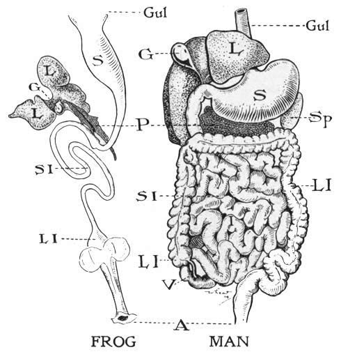
The digestive tract of the frog and man. Gul, gullet; S, stomach; L, liver; G, gall bladder; P, pancreas; Sp, spleen; SI, small intestine; LI, large intestine; V, appendix; A, anus.
Alimentary Canal.—In all vertebrate animals, including man, food is taken in the mouth and passed through a food tube in which it is digested. This tube is composed of different portions, named, respectively, as we pass from the mouth downward, the gullet, stomach, small and large intestine, and rectum.
Comparison of Food Tube of a Frog and Man.—If we compare the food tube of a dissected frog with the food tube of man (as shown by a manikin or chart), we find part for part they are much the same. But we notice that the intestines of man, both small and large, are relatively longer than in the frog. We also notice in man the body cavity or space in which the internal organs rest is divided in two parts by a wall of muscle, the diaphragm, which separates the heart and lungs from the other internal organs. In the frog no muscular diaphragm exists. In the frog we can see plainly the silvery transparent mesentery or double fold of the lining of the body cavity in which the organs of digestion are suspended. Numerous blood vessels can be found especially in the walls of the food tube.
Glands.—In addition to the alimentary canal proper, we find a number of digestive glands, varying in size and position, connected with the canal.
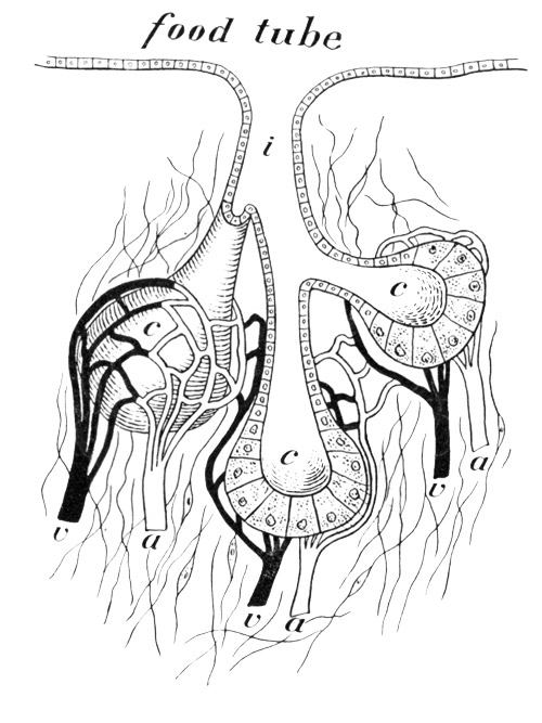
Diagram of a gland. i, the common tube which carries off the secretions formed in the cells lining the cavity c; a, arteries carrying blood to the glands; v, veins taking blood away from the glands.
What a Gland Does. Enzymes.—In man there are the saliva gland of the mouth, the gastric glands of the stomach, the pancreas and liver, the two latter connected with the small intestine, and the intestinal glands in the walls of the intestine. Besides glands which aid in digestion there are several others of which we will speak later. As we have already learned, a gland is a collection of cells which takes up material from within the body and manufactures from it something which is later poured out as a secretion. An example of a gland in plants is found in the nectar-secreting cells of a flower.
Certain substances, called enzymes, formed by glands cause the digestion of food. The enzymes secreted by the cells of the glands and poured out into the food tube act upon insoluble foods so as to change them to a soluble form. They are the product of the activity of the cell, although they are not themselves alive. We do not know much about enzymes themselves, but we can observe what they do. Some enzymes render soluble different foods, others work in the blood, still others probably act within any cell of the body as an aid to oxidation, when work is done. Enzymes are very sensitive to changes in temperature and to the degree of acidity or alkalinity[42] of the material in which they act. We will find that the enzymes found in glands in the mouth will not act long in the stomach because of the change from an alkaline surrounding in the mouth to that of an acid in the stomach. Enzymes seem to be able to work indefinitely, providing the surroundings are favorable. A small amount of digestive fluid, if it had long enough to work, could therefore digest an indefinite amount of food.
Gland Structure.—The entire inner surface of the food tube is covered with a soft lining of mucous membrane. This is always moist because certain cells, called mucus cells, empty out their contents into the food tube, thus lubricating its inner surface. When a large number of cells which have the power to secrete fluids are collected together, the surface of the food tube may become indented at this point to form a pitlike gland. Often such depressions are branched, thus giving a greater secreting surface, as is seen in the figure on page 298. The cells of the gland are always supplied with blood vessels and nerves, for the secretions of the glands are under the control of the nervous system.
How a Gland Secretes.—We must therefore imagine that as the blood goes to the cells of a gland it there loses some substances which the gland cells take out and make over into the particular enzyme that they are called upon to manufacture. Under certain conditions, such as the sight or smell of food, or even the desire for it, the activity of the gland is stimulated. It then pours out its secretion containing the digestive enzyme. Thus a gland does its work.
Salivary Glands.—We are all familiar with the substance called saliva which acts as a lubricant in the mouth. Saliva is manufactured in the cells of three pairs of glands which empty into the mouth, and which are called, according to their position, the parotid (beside the ear), the submaxillary (under the jawbone), and the sublingual (under the tongue).
Digestion of Starch.—If we collect some saliva in a test tube, add to it a little starch paste, place the tube containing the mixture for a few minutes in tepid water, and then test with Fehling's solution, we shall find grape sugar present. Careful tests of the starch paste and of the saliva made separately will usually show no grape sugar in either.
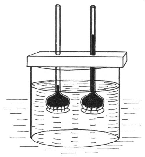
Experiment showing non-osmosis of starch in tube A, and osmosis of sugar in tube B.
If another test be made for grape sugar, in a test tube containing starch paste, saliva, and a few drops of any weak acid, the starch will be found not to have changed. The digestion or change of starch to grape sugar is caused by the presence in the saliva of an enzyme, or digestive ferment. You will remember that starch in the growing corn grain was changed to grape sugar by an enzyme called diastase. Here a similar action is caused by an enzyme called ptyalin. This ferment acts only in an alkaline medium at about the temperature of the body.
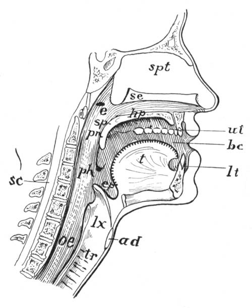
The mouth cavity of man. e, Eustachian tube; hp, hard palate; sp, soft palate; ut, upper teeth; bc, buccal cavity; lt, lower teeth; t, tongue; ph, pharynx; ep, epiglottis; lx, voice box; oe, gullet; tr, trachea.
Mouth Cavity in Man.—In our study of a frog we find that the mouth cavity has two unpaired and four paired tubes leading from it. These are (a) the gullet or food tube, (b) the windpipe (in the frog opening through the glottis), (c) the paired nostril holes (posterior nares), (d) the paired Eustachian tubes, leading to the ear. All of these openings are found in man.
In man the mouth cavity, and all internal surfaces of the food tube, are lined with a mucous membrane. The mucus secreted from gland cells in this lining makes a slippery surface so that the food may slip down easily. The roof of the mouth is formed in front by a plate of bone called the hard palate, and a softer continuation to the back of the mouth, the soft palate. These separate the nose cavity from that of the mouth proper. The part of the space back of the soft palate is called the pharynx, or throat cavity. From the pharynx lead off the gullet and windpipe, the former back of the latter. The lower part of the mouth cavity is occupied by a muscular tongue. Examination of its surface with a looking-glass shows it to be almost covered in places by tiny projections called papillæ. These papillæ contain organs known as taste buds, the sensory endings of which determine the taste of substances. The tongue is used in moving food about in the mouth, and in starting it on its way to the gullet; it also plays an important part in speaking.
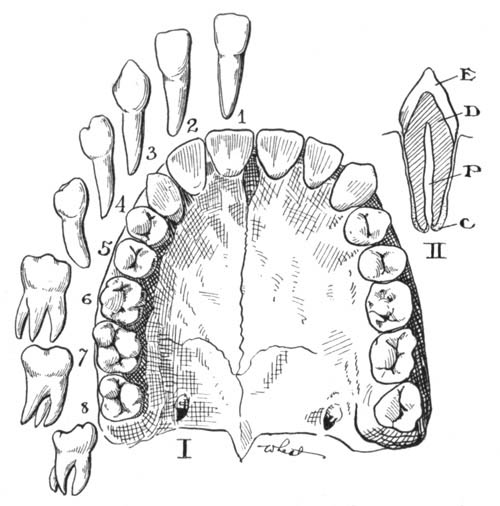
I. Teeth of the upper jaw, from below. 1, 2, incisors; 3, canine; 4, 5, premolars; 6, 7, 8, molars. II. longitudinal section of a tooth. E, enamel; D, dentine; C, cement; P, pulp cavity.
The Teeth.—In man the teeth, unlike those of the frog, are used in the mechanical preparation of the food for digestion. Instead of holding prey, they crush, grind, or tear food so that more surface may be given for the action of the digestive fluids. The teeth of man are divided, according to their functions, into four groups. In the center of both the upper and lower jaw in front are found eight teeth with chisel-like edges, four in each jaw; these are the incisors, or cutting teeth. Next is found a single tooth on each side (four in all); these have rather sharp points and are called the canines. Then come two teeth on each side, eight in all, called premolars. Lastly, the flat-top molars, or grinding teeth, of which there are six in each jaw. Food is caught between irregular projections on the surface of the molars and crushed to a pulpy mass.
Hygiene of the Mouth.—Food should simply be chewed and relished, with no thought of swallowing. There should be no more effort to prevent than to force swallowing. It will be found that if you attend only to the agreeable task of extracting the flavors of your food, Nature will take care of the swallowing, and this will become, like breathing, involuntary. The instinct by which most people eat is perverted through the "hurry habit" and the use of abnormal foods. Thorough mastication takes time, and therefore one must not feel hurried at meals if the best results are to be secured. The stopping point for eating should be at the earliest moment after one is really satisfied.
Care of the Teeth.—It has been recently found that fruit acids are very beneficial to the teeth. Vinegar diluted to about half strength with water makes an excellent dental wash. Clean your teeth carefully each morning and before going to bed. Use dental silk after meals. We must remember that the bacteria which cause decay of the teeth are washed down into the stomach and may do even more harm there than in the mouth.
How Food is Swallowed.—After food has been chewed and mixed with saliva, it is rolled into little balls and pushed by the tongue into such position that the muscles of the throat cavity may seize it and force it downward. Food, in order to reach the gullet from the mouth cavity, must pass over the opening into the windpipe. When food is in the course of being swallowed, the upper part of this tube forms a trapdoor over the opening. When this trapdoor is not closed, and food "goes down the wrong way," we choke, and the food is expelled by coughing.
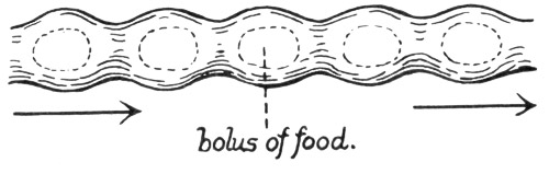
Peristaltic waves on the gullet of man. (A bolus means little ball.)
The Gullet, or Esophagus.—Like the rest of the food tube the gullet is lined by soft and moist mucous membrane. The wall is made up of two sets of muscles,—the inside ones running around the tube; the outer layer of muscle taking a longitudinal course. After food leaves the mouth cavity, it gets beyond our direct control, and the muscles of the gullet, stimulated to activity by the presence of food in the tube, push the food down to the stomach by a series of contractions until it reaches the stomach. These wavelike movements (called peristaltic movements) are characteristic of other parts of the food tube, food being pushed along in the stomach and the small intestine by a series of slow-moving muscular waves. Peristaltic movement is caused by muscles which are not under voluntary nervous control, although anger, fear, or other unpleasant emotions have the effect of slowing them up or even stopping them entirely.
Stomach of Man.—The stomach is a pear-shaped organ capable of holding about three pints. The end opposite to the gullet, which empties into the small intestine, is provided with a ring of muscle forming a valve called the pylorus. There is also another ring of muscle guarding the entrance to the stomach.
Gastric Glands.—If we open the stomach of the frog, and remove its contents by carefully washing, its wall is seen to be thrown into folds internally. Between the folds in the stomach of man, as well as in the frog, are located a number of tiny pits. These form the mouths of the gastric glands, which pour into the stomach a secretion known as the gastric juice. The gastric glands are little tubes, the lining of which secretes the fluid. When we think of or see appetizing food, this secretion is given out in considerable quantity. The stomach, like the mouth, "waters" at the sight of food. Gastric juice is slightly acid in its chemical reaction, containing about .2 per cent free hydrochloric acid. It also contains two very important enzymes, one called pepsin, and another less important one called rennin.
Action of Gastric Juice.—If protein is treated with artificial gastric juice at the temperature of the body, it will be found to become swollen and then gradually to change to a substance which is soluble in water. This is like the action of the gastric juice upon proteins in the stomach.
The other enzyme of gastric juice, called rennin, curdles or coagulates a protein found in milk; after the milk is curdled, the pepsin is able to act upon it. "Junket" tablets, which contain rennin, are used in the kitchen to cause this change.
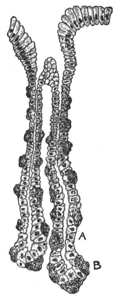
A peptic gland, from the stomach, very much magnified. A, central or chief cell, which makes pepsin; B, border cells, which make acid. (From Miller's Histology.)
The hydrochloric acid found in the gastric juice acts upon lime and some other salts taken into the stomach with food, changing them so that they may pass into the blood and eventually form the mineral part of bone or other tissue. The acid also has a decided antiseptic influence in preventing growth of bacteria which cause decay, and some of which might cause disease.
Movement of Walls of Stomach.—The stomach walls, provided with three layers of muscle which run in an oblique, circular, and longitudinal direction (taken from the inside outward), are well fitted for the constant churning of the food in that organ. Here, as elsewhere in the digestive tract, the muscles are involuntary, muscular action being under the control of the so-called sympathetic nervous system. Food material in the stomach makes several complete circuits during the process of digestion in that organ. Contrary to common belief, the greatest amount of food is digested after it leaves the stomach. But this organ keeps the food in it in almost constant motion for a considerable time, a meal of meat and vegetables remaining in the stomach for three or four hours. While movement is taking place, the gastric juice acts upon proteins, softening them, while the constant churning movement tends to separate the bits of food into finer particles. Ultimately the semifluid food, much of it still undigested, is allowed to pass in small amounts through the pyloric valve, into the small intestine. This is allowed by the relaxation of the ringlike muscles of the pylorus.
Experiments on Digestion in the Stomach.—Some very interesting experiments have recently been made by Professor Cannon of Harvard with reference to movements of the stomach contents. Cats were fed with material having in it bismuth, a harmless chemical that would be visible under the X-ray. It was found that shortly after food reached the stomach a series of waves began which sent the food toward the pyloric end of the stomach. If the cat was feeling happy and well, these contractions continued regularly, but if the cat was cross or bad tempered, the movements would stop. This shows the importance of cheerfulness at meals. Other experiments showed that food which was churned into a soft mass was only permitted to leave the stomach when it became thoroughly permeated by the gastric juice. It is the acid in the partly digested food that causes the stomach valve to open and allow its contents to escape little by little into the small intestine.
The partly digested food in the small intestine almost immediately comes in contact with fluids from two glands, the liver and pancreas. We shall first consider the function of the pancreas.
Position and Structure of the Pancreas.—The most important digestive gland in the human body is the pancreas. The gland is a rather diffuse structure; its duct empties by a common opening with the bile duct into the small intestine, a short distance below the pylorus. In internal structure, the pancreas resembles the salivary glands.
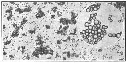
Appearance of milk under the microscope, showing the natural grouping of the fat globules. In the circle a single group is highly magnified. Milk is one form of an emulsion. (S. M. Babcock, Wis. Bul. No. 61.)
Work done by the Pancreas.—Starch paste added to artificial pancreatic fluid and kept at blood heat is soon changed to sugar. Protein, under the same conditions, is changed to a peptone. Fats, which so far have been unchanged except to be melted by the heat of the body, are changed by the action of the pancreas into a form which can pass through the walls of the food tube. If we test pancreatic fluid, we find it strongly alkaline in its reaction. If two test tubes, one containing olive oil and water, the other olive oil and a weak solution of caustic soda, an alkali, be shaken violently and then allowed to stand, the oil and water will quickly separate, while the oil, caustic soda, and water will remain for some time in a milky emulsion. If this emulsion be examined under the microscope, it will be found to be made of millions of little droplets of fat, floating in the liquid. The presence of the caustic soda helped the forming of the emulsion. Pancreatic fluid similarly emulsifies fats and changes them into soft soaps and fatty acids. Fat in this form may be absorbed. The process of this transformation is not well understood.
Conditions under which the Pancreas does its Work.—The secretion from this gland seems to be influenced by the overflow of acid material from the stomach. This acid, on striking the lining of the small intestine, causes the formation in its walls of a substance known as secretin. This secretin reaches the blood and seems to stimulate all the glands pouring fluid into the intestine to do more work. A pint or more of pancreatic fluid is secreted every day.
The Intestinal Fluid.—Three different pancreatic enzymes do the work of digestion, one acting on starch, another on protein, and a third on fats. It has been found that some of these enzymes will not do their work unless aided by the intestinal fluid, a secretion formed in glands in the walls of the small intestine. This fluid, though not much is known about it, is believed to play an important part in the digestion of all kinds of foods left undigested in the small intestine.
Liver.—The liver is the largest gland in the body. In man, it hangs just below the diaphragm, a little to the right side of the body. During life, its color is deep red. It is divided into three lobes, between two of which is found the gall bladder, a thin-walled sac which holds the bile, a secretion of the liver. Bile is a strongly alkaline fluid of greenish color. It reaches the intestine through the same opening as the pancreatic fluid. Almost one quart of bile is passed daily into the digestive canal. The color of bile is due to certain waste substances which come from the destruction of worn-out red corpuscles of the blood. This destruction takes place in the liver.
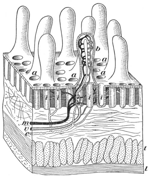
Diagram of a bit of the wall of the small intestine, greatly magnified, a, mouths of intestinal glands; b, villus cut lengthwise to show blood vessels and lacteal (in center); e, lacteal sending branches to other villi; i, intestinal glands; m, artery; v, vein; l, t, muscular coats of intestine wall.
Functions of Bile.—The action of bile is not very well known. It has the very important faculty of aiding the pancreatic fluid in digestion, though alone it has slight if any digestive power. Certain substances in the bile aid especially in the absorption of fats. Bile seems to be mostly a waste product from the blood and as such incidentally serves to keep the contents of the intestine in a more or less soft condition, thus preventing extreme constipation.
The Liver a Storehouse.—Perhaps the most important function of the liver is the formation within it of a material called glycogen, or animal starch. The liver is supplied by blood from two sources. The greater amount of blood received by the liver comes directly from the walls of the stomach and intestine to this organ. It normally contains about one fifth of all the blood in the body. This blood is very rich in food materials, and from it the cells of the liver take out sugars to form glycogen.[43] Glycogen is stored in the liver until such a time as a food is needed that can be quickly oxidized; then it is changed to sugar and carried off by the blood to the tissue which requires it, and there used for this purpose. Glycogen is also stored in the muscles, where it is oxidized to release energy when the muscles are exercised.
The Absorption of Digested Food into the Blood.—The object of digestion is to change foods from an insoluble to a soluble form. This has been seen in the study of the action of the various digestive fluids in the body, each of which is seen to aid in dissolving solid foods, changing them to a fluid, and, in case of the bile, actually assisting them to pass through the wall of the intestine. A small amount of digested food may be absorbed by the blood in the blood vessels of the walls of the stomach. Most of the absorption, however, takes place through the walls of the small intestine.
Structure of the Small Intestine.—The small intestine in man is a slender tube nearly twenty feet in length and about one inch in diameter. If the chief function of the small intestine is that of absorption, we must look for adaptations which increase the absorbing surface of the tube. This is gained in part by the inner surface of the tube being thrown into transverse folds which not only retard the rapidity with which food passes down the intestine, but also give more absorbing surface. But far more important for absorption are millions of little projections which cover the inner surface of the small intestine.
The Villi.—So numerous are these projections that the whole surface presents a velvety appearance. Collectively, these structures are called the villi (singular villus). They form the chief organs of absorption in the intestine, several thousand being distributed over every square inch of surface. By means of the folds and villi the small intestine is estimated to have an absorbing surface equal to twice that of the surface of the body. Between the villi are found the openings of the intestinal glands.
Internal Structure of a Villus.—The internal structure of a villus is best seen in a longitudinal section. We find the outer wall made up of a thin layer of cells, the epithelial layer. It is the duty of these cells to absorb the semifluid food from within the intestine. Underneath these cells lies a network of very tiny blood vessels, while inside of these, occupying the core of the villus, are found spaces which, because of their white appearance after absorption of fats, have been called lacteals. (See figure, page 307.[TN6])
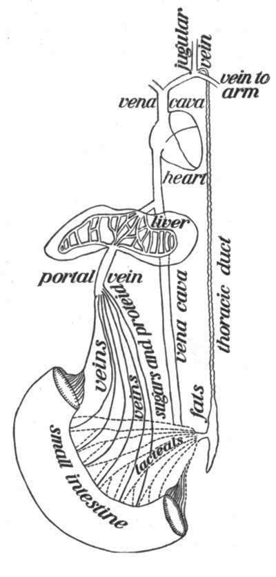
Diagram to show how the nutrients reach the blood.
Absorption of Foods.—Let us now attempt to find out exactly how foods are passed from the intestines into the blood. Food substances in solution may be soaked up as a sponge would take up water, or they may pass by osmosis into the cells lining the villus. These cells break down the peptones into a substance that will pass into and become part of the blood. Once within the villus, the sugars and digested proteins pass through tiny blood vessels into the larger vessels comprising the portal circulation. These pass through the liver, where, as we have seen, sugar is taken from the blood and stored as glycogen. From the liver, the food within the blood is sent to the heart, from there is pumped to the lungs, from there returns to the heart, and is pumped to the tissues of the body. A large amount of water and some salts are also absorbed through the walls of the stomach and intestine as the food passes on its course. The fats in the form of soaps and fatty acids pass into the space in the center of the villus. Later they are changed into fats again, probably in certain groups of gland cells known as mesenteric glands, and eventually reach the blood by way of the thoracic duct without passing through the liver.
Large Intestine.—The large intestine has somewhat the same structure as the small intestine, except that it lacks the villi and has a greater diameter. Considerable absorption, however, takes place through its walls as the mass of food and refuse material is slowly pushed along by the muscles within its walls.
Vermiform Appendix.—At the point where the small intestine widens to form the large intestine, a baglike pouch is formed. From one side of this pouch is given off a small tube about four inches long, closed at the lower end. This tube, the rudiment of what is an important part of the food tube in the lower vertebrates, is called the vermiform appendix. It has come to have unpleasant notoriety in late years, as the site of serious inflammation.
Constipation.—In the large intestine live millions of bacteria, some of which make and give off poisonous substances known as toxins. These substances are easily absorbed through the walls of the large intestine, and, when they pass into the blood, cause headaches or sometimes serious trouble. Hence it follows that the lower bowel should be emptied of this matter as frequently as possible, at least once a day. Constipation is one of the most serious evils the American people have to deal with, and it is largely brought about by the artificial life which we lead, with its lack of exercise, fresh air, and sleep. Fruit with meals, especially at breakfast, plenty of water between meals and before breakfast, exercise, particularly of the abdominal muscles, and regular habits will all help to correct this evil.
Hygienic Habits of Eating; the Causes and Prevention of Dyspepsia.—From the contents of the foregoing chapter it is evident that the object of the process of digestion is to break up solid food so that it may be absorbed to form part of the blood. Any habits we may form of thoroughly chewing our food will evidently aid in this process. Undoubtedly much of the distress known as dyspepsia is due to too hasty meals with consequent lack of proper mastication of food. The message of Mr. Horace Fletcher in bringing before us the need of proper mastication of food and the attendant evils of overeating is one which we cannot afford to ignore. It is a good rule to go away from the table feeling a little hungry. Eating too much overtaxes the digestive organs and prevents their working to the best advantage. Still another cause of dyspepsia is eating when in a fatigued condition. It is always a good plan to rest a short time before eating, especially after any hard manual work. We have seen how great a part unpleasant emotions play in preventing peristaltic movements of the food tube. Conversely, pleasant conversation, laughter, and fun will help you to digest your meal. Eating between meals is condemned by physicians because it calls the blood to the digestive organs at a time when it should be more active in other parts of the body.
Effect of Alcohol on Digestion.—It is a well-known fact that alcohol extracts water from tissues with which it is in contact. This fact works much harm to the interior surface of the food tube, especially the walls of the stomach, which in the case of a hard drinker are likely to become irritated and much toughened. In very small amounts alcohol stimulates the secretion of the salivary and gastric glands, and thus appears to aid in digestion.
The following results of experiments on dogs, published in the American Journal of Physiology, Vol. I, Professor Chittenden of Yale University gives as "strictly comparable," because "they were carried out in succession on the same day." They show that alcohol retards rather than aids in digestion:—
Number of Experiment 1/16 Lb. Meat With Water 1/16 Lb. Meat With Dilute Alcohol
XVII α 9:15 A.M. Digested in 3 hours
XVII β 3:00 P.M. Digested in 3:15 hours
XVIII α 8:30 A.M. Digested in 2:30 hours
XVIII β 2:10 P.M. Digested in 3:00 hours
XIX α 9:00 A.M. Digested in 2:30 hours
XIX β 2:30 P.M. Digested in 3:00 hours
XX α 9:15 A.M. Digested in 2:45 hours
XX β 2:30 P.M. Digested in 2:15 hours
VI α 9:15 A.M. Digested in 3:45 hours
VI β 1:00 P.M. Digested in 3:15 hours
Average 2:42 hours 3:09 hours
As a result of his experiments, Professor Chittenden remarks: "We believe that the results obtained justify the conclusion that gastric digestion as a whole is not materially modified by the introduction of alcoholic fluids with the food. In other words, the unquestionable acceleration of gastric secretion which follows the ingestion of alcoholic beverages is, as a rule, counterbalanced by the inhibitory effect of the alcoholic fluids upon the chemical process of gastric digestion, with perhaps at times a tendency towards preponderance of inhibitory action." Others have come to the same or stronger conclusions as to the undesirable action of alcohol on digestion, as a result of their own experiments.
Effect of Alcohol on the Liver.—The effect of heavy drinking upon the liver is graphically shown in the following table prepared by the Scientific Temperance Federation of Boston, Mass.:—
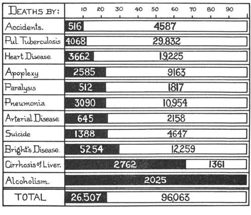
Proportion of deaths from disease in a certain area due to alcohol. The black area shows deaths due to alcohol.[44]
"Alcoholic indulgence stands almost if not altogether in the front rank of the enemies to be combated in the battle for health."—Professor William T. Sedgwick.
XXI. THE BLOOD AND ITS CIRCULATION
Problems.—To discover the composition and uses of the different parts of the blood.
To find out the means by which the blood is circulated about the body.
Laboratory Suggestions
Demonstration.—Structure of blood, fresh frog's blood and human blood. Drawings.
Demonstration.—Clotting of blood.
Demonstration.—Use of models to demonstrate that the heart is a force pump.
Demonstration.—Capillary circulation in web of frog's foot or tadpole's tail. Drawing.
Home or laboratory exercise.—On relation of exercise on rate of heart beat.
Function of the Blood.—The chief function of the digestive tract is to change foods to such form that they can be absorbed through the walls of the food tube and become part of the blood.[45]
If we examine under the microscope a drop of blood taken from the frog or man, we find it made up of a fluid called plasma and two kinds of bodies, the so-called red corpuscles and colorless corpuscles, floating in this plasma.
Composition of Plasma.—The plasma of blood is found to be largely (about 90 per cent) water. It also contains a considerable amount of protein, some sugar, fat, and mineral material. It is, then, the medium which holds the fluid food that has been absorbed from within the intestine. This food is pumped to the body cells where, as work is performed, oxidation takes place and heat is given off as a form of energy. The almost constant temperature of the body is also due to the blood, which brings to the surface of the body much of the heat given off by oxidation of food in the muscles and other tissues. When the blood returns from the tissues where the food is oxidized, the plasma brings back with it to the lungs part of the carbon dioxide liberated where oxidation has taken place. Some waste products, to be spoken of later, are also found in the plasma.
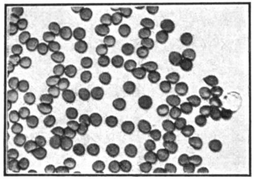
Human blood as seen under the high power of the compound microscope; at the extreme right is a colorless corpuscle.
The Red Blood Corpuscle; its Structure and Functions.—The red corpuscle in the blood of the frog is a true cell of disklike form, containing a nucleus. The red corpuscle of man is made in the red marrow of bones and in its young stages has a nucleus. In its adult form, however, it lacks a nucleus. Its form is that of a biconcave disk. So small and so numerous are these corpuscles that over five million are found in a cubic centimeter of normal blood. They make up almost one half the total volume of the blood. The color, which is found to be a dirty yellow when separate corpuscles are viewed under the microscope, is due to a protein material called hæmoglobin. Hæmoglobin contains a large amount of iron. It has the power of uniting very readily with oxygen whenever that gas is abundant, and, after having absorbed it, of giving it up to the surrounding media, when oxygen is there present in smaller amounts than in the corpuscle. This function of carrying oxygen is the most important function of the red corpuscle, although the red corpuscle also removes part of the carbon dioxide from the tissues on their return to the lungs. The taking up of oxygen is accompanied by a change in color of the mass of corpuscles from a dull red to a bright scarlet.
Clotting of Blood.—If fresh beef blood is allowed to stand overnight, it will be found to have separated into two parts, a dark red, almost solid clot and a thin, straw-colored liquid called serum. Serum is found to be made up of about 90 per cent water, 8 per cent protein, 1 per cent other organic foods, and 1 per cent mineral substances. In these respects it very closely resembles the fluid food that is absorbed from the intestines.
If another jar of fresh beef blood is poured into a pan and briskly whipped with a bundle of little rods (or with an egg beater), a stringy substance will be found to stick to the rods. This, if washed carefully, is seen to be almost colorless. Tested with nitric acid and ammonia, it is found to contain a protein substance which is called fibrin.
Blood plasma, then, is made up of a fluid portion of serum, and fibrin, which, although in a fluid state in the blood vessels within the body, coagulates when blood is removed from the blood vessels. This coagulation aids in making a blood clot. A clot is simply a mass of fibrin threads with a large number of corpuscles tangled within. The clotting of blood is of great physiological importance, for otherwise we might bleed to death even from a small wound.
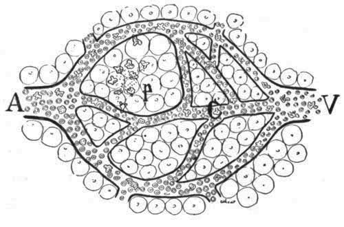
A small artery (A) breaking up into capillaries (c) which unite to form a vein (V). Note at (P) several colorless corpuscles, which are fighting bacteria at that point.
Blood Plates.—In blood within the circulatory system of the body, the fibrin is held in a fluid state called fibrinogen. An enzyme, acting upon this fibrinogen, the soluble protein in the blood, causes it to change to an insoluble form, the fibrin of the clot. This change seems to be due to the action of minute bodies in the blood known as blood plates. Under abnormal conditions these blood plates break down, releasing some substances which eventually cause this enzyme to do its work.
The Colorless Corpuscle; Structure and Functions.—A colorless corpuscle is a cell irregular in outline, the shape of which is constantly changing. These corpuscles are somewhat larger than the red corpuscles, but less numerous, there being about one colorless corpuscle to every three hundred red ones. They have the power of movement, for they are found not only inside but outside the blood vessels, showing that they have worked their way between the cells that form the walls of the blood tubes.
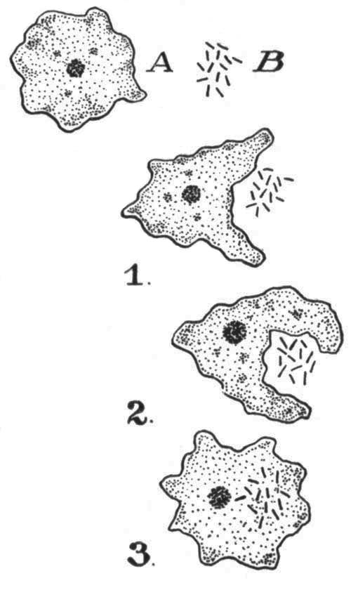
A colorless corpuscle catching and eating germs.
A Russian zoölogist, Metchnikoff, after studying a number of simple animals, such as medusæ and sponges, found that in such animals some of the cells lining the inside of the food cavity take up or engulf minute bits of food. Later, this food is changed into the protoplasm of the cell. Metchnikoff believed that the colorless corpuscles of the blood have somewhat the same function. This he later proved to be true. Like the amœba, they feed by engulfing their prey. This fact has a very important bearing on the relation of colorless corpuscles to certain diseases caused by bacteria within the body. If, for example, a cut becomes infected by bacteria, inflammation may set in. Colorless corpuscles at once surround the spot and attack the bacteria which cause the inflammation. If the bacteria are few in number, they are quickly eaten by certain of the colorless corpuscles, which are known as phagocytes. If bacteria are present in great quantities, they may prevail and kill the phagocytes by poisoning them. The dead bodies of the phagocytes thus killed are found in the pus, or matter, which accumulates in infected wounds. In such an event, we must come to the aid of nature by washing the wound with some antiseptic, as weak carbolic acid or hydrogen peroxide.
Antibodies and their Uses.—In case of disease where, for example, fever is caused by poison given off from bacteria we find the cells of the body manufacture and pour into the blood a substance known as an antibody. This substance does not of necessity kill the harmful germs or even stop their growth. It does, however, unite with the toxin or poison given off by the germs and renders it entirely harmless.
Function of Lymph.—The tissues and organs of the body are traversed by a network of tubes which carry the blood. Inside these tubes is the blood proper, consisting of a fluid plasma, the colorless corpuscles, and the red corpuscles. Outside the blood tubes, in spaces between the cells which form tissues, is found another fluid, which is in chemical composition very much like plasma of the blood. This is the lymph. It is, in fact, fluid food in which some colorless amœboid corpuscles are found. Blood gives up its food material to the lymph. This it does by passing it through the walls of the capillaries. The food is in turn given up to the tissue cells, which are bathed by the lymph.
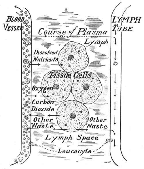
The exchange between blood and the cells of the body.
Some of the amœboid corpuscles from the blood make their way between the cells forming the walls of the capillaries. Lymph, then, is practically blood plasma plus some colorless corpuscles. It acts as the medium of exchange between the blood proper and the cells in the tissues of the body. By means of the food supply thus brought, the cells of the body are able to grow, the fluid food being changed to the protoplasm of the cells. By means of the oxygen passed over by the lymph, oxidation may take place within the cells. Lymph not only gives food to the cells of the body, but also takes away carbon dioxide and other waste materials, which are ultimately passed out of the body by means of the lungs, skin, and kidneys.
Internal Secretions.—In addition to all the functions given above, the blood has recently been shown to carry the secretions of a number of glands through which it passes, although these glands have no ducts to carry off their secretions. These internal secretions seem absolutely necessary for the health of the body. Several glands, the thyroid, adrenal bodies, the testes, and ovaries, as well as the pancreas, give off these remarkable substances.
The Amount of Blood and its Distribution.—Blood forms, by weight, about one sixteenth of the body. This would be about four quarts to a body weight of 130 pounds. Normally, about one half of the blood of the body is found in or near the organs lying in the body cavity below the diaphragm, about one fourth in the muscles, and the rest in the head, heart, lungs, large arteries, and veins.
Blood Temperature.—The temperature of blood in the human body is normally about 98.6° Fahrenheit when tested under the tongue by a thermometer, although the temperature drops almost two degrees after we have gone to sleep at night. It is highest about 5 P.M. and lowest about 4 A.M. In fevers, the temperature of the body sometimes rises to 107°; but unless this temperature is soon reduced, death follows. Any considerable drop in temperature below the normal also means death. Body heat results from the oxidation of food, and the circulation of blood keeps the temperature nearly uniform in all parts of the body.
Cold-blooded Animals.—In animals which are called cold-blooded, the blood has no fixed temperature, but varies with the temperature of the medium in which the animal lives. Frogs, in the summer, may sit for hours in water with a temperature of almost 100°. In winter, they often endure freezing so that the blood and lymph within the spaces under the loose skin are frozen into ice crystals. This change in body temperature is evidently an adaptation to the mode of life.
Circulation of the Blood in Man.—The blood is the carrying agent of the body. Like a railroad or express company, it takes materials from one part of the human organism to another. This it does by means of the organs of circulation,—the heart and blood vessels. These blood vessels are called arteries where they carry blood away from the heart, veins where they bring blood back to the heart, and capillaries where they connect the larger blood vessels. The organs of circulation thus form a system of connected tubes through which the blood flows.
The Heart; Position, Size, Protection.—The heart is a cone-shaped muscular organ about the size of a man's fist. It is located immediately above the diaphragm, and lies so that the muscular apex, which points downward, moves while beating against the fifth and sixth ribs, just a little to the left of the midline of the body. This fact gives rise to the notion that the heart is on the left side of the body. The heart is surrounded by a loose membranous bag called the pericardium, the inner lining of which secretes a fluid in which the heart lies. When, for any reason, the pericardial fluid is not secreted, inflammation arises in that region.
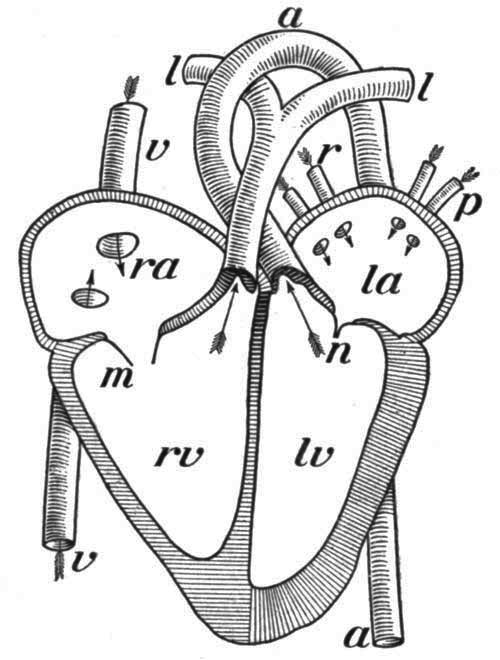
Diagram showing the front half of the heart cut away: a, aorta; l, arteries to the lungs; la, left auricle; lv, left ventricle; m, tricuspid valve open; n, bicuspid or mitral valve closed; p and r, veins from the lungs; ra, right auricle; rv, right ventricle; v, vena cava. Arrows show direction of circulation.
Internal Structure of Heart.—If we should cut open the heart of a mammal down the midline, we could divide it into a right and a left side, each of which would have no internal connection with the other. Each side is made up of an upper thin-walled portion with a rather large internal cavity, the auricle, which opens into a lower smaller portion with heavy muscular walls, the ventricle. Communication between auricles and ventricles is guarded by little flaps or valves. The auricles receive blood from the veins. The ventricles pump the blood into the arteries.
The Heart in Action.—The heart is constructed on the same plan as a force pump, the valves preventing the reflux of blood into the auricle when it is forced out of the ventricle. Blood enters the auricles from the veins because the muscles of that part of the heart relax; this allows the space within the auricles to fill. Almost immediately the muscles of the ventricles relax, thus allowing blood to pass into the chambers within the ventricles. Then, after a short pause, during which time the muscles of the heart are resting, a wave of muscular contraction begins in the auricles and ends in the ventricles, with a sudden strong contraction which forces the blood out into the arteries. Blood is kept on its course by the valves, which act in the same manner as do the valves in a pump. The blood is thus made to pass into the arteries upon the contraction of the ventricle walls.
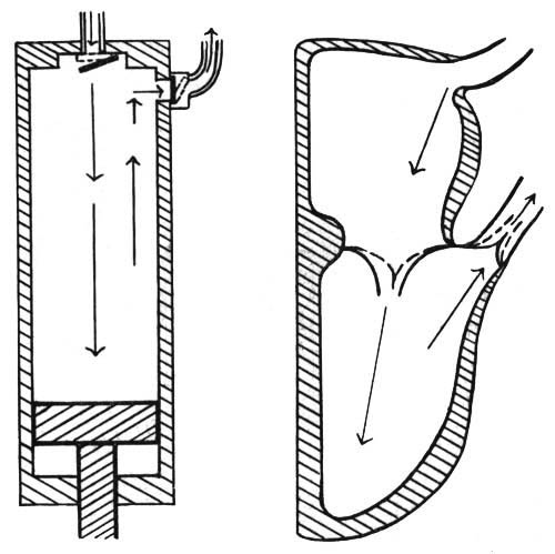
The heart is a force pump; prove it from these diagrams.
The Course of the Blood in the Body.—Although the two sides of the heart are separate and distinct from each other, yet every drop of blood that passes through the right heart likewise passes later through the left heart. There are two distinct systems of circulation in the body. The pulmonary circulation takes the blood through the right auricle and ventricle, to the lungs, and passes it back to the left auricle. This is a relatively short circulation, the blood receiving in the lungs its supply of oxygen, and there giving up some of its carbon dioxide. The greater circulation is known as the systemic circulation; in this system, the blood leaves the left ventricle through the great dorsal aorta. A large part of the blood passes directly to the muscles; some of it goes to the nervous system, kidneys, skin, and other organs of the body. It gives up its supply of food and oxygen in these tissues, receives the waste products of oxidation while passing through the capillaries, and returns to the right auricle through two large vessels known as the venæ cavæ. It requires only from twenty to thirty seconds for the blood to make the complete circulation from the ventricle back again to the starting point. This means that the entire volume of blood in the human body passes three or four thousand times a day through the various organs of the body.[46]
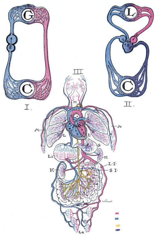
I. Circulation in a fish. G, gills; C, capillaries of the body. Notice the two-chambered heart.
II. The circulation in a frog. L, the lungs; C, the capillaries. Notice the heart has three chambers. What is the condition of blood leaving the ventricle to go to the cells of the body?
III. The circulation in man. H, head; A, arms; L, lungs; S, stomach; Li, liver; K, kidney; S.I., small intestine; L.I., large intestine; Le, legs; 1, right auricle; 2, right ventricle; 3, left ventricle; 4, left auricle; 5, dorsal aorta; 6, vein to lungs.
Portal Circulation.—Some of the blood, on its way back to the heart, passes to the walls of the food tube and to its glands. From there it is sent with its load of absorbed food to the liver. Here the vein which carries the blood (called the portal vein) breaks up into capillaries around the cells of the liver, when it gives up sugar to be stored as glycogen. From the liver, blood passes directly to the right auricle. The portal circulation, as it is called, is the only part of the circulation where the blood passes through two sets of capillaries on its way from auricle to auricle.
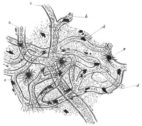
Capillary circulation in the web of a frog's foot, as seen under the compound microscope. a, b, small veins; c, pigment cells in the skin; d, capillaries in which the oval corpuscles are seen to follow one another in single series.
Circulation in the Web of a Frog's Foot.—If the web of the foot of a live frog or the tail of a tadpole is examined under the compound microscope, a network of blood vessels will be seen. In some of the larger vessels the corpuscles are moving rapidly and in spurts; these are arteries. The arteries lead into smaller vessels hardly greater in diameter than the width of a single corpuscle. This network of capillaries may be followed into larger veins in which the blood moves regularly. This illustrates the condition in any tissue of man where the arteries break up into capillaries, and these in turn unite to form veins.
Structure of the Arteries.—A distinct difference in structure exists between the arteries and the veins in the human body. The arteries, because of the greater strain received from the blood which is pumped from the heart, have thicker muscular walls, and in addition are very elastic.
Cause of the Pulse.—The pulse, which can easily be detected by pressing the large artery in the wrist or the small one in front of and above the external ear, is caused by the gushing of blood through the arteries after each pulsation of the heart. As the large arteries pass away from the heart, the diameter of each individual artery becomes smaller. At the very end of their course, these arteries are so small as to be almost microscopic in size and are very numerous. There are so many that if they were placed together, side by side, their united diameter would be much greater than the diameter of the large artery (aorta) which passes blood from the left side of the heart. This fact is of very great importance, for the force of the blood as it gushes through the arteries becomes very much less when it reaches the smaller vessels. This gushing movement is quite lost when the capillaries are reached, first, because there is so much more space for the blood to fill, and second, because there is considerable friction caused by the very tiny diameter of the capillaries.
Capillaries.—The capillaries form a network of minute tubes everywhere in the body, but especially near the surface and in the lungs. It is through their walls that the food and oxygen pass to the tissues, and carbon dioxide is given up to the plasma. They form the connection that completes the system of circulation of blood in the body.
Function and Structure of the Veins.—If the arteries are supply pipes which convey fluid food to the tissues, then the veins may be likened to drain pipes which carry away waste material from the tissues. Extremely numerous in the extremities and in the muscles and among other tissues of the body, they, like the branches of a tree, become larger and unite with each other as they approach the heart.
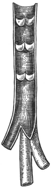
Valves in a vein. Notice the thin walls of the vein.
If the wall of a vein is carefully examined, it will be found to be neither so thick nor so tough as an artery wall. When empty, a vein collapses; the wall of an artery holds its shape. If you hold your hand downward for a little time and then examine it, you will find that the veins, which are relatively much nearer the surface than are the arteries, appear to be very much knotted. This appearance is due to the presence of tiny valves within. These valves open in the direction of the blood current, but would close if the direction of the blood flow should be reversed (as in case a deep cut severed a vein). As the pressure of blood in the veins is much less than in the arteries, the valves thus aid in keeping the flow of blood in the veins toward the heart. The higher pressure in arteries and the suction in the veins (caused by the enlargement of the chest cavity in breathing) are the chief factors which cause a steady flow of blood through the veins in the body.
Lymph Vessels.—The lymph is collected from the various tissues of the body by means of a number of very thin-walled tubes, which are at first very tiny, but after repeated connection with other tubes ultimately unite to form large ducts. These lymph ducts are provided, like the veins, with valves. The pressure of the blood within the blood vessels forces continually more plasma into the lymph; thus a slow current is maintained. On its course the lymph passes through many collections of gland cells, the lymph glands. In these glands some impurities appear to be removed and colorless corpuscles made. The lymph ultimately passes into a large tube, the thoracic duct, which flows upward near the ventral side of the spinal column, and empties into the large subclavian vein in the left side of the neck. Another smaller lymph duct enters the right subclavian vein.
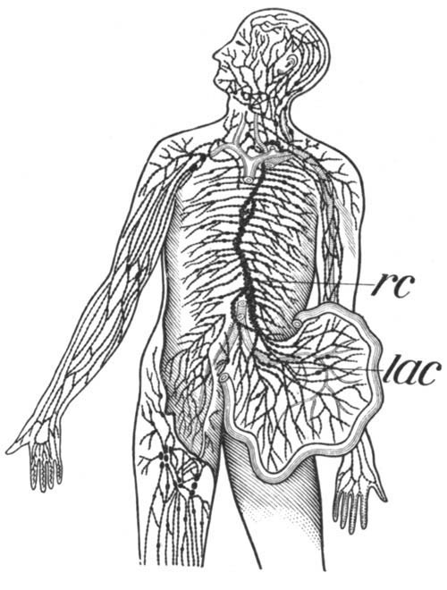
The lymph vessels; the dark spots are lymph glands: lac, lacteals; rc, thoracic duct.
The Lacteals.—We have already found that part of the digested food (chiefly carbohydrates, proteins, salts, and water) is absorbed directly into the blood through the walls of the villi and carried to the liver. Fat, however, is passed into the spaces in the central part of the villi, and from there into other spaces between the tissues, known as the lacteals. The lacteals carry the fats into the blood by way of the thoracic duct. The lacteals and lymph vessels have in part the same course. It will be thus seen that lymph at different parts of its course would have a very different composition.
The Nervous Control of the Heart and Blood Vessels.—Although the muscles of the heart contract and relax without our being able to stop them or force them to go faster, yet in cases of sudden fright, or after a sudden blow, the heart may stop beating for a short interval. This shows that the heart is under the control of the nervous system. Two sets of nerve fibers, both of which are connected with the central nervous system, pass to the heart. One set of fibers accelerates, the other slows or inhibits, the heart beat. The arteries and veins are also under the control of the sympathetic nervous system. This allows of a change in the diameter of the blood vessels. Thus, blushing is due to a sudden rush of blood to the surface of the body caused by an expansion of the blood vessels at the surface. The blood vessels of the body are always full of blood. This results from an automatic regulation of the diameter of the blood tubes by a part of the nervous system called the vasomotor nerves. These nerves act upon the muscles in the walls of the blood vessels. In this way, each vessel adapts itself to the amount of blood in it at a given time. After a hearty meal, a large supply of blood is needed in the walls of the stomach and intestines. At this time, the arteries going to this region are dilated so as to receive an extra supply. When the brain performs hard work, blood is supplied in the same manner to that region. Hence, one should not study or do mental work immediately after a hearty meal, for blood will be drawn away to the brain, leaving the digestive tract with an insufficient supply. Indigestion may follow as a result.
The Effect of Exercise on the Circulation.—It is a fact familiar to all that the heart beats more violently and quickly when we are doing hard work than when we are resting. Count your own pulse when sitting quietly, and then again after some brisk exercise in the gymnasium. Exercise in moderation is of undoubted value, because it sends the increased amount of blood to such parts of the body where increased oxidation has been taking place as the result of the exercise. The best forms of exercise are those which give as many muscles as possible work—walking, out-of-door sports, any exercise that is not violent. Exercise should not be attempted immediately after eating, as this causes a withdrawal of blood from the digestive tract to the muscles of the body. Neither should exercise be continued after becoming tired, as poisons are then formed in the muscles, which cause the feeling we call fatigue. Remember that extra work given to the heart by extreme exercise may injure it, causing possible trouble with the valves.
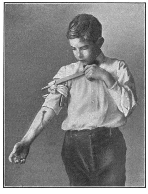
Stopping flow of blood from an artery by applying a tight bandage (ligature) between the cut and the heart.
Treatment of Cuts and Bruises.—Blood which oozes slowly from a cut will usually stop flowing by the natural means of the formation of a clot. A cut or bruise should, however, be washed in a weak solution of carbolic acid or some other antiseptic in order to prevent bacteria from obtaining a foothold on the exposed flesh. If blood, issuing from a wound, gushes in distinct pulsations, then we know that an artery has been severed. To prevent the flow of blood, a tight bandage known as a tourniquet must be tied between the cut and the heart. A handkerchief with a knot placed over the artery may stop bleeding if the cut is on one of the limbs. If this does not serve, then insert a stick in the handkerchief and twist it so as to make the pressure around the limb still greater. Thus we may close the artery until the doctor is called, who may sew up the injured blood vessel.
The Effect of Alcohol upon the Blood.—It has recently been discovered that alcohol has an extremely injurious effect upon the colorless corpuscles of the blood, lowering their ability to fight disease germs to a marked degree. This is well seen in a comparison of deaths from certain infectious diseases in drinkers and abstainers, the percentage of mortality being much greater in the former.
Dr. T. Alexander MacNichol, in a recent address, said:—
"Massart and Bordet, Metchnikoff and Sims Woodhead, have proved that alcohol, even in very dilute solution, prevents the white blood corpuscles from attacking invading germs, thus depriving the system of the coöperation of these important defenders, and reducing the powers of resisting disease. The experiments of Richardson, Harley, Kales, and others have demonstrated the fact that one to five per cent of alcohol in the blood of the living human body in a notable degree alters the appearance of the corpuscular elements, reduces the oxygen bearing elements, and prevents their reoxygenation."
Alcohol weakens Resistance to Disease.—In acute illnesses, grippe, fevers, blood poisoning, etc., substances formed in the blood termed "antibodies" antagonize the action of bacteria, facilitating their destruction by the white blood cells and neutralizing their poisonous influence. In a person with good "resistance" this protective machinery, which we do not yet thoroughly understand, works with beautiful precision, and the patient "gets well." Experiments by scientific experts have demonstrated that alcohol restrains the formation of these marvelous antibodies. Alcohol puts to sleep the sentinels that guard your body from disease.
The Effect of Alcohol on the Circulation.—Alcoholic drinks affect the very delicate adjustment of the nervous center's[TN7] controlling the blood vessels and heart. Even very dilute alcohol acts upon the muscles of the tiny blood vessels; consequently, more blood is allowed to enter them, and, as the small vessels are usually near the surface of the body, the habitual redness seen in the face of hard drinkers is the ultimate result.
"The first effect of diluted alcohol is to make the heart beat faster. This fills the small vessels near the surface. A feeling of warmth is produced which causes the drinker to feel that he was warmed by the drink. This feeling, however, soon passes away, and is succeeded by one of chilliness. The body temperature, at first raised by the rather rapid oxidation of the alcohol, is soon lowered by the increased radiation from the surface.
"The immediate stimulation to the heart's action soon passes away and, like other muscles, the muscles of the heart lose power and contract with less force after having been excited by alcohol."—Macy, Physiology.
Alcohol, when brought to act directly on heart muscle, lessens the force of the beat. It may even cause changes in the tissues, which eventually result in the breaking of the walls of a blood vessel or the plugging of a vessel with a blood clot. This condition may cause the disease known as apoplexy.
Effects of Tobacco upon the Circulation.—"The frequent use of cigars or cigarettes by the young seriously affects the quality of the blood. The red blood corpuscles are not fully developed and charged with their normal supply of life-giving oxygen. This causes paleness of the skin, often noticed in the face of the young smoker. Palpitation of the heart is also a common result, followed by permanent weakness, so that the whole system is enfeebled, and mental vigor is impaired as well as physical strength."—Macy, Physiology.
XXII. RESPIRATION AND EXCRETION
Problems.—A study of respiration to find out:—
(a) What changes in blood and air take place within the lungs.
(b) The mechanics of respiration.
A study of ventilation to discover:—
(a) The reason for ventilation.
(b) The best method of ventilation.
A study of the organs of excretion.
Laboratory Suggestions
Demonstration.—Comparison of lungs of frog with those of bird or mammal.
Experiment.—The changes of blood within the lungs.
Experiment.—Changes taking place in air in the lungs.
Experiment.—The use of the ribs in respiration.
Demonstration experiment.—What causes the filling of air sacs of the lungs?
Demonstration experiment.—What are the best methods of ventilating a room?
Demonstration.—Best methods of dusting and cleaning.
Demonstration.—Beef or sheep's kidney to show areas.
Necessity for Respiration.—We have seen that plants and animals need oxygen in order that the life processes may go on. Food is oxidized to release energy, just as coal is burned to give heat to run an engine. As a draft of air is required to make fire under the boiler, so, in the human body, oxygen must be given so that food in tissues may be oxidized to release energy used in work. This oxidation takes place in the cells of the body, be they part of a muscle, a gland, or the brain. Blood, in its circulation to all parts of the body, is the medium which conveys the oxygen to that place in the body where it will be used.
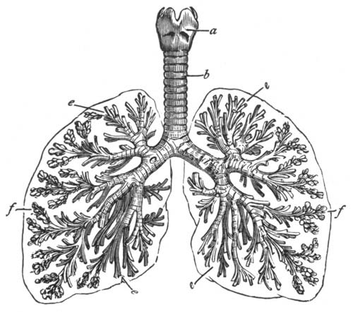
Air passages in the human lungs. a, larynx; b, trachea (or windpipe); c, d, bronchi; e, bronchial tubes; f, cluster of air cells.
The Organs of Respiration in Man.—We have alluded to the fact that the lungs are the organs which give oxygen to the blood and take from it carbon dioxide. The course of the air passing to the lungs in man is much the same as in the frog. Air passes through the nose, and into the windpipe. This cartilaginous tube, the top of which may easily be felt as the Adam's apple of the throat, divides into two bronchi. The bronchi within the lungs break up into a great number of smaller tubes, the bronchial tubes, which divide somewhat like the small branches of a tree. The bronchial tubes, indeed all the air passages, are lined with ciliated cells. The cilia of these cells are constantly in motion, beating with a quick stroke toward the outer end of the tube, that is, toward the mouth. Hence any foreign material will be raised from the throat first by the action of the cilia and then by coughing or "clearing the throat." The bronchi end in very minute air sacs, little pouches having elastic walls, into which air is taken when we inspire, or take a deep breath. In the walls of these pouches are numerous capillaries, the ends of arteries which pass from the heart into the lung. It is through the very thin walls of the air sacs that an interchange of gases takes place which results in the blood giving up part of its load of carbon dioxide, and taking up oxygen in its place. This exchange appears to be aided by the presence of an enzyme in the lung tissues. This is another example of the various kinds of work done by the enzymes of the body.
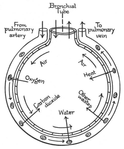
Diagram to show what the blood loses and gains in one of the air sacs of the lungs.
Changes in the Blood within the Lungs.—Blood, after leaving the lungs, is much brighter red than just before entering them. The change in color is due to a taking up of oxygen by the hæmoglobin of the red corpuscles. Changes taking place in blood are obviously the reverse of those which take place in air in the lungs. Every hundred cubic centimeters of blood going into the lungs contains 8 to 12 c.c. of oxygen, 45 to 50 c.c. of carbon dioxide, and 1 to 2 c.c. of nitrogen. The same amount of blood passing out of the lungs contains 20 c.c. of oxygen, 38 c.c. of carbon dioxide, and 1 to 2 c.c. of nitrogen. The water, of which about half a pint is given off daily, is mostly lost from the blood.
Changes in Air in the Lungs.—Air is much warmer after leaving the lungs than before it enters them. Breathe on the bulb of a thermometer to prove this. Expired air contains a considerable amount of moisture, as may be proved by breathing on a cold polished surface. This it has taken up in the air sacs of the lungs. The presence of carbon dioxide in expired air may easily be detected by the limewater test. Air such as we breathe out of doors contains, by volume:—
Nitrogen 76.95
Oxygen 20.61
Carbon dioxide .03
Argon 1.00
Water vapor (average) 1.40
Air expired from the lungs contains:—
Nitrogen 76.95
Oxygen 15.67
Carbon dioxide 4.38
Water vapor 2.00
Argon 1.00
In other words, there is a loss between 4 and 5 per cent oxygen, and nearly a corresponding gain in carbon dioxide, in expired air. There are also some other organic substances present.
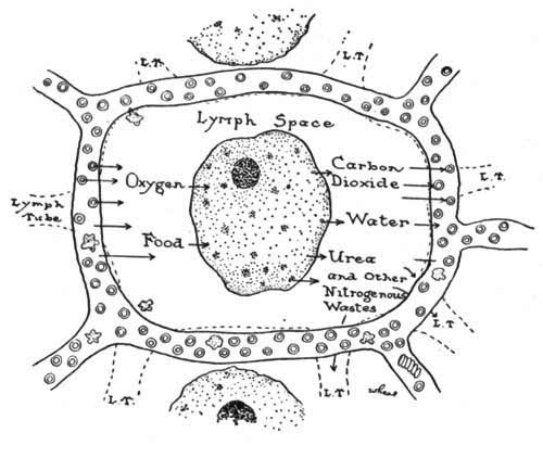
The respiration of cells.
Cell Respiration.—It has been shown, in the case of very simple animals, such as the amœba, that when oxidation takes place in a cell, work results from this oxidation. The oxygen taken into the lungs is not used there, but is carried by the blood to such parts of the body as need oxygen to oxidize food materials in the cells. Since work is done in the cells of the body, food and oxygen are therefore required. The quantity of oxygen used by the body is nearly dependent on the amount of work performed. Oxygen is constantly taken from the blood by tissues in a state of rest and is used up when the body is at work. This is suggested by the fact that in a given time a man, when working, gives off more oxygen (in carbon dioxide) than he takes in during that time.
While work is being done certain wastes are formed in the cell. Carbon dioxide is given off when carbon is burned. But when proteins are burned, another waste product containing nitrogen is formed. This must be passed off from the cells, as it is a poison. Here again the lymph and blood, the common carriers, take the waste material to points where it may be excreted or passed out of the body.
The Mechanics of Respiration. The Pleura.—The lungs are covered with a thin elastic membrane, the pleura. This forms a bag in which the lungs are hung. Between the walls of the bag and the lungs is a space filled with lymph. By this means the lungs are prevented from rubbing against the walls of the chest.
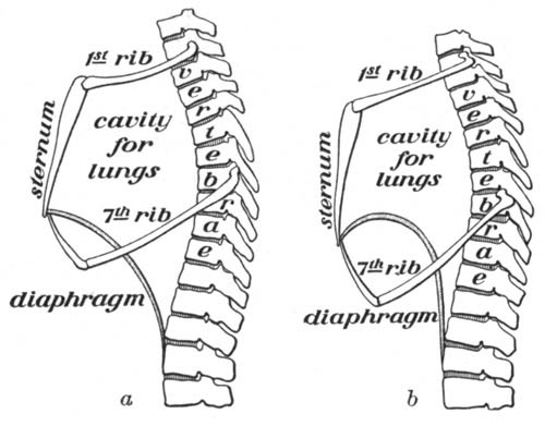
The chest cavity (a) at the time of a full breath; (b), after an expiration. Explain how the cavity for lungs is made larger.
Breathing.—In every full breath there are two distinct movements, inspiration (taking air in) and expiration (forcing air out). In man an inspiration is produced by the contraction of the muscles between the ribs, together with the contraction of the diaphragm, the muscular wall just below the heart and lungs; this results in pulling down the diaphragm and pulling upward and outward of the ribs, thus making the space within the chest cavity larger. The lungs, which lie within this cavity, are filled by the air rushing into the larger space thus made. That this cavity is larger than it was at first may be demonstrated by a glance at the accompanying figure. An expiration is simpler than an inspiration, for it requires no muscular effort; the muscles relax, the breastbone and ribs sink into place, while the diaphragm returns to its original position.
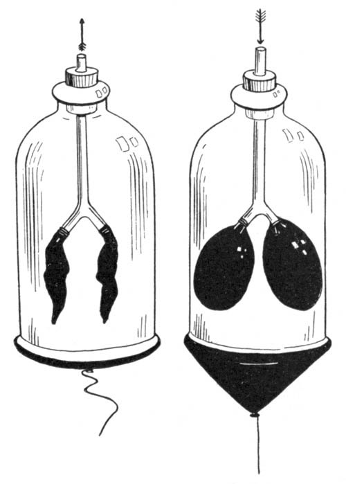
Apparatus to show the mechanics of breathing.
A piece of apparatus which illustrates to a degree the mechanics of breathing may be made as follows: Attach a string to the middle of a piece of sheet rubber. Tie the rubber over the large end of a bell jar. Pass a glass Y-tube through a rubber stopper. Fasten two small toy balloons to the branches of the tube. Close the small end of the jar with the stopper. Adjust the tube so that the balloons shall hang free in the jar. If now the rubber sheet is pulled down by means of the string, the air pressure in the jar is reduced and the toy balloons within expand, owing to the air pressure down the tube. When the rubber is allowed to go back to its former position, the balloons collapse.
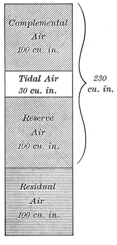
Diagram showing the relative amounts of tidal, complemental, reserve, and residual air. The brace shows the average lung capacity for the adult man.
Rate of Breathing and Amount of Air Breathed.—During quiet breathing, the rate of inspiration is from fifteen to eighteen times per minute; this rate largely depends on the amount of physical work performed. About 30 cubic inches of air are taken in and expelled during the ordinary quiet respiration. The air so breathed is called tidal air. In a "long" breath, we take in about 100 cubic inches in addition to the tidal air. This is called complemental air. By means of a forced expiration, it is possible to expel from 75 to 100 cubic inches more than tidal air; this air is called reserve air. What remains in the lungs, amounting to about 100 cubic inches, is called the residual air. The value of deep breathing is seen by a glance at the diagram. It is only by this means that we clear the lungs of the reserve air with its accompanying load of carbon dioxide.
Respiration under Nervous Control.—The muscular movements which cause an inspiration are partly under the control of the will, but in part the movement is beyond our control. The nerve centers which govern inspiration are part of the sympathetic nervous system. Anything of an irritating nature in the trachea or larynx will cause a sudden expiration or cough. When a boy runs, the quickened respiration is due to the fact that oxygen is used up rapidly and a larger quantity of carbon dioxide is formed. The carbon dioxide in the blood stimulates the nervous center which has control of respiration to greater activity, and quickened inspiration follows.
Need of Ventilation.—During the course of a day the lungs lose to the surrounding air nearly two pounds of carbon dioxide. This means that about three fifths of a cubic foot is given off by each person during an hour. When we are confined for some time in a room, it becomes necessary to get rid of this carbon dioxide. This can be done only by means of proper ventilation. A considerable amount of moisture is given off from the body, and this moisture in a crowded room is responsible for much of the discomfort. The air becomes humid and uncomfortable. It has been found that by keeping the air in motion in such a room (as through the use of electric fans) much of this discomfort is obviated.
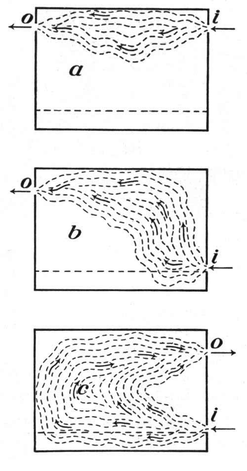
Three ways of ventilating a room. i, inlet for air; o, outlet for air. Which is the best method of ventilation? Explain.
The presence of impurities in the air of a room may easily be determined by its odor. The odor of a poorly ventilated room is due to organic impurities given off with the carbon dioxide. This, fortunately, gives us an index of the amount of waste material in the air. Among the factors which take oxygen from the air in a closed room and produce carbon dioxide are burning gas or oil lamps and stoves, and the presence of a number of people.
Proper Ventilation.—Ventilation consists in the removal of air that has been used, and the introduction of a fresh supply to take its place. Heated air rises, carrying with it much of the carbon dioxide and other impurities. A good method of ventilation for the home is to place a board two or three inches high between the lower sash and the frame of a window or to have the window open an inch or so at the top and the bottom. An open fireplace in a room aids in ventilation because of the constant draft up the flue.
Sweeping and Dusting.—It is very easy to demonstrate the amount of dust in the air by following the course of a beam of light in a darkened room. We have already proved that spores of mold and yeast exist in the air. That bacteria are also present can be proved by exposing a sterilized gelatin plate to the air in a schoolroom for a few moments.[47]
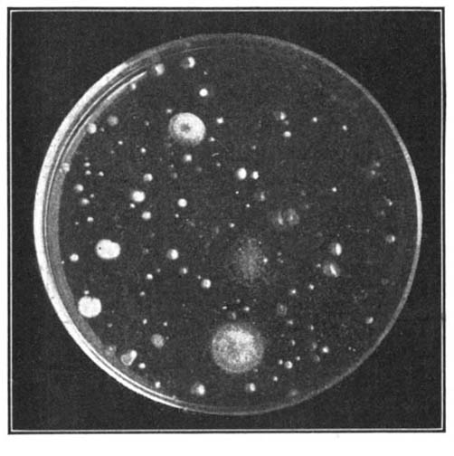
Plate culture exposed for five minutes in a school hall where pupils were passing to recitations. Each spot is a colony of bacteria or mold.
Many of the bacteria present in the air are active in causing diseases of the respiratory tract, such as diphtheria, membranous croup, and tuberculosis. Other diseases, as colds, bronchitis (inflammation of the bronchial tubes), and pneumonia (inflammation of the tiny air sacs of the lungs), are also caused by bacteria.
Dust, with its load of bacteria, will settle on any horizontal surface in a room not used for three or four hours. Dusting and sweeping should always be done with a damp cloth or broom, otherwise the bacteria are simply stirred up and sent into the air again. The proper watering of streets before they are swept is also an important factor in health. Much dust is composed largely of dried excreta of animals. Soft-coal smoke does its share to add to the impurities of the air, while sewer gas and illuminating gas are frequently found in sufficient quantities to poison people. Pure air is, as can be seen, almost an impossibility in a great city.
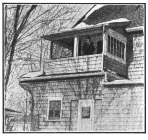
A sleeping porch, an ideal way to get fresh air at night.
How to get Fresh Air.—As we know, green plants give off in the sunlight considerable more oxygen than they use, and they use up carbon dioxide. The air in the country is naturally purer than in the city, as smoke and bacteria are not so prevalent there, and the plants in abundance give off oxygen. In the city the night air is purer than day air, because the factories have stopped work, the dust has settled, and fewer people are on the streets. The old myth of "night air" being injurious has long since been exploded, and thousands of people of delicate health, especially those who have weak throat or lungs, are regaining health by sleeping out of doors or with the windows wide open. The only essential in sleeping out of doors or in a room with a low temperature is that the body be kept warm and the head be protected from strong drafts by a nightcap or hood. Proper ventilation at all times is one of the greatest factors in good health.
Change of Air.—Persons in poor health, especially those having tuberculosis, are often cured by a change of air. This is not always so much due to the composition of the air as to change of occupation, rest, and good food. Mountain air is dry, and relatively free from dust and bacteria, and often helps a person having tuberculosis. Air at the seaside is beneficial for some forms of disease, especially hay fever and bone tuberculosis. Many sanitariums have been established for this latter disease near the ocean, and thousands of lives are being annually saved in this way.
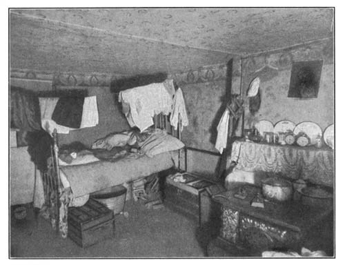
Unfavorable sleeping conditions. Explain why unfavorable.
Ventilation of Sleeping Rooms.—Sleeping in close rooms is the cause of much illness. Beds ought to be placed so that a constant supply of fresh air is given without a direct draft. This may often be managed with the use of screens. Bedroom windows should be thrown open in the morning to allow free entrance of the sun and air, bedclothes should be washed frequently, and sheets and pillow covers often changed. Bedroom furniture should be simple, and but little drapery allowed in the room.
Hygienic Habits of Breathing.—Every one ought to accustom himself upon going into the open air to inspire slowly and deeply to the full capacity of the lungs. A slow expiration should follow. Take care to force the air out. Breathe through the nose, thus warming the air you inspire before it enters the lungs and chills the blood. Repeat this exercise several times every day. You will thus prevent certain of the air sacs which are not often used from becoming hardened and permanently closed.
Relation of Proper Exercise to Health.—We are all aware that exercise in moderation has a beneficial effect upon the human organism. The pale face, drooping shoulders, and narrow chest of the boy or girl who takes no regular exercise is too well known. Exercise, besides giving direct use of the muscles, increases the work of the heart and lungs, causing deeper breathing and giving the heart muscles increased work; it liberates heat and carbon dioxide from the tissues where the work is taking place, thus increasing the respiration of the tissues themselves, and aids mechanically in the removal of wastes from tissues. It is well known that exercise, when taken some little time after eating, has a very beneficial effect upon digestion. Exercise and especially games are of immense importance to the nervous system as a means of rest. The increasing number of playgrounds in this country is due to this acknowledged need of exercise, especially for growing children.
Proper exercise should be moderate and varied. Walking in itself is a valuable means of exercising certain muscles, so is bicycling, but neither is ideal as the only form to be used. Vary your exercise so as to bring different muscles into play, take exercise that will allow free breathing out of doors if possible, and the natural fatigue which follows will lead you to take the rest and sleep that every normal body requires.
Exercise should always be limited by fatigue, which brings with it fatigue poisons. This is nature's signal when to rest. If one's use of diet and air is proper, the fatigue point will be much further off than otherwise. One should learn to relax when not in activity. The habit produces rest, even between exertions very close together, and enables one to continue to repeat those exertions for a much longer time than otherwise. The habit of lying down when tired is a good one.
The Relation of Tight Clothing to Correct Breathing.—It is impossible to breathe correctly unless the clothing is worn loosely over the chest and abdomen. Tight corsets and tight belts prevent the walls of the chest and the abdomen from pushing outward and interfere with the drawing of air into the lungs. They may also result in permanent distortion of parts of the skeleton directly under the pressure. Other organs of the body cavity, as the stomach and intestines, may be forced downward, out of place, and in consequence cannot perform their work properly.
Suffocation and Artificial Respiration.—Suffocation results from the shutting off of the supply of oxygen from the lungs. It may be brought about by an obstruction in the windpipe, by a lack of oxygen in the air, by inhaling some other gas in quantity, or by drowning. A severe electric shock may paralyze the nervous centers which control respiration, thus causing a kind of suffocation. In the above cases, death often may be prevented by prompt recourse to artificial respiration. To accomplish this, place the patient on his back with the head lower than the body; grasp the arms near the elbows and draw them upward and outward until they are stretched above the head, on a line with the body. By this means the chest cavity is enlarged and an inspiration produced. To produce an expiration, carry the arms downward, and press them against the chest, thus forcing the air out of the lungs. This exercise, regularly repeated every few seconds, if necessary for hours, has been the source of saving many lives.
Common Diseases of the Nose and Throat.—Catarrh is a disease to which people with sensitive mucous membrane of the nose and throat are subject. It is indicated by the constant secretion of mucus from these membranes. Frequent spraying of the nose and throat with some mild antiseptic solutions is found helpful. Chronic catarrh should be attended to by a physician. Often we find children breathing entirely through the mouth, the nose being seemingly stopped up. When this goes on for some time the nose and throat should be examined by a physician for adenoids, or growths of soft masses of tissue which fill up the nose cavity, thus causing a shortage of the air supply for the body. Many a child, backward at school, thin and irritable, has been changed to a healthy, normal, bright scholar by the removal of adenoids. Sometimes the tonsils at the back of the mouth cavity may become enlarged, thus shutting off the air supply and causing the same trouble as we see in a case of adenoids. The simple removal of the obstacle by a doctor soon cures this condition. (See page 395.)
Organs of Excretion.—All the life processes which take place in a living thing result ultimately, in addition to giving off of carbon dioxide, in the formation of organic wastes within the body. The retention of these wastes which contain nitrogen, is harmful to animals. In man, the skin and kidneys remove this waste from the body, hence they are called the organs of excretion.
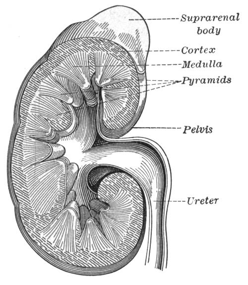
Longitudinal section through a kidney.
The Human Kidney.—The human kidney is about four inches long, two and one half inches wide, and one inch in thickness. Its color is dark red. If the structure of the medulla and cortex (see figure above) is examined under the compound microscope, you will find these regions to be composed of a vast number of tiny branched and twisted tubules. The outer end of each of these tubules opens into the pelvis, the space within the kidney; the inner end, in the cortex, forms a tiny closed sac. In each sac, the outer wall of the tube has grown inward and carried with it a very tiny artery. This artery breaks up into a mass of capillaries. These capillaries, in turn, unite to form a small vein as they leave the little sac. Each of these sacs with its contained blood vessels is called a glomerulus.
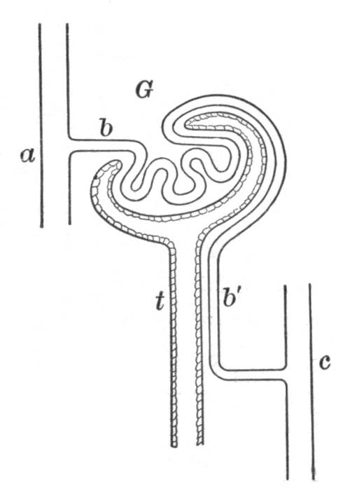
Diagram of kidney circulation, showing a glomerulus and tubule: a, artery bringing blood to part; b, capillary bringing blood to glomerulus; b', vessel continuing with blood to vein; c, vein; t, tubule; G, glomerulus.
Wastes given off by the Blood in the Kidney.—In the glomerulus the blood loses by osmosis, through the very thin walls of the capillaries, first, a considerable amount of water (amounting to nearly three pints daily); second, a nitrogenous waste material known as urea; third, salts and other waste organic substances, uric acid among them.
These waste products, together with the water containing them, are known as urine. The total amount of nitrogenous waste leaving the body each day is about twenty grams. It is passed through the ureter to the urinary bladder; from this reservoir it is passed out of the body, through a tube called the urethra. After the blood has passed through the glomeruli of the kidneys it is purer than in any other place in the body, because, before coming there, it lost a large part of its burden of carbon dioxide in the lungs. After leaving the kidney it has lost much of its nitrogenous waste. So dependent is the body upon the excretion of its poisonous material that, in cases where the kidneys do not do their work properly, death may ensue within a few hours.
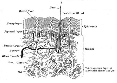
Diagram of a section of the skin. (Highly magnified.)
Structure and Use of Sweat Glands.—If you examine the palm of your hand with a lens, you will notice the surface is thrown into little ridges. In these ridges may be found a large number of very tiny pits; these are the pores or openings of the sweat-secreting glands. From each opening a little tube penetrates deep within the epidermis; there, coiling around upon itself several times, it forms the sweat gland. Close around this coiled tube are found many capillaries. From the blood in these capillaries, cells lining the wall of the gland take water, and with it a little carbon dioxide, urea, and some salts (common salt among others). This forms the excretion known as sweat. The combined secretions from these glands amount normally to a little over a pint during twenty-four hours. At all times, a small amount of sweat is given off, but this is evaporated or is absorbed by the underwear; as this passes off unnoticed, it is called insensible perspiration. In hot weather or after hard manual labor the amount of perspiration is greatly increased.
Regulation of Heat of the Body.—The bodily temperature of a person engaged in manual labor will be found to be but little higher than the temperature of the same person at rest. We know from our previous experiments that heat is released. Muscles, nearly one half the weight of the body, release about five sixths of their energy as heat. At all times they are giving up some heat. How is it that the bodily temperature does not differ greatly at such times? The temperature of the body is largely regulated by means of the activity of the sweat glands. The blood carries much of the heat, liberated in the various parts of the body by the oxidation of food, to the surface of the body, where it is lost in the evaporation of sweat. In hot weather the blood vessels of the skin are dilated; in cold weather they are made smaller by the action of the nervous system. The blood thus loses water in the skin, the water evaporates, and we are cooled off. The object of increased perspiration, then, is to remove heat from the body. With a large amount of blood present in the skin, perspiration is increased; with a small amount, it is diminished. Hence, we have in the skin an automatic regulator of bodily temperature.
Sweat Glands under Nervous Control.—The sweat glands, like the other glands in the body, are under the control of the sympathetic nervous system. Frequently the nerves dilate the blood vessels of the skin, thus helping the sweat glands to secrete, by giving them more blood.
"Thus regulation is carried out by the nervous system determining, on the one hand, the loss by governing the supply of blood to the skin and the action of the sweat glands; and on the other, the production by diminishing or increasing the oxidation of the tissues."—Foster and Shore, Physiology.
Colds and Fevers.—The regulation of blood passing through the blood vessels is under control of the nervous system. If this mechanism is interfered with in any way, the sweat glands may not do their work, perspiration may be stopped, and the heat from oxidation held within the body. The body temperature goes up, and a fever results.
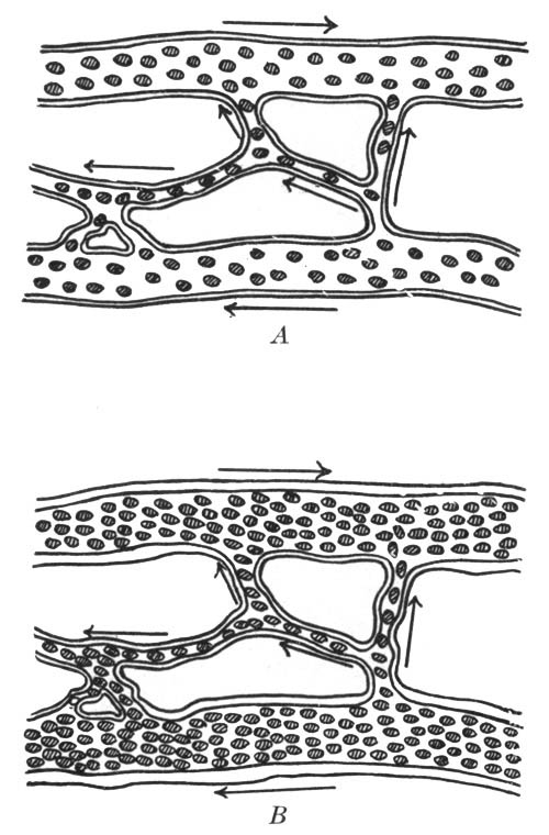
A, blood vessels in skin normal; B, when congested.
If the blood vessels in the skin are suddenly cooled when full of blood, they contract and send the blood elsewhere. As a result a congestion or cold may follow. Colds are, in reality, a congestion of membranes lining certain parts of the body, as the nose, throat, windpipe, or lungs.
When suffering from a cold, it is therefore important not to chill the skin, as a full blood supply should be kept in it and so kept from the seat of the congestion. For this reason hot baths (which call the blood to the skin), the avoiding of drafts (which chill the skin), and warm clothing are useful factors in the care of colds.
Hygiene of the Skin.—The skin is of importance both as an organ of excretion and as a regulator of bodily temperature. The skin of the entire body should be bathed frequently so that this function of excretion may be properly performed. Pride in one's own appearance forbids a dirty skin. For those who can stand it, a cold sponge bath is best. Soap should be used daily on parts exposed to dirt. Exercise in the open air is important to all who desire a good complexion. The body should be kept at an even temperature by the use of proper underclothing. Wool, a poor conductor of heat, should be used in winter, and cotton, which allows of a free escape of heat, in summer.
Cuts, Bruises, and Burns.—In case the skin is badly broken, it is necessary to prevent the entrance and growth of bacteria. This may be done by washing the wound with weak antiseptic solutions such as 3 per cent carbolic acid, 3 per cent lysol, or peroxide of hydrogen (full strength). These solutions should be applied immediately. A burn or scald should be covered at once with a paste of baking soda, with olive oil, or with a mixture of limewater and linseed oil. These tend to lessen the pain by keeping out the air and reducing the inflammation.
Summary of Changes in Blood within the Body.—We have already seen that red corpuscles in the lungs lose part of their load of carbon dioxide that they have taken from the tissues, replacing it with oxygen. This is accompanied by a change of color from purple (in blood which is poor in oxygen) to that of bright red (in richly oxygenated blood). Other changes take place in other parts of the body. In the walls of the food tube, especially in the small intestine, the blood receives its load of fluid food. In the muscles and other working tissues the blood gives up food and oxygen, receiving carbon dioxide and organic waste in return. In the liver, the blood gives up its sugar, and the worn-out red corpuscles which break down are removed (as they are in the spleen) from the circulation. In glands, it gives up materials used by the gland cells in their manufacture of secretions. In the kidneys, it loses water and nitrogenous wastes (urea). In the skin, it also loses some waste materials, salts, and water.
"The Effect of Alcohol on Body Heat.—It is usually believed that 'taking a drink' when cold makes one warmer. But such is not the case. In reality alcohol lowers the temperature of the body by dilating the blood vessels of the skin. It does this by means of its influence on the nervous system. It is, therefore, a mistake to drink alcoholic beverages when one is extremely cold, because by means of this more bodily heat is allowed to escape.
"Because alcohol is quickly oxidized, and because heat is produced in the process, it was long believed to be of value in maintaining the heat of the body. A different view now prevails as the result of much observation and experiment. Physiologists show by careful experiments that though the temperature of the body rises during digestion of food, it is lowered for some hours when alcohol is taken. The flush which is felt upon the skin after a drink of wine or spirits is due in part to an increase of heat in the body, but also to the paralyzing effect of the alcohol upon the capillary walls, allowing them to dilate, and so permitting more of the warm blood of the interior of the body to reach the surface. There it is cooled by radiation, and the general temperature is lowered."—Macy, Physiology.
Effect of Alcohol on Respiration.—Alcohol tends to congest the membrane of the throat and lungs. It does this by paralyzing the nerves which take care of the tiny blood vessels in the walls of the air tubes and air sacs. The capillaries become full of blood, the air spaces are lessened, and breathing is interfered with. The use of alcohol is believed by many physicians to predispose a person to tuberculosis. Certainly this disease attacks drinkers more readily than those who do not drink. Alcohol interferes with the respiration of the cells because it is oxidized very quickly within the body as it is quickly absorbed and sent to the cells. So rapid is this oxidation that it interferes with the oxidation of other substances. Using alcohol has been likened to burning kerosene in a stove; the operation is a dangerous one.
Effects of Tobacco on Respiration.—Tobacco smoke contains the same kind of poisons as the tobacco, with other irritating substances added. It is extremely irritating to the throat; it often causes a cough, and renders it more liable to inflammation. If the smoke is inhaled more deeply, the vaporized nicotine is still more readily absorbed and may thus produce greater irritation in the bronchi and lungs. Cigarettes are worse than other forms of tobacco, for they contain the same poisons with others in addition.
Effect of Alcohol on the Kidneys.—It is said that alcohol is one of the greatest causes of disease in the kidneys. The forms of disease known as "fatty degeneration of the kidney" and "Bright's disease" are both frequently due to this cause. The kidneys are the most important organs for the removal of nitrogenous waste.