Chapter XII
Reading edition
The Organs of Excretion.
The earliest stages in the development of the excretory system have already been described in a previous chapter[335] of this memoir, and up to the present time no investigator, with the exception of Dr Alex. Schultz[336], has gone over the same ground. Dr Schultz' descriptions are somewhat brief, but differ from my own mainly in stating that the segmental duct arises from an involution instead of as a solid knob. This discrepancy is, I believe, due to Dr Schultz drawing his conclusions as to the development of the segmental duct from its appearance at a comparatively late stage. He appears to have been unacquainted with my earlier descriptions.
The adult anatomy and later stages in the development of the excretory organs form the subject of the present chapter, and stand in marked contrast to the earlier stages in that they have been dealt with in a magnificent monograph[337] by Professor Semper, whose investigations have converted this previously almost unknown field of vertebrate embryology into one of the most fully explored parts of the whole subject. Reference is frequently made to this monograph in the succeeding pages, but my references, numerous as they are, give no adequate idea of the completeness and thoroughness of Professor Semper's investigations. In Professor Semper's monograph are embodied the results of a considerable number of preliminary papers published by him in his Arbeiten and in the Centralblatt. The excretory organs of Elasmobranchii have also formed the subject of some investigations by Dr Meyer[338] and by myself[339]. Their older literature is fully given by Professor Semper. In addition to the above-cited works, there is one other paper by Dr Spengel[340] on the Urinogenital System of Amphibians, to which reference will frequently be made in the sequel, and which, though only indirectly connected with the subject of this chapter, deserves special mention both on account of the accuracy of the investigations of which it forms the record, and of the novel light which it throws on many of the problems of the constitution of the urinogenital system of Vertebrates.
Excretory organs and genital ducts in the adult.
The kidneys of Scyllium canicula are paired bodies in contact along the median line. They are situated on the dorsal wall of the abdominal cavity, and extend from close to the diaphragm to a point a short way behind the anus. Externally, each appears as a single gland, but by the arrangement of its ducts may be divided into two distinct parts, an anterior and a posterior. The former will be spoken of as the Wolffian body, and the latter as the kidney, from their respective homology with the glands so named in higher Vertebrates. The grounds for these determinations have already been fully dealt with both by Semper[341] and by myself.
Externally both the Wolffian body and the kidney are more or less clearly divided into segments, and though the breadth of both glands as viewed from the ventral surface is fairly uniform, yet the hinder part of the kidney is very much thicker and bulkier than the anterior part and than the whole of the Wolffian body. In both sexes the Wolffian body is rather longer than the kidney proper. Thus in a male example, 33 centimetres long, the two glands together measured 8¼ centimetres and the kidney proper only 3½. In the male the Wolffian bodies extend somewhat further forwards than in the female. Leaving the finer details of the glands for subsequent treatment, I pass at once to their ducts. These differ slightly in the two sexes, so that it will be more convenient to take the male and female separately.
A partly diagrammatic representation of the kidney and Wolffian body of the male is given on Pl. 20, fig. 1. The secretion of the Wolffian body is carried off by a duct, the Wolffian duct (w.d.), which lies on the ventral surface of the gland, and receives a separate ductule from each segment (Pl. 20, fig. 5). The main function of the Wolffian duct in the male is, however, that of a vas deferens. The testicular products are brought to it through the coils of the anterior segments of the Wolffian body by a number of vasa efferentia, the arrangement of which is treated of on pp. 487, 488. The section of the Wolffian duct which overlies the Wolffian body is much contorted, and in adult individuals at the generative period enormously so. The duct often presents one or two contortions beyond the hind end of the Wolffian body, but in the normal condition takes a straight course from this point to the unpaired urinogenital cloaca, into which it falls independently of its fellow of the opposite side. It receives no feeders from the kidney proper.
The excretion of the kidney proper is carried off not by a single duct, but by a series of more or less independent ducts, which, in accordance with Prof. Semper's nomenclature, will be spoken of as ureters. These are very minute, and their investigation requires some care. I have reason, from my examinations of this and other species of Elasmobranchii, to believe that they are, moreover, subject to considerable variations, and the following description applies to a definite individual. Nine or possibly ten distinct ureters, whose arrangement is diagrammatically represented in fig. 1, Pl. 20, were present on each side. It will be noticed that, whereas the five hindermost are distinct till close to their openings into the urinogenital cloaca, the four anterior ones appear to unite at once into a single duct, but are probably only bound up in a common sheath. The ureters fall into the common urinogenital cloaca, immediately behind the opening of the Wolffian duct (so far as could be determined), by four apertures on each side. In a section made through the part of the wall of the cloaca containing the openings of the ureters of both sides, there were present on the left side (where the section passed nearer to the surface than on the right) four small openings posteriorly, viz. the openings of the ureters and one larger one anteriorly, viz. the opening of the Wolffian duct. On the other side of the section where the level was rather deeper, there were five distinct ducts cut through, one of which was almost on the point of dividing into two. This second section proves that, in this instance at least, the two ureters did not unite till just before opening into the urinogenital cloaca. The same section also appeared to shew that one of the ureters fell not into the cloaca but into the Wolffian duct.
As stated above both the Wolffian duct and the ureters fall into an unpaired urinogenital cloaca. This cloaca communicates at one end with the general cloaca by a single aperture situated at the point of a somewhat conspicuous papilla, just behind the anus (Pl. 20, fig. 1, o), and on the other it opens freely into a pair of bladders, situated in close contact with each other, on the ventral side of the kidney (Pl. 20, fig. 1, sb). To these bladders Professor Semper has given the name uterus masculinus, from having supposed them to correspond with the lower part of the oviducts of the female. This homology he now admits to be erroneous, and it will accordingly be better to drop the name uterus masculinus, for which may be substituted seminal bladder—a name which suits their function, since they are usually filled with semen at the generation season. The seminal bladders communicate with the urinogenital cloaca by wide openings, and it is on the borders of these openings that the mouths of the Wolffian duct and ureters must be looked for. My embryological investigations, though they have not been specially directed to this point, seem to shew that the seminal bladders do not arise during embryonic life, and are still absent in very young individuals. It seems probable that both the bladders and the urinogenital cloaca are products of the lower extremities of the Wolffian duct. The only other duct requiring any notice in the male is the rudimentary oviduct. As was first shewn by Semper, rudiments of the upper extremities of the oviducts, with their abdominal openings, are to be found in the male in the same position as in the female, on the front surface of the liver.
In the female the same ducts are present as in the male, viz. the Wolffian duct and the ureters. The part of the Wolffian duct which receives the secretion of the Wolffian body is not contorted, but is otherwise similar to the homologous part of the Wolffian duct in the male. The Wolffian ducts of the two sides fall independently into an unpaired urinal cloaca, but their lower ends, instead of remaining simple as in the male, become dilated into urinary bladders. Vide Pl. 20, fig. 2. There were nine ureters in the example dissected, whose arrangement did not differ greatly from that in the male—the hinder ones remaining distinct from each other, but a certain amount of fusion, the extent of which could not be quite certainly ascertained, taking place between the anterior ones. The arrangement of the openings of these ducts is not quite the same as in the male. A somewhat magnified representation of it is given in Pl. 20, fig. 3, o.u. The two Wolffian ducts meet at so acute an angle that their hindermost extremities are only separated by a septum. In the region of this septum on the inner walls of the two Wolffian ducts were situated the openings of the ureters, of which there were five on each side arranged linearly. In a second example, also adult, I found four distinct openings on each side similarly arranged to those in the specimen described. Professor Semper states that all the ureters in the female unite into a single duct before opening into the Wolffian duct. It will certainly surprise me to find such great variations in different individuals of this species as is implied by the discrepancy between Professor Semper's description and my own.
The main difference between the ureters in the male and female consists in their falling into the urinogenital cloaca in the former and into the Wolffian duct in the latter. Since, however, the urinogenital cloaca is a derivative of the Wolffian duct, this difference between the two sexes is not a very important one. The urinary cloaca opens, in the female, into the general cloaca by a median papilla of somewhat smaller dimensions than the corresponding papilla in the male. Seminal bladders are absent in the female, though possibly represented by the bladder-like dilatations of the Wolffian duct. The oviducts, whose anatomy is too well known to need description, open independently into the general cloaca.
Since the publication of Professor Semper's researches on the urinogenital system of Elasmobranch fishes, it has been well known that, in most adult Elasmobranchii, there are present a series of funnel-shaped openings, leading from the perivisceral cavity, by the intermediation of a short canal, into the glandular tubuli of the kidney. These openings are called by Professor Semper, Segmentaltrichter, and by Dr Spengel, in his valuable work on the urogenital system of Amphibia, Nephrostomen. In the present work the openings will be spoken of as segmental openings, and the tubes connected with them as segmental tubes. Of these openings there are a considerable number in the adults of both sexes of Scy. canicula, situated along the inner border of each kidney. The majority of them belong to the Wolffian body, though absent in the extreme anterior part of this. In very young examples a few certainly belong to the region of the kidney proper. Where present, there is one for each segment[342]. It is not easy to make certain of their exact number. In one male I counted thirteen. In the female it is more difficult than in the male to make this out with certainty, but in one young example, which had left the egg but a short time, there appeared to be at least fourteen present. According to Semper there are thirteen funnels in both sexes—a number which fairly well agrees with my own results. In the male, rudiments of segmental tubes are present in all the anterior segments of the Wolffian body behind the vasa efferentia, but it is not till about the tenth segment that the first complete one is present. In the female a somewhat smaller number of the anterior segments, six or seven, are without segmental tubes, or only possess them in a rudimentary condition.
A typical segment of the Wolffian body or kidney, in the sense in which this term has been used above, consists of a number of factors, each of which will be considered in detail with reference to its variations. On Pl. 20, fig. 5, is represented a portion of the Wolffian body with three complete segments and part of a fourth. If one of these be selected, it will be seen to commence with (1) a segmental opening, somewhat oval in form (st.o) and leading directly into (2) a narrow tube, the segmental tube, which takes a more or less oblique course backwards, and, passing superficially to the Wolffian duct (w.d), opens into (3) a Malpighian body (p.mg) at the anterior extremity of an isolated coil of glandular tubuli. This coil forms the fourth section of each segment, and starts from the Malpighian body. It consists of a considerable number of rather definite convolutions, and after uniting with tubuli from one or two (according to size of the segment) accessory Malpighian bodies (a.mg), smaller than the one into which the segmental tube falls, eventually opens by a (5) narrowish tube into the Wolffian duct at the posterior end of the segment. Each segment is completely isolated (except for certain rudimentary structures to be alluded to shortly) from the adjoining ones, and never has more than one segmental tube and one communication with the Wolffian duct.
The number and general arrangement of the segmental tubes have already been spoken of. Their openings into the body-cavity are, in Scyllium, very small, much more so than in the majority of Elasmobranchii. The general appearance of a segmental tube and its opening is somewhat that of a spoon, in which the handle represents the segmental tube, and the bowl the segmental opening. Usually amongst Elasmobranchii the openings and tubes are ciliated, but I have not determined whether this is the case in Scy. canicula, and Semper does not speak definitely on this point. From the segmental openings proceed the segmental tubes, which in the front segments have nearly a transverse direction, but in the posterior ones are directed more and more obliquely backwards. This statement applies to both sexes, but the obliquity is greater in the female than in the male.
As has been said, each segmental tube normally opens into a Malpighian body, from which again there proceeds the tubulus, the convolutions of which form the main mass of each segment. This feature can be easily seen in the case of the Malpighian bodies of the anterior part of the Wolffian gland in young examples, and sometimes fairly well in old ones, of either sex[343]. There is generally in each segment a second Malpighian body, which forms the commencement of a tubulus joining that from the primary Malpighian body, and, where the segments are larger, there are three, and possibly in the hinder segments of the Wolffian gland and segments of the kidney proper, more than three Malpighian bodies.
The accessory Malpighian bodies, or at any rate one of them, appear to have curious relations to the segmental tubes. The necks of some of the anterior segmental tubes (Pl. 20, fig. 5) close to their openings into the primary Malpighian bodies are provided with a small knob of cells which points towards the preceding segment and is usually connected with it by a fibrous band. This knob is most conspicuous in the male, and in very young animals or almost ripe embryos. In several instances in a ripe male embryo it appeared to me to have a lumen, and to be continued directly forwards into the accessory Malpighian body of the preceding segment. One such case is figured in the middle segment on Pl. 20, fig. 5. In this embryo segmental tubes were present in the segments immediately succeeding those connected with the vasa efferentia, and at the same time these segments contained ordinary and accessory Malpighian bodies. The segmental tubes of these segments were not, however, connected with the Malpighian body of their proper segment, but instead, turned forwards and entered the segment in front of that to which they properly belonged. I failed to trace them quite definitely to the accessory Malpighian body of the preceding segment, but, in one instance at least, there appeared to me to be present a fibrous connection, which is shewn in the figure already referred to, Pl. 20, fig. 5, r.st. In any case it can hardly be doubted that this peculiarity of the foremost segmental tubes is related to what would seem to be the normal arrangement in the next few succeeding segments, where each segmental tube is connected with a Malpighian body in its own segment, and more or less distinctly with an accessory Malpighian body in the preceding segment.
In the male the anterior segmental tubes, which even in the embryo exhibit signs of atrophy, become in the adult completely aborted (as has been already shewn by Semper), and remain as irregular tubes closed at both ends, which for the most part do not extend beyond the Wolffian duct (Pl. 20, fig. 4, r.st). In the adult, the first two or three segments with these aborted tubes contain only accessory Malpighian bodies; the remaining segments, with aborted segmental tubes, both secondary and primary Malpighian bodies. In neither case are the Malpighian bodies connected with the aborted tubes.
The Malpighian bodies in Scyllium present no special peculiarities. The outer layer of their capsule is for the most part formed of flattened cells; but, between the opening of the segmental tube and the efferent tubulus of the kidney, their cells become columnar. Vide Pl. 20, fig. 5. The convoluted tubuli continuous with them are, I believe, ciliated in their proximal section, but I have not made careful investigations with reference to their finer structure. Each segment is connected with the Wolffian duct by a single tube at the hinder end of the segment. In the kidney proper, these tubes become greatly prolonged, and form the ureters.
It has already been stated that the semen is carried by vasa efferentia from the testes to the anterior segments of the Wolffian body, and thence through the coils of the Wolffian body to the Wolffian duct. The nature of the vasa will be discussed in the embryological section of this chapter: I shall here confine myself to a simple description of their anatomical relations. The consideration of their connections naturally falls under three heads: (1) the vasa efferentia passing from the testes to the Wolffian body, (2) the mode in which these are connected with the Wolffian body, and (3) with the testis.
In Pl. 20, fig. 4, drawn for me from nature by my friend Mr Haddon, are shewn the vasa efferentia and their junctions both with the testes and the kidney. This figure illustrates better than any description the anatomy of the various parts. Behind there are two simple vasa efferentia (v.e.) and in front a complicated network of vasa, which might be regarded as formed of either two or four main vessels. It will be shewn in the sequel that it is really formed of four distinct vessels. Professor Semper states that there is but a single vas efferens in Scyllium canicula, a statement which appears to me unquestionably erroneous. All the vasa efferentia fall into a longitudinal duct (l.c), which is connected in succession with the several segments of the Wolffian body (one for each vas efferens) which appertain to the testis. The hind end of the longitudinal duct is simple, and ends blindly close to its junction with the last vas efferens; but in front, where the vasa efferentia are complicated, the longitudinal duct also has a complicated constitution, and forms a network rather than a simple tube. It typically sends off a duct to join the coils of the Wolffian body between each pair of vasa efferentia, and is usually swollen where this duct parts from it. A duct similar to this has been described by Semper as Nierenrandcanal in several Elasmobranchii, but its existence is expressly denied in the case of Scyllium! It is usually found in Amphibia, as we know from Bidder and Spengel's researches. Spengel calls it Längscanal des Hoden; the vessels from it into the kidney he calls vasa efferentia, and the vessels to it, which I speak of as vasa efferentia, he calls Quercanale.
The exact mode of junction of the separate vasa efferentia with the testis is difficult to make out on account of the opacity of the basal portion of the testis. My figure shews that there is a network of tubes (formed of four main tubes connected by transverse branches) which is a continuation of the anterior vasa efferentia, and joined by the two posterior ones. These tubes receive the tubuli coming from the testicular ampullæ. The whole network may be called, with Semper, the testicular network. While its general relations are represented in my figure, the opacity of the testes was too great to allow of all the details being with certainty filled in.
The kidneys of Scyllium stellare, as might be expected, closely resemble those of Scy. canicula. The ducts of the kidney proper, have, in the former species, a larger number of distinct openings into the urinogenital cloaca. In two male examples I counted seven distinct ureters, though it is not impossible that there may have been one or two more present. In one of my examples the ureters had seven distinct openings into the cloaca, in the other five openings. In a female I counted eleven ureters opening into the Wolffian duct by seven distinct openings. In the remaining parts of the excretory organs the two species of Scyllium resemble each other very closely.
As may be gathered from Prof. Semper's monograph, the excretory organs of Scyllium canicula are fairly typical for Elasmobranchii generally. The division into kidney and Wolffian body is universal. The segmental openings may be more numerous and larger, e.g. Acanthias and Squatina, or absent in the adult, e.g. Mustelus and Raja. Bladder-like swellings of the Wolffian duct in the female appear to be exceptional, and seminal bladders are not always present. The variations in the ureters and their openings are considerable, and in some cases all the ureters are stated to fall into a single duct, which may be spoken of as the ureter par excellence[344], with the same relations to the kidneys as the Wolffian duct bears to the Wolffian body. In some cases Malpighian corpuscles are completely absent in the Wolffian body, e.g. Raja.
The vasa efferentia of the testes in Scyllium are very typical, but there are some forms in which they are more numerous as well as others in which they are less so. Perhaps the vasa efferentia are seen in their most typical form in Centrina as described and figured (Pl. XXI) by Professor Semper, or in Squatina vulgaris, as I find it, and have represented it on Pl. 20, fig. 8. From my figure, representing the anterior part of the Wolffian body of a nearly ripe embryo, it will be seen that there are five vasa efferentia (v.e) connected on the one hand with a longitudinal canal at the base of the testes (n.t) and on the other with a longitudinal canal in the Wolffian body. Connected with the second longitudinal canal are four Malpighian bodies, three of them stalked and one sessile; from which again proceed tubes forming the commencements of the coils of the anterior segments of the Wolffian body. These Malpighian bodies are clearly my primary Malpighian bodies, but there are in Squatina, even in the generative segments, secondary Malpighian bodies. What Semper has described for Centrina and one or two other genera, closely correspond with what is present in Squatina.
Development of the Segmental Tubes.
On p. 345, et seq. an account was given of the first formation of the segmental tubes and the segmental duct, and the history of these bodies was carried on till nearly the period at which it is taken up in the exhaustive Memoir of Professor Semper. Though the succeeding narration traverses to a great extent the same ground as Semper's Memoir, yet many points are treated somewhat differently, and others are dealt with which do not find a place in the latter. In the majority of instances, attention is called to points on which my results either agree with, or are opposed to, those of Professor Semper.
From previous statements it has been rendered clear that at first the excretory organs of Elasmobranchii exhibit no division into Wolffian body or kidney proper. Since this distinction is merely a question of the ducts, and does not concern the glandular tubuli, no allusion is made to its appearance in the present section, which deals only with the glandular part of the kidneys and not with their ducts.
Up to the close of stage K the urinogenital organs consist of a segmental duct opening in front into the body-cavity, and terminating blindly behind in close contact with the cloaca, and of a series of segmental tubes, each opening into the body-cavity on the inner side of the segmental duct, but ending blindly at their opposite extremities. It is with these latter that we have at present to deal. They are from the first directed obliquely backwards, and coil close round the inner and dorsal sides of the segmental duct. Where they are in contact (close to their openings into the body-cavity) with the segmental duct, the lumen of the latter diminishes and so comes to exhibit regular alternations of size. This is shewn in Pl. 12, fig. 18, s.d. At the points where the segmental duct has a larger lumen, it eventually unites with the segmental tubes.
The segmental tubes rapidly undergo a series of changes, the character of which may be investigated, either by piecing together transverse sections, or more easily from longitudinal and vertical sections. They acquire a Λ-shaped form with an anterior limb opening into the body-cavity and posterior limb, resting on a dilated portion of the segmental duct. The next important change which they undergo consists in a junction being effected between their posterior limbs and the segmental duct. In the anterior part of the body these junctions appear before the commencement of stage L. A segmental tube at this stage is shewn in longitudinal section on Pl. 21, fig. 7a, and in transverse section on Pl. 18, fig. 2. In the former the actual openings into the body-cavity are not visible. In the transverse section only one limb of the Λ is met with on either side of the section; the limb opening into the body-cavity is seen on the left side, and that opening into the segmental duct on the right side. This becomes quite intelligible from a comparison with the longitudinal section, which demonstrates that it is clearly not possible to see more than a single limb of the Λ in any transverse section.
After the formation of their junctions with the segmental duct, other changes soon take place in the segmental tubes. By the close of stage L four distinct divisions may be noticed in each tube. Firstly, there is the opening into the body-cavity, with a somewhat narrow stalk, to which the name segmental tube will be strictly confined in the future, while the whole products of the original segmental tube will be spoken of as a segment of the kidney. This narrow stalk opens into a vesicle (Pl. 18, fig. 2, and 21, fig. 6), which forms the second division. From the vesicle proceeds a narrower section forming the third division, which during stage L remains very short, though in later stages it grows with great rapidity. It leads into the fourth division, which constitutes the posterior limb of the Λ, and has the form of a dilated tube with a narrow opening into the segmental duct.
The subsequent changes of each segment do not for the most part call for much attention. They consist mainly in the elongation of the third division, and its conversion into a coiled tubulus, which then constitutes the main mass of each segment of the kidney. There are, however, two points of some interest, viz. (1) the formation of the Malpighian bodies, and (2) the establishment of the connection between each segmental tube and the tubulus of the preceding segment which was alluded to in the description on p. 486. The development of the Malpighian body is intimately linked with that of the secondary connection between two segments. They are both products of the metamorphosis of the vesicle which forms the termination of the segmental tube proper.
At about stage O this vesicle grows out in two directions (Pl. 21, fig. 10), viz. towards the segment in front (p.x) and posteriorly into the segment of which it properly forms a part (mg). That portion which grows backward remains continuous with the third division of its proper segment, and becomes converted into a Malpighian body. It assumes (Pl. 21, figs. 6 and 10) a hemispherical form, while near one edge of it is the opening from a segmental tube, and near the other the opening leading into a tubulus of the kidney. The two-walled hemisphere soon grows into a nearly closed sphere, with a central cavity into which projects a vascular tuft. For this tuft the thickened inner wall of cells forms a lining, and at the same time the outer wall becomes thinner, and formed of flattened cells, except in the interval between the openings of the segmental tube and kidney tubulus, where its cells remain columnar.
The above account of the formation of the Malpighian bodies agrees very well with the description which Pye[345] has given of the formation of these bodies in the embryonic Mammalian kidney. My statements also agree with those of Semper, in attributing the formation of the Malpighian body to a metamorphosis of part of the vesicle at the end of the segmental tube. Semper does not however enter into full details on this subject.
The elucidation of the history of the second outgrowth from the original vesicle towards the preceding segment is fraught with considerable difficulties, which might no doubt be overcome by a patient investigation of ample material, but which I have not succeeded in fully accomplishing.
The points which I believe myself to have determined are illustrated by fig. 10, Pl. 21, a longitudinal vertical section through a portion of the kidney between stages O and P. In this figure parts of three segments of the kidney are represented. In the hindermost of the three—the one to the right—there is a complete segmental tube (s.t) which opens at its upper extremity into an irregular vesicle, prolonged behind into a body which is obviously a developing Malpighian body, m.g, and in front into a wide tube cut obliquely in the section and ending apparently blindly (p.x). In the preceding segment there is also a segmental tube (s.t) whose opening into the body-cavity passes out of the plane of the section, but which is again connected with a vesicle dilating behind into a Malpighian body (m.g) and in front into the irregular tube (p.x), as in the succeeding segment, but this tube is now connected (and this could be still more completely seen in the segment in front of this) with a vesicle which opens into the thick-walled collecting tube (fourth division) of the preceding segment close to the opening of the latter into the Wolffian duct. The fact that the anterior prolongation of the vesicle ends blindly in the hinder-most segment is due of course to its terminal part passing out of the plane of the section. Thus we have established between stages O and P a connection between each segmental tube and the collecting tube of the segment in front of that to which it properly belongs; and it further appears that in consequence of this each segment of the kidney contains two distinct coils of tubuli which only unite close to their common opening into the Wolffian duct!
This remarkable connection is not without morphological interest, but I am unfortunately only able to give in a fragmentary manner its further history. During the greater part of embryonic life a large amount of interstitial tissue is present in the embryonic kidneys, and renders them too opaque to be advantageously studied as a whole; and I have also, so far, failed to prepare longitudinal sections suitable for the study of this connection. It thus results that the next stage I have satisfactorily investigated is that of a nearly ripe embryo already spoken of in connection with the adult, and represented on Pl. 20, fig. 5. This figure shews that each segmental tube, while distinctly connected with the Malpighian body of its own segment, also sends out a branch towards the secondary Malpighian body of the preceding segment. This branch in most cases appeared to be rudimentary, and in the adult is certainly not represented by more than a fibrous band, but I fancy that I have been able to trace it (though not with the distinctness I could desire) in surface views of the embryonic kidney of stage Q. The condition of the Wolffian body represented on Pl. 20, fig. 5 renders it probable that the accessory Malpighian body in each segment is developed in connection with the anterior growth from the original vesicle at the end of the segmental tube of the succeeding segment. How the third or fourth accessory Malpighian bodies, when present, take their origin I have not made out. It is, however, fairly certain that they form the commencement of two additional coils which unite, like the coil connected with the first accessory Malpighian body, with the collecting tube of the primitive coil close to its opening into the Wolffian duct or ureter.
The connection above described between two successive kidney segments appears to have escaped Professor Semper's notice, though I fancy that the peculiar vesicle he describes, loc. cit. p. 303, as connected with the end of each segmental tube, is in some way related to it. It seems possible that the secondary connection between the segmental tube and the preceding segment may explain a peculiar observation of Dr Spengel[346] on the kidney of the tailless Amphibians. He finds that, in this group, the segmental tubes do not open into Malpighian bodies, but into the fourth division of the kidney tube. Is it not just possible that in this case the primitive attachment of the segmental tubes may have become lost, and a secondary attachment, equivalent to that above described, though without the development of a secondary Malpighian body, have been developed? In my embryos the secondary coil of the segmental tubes opens, as in the Anura, into the fourth section of a kidney tubulus.
Development of the Müllerian and Wolffian ducts.
The formation of the Müllerian and Wolffian ducts out of the original segmental duct has been dealt with in a masterly manner by Professor Semper, but though I give my entire assent to his general conclusions, yet there are a few points on which I differ from him. These are for the most part of a secondary importance; but they have a certain bearing on the homology between the Müllerian duct of higher Vertebrates and that of Elasmobranchii. The following account refers to Scy. canicula, but so far as my observations go, the changes in Scy. stellare are nearly identical in character.
I propose treating the development of these ducts in the two sexes separately, and begin with the female.
Shortly before stage N a horizontal split arises in the segmental duct[347], commencing some little distance from its anterior extremity, and extending backwards. This split divides the duct into a dorsal section and a ventral one. The dorsal section forms the Wolffian duct, and receives the openings of the segmental tubes, and the ventral one forms the Müllerian duct or oviduct, and is continuous with the unsplit anterior part of the primitive segmental duct, which opens into the body-cavity. The nature of the splitting may be gathered from the woodcut, fig. 6, p. 511, where x represents the line along which the segmental duct is divided. The splitting of the primitive duct extends slowly backwards, and thus there is for a considerable period a single duct behind, which bifurcates in front. A series of transverse sections through the point of bifurcation always exhibits the following features. Anteriorly two separate ducts are present, next two ducts in close juxtaposition, and immediately behind this a single duct. A series of sections through the junction of two ducts is represented on Plate 21, figs. 1A, 1B, 1C, 1D.
In my youngest example, in which the splitting had commenced, there were two separate ducts for only 14 sections, and in a slightly older one for about 18. In the second of these embryos the part of the segmental duct anterior to the front end of the Wolffian duct, which is converted directly into the oviduct, extended through 48 sections. In the space included in these 48 sections at least five, and I believe six, segmental tubes with openings into the body-cavity were present. These segmental tubes did not however unite with the oviduct, or at best, but one or two rudimentary junctions were visible, and the evidence of my earlier embryos appears to shew that the segmental tubes in front of the Wolffian duct never become in the female united with the segmental duct. The anterior end of the Wolffian duct is very much smaller than the oviduct adjoining it, and as the reverse holds good in the male, an easy method is afforded of distinguishing the two sexes even at the earliest period of the formation of the Wolffian duct.
Hitherto merely the general features of the development of the oviduct and Wolffian duct have been alluded to, but a careful inspection of any good series of sections, shewing the junction of these two ducts, brings to light some features worth noticing in the formation of the oviduct. It might have been anticipated that, where the two ducts unite behind as the segmental duct, their lumens would have nearly the same diameter, but normally this appears to be far from the case.
To illustrate the formation of the oviduct I have represented a series of sections through a junction in an embryo in which the splitting into two ducts had only just commenced (Pl. 21, fig. 1), but I have found that the features of this series of sections are exactly reproduced in other series in which the splitting has extended as far back as the end of the small intestine. In the series represented (Pl. 21) 1A is the foremost section, and 1D the hindermost. In 1A the oviduct (od) is as large or slightly larger than the Wolffian duct (w.d), and in the section in front of this (which I have not represented) was considerably the larger of the two ducts. In 1B the oviduct has become markedly smaller, but there is no indication of its lumen becoming united with that of the Wolffian duct—the two ducts, though in contact, are distinctly separate. In 1C the walls of the two ducts have fused, and the oviduct appears merely as a ridge on the under surface of the Wolffian duct, and its lumen, though extremely minute, shews no sign of becoming one with that of the Wolffian duct. Finally, in 1D the oviduct can merely be recognised as a thickening on the under side of the segmental duct, as we must now call the single duct, but a slight bulging downwards of the lumen of the segmental duct appears to indicate that the lumens of the two ducts may perhaps have actually united. But of this I could not be by any means certain, and it seems quite possible that the lumen of the oviduct never does open into that of the segmental duct.
The above series of sections goes far to prove that the posterior part of the oviduct is developed as a nearly solid ridge split off from the under side of the segmental duct, into which at the utmost a very small portion of the lumen of the latter is continued. One instance has however occurred amongst my sections which probably indicates that the lumen of the segmental duct may sometimes, in the course of the formation of the oviduct and Wolffian duct, become divided into two parts, of which that for the oviduct, though considerably smaller than that for the Wolffian duct, is not so markedly so as in normal cases (Pl. 21, fig. 2).
Professor Semper states that the lumen of the part of the oviduct split off from the hindermost end of the segmental duct becomes continuously smaller, till at last close to the cloaca it is split off as a solid rod of cells without a lumen, and thus it comes about that the oviduct, when formed, ends blindly, and does not open into the cloaca till the period of sexual maturity. My own sections do not include a series shewing the formation of a terminal part of the oviduct, but Semper's statements accord precisely with what might probably take place if my account of the earlier stages in the development of the oviduct is correct. The presence of a hymen in young female Elasmobranchii was first made known by Putmann and Garman[348], and subsequently discovered independently by Semper[349].
The Wolffian duct appears to receive its first segmental tube at its anterior extremity.
In the male the changes of the original segmental duct have a somewhat different character to those in the female, although there is a fundamental agreement between the two sexes. As in the female, a horizontal split makes its appearance a short way behind the front end of the segmental duct, and divides this into a dorsal Wolffian duct and a ventral Müllerian duct, the latter continuous with the anterior section of the segmental duct, which carries the abdominal opening. The differences in development between the two sexes are, in spite of a general similarity, very obvious. In the first place, the ventral portion split off from the segmental duct, instead of being as in the female larger in front than the Wolffian duct, is very much smaller; while behind it does not form a continuous duct, but in some parts a lumen is present, and in others again absent (Pl. 21, fig. 6). It does not even form an unbroken cord, but is divided in disconnected portions. Those parts with a lumen do not appear to open into the Wolffian duct.
The process of splitting extends gradually backwards, so that there is a much longer rudimentary Müllerian duct by stage O than by stage N. By stage P the posterior portions of the Müllerian ducts have vanished. The anterior parts remain, as has been already stated, till adult life. A second difference between the male and female depends on the fact that, in the male, the splitting of the segmental duct into Müllerian duct and Wolffian duct never extends beyond the hinder extremity of the small intestine. A third and rather important point of difference consists in the splitting commencing far nearer the front end of the segmental duct in the male than in the female. In the female it was shewn that about 48 sections intervened between the front end of the segmental duct and the point where this became split, and that this region included five or six segmental tubes. In the male the homologous space only occupies about 7 to 12 sections, and does not contain the rudiment of more than a single segmental tube. Although my sections have not an absolutely uniform thickness, yet the above figures suffice to shew in a conclusive manner that the splitting of the segmental duct commences far further forwards in the male than in the female. This difference accounts for two facts which were mentioned in connection with the excretory organs of the adult, viz. (1) the greater length of the Wolffian body in the male than in the female, and (2) the fact that although a nearly similar number of segmental tubes persist in the adults of both sexes, yet that in the male there are five or six more segments in front of the first fully developed segmental opening than in the female.
The above description of the formation of the Müllerian duct in the male agrees very closely with that of Professor Semper for Acanthias. For Scyllium however he denies, as it appears to me erroneously, the existence of the posterior rudimentary parts of the Müllerian duct. He further asserts that the portions of the Müllerian duct with a lumen open into the Wolffian duct. The most important difference, however, between Professor Semper's and my own description consists in his having failed to note that the splitting of the segmental duct commences much further forwards in the male than in the female.
I have attempted to shew that the oviduct in the female, with the exception of the front extremity, is formed as a nearly solid cord split off from the ventral surface of the segmental duct, and not by a simple splitting of the segmental duct into two equal parts. If I am right on this point, it appears to me far easier to understand the relationship between the oviduct or Müllerian duct of Elasmobranchii and the Müllerian duct of Birds, than if Professor Semper's account of the development of the oviduct is the correct one. Both Professor Semper and myself have stated our belief in the homology of the ducts in the two cases, but we have treated their relationship in a very different way. Professor Semper[350] finds himself compelled to reject, on theoretical grounds, the testimony of recent observers on the development of the Müllerian duct in Birds, and to assert that it is formed out of the Wolffian duct, or, according to my nomenclature, 'the segmental duct.' In my account[351], the ordinary statements with reference to the development of the Müllerian duct in Birds are accepted; but it is suggested that the independent development of the Müllerian duct may be explained by the function of this duct in the adult having, as it were, more and more impressed itself upon the embryonic development, till finally all connection, even during embryonic life, between the oviduct and the segmental duct (Wolffian duct) became lost.
Since finding what a small portion of the segmental duct became converted into the Müllerian duct in Elasmobranchii, I have reexamined the development of the Müllerian duct in the Fowl, in the hope of finding that its posterior part might develop nearly in the same manner as in Elasmobranchii, at the expense of a thickening of cells on the outer surface of the Wolffian duct. I have satisfied myself, in conjunction with Mr Sedgwick, that this is not the case, and that the general account is in the main true; but at the same time we have obtained evidence which tends to shew that the cells which form the Müllerian duct are in part derived from the walls of the Wolffian duct. We propose giving a full account of our observations on this point, so that I refrain from mentioning further details here. It may however be well to point out that, apart from observations on the actual development of the Müllerian duct in the Bird, the fact of its abdominal opening being situated some way behind the front end of the Wolffian duct, is of itself a sufficient proof that it cannot be the metamorphosed front extremity of the Wolffian (= segmental) duct, in the same way that the abdominal opening of the Müllerian duct is the front extremity of the segmental duct in Elasmobranchii.
Although the evidence I can produce in the case of the Fowl of a direct participation of the Wolffian duct in the formation of the Müllerian is not of an absolutely conclusive kind, yet I am inclined to think that the complete independence of the two ducts, if eventually established as a fact, would not of itself be sufficient (as Semper is inclined to think) to disprove the identity of the Müllerian duct in Birds and Elasmobranchii.
We have, no doubt, almost no knowledge of the magnitude of the changes which can take place in the mode of development of the same organ in different types, yet this would have to be placed at a very low figure indeed in order to exclude the possibility of a change from the mode of development of the Müllerian duct in Elasmobranchii to that in Birds. We have, it appears to me, in the smallness of the portion of the segmental duct which goes to form the Müllerian duct in Elasmobranchii, evidence that a change has already appeared in this group in the direction of a development of the Müllerian duct independent of the segmental duct, and therefore of the Wolffian duct; and it has been in view of this consideration, that I have devoted so much attention to the apparently unimportant point of how much of the segmental duct was concerned in the formation of the Müllerian duct. An analogous change, in a somewhat different direction, would seem to be taking place in the development of the rudimentary Müllerian duct in the male Elasmobranchii.
It is, perhaps, just worth pointing out, that the blindness of the oviduct of female Elasmobranchii, and its mode of development from an imperfect splitting of the segmental duct, may probably be brought into connection with the blindness of the extremity of the Müllerian duct or oviduct which so often occurs in both sexes of Sturgeons (Accipenser).
I may, perhaps, at this point, be permitted to say a few words about my original account of the development of the Wolffian duct This account was incorrect, and based upon a false interpretation of an imperfect series of sections, and I took the opportunity, in a general account of the urinogenital system of Vertebrates, to point out my mistake[352]. Professor Semper has, however, subsequently done me the honour to discuss, at considerable length, my original errors, and to attempt to explain them. Since it appears to me improbable that the continuation of such a discussion can be of much general interest, it will suffice to say now, that both Professor Semper's and my own original statements on the development of the Wolffian duct were erroneous; but that both of us have now recognised our mistakes; and that the first morphologically correct account of the development was given by him.
* * * * *
With reference to the formation of the urinal cloaca there is not much to say. The originally widely separated openings of the two Wolffian ducts gradually approximate in both sexes. By stage O (Pl. 19, fig. 1b) they are in close contact, and the lower ends of the two ducts actually coalesce at a somewhat later period, and open by a single aperture into the common cloaca. The papilla on which this is situated begins to make its appearance considerably before the actual fusion of the lower extremities of the two ducts.
Formation of Wolffian Body and Kidney proper.
Between stages L and M the hindermost ten or eleven segments of the primitive undivided excretory organ commence to undergo changes which result in their separation from the anterior segments as a distinct gland, which was spoken of in the description of the adult as the kidney proper, while the unaltered preceding segments of the kidney were spoken of as the Wolffian body.
It will be remembered that each segment of the embryonic kidney consists of four divisions, the last or fourth of which opens into the Wolffian duct. The changes which take place in the hindermost ten or eleven segments, and cause them to become distinguished as the kidney proper, concern alone the fourth division of each segment, which becomes prolonged backwards, and its opening into the Wolffian duct proportionately shifted. These changes affect the foremost segments of the kidney much more than the hindermost, so that the fourth division in the foremost segments becomes very much longer than in the hindermost, and at last all the prolongations of the kidney segments come to open nearly on the same level, close to the cloacal termination of the Wolffian duct (Pl. 21, fig. 8). The prolongations of the fourth division of the kidney-segments have already (p. 481) been spoken of in the description of the adult as ureters, and this name will be employed for them in the present section.
The exact manner in which the changes, that have been briefly related, take place is rather curious, and very difficult to unravel without the aid of longitudinal sections. First of all, the junction between each segment of the kidney and the Wolffian duct becomes so elongated as to occupy the whole interval between the junctions of the two neighbouring segments. The original opening of each tube into the Wolffian duct is situated at the anterior end of this elongated attachment, the remaining part of the attachment being formed solely of a ridge of cells on the dorsal side of the Wolffian duct. The general character of this growth will be understood by comparing figs. 7a and 7b, Pl. 21—two longitudinal vertical sections through part of the kidneys. Fig. 7 a shews the normal junction of a segmental tube with the Wolffian duct in the Wolffian body, while in figure 7b (r.u) is shewn the modified junction in the region of the kidney proper in the same embryo. The latter of these figures (fig. 7b) appears to me to prove that the elongation of the attachments between the segmental tubes and Wolffian duct takes place entirely at the expense of the former. Owing to the length of this attachment, every transverse section through the kidney proper at this stage either presents a solid ridge of cells closely adhering to the dorsal side of the Wolffian duct, or else passes through one of the openings into the Wolffian duct.
During stage M the original openings of the segmental tubes into the Wolffian duct appear to me to become obliterated, and at the same time the lumen of each ureter is prolonged into the ridge of cells on the dorsal wall of the duct.
Both of these changes are illustrated in my figures. The fact of the obliteration of the original opening into the Wolffian duct is shewn in longitudinal section in Pl. 21, fig. 9, u, but more conclusively in the series of transverse sections represented on Pl. 21, figs. 3A, 3B, 3C. In the hindermost of these (3C) is seen the solid terminal point of a ureter, while the same ureter possesses a lumen in the two previous sections, but exhibits no signs of opening into the Wolffian duct. Sections may however be met with which appear to shew that in some instances the ureters still continue to open into the Wolffian duct, but these I find to be rare and inconclusive, and am inclined to regard them as abnormalities. The prolongation of the lumen of the ureters takes place in a somewhat peculiar fashion. The lumen is not, as might be expected, completely circumscribed by the wall of the ureter, but only dorsally and to the sides. Ventrally it is closed in by the dorsal wall of the Wolffian duct. In other words, each ureter is at first an incomplete tube. This peculiarity is clearly shewn in the middle figure of the series on Pl. 21, fig. 3B.
During stages M and N the ureters elongate considerably, and, since the foremost ones grow the most rapidly, they soon come to overlap those behind. As each ureter grows in length it remains an incomplete tube, and its lumen, though proportionately prolonged, continues to present the same general relations as at first. It is circumscribed by its proper walls only dorsally and laterally; its floor being formed in the case of the front ureter by the Wolffian duct, and in the case of each succeeding ureter by the dorsal wall of the ureter in front. This is most easily seen in longitudinal sections, and is represented on Pl. 21, fig. 9, or on a larger scale in fig. 9A. In the latter figure it is especially clear that while the wall on the dorsal side of the lumen of each ureter is continuous with the dorsal wall of the tubulus of its own segment, the wall on the ventral side is continuous with the dorsal wall of the ureter of the preceding segment. This feature in the ureters explains the appearance of transverse sections in which the ureters are not separate from each other, but form together a kind of ridge on the dorsal side of the Wolffian duct, in which there are a series of perforations representing the separate lumens of the ureters (Pl. 21, fig. 4). The peculiarities in the appearance of the dorsal wall of the Wolffian duct in fig. 9A, and the difference between the cells composing it and those of the ventral wall, become intelligible on comparing this figure with the representation of transverse section in figs. 3B and 3C, and especially in fig. 4. Most of the ureters continue to end blindly at the close of stage N, and appear to have solid posterior terminations like that of the Müllerian duct in Birds.
By stage O all the ureters have become prolonged up to the cloacal end of the Wolffian duct, so that the anterior one has a length equal to that of the whole kidney proper. For the most part they acquire independent openings into the end section of the Wolffian duct, though some of them unite together before reaching this. The general appearance of the hindermost of them between stages N and O is shewn in longitudinal and vertical section in Pl. 21, fig. 8, u.
They next commence to develop into complete and independent tubes by their side walls growing inwards and meeting below so as to completely enclose their lumen. This is seen already to have occurred in most of the posterior ureters in Pl. 21, fig. 8.
Before stage P the ureters cease to be united into a continuous ridge, and each becomes separated from its neighbours by a layer of indifferent tissue: by this stage, in fact, the ureters have practically attained very nearly their adult condition. The general features of a typical section through them are shewn on Pl. 21, fig. 5. The figure represents the section of a female embryo, not far from the cloaca. Below is the oviduct (od). Above this again is the Wolffian duct (w.d), and still dorsal to this are four ureters (u). In female embryos more than four ureters are not usually to be seen in a single section. This is probably owing to the persistence, in some instances, of the intimate connection between the ureters found at an earlier stage of development, and results in a single ureter coming to serve as the collecting duct for several segments. A section through a male embryo of stage P would mainly differ from that through a female in the absence of the oviduct, and in the presence of probably six[353], instead of four, ureters.
The exact amount of fusion which takes place between the ureters, and the exact number of the ureters, cannot easily be determined from sections, but the study of sections is chiefly of value in shewing the general nature of the changes which take place in the process of attaining the adult condition.
It may be noticed, as a consequence of the above account, that the formation of the ureters takes place by a growth of the original segmental tubes, and not by a splitting off of parts of the wall of the Wolffian duct.
The formation of ureters in Scyllium, which has been only very cursorily alluded to by Professor Semper, appears to differ very considerably from that in Acanthias as narrated by him.
The Vasa Efferentia.
A comparison of the results of Professor Semper on Elasmobranchii, and Dr Spengel on Amphibians, suggests several interesting questions with reference to the development of the vasa efferentia, and the longitudinal canal of the Wolffian body.
Professor Semper was the first to describe the adult anatomy and development of vasa efferentia in Elasmobranchii, and the following extracts will fully illustrate his views with reference to them.
In[354] dem frühesten Stadium finden sich wie früher angegeben ungefahr 34 Trichter in der Leibeshöhle, von diesen gehen die 27 hintersten in die persistirenden Segmentaltrichter über, von denen 4 beim erwachsenen Thiere auf dem Mesorchium stehen. Die übrigen 7 schliessen sich vollständig ab zu den erwähnten länglichen und später mannigfach auswachsenden varicösen Trichterblasen; von diesen sind es wiederum 3-4 welche untereinander in der Längsrichtung verwachsen und dadurch den in der Basis der Hodenfalte verlaufenden Centralcanal des Hodens bilden. Ehe aber diese Verwachsung zu einem mehr oder minder geschlängelten Centralcanal vollständig wird, hat sich einmal das Lumen der Trichterblasen fast vollständig geschlossen und ausserdem von ihnen aus durch Verwachsung und Knospung die erste Anlage des rete vasculosum Halleri gebildet (Taf. XX. Figs. 1, 2c). Es erstreckt sich nämlich mehr oder minder weit in die Genitalfalte hinein ein unregelmässiges von kleinen Zellen begränztes Canalnetz welches zweifellos mit dem noch nicht ganz vollständigen Centralcanale des Hodens (Taf. XX. Fig. 2c) in Verbindung steht. Von diesem letzteren aus gehen in regelmässigen Abständen die Segmentalgänge (Taf. XX. Fig. 2 sg.) gegen die Niere hin; da sie meist stark geneigt oder selbst geschlängelt (bei 6ctm langen Embryonen) gegen die Niere zu verlaufen, wo sie sich an die primären Malpighi'schen Körperchen und deren Bildungsblasen ansetzen, so kann ein verticaler Querschnitt auch nie einen solchen nun zum vas efferens gewordenen Segmentalgang seiner ganzen Länge nach treffen. Gegen die Trichterfurche zu aber steht namentlich am hinteren Theile der Genitalfalte der Centralcanal häufig noch durch einen kurzen Zellstrang mit dem Keimepithel der Trichterfurche in Verbindung; mitunter findet sich hier sogar noch eine kleine Höhlung, Rest des ursprünglich hier vorhandenen weiten Trichters (Taf. XX. Fig. 3c).
And again: Dieser[355] Gegensatz in der Umbildung der Segmentalgänge an der Hodenbasis scheint nun mit einem anderen Hand in Hand zu gehen. Es bildet sich nämlich am Innenrande der Niere durch Sprossung und Verwachsung der Segmentalgänge vor ihrer Insertion an das primäre Malpighi'sche Körperchen ein Canal beim Männchen aus, den ich als Nierenrandcanal oben bezeichnet habe. Ich habe denselben bei Acanthias Centrina (Taf. XXI. Fig. 13) und Mustelus (Taf. XV. Fig. 8) gefunden. Bei Centrina ist er ziemlich lang und vereinigt mindestens 7 Segmentalgänge, aber von diesen letzteren stehen nur 5 mit dem Hodennetz in Verbindung. Dort nun wo diese letzteren sich an den Nierenrandcanal ansetzen (Taf. XXI. Fig. 13 sg.1-sg.5) findet sich jedesmal ein typisch ausgebildetes Malpighi'sches Körperchen, mit dem aber nun nicht mehr wie ursprünglich nur 2 Canäle verbunden sind (Taf. XXI. Fig. 14) sondern 3. Einer dieser letzteren ist derjenige Ast des Nierenrandcanals welcher die Verbindung mit dem nächst folgenden Segmentalgang zu besorgen hat. An den Stellen aber wo sich an den Nierenrandcanal die hinteren blind gegen den Hoden hin endenden Segmentalgänge ansetzen fehlen diese Malpighi'schen Körperchen (Taf. XXI. Fig. 13 sg7) vollständig. Auch bei Mustelus (Taf. XV. Figs. 8, 10) findet genau dasselbe Verhältniss statt; da aber hier nur 2 (oder 3) Segmentalgänge zu vasa efferentia umgewandelt werden, so stehen hier am kurzen Randcanal der Niere auch nur 2 oder 3 Malpighi'sche Körperchen. Diese aber sind typisch ausgebildet (Taf. XV. Fig. 10).
From these two extracts it is clear that Semper regards both the vasa efferentia, and central canal of the testis network, as well as the longitudinal canal of the Wolffian body, as products of the anterior segmental tubes.
The appearance of these various parts in the fully grown embryos or adults of such genera as Acanthias and Squatina strongly favours this view, but Semper appears to have worked out the development of these structures somewhat partially and by means of sections, a method not, in Scyllium at least, very suitable for this particular investigation. I myself at first unhesitatingly accepted Semper's views, and it was not till after the study of the paper of Dr Spengel on the Amphibian kidney that I came to have my doubts as to their accuracy. The arrangement of the parts in most Amphibians is strikingly similar to that in Elasmobranchii. From the testis come transverse canals corresponding with my vasa efferentia; these fall into a longitudinal canal of the kidneys, from which again, as in Squatina (Pl. 20, fig. 8), Mustelus and Centrina, canals (the vasa efferentia of Spengel) pass off to Malpighian bodies. So far there is no difficulty, but Dr Spengel has made the extremely important discovery, that in young Amphibians each Malpighian body in the region of the generative ducts, in addition to receiving the vasa efferentia, is connected with a fully developed segmental tube opening into the body-cavity. In Amphibians, therefore, it is improbable that the vasa efferentia are products of the open extremities of the segmental tubes, considering that these latter are found in their unaltered condition at the same time as the vasa efferentia. When it is borne in mind how strikingly similar in most respects is the arrangement of the testicular ducts in Amphibia and Elasmobranchii, it will not easily be credited that they develop in entirely different methods. Since then we find in Amphibians fully developed segmental tubes in the same segments as the vasa efferentia, it is difficult to believe that in Elasmobranchii the same vasa efferentia have been developed out of the segmental tubes by the obliteration of their openings.
I set myself to the solution of the origin of the vasa efferentia by means of surface views, after the parts had been made transparent in creosote, but I have met with great difficulties, and so far my researches have only been partially successful. From what I have been able to see of Squatina and Acanthias, I am inclined to think that the embryos of either of these genera would form far more suitable objects for this research than Scyllium. I have had a few embryos of Squatina which were unfortunately too old for my purpose.
Very early the vasa efferentia are fully formed, and their arrangement in an embryo eight centimetres long is shewn in Pl. 20, fig. 6, v.e. It is there seen that there are six if not seven vasa efferentia connected with a longitudinal canal along the base of the testes (Semper's central canal of the testis), and passing down like the segmental tubes to spaces between the successive segments of the Wolffian body. They were probably connected by a longitudinal canal in the Wolffian body, but this could not be clearly seen. In the segment immediately behind the last vas efferens was a fully developed segmental tube. This embryo clearly throws no light on the question at issue except that on the whole it supports Semper's views. I further failed to make out anything from an examination of still younger embryos.
In a somewhat older embryo there was connected with the anterior vas efferens a peculiar structure represented on Pl. 20, fig. 7, r.st? which strangely resembled the opening of an ordinary segmental tube, but as I could not find it in the younger embryo, this suggestion as to its nature, is, at the best, extremely hazardous. If, however, this body really is the remnant of a segmental opening, it would be reasonable to conclude that the vasa efferentia are buds from the segmental tubes as opposed to their openings; a mode of origin which is not incompatible with the discoveries of Dr Spengel. I have noticed a remnant, somewhat similar to that in the Scyllium embryo, close to the hindermost vas efferens in an embryo Squatina (Pl. 20, fig. 8, r.st?).
With reference to the development of the longitudinal canal of the Wolffian body, I am without observations, but it appears to me to be probably a further development of the outgrowths of the vesicles of each segmental tube, which were described in connection with the development of the segmental tubes, p. 492. Were an anterior outgrowth of one vesicle to meet and coalesce with the posterior outgrowth of the preceding vesicle, a longitudinal canal such as actually exists would be the result. The central canal of the base of the testes and the network connected with it in the adult (Pl. 20, fig. 4), appear to be derivatives of the vasa efferentia.
I am thus compelled to leave open the question of the real nature of the vasa efferentia, but am inclined to regard them as outgrowths from the anterior segmental tubes, though not from their open terminations.
* * * * *
My views upon the homologies of the various parts of the urinogenital system, the development of which has been described in the present chapter, have already been expressed in a paper on Urinogenital organs of Vertebrates[356]. Although Kölliker's[357] discovery of the segmental tubes in Aves, and the researches of Spengel[358], Gasser[359], Ewart[360] and others, have rendered necessary a few corrections in my facts, I still adhere in their entirety to the views expressed in that paper, and feel it unnecessary to repeat them in this place. I conclude the chapter with a résumé of the development of the urinogenital organs in Elasmobranchii from their first appearance to their permanent condition.
* * * * *
Résumé.—The first trace of the urinary system makes its appearance as a knob springing from the intermediate cell-mass opposite the fifth protovertebra (woodcut, fig. 5A, p.d). This knob is the rudiment of the abdominal opening of the segmental duct, and from it there grows backwards to the level of the anus a solid column of cells, which constitutes the rudiment of the segmental duct itself (woodcut, fig. 5B, p.d). The knob projects towards the epiblast, and the column connected with it lies between the mesoblast and epiblast. The knob and column do not long remain solid, but the former acquires an opening into the body-cavity continuous with a lumen, which makes its appearance in the latter.
Fig. 5.
Two Sections of a Pristiurus Embryo with three visceral clefts.
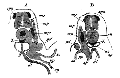
The sections illustrate the development of the segmental duct (pd) or primitive duct of the kidneys. In A (the anterior of the two sections) this appears as a solid knob (pd) projecting towards the epiblast. In B is seen a section of the column which has grown backwards from the knob in A.
spn. rudiment of a spinal nerve; mc. medullary canal; ch. notochord; X. string of cells below the notochord; mp. muscle-plate; mp´. specially developed portion of muscle-plate; ao. dorsal aorta; pd. segmental duct; so. somatopleure; sp. splanchnopleure; pp. pleuro-peritoneal or body-cavity; ep. epiblast; al. alimentary canal.
While the lumen is gradually pushing its way backwards along the solid rudiment of the segmental duct, the first traces of the segmental tubes, or proper excretory organs, make their appearance in the form of solid outgrowths of the intermediate cell-mass, which soon become hollow and open into the body-cavity. Their blind ends curl obliquely backwards round the inner and dorsal side of the segmental duct. One segmental tube makes its appearance for each protovertebra, commencing with that immediately behind the abdominal opening of the segmental duct, the last tube being situated a short way behind the anus. Soon after their formation the blind ends of the segmental tubes open into the segmental duct, and each of them becomes divided into four parts. These are (woodcut 7) (1) a section carrying the abdominal opening or segmental tube proper, (2) a dilated vesicle into which this opens, (3) a coiled tubulus proceeding from (2) and terminating in (4), a wider portion opening into the segmental duct. At the same time, or shortly before this, each segmental duct unites with and opens into one of the horns of the cloaca, and also retires from its primitive position between the epiblast and mesoblast, and assumes a position close to the epithelium lining the body-cavity. The general features of the excretory organs at this period are diagrammatically represented on the woodcut, fig. 6. In this fig. p.d is the segmental duct and o its abdominal opening. s.t points to the segmental tubes, the finer details of whose structure are not represented in the diagram. The kidneys thus form at this period an unbroken gland composed of a series of isolated coiled tubes, one extremity of each of which opens into the body-cavity, and the other into the segmental duct, which forms the only duct of the kidney, and communicates at one end with the body-cavity, and at the other with the cloaca.
Fig. 6.
Diagram of the primitive condition of the Kidney in an Elasmobranch Embryo.
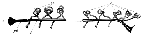
pd. segmental duct. It opens at o into the body-cavity and at its other extremity into the cloaca; x. line along which the division appears which separates the segmental duct into the Wolffian duct above and the Müllerian duct below; st. segmental tubes. They open at one end into the body-cavity, and at the other into the segmental duct.
The next important change concerns the segmental duct, which becomes longitudinally split into two complete ducts in the female, and one complete duct and parts of a second in the male. The manner in which this takes place is diagrammatically represented in woodcut 6 by the clear line x, and in transverse section in woodcut 7. The resulting ducts are the (1) Wolffian duct dorsally, which remains continuous with the excretory tubules of the kidney, and ventrally (2) the oviduct or Müllerian duct in the female, and the rudiments of this duct in the male. In the female the formation of these ducts takes place by a nearly solid rod of cells, being gradually split off from the ventral side of all but the foremost part of the original segmental duct, with the short undivided anterior part of which duct it is continuous in front. Into it a very small portion of the lumen of the original segmental duct is perhaps continued (Pl. 21, fig. 1A, etc.). The remainder of the segmental duct (after the loss of its anterior section and the part split off from its ventral side) forms the Wolffian duct. The process of formation of the ducts in the male chiefly differs from that in the female in the fact of the anterior undivided part of the segmental duct, which forms the front end of the Müllerian duct, being shorter, and in the column of cells with which it is continuous being from the first incomplete.
Fig. 7.
Diagrammatic representation of a transverse section of a Scyllium Embryo illustrating the formation of the Wolffian and Müllerian ducts by the longitudinal splitting of the segmental duct.
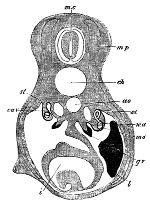
mc. medullary canal; mp. muscle-plate; ch. notochord; ao. aorta; cav. cardinal vein; st. segmental tube. On the one side the section passes through the opening of a segmental tube into the body-cavity. On the other this opening is represented by dotted lines, and the opening of the segmental tube into the Wolffian duct has been cut through; w.d. Wolffian duct; m.d. Müllerian duct. The section is taken through the point where the segmental duct and Wolffian duct have just become separate; gr. The germinal ridge with the thickened germinal epithelium; l. liver; i. intestine with spiral valve.
The tubuli of the primitive excretory organ undergo further important changes. The vesicle at the termination of each segmental tube grows forwards towards the preceding tubulus, and joins the fourth section of it close to the opening into the Wolffian duct (Pl. 21, fig. 10). The remainder of the vesicle becomes converted into a Malpighian body. By the first of these changes a connection is established between the successive segments of the kidney, and though this connection is certainly lost (or only represented by fibrous bands) in the anterior part of the excretory organs in the adult, and very probably in the hinder part, yet it seems most probable that traces of it are to be found in the presence of the secondary Malpighian bodies of the majority of segments, which are most likely developed from it.
Up to this time there has been no distinction between the anterior and posterior tubuli of the primitive excretory organ which alike open into the Wolffian duct. The terminal division of the tubuli of a considerable number of the hindermost of these (ten or eleven in Scyllium canicula), either in some species elongate, overlap, and eventually open by apertures (not usually so numerous as the separate tubes), on nearly the same level, into the hindermost section of the Wolffian duct in the female, or into the urinogenital cloaca, formed by the coalesced terminal parts of the Wolffian ducts, in the male; or in other species become modified in such a manner as to pour their secretion into a single duct on each side, which opens in a position corresponding with the numerous ducts of the other type (woodcut, fig. 8). It seems that both in Amphibians and Elasmobranchii the type with a single duct, or approximations to it, are more often found in the females than in the males. The subject requires however to be more worked out in Elasmobranchii[361]. In both groups the modified posterior kidney-segments are probably equivalent to the permanent kidney of the amniotic Vertebrates, and for this reason the numerous ducts of the first group or single duct of the second were spoken of as ureters. The anterior tubuli of the primitive excretory organ retain their early relation to the Wolffian duct, and form the Wolffian body.
The originally separate terminal extremities of the Wolffian ducts always coalesce, and form a urinal cloaca, opening by a single aperture situated at the extremity of a median papilla behind the anus. Some of the abdominal openings of the segmental tubes in Scyllium, or in other cases all the openings, become obliterated.
In the male the anterior segmental tubes undergo remarkable modifications. There appear to grow from the first three or four or more of them (though the point is still somewhat obscure) branches, which pass to the base of the testis and there unite into a longitudinal canal, form a network, and receive the secretion of the testicular ampullæ (woodcut 9, nt). These ducts, the vasa efferentia, carry the semen to the Wolffian body, but before opening into the tubuli of this they unite into the longitudinal canal of the Wolffian body (l.c), from which pass off ducts equal in number to the vasa efferentia, each of which normally ends in a Malpighian body. From the Malpighian body so connected start the convoluted tubuli of what may be called the generative segments of the Wolffian body along which the semen is conveyed to the Wolffian duct (v.d). The Wolffian duct itself becomes much contorted and acts as vas deferens.
Fig. 8.
Diagram of the arrangement of the Urinogenital Organs in an adult Female Elasmobranch.
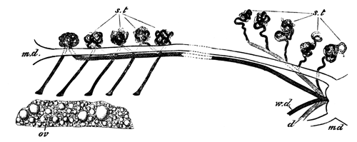
m.d. Müllerian duct; w.d. Wolffian duct; s.t. glandular tubuli; five of them are represented with openings into the body-cavity; d. duct of the posterior segmental tubes; ov. ovary.
In the woodcuts, figs. 8 and 9, are diagrammatically represented the chief constituents of the adult urinogenital organs in the two sexes. In the adult female, fig. 8, there are present the following parts:
(1) The oviduct or Müllerian duct (m.d) split off from the segmental duct of the kidneys. Each oviduct opens at its anterior extremity into the body-cavity, and behind the two oviducts have independent communications with the general cloaca.
(2) The Wolffian ducts (w.d), the other product of the segmental ducts of the kidneys. They end in front by becoming continuous with the tubulus of the anterior segment of the Wolffian body on each side, and unite behind to open by a common papilla into the cloaca. The Wolffian duct receives the secretion of the anterior part of the primitive kidney which forms the Wolffian body.
(3) The ureter (d) which carries off the secretion of the kidney proper. It is represented in my diagram in its most rare and differentiated condition as a single duct.
(4) The glandular tubuli (s.t), some of which retain their original openings into the body-cavity, and others are without them. They are divided into two groups, an anterior forming the Wolffian body, which pour their secretion into the Wolffian duct, and a posterior group forming the kidney proper, which are connected with the ureter.
Fig. 9.
Diagram of the arrangement of the Urinogenital Organs in an adult Male Elasmobranch.
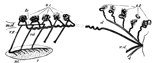
m.d. rudiment of Müllerian duct; w.d. Wolffian duct, marked vd in front and serving as vas deferens; st. glandular tubuli; two of them are represented with openings into the body-cavity; d. ureter; t. testis; nt. central canal at the base of the testis; VE. vasa efferentia; lc. longitudinal canal of the Wolffian body.
In the male the following parts are present (woodcut 9):
(1) The Müllerian duct (md), consisting of a small rudiment attached to the liver representing the foremost end of the oviduct of the female.
(2) The Wolffian duct (w.d) which precisely corresponds to the Wolffian duct of the female, but, in addition to functioning as the duct of the Wolffian body, also acts as a vas deferens (vd). In the adult male its foremost part has a very tortuous course.
(3) The ureter (d), which has the same fundamental constitution as in the female.
(4) The segmental tubes (st). The posterior of these have the same arrangement in both sexes, but in the male modifications take place in connection with the anterior ones to fit them to act as transporters of the testicular products.
Connected with the anterior ones there are present (1) the vasa efferentia (VE), united on the one hand with (2) the central canal in the base of the testis (nt), and on the other with the longitudinal canal of the Wolffian body (l.c). From the latter are seen passing off the successive tubuli of the anterior segments of the Wolffian body in connection with which Malpighian bodies are typically present, though not represented in my diagram.
Postscript.
It was my original intention to have given an account of the development of the generative organs. In the course, however, of my work a number of novel and unexpected points turned up, which have considerably protracted my investigations, and it has appeared to me better no longer to delay the appearance of this monograph, but to publish elsewhere my results on the generative organs. In chapter VI. p. 349 et seq. the early stages of the generative organs are described, but in contemplation of the completion of the account no allusion was made to their literature, and more especially to Professor Semper's important contributions. I may perhaps say that I have been able to confirm the most important result to which he and other anatomists have nearly simultaneously arrived with respect to Vertebrates, viz. that the primitive ova give rise to both the male and female generative products.
EXPLANATION OF PLATES 20 AND 21.
Complete List of Reference Letters.
amg. Accessory Malpighian body. cav. Cardinal vein. ge. Germinal epithelium. k. True kidney. l.c. Longitudinal canal of the Wolffian body connected with vasa efferentia. mg. Malpighian body. nt. Network and central canal at the base of the testis. o. External aperture of urinal cloaca. od. Oviduct or Müllerian duct of the female. od´. Müllerian duct of the male. ou. Openings of ureters in Wolffian duct in the female (fig. 3). pmg. Primary Malpighian body. px. Growth from vesicle at the end of a segmental tube to join the collecting tube of the preceding segment. rst. Rudimentary segmental tube. ru. Ureter commencing to be formed. sb. Seminal bladder. sd. Segmental duct. st. Segmental tube. sto. Opening of segmental tube into body-cavity. sur. Suprarenal body. t. Testis. u. Ureters. ve. Vas efferens. wb. Wolffian body. wd. Wolffian duct.
Plate 20.
Fig. 1. Diagrammatic representation of excretory organs on one side of a male Scyllium canicula, natural size.
Fig. 2. Diagrammatic representation of the kidney proper on one side of a female Scyllium canicula, natural size, shewing the ducts of the kidney and the dilated portion of the Wolffian duct.
Fig. 3. Opening of the ureters into the Wolffian duct of a female Scyllium canicula. The figure represents the Wolffian ducts (wd) with ventral portion removed so as to expose their inner surface, and shews the junction of the two W. ducts to form the common urinal cloaca, the single external opening of this (o), and openings of ureters into one Wolffian duct (ou).
Fig. 4. Anterior extremity of Wolffian body of a young male Scyllium canicula shewing the vasa efferentia and their connection with the kidneys and the testis. The vasa efferentia and longitudinal canal are coloured to render them distinct. They are intended to be continuous with the uncoloured coils of the Wolffian body, though this connection has not been very successfully rendered by the artist.
Fig. 5. Part of the Wolffian body of a nearly ripe male embryo of Scyllium canicula as a transparent object. Zeiss a a, ocul. 3. The figure shews two segmental tubes opening into the body-cavity and connected with a primary Malpighian body, and also, by a fibrous connection, with a secondary Malpighian body of the preceding segment. It also shews one segmental tube (rst) imperfectly connected with the accessory Malpighian body of the preceding segment of the kidney. The coils of the kidney are represented somewhat diagrammatically.
Fig. 6. Vasa efferentia of a male embryo of Scyllium canicula eight centimetres in length. Zeiss a a, ocul. 2.
There are seen to be at the least six and possibly seven distinct vasa going to as many segments of the Wolffian body and connected with a longitudinal canal in the base of the testis. They were probably also connected with a longitudinal canal in the Wolffian body, but this could not be clearly made out.
Fig. 7. The anterior four vasa efferentia of a nearly ripe embryo. Connected with the foremost one is seen a body which looks like the remnant of a segmental tube and its opening (rst?).
Fig. 8. Testis and anterior part of Wolffian body of an embryo of Squatina vulgaris.
The figure is intended to illustrate the arrangement of the vasa efferentia. There are five of these connected with a longitudinal canal in the base of the testis, and with another longitudinal canal in the Wolffian body. From the second longitudinal canal there pass off four ducts to as many Malpighian bodies. Through the Malpighian bodies these ducts are continuous with the several coils of the Wolffian body, and so eventually with the Wolffian duct. Close to the hindermost vas efferens is seen a body which resembles a rudimentary segmental tube (rst?).
Plate 21.
Figs. 1A, 1B, 1C, 1D. Four sections from a female Scyllium canicula of a stage between M and N through the part where the segmental duct becomes split into Wolffian duct and oviduct. Zeiss B, ocul. 2. 1A is the foremost section.
The sections shew that the oviduct arises as a thickening on the under surface of the segmental duct into which at the utmost a very narrow prolongation of the lumen of the segmental duct is carried. The small size of the lumen of the Wolffian duct in the foremost section is due to the section passing through nearly its anterior blind extremity.
Fig. 2. Section close to the junction of the Wolffian duct and oviduct in a female embryo of Scyllium canicula belonging to stage N. Zeiss B, ocul. 2.
The section represented shews that in some instances the formation of the oviduct and Wolffian duct is accompanied by a division of the lumen of the segmental duct into two not very unequal parts.
Figs. 3A, 3B, 3C. Three sections illustrating the formation of a ureter in a female embryo belonging to stage N. Zeiss B, ocul. 2.
3A is the foremost section.
The figures shew that the lumen of the developing ureter is enclosed in front by an independent wall (fig. 3A), but that further back the lumen is partly shut in by the subjacent Wolffian duct, while behind no lumen is present, but the ureter ends as a solid knob of cells without an opening into the Wolffian duct.
Fig. 4. Section through the ureters of the same embryo as fig. 3, but nearer the cloaca. Zeiss B, ocul. 2.
The figure shews the appearance of a transverse section through the wall of cells above the Wolffian duct formed by the overlapping ureters, the lumens of which appear as perforations in it. It should be compared with fig. 9A, which represents a longitudinal section through a similar wall of cells.
Fig. 5. Section through the ureters, the Wolffian duct and the oviduct of a female embryo of Scy. canicula belonging to stage P. Zeiss B, ocul. 2.
Fig. 6. Section of part of the Wolffian body of a male embryo of Scyllium canicula belonging to stage O. Zeiss B, ocul. 2.
The section illustrates (1) the formation of a Malpighian body (mg) from the dilatation at the end of a segmental tube, (2) the appearance of the rudiment of the Müllerian duct in the male (od´).
Figs. 7a, 7b. Two longitudinal and vertical sections through part of the kidney of an embryo between stages L and M. Zeiss B, ocul. 2.
7a illustrates the parts of a single segment of the Wolffian body at this stage, vide p. 491. The segmental tube and opening are not in the plane of the section, but the dilated vesicle is shewn into which the segmental tube opens.
7b is taken from the region of the kidney proper. To the right is seen the opening of a segmental tube into the body-cavity, and in the segment to the left the commencing formation of a ureter, vide p. 502.
Fig. 8. Longitudinal and vertical section through the posterior part of the kidney proper of an embryo of Scyllium canicula at a stage between N and O. Zeiss A, ocul. 2.
The section shews the nearly completed ureters, developing Malpighian bodies, &c.
Fig. 9. Longitudinal and vertical section through the anterior part of the kidney proper of the same embryo as fig. 8. Zeiss A, ocul. 2.
The figure illustrates the mode of growth of the developing ureters.
9A. More highly magnified portion of the same section as fig. 9.
Compare with transverse section fig. 4.
Fig. 10. Longitudinal and vertical section through part of the Wolffian body of an embryo of Scyllium canicula at a stage between O and P.
The section contains two examples of the budding out of the vesicle of a segmental tube to form a Malpighian body in its own segment and to unite with the tubulus of the preceding segment close to its opening into the Wolffian duct.
XI. On the Phenomena accompanying the Maturation and Impregnation of the Ovum[362].
The brilliant discoveries of Strasburger and Auerbach have caused the attention of a large number of biologists to be turned to the phenomena accompanying the division of nuclei and the maturation and impregnation of the ovum. The results of the recent investigations on the first of these points formed the subject of an article by Mr Priestley in the sixteenth volume of this Journal, and the object of the present article is to give some account of what has so far been made out with reference to the second of them. The matters to be treated of naturally fall under two heads: (1) the changes attending the ripening of the ovum, which are independent of impregnation; (2) the changes which are directly due to impregnation.
Fig. 1.—Unripe ovum of Toxopneustes lividus (copied from Hertwig).
Every ovum as it approaches maturity is found to be composed (Fig. 1) of (1) a protoplasmic body or vitellus usually containing yolk-spherules in suspension; (2) of a germinal vesicle or nucleus, containing (3) one or more germinal spots or nucleoli. It is with the germinal vesicle and its contents that we are especially concerned. This body at its full development has a more or less spherical shape, and is enveloped by a distinct membrane. Its contents are for the most part fluid, but may be more or less granular. Their most characteristic component is, however, a protoplasmic network which stretches from the germinal spot to the investing membrane, but is especially concentrated round the former (Fig. 1). The germinal spot forms a nearly homogeneous body, with frequently one or more vacuoles. It occupies an often excentric position within the germinal vesicle, and is usually rendered very conspicuous by its high refrangibility. In many instances it has been shewn to be capable of amœboid movements (Auerbach, and Os. Hertwig), and is moreover more solid and more strongly tinged by colouring reagents than the remaining constituents of the germinal vesicle. These peculiarities have caused the matter of which it is composed to be distinguished by Auerbach and Hertwig as nuclear substance.
In many instances there is only one germinal spot, or one main spot, and two or three accessory smaller spots. In other cases, e.g. Osseous Fish, there are a large number of nearly equal germinal spots. The eggs which have been most investigated with reference to the changes of germinal vesicle are those with a single germinal spot, and it is with these that I shall have more especially to deal in the sequel.
The germinal vesicle occupies in the first instance a central position in the ovum, but at maturity is almost always found in close proximity to the surface. Its change of position in a large number of instances is accomplished during the growth of the ovum in the ovary, but in other cases does not take place till the ovum has been laid.
The questions which many investigators have recently set themselves to answer are the two following:—(1) What becomes of the germinal vesicle when the ovum is ready to be impregnated? (2) Is any part of it present in the ovum at the commencement of segmentation? According to their answers to these questions the older embryologists roughly fall into two groups: (1) By one set the germinal vesicle is stated to completely disappear and not to be genetically connected with the subsequent nuclei of the embryo. (2) According to the other set it remains in the ovum and by successive divisions forms the parent nucleus of all the nuclei in the body of the embryo. Though the second of these views has been supported by several very distinguished names the first view was without doubt the one most generally entertained, and Haeckel (though from his own observations he was originally a supporter of the second view) has even enunciated the theory that there exists an anuclear stage, after the disappearance of the germinal vesicle, which he regards as an embryonic repetition of the monad condition of the Protozoa.