Chapter XII · (continued 8)
Reading edition
Part III.—The ribs of Fishes.
The nature and homologies of the ribs of Fishes have long been a matter of controversy; but the subject has recently been brought forward in the important memoirs of Götte[524] on the Vertebrate skeleton. The alternatives usually adopted are, roughly speaking, these:—Either the hæmal arches of the tail are homologous throughout the piscine series, while the ribs of Ganoids and Teleostei are not homologous with those of Elasmobranchii; or the ribs are homologous in all the piscine groups, and the hæmal arches in the tail are differently formed in the different types. Götte has brought forward a great body of evidence in favour of the first view; while Gegenbaur[525] may be regarded as more especially the champion of the second view.
One of us held in a recent publication[526] that the question was not yet settled, though the view that the ribs are homologous throughout the series was provisionally accepted.
It is admitted by both Gegenbaur and Götte that in Lepidosteus the ribs, in the transition from the trunk to the tail, bend inwards, and finally unite in the region of the tail to form the ventral parts of the hæmal arches, and our researches have abundantly confirmed this conclusion.
Are the hæmal arches, the ventral parts of which are thus formed by the coalescence of the ribs, homologous with the hæmal arches in Elasmobranchii? The researches recorded in the preceding pages appear to us to demonstrate in a conclusive manner that they are so.
The development of the hæmal arches in the tail in these two groups is practically identical; they are formed in both as simple elongations of the primitive hæmal processes, which meet below the caudal vein. In the adult there is an apparent difference between them, arising from the fact that in Lepidosteus the peripheral parts of the hæmal processes are only articulated with the basal portions, and not, as in Elasmobranchii, continuous with them. This difference does not, however, exist in the early larva, since in the larval Lepidosteus the hæmal arches of the tail are unsegmented cartilaginous arches, as they permanently are in Elasmobranchii. If, however, the homology between the hæmal arches of the two types should still be doubted, the fact that in both types the hæmal arches are similarly modified to support the fin-rays of the ventral lobe of the caudal fin, while in neither type are they modified to support the anal fin, may be pointed out as a very strong argument in confirmation of their homology.
The demonstration of the homology of the hæmal arches of the tail in Lepidosteus and Elasmobranchii might at first sight be taken as a conclusive argument in favour of Götte's view, that the ribs of Elasmobranchii are not homologous with those of Ganoidei. This view is mainly supported by two facts:—
(1) In the first place, the ribs in Elasmobranchii do not at first sight appear to be serially homologous with the ventral parts of the hæmal arches of the tail, but would rather seem to be lateral offshoots of the hæmal processes, while the hæmal arches of the tail appear to be completed by the coalescence of independent ventral prolongations of the hæmal processes.
(2) In the second place, the position of the ribs is different in the two groups. In Elasmobranchii they are situated between the dorso-lateral and ventro-lateral muscles (woodcut, fig. 1, rb.), while in Lepidosteus and other Ganoids they immediately girth the body-cavity.
Fig. 1.
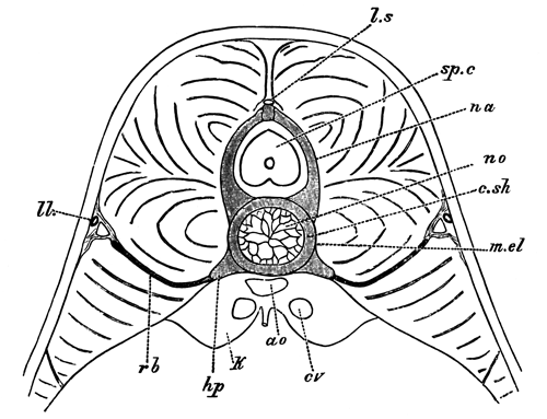
Diagrammatic section through the trunk of an advanced embryo of Scyllium, to shew the position of the ribs.
ao., aorta; c.sh., cartilaginous notochordal sheath; cv., cardinal vein; hp., hæmal process; k., kidney; l.s., ligamentum longitudinale superius; m.el., membrana elastica externa; na., neural arch; no., notochord; ll., lateral line; rb., rib; sp.c., spinal cord.
There is much, therefore, to be said in favour of Götte's view. At the same time, there is another possible interpretation of the facts which would admit the homology of the ribs as well as of the hæmal arches throughout the Pisces.
Let us suppose, to start with, that the primitive arrangement of the parts is more or less nearly that found in Lepidosteus, where we have well-developed ribs in the region of the trunk, girthing the body-cavity, and uniting in the caudal region to form the ventral parts of the hæmal arches. It is easy to conceive that the ribs in the trunk might somewhat alter their position by passing into the muscles, along the inter-muscular septa, till they come to lie between the dorso-lateral and ventro-lateral muscles, as in Elasmobranchii. Lepidosteus itself affords a proof that such a change in the position of the ribs is not impossible, in that it differs from other Ganoids and from Teleostei in the fact that the free ends of the ribs leave the neighbourhood of the body-cavity and penetrate into the muscles.
If it be granted that the mere difference in position between the ribs of Ganoids and Elasmobranchii is not of itself sufficient to disprove their homology, let us attempt to picture what would take place at the junction of the trunk and tail in a type in which the ribs had undergone the above change in position. On nearing the tail it may be supposed that the ribs would gradually become shorter, and at the same time alter their position, till finally they shaded off into ordinary hæmal processes. If, however, the hæmal canal became prolonged forwards by the formation of some additional complete or nearly complete hæmal arches, an alteration in the relation of the parts would necessarily take place. Owing to the position of the ribs, these structures could hardly assist in the new formation of the anterior part of the hæmal canal, but the continuation forwards of the canal would be effected by prolongations of the hæmal processes supporting the ribs. The new arches so formed would naturally be held to be homologous with the hæmal arches of the tail, though really not so, while the true nature of the ribs would also be liable to be misinterpreted, in that the ribs would appear to be lateral outgrowths of the hæmal processes of a wholly different nature to the ventral parts of the hæmal arches of the tail.
In some Elasmobranchii, as shewn in the accompanying woodcut (fig. 2), in the transitional vertebræ between the trunk and the tail, the ribs are supported by lateral outgrowths of the hæmal processes, while the wholly independent prolongations of the hæmal processes appear to be about to give rise to the hæmal arches of the tail.
This peculiar state of things led Götte, and subsequently one of us, to deny for Elasmobranchii all homology between the ribs and any part of the hæmal arches of the tail; but in view of the explanation just suggested, this denial was perhaps too hasty.
Fig. 2.
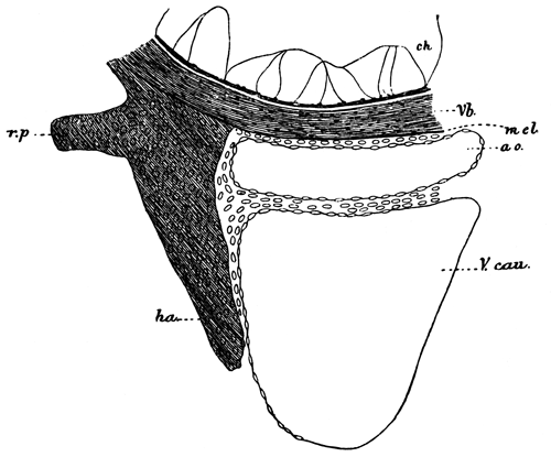
Transverse section through the ventral part of the notochord, and adjoining structures of an advanced Scyllium embryo at the root of the tail.
Vb., cartilaginous sheath of the notochord; ha., hæmal process; r.p., process to which the rib is articulated; m.el., membrana elastica externa; ch., notochord; ao., aorta; V.cau., caudal vein.
We are the more inclined to take this view because the researches of Götte appear to shew that an occurrence, in many respects analogous, has taken place in some Teleostei.
In Teleostei, Johannes Müller, and following him Gegenbaur, do not admit that the hæmal arches of the tail are in any part formed by the ribs. Gegenbaur (Elements of Comp. Anat., translation, p. 431) says, In the Teleostei, the costiferous transverse processes (what we have called the hæmal processes) gradually converge in the caudal region, and form inferior arches, which are not homologous with those of Selachii and Ganoidei, although they also form spinous processes.
The opposite view, that the hæmal arches of the tail in Teleostei contain parts serially homologous with the basal parts of the hæmal processes as well as with the ribs, has been also maintained by many anatomists, e.g., Meckel, Aug. Müller, &c., and has recently found a powerful ally in Götte.
In many cases, the relations of the parts appear to be fundamentally those found in Lepidosteus and Amia, and Götte has shewn by his careful embryological investigations on Esox and Anguilla, that in these two forms there is practically conclusive evidence that the ribs as well as the hæmal costiferous processes of Gegenbaur, which support them, enter into the formation of the hæmal arches of the tail.
In a great number of Teleostei, e.g., the Salmon and most Cyprinoids, &c., the hæmal arches in the region of transition from the trunk to the tail have a structure which at first sight appears to support Johannes Müller's and Gegenbaur's view. The hæmal processes grow larger and meet each other ventrally; while the ribs articulated to them gradually grow smaller and disappear.
The Salmon is typical in this respect, and has been carefully studied by Götte, who attempts to shew (with, in our opinion, complete success) that the anterior hæmal arches are really not entirely homologous with the true hæmal arches behind, but that in the latter, the closure of the arch below is effected by the hæmal spine, which is serially homologous with a pair of coalesced ribs, while in the anterior hæmal arches, i.e., those of the trunk, the closure of the arch is effected by a bridge of bone uniting the hæmal processes.
The arrangement of the parts just described, as well as the view of Götte with reference to them, will be best understood from the accompanying woodcut (fig. 3), copied from Götte's memoir.
Götte sums up his own results on this point in the following words (p. 138): It follows from this, that the half rings, forming the hæmal canal in the hindermost trunk vertebræ of the Salmon, are not (with the exception of the last) completely homologous with those of the tail, but are formed by a connecting piece between the basal stumps (hæmal processes), which originates as a paired median process of these stumps.
The incomplete homology between the anterior hæmal arches and the true caudal hæmal arches which follow them is exactly what we suggest may be the case in Elasmobranchii, and if it be admitted in the one case, we see no reason why it should not also be admitted in the other.
Fig. 3.
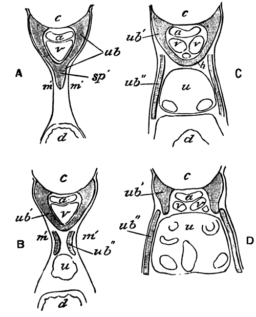
Semi-diagrammatic transverse sections through the first caudal vertebra (A), the last trunk vertebra (B), and the two trunk vertebræ in front (C and D), of a Salmon embryo of 2-3 centims. (From Götte.)
ub., hæmal arch; ub´., hæmal process; ub´´., rib; c., notochord; a., aorta; v., vein; h., connecting pieces between hæmal processes; u., kidney; d., intestine; sp´., hæmal spine; m´., muscles.
If this admission is made, the only ground for not regarding the ribs of Elasmobranchii as homologous with those of Ganoids is their different position, and we have already attempted to prove that this is not a fundamental point.
The results of our researches appear to us, then, to leave two alternatives as to the ribs of Fishes. One of these, which may be called Götte's view, may be thus stated:—The hæmal arches are homologous throughout the Pisces: in Teleostei, Ganoidei, and Dipnoi[527], the ribs, placed on the inner face of the body-wall, are serially homologous with the ventral parts of the hæmal arches of the tail; in Elasmobranchii, on the other hand, the ribs are neither serially homologous with the hæmal arches of the tail nor homologous with the ribs of Teleostei and Ganoidei, but are outgrowths of the hæmal processes into the space between the dorso-lateral and ventro-lateral muscles, which may perhaps have their homologues in Teleostei and Ganoids in certain accessory processes of the vertebræ.
The other view, which we are inclined to adopt, and the arguments for which have been stated in the preceding pages, is as follows:—The Teleostei, Ganoidei, Dipnoi, and Elasmobranchii are provided with homologous hæmal arches, which are formed by the coalescence below the caudal vein of simple prolongations of the primitive hæmal processes of the embryo. The canal enclosed by the hæmal arches can be demonstrated embryologically to be the aborted body-cavity.
In the region of the trunk the hæmal processes and their prolongations behave somewhat differently in the different types.
In Ganoids and Dipnoi, in which the most primitive arrangement is probably retained, the ribs are attached to the hæmal processes, and are placed immediately without the peritoneal membrane at the insertions of the intermuscular septa. These ribs are in many instances (Lepidosteus, Acipenser), and very probably in all, developed continuously with the hæmal processes, and become subsequently segmented from them. They are serially homologous with the ventral parts of the hæmal arches of the tail, which, like them, are in many instances (Ceratodus, Lepidosteus, Polypterus, and to some extent in Amia) segmented off from the basal parts of the hæmal arches.
In Teleostei the ribs have the same position and relations as those in Ganoids and Dipnoi, but their serial homology with the ventral parts of the hæmal processes of the tail, is often (e.g., the Salmon) obscured by some of the anterior hæmal arches in the posterior part of the trunk being completed, not by the ribs, but by independent outgrowths of the basal parts of the hæmal processes.
In Elasmobranchii a still further divergence from the primitive arrangement is present. The ribs appear to have passed outwards along the intermuscular septa into the muscles, and are placed between the dorso-lateral and ventro-lateral muscles (a change of position of the ribs of the same nature, but affecting only their ends, is observable in Lepidosteus). This change of position, combined probably with the secondary formation of a certain number of anterior hæmal arches similar to those in the Salmon, renders their serial homology with the ventral parts of the hæmal processes of the tail far less clear than in other types, and further proof is required before such homology can be considered as definitely established.
This is not the place to enter into the obscure question as to how far the ribs of the Amphibia and Amniota are homologous with those of Fishes. It is to be remarked, however, that the ribs of the Urodela (1) occupy the same position in relation to the muscles as the Elasmobranch ribs, (2) that they are connected with the neural arches, and (3) that they coexist in the tail with the hæmal arches, and seem, therefore, to be as different as possible from the ribs of the Dipnoi.
Part IV.—The skeleton of the ventral lobe of the tail fin, and its bearing on the nature of the tail fin of the various types of Pisces.
In the embryos or larvæ of all the Elasmobranchii, Ganoidei, and Teleostei which have up to this time been studied, the unpaired fins arise as median longitudinal folds of the integument on the dorsal and ventral sides of the body, which meet at the apex of the tail. The tail at first is symmetrical, having a form which has been called diphycercal or protocercal. At a later stage, usually, though not always, parts of these fins atrophy, while other parts undergo a special development and constitute the permanent unpaired fins.
Since the majority of existing as well as extinct Fishes are provided with discontinuous fins, those forms, such as the Eel (Anguilla), in which the fins are continuous, have probably reverted to an embryonic condition: an evolutional process which is of more frequent occurrence than has usually been admitted.
In the caudal region there is almost always developed in the larvæ of the above groups a special ventral lobe of the embryonic fin a short distance from the end of the tail. In Elasmobranchii and Chondrostean Ganoids the portion of the embryonic tail behind this lobe persists through life, and a special type of caudal fin, which is usually called heterocercal, is thus produced. This type of caudal fin appears to have been the most usual in the earlier geological periods.
Simultaneously with the formation of the ventral lobe of the heterocercal caudal fin, the notochord with the vertebral tissues surrounding it, becomes bent somewhat dorsalwards, and thus the primitive caudal fin forms a dorsally directed lobe of the heterocercal tail. We shall call this part the dorsal lobe of the tail-fin, and the secondarily formed lobe the ventral lobe.
Lepidosteus and Amia (Wilder, No. 15) amongst the bony Ganoids, and, as has recently been shewn by A. Agassiz[528], most Teleostei acquire at an early stage of their development heterocercal caudal fins, like those of Elasmobranchii and the Chondrostean Ganoids; but in the course of their further growth the dorsal lobe partly atrophies, and partly disappears as such, owing to the great prominence acquired by the ventral lobe. A portion of the dorsally flexed notochord and of the cartilage or bone replacing or investing it remains, however, as an indication of the original dorsal lobe, though it does not project backwards beyond the level of the end of the ventral lobe, which in these types forms the terminal caudal fin.
The true significance of the dorsally flexed portion of the vertebral axis was first clearly stated by Huxley[529], but as A. Agassiz has fairly pointed out in the paper already quoted, this fact does not in any way militate against the view put forward by L. Agassiz that there is a complete parallelism between the embryonic development of the tail in these Fishes and the palæontological development of this organ. We think that it is moreover convenient to retain the term homocercal for those types of caudal fin in which the dorsal lobe has atrophied so far as not to project beyond the ventral lobe.
We have stated these now well-known facts to enable the reader to follow us in dealing with the comparison between the skeleton supporting the fin-rays of the ventral lobe of the caudal fin, and that supporting the fin-rays of the remaining unpaired fins.
It has been shewn that in Lepidosteus the unpaired fins fall into two categories, according to the nature of the skeletal parts supporting them. The fin-rays of the true ventral lobe of the caudal fin are supported by the spinous processes of certain of the hæmal arches. The remaining unpaired fins, including the anal fin, are supported by the so-called interspinous bones, which are developed independently of the vertebral column and its arches.
The question which first presents itself is, how far does this distinction hold good for other Fishes? This question, though interesting, does not appear to have been greatly discussed by anatomists. Not unfrequently the skeletal supports of the ventral lobe of the caudal fin are assumed to be the same as those of the other fins.
Davidoff[530], for instance, in speaking of the unpaired fins of Elasmobranch embryos, says (p. 514): The cartilaginous rays of the dorsal fins agreed not only in number with the spinous processes (as indeed is also found in the caudal fin of the full-grown Dog-fish), &c.
Thacker[531], again, in his memoir on the Median and Paired Fins, states at p. 284: We shall here consider the skeleton of the dorsal and anal fins alone. That of the caudal fin has undergone peculiar modifications by the union of fin-rays with hæmal spines.
Mivart[532] goes into the question more fully. He points out (p. 471) that there is an essential difference between the dorsal and ventral parts of the caudal fin in Elasmobranchii, in that in the former the radials are more numerous than the vertebræ and unconformable to them, while in the latter they are equal in number to the vertebræ and continuous with them. This, he goes on to say, seems to point to a difference in nature between the dorsal and ventral portions of the caudal fin, in at least most Elasmobranchii. He further points out that Polyodon resembles Elasmobranchii. As to Teleostei, he does not express himself decidedly except in the case of Muræna, to which we shall return.
Mivart expresses himself as very doubtful as to the nature of the supports of the caudal fin, and thinks that the caudal fin of different kinds of Fishes may have arisen in different ways in different cases.
An examination of the ventral part of the caudal fin in various Ganoids, Teleostei, and Elasmobranchii appears to us to shew that there can be but little doubt that, in the majority of the members of these groups at any rate, and we believe in all, the same distinction between the ventral lobe of the caudal fin and the remaining unpaired fins is found as in Lepidosteus.
In the case of most Elasmobranchii, a simple inspection of the caudal fin suffices to prove this, and the anatomical features involved in this fact have usually been recognized; though, in the absence of embryological evidence, the legitimate conclusion has not always been drawn from them.
The difference between the ventral lobe of the caudal fin and the other fins in the mode in which the fin-rays are supported is as obvious in Chondrostean Ganoids as it is in Elasmobranchii; it would appear also to hold good for Amia. Polypterus we have had no opportunity of examining, but if, as there is no reason to doubt, the figure of its skeleton given by Agassiz (Poissons Fossiles) is correct, there can be no question that the ventral lobe of the caudal fin is supported by the hæmal arches, and not by interspinous bones. In Calamoicthys, the tail of which we have had an opportunity of dissecting through the kindness of Professor Parker, the fin-rays of the ventral lobe of the true caudal fin are undoubtedly supported by true hæmal arches.
There is no unanimity of opinion as to the nature of the elements supporting the fin-rays of the caudal fin of Teleostei.
Huxley[533] in his paper on the development of the caudal fin of the Stickleback, holds that these elements are of the nature of interhæmal bones. He says (p. 39): "The last of these rings lay just where the notochord began to bend up. It was slightly longer than the bony ring which preceded it, and instead of having its posterior margin parallel with the anterior, it sloped from above downwards and backwards. Two short osseous plates, attached to the anterior part of the inferior surface of the penultimate ring, or rudimentary vertebral centrum, passed downwards and a little backwards, and abutted against a slender elongated mass of cartilage. Similar cartilaginous bodies occupy the same relation to corresponding plates of bone in the anterior vertebræ in the region of the anal fin; and it is here seen, that while the bony plates coalesce and form the inferior arches of the caudal vertebræ, the cartilaginous elements at their extremities become the interhæmal bones. The cartilage connected with the inferior arch of the penultimate centrum is therefore an interhæmal cartilage. The anterior part of the inferior surface of the terminal ossification likewise has its osseous inferior arch, but the direction of this is nearly vertical, and though it is connected below with an element which corresponds in position with the interhæmal cartilage, this cartilage is five or six times as large, and constitutes a broad vertical plate, longer than it is deep, and having its longest axis inclined downwards and backwards....
Immediately behind and above this anterior hypural apophysis (as it may be termed) is another very much smaller vertical cartilaginous plate, which may be called the posterior hypural apophysis.
We have seen that Mivart expresses himself doubtful on the subject. Gegenbaur[534] appears to regard them as hæmal arches.
The latter view appears to us without doubt the correct one. An examination of the tail of normal Teleostei shews that the fin-rays of that part of the caudal fin which is derived from the ventral lobe of the larva are supported by elements serially homologous with the hæmal arches, but in no way homologous with the interspinous bones of the anal fin. The elements in question formed of cartilage in the larva, become ossified in the adult, and are known as the hypural bones. They may appear in the form of a series of separate hæmal arches, corresponding in number with the primitive somites of this region, which usually, however, atrophy in the adult, or more often are from the first imperfectly segmented, and have in the adult the form of two or three or even of a single broad bony plate. The transitional forms between this state of things and that, for instance, in Lepidosteus are so numerous, that there can be no doubt that even the most peculiar forms of the hypural bones of Teleostei are simply modified hæmal arches.
This view of the hypural bones is, moreover, supported by embryological evidence, since Aug. Müller[535] (p. 205) describes their development in a manner which, if his statements are to be trusted, leaves no doubt on this point.
There are a considerable number of Fishes which are not provided with an obvious caudal fin as distinct from the remaining unpaired fins, i.e. Chimæra, Eels, and various Eel-like forms amongst Teleostei, and the Dipnoi. Gegenbaur appears to hold that these Fishes ought to be classed together in relation to the structure of the caudal portion of their vertebral column, as he says on p. 431 of his Comparative Anatomy (English Translation): In the Chimæræ, Dipnoi, and many Teleostei, the caudal portion of the vertebral column ends by gradually diminishing in size, but in most Fishes, &c.
For our purpose it will, however, be advisable to treat them separately.
The tail of Chimæra appears to us to be simply a peculiar modification of the typical Elasmobranch heterocercal tail, in which the true ventral lobe of the caudal fin may be recognized in the fin-fold immediately in front of the filamentous portion of the tail. In the allied genus Callorhynchus this feature is more distinct. The filamentous portion of the tail of Chimæra constitutes, according to the nomenclature adopted above, the true dorsal lobe, and may be partially paralleled in the filamentous dorsal lobe of the tail of the larval Lepidosteus (Plate 34, fig. 16).
The tail of the eel-like Teleostei is again undoubtedly a modification of the normal form of tail characteristic of the Teleostei, in which, however, the caudal fin has become very much reduced and merged into the prolongations of the anal and dorsal fins.
This can be very clearly seen in Siluroid forms with an Eel-like tail, such as Cnidoglanis. Although the dorsal and ventral fins appear to be continuous round the end of the tail, and there is superficially no distinct caudal fin, yet an examination of the skeleton of Cnidoglanis shews that the end of the vertebral column is modified in the usual Teleostean fashion, and that the hæmal arches of the modified portion of the vertebral column support a small number of fin-rays; the adjoining ventral fin-rays being supported by independent osseous fin-supports (interspinous bones).
In the case of the Eel (Anguilla anguilla) Huxley (loc. cit.) long ago pointed out that the terminal portion of the vertebral column was modified in an analogous fashion to that of other Teleostei, and we have found that the modified hæmal arches of this part support a few fin-rays, though a still smaller number than in Cnidoglanis. The fin-rays so supported clearly constitute an aborted ventral lobe of the caudal fin.
Under these circumstances we think that the following statement by Mivart (Zool. Trans. Vol. X., p. 471) is somewhat misleading:—
As to the condition of this part (i.e. the ventral lobe of the tail-fin) in Teleosteans generally, I will not venture as yet to say anything generally, except that it is plain that in such forms as Muræna, the dorsal and ventral parts of the caudal fin are similar in nature and homotypal with ordinary dorsal and anal fins[536].
The italicized portion of this sentence is only true in respect to that part of the fringe of fin surrounding the end of the body, which is not only homotypal with, but actually part of, the dorsal and anal fins.
Having settled, then, that the tails of Chimæra and of Eel-like Teleostei are simply special modifications of the typical form of tail of the group of Fishes to which they respectively belong, we come to the consideration of the Dipnoi, in which the tail-fin presents problems of more interest and greater difficulty than those we have so far had to deal with.
The undoubtedly very ancient and primitive character of the Dipnoi has led to the view, implicitly if not definitely stated in most text-books, that their tail-fin retains the character of the piscine tail prior to the formation of the ventral caudal lobe, a stage which is repeated embryologically in the pre-heterocercal condition of the tail in ordinary Fishes.
Through the want of embryological data, and in the absence of really careful histological examination of the tail of any of the Dipnoi, we are not willing to speak with very great confidence as to its nature; we are nevertheless of the opinion that the facts we can bring forward on this head are sufficient to shew that the tail of the existing Dipnoi is largely aborted, so that it is more or less comparable with that of the Eel.
We have had opportunities of examining the structure of the tail of Ceratodus and Protopterus in dissected specimens in the Cambridge Museum. The vertebral axis runs to the ends of the tail without shewing any signs of becoming dorsally flexed. At some distance from the end of the tail the fin-rays are supported by what are apparently segmented spinous prolongations of the neural and hæmal arches. The dorsal elements are placed above the longitudinal dorsal cord, and occupy therefore the same position as the independent elements of the neural arches of Lepidosteus. They are therefore to be regarded as homologous with the dorsal fin-supports or interspinous bones of other types. The corresponding ventral elements are therefore also to be regarded as interspinous bones.
In view of the fact that the fin-supports, whenever their development has been observed, are found to be formed independently of the neural and hæmal arches, we may fairly assume that this is also true for what we have identified as the interspinous elements in the Dipnoi.
The interspinous elements become gradually shorter as the end of the tail is approached, and it is very difficult from a simple examination of dissected specimens to make out how far any of the posterior fin-rays are supported by the hæmal arches only. To this question we shall return, but we may remark that, although there is a prolongation backwards of the vertebral axis beyond the last interspinous elements, composed it would seem of the coalesced neural and hæmal arches but without the notochord, yet by far the majority of the fin-rays which constitute the apparent caudal fin are supported by interspinous elements.
The grounds on which we hold that the tail of the Dipnoi is to be regarded as a degenerate rather than primitive type of tail are the following:—
(1) If it be granted that a diphycercal or protocercal form of tail must have preceded a heterocercal form, it is also clear that the ventral fin-rays of such a tail must have been supported, as in Polypterus and Calamoicthys, by hæmal arches, and not by interspinous elements; otherwise, a special ventral lobe, giving a heterocercal character to the tail, and provided with fin-rays supported only by hæmal arches, could never have become evolved from the protocercal tail-fin. Since the ventral fin-rays of the tail of the Dipnoi are supported by interspinous elements and not by hæmal arches, this tail-fin cannot claim to have the character of that primitive type of diphycercal or protocercal tail from which the heterocercal tail must be supposed to have been evolved.
(2) Since the nearest allies of the Dipnoi are to be found in Polypterus and the Crossopterygidæ of Huxley, and since in these forms (as evinced by the structure of the tail-fin of Polypterus, and the transitional type between a heterocercal and diphycercal form of fin observable in fossil Crossopterygidæ) the ventral fin-rays of the caudal fin were clearly supported by hæmal arches and not by interspinous elements, it is rendered highly probable that the absence of fin-rays so supported in the Dipnoi is a result of degeneration of the posterior part of the tail.
[We use this argument without offering any opinion as to whether the diphycercal character of the tail of many Crossopterygidæ is primary or secondary.]
(3) The argument just used is supported by the degenerate and variable state of the end of the vertebral axis in the Dipnoi—a condition most easily explained by assuming that the terminal part of the tail has become aborted.
(4) We believe that in Ceratodus we have been able to trace a small number of the ventral fin-rays supported by hæmal arches only, but these rays are so short as not to extend so far back as some of the rays attached to the interspinous elements in front. These rays may probably be interpreted, like the more or less corresponding rays in the tail of the Eel, as the last remnant of a true caudal fin.
The above considerations appear to us to shew with very considerable probability that the true caudal fin of the Dipnoi has become all but aborted like that of various Teleostei; and that the apparent caudal fin is formed by the anal and dorsal fins meeting round the end of the stump of the tail.
From the adult forms of Dipnoi we are, however, of opinion that no conclusion can be drawn as to whether their ancestors were provided with a diphycercal or a heterocercal form of caudal fin.
The general conclusions with reference to the tail-fin at which we have arrived are the following:—
(1) The ventral lobe of the tail-fin of Pisces differs from the other unpaired fins in the fact that its fin-rays are directly supported by spinous processes of certain of the hæmal arches instead of independently developed interspinous bones.
(2) The presence or absence of fin-rays in the tail-fin supported by hæmal arches may be used in deciding whether apparently diphycercal tail-fins are aborted or primitive.
Excretory and Generative Organs.
I.—Anatomy.
The excretory organs of Lepidosteus have been described by Müller (No. 13) and Hyrtl (No. 11). These anatomists have given a fairly adequate account of the generative ducts in the female, and Hyrtl has also described the male generative ducts and the kidney and its duct, but his description is contradicted by our observations in some of the most fundamental points.
In the female example of 100.5 centims. which we dissected, the kidney forms a paired gland, consisting of a narrow strip of glandular matter placed on each side of the vertebral column, on the dorsal aspect of the body-cavity. It is covered on its ventral aspect by the oviduct and by its own duct, but is separated from both of these by a layer of the tough peritoneal membrane, through which the collecting tubes pass. It extends forwards from the anus for about three-fifths of the length of the body-cavity, and in our example had a total length of about 28 centims. (Plate 39, fig. 60, k). Anteriorly the two kidneys are separated by a short interval in the median line, but posteriorly they come into contact, and are so intimately united as almost to constitute a single gland.
A superficial examination might lead to the supposition that the kidney extended forwards for the whole length of the body-cavity up to the region of the branchial arches, and Hyrtl appears to have fallen into this error; but what appears to be its anterior continuation is really a form of lymphatic tissue, something like that of the spleen, filled with numerous cells. This matter (Plate 39, fig. 60, ly.) continues from the kidney forwards without any break, and has a colour so similar to that of the kidney as to be hardly distinguishable from it with the naked eye. The true anterior end of the kidney is placed about 3 centims. in front on the left side, and on the same level on the right side as the wide anterior end of the generative duct (Plate 39, fig. 60, od.). It is not obviously divided into segments, and is richly supplied with malpighian bodies.
It is clear from the above description that there is no trace of head-kidney or pronephros visible in the adult. To this subject we shall, however, again return.
As will appear from the embryological section, the ducts of the kidneys are probably simply the archinephric ducts, but to avoid the use of terms involving a theory, we propose in the anatomical part of our work to call them kidney ducts. They are thin-walled widish tubes coextensive with the kidneys. If cut open there may be seen on their inner aspect the numerous openings of the collecting tubes of the kidneys. They are placed ventrally to and on the outer border of the kidneys (Plate 39, fig. 60, s.g.). Posteriorly they gradually enlarge, and approaching each other in the median line, coalesce, forming an unpaired vesicle or bladder (bl.)—about 6 centims. long in our example—opening by a median pore on a more or less prominent papilla (u.g.) behind the anus. The dilated portions of the two ducts are called by Hyrtl the horns of the bladder.
The sides of the bladder and its so-called horns are provided with lateral pockets into which the collecting tubes of the kidney open. These pockets, which we have found in two female examples, are much larger in the horns of the bladder than in the bladder itself. Similar pockets, but larger than those we have found, have been described by Hyrtl in the male, but are stated by him to be absent in the female. It is clear from our examples that this is by no means always the case.
Hyrtl states that the wide kidney ducts, of which his description differs in no material point from our own, suddenly narrow in front, and, perforating the peritoneal lining, are continued forwards to supply the anterior part of the kidney. We have already shewn that the anterior part of the kidney has no existence, and the kidney ducts supplying it are, according to our investigations, equally imaginary.
It was first shewn by Müller, whose observations on this point have been confirmed by Hyrtl, &c., that the ovaries of Lepidosteus are continuous with their ducts, forming in this respect an exception to other Ganoids.
In our example of Lepidosteus the ovaries (Plate 39, fig. 60, ov.) were about 18 centims. in length. They have the form of simple sacks, filled with ova, and attached about their middle to their generative duct, and continued both backwards and forwards from their attachment into a blind process.
With reference to these sacks Müller has pointed out—and the importance of this observation will become apparent when we deal with the development—that the ova are formed in the thickness of the inner wall of the sack. We hope to shew that the inner wall of the sack is alone equivalent to the genital ridge of, for instance, the ovary of Scyllium. The outer aspect of this wall—i.e., that turned towards the interior of the sack—is equivalent to the outer aspect of the Elasmobranch genital ridge, on which alone the ova are developed[537]. The sack into which the ova fall is, as we shall shew in the embryological section, a special section of the body-cavity shut off from the remainder, and the dehiscence of the ova into this cavity is equivalent to their discharge into the body-cavity in other forms.
The oviduct (Plate 39, fig. 60, od.) is a thin-walled duct of about 21 centims. in length in the example we are describing, continuous in front with the ovarian sack, and gradually tapering behind, till it ends (od´.) by opening into the dilated terminal section of the kidney duct on the inner side, a short distance before the latter unites with its fellow. It is throughout closely attached to the ureter and placed on its inner, and to some extent on its ventral, aspect. The hindermost part of the oviduct which runs beside the enlarged portion of the kidney duct—that portion called by Hyrtl the horn of the urinary bladder—is so completely enveloped by the wall of the horn of the urinary bladder as to appear like a projection into the lumen of the latter structure, and the somewhat peculiar appearance which it presents in Hyrtl's figure is due to this fact. In our examples the oviduct was provided with a simple opening into the kidney duct, on a slight papilla; the peculiar dilatations and processes of the terminal parts of the oviduct, which have been described by Hyrtl, not being present.
The results we have arrived at with reference to the male organs are very different indeed from those of our predecessor, in that we find the testicular products to be carried off by a series of vasa efferentia, which traverse the mesorchium, and are continuous with the uriniferous tubuli; so that the semen passes through the uriniferous tubuli into the kidney duct and so to the exterior. We have moreover been unable to find in the male a duct homologous with the oviduct of the female.
This mode of transportation outwards of the semen has not hitherto been known to occur in Ganoids, though found in all Elasmobranchii, Amphibia, and Amniota. It is not, however, impossible that it exists in other Ganoids, but has hitherto been overlooked.
Our male example of Lepidosteus was about 60 centims. in length, and was no doubt mature. It was smaller than any of our female examples, but this according to Garman (vide, p. 361) is usual. The testes (Plate 39, fig. 58A., t.) occupied a similar position to the ovaries, and were about 21 centims. long. They were, as is frequently the case with piscine testes, divided into a series of lobes (10-12), and were suspended by a delicate mesentery (mesorchium) from the dorsal wall of the abdomen on each side of the dorsal aorta. Hyrtl (No. 11) states that air or quicksilver injected between the limbs of the mesentery, passed into a vas deferens homologous with the oviduct which joins the ureter. We have been unable to find such a vas deferens; but we have found in the mesorchium a number of tubes of a yellow colour, the colour being due to a granular substance quite unlike coagulated blood, but which appeared to us from microscopic examination to be the remains of spermatozoa[538]. These tubes to the number of 40-50 constitute, we believe, the vasa efferentia. Along the line of suspension of the testis on its inner border these tubes unite to form an elaborate network of tubes placed on the inner face of the testis—an arrangement very similar to that often found in Elasmobranchii (vide F. M. Balfour, Monograph on the Development of Elasmobranch Fishes, plate 20, figs. 4 and 8).
We have figured this network on the posterior lobe of the testis (fig. 58B), and have represented a section through it (fig. 59A, n.v.e.), and through one of the vasa efferentia (v.e.) in the mesorchium. Such a section conclusively demonstrates the real nature of these passages: they are filled with sperm like that in the body of the testis, and are, as may be seen from the section figured, continuous with the seminal tubes of the testis itself.
At the attached base of the mesorchium the vasa efferentia unite into a longitudinal canal, placed on the inner side of the kidney duct (Plate 39, fig. 58A, l.c., also shewn in section in Plate 39, fig. 59B, l.c.). From this canal tubules pass off which are continuous with the tubuli uriniferi, as may be seen from fig. 59B, but the exact course of these tubuli through the kidney could not be made out in the preparations we were able to make of the badly conserved kidney. Hyrtl describes the arrangement of the vascular trunks in the mesorchium in the following way (No. 11, p. 6): The mesorchium contains vascular trunks, viz., veins, which through their numerous anastomoses form a plexus at the hilus of the testis, whose efferent trunks, 13 in number, again unite into a plexus on the vertebral column, which is continuous with the cardinal veins. The arrangement (though not the number) of Hyrtl's vessels is very similar to that of our vasa efferentia, and we cannot help thinking that a confusion of the two may have taken place; which, in badly conserved specimens, not injected with semen, would be very easy.
We have, as already stated, been unable to find in our dissections any trace of a duct homologous with the oviduct of the female, and our sections through the kidney and its ducts equally fail to bring to light such a duct. The kidney ducts are about 19 centims. in length, measured from the genital aperture to their front end. These ducts are generally similar to those in the female; they unite about 2 centims. from the genital pore to form an unpaired vesicle. Their posterior parts are considerably enlarged, forming what Hyrtl calls the horns of the urinary bladder. In these enlarged portions, and in the wall of the unpaired urinary bladder, numerous transverse partitions are present, as correctly described by Hyrtl, which are similar to those in the female, but more numerous. They give rise to a series of pits, at the blind ends of which are placed the openings of the kidney tubules. The kidney duct without doubt serves as vas deferens, and we have found in it masses of yellowish colour similar to the substance in the vasa efferentia identified by us as remains of spermatozoa.
II.—Development.
In the general account of the development we have already called attention to the earliest stages of the excretory system.
We may remind the reader that the first part of the system to be formed is the segmental or archinephric duct (Plate 36, figs. 28 and 29, sg.). This duct arises, as in Teleostei and Amphibia, by the constriction of a hollow ridge of the somatic mesoblast into a canal, which is placed in contiguity with the epiblast, along the line of junction between the mesoblastic somites and the lateral plates of mesoblast. Anteriorly the duct does not become shut off from the body-cavity, and also bends inwards towards the middle line. The inflected part of the duct is the first rudiment of the pronephros, and very soon becomes considerably dilated relatively to the posterior part of the duct.
The posterior part of each segmental duct acquires an opening into the cloacal section of the alimentary tract. Apart from this change, the whole of the ducts, except their pronephric sections, remain for a long time unaltered, and the next changes we have to speak of concern the definite establishment of the pronephros.
The dilated incurved portion of each segmental duct soon becomes convoluted, and by the time the embryo is about 10 millims. in length, but before the period of hatching, an important change is effected in the relations of their peritoneal openings[539].
Instead of leading into the body-cavity, they open into an isolated chamber on each side (Plate 38, fig. 51, pr.c.), which we will call the pronephric chamber. The pronephric chamber is not, however, so far as we can judge, completely isolated from the body-cavity. We have not, it is true, detected with certainty at this stage a communication between the two; but in later stages, in larvæ of from 11 to 26 millims., we have found a richly ciliated passage leading from the body-cavity into the pronephros on each side (Plate 38, fig. 52, p.f.p.). We have not succeeded in determining with absolute certainty the exact relations between this passage and the tube of the pronephros, but we are inclined to believe that it opens directly into the pronephric chamber just spoken of.
As we hope to shew, this chamber soon becomes largely filled by a vascular glomerulus. On the accomplishment of these changes, the pronephros is essentially provided with all the parts typically present in a segment of the mesonephros (woodcut, fig. 4). There is a peritoneal tube (f)[540], opening into a vesicle (v); from near the neck of the peritoneal tube there comes off a convoluted tube (pr.n.), forming the main mass of the pronephros, and ending in the segmental duct (sd.).
Fig. 4.
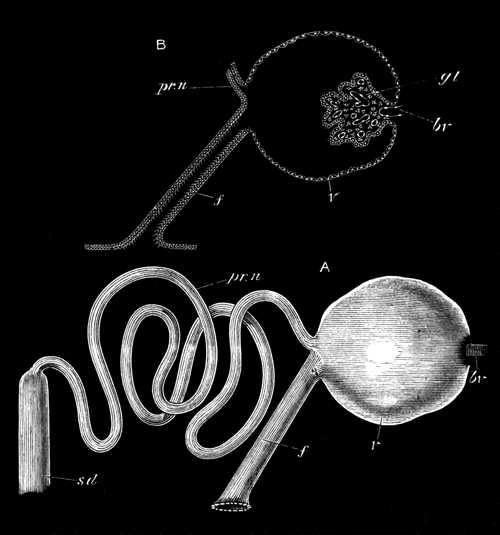
Diagrammatic views of the pronephros of Lepidosteus.
A, pronephros supposed to be isolated and seen from the side; B, section through the vesicle of the pronephros and the ciliated peritoneal funnel leading into it; pr.n., coiled tube of pronephros; sd., segmental or archinephric duct; f., peritoneal funnel; v., vesicle of pronephros; bv., blood vessel of glomerulus; gl., glomerulus.
The different parts do not, however, appear to have the same morphological significance as those in the mesonephros.
Judging from the analogy of Teleostei, the embryonic structure of whose pronephros is strikingly similar to that of Lepidosteus, the two pronephric chambers into which the segmental ducts open are constricted off sections of the body-cavity.
With the formation of the convoluted duct opening into the isolated section of the body-cavity we may speak of a definite pronephros as having become established. The pronephros is placed, as can be made out in later stages, on the level of the opening of the air-bladder into the throat.
The pronephros increases in size, so far as could be determined, by the further convolution of the duct of which it is mainly formed; and the next change of importance which we have noticed is the formation of a vascular projection into the pronephric chamber, forming the glomerulus already spoken of (vide woodcut, fig. 4, gl.), which is similar to that of the pronephros of Teleostei. We first detected these glomeruli in an embryo of about 15 millims., some days after hatching (Plate 38, fig. 52, gl.), but it is quite possible that they may be formed considerably earlier.
In the same embryo in which the glomeruli were found we also detected for the first time a mesonephros consisting of a series of isolated segmental or nephridial tubes, placed posteriorly to the pronephros along the dorsal wall of the abdomen.
These were so far advanced at this stage that we are not in a position to give any account of their mode of origin. They are, however, formed independently of the segmental ducts, and in the establishment of the junction between the two structures, there is no outgrowth from the segmental duct to meet the segmental tubes. We could not at this stage find peritoneal funnels of the segmental tubes, though we have met with them at a later stage (Plate 38, fig. 53, p.f.), and our failure to find them at this stage is not to be regarded as conclusive against their existence.
A very considerable space exists between the pronephros and the foremost segmental tube of the mesonephros. The anterior mesonephric tubes are, moreover, formed earlier than the posterior.
In the course of further development, the mesonephric tubules increase in size, so that there ceases to be an interval between them, the mesonephros thus becoming a continuous gland. In an embryo of 26 millims. there was no indication of the formation of segmental tubes to fill up the space between the pronephros and mesonephros.
The two segmental ducts have united behind into an unpaired structure in an embryo of 11 millims. This structure is no doubt the future unpaired urinogenital chamber (Plate 39, figs. 58A, and 60, bl.). Somewhat later, the hypoblastic cloaca becomes split into two sections, the hinder one receiving the coalesced segmental ducts, and the anterior remaining continuous with the alimentary tract. The opening of the hinder one forms the urinogenital opening, and that of the anterior the anus.
In an older larva of about 5.5 centims. the pronephros did not exhibit any marked signs of atrophy, though the duct between it and the mesonephros was somewhat reduced and surrounded by the trabecular tissue spoken of in connection with the adult. In the region between the pronephros and the front end of the fully developed part of the mesonephros very rudimentary tubules had become established.
The latest stage of the excretory system which we have studied is in a young Fish of about 11 centims. in length. The special interest of this stage depends upon the fact that the ovary is already developed, and not only so, but the formation of the oviducts has commenced, and their condition at this stage throws considerable light on the obscure problem of their nature in the Ganoids.
Unfortunately, the head of the young Fish had been removed before it was put into our hands, so that it was impossible for us to determine whether the pronephros was still present; but as we shall subsequently shew, the section of the segmental duct, originally present between the pronephros and the front end of the permanent kidney or mesonephros, has in any case disappeared.
In addition to an examination of the excretory organs in situ, which shewed little except the presence of the generative ridges, we made a complete series of sections through the excretory organs for their whole length (Plate 39, figs. 54-57).
Posteriorly these sections shewed nothing worthy of note, the excretory organs and their ducts differing in no important particular from these organs as we have described them in the adult, except in the fact that the segmental ducts are not joined by the oviducts.
Some little way in front of the point where the two segmental ducts coalesce to form the urinary bladder, the genital ridge comes into view. For its whole extent, except near its anterior part (of which more hereafter) this ridge projects freely into the body-cavity, and in this respect the young Fish differs entirely from the adult. As shewn in Plate 39, figs. 56 and 57 (g.r.), it is attached to the abdominal wall on the ventral side of, and near the inner border of each kidney. The genital ridge itself has a structure very similar to that which is characteristic of young Elasmobranchii, and it may be presumed of young Fishes generally. The free edge of the ridge is swollen, and this part constitutes the true generative region of the ridge, while its dorsal portion forms the supporting mesentery. The ridge itself is formed of a central stroma and a germinal epithelium covering it. The epithelium is thin on the whole of the inner aspect of the ridge, but, just as in Elasmobranchii, it becomes greatly thickened for a band-like strip on the outer aspect. Here, the epithelium is several layers deep, and contains numerous primitive germinal cells (p.o.).
Though the generative organs were not sufficiently advanced for us to decide the point with certainty, the structure of the organ is in favour of the view that this specimen was a female, and, as will be shewn directly, there can on other grounds be no doubt that this is so. The large size of the primitive germinal cells (primitive ova) reminded us of these bodies in Elasmobranchii.
In the region between the insertion of the genital ridge (or ovary, as we may more conveniently call it) and the segmental duct we detected the openings of a series of peritoneal funnels of the excretory tubes (Plate 39, fig. 57, p.f.), which clearly therefore persist till the young Fish has reached a very considerable size.
As we have already said, the ovary projects freely into the body-cavity for the greater part of its length. Anteriorly, however, we found that a lamina extended from the free ventral edge of the ovary to the dorsal wall of the body-cavity, to which it was attached on the level of the outer side of the segmental duct. A somewhat triangular channel was thus constituted, the inner wall of which was formed by the ovary, the outer by the lamina just spoken of, and the roof by the strip of the peritoneum of the abdominal wall covering that part of the ventral surface of the kidney in which the openings of the peritoneal funnels of the excretory tubes are placed. The structure of this canal will be at once understood by the section of it shewn in Plate 39, fig. 55.
There can be no doubt that this canal is the commencing ovarian sack. On tracing it backwards we found that the lamina forming its outer wall arises as a fold growing upwards from the free edge of the genital ridge meeting a downward growth of the peritoneal membrane from the dorsal wall of the abdomen; and in Plate 39, fig. 56, these two laminæ may be seen before they have met. Anteriorly the canal becomes gradually smaller and smaller in correlation with the reduced size of the ovarian ridge, and ends blindly nearly on a level with the front end of the excretory organs.
It should be noted that, owing to the mode of formation of the ovarian sack, the outer side of the ovary with the band of thickened germinal epithelium is turned towards the lumen of the sack; and thus the fact of the ova being formed on the inner wall of the genital sack in the adult is explained, and the comparison which we instituted in our description of the adult between the inner wall of the genital sack and the free genital ridge of Elasmobranchii receives its justification.
It is further to be noticed that, from the mode of formation of the ovarian sack, the openings of the peritoneal funnels of the excretory organs ought to open into its lumen; and if these openings persist in the adult, they will no doubt be found in this situation.
Before entering on further theoretical considerations with reference to the oviduct, it will be convenient to complete our description of the excretory organs at this stage.
When we dissected the excretory organs out, and removed them from the body of the young Fish, we were under the impression that they extended for the whole length of the body-cavity. Great was our astonishment to find that slightly in front of the end of the ovary both excretory organs and segmental ducts grew rapidly smaller and finally vanished, and that what we had taken to be the front part of the kidney was nothing else but a linear streak of tissue formed of cells with peculiar granular contents supported in a trabecular work (Plate 39, fig. 54). This discovery first led us to investigate histologically what we, in common with previous observers, had supposed to be the anterior end of the kidneys in the adult, and to shew that they were nothing else but trabecular tissue with cells like that of lymphatic glands. The interruption of the segmental duct at the commencement of this tissue demonstrates that if any rudiment of the pronephros still persists, it is quite functionless, in that it is not provided with a duct.
III.—Theoretical considerations.
There are three points in our observations on the urinogenital system which appear to call for special remark. The first of these concerns the structure and fate of the pronephros, the second the nature of the oviduct, and the third the presence of vasa efferentia in the male.
Although the history we have been able to give of the pronephros is not complete, we have nevertheless shewn that in most points it is essentially similar to the pronephros of Teleostei. In an early stage we find the pronephros provided with a peritoneal funnel opening into the body-cavity. At a later stage we find that there is connected with the pronephros on each side, a cavity—the pronephric cavity—into which a glomerulus projects. This cavity is in communication on the one hand with the lumen of the coiled tube which forms the main mass of the pronephros, and on the other hand with the body-cavity by means of a richly ciliated canal (woodcut, fig. 4, p. 817).
In Teleostei the pronephros has precisely the same characters, except that the cavity in which the glomerulus is placed is without a peritoneal canal.
The questions which naturally arise in connection with the pronephros are: (1) what is the origin of the above cavity with its glomerulus; and (2) what is the meaning of the ciliated canal connecting this cavity with the peritoneal cavity?
We have not from our researches been able to answer the first of these questions. In Teleostei, however, the origin of this cavity has been studied by Rosenberg[541] and Götte[542]. According to the account of the latter, which we have not ourselves confirmed but which has usually been accepted, the front end of the segmental duct, instead of becoming folded off from the body-cavity, becomes included in a kind of diverticulum of the body-cavity, which only communicates with the remainder of the body-cavity by a narrow opening. On the inner wall of this diverticulum a projection is formed which becomes a glomerulus. At this stage in the development of the pronephros we have essentially the same parts as in the fully formed pronephros of Lepidosteus, the only difference being that the passage connecting the diverticulum containing the glomerulus with the remainder of the body-cavity is short in Teleostei, and in Lepidosteus forms a longish ciliated canal. In Teleostei the opening into the body-cavity becomes soon closed. If the above comparison is justified, and if the development of these parts in Lepidosteus takes place as it is described as doing in Teleostei, there can, we think, be no doubt that the ciliated canal of Lepidosteus, which connects the pronephric cavity with the body-cavity, is a persisting communication between this cavity and the body-cavity; and that Lepidosteus presents in this respect a more primitive type of pronephros than Teleostei.
It may be noted that in Lepidosteus the whole pronephros has exactly the character of a single segmental tube of the mesonephros. The pronephric cavity with its glomerulus is identical in structure with a malpighian body. The ciliated canal is similar in its relations to the peritoneal canal of such a segmental tube, and the coiled portion of the pronephros resembles the secreting part of the ordinary segmental tube. This comparison is no doubt an indication that the pronephros is physiologically very similar to the mesonephros, and so far justifies Sedgwick's[543] comparison between the two, but it does not appear to us to justify the morphological conclusions at which he has arrived, or to necessitate any modification in the views on this subject expressed by one of us[544].
The genital ducts of Ganoids and Teleostei have for some time been a source of great difficulty to morphologists; and any contributions with reference to the ontogeny of these structures are of interest.
The essential point which we have made out is that the anterior part of the oviduct of Lepidosteus arises by a fold of the peritoneum attaching itself to the free edge of the genital ridge. We have not, unfortunately, had specimens old enough to decide how the posterior part of the oviduct is formed; and although in the absence of such stages it would be rash in the extreme to speak with confidence as to the nature of this part of the duct, it may be well to consider the possibilities of the case in relation to other Ganoids and Teleostei.
The simplest supposition would be that the posterior part of the genital duct had the same origin as the anterior, i.e., that it was formed for its whole length by the concrescence of a peritoneal fold with the genital ridge, and that the duct so formed opened into the segmental duct.
The other possible supposition is that a true Müllerian duct—i.e., a product of the splitting of the segmental duct—is subsequently developed, and that the open end of this duct coalesces with the duct which has already begun to be formed in our oldest larva.
In attempting to estimate the relative probability of these two views, one important element is the relation of the oviducts of Lepidosteus to those of other Ganoids.
In all other Ganoids (vide Hyrtl, No. II) there are stated to be genital ducts in both sexes which are provided at their anterior extremities with a funnel-shaped mouth open to the abdominal cavity. At first sight, therefore, it might be supposed that they had no morphological relationship with the oviducts of Lepidosteus, but, apart from the presence of a funnel-shaped mouth, the oviducts of Lepidosteus are very similar to those of Chondrostean Ganoids, being thin-walled tubes opening on a projecting papilla into the dilated kidney ducts (horns of the urinary bladder, Hyrtl). These relations seem to prove beyond a doubt that the oviduct of Lepidosteus is for its major part homologous with the genital ducts of other Ganoids.
The relationship of the genital ducts to the kidney ducts in Amia and Polypterus is somewhat different from that in the Chondrostei and Lepidosteus. In Amia the ureters are so small that they may be described rather as joining the coalesced genital ducts than vice versâ, although the apparent coalesced portion of the genital ducts is shewn to be really part of the kidney ducts by receiving the secretion of a number of mesonephric tubuli. In Polypterus the two ureters are stated to unite, and open by a common orifice into a sinus formed by the junction of the two genital ducts, which has not been described as receiving directly the secretion of any part of the mesonephros.
It has been usual to assume that the genital ducts of Ganoids are true Müllerian ducts in the sense above defined, on the ground that they are provided with a peritoneal opening and that they are united behind with the kidney ducts. In the absence of ontological evidence this identification is necessarily provisional. On the assumption that it is correct we should have to accept the second of the two alternatives above suggested as to the development of the posterior parts of the oviduct in Lepidosteus.
There appear to us, however, to be sufficiently serious objections to this view to render it necessary for us to suspend our judgment with reference to this point. In the first place, if the view that the genital ducts are Müllerian ducts is correct, the true genital ducts of Lepidosteus must necessarily be developed at a later period than the secondary attachment between their open mouths and the genital folds, which would, to say the least of it, be a remarkable inversion of the natural order of development. Secondly, the condition of our oldest larva shews that the Müllerian duct, if developed later, is only split off from quite the posterior part of the segmental duct; yet in all types in which the development of the Müllerian duct has been followed, its anterior extremity, with the abdominal opening, is split off from either the foremost or nearly the foremost part of the segmental duct.
Judging from the structure of the adult genital ducts of other Ganoids they must also be developed only from the posterior part of the segmental duct, and this peculiarity so struck one of us that in a previous paper[545] the suggestion was put forward that the true Ganoid genital ducts were perhaps not Müllerian ducts, but enlarged segmental tubes with persisting abdominal funnels belonging to the mesonephros.
If the possibility of the oviduct of Lepidosteus not being a Müllerian duct is admitted, a similar doubt must also exist as to the genital ducts of other Ganoids, and we must be prepared to shew that there is a reasonable ground for scepticism on this point. We would in this connexion point out that the second of the two arguments urged against the view that the genital duct of Lepidosteus is not a Müllerian duct applies with equal force to the case of all other Ganoids.
The short funnel-shaped genital duct of the Chondrostei is also very unlike undoubted Müllerian ducts, and could moreover easily be conceived as originating by a fold of the peritoneum, a slight extension of which would give rise to a genital duct like that of Lepidosteus.
The main difficulty of the view that the genital ducts of Ganoids are not Müllerian ducts lies in the fact that they open into the segmental duct. While it is easy to understand the genesis of a duct from a folding of the peritoneum, and also easy to understand how such a duct might lead to the exterior by coalescing, for instance, with an abdominal pore, it is not easy to see how such a duct could acquire a communication with the segmental duct.
We do not under these circumstances wish to speak dogmatically, either in favour of or against the view that the genital ducts of Ganoids are Müllerian ducts. Their ontogeny would be conclusive on this matter, and we trust that some of the anatomists who have the opportunity of studying the development of the Sturgeon will soon let us know the facts of the case. If there are persisting funnels of the mesonephric segmental tubes in adult Sturgeons, some of them ought to be situated within the genital ducts, if the latter are not Müllerian ducts; and naturalists who have the opportunity ought also to look out for such openings.
The mode of origin of the anterior part of the genital duct of Lepidosteus appears to us to tell strongly in favour of the view, already regarded as probable by one of us[546], that the Teleostean genital ducts are derived from those of Ganoids; and if, as appears to us indubitable, the most primitive type of Ganoid genital ducts is found in the Chondrostei, it is interesting to notice that the remaining Ganoids present in various ways approximations to the arrangement typically found in Teleostei. Lepidosteus obviously approaches Teleostei in the fact of the ovarian ridge forming part of the wall of the oviduct, but differs from the Teleostei in the fact of the oviduct opening into the kidney ducts, instead of each pair of ducts having an independent opening in the cloaca, and in the fact that the male genital products are not carried to the exterior by a duct homologous with the oviduct. Amia is closer to the Teleostei in the arrangement of the posterior part of the genital ducts, in that the two genital ducts coalesce posteriorly; while Polypterus approaches still nearer to the Teleostei in the fact that the two genital ducts and the two kidney ducts unite with each other before they join; and in order to convert this arrangement into that characteristic of the Teleostei we have only to conceive the coalesced ducts of the kidneys acquiring an independent opening into the cloaca behind the genital opening.
The male genital ducts.—The discovery of the vasa efferentia in Lepidosteus, carrying off the semen from the testis, and transporting it to the mesonephros, and thence through the mesonephric tubes to the segmental duct, must be regarded as the most important of our results on the excretory system.
It proves in the first place that the transportation outwards of the genital products of both sexes by homologous ducts, which has been hitherto held to be universal in Ganoids, and which, in the absence of evidence to the contrary, must still be assumed to be true for all Ganoids except Lepidosteus, is a secondary arrangement. This conclusion follows from the fact that in Elasmobranchii, &c., which are not descendants of the Ganoids, the same arrangement of seminal ducts is found as in Lepidosteus, and it must therefore have been inherited from an ancestor common to the two groups.
If, therefore, the current statements about the generative ducts of Ganoids are true, the males must have lost their vasa efferentia, and the function of vas deferens must have been taken by the homologue of the oviduct, presumably present in the male. The Teleostei must, moreover, have sprung from Ganoidei in which the vasa efferentia had become aborted.
Considerable phylogenetic difficulties as to the relationships of Ganoidei and Elasmobranchii are removed by the discovery that Ganoids were originally provided with a system of vasa efferentia like that of Elasmobranchii.
The Alimentary Canal and its Appendages.
I.—Anatomy.
Agassiz (No. 2) gives a short description with a figure of the viscera of Lepidosteus as a whole. Van der Hœven has also given a figure of them in his memoir on the air-bladder of this form (No. 8), and Johannes Müller first detected the spiral valve and gave a short account of it in his memoir (No. 13). Stannius, again, makes several references to the viscera of Lepidosteus in his anatomy of the Vertebrata, and throws some doubt on Müller's determination of the spiral valve.
The following description refers to a female Lepidosteus of 100.5 centims. (Plate 40, fig. 66).
With reference to the mouth and pharynx, we have nothing special to remark. Immediately behind the pharynx there comes an elongated tube, which is not divisible into stomach and œsophagus, and may be called the stomach (st.). It is about 44.6 centims. long, and gradually narrows from the middle towards the hinder or pyloric extremity. It runs straight backwards for the greater part of its length, the last 3.8 centims., however, taking a sudden bend forwards. For about half its length the walls are thin, and the mucous membrane is smooth; in the posterior half the walls are thick, and the mucous membrane is raised into numerous longitudinal ridges. The peculiar glandular structure of the epithelium of this part in the embryo is shewn in Plate 40, fig. 62 (st.). Its opening into the duodenum is provided with a very distinct pyloric valve (py.). This valve projects into a kind of chamber, freely communicating with the duodenum, and containing four large pits (c´), into each of which a group of pyloric cæca opens. These cæca form a fairly compact gland (c.) about 6.5 centims. long, which overlaps the stomach anteriorly, and the duodenum posteriorly.
Close to the pyloric valve, on its right side, is a small papilla, on the apex of which the bile duct opens (b.d´).
A small, apparently glandular, mass closely connected with the bile duct, in the position in which we have seen the pancreas in the larva (Plate 40, figs. 62 and 63, p.), is almost certainly a rudimentary pancreas, like that of many Teleostei; but its preservation was too bad for histological examination. We believe that the pancreas of Lepidosteus has hitherto been overlooked.
The small intestine passes straight backwards for about 8 centims., and then presents three compact coils. From the end of these a section, about 5 centims. long, the walls of which are much thicker, runs forwards. The intestine then again turns backwards, making one spiral coil. This spiral part passes directly, without any sharp line of demarcation, into a short and straight tube, which tapers slightly from before backwards, and ends at the anus. The mucous membrane of the intestine for about the first 3.5 centims. is smooth, and the muscular walls thin: the rest of the small intestine has thick walls, and the mucous membrane is reticulated.
A short spiral valve (sp.v.), with a very rudimentary epithelial fold, making nearly two turns, begins in about the posterior half of the spiral coil of the intestine, extending backwards for slightly less than half the straight terminal portion of the intestine, and ending 4 centims. in front of the anus. Its total length in one example was about 4.5 centims.
The termination of the spiral valve is marked by a slight constriction, and we may call the straight portion of the intestine behind it the rectum (rc.).
The posterior part of the intestine, from the beginning of the spiral valve to the anus, is connected with the ventral wall of the abdomen by a mesentery.
The air-bladder (a.b.) is 45 centims. long, and opens into the alimentary canal by a slit-like aperture (a.b´.) on the median dorsal line, immediately behind the epipharyngeal teeth. Each lip of this aperture is largely formed by a muscular cushion, thickest at its posterior end, and extending about 6 millims. behind the aperture itself. A narrow passage is bounded by these muscular walls, which opens dorsally into the air-bladder.
The air-bladder is provided with two short anterior cornua, and tapers to a point behind: it shews no indication of any separation into two parts. A strong band of connective tissue runs along the inner aspect of its whole dorsal region, from which there are given off on each side—at intervals of about 12 millims. anteriorly, gradually increasing to 18 millims. posteriorly—bands of muscle, which pass outwards towards its side walls, and then spread out into the numerous reticulations with which the air-bladder is lined throughout. By the contraction of these muscles the cavity of the air-bladder can doubtless be very much diminished.
The main muscular bands circumscribe a series of more or less complete chambers, which were about twenty-seven in number on each side in our example. The chambers are confined to the sides, so that there is a continuous cavity running through the central part of the organ. The whole organ has the characteristic structure of a simple lung.
The liver (lr.) consists of a single elongated lobe, about 32 centims. long, tapering anteriorly and posteriorly, the anterior half being on the average twice as thick as the posterior half. The gall-bladder (g.b.) lies at its posterior end, and is of considerable size, tapering gradually so as to pass insensibly into the bile duct. The hepatic duct (hp.d.) opens into the gall-bladder at its anterior end.
The spleen (s.) is a large, compact, double gland, one lobe lying in the turn of the intestine immediately above the spiral valve, and the other on the opposite side of the intestine, so that the intestine is nearly embraced between the two lobes.
II.—Development.
We have already described in detail the first formation of the alimentary tract so far as we have been able to work it out, and we need only say here that the anterior and posterior ends of the canal become first formed, and that these two parts gradually elongate, so as to approach each other; the growth of the posterior part is, however, the most rapid. The junction of the two parts takes place a very short distance behind the opening of the bile duct into the intestine.
For some time after the two parts of the alimentary tract have nearly met, the ventral wall of the canal at this point is not closed; so that there is left a passage between the alimentary canal and the yolk-sack, which forms a vitelline duct.
After the yolk-sack has ceased to be visible as an external appendage it still persists within the abdominal cavity. It has, however, by this stage ceased to communicate with the gut, so that the eventual absorption of the yolk is no doubt entirely effected by the vitelline vessels. At these later stages of development we have noticed that numerous yolk nuclei, like those met with in Teleostei and Elasmobranchii[547], are still to be found in the yolk.
It will be convenient to treat the history of sections of the alimentary tract in front of and behind the vitelline duct separately. The former gives rise to the pharyngeal region, the œsophagus, the stomach, and the duodenum.
The pharyngeal region, immediately after it has become established, gives rise to a series of paired pouches. These may be called the branchial pouches, and are placed between the successive branchial arches. The first or hyomandibular pouch, placed between the mandibular and hyoid arches, has rather the character of a double layer of hypoblast than of a true pouch, though in parts a slight space is developed between its two walls. It is shewn in section in Plate 37, fig. 43 (h.m.), from an embryo of about 10 millims., shortly before hatching. It does not appear to undergo any further development, and, so far as we can make out, disappears shortly after the embryo is hatched, without acquiring an opening to the exterior.
It is important to notice that this cleft, which in the cartilaginous Ganoids and Polypterus remains permanently open as the spiracle, is rudimentary even in the embryo of Lepidosteus.
The second pouch is the hyobranchial pouch: its outer end meets the epiblast before the larva is hatched, and a perforation is effected at the junction of the two layers, converting the pouch into a visceral cleft.
Behind the hyobranchial pouch there are four branchial pouches, which become perforated and converted into branchial clefts shortly after hatching.
The region of the œsophagus following the pharynx is not separated from the stomach, unless a glandular posterior region (vide description of adult) be regarded as the stomach, a non-glandular anterior region forming the œsophagus. The lumen of this part appears to be all but obliterated in the stages immediately before hatching, giving rise for a short period to a solid œsophagus like that of Elasmobranchii and Teleostei[548].
From the anterior part of the region immediately behind the pharynx the air-bladder arises as a dorsal unpaired diverticulum. From the very first it has an elongated slit-like mouth (Plate 40, fig. 64, a.b´.), and is placed in the mesenteric attachment of the part of the throat from which it springs.
We have first noticed it in the stages immediately after hatching. At first very short and narrow, it grows in succeeding stages longer and wider, making its way backwards in the mesentery of the alimentary tract (Plate 40, fig. 65, a.b.). In the larva of a month and a half old (26 millims.) it has still a perfectly simple form, and is without traces of its adult lung-like structure; but in the larva of 11 centims. it has the typical adult structure.
The stomach is at first quite straight, but shortly after the larva is hatched its posterior end becomes bent ventralwards and forwards, so that the flexure of its posterior end (present in the adult) is very early established. The stomach is continuous behind with the duodenum, the commencement of which is indicated by the opening of the bile duct.
The liver is the first-formed alimentary gland, and is already a compact body before the larva is hatched. We have nothing to say with reference to its development, except that it exhibits the same simple structure in the embryo that it does in the adult.
A more interesting glandular body is the pancreas. It has already been stated that in the adult we have recognized a small body which we believe to be the pancreas, but that we were unable to study its histological characters.
In the embryo there is a well-developed pancreas which arises in the same position and the same manner as in those Vertebrata in which the pancreas is an important gland in the adult.
We have first noticed the pancreas in a stage shortly after hatching (Plate 40, fig. 61, p.). It then has the form of a funnel-shaped diverticulum of the dorsal wall of the duodenum, immediately behind the level of the opening of the bile duct. From the apex of this funnel numerous small glandular tubuli soon sprout out.
The similarity in the development of the pancreas in Lepidosteus to that of the same gland in Elasmobranchii is very striking[549].
The pancreas at a later stage is placed immediately behind the end of the liver in a loop formed by the pyloric section of the stomach (Plate 40, fig. 62, p.). During larval life it constitutes a considerable gland, the anterior end of which partly envelopes the bile duct (Plate 40, fig. 63, p.).
Considering the undoubted affinities between Lepidosteus and the Teleostei, the facts just recorded with reference to the pancreas appear to us to demonstrate that the small size and occasional absence (?) of this gland in Teleostei is a result of the degeneration of this gland; and it seems probable that the pancreas will be found in the larvæ of most Teleostei. These conclusions render intelligible, moreover, the great development of the pancreas in the Elasmobranchii.
We have first noticed the pyloric cæca arising as outgrowths of the duodenum in larvæ of about three weeks old, and they become rapidly longer and more prominent (Plate 40, fig. 62, c.).
The portion of the intestine behind the vitelline duct is, as in all the Vertebrata, at first straight. In Elasmobranchii the lumen of the part of the intestine in which a spiral valve is present in the adult, very early acquires a more or less semilunar form by the appearance of a fold which winds in a long spiral. In Lepidosteus there is a fold similar in every respect (Plate 38, fig. 53, sp.v.), forming an open spiral round the intestine. This fold is the first indication of the spiral valve, but it is relatively very much later in its appearance than in Elasmobranchii, not being formed till about three weeks after hatching. It is, moreover, in correlation with the small extent of the spiral valve of the adult, confined to a much smaller portion of the intestine than in Elasmobranchii, although owing to the relative straightness of the anterior part of the intestine it is proportionately longer in the embryo than in the adult.
The similarity of the embryonic spiral valve of Lepidosteus to that of Elasmobranchii shews that Stannius' hesitation in accepting Müller's discovery of the spiral valve in Lepidosteus is not justified.
J. Müller (Bau u. Entwick. d. Myxinoiden) holds that the so-called bursa entiana of Elasmobranchii (i.e., the chamber placed between the part of the intestine with the spiral valve and the end of the pylorus) is the homologue of the more elongated portion of the small intestine which occupies a similar position in the Sturgeon. This portion of the small intestine is no doubt homologous with the still more elongated and coiled portion of the small intestine in Lepidosteus placed between the chamber into which the pyloric cæca, &c., open and the region of the spiral valve. The fact that the vitelline duct in the embryo Lepidosteus is placed close to the pyloric end of the stomach, and that the greater portion of the small intestine is derived from part of the alimentary canal behind this, shews that Müller is mistaken in attempting to homologise the bursa entiana of Elasmobranchii, which is placed in front of the vitelline duct, with the coiled part of the small intestine of the above forms. The latter is either derived from an elongation of the very short portion of the intestine between the vitelline duct and the primitive spiral valve, or more probably by the conversion of the anterior part of the intestine, originally provided with a spiral valve into a coiled small intestine not so provided.
We have already called attention to the peculiar mesentery present in the adult attaching the posterior straight part of the intestine to the ventral wall of the body. This mesentery, which together with the dorsal mesentery divides the hinder section of the body-cavity into two lateral compartments is, we believe, a persisting portion of the ventral mesentery which, as pointed out by one of us[550], is primitively present for the whole length of the body-cavity. The persistence of such a large section of it as that found in the adult Lepidosteus is, so far as we know, quite exceptional. This mesentery is shewn in section in the embryo in Plate 38, fig. 53 (v.mt.). The small vessel in it appears to be the remnant of the subintestinal vein.
The Gill on the Hyoid Arch.
It is well known that Lepidosteus is provided with a gill on the hyoid arch, divided on each side into two parts. An excellent figure of this gill is given by Müller (No. 13, plate 5, fig. 6), who holds from a consideration of the vascular supply that the two parts of this gill represent respectively the hyoid gill and the mandibular gill (called by Müller pseudobranch). Müller's views on this subject have not usually been accepted, but it is the fashion to regard the whole of the gill as the hyoid gill divided into two parts. It appeared to us not improbable that embryology might throw some light on the history of this gill, and accordingly we kept a look out in our embryos for traces of gills on the hyoid and mandibular arches. The results we have arrived at are purely negative, but are not the less surprising for this fact. The hyomandibular cleft as shewn above, is never fully developed, and early undergoes a complete atrophy—a fact which is, on the whole, against Müller's view; but what astonished us most in connection with the gill in question is that we have been unable to find any trace of it even in the oldest larva whose head we have had (26 millims.), and at a period when the gills on the hinder arches have reached their full development.
We imagined the gill in question to be the remnant of a gill fully formed in extinct Ganoid types, and therefore expected to find it better developed in the larva than in the adult. That the contrary is the fact appears to us fairly certain, although we cannot at present offer any explanation of it.
Systematic Position of Lepidosteus.
A. Agassiz concludes his memoir on the development of Lepidosteus by pointing out that in spite of certain affinities in other directions this form is not so far removed from the bony Fishes as has been supposed. Our own observations go far to confirm Agassiz' opinion.
Apart from the complete segmentation, the general development of Lepidosteus is strikingly Teleostean. In addition to the general Teleostean features of the embryo and larva, which can only be appreciated by those who have had an opportunity of practically working at the subject, we may point to the following developmental features[551] as indicative of Teleostean affinities:—
(1) The formation of the nervous system as a solid keel of the epiblast.
(2) The division of the epiblast into a nervous and epidermic stratum.
(3) The mode of development of the gut (vide pp. 752-754).
(4) The mode of development of the pronephros; though, as shewn on p. 822, the pronephros of Lepidosteus has primitive characters not retained by Teleostei.
(5) The early stages in the development of the vertebral column (vide p. 779).
In addition to these, so to speak, purely embryonic characters there are not a few important adult characters:—
(1) The continuity of the oviducts with the genital glands.
(2) The small size of the pancreas, and the presence of numerous so-called pancreatic cæca.
(3) The somewhat coiled small intestine.
(4) Certain characters of the brain, e.g., the large size of the cerebellum; the presence of the so-called lobi inferiores on the infundibulum; and of tori semicirculares in the mid-brain.
In spite of the undoubtedly important list of features to which we have just called attention, a list containing not less important characters, both embryological and adult, separating Lepidosteus from the Teleostei, can be drawn up:—
(1) The character of the truncus arteriosus.
(2) The fact of the genital ducts joining the ureters.
(3) The presence of vasa efferentia in the male carrying the semen from the testes to the kidney, and through the tubules of the latter into the kidney duct.
(4) The presence of a well-developed opercular gill.
(5) The presence of a spiral valve; though this character may possibly break down with the extension of our knowledge.
(6) The typical Ganoid characters of the thalamencephalon and the cerebral hemispheres (vide pp. 769 and 770).
(7) The chiasma of the optic nerves.
(8) The absence of a pecten, and presence of a vascular membrane between the vitreous humour and the retina.
(9) The opisthocœlous form of the vertebræ.
(10) The articulation of the ventral parts of the hæmal arches of the tail with processes of the vertebral column.
(11) The absence of a division of the muscles into dorso-lateral and ventro-lateral divisions.
(12) The complete segmentation of the ovum.
The list just given appears to us sufficient to demonstrate that Lepidosteus cannot be classed with the Teleostei; and we hold that Müller's view is correct, according to which Lepidosteus is a true Ganoid.
The existence of the Ganoids as a distinct group has, however, recently been challenged by so distinguished an Ichthyologist as Günther, and it may therefore be well to consider how far the group as defined by Müller is a natural one for living forms[552], and how far recent researches enable us to improve upon Müller's definitions. In his classical memoir (No. 13) the characters of the Ganoids are thus shortly stated:—
"These Fishes are either provided with plate-like angular or rounded cement-covered scales, or they bear osseous plates, or are quite naked. The fins are often, but not always, beset with a double or single row of spinous plates or splints. The caudal fin occasionally embraces in its upper lobe the end of the vertebral column, which may be prolonged to the end of the upper lobe. Their double nasal openings resemble those of Teleostei. The gills are free, and lie in a branchial cavity under an operculum, like those of Teleostei. Many of them have an accessory organ of respiration, in the form of an opercular gill, which is distinct from the pseudobranch, and can be present together with the latter; many also have spiracles like Elasmobranchii. They have many valves in the stem of the aorta like the latter, also a muscular coat in the stem of the aorta. Their ova are transported from the abdominal cavity by oviducts. Their optic nerves do not cross each other. The intestine is often provided with a spiral valve, like Elasmobranchii. They have a swimming-bladder with a duct, like many Teleostei. Their pelvic fins are abdominal.
If we include in a definition only those characters which are invariable, the Ganoids may be shortly defined as being those Fish with numerous valves to the stem of the aorta, which is also provided with a muscular coat; with free gills and an operculum, and with abdominal pelvic fins.
To these distinctive characters, he adds in an appendix to his paper, the presence of the spiral valve, and the absence of a processus falciformis and a choroid gland.
To the distinctive set of characters given by Müller we may probably add the following:—
(1) Oviducts and urinary ducts always unite, and open by a common urinogenital aperture behind the anus.
(2) Skull hyostylic.
(3) Segmentation complete in the types so far investigated, though perhaps Amia may be found to resemble the Teleostei in this particular.
(4) A pronephros of the Teleostean type present in the larva.
(5) Thalamencephalon very large and well developed.
(6) The ventricle in the posterior part of the cerebrum is not divided behind into lateral halves, the roof of the undivided part being extremely thin.
(7) Abdominal pores always present.
The great number of characters just given are amply sufficient to differentiate the Ganoids as a group; but, curiously enough, the only characters amongst the whole series which have been given, which can be regarded as peculiar to the Ganoids, are (1) the characters of the brain, and (2) the fact of the oviducts and kidney ducts uniting together and opening by a common pore to the exterior.
This absence of characters peculiar to the Ganoids is an indication of how widely separated in organization are the different members of this great group.