ADAM SEDGWICK, M.A., (continued 4)
Reading edition
In the development of a yolk-sac as a distinct appendage, and its absorption within the body, at a later period, the bird fundamentally resembles the Dog-fish.
Although there are some difficulties in deriving the type of development exhibited by the Bird directly from that of the Selachian, it is not very difficult to do so directly from Amphioxus. Were the alimentary involution to remain symmetrical as in Amphioxus, and the yolk-containing part of the egg to assume the proportions it does in the Bird, we should obtain a mode of development which would not be very dissimilar to that of the Bird. The epiblast would necessarily overgrow the yolk uniformly on all sides and not in the unsymmetrical fashion of the Selachian egg. A confirmation of this view might perhaps be sought for in the complete difference between the types of circulation of the yolk-sac in Birds and Selachians; but this is not so important as might at first sight appear, since it is not from the Selachian egg but from some Batrachian that it would be necessary to derive the Reptiles' and Birds' eggs.
If this view of the Bird's egg be correct, we are compelled to suppose that the line of ancestors of Birds and Reptiles did not include amongst them the Selachians and the Batrachians, or at any rate Selachians and Batrachians which develop on the type we now find.
The careful investigation of the development of some Reptiles might very probably throw light upon this important point. In the meantime it is better to assume that the type of development of Birds is to be derived from that of the Frog and Selachians.
Summary.—If the views expressed in this paper are correct, all the modes of development found in the higher vertebrates are to be looked upon as modifications of that of Amphioxus. It is, however, rather an interesting question whether it is possible to suppose that the original type was not that of Amphioxus, but of some other animal, say, for instance, that of the Frog, and that this varied in two directions,—on the one hand towards Amphioxus, in the reverse direction to the course of variation presupposed in the text; and on the other hand in the direction towards the Selachians as before.
The answer to this question must in my opinion be in the negative. It is quite easy to conceive the food material of the Frog's egg completely vanishing, but although this would entail simplifications of development and possibly even make segmentation uniform, there would, as far as I can see, be no cause why the essential features of difference between the Frog's mode of development and that of Amphioxus should change. The asymmetrical and slit-like form of involution on the one side and the growth of the epiblast over the mesoblast on the other side, both characteristics of the present Frog's egg, would still be features in the development of the simplified egg.
In the Mammal's egg we probably have an example of a Reptile's egg simplified by the disappearance of the food material; and when we know more of Mammalian embryology it will be very interesting to trace out the exact manner in which this simplification has affected the development. It is also probable that the eggs of Osseous fish are fundamentally simplified Selachian eggs; in which case we already know that the diminution of food material has affected but very slightly the fundamental features of development.
One common feature which appears prominently in reviewing the embryology of vertebrates as a whole is the derivation of the mesoblast from the hypoblast; in other words, we find that it is from the layer corresponding to that which becomes involuted in Amphioxus so as to line the alimentary cavity that the mesoblast is split off.
That neither the hypoblast or mesoblast can in any sense be said to be derived from the epiblast is perfectly clear. When the egg of Amphioxus is in the blastosphere stage we cannot speak of either an epiblast or hypoblast. It is not till the involution or what is equivalent has occurred, converting the single-walled vesicle into a double-walled one, that we can speak of these two layers. It might seem scarcely necessary to insist upon this point, so clear is it without explanation, were it not that certain embryologists have made a confusion about it.
The derivation of the mesoblast from the hypoblast is the more interesting, since it is not confined to the vertebrates, but has a very wide extension amongst the invertebrates. In the cases (whose importance has been recently insisted upon by Professor Huxley), of the Asteroids, the Echinoids, Sagitta, and others, in which the body-cavity arises as an outgrowth of the alimentary canal and the somatopleure and splanchnopleure are formed from that outgrowth, it is clear without further remark that the mesoblast is derived from the hypoblast. For the Echinoderms in which the water-vascular system and muscular system arise as a solid outgrowth of the wall of the alimentary canal there can also be no question as to the derivation of the mesoblast from the hypoblast.
Amongst other worms, in addition to Sagitta, the investigations of Kowalevsky seem to shew that in Lumbricus the mesoblast is derived from the hypoblast.
Amongst Crustaceans, Bobretsky's[22] observations on Oniscus (Zeitschrift für wiss. Zoologie, 1874) lead to the same conclusion.
In Insects Kowalevsky's observations lead to the conclusion that mesoblast and hypoblast arise from a common mass of cells; Ulianin's observations bring out the same result for the abnormal Poduridæ, and Metschnikoff's observations shew that this also holds for Myriapods.
In Molluscs the point is not so clear.
In Tunicates, even if we are not to include them amongst vertebrates[23], the derivation of mesoblast from hypoblast is without doubt.
Without going further into details it is quite clear that the derivation of the mesoblast from the hypoblast is very general amongst invertebrates.
It will hardly be disputed that primitively the muscular system of the body-wall could not have been derived from the layer of cells which lines the alimentary canal. We see indeed in Hydra and the Hydrozoa that in its primitive differentiation, as could have been anticipated beforehand, the muscular system of the body is derived from the epiblast cells. What, then, is the explanation of the widespread derivation of the mesoblast, including the muscular system of the body, from the hypoblast?
The explanation of it may, I think, possibly be found, and at all events the suggestion seems to me sufficiently plausible to be worth making, in the fact that in many cases, and probably this applies to the ancestors of the vertebrates, the body-cavity was primitively a part of the alimentary.
Mr Lankester, who has already entered into this line of speculation, even suggests (Q. J. of Micr. Science, April, 1875) that this applies to all higher animals. It might then be supposed that the muscular system of part of the alimentary canal took the place of the primitive muscular system of the body; so that the whole muscular system of higher animals would be primitively part of the muscular system of the digestive tract.
I put this forward merely as a suggestion, in the truth of which I feel no confidence, but which may perhaps induce embryologists to turn their attention to the point. If we accept it for the moment, the supplanting of the body muscular system by that of the digestive tract may hypothetically be supposed to have occurred in the following way.
When the diverticulum or rather paired diverticula were given off from the alimentary canal they would naturally become attached to the body-wall, and any contractions of their intrinsic muscles would tend to cause movements in the body-wall. So far there is no difficulty, but there is a physiological difficulty in explaining how it can have happened that this secondary muscular system can have supplanted the original muscular system of the body.
The following suggestions may lessen this difficulty, though perhaps they hardly remove it completely. If we suppose that the animal in which these diverticula appeared had a hard test and was not locomotive, the intrinsic muscular system of the body would naturally completely atrophy. But since the muscular system of the diverticula from the stomach would be required to keep up the movement of the nutritive fluid, it would not atrophy, and were the test subsequently to become soft and the animal locomotive, would naturally form the muscular system of the body. Or even were the animal locomotive in which the diverticula appeared, it is conceivable that the two systems might at first coexist together; that either (1) subsequently owing to the greater convenience of early development, the two systems might acquire a development from the same mass of cells and those the cells of the inner or hypoblast layer, so that the derivation of the body muscles from the hypoblast would only be apparent and not real, or (2) owing to their being better nourished as they would necessarily be, and to their possibly easier adaptability to some new form of movement of the animal, the muscle-cells of the alimentary canal might become developed exclusively whilst the original muscular system atrophied.
I only hold this view provisionally till some better explanation is given of the cases of Sagitta and the Echinoderms, as well as of the nearly universal derivation of the mesoblast from the hypoblast. The cases of this kind may be due to some merely embryonic changes and have no meaning in reference to the adult condition, but I think that we have no right to assume this till some explanation of the embryonic can be suggested.
For vertebrates, I have shewn that in Selachians the body-cavity at first extends quite to the top of what becomes the muscle plate, so that the line or space separating the two layers of the muscle plate (vide Balfour, 'Development of Elasmobranch Fishes[24],' Quart. Journ. of Micro. Science for Oct., 1874. Plate XV, fig. 11a, 11b, 12a, mp.) is a portion of the original body-cavity. If this is a primitive condition, which is by no means certain, we have a condition which we might expect, in which both the inner and the outer wall of the primitive body-cavity assists in forming the muscular system of the body.
It is very possible that the formation of the mesoblast as two masses, one on each side of the middle line as occurs in Selachians, and which as I pointed out in the paper quoted above also takes place in some worms, is a remnant of the primitive formation of the body-cavity as paired outgrowth of the alimentary canal. This would also explain the fact that in Selachians the body-cavity consists at first of two separate portions, one on each side of the alimentary canal, which only subsequently become united below and converted into a single cavity (vide loc. cit.[25], Plate XIV, fig. 8b, pp).
In the Echinoderms we find instances where the body-cavity and water-vascular system arise as an outgrowth from the alimentary canal, which subsequently becomes constricted off from the latter (Asteroids and Echinoids), together with other instances (Ophiura, Synapta) where the water-vascular system and body-cavity are only secondarily formed in a solid mass of mesoblast originally split off from the walls of the alimentary canal.
These instances shew us how easily a change of this kind may take place, and remove the difficulty of understanding why in vertebrates the body-cavity never communicates with the alimentary.
The last point which I wish to call attention to is the blastopore or anus of Rusconi.
This is the primitive opening by which the alimentary canal communicates with the exterior, or, in other words, the opening of the alimentary involution. It is a distinctly marked structure in Amphioxus and the Batrachians, and is also found in a less well-marked form in the Selachians; in Birds no trace of it is any longer to be seen. In all those vertebrates in which it is present, it closes up and does not become the anus of the adult. The final anus nevertheless corresponds very closely in position with the anus of Rusconi. Mr Lankester has shewn (Quart. Journ. of Micro. Science for April, 1875) that in invertebrates as well as vertebrates the blastopore almost invariably closes up. It nevertheless corresponds as a rule very nearly in position either with the mouth or with the anus.
If this opening is viewed, as is generally done, as really being the mouth in some cases and the anus in others, it becomes very difficult to believe that the blastopore can in all cases represent the same structure. In a single branch of the animal kingdom it sometimes forms the mouth and sometimes the anus: thus for instance in Lumbricus it is the mouth (according to Kowalevsky), in Palæmon (Bobretzky) the anus. Is it credible that the mouth and anus have become changed, the one for the other?
If, on the other hand, we accept the view that the blastopore never becomes either the one or the other of these openings, it is, I think, possible to account for its corresponding in position with the mouth in some cases or the anus in others.
That it would soon come to correspond either with the mouth or anus (probably with the earliest formed of these in the embryo), wherever it was primitively situated, follows from the great simplification which would be effected by its doing so. This simplification consists in the greater facility with which the fresh opening of either mouth or anus could be made where the epiblast and hypoblast were in continuity than elsewhere. Even a change of correspondence from the position of the mouth to that of the anus or vice versa could occur. The mode in which this might happen is exemplified by the case of the Selachians. I pointed out in the course of this paper how the final point of envelopment of the yolk became altered in Selachians so as to cease to correspond with the anus of Rusconi; in other words, how the position of the blastopore became changed. In such a case, if the yolk material again became diminished, the blastopore would correspond in position with neither mouth nor anus, and the causes which made it correspond in position with the anus before, would again operate, and make it correspond in position perhaps with the mouth. Thus the blastopore might absolutely cease to correspond in position with the anus and come to correspond in position with the mouth.
It is hardly possible to help believing that the blastopore primitively represented a mouth. It may perhaps have lost this function owing to an increase of food yolk in the ovum preventing its being possible for the blastopore to develop directly into a mouth, and necessitating the formation of a fresh mouth. If such were the case, there would be no reason why the blastopore should ever again serve functionally as a mouth in the descendants of the animal which developed this fresh mouth.
EXPLANATION OF PLATE 5.
COMPLETE LIST OF REFERENCES.
al. Cavity of alimentary canal. bl. Blastoderm. ch. Notochord. ep. Epiblast. em. Embryo. f. Formative cells. hy. Hypoblast. ll. Lower layer cells. m. Mesoblast. n. Nuclei of yolk of Selachian egg. nc. Neural canal. sg. Segmentation cavity. x. Point where epiblast and hypoblast are continuous at the mouth of the alimentary involution. This point is always situated at the tail end of the embryo. yk. Yolk.
Epiblast is coloured blue, mesoblast red, and hypoblast yellow. The lower layer cells before their separation into hypoblast and mesoblast are also coloured green.
A I, A II, A III. Diagrammatic sections of Amphioxus in its early stages (founded upon Kowalevsky's observations).
B I, B II, B III. Diagrammatic longitudinal sections of an hypothetical animal, intermediate between Amphioxus and Batrachians, in its early stages.
C I, C II, C III. Diagrammatic longitudinal sections of Bombinator igneus in its early stages (founded upon Götte's observations). in C III the neural canal is completed, which was not the case in B III. The epiblast in C III has been diagrammatically represented as a single layer.
D I, D II, D III. Diagrammatic longitudinal sections of an animal, intermediate between Batrachians and Selachians, in its early stages.
E I, E II, E III. Diagrammatic longitudinal sections of a Selachian in its early stages.
E´. Surface view of the yolk of a Selachian's egg to shew the manner in which it is enclosed by the Blastoderm. The yolk is represented yellow and the Blastoderm blue.
F I, F II, F III. Diagrammatic longitudinal sections of a Bird in its early stages.
VII. On the Origin and History of the Urinogenital Organs of Vertebrates[26].
Recent discoveries[27] as to the mode of development and anatomy of the urinogenital system of Selachians, Amphibians, and Cyclostome fishes, have greatly increased our knowledge of this system of organs, and have rendered more possible a comparison of the types on which it is formed in the various orders of vertebrates.
The following paper is an attempt to give a consecutive history of the origin of this system of organs in vertebrates and of the changes which it has undergone in the different orders.
For this purpose I have not made use of my own observations alone, but have had recourse to all the Memoirs with which I am acquainted, and to which I have access. I have commenced my account with the Selachians, both because my own investigations have been directed almost entirely to them, and because their urinogenital organs are, to my mind, the most convenient for comparison both with the more complicated and with the simpler types.
On many points the views put forward in this paper will be found to differ from those which I expressed in my paper (loc. cit.) which give an account of my original[28] discovery of the segmental organs of Selachians, but the differences, with the exception of one important error as to the origin of the Wolffian duct, are rather fresh developments of my previous views from the consideration of fresh facts, than radical changes in them.
* * * * *
In Selachian embryos an intermediate cell-mass, or middle plate of mesoblast is formed, as in birds, from a partial fusion of the somatic and splanchnic layers of the mesoblast at the outer border of the protovertebræ. From this cell-mass the whole of the urinogenital system is developed.
At about the time when three visceral clefts have appeared, there arises from the intermediate cell-mass, opposite the fifth protovertebra, a solid knob, from which a column of cells grows backwards to opposite the position of the future anus (Fig. 1. pd.).
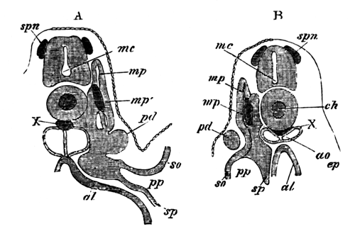
Fig. 1. Two sections of a Pristiurus Embryo with three visceral clefts.
The sections are to shew the development of the segmental duct (pd) or primitive duct of the kidneys. In A (the anterior of the two sections) this appears as a solid knob projecting towards the epiblast. In B is seen a section of the column which has grown backwards from the knob in A.
spn. rudiment of a spinal nerve; mc. medullary canal; ch. notochord; X. string of cells below the notochord; mp. muscle-plate; mp´. specially developed portion of muscle-plate; ao. dorsal aorta; pd. segmental duct; so. somatopleura; sp. splanchnopleura; pp. pleuro-peritoneal or body-cavity; ep. epiblast; al. alimentary canal.
This knob projects outwards toward the epiblast, and the column lies at first between the mesoblast and epiblast. The knob and column do not long remain solid. The knob becoming hollow acquires a wide opening into the pleuro-peritoneal or body-cavity, and the column a lumen; so that by the time that five visceral clefts have appeared, the two together form a duct closed behind, but communicating in front by a wide opening with the pleuro-peritoneal cavity.
Before these changes are accomplished, a series of solid[29] outgrowths of elements of the 'intermediate cell-mass' appear at the uppermost corner of the body-cavity. These soon become hollow and appear as involutions from the body-cavity, curling round the inner and dorsal side of the previously formed duct.
One involution of this kind makes its appearance for each protovertebra, and the first belongs to the protovertebra immediately behind the anterior end of the duct whose development has just been described. In Pristiurus there are in all 29 of these at this period. The last two or three arise from that portion of the body-cavity, which at this stage still exists behind the anus. The first-formed duct and the subsequent involutions are the rudiments of the whole of the urinary system. The duct is the primitive duct of the kidney[30]; I shall call it in future the segmental duct; and the involutions are the commencements of the segmental tubes which constitute the body of the kidney. I shall call them in future segmental tubes.
Soon after their formation the segmental tubes become convoluted, and their blind ends become connected with the segmental duct of the kidney. At the same time, or rather before this, the blind posterior termination of each of the segmental ducts of the kidneys unites with and opens into one of the horns of the cloaca. At this period the condition of affairs is represented in Fig. 2.
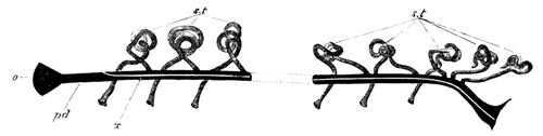
Fig. 2. Diagram of the primitive condition of the Kidney in a Selachian Embryo.
pd. segmental duct. It opens at o into the body-cavity and at its other extremity into the cloaca; x. line along which the division appears which separates the segmental duct into the Wolffian duct above and the Müllerian duct below; st. segmental tubes. They open at one end into the body-cavity, and at the other into the segmental duct.
There is at pd, the segmental duct of the kidneys, opening in front (o) into the body-cavity, and behind into the cloaca, and there are a series of convoluted segmental tubes (st), each opening at one end into the body-cavity, and at the other into the duct (pd).
The next important change which occurs is the longitudinal division of the segmental duct of the kidneys into Müller's duct, or the oviduct, and the duct of the Wolffian bodies or Leydig's duct. The splitting[31] is effected by the growth of a wall of cells which divides the duct into two parts (fig. 3, wd. and md.). It takes place in such a way that the front end of the segmental duct, anterior to the entrance of the first segmental tube, together with the ventral half of the rest of the duct, is split off from its dorsal half as an independent duct (vide fig. 2, x).
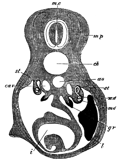
Fig. 3. Transverse section of a Selachian Embryo illustrating the formation of the Wolffian and Müllerian ducts by the longitudinal splitting of the segmental duct.
mc. medullary canal; mp. muscle-plate; ch. notochord; ao. aorta; cav. cardinal vein; st. segmental tube. On the one side the section passes through the opening of a segmental tube into the body-cavity. On the other this opening is represented by dotted lines, and the opening of the segmental tube into the Wolffian duct has been cut through; wd. Wolffian duct; md. Müllerian duct. The Müllerian duct and the Wolffian duct together constitute the primitive segmental duct; gr. The germinal ridge with the thickened germinal epithelium; l. liver; i. intestine with spiral valve.
The dorsal portion also forms an independent duct, and into it the segmental tubes continue to open. Such at least is the method of splitting for the female—for the male the splitting is according to Professor Semper, of a more partial character, and consists for the most part in the front end of the duct only being separated off from the rest. The result of these changes is the formation in both sexes of a fresh duct which carries off the excretions of the segmental involutions, and which I shall call the Wolffian duct—while in the female there is formed another complete and independent duct, which I shall call the Müllerian duct, or oviduct, and in the male portions only of such a duct.
The next change which takes place is the formation of another duct from the hinder portion of the Wolffian duct, which receives the secretion of the posterior segmental tubes. This secondary duct unites with the primary or Wolffian duct near its termination, and the primary ducts of the two sides unite together to open to the exterior by a common papilla.
Slight modifications of the posterior terminations of these ducts are found in different genera of Selachians (vide Semper, Centralblatt für Med. Wiss. 1874, No. 59), but they are of no fundamental importance.
These constitute the main changes undergone by the segmental duct of the kidneys and the ducts derived from it; but the segmental tubes also undergo important changes. In the majority of Selachians their openings into the body-cavity, or, at any rate, the openings of a large number of them, persist through life; but the investigations of Dr Meyer[32] render it very probable that the small portion of each segmental tube adjoining the opening becomes separated from the rest and becomes converted into a sort of lymph organ, so that the openings of the segmental tubes in the adult merely lead into lymph organs and not into the gland of the kidneys.
These constitute the whole changes undergone in the female, but in the male the open ends of a varying number (according to the species) of the segmental tubes become connected with the testis and, uniting with the testicular follicles, serve to carry away the seminal fluid[33]. The spermatozoa have therefore to pass through a glandular portion of the kidneys before they enter the Wolffian duct, by which they are finally carried away to the exterior.
In the adult female, then, there are the following parts of the urinogenital system (fig. 4):
(1) The oviduct, or Müller's duct (fig. 4, md.), split off from the segmental duct of the kidneys. Each oviduct opens at its upper end into the body-cavity, and behind the two oviducts have independent communications with the cloaca. The oviducts serve simply to carry to the exterior the ova, and have no communication with the glandular portion of the kidneys.
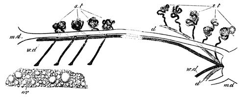
Fig. 4. Diagram of the arrangement of the Urinogenital Organs in an adult Female Selachian.
md. Müllerian duct; wd. Wolffian duct; st. segmental tubes; d. duct of the posterior segmental tubes; ov. ovary.
(2) The Wolffian ducts (fig. 4, wd.) or the remainder of the segmental ducts of the kidneys. Each Wolffian duct ends blindly in front, and the two unite behind to open by a common papilla into the cloaca.
This duct receives the secretion of the whole anterior end of the kidneys[34], that is to say, of all the anterior segmental tubes.
(3) The secondary duct (fig. 4, d.) belonging to the lower portion of the kidneys opening into the former duct near its termination.
(4) The segmental tubes (fig. 4, st) from whose convolutions and outgrowths the kidney is formed. They may be divided into two parts, according to the duct by which their secretion is carried off.
In the male the following parts are present:
(1) The Müllerian duct (fig. 5, md.), consisting of a small remnant, attached to the liver, which represents the foremost end of the oviduct of the female.
(2) The Wolffian duct (fig. 5, wd), which precisely corresponds to the Wolffian duct of the female, except that, in addition to functioning as the duct of the anterior part of the kidneys, it also serves to carry away the semen. In the female it is straight, but has in the adult male a very tortuous course (vide fig. 5).
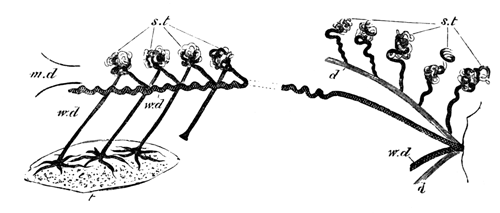
Fig. 5. Diagram of the arrangement of the Urinogenital Organs in an adult male selachian.
md. rudiment of Müllerian duct; wd. Wolffian duct, which also serves as vas deferens; st. segmental tubes. The ends of three of those which in the female open into the body-cavity, have in the male united with the testicular follicles, and serve to carry away the products of the testis; d. duct of the posterior segmental tubes; t. testis.
(3) the duct (fig. 5, d.) of the posterior portion of the kidneys, which has the same relations as in the female.
(4) The segmental tubes (fig. 5, st.). These have the same relations as in the female, except that the most anterior two, three or more, unite with the testicular follicles, and carry away the semen into the Wolffian duct.
* * * * *
The mode of arrangement and the development of these parts suggest a number of considerations.
In the first place it is important to notice that the segmental tubes develop primitively as completely independent organs[35], one of which appears in each segment. If embryology is in any way a repetition of ancestral history, it necessarily follows that these tubes were primitively independent of each other. Ancestral history, as recorded in development, is often, it is true, abridged; but it is clear that though abridgement might prevent a series of primitively separate organs from appearing as such, yet it would hardly be possible for a primitively compound organ, which always retained this condition, to appear during development as a series of separate ones. These considerations appear to me to prove that the segmented ancestors of vertebrates possessed a series of independent and segmental excretory organs.
Both Professor Semper and myself, on discovering these organs, were led to compare them and state our belief in their identity with the so-called segmental organs of Annelids.
This view has since been fairly generally accepted. The segmental organs of annelids agree with those of vertebrates in opening at one end into the body-cavity, but differ in the fact that each also communicates with the exterior by an independent opening, and that they are never connected with each other.
On the hypothesis of the identity of the vertebrate segmental tubes with the annelid segmental organs, it becomes essential to explain how the external openings of the former may have become lost.
This brings us at once to the origin of the segmental duct of the kidneys, by which the secretion of all the segmental tubes was carried to the exterior, and it appears to me that a right understanding of the vertebrate urinogenital system depends greatly upon a correct view of the origin of this duct. I would venture to repeat the suggestion which I made in my original paper (loc. cit.) that this duct is to be looked upon as the most anterior of the segmental tubes which persist in vertebrates. In favour of this view are the following anatomical and embryological facts. (1) It develops in nearly the same manner as the other segmental tubes, viz. in Selachians as a solid outgrowth from the intermediate cell-mass, which subsequently becomes hollowed so as to open into the body-cavity: and in Amphibians and Osseous and Cyclostome fishes as a direct involution from the body-cavity. (2) In Amphibians, Cyclostomes and Osseous fishes its upper end develops a glandular portion, by becoming convoluted in a manner similar to the other segmental tubes. This glandular portion is often called either the head-kidney or the primitive kidney. It is only an embryonic structure, but is important as demonstrating the true nature of the primitive duct of the kidneys.
We may suppose that some of the segmental tubes first united, possibly in pairs, and that then by a continuation of this process the whole of them coalesced into a common gland. One external opening sufficed to carry off the entire secretion of the gland, and the other openings therefore atrophied.
This history is represented in the development of the dog-fish in an abbreviated form, by the elongation of the first segmental tube (segmental duct of the kidney) and its junction with each of the posterior segmental tubes. Professor Semper looks upon the primitive duct of the kidneys as a duct which arose independently, and was not derived from metamorphosis of the segmental organs. Against this view I would on the one hand urge the consideration, that it is far easier to conceive of the transformation by change of function (comp. Dohrn, Functionswechsel, Leipzig, 1875) of a segmental organ into a segmental duct, than to understand the physiological cause which should lead, in the presence of so many already formed ducts, to the appearance of a totally new one. By its very nature a duct is a structure which can hardly arise de novo. We must even suppose that the segmental organs of Annelids were themselves transformations of still simpler structures. On the other hand I would point to the development in this very duct amongst Amphibians and Osseous fishes of a glandular portion similar to that of a segmental tube, as an à posteriori proof of its being a metamorphosed segmental tube. The development in insects of a longitudinal tracheal duct by the coalescence of a series of transverse tracheal tubes affords a parallel to the formation of a duct from the coalescence of a series of segmental tubes.
Though it must be admitted that the loss of the external openings of the segmental organs requires further working out, yet the difficulties involved in their disappearance are not so great as to render it improbable that the vertebrate segmental organs are descended from typical annelidan ones.
The primitive vertebrate condition, then, is probably that of an early stage of Selachian development while there is as yet a segmental duct,—the original foremost segmental tube opening in front into the body-cavity and behind into the cloaca; with which duct all the segmental tubes communicate. Vide Fig. 2.
The next condition is to be looked upon as an indirect result of the segmental duct serving as well for the products of the generative organs as the secretions of the segmental tubes.
As a consequence of this, the segmental duct became split into a ventral portion, which served alone for the ova, and a dorsal portion which received the secretion of the segmental tubes. The lower portion, which we have called the oviduct, in some cases may also have received the semen as well as the ova. This is very possibly the case with Ceratodus (vide Günther, Trans. of Royal Society, 1871), and the majority of Ganoids (Hyrtl, Denkschriften Wien, Vol. VIII.). In the majority of other cases the oviduct exists in the male in a completely rudimentary form; and the semen is carried away by the same duct as the urine.
In Selachians the transportation of the semen from the testis to the Wolffian duct is effected by the junction of the open ends of two or three or more segmental tubes with the testicular follicles, and the modes in which this junction is effected in the higher vertebrates seem to be derivatives from this. If the views here expressed are correct it is by a complete change of function that the oviduct has come to perform its present office. And in the bird and higher vertebrates no trace, or only the very slightest (vide p. 165) of the primitive urinary function is retained during embryonic or adult life.
The last feature in the anatomy of the Selachians which requires notice is the division of the kidney into two portions, an anterior and posterior. The anatomical similarity between this arrangement and that of higher vertebrates (birds, &c.) is very striking. The anterior one precisely corresponds, anatomically, to the Wolffian body, and the posterior one to the true permanent kidney of higher vertebrates: and when we find that in the Selachians the duct for the anterior serves also for the semen as does the Wolffian duct of higher vertebrates, this similarity seems almost to amount to identity. A discussion of the differences in development in the two cases will come conveniently with the account of the bird; but there appear to me the strongest grounds for looking upon the kidneys of Selachians as equivalent to both the Wolffian bodies and the true kidneys of the higher vertebrates.
The condition of the urinogenital organs in Selachians is by no means the most primitive found amongst vertebrates.
The organs of both Cyclostomous and Osseous fishes, as well as those of Ganoids, are all more primitive; and in the majority of points the Amphibians exhibit a decidedly less differentiated condition of these organs than do the Selachians.
In Cyclostomous fishes the condition of the urinary system is very simple. In Myxine (vide Joh. Müller Myxinoid fishes, and Wilhelm Müller, Jenaische Zeitschrift, 1875, Das Urogenitalsystem des Amphioxus u. d. Cyclostomen) there is a pair of ducts which communicate posteriorly by a common opening with the abdominal pore. From these ducts spring a series of transverse tubules, each terminating in a Malpighian corpuscle. These together constitute the mass of the kidneys. About opposite the gall-bladder the duct of the kidney (the segmental duct) narrows very much, and after a short course ends in a largish glandular mass (the head-kidney), which communicates with the pericardial cavity by a number of openings.
In Petromyzon the anatomy of the kidneys is fundamentally the same as in Myxine. They consist of the two segmental ducts, and a number of fine branches passing off from these, which become convoluted but do not form Malpighian tufts. The head-kidney is absent in the adult.
W. Müller (loc. cit.) has given a short but interesting account of the development of the urinary system of Petromyzon. He finds that the segmental ducts develop first of all as simple involutions from the body-cavity. The anterior end of each then develops a glandular portion which comes to communicate by a number of openings with the body-cavity. Subsequently to the development of this glandular portion the remainder of the kidneys appears in the posterior portion of the body-cavity; and before the close of embryonic life the anterior glandular portion atrophies.
The comparison of this system with that of a Selachian is very simple. The first developed duct is the segmental duct of a Selachian, and the glandular portion developed at its anterior extremity, which is permanent in Myxine but embryonic in Petromyzon, is, as W. Müller has rightly recognized, equivalent to the head-kidney of Amphibians, which remains undeveloped in Selachians. It is, according to my previously stated view, the glandular portion of the first segmental organ or the segmental duct. The series of orifices by which this communicates with the body-cavity are due to the division of the primary opening of the segmental duct. This is shewn both by the facts of their development in Petromyzon given by Müller, as well as by the occurrence of a similar division of the primary orifice in Amphibians, which is mentioned later in this paper. In a note in my original paper (loc. cit.) I stated that these openings were equivalent to the segmental involutions of Selachians. This is erroneous, and was due to my not having understood the description given in a preliminary paper of Müller (Jenaische Zeitschrift, 1873). The large development of this glandular mass in the Cyclostome and Osseous fishes and in embryo Amphibians, implies that it must at one time have been important. Its earlier development than the remainder of the kidneys is probably a result of the specialized function of the first segmental organ.
The remainder of the kidney in Cyclostomes is equivalent to the kidney of Selachians. Its development from segmental involutions has not been recognized. If these segmental involutions are really absent it may perhaps imply that the simplicity of the Cyclostome kidneys, like that of so many other of their organs, is a result of degeneration rather than a primitive condition.
In Osseous fishes the segmental duct of the kidneys develops, as the observations of Rosenberg[36] (Teleostierniere, Inaug. Disser. Dorpat, 1867) and Oellacher (Zeitschrift für Wiss. Zool. 1873) clearly prove, by an involution from the body-cavity. This involution grows backwards in the form of a duct and opens into the cloaca. The upper end of this duct (the most anterior segmental tube) becomes convoluted, and forms a glandular body, which has no representative in the urinary apparatus of Selachians, but whose importance, as indicating the origin of the segmental duct of the kidneys, I have already insisted upon.
The rest of the kidney becomes developed at a later period, probably in the same way as in Selachians; but this, as far as I know, has not been made out.
The segmental duct of the kidneys forms the duct for this new gland, as in embryo Selachians (Fig. 2), but, unlike what happens in Selachians, undergoes no further changes, with the exception of a varying amount of retrogressive metamorphosis of its anterior end. The kidneys of Osseous fish usually extend from just behind the head to opposite the anus, or even further back than this. They consist for the most part of a broader anterior portion, an abdominal portion reaching from this to the anus, and, as in those cases in which the kidneys extend further back than the anus, of a caudal portion.
The two ducts (segmental ducts of the kidneys) lie, as a rule, in the lower part of the kidneys on their outer borders, and open almost invariably into a urinary bladder. In some cases they unite before opening into the bladder, but generally have independent openings.
This bladder, which is simply a dilatation of the united lower ends of the primitive kidney-ducts, and has no further importance, is almost invariably present, but in many cases lies unsymmetrically either to the right or the left. It opens to the exterior by a very minute opening in the genito-urinary papilla, immediately behind the genital pore. There are, however, a few cases in which the generative and urinary organs have a common opening. For further details vide Hyrtl, Denk. der k. Akad. Wien, Vol. II.
It is possible that the generative ducts of Osseous fishes are derived from a splitting from the primitive duct of the kidney, but this is discussed later in the paper.
In Osseous fishes we probably have an embryonic condition of the Selachian kidneys retained permanently through life.
* * * * *
In the majority of Ganoids the division of the segmental duct of the kidney into two would seem to occur, and the ventral duct of the two (Müllerian duct), which opens at its upper end into the body-cavity, is said to serve as an excretory duct for both male and female organs.
The following are the more important facts which are known about the generative and urinary ducts of Ganoids.
In Spatularia (vide Hyrtl, Geschlechts u. Harnwerkzeuge bei den Ganoiden, Denkschriften der k. Akad. Wien, Vol. VIII.) the following parts are found in the female.
(1) The ovaries stretching along the whole length of the abdominal cavity.
(2) The kidneys, which are separate and also extend along the greater part of the abdominal cavity.
(3) The ureters lying on the outer borders of the kidneys. Each ureter dilates at its lower end into an elongated wide tube, which continues to receive the ducts from the kidneys. The two ureters unite before terminating and open behind the anus.
(4) The two oviducts (Müllerian ducts). These open widely into the abdominal cavity, at about two-thirds of the distance from the anterior extremity of the body-cavity. Each opens by a narrow pore into the dilated ureter of its side.
In the male the same parts are found as in the female, but Hyrtl found that the Müllerian duct of the left side at its entrance into the ureter became split into two horns, one of which ended blindly. On the right side the opening of the Müllerian duct was normal.
In the Sturgeon (vide J. Müller, Bau u. Grenzen d. Ganoiden, Berlin Akad. 1844; Leydig, Fischen u. Reptilien, and Hyrtl, Ganoiden) the same parts are found as in Spatularia.
The kidneys extend along the whole length of the body-cavity; and the ureter, which does not reach the whole length of the kidneys, is a thin-walled wide duct lying on the outer side. On laying it open the numerous apertures of the tubules for the kidney are exposed. The Müllerian duct, which opens in both sexes into the abdominal cavity, ends, according to Leydig, in the cases of some males, blindly behind without opening into the ureter, and Müller makes the same statement for both sexes. It was open on both sides in a female specimen I examined[37], and Hyrtl found it invariably so in both sexes in all the specimens he examined.
Both Rathke and Stannius (I have been unable to refer to the original papers) believed that the semen was carried off by transverse ducts directly into the ureter, and most other observers have left undecided the mechanism of the transportation of the semen to the exterior. If we suppose that the ducts Rathke saw really exist they might perhaps be supposed to enter not directly into the ureter, but into the kidney, and be in fact homologous with the vasa efferentia of the Selachians. The frequent blind posterior termination of the Müllerian duct is in favour of the view that these ducts of Rathke are really present.
In Polypterus (vide Hyrtl, Ganoiden) there is, as in other Ganoids, a pair of Müllerian ducts. They unite at their lower ends. The ureters are also much narrower than in previously described Ganoids and, after coalescing, open into the united oviducts. The urinogenital canal, formed by coalescence of the Müllerian ducts and ureters, has an opening to the exterior immediately behind the anus.
In Amia (vide Hyrtl) there is a pair of Müllerian ducts which, as well as the ureters, open into a dilated vesicle. This vesicle appears as a continuation of the Müllerian ducts, but receives a number of the efferent ductules of the kidneys. There is a single genito-urinary pore behind the anus.
In Ceratodus (Günther, Phil. Trans. 1871) the kidneys are small and confined to the posterior extremity of the abdomen. The generative organs extend however along the greater part of the length of the abdominal cavity. In both male and female there is a long Müllerian duct, and the ducts of the two sides unite and open by a common pore into a urinogenital cloaca which communicates with the exterior by the same opening as the alimentary canal. In both sexes the Müllerian duct has a wide opening near the anterior extremity of the body-cavity. The ureters coalesce and open together into the urinogenital cloaca dorsal to the Müllerian ducts. It is not absolutely certain that the semen is transported to the exterior by the Müllerian duct of the male, which is perhaps merely a rudiment as in Amphibia. Dr Günther failed however to find any other means by which it could be carried away.
The genital ducts of Lepidosteus differ in important particulars from those of the other Ganoids (vide Müller, loc. cit. and Hyrtl, loc. cit.).
In both sexes the genital ducts are continuous with the investments of the genital organs.
In the female the dilated posterior extremities of the ureters completely invest for some distance the generative ducts, whose extremities are divided into several processes, and end in a different way on the two sides. A similar division and asymmetry of the ducts is mentioned by Hyrtl as occurring in the male of Spatularia, and it seems not impossible that on the hypothesis of the genital ducts being segmental tubes these divisions may be remnants of primitive glandular convolutions. The ureters in both sexes dilate as in other Ganoids at their posterior extremities, and unite with one another. The unpaired urinogenital opening is situated behind the anus. In the male the dilated portion of the ureters is divided into a series of partitions which are not present in the female.
Till the embryology of the secretory system of Ganoids has been worked out, the homologies of their generative ducts are necessarily a matter of conjecture. It is even possible that what I have called the Müllerian duct in the male is functionless, as with Amphibians, but that, owing to the true ducts of the testis having been overlooked, it has been supposed to function as the vas deferens. Günther's (loc. cit.) injection experiments on Ceratodus militate against this view, but I do not think they can be considered as conclusive as long as the mechanism for the transportation of the semen to the exterior has not been completely made out. Analogy would certainly lead us to expect the ureter to serve in Ganoids as the vas deferens.
The position of the generative ducts might in some cases lead to the supposition that they are not Müllerian ducts, or, in other words, the most anterior pair of segmental organs but a pair of the posterior segmental tubes.
What are the true homologies of the generative ducts of Lepidosteus, which are continuous with the generative glands, is somewhat doubtful. It is very probable that they may represent the similarly functioning ducts of other Ganoids, but that they have undergone further changes as to their anterior extremities.
It is, on the other hand, possible that their generative ducts are the same structures as those ducts of Osseous fishes, which are continuous with the generative organs. These latter ducts are perhaps related to the abdominal pores, and had best be considered in connection with these; but a completely satisfactory answer to the questions which arise in reference to them can only be given by a study of their development.
In the Cyclostomes the generative products pass out by an abdominal pore, which communicates with the peritoneal cavity by two short tubes[38], and which also receives the ducts of the kidneys.
Gegenbaur suggests that these are to be looked upon as Müllerian ducts, and as therefore developed from the segmental ducts of the kidneys. Another possible view is that they are the primitive external openings of a pair of segmental organs. In Selachians there are usually stated to be a pair of abdominal pores. In Scyllium I have only been able to find, on each side, a large deep pocket opening to the exterior, but closed below towards the peritoneal cavity, so that in it there seem to be no abdominal pores[39]. In the Greenland Shark (Læmargus Borealis) Professor Turner (Journal of Anat. and Phys. Vol. VIII.) failed to find either oviduct or vas deferens, but found a pair of large open abdominal pores, which he believes serve to carry away the generative products of both sexes. Whether the so-called abdominal pores of Selachians usually end blindly as in Scyllium, or, as is commonly stated, open into the body-cavity, there can be no question that they are homologous with true abdominal powers.
The blind pockets of Scyllium appear very much like the remains of primitive involutions from the exterior, which might easily be supposed to have formed the external opening of a pair of segmental organs, and this is probably the true meaning of abdominal pores. The presence of abdominal pores in all Ganoids in addition to true genital ducts and of these pockets or abdominal pores in Selachians, which are almost certainly homologous with the abdominal pores of Ganoids and Cyclostomes, and also occur in addition to true Müllerian ducts, speak strongly against the view that the abdominal pores have any relation to Müllerian ducts. Probably therefore the abdominal pores of the Cyclostomous fishes (which seem to be of the same character as other abdominal pores) are not to be looked on as rudimentary Müllerian ducts.
We next come to the question which I reserved while speaking of the kidneys of Osseous fishes, as to the meaning of their genital ducts.
In the female Salmon and the male and female Eel, the generative products are carried to the exterior by abdominal pores, and there are no true generative ducts. In the case of most other Osseous fish there are true generative ducts which are continuous with the investment of the generative organs[40] and have generally, though not always, an opening or openings independent of the ureter close behind the rectum, but no abdominal pores are present. It seems, therefore, that in Osseous fish the generative ducts are complementary to abdominal pores, which might lead to the view that the generative ducts were formed by a coalescence of the investment of the generative glands with the short duct of abdominal pore.
Against this view there are, however, the following facts:
(1) In the cases of the salmon and the eel it is perfectly true that the abdominal pore exactly corresponds with the opening of the genital duct in other Osseous fishes, but the absence of genital ducts in these cases must rather be viewed, as Vogt and Pappenheim (loc. cit.) have already insisted, as a case of degeneration than of a primitive condition. The presence of genital ducts in the near allies of the Salmonidæ, and even in the male salmon, are conclusive proofs of this. If we admit that the presence of an abdominal pore in Salmonidæ is merely a result of degeneration, it obviously cannot be used as an argument for the complementary nature of abdominal pores and generative ducts.
(2) Hyrtl (Denkschriften der k. Akad. Wien, Vol. 1) states that in Mormyrus oxyrynchus there is a pair of abdominal pores in addition to true generative ducts. If his statements are correct, we have a strong argument against the generative ducts of Osseous fishes being related to abdominal pores. For though this is the solitary instance of the presence of both a genital opening and abdominal pores known to me in Osseous fishes, yet we have no right to assume that the abdominal pores of Mormyrus are not equivalent to those of Ganoids and Selachians. It must be admitted, with Gegenbaur, that embryology alone can elucidate the meaning of the genital ducts of Osseous fishes.
In Lepidosteus, as was before mentioned, the generative ducts, though continuous with the investment of the generative bodies, unite with the ureters, and in this differ from the generative ducts of Osseous fishes. The relation, indeed, of the generative ducts of Lepidosteus to the urinary ducts is very similar to that existing in other Ganoid fishes; and this, coupled with the fact that Lepidosteus possesses a pair of abdominal pores on each side of the anus[41], makes it most probable that its generative ducts are true Müllerian ducts.
* * * * *
In the Amphibians the urinary system is again more primitive than in the Selachians.
The segmental duct of the kidneys is formed[42] by an elongated fold arising from the outer wall of the body-cavity, in the same position as in Selachians. This fold becomes constricted into a canal, closed except at its anterior end, which remains open to the body-cavity. This anterior end dilates, and grows out into two horns, and at the same time its opening into the body-cavity becomes partly constricted, and so divided into three separate orifices, one for each horn and a central one between the two. The horns become convoluted, blood channels appearing between their convolutions, and a special coil of vessels is formed arising from the aorta and projecting into the body-cavity near the openings of the convolutions. These formations together constitute the glandular portion[43] of the original anterior segmental tube or segmental duct of the kidneys. I have already pointed out the similarity which this organ exhibits to the head-kidneys of Cyclostome fishes in its mode of formation, especially with reference to the division of the primitive opening. The lower end of the segmental duct unites with a horn of the cloaca.
After the formation of the gland just described the remainder of the kidney is formed.
This arises in the same way as in Selachians. A series of involutions from the body-cavity are developed; these soon form convoluted tubes, which become branched and interlaced with one another, and also unite with the primitive duct of the kidneys. Owing to the branching and interlacing of the primitive segmental tubes, the kidney is not divided into distinct segments in the same way as with the Selachians. The mode of development of these segmental tubes was discovered by Götte. Their openings are ciliated, and, as Spengel (loc. cit.) and Meyer (loc. cit.) have independently discovered, persist in most adult Amphibians. As both these investigators have pointed out, the segmental openings are in the adult kidneys of most Amphibians far more numerous than the vertebral segments to which they appertain. This is due to secondary changes, and is not to be looked upon as the primitive state of things. At this stage the Amphibian kidneys are nearly in the same condition as the Selachian, in the stage represented in Fig. 2. In both there is the segmental duct of the kidneys, which is open in front, communicates with the cloaca behind, and receives the whole secretion from the kidneys. The parallelism between the two is closely adhered to in the subsequent modifications of the Amphibian kidney, but the changes are not completed so far in Amphibians as in Selachians. The segmental duct of the Amphibian kidney becomes, as in Selachians, split into a Müllerian duct or oviduct, and a Wolffian duct or duct for the kidney.
The following points about this are noteworthy:
(1) The separation of the two ducts is never completed, so that they are united together behind, and for a short distance, blend and form a common duct; the ducts of the two sides so formed also unite before opening to the exterior.
(2) The separation of the two ducts does not occur in the form of a simple splitting, as in Selachians. But the efferent ductules from the kidney gradually alter their points of entrance into the primitive duct. Their points of entrance become carried backwards further and further, and since this process affects the anterior ducts proportionally more than the posterior, the efferent ducts finally all meet and form a common duct which unites with the Müllerian duct near its posterior extremity. This process is not always carried out with equal completeness. In the tailless Amphibians, however, the process is generally[44] completed, and the ureters (Wolffian ducts) are of considerable length. Bufo cinereus, in the male of which the Müllerian ducts are very conspicuous, serves as an excellent example of this.
In the Salamander (Salamandra maculosa), Figs. 6 and 7, the process is carried out with greater completeness in the female than in the male, and this is the general rule in Amphibians. In the male Proteus, the embryonic condition would seem to be retained almost in its completeness so that the ducts of the kidney open directly and separately into the still persisting primitive duct of the kidney. The upper end of the duct nevertheless extends some distance beyond the end of the kidney and opens into the abdominal cavity. In the female Proteus, on the other hand, the separation into a Müllerian duct and a ureter is quite complete. The Newt (Triton) also serves as an excellent example of the formation of distinct Müllerian and Wolffian ducts being much more complete in the female than the male. In the female Newt all the tubules from the kidney open into a duct of some length which unites with the Müllerian duct near its termination, but in the male the anterior segmental tubes, including those which, as will be afterwards seen, serve as vasa efferentia of the testis, enter the Müllerian duct directly, while the posterior unite as in the female into a common duct before joining the Müllerian duct. For further details as to the variations exhibited in the Amphibians, the reader is referred to Leydig, Anat. Untersuchung, Fischen u. Reptilien. Ditto, Lehrbuch der Histologie, Menschen u. Thiere. Von Wittich, Siebold u. Kölliker, Zeitschrift, Vol. IV. p. 125.
The different conditions of completeness of the Wolffian ducts observable amongst the Amphibians are instructive in reference to the manner of development of the Wolffian duct in Selachians. The mode of division in the Selachians of the segmental duct of the kidney into a Müllerian and Wolffian duct is probably to be looked upon as an embryonic abbreviation of the process by which these two ducts are formed in Amphibians. The fact that this separation into Müllerian and Wolffian ducts proceeds further in the females of most Amphibians than in the males, strikingly shews that it is the oviductal function of the Müllerian duct which is the indirect cause of its separation from the Wolffian duct. The Müllerian duct formed in the way described persists almost invariably in both sexes, and in the male sometimes functions as a sperm reservoir; e.g. Bufo cinereus. In the embryo it carries at its upper end the glandular mass described above (Kopfniere), but this generally atrophies, though remnants of it persist in the males of some species (e.g. Salamandra). Its anterior end opens, in most cases by a single opening, into the perivisceral cavity in both sexes, and is usually ciliated. As the female reaches maturity, the oviduct dilates very much; but it remains thin and inconspicuous in the male.
The only other developmental change of importance is the connection of the testes with the kidneys. This probably occurs in the same manner as in Selachians, viz. from the junction of the open ends of the segmental tubes with the follicles of the testes. In any case the vessels which carry off the semen constitute part of the kidney, and the efferent duct of the testis is also that of the kidney. The vasa efferentia from the testis either pass through one or two nearly isolated anterior portions of the kidney (Proteus, Triton) or else no such special portion of the kidney becomes separated from the rest, and the vasa efferentia enter the general body of the kidney.
* * * * *
In the male Amphibian, then, the urinogenital system consists of the following parts (Fig. 6):
(1) Rudimentary Müllerian ducts, opening anteriorly into the body-cavity, which sometimes carry aborted Kopfnieren.
(2) The partially or completely formed Wolffian ducts (ureters) which also serve as the ducts for the testes.
(3) The kidneys, parts of which also serve as the vasa efferentia, and whose secretion, together with the testicular products, is carried off by the Wolffian ducts.
(4) The united lower parts of Wolffian and Müllerian ducts which are really the lower unsplit part of the segmental ducts of the kidneys.
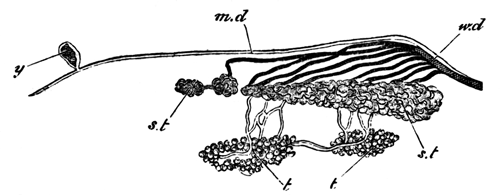
Fig. 6. Diagram of the Urinogenital Organs of a Male Salamander.
(Copied from Leydig's Histologie des Menschen u. der Thiere.)
md. Müller's duct (rudimentary); y. remnant of the secretory portion of the segmental duct Kopfniere; Wd. Wolffian duct; a less complete structure in the male than in the female; st. segmental tubes or kidney. The openings of these into the body-cavity are not inserted in the figure; t. testis. Its efferent ducts form part of the kidney.
In the female, there are (Fig. 7)
(1) The Müllerian ducts which function as the oviducts.
(2) The Wolffian ducts.
(3) The kidneys.
(4) The united Müllerian and Wolffian ducts as in the male.
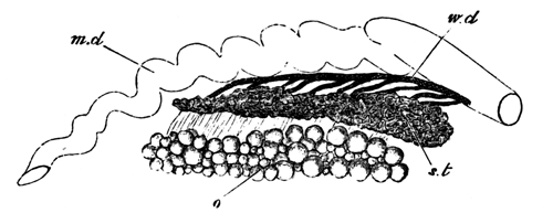
Fig. 7. Diagram of the Urinogenital Organs of a Female Salamander.
(Copied from Leydig's Histologie des Menschen u. der Thiere.)
Md. Müller's duct or oviduct; Wd. Wolffian duct or the duct of the kidneys; st. segmental tubes or kidney. The openings of these into the body-cavity are not inserted in the figure; o. ovary.
The urinogenital organs of the adult Amphibians agree in almost all essential particulars with those of Selachians. The ova are carried off in both by a specialized oviduct. The Wolffian duct, or ureter, is found both in Selachians and Amphibians, and the relations of the testis to it are the same in both, the vasa efferentia of the testes having in both the same anatomical peculiarities.
The following points are the main ones in which Selachians and Amphibians differ as to the anatomy of the urinogenital organs; and in all but one of these, the organs of the Amphibian exhibit a less differentiated condition than do those of the Selachian.
(1) A glandular portion (Kopfniere) belonging to the first segmental organ (segmental duct of the kidneys) is found in all embryo Amphibians, but usually disappears, or only leaves a remnant in the adult. It has not yet been found in any Selachian.
(2) The division of the primitive duct of the kidney into the Müllerian duct and the Wolffian duct is not completed so far in Amphibians as Selachians, and in the former the two ducts are confluent at their lower ends.
(3) The permanent kidney exhibits in Amphibians no distinction into two glands (foreshadowing the Wolffian bodies and true kidneys of higher vertebrates), as it does in the Selachians.
(4) The Müllerian duct persists in its entirety in male Amphibians, but only its upper end remains in male Selachians.
(5) The openings of the segmental tubes into the body-cavity correspond in number with the vertebral segments in most Selachians, but are far more numerous than these in Amphibians. This is the chief point in which the Amphibian kidney is more differentiated than the Selachian.
* * * * *
The modifications in development which the urinogenital system has suffered in higher vertebrates (Sauropsida and Mammalia) are very considerable; nevertheless it appears to me to be possible with fair certainty to trace out the relationship of its various parts in them to those found in the Ichthyopsida. The development of urinogenital organs has been far more fully worked out for the bird than for any other member of the amniotic vertebrates; but, as far as we know, there are no essential variations except in the later periods of development throughout the division. These later variations, concerning for the most part the external apertures of the various ducts, are so well known and have been so fully described as to require no notice here. The development of these parts in the bird will therefore serve as the most convenient basis for comparison.
In the bird the development of these parts begins by the appearance of a column of cells on the upper surface of the intermediate cell-mass (Fig. 8, W.d). As in Selachians, the intermediate cell-mass is a group of cells between the outer edge of the protovertebræ and the upper end of the body-cavity. The column of cells thus formed is the commencement of the duct of the Wolffian body. Its development is strikingly similar to that of the segmental duct of the kidney in Selachians. I shall attempt when I have given an account of the development of the Müllerian duct to speak of the relations between the Selachian duct and that of the bird.
Romiti (Archiv f. Micr. Anat. Vol. X.) has recently stated that the Wolffian duct develops as an involution from the body-cavity. The fact that the specimens drawn by Romiti to support this view are too old to determine such a point, and the inspection of a number of specimens made by my friend Mr Adam Sedgwick of Trinity College, who, at my request, has been examining the urinogenital organs of the fowl, have led me to the conclusion that Romiti is in error in differing from his predecessors as to the development of the Wolffian duct. The solid string of cells to form the Wolffian duct lies at first close to the epiblast, but, by the alteration in shape which the protovertebræ undergo and the general growth of cells around it, becomes gradually carried downwards till it lies close to the germinal epithelium which lines the body-cavity. While undergoing this change of position it also acquires a lumen, but ends blindly both in front and behind. Towards the end of the fourth day the Wolffian duct opens into a horn of the cloaca. The cells adjoining its inner border commence, as it passes down on the third day, to undergo histological changes, which, by the fourth day, result in the formation of a series of ducts and Malpighian tufts which form the mass of the Wolffian body[45].
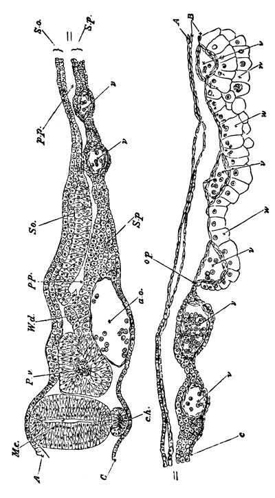
Fig. 8. Transverse section through the Dorsal region of an Embryo Fowl of 45 h. To shew the mode of Formation of the Wolffian Duct.
A. epiblast; B. mesoblast; C. hypoblast; M.c. medullary canal; Pv. Protovertebræ; W.d. Wolffian duct; so. Somatopleure; Sp. Splanchnopleure; pp. pleuro-peritoneal cavity; ch. notochord; ao. dorsal aorta; v. blood-vessels.
The Müllerian duct arises in the form of an involution, whether at first solid or hollow, of the germinal epithelium, and, as I am satisfied, quite independently of the Wolffian duct. It is important to notice that its posterior end soon unites with the Wolffian duct, from which however it not long after becomes separated and opens independently into the cloaca. The upper end remains permanently open to the body-cavity, and is situated nearly opposite the extreme front end of the Wolffian body.
Between the 80th and 100th hour of incubation the ducts of the permanent kidneys begin to make their appearance. Near its posterior extremity each Wolffian duct becomes expanded, and from the dorsal side of this portion a diverticulum is constricted off, the blind end of which points forwards. This is the duct of the permanent kidneys, and around its end the kidneys are found. It is usually stated that the tubules of the permanent kidneys arise as outgrowths from the duct, but this requires to be worked over again.
The condition of the urinogenital system in birds immediately after the formation of the permanent kidneys is strikingly similar to its permanent condition in adult Selachians. There is the Müllerian duct in both opening in front into the body-cavity and behind into the cloaca. In both the kidneys consist of two parts—an anterior and posterior—which have been called respectively Wolffian bodies and permanent kidneys in birds and Leydig's glands and the kidneys in Selachians.
The duct of the permanent kidney, which at first opens into that of the Wolffian body, subsequently becomes further split off from the Wolffian duct, and opens independently into the cloaca.
The subsequent changes of these parts are different in the two sexes.
In the female the Müllerian ducts[46] persist and become the oviducts. Their anterior ends remain open to the body-cavity. The changes in their lower ends in the various orders of Sauropsida and Mammalia are too well known to require repetition here. The Wolffian body and duct atrophy: there are left however in many cases slight remnants of the anterior extremity of the body forming the parovarium of the bird, and also frequently remnants of the posterior portion of the gland as well as of the duct. The permanent kidney and its duct remain unaltered.
In the male the Müllerian duct becomes almost completely obliterated. The Wolffian duct persists and forms the vas deferens, and the anterior so-called sexual portion of the Wolffian body also persists in an altered form. Its tubules unite with the seminiferous tubules, and also form the epididymis. Unimportant remnants of the posterior part of the Wolffian body also persist, but are without function. In both sexes the so-called permanent kidneys form the sole portion of the primitive uriniferous system which persists in the adult.
In considering the relations between the modes of development of the urinogenital organs of the bird and of the Selachians, the first important point to notice is, that whereas in the Selachians the segmental duct of the kidneys is first developed and subsequently becomes split into the Müllerian and Wolffian ducts; in the bird these two ducts develop independently. This difference in development would be accurately described by saying that in birds the segmental duct of the kidneys develops as in Selachians, but that the Müllerian duct develops independently of it.
Since in Selachians the Wolffian duct is equivalent to the segmental duct of the kidneys with the Müllerian removed from it, when in birds the Müllerian duct develops independently of the segmental kidney duct, the latter becomes the same as the Wolffian duct.
The second mode of stating the difference in development in the two cases represents the embryological facts of the bird far better than the other method.
It explains why the Wolffian duct appears earlier than the Müllerian and not at the same time, as one might expect according to the other way of stating the case. If the Wolffian duct is equivalent to the segmental duct of Selachians, it must necessarily be the first duct to develop; and not improbably the development of the Müllerian duct would in birds be expected to occur at the time corresponding to that at which the primitive duct in Selachians became split into two ducts.
It probably also explains the similarity in the mode of development of the Wolffian duct in birds and the primitive duct of the kidneys in Selachians.
This way of stating the case is also in accordance with theoretical conclusions. As the egg-bearing function of the Müllerian duct became more and more confirmed we might expect that the adult condition would impress itself more and more upon the embryonic development, till finally the Müllerian duct ceased to be at any period connected with the kidneys, and the history of its origin ceased to be traceable in its development. This seems to have actually occurred in the higher vertebrates, so that the only persisting connection between the Müllerian duct and the urinary system is the brief but important junction of the two at their lower ends on the sixth or seventh day. This junction justly surprised Waldeyer (Eierstock u. Ei, p. 129), but receives a complete and satisfactory explanation on the hypothesis given above.
The original development of the segmental tubes is in the bird solely retained in the tubules of the Wolffian body arising independently of the Wolffian duct, and I have hitherto failed to find that there is a distinct division of the Wolffian bodies into segments corresponding with the vertebral segments.
I have compared the permanent kidneys to the lower portion of the kidneys of Selachians. The identity of the anatomical condition of the adult Selachian and embryonic bird which has been already pointed out speaks strongly in favour of this view; and when we further consider that the duct of the permanent kidneys is developed in nearly the same way as the supposed homologous duct in Selachians, the suggested identity gains further support. The only difficulty is the fact that in Selachians the tubules of the part of the kidneys under comparison develop as segmental involutions in point of time anteriorly to their duct, while in birds they develop in a manner not hitherto certainly made out but apparently in point of time posteriorly to their duct. But when the immense modifications in development which the whole of the gland of the excretory organ has undergone in the bird are considered, I do not think that the fact I have mentioned can be brought forward as a serious difficulty.[TN5]
The further points of comparison between the Selachian and the bird are very simple. The Müllerian duct in its later stages behaves in the higher vertebrates precisely as in the lower. It becomes in fact the oviduct in the female and atrophies in the male. The behaviour of the Wolffian duct is also exactly that of the duct which I have called the Wolffian duct in Ichthyopsida, and in the tubules of the Wolffian body uniting with the tubuli seminiferi we have represented the junction of the segmental tubes with the testis in Selachians and Amphibians. It is probably this junction of two independent organs which led Waldeyer to the erroneous view that the tubuli seminiferi were developed from the tubules of the Wolffian body.
With the bird I conclude the history of the origin of the urinogenital system of vertebrates. I have attempted, and I hope succeeded, in tracing out by the aid of comparative anatomy and embryology the steps by which a series of independent and simple segmental organs like those of Annelids have become converted into the complicated series of glands and ducts which constitute the urinogenital system of the higher vertebrates. There are no doubt some points which require further elucidation amongst the Ganoid and Osseous fishes. The most important points which appear to me still to need further research, both embryological and anatomical, are the abdominal pores of fishes, the generative ducts of Ganoids, especially Lepidosteus, and the generative ducts of Osseous fishes.
The only further point which requires discussion is the embryonic layer from which these organs are derived.
I have shewn beyond a doubt (loc. cit.) that in Selachians these organs are formed from the mesoblast. The unanimous testimony of all the recent investigators of Amphibians leads to the same conclusion. In birds, on the other hand, various investigators have attempted to prove that these organs are derived from the epiblast. The proof they give is the following: the epiblast and mesoblast appear fused in the region of the axis cord. From this some investigators have been led to the conclusion that the whole of the mesoblast is derived from the upper of the two primitive embryonic layers. To these it may be replied that, even granting their view to be correct, it is no proof of the derivation of the urinogenital organs from the epiblast, since it is not till the complete formation of the three layers that any one of them can be said to exist. Others look upon the fusion of the two layers as a proof of the passage of cells from the epiblast into the mesoblast. An assumption in itself, which however is followed by the further assumption that it is from these epiblast cells that the urinogenital system is derived! Whatever may have been the primitive origin of the system, its mesoblastic origin in vertebrates cannot in my opinion be denied.
Kowalewsky (Embryo. Stud. an Vermen u. Arthropoda, Mem. Akad. St Petersbourg, 1871) finds that the segmental tubes of Annelids develop from the mesoblast. We must therefore look upon the mesoblastic origin of the excretory system as having an antiquity greater even than that of vertebrates.
VIII. On the Development of the Spinal Nerves in Elasmobranch Fishes[47].
With Plates 22 and 23.
In the course of an inquiry into the development of Elasmobranch Fishes, my attention has recently been specially directed to the first appearance and early stages of the spinal nerves, and I have been led to results which differ so materially from those of former investigators, that I venture at once to lay them before the Society. I have employed in my investigations embryos of Scyllium canicula, Scyllium stellare, Pristiurus, and Torpedo. The embryos of the latter animal, especially those hardened in osmic acid, have proved by far the most favourable for my purpose, though, as will be seen from the sequel, I have been able to confirm the majority of my conclusions on embryos of all the above-mentioned genera.
A great part of my work was done at the Zoological Station founded by Dr Dohrn at Naples; and I have to thank both Dr Dohrn and Dr Eisig for the uniformly obliging manner in which they have met my requirements for investigation. I have more recently been able to fill up a number of lacunæ in my observations by the study of embryos bred in the Brighton Aquarium; for these I am indebted to the liberality of Mr Lee and the directors of that institution.
The first appearance of the Spinal Nerves in Pristiurus.
In a Pristiurus-embryo, at the time when two visceral clefts become visible from the exterior (though there are as yet no openings from without into the throat), a transverse section through the dorsal region exhibits the following features (Pl. 22, fig. A):—
The external epiblast is formed of a single row of flattened elongated cells. Vertically above the neural canal the cells of this layer are more columnar, and form the rudiment of the primitively continuous dorsal fin.
The neural canal (nc) is elliptical in section, and its walls are composed of oval cells two or three deep. The wall at the two sides is slightly thicker than at the ventral and dorsal ends, and the cells at the two ends are also smaller than elsewhere. A typical cell from the side walls of the canal is about 1/1900 inch in its longest diameter. The outlines of the cells are for the most part distinctly marked in the specimens hardened in either chromic or picric acid, but more difficult to see in those prepared with osmic acid; their protoplasm is clear, and in the interior of each is an oval nucleus very large in proportion to the size of its cell. The long diameter of a typical nucleus is about 1/3000 inch, or about two-thirds of that of the cell.
The nuclei are granular, and very often contain several especially large and deeply stained granules; in other cases only one such is present, which may then be called a nucleolus.
In sections there may be seen round the exterior of the neural tube a distinct hyaline membrane: this becomes stained of a brown colour with osmic acid, and purple or red with hæmatoxylin or carmine respectively. Whether it is to be looked upon as a distinct membrane differentiated from the outermost portion of the protoplasm of the cells, or as a layer of albumen coagulated by the reagents applied, I am unable to decide for certain. It makes its appearance at a very early period, long before that now being considered; and similar membranes are present around other organs as well as the neural tube. The membrane is at this stage perfectly continuous round the whole exterior of the neural tube as well on the dorsal surface as on the ventral.
The section figured, whose features I am describing, belongs to the middle of the dorsal region. Anteriorly to this point the spinal cord becomes more elliptical in section, and the spinal canal more lanceolate; posteriorly, on the other hand, the spinal canal and tube become more nearly circular in section. Immediately beneath the neural tube is situated the notochord (ch). It exhibits at this stage a central area rich in protoplasm, and a peripheral layer very poor in protoplasm; externally it is invested by a distinct cuticular membrane.
Beneath the notochord is a peculiar rod of cells, constricted from the top of the alimentary canal[48]. On each side and below this are the two aortæ, just commencing to be formed, and ventral to these is the alimentary canal.
On each side of the body two muscle-plates are situated; their upper ends reach about one-third of the way up the sides of the neural tube. The two layers which together constitute the muscle-plates are at this stage perfectly continuous with the somatic and splanchnic layers of the mesoblast, and the space between the two layers is continuous with the body-cavity. In addition to the muscle-plates and their ventral continuations, there are no other mesoblast-cells to be seen. The absence of all mesoblastic cells dorsal to the superior extremities of the muscles is deserving of special notice.
Very shortly after this period and, as a rule, before a third visceral cleft has become visible, the first traces of the spinal nerves make their appearance.
First Stage.—The spinal nerves do not appear at the same time along the whole length of the spinal canal, but are formed first of all in the neck and subsequently at successive points posterior to this.
Their mode of formation will be most easily understood by referring to Pl. 22, figs. B I, B II, B III, which are representations of three sections taken from the same embryo. B I is from the region of the heart; B II belongs to a part of the body posterior to this, and B III to a still posterior region.
In most points the sections scarcely differ from Pl. 22, fig. A, which, indeed, might very well be a posterior section of the embryo to which these three sections belong.
The chief point, in addition to the formation of the spinal nerves, which shews the greater age of the embryo from which the sections were taken is the complete formation of the aortæ.
The upper ends of the muscle-plates have grown no further round the neural canal than in fig. A, and no scattered mesoblastic connective-tissue cells are visible.
In fig. A the dorsal surface of the neural canal was as completely rounded off as the ventral surface; but in fig. B III this has ceased to be the case. The cells at the dorsal surface of the neural canal have become rounder and smaller and begun to proliferate, and the uniform outline of the neural canal has here become broken (fig. B III, pr). The peculiar membrane completely surrounding the canal in fig. A now terminates just below the point where the proliferation of cells is taking place.
The prominence of cells which springs in this way from the top of the neural canal is the commencing rudiment of a pair of spinal nerves. In fig. B II, a section anterior to fig. B III, this formation has advanced much further (fig. B II, pr). From the extreme top of the neural canal there have now grown out two club-shaped masses of cells, one on each side; they are perfectly continuous with the cells which form the extreme top of the neural canal, and necessarily also are in contact with each other dorsally. Each grows outwards in contact with the walls of the neural canal; but, except at the point where they take their origin, they are not continuous with its walls, and are perfectly well separated by a sharp line from them.
In fig. B I, though the club-shaped processes still retain their attachment to the summit of the neural canal, they have become much longer and more conspicuous.
Specimens hardened in both chromic acid (Pl. 22, fig. C) and picric acid give similar appearances as to the formation of these bodies.
In those hardened in osmic acid, though the mutual relations of the masses of cells are very clear, yet it is difficult to distinguish the outlines of the individual cells.
In the chromic acid specimens (fig. C) the cells of these rudiments appear rounded, and each of them contains a large nucleus.
I have been unable to prepare longitudinal sections of this stage, either horizontal or vertical, to shew satisfactorily the extreme summit of the spinal cord; but I would call attention to the fact that the cells forming the proximal portion of the outgrowth are seen in every transverse section at this stage, and therefore exist the whole way along, whereas the distal portion is seen only in every third or fourth section, according to the thickness of the sections. It may be concluded from this that there appears a continuous outgrowth from the spinal canal, from which discontinuous processes grow out.
In specimens of a very much later period (Pl. 23, fig. I) the proximal portions of the outgrowth are unquestionably continuous with each other, though their actual junctions with the spinal cord are very limited in extent. The fact of this continuity at a later period is strongly in favour of the view that the posterior branches of the spinal nerves arise from the first as a continuous outgrowth of the spinal cord, from which a series of distal processes take their origin. I have, however, failed to demonstrate this point absolutely. The processes, which we may call the nerve-rudiments, are, as appears from the later stages, equal in number to the muscle-plates.
It may be pointed out, as must have been gathered from the description above, that the nerve-rudiments have at this stage but one point of attachment to the spinal cord, and that this one corresponds with the dorsal or posterior root of the adult nerve.
The rudiments are, in fact, those of the posterior root only.
The next or second stage in the formation of these structures to which I would call attention occurs at about the time when three to five visceral clefts are present. The disappearance from the notochord in the anterior extremity of the body of a special central area rich in protoplasm serves as an excellent guide to the commencement of this epoch.
Its investigation is beset with far greater difficulties than the previous one. This is owing partly to the fact that a number of connective-tissue cells, which are only with great difficulty to be distinguished from the cells which compose the spinal nerves, make their appearance around the latter, and partly to the fact that the attachment of the spinal nerves to the neural canal becomes much smaller, and therefore more difficult to study.
Fortunately, however, in Torpedo these peculiar features are not present to nearly the same extent as in Pristiurus and Scyllium.
The connective-tissue cells, though they appear earlier in Torpedo than in the two other genera, are much less densely packed, and the large attachment of the nerves to the neural canal is retained for a longer period.
Under these circumstances I consider it better, before proceeding with this stage, to give a description of the occurrences in Torpedo, and after that to return to the history of the nerves in the genera Pristiurus and Scyllium.
The development of the Spinal Nerves in Torpedo.
The youngest Torpedo-embryo in which I have found traces of the spinal nerves belongs to the earliest part of what I called the second stage.
The segmental duct[49] is just appearing, but the cells of the notochord have not become completely vacuolated. The rudiments of the spinal nerves extend half of the way towards the ventral side of the spinal cord; they grow out in a most distinct manner from the dorsal surface of the spinal cord (Pl. 22, fig. D a, pr); but the nerve-rudiments of the two sides are no longer continuous with each other at the dorsal median line, as in the earlier Pristiurus-embryos. The cells forming the proximal portion of the rudiment have the same elongated form as the cells of the spinal cord, but the remaining cells are more circular.
From the summit of the muscle-plates (mp) an outgrowth of connective tissue has made its appearance (c), which eventually fills up the space between the dorsal surface of the cord and the external epiblast. There is not the slightest difficulty in distinguishing the connective-tissue cells from the nerve-rudiment. I believe that in this embryo the origin of the nerves from the neural canal was a continuous one, though naturally the peripheral ends of the nerve-rudiments were separate from each other.
The most interesting feature of the stage is the commencing formation of the anterior roots. Each of these arises (Pl. 22, fig. D a, ar) as a small but distinct outgrowth from the epiblast of the spinal cord, near the ventral corner of which it appears as a conical projection. Even from the very first it has an indistinct form of termination and a fibrous appearance, while the protoplasm of which it is composed becomes very attenuated towards its termination.
The points of origin of the anterior roots from the spinal cord are separated from each other by considerable intervals. In this fact, and also in the nerves of the two sides never being united with each other in the ventral median line, the anterior roots exhibit a marked contrast to the posterior.
There exists, then, in Torpedo-embryos by the end of this stage distinct rudiments of both the anterior and posterior roots of the spinal nerves. These rudiments are at first quite independent of and disconnected with each other, and both take their rise as outgrowths of the epiblast of the neural canal.
The next Torpedo-embryo (Pl. 22, fig. D b), though taken from the same female, is somewhat older than the one last described. The cells of the notochord are considerably vacuolated; but the segmental duct is still without a lumen. The posterior nerve-rudiments are elongated, pear-shaped bodies of considerable size, and, growing in a ventral direction, have reached a point nearly opposite the base of the neural canal. They still remain attached to the top of the neural canal, though the connexion has in each case become a pedicle so narrow that it can only be observed with great difficulty.
It is fairly certain that by this stage each posterior nerve-rudiment has its own separate and independent junction with the spinal cord; their dorsal extremities are nevertheless probably connected with each other by a continuous commissure.
The cells composing the rudiments are still round, and have, in fact, undergone no important modifications since the last stage.
The important feature of the section figured (fig. D b), and one which it shares with the other sections of the same embryo, is the appearance of connective-tissue cells around the nerve-rudiment. These cells arise from two sources; one of these is supplied by the vertebral rudiments, which at the end of the last stage (Pl. 22, fig. C, vr) become split off from the inner layer of the muscle-plates. The vertebral rudiments have in fact commenced to grow up on each side of the neural canal, in order to form the mass of cells out of which the neural arches are subsequently developed.
The dorsal extremities of the muscle-plates form the second source of these connective-tissue cells. These latter cells lie dorsal and external to the nerve-rudiments.
The presence of this connective tissue, in addition to the nerve-rudiments, removes the possibility of erroneous interpretations in the previous stages of the Pristiurus-embryo.
It might be urged that the two masses which I have called nerve-rudiments are nothing else than mesoblastic connective tissue commencing to develop around the neural canal, and that the appearance of attachment to the neural canal which they present is due to bad preparation or imperfect observation. The sections of both this and the last Torpedo-embryo which I have been describing clearly prove that this is not the case. We have, in fact, in the same sections the developing connective tissue as well as the nerve-rudiments, and at a time when the latter still retains its primitive attachment to the neural canal. The anterior root (fig. D b, ar) is still a distinct conical prominence, but somewhat larger than in the previously described embryo; it is composed of several cells, and the cells of the spinal cord in its neighbourhood converge towards its point of origin.
In a Torpedo-embryo (Pl. 22, fig. D c) somewhat older than the one last described, though again derived from the oviduct of the same female, both the anterior and the posterior rudiments have made considerable steps in development.
In sections taken from the hinder part of the body I found that the posterior rudiments nearly agreed in size with those in fig. D b.
It is, however, still less easy than there to trace the junction of the posterior rudiments with the spinal cord, and the upper ends of the rudiments of the two sides do not nearly meet.
In a considerable series of sections I failed to find any case in which I could be absolutely certain that a junction between the nerve and the spinal cord was effected; and it is possible that in course of the change of position which this junction undergoes there may be for a short period a break of continuity between the nerve and the cord. This, however, I do not think probable. But if it takes place at all, it takes place before the nerve becomes functionally active, and so cannot be looked upon as possessing any physiological significance.