Chapter XII · (continued 6)
Reading edition
A considerable number of early developmental processes are common to the whole of the Metazoa.
In the first place every Metazoon commences its existence as a simple cell, in the sense above defined; this cell is known as the ovum. The first developmental process which takes place consists in the division or segmentation of the single cell into a number of smaller cells. The cells then arrange themselves into two groups or layers known to embryologists as the primary germinal layers. These two layers are usually placed one within the other round a central cavity. The inner of the two is called the hypoblast, the outer the epiblast. The existence of these two layers in the embryos of vertebrated animals was made out early in the present century by Pander, and his observations were greatly extended by Von Baer and Remak. But it was supposed that these layers were confined to vertebrated animals. In the year 1849, and at greater length in 1859, Huxley demonstrated that the bodies of all the polype tribe or Cœlenterata—that is to say of the group to which the common polype, jelly-fish and the sea-anemone belong—were composed of two layers of cells, and stated that in his opinion these two layers were homologous with the epiblast and hypoblast of vertebrate embryos. This very brilliant discovery came before its time. It fell upon barren ground, and for a long time bore no fruit. In the year 1866 a young Russian naturalist named Kowalevsky began to study by special histological methods the development of a number of invertebrated forms of animals, and discovered that at an early stage of development the bodies of all these animals were divided into germinal layers like those in vertebrates. Biologists were not long in recognizing the importance of these discoveries, and they formed the basis of two remarkable essays, one by our own countryman, Professor Lankester, and the other by a distinguished German naturalist, Professor Haeckel, of Jena.
In these essays the attempt was made to shew that the stage in development already spoken of, in which the cells are arranged in the form of two layers enclosing a central cavity has an ancestral meaning, and that it is to be interpreted to signify that all the Metazoa are descended from an ancestor which had a more or less oval form, with a central digestive cavity provided with a single opening, serving both for the introduction of food and for the ejection of indigestible substances. The body of this ancestor was supposed to have been a double-walled sack formed of an inner layer, the hypoblast, lining the digestive cavity, and an outer layer, the epiblast. To this form Haeckel gave the name of gastræa or gastrula.
There is every reason to think that Lankester and Haeckel were quite justified in concluding that a form more or less like that just described was the ancestor of the Metazoa; but the further speculations contained in their essays as to the origin of this form from the Protozoa can only be regarded as suggestive feelers, which, however, have been of great importance in stimulating and directing embryological research. It is, moreover, very doubtful whether there are to be found in the developmental histories of most animals any traces of this gastræa ancestor, other than the fact of their passing through a stage in which the cells are divided into two germinal layers.
The key to the nature of the two germinal layers is to be found in Huxley's comparison between them, and the two layers in the fresh-water polype and the sea-anemone. The epiblast is the primitive skin, and the hypoblast is the primitive epithelial wall of the alimentary tract.
In the whole of the polype group, or Cœlenterata, the body remains through life composed of the two layers, which Huxley recognized as homologous with the epiblast and hypoblast of the Vertebrata; but in all the higher Metazoa a third germinal layer, known as the mesoblast, early makes its appearance between the two primary layers. The mesoblast originates as a differentiation of one or of both the primary germinal layers; but although the different views which have been held as to its mode of origin form an important section of the history of recent embryological investigations, I must for the moment confine myself to saying that from this layer there take their origin—the whole of the muscular system, of the vascular system, and of that connective-tissue system which forms the internal skeleton, tendons, and other parts.
We have seen that the epiblast represents the skin or epidermis of the simple sack-like ancestor common to all the Metazoa. In all the higher Metazoa it gives rise, as might be expected, to the epidermis, but it gives rise at the same time to a number of other organs; and, in accordance with the principles laid down in the earlier part of my address, it is to be concluded that the organs so derived have been formed as differentiations of the primitive epidermis. One of the most interesting of recent embryological discoveries is the fact that the nervous system is, in all but a very few doubtful cases, derived from the epiblast. This fact was made out for vertebrate animals by the great embryologist Von Baer; and the Russian naturalist Kowalevsky, to whose researches I have already alluded, shewed that this was true for a large number of invertebrate animals. The derivation of the nervous system from the epiblast has since been made out for a sufficient number of forms satisfactorily to establish the generalization that it is all but universally derived from the epiblast.
In any animal in which there is no distinct nervous system, it is obvious that the general surface of the body must be sensitive to the action of its surroundings, or to what are technically called stimuli. We know experimentally that this is so in the case of the Protozoa, and of some very simple Metazoa, such as the freshwater Polype or Hydra, where there is no distinct nervous system. The skin or epidermis of the ancestor of the Metazoa was no doubt similarly sensitive; and the fact of the nervous system being derived from the epiblast implies that the functions of the central nervous system, which were originally taken by the whole skin, became gradually concentrated in a special part of the skin which was step by step removed from the surface, and finally became a well-defined organ in the interior of the body.
What were the steps by which this remarkable process took place? How has it come about that there are nerves passing from the central nervous system to all parts of the skin, and also to the muscles? How have the arrangements for reflex actions arisen by which stimuli received on the surface of the body are carried to the central part of the nervous system, and are thence transmitted to the appropriate muscles, and cause them to contract? All these questions require to be answered before we can be said to possess a satisfactory knowledge of the origin of the nervous system. As yet, however, the knowledge of these points derived from embryology is imperfect, although there is every hope that further investigation will render it less so. Fortunately, however, a study of comparative anatomy, especially that of the Cœlenterata, fills up some of the gaps left from our study of embryology.
From embryology we learn that the ganglion-cells of the central part of the nervous system are originally derived from the simple undifferentiated epithelial cells of the surface of the body. We further learn that the nerves are out-growths of the central nervous system. It was supposed till quite recently that the nerves in Vertebrates were derived from parts of the middle germinal layer or mesoblast, and that they only became secondarily connected with the central nervous system. This is now known not to be the case, but the nerves are formed as processes growing out from the central part of the nervous system.
Another important fact shewn by embryology is that the central nervous system, and percipient portion of the organs of special sense, are often formed from the same part of the primitive epidermis. Thus, in ourselves and in other vertebrate animals the sensitive part of the eye, known as the retina, is formed from two lateral lobes of the front part of the primitive brain. The crystalline lens and cornea of the eye are, however, subsequently formed from the skin.
The same is true for the peculiar compound eyes of crabs or Crustacea. The most important part of the central nervous system of these animals is the supra-œsophageal ganglia, often known as the brain, and these are formed in the embryo from two thickened patches of the skin at the front end of the body. These thickened patches become gradually detached from the surface, remaining covered over by a layer of skin. They then constitute the supra-œsophageal ganglia; but they form not only the ganglia, but also the rhabdons or retinal elements of the eye—the parts in fact which correspond to the rods and cones in our own retina. The layer of epidermis or skin which lies immediately above the supra-œsophageal ganglia becomes gradually converted into the refractive media of the crustacean eye. A cuticle which lies on its surface forms the peculiar facets on the surface of the eye, which are known as the corneal lenses, while the cells of the epidermis give rise to lens-like bodies known as the crystalline cones.
It would be easy to quote further instances of the same kind, but I trust that the two which I have given will be sufficient to shew the kind of relation which often exists between the organs of special sense, especially those of vision, and the central nervous system. It might have been anticipated à priori that organs of special sense would only appear in animals provided with a well-developed central nervous system. This, however, is not the case. Special cells, with long delicate hairs, which are undoubtedly highly sensitive structures, are present in animals in which as yet nothing has been found which could be called a central nervous system; and there is every reason to think that the organs of special sense originated pari passu with the central nervous system. It is probable that in the simplest organisms the whole body is sensitive to light, but that with the appearance of pigment-cells in certain parts of the body, the sensitiveness to light became localised to the areas where the pigment-cells were present. Since, however, it was necessary that stimuli received by such organs should be communicated to other parts of the body, some of the epidermic cells in the neighbourhood of the pigment-spots, which were at first only sensitive, in the same manner as other cells of the epidermis, became gradually differentiated into special nerve-cells. As to the details of this differentiation, embryology does not as yet throw any great light; but from the study of comparative anatomy there are grounds for thinking that it was somewhat as follows:—Cells placed on the surface sent protoplasmic processes of a nervous nature inwards, which came into connection with nervous processes from similar cells placed in other parts of the body. The cells with such processes then became removed from the surface, forming a deeper layer of the epidermis below the sensitive cells of the organ of vision. With these cells they remained connected by protoplasmic filaments, and thus they came to form a thickening of the epidermis underneath the organ of vision, the cells of which received their stimuli from those of the organ of vision, and transmitted the stimuli so received to other parts of the body. Such a thickening would obviously be the rudiment of a central nervous system, and it is easy to see by what steps it might become gradually larger and more important, and might gradually travel inwards, remaining connected with the sense organ at the surface by protoplasmic filaments, which would then constitute nerves. The rudimentary eye would at first merely consist partly of cells sensitive to light, and partly of optical structures constituting the lens, which would throw an image of external objects upon it, and so convert the whole structure into a true organ of vision. It has thus come about that, in the development of the individual, the retina or sensitive part of the eye is first formed in connection with the central nervous system, while the lenses of the eye are independently evolved from the epidermis at a later period.
The general features of the origin of the nervous system which have so far been made out by means of the study of embryology are the following:—
(1) That the nervous system of the higher Metazoa has been developed in the course of a long series of generations by a gradual process of differentiation of parts of the epidermis.
(2) That part of the central nervous system of many forms arose as a local collection of nerve-cells in the epidermis, in the neighbourhood of rudimentary organs of vision.
(3) That ganglion cells have been evolved from simple epithelial cells of the epidermis.
(4) That the primitive nerves were outgrowths of the original ganglion cells; and that the nerves of the higher forms are formed as outgrowths of the central nervous system.
The points on which embryology has not yet thrown a satisfactory light are:—
(1) The steps by which the protoplasmic processes, from the primitive epidermic cells, became united together so as to form a network of nerve-fibres, placing the various parts of the body in nervous communication.
(2) The process by which nerves became connected with muscles, so that a stimulus received by a nerve-cell could be communicated to and cause a contraction in a muscle.
Recent investigations on the anatomy of the Cœlenterata, especially of jelly-fish and sea-anemones, have thrown some light on these points, although there is left much that is still obscure.
In our own country Mr Romaines has conducted some interesting physiological experiments on these forms; and Professor Schäfer has made some important histological investigations upon them. In Germany a series of interesting researches have also been made on them by Professors Kleinenberg, Claus and Eimer, and more especially by the brothers Hertwig, of Jena. Careful histological investigations, especially those of the last-named authors, have made us acquainted with the forms of some very primitive types of nervous system. In the common sea-anemones there are, for instance, no organs of special sense, and no definite central nervous system. There are, however, scattered throughout the skin, and also throughout the lining of the digestive tract, a number of specially modified epithelial cells, which are no doubt delicate organs of sense. They are provided at their free extremity with a long hair, and are prolonged on their inner side into a fine process which penetrates the deeper part of the epithelial layer of the skin or digestive wall. They eventually join a fine network of protoplasmic fibres which forms a special layer immediately within the epithelium. The fibres of this network are no doubt essentially nervous. In addition to fibres there are, moreover, present in the network cells of the same character as the multipolar ganglion-cells in the nervous system of Vertebrates, and some of these cells are characterized by sending a process into the superjacent epithelium. Such cells are obviously epithelial cells in the act of becoming nerve-cells; and it is probable that the nerve-cells are, in fact, sense-cells which have travelled inwards and lost their epithelial character.
There is every reason to think that the network just described is not only continuous with the sense-cells in the epithelium, but that it is also continuous with epithelial cells which are provided with muscular prolongations. The nervous system thus consists of a network of protoplasmic fibres, continuous on the one hand with sense-cells in the epithelium, and on the other with muscular cells. The nervous network is generally distributed both beneath the epithelium of the skin and that of the digestive tract, but is especially concentrated in the disc-like region between the mouth and tentacles. The above observations have thrown a very clear light on the characters of the nervous system at an early stage of its evolution, but they leave unanswered the questions (1) how the nervous network first arose, and (2) how its fibres became continuous with muscles. It is probable that the nervous network took its origin from processes of the sense-cells. The processes of the different cells probably first met and then fused together, and, becoming more arborescent, finally gave rise to a complicated network.
The connection between this network and the muscular cells also probably took place by a process of contact and fusion.
Epithelial cells with muscular processes were discovered by Kleinenberg before epithelial cells with nervous processes were known, and he suggested that the epithelial part of such cells was a sense-organ, and that the connecting part between this and the contractile processes was a rudimentary nerve. This ingenious theory explained completely the fact of nerves being continuous with muscles; but on the further discoveries being made which I have just described, it became obvious that this theory would have to be abandoned, and that some other explanation would have to be given of the continuity between nerves and muscles. The hypothetical explanation just offered is that of fusion.
It seems very probable that many of the epithelial cells were originally provided with processes the protoplasm of which, like that of the Protozoa, carried on the functions of nerves and muscles at the same time, and that these processes united amongst themselves into a network. By a process of differentiation parts of this network may have become specially contractile, and other parts may have lost their contractility and become solely nervous. In this way the connection between nerves and muscles might be explained, and this hypothesis fits in very well with the condition of the neuro-muscular system as we find it in the Cœlenterata.
The nervous system of the higher Metazoa appears then to have originated from a differentiation of some of the superficial epithelial cells of the body, though it is possible that some parts of the system may have been formed by a differentiation of the alimentary epithelium. The cells of the epithelium were most likely at the same time contractile and sensory, and the differentiation of the nervous system may very probably have commenced, in the first instance, from a specialization in the function of part of a network formed of neuro-muscular prolongations of epithelial cells. A simultaneous differentiation of other parts of the network into muscular fibres may have led to the continuity at present obtaining between nerves and muscles.
Local differentiations of the nervous network, which was no doubt distributed over the whole body, took place on the formation of organs of special sense, and such differentiations gave rise to the formation of a central nervous system. The central nervous system was at first continuous with the epidermis, but became separated from it and travelled inwards. Ganglion-cells took their origin from sensory epithelial cells, provided with prolongations, continuous with the nervous network. Such epithelial cells gradually lost their epithelial character, and finally became completely detached from the epidermis.
Nerves, such as we find them in the higher types, originated from special differentiations of the nervous network, radiating from the parts of the central nervous system.
Such, briefly, is the present state of our knowledge as to the genesis of the nervous system. I ought not, however, to leave this subject without saying a few words as to the hypothetical views which the distinguished evolutionist Mr Herbert Spencer has put forward on this subject in his work on Psychology.
For Herbert Spencer nerves have originated, not as processes of epithelial cells, but from the passage of motion along the lines of least resistance. The nerves would seem, according to this view, to have been formed in any tissue from the continuous passage of nervous impulses through it. A wave of molecular disturbance, he says, passing along a tract of mingled colloids closely allied in composition, and isomerically transforming the molecules of one of them, will be apt at the same time to form some new molecules of the same type, and thus a nerve becomes established.
A nervous centre is formed, according to Herbert Spencer, at the point in the colloid in which nerves are generated, where a single nervous wave breaks up, and its parts diverge along various lines of least resistance. At such points some of the nerve-colloid will remain in an amorphous state, and as the wave of molecular motion will there be checked, it will tend to cause decompositions amongst the unarranged molecules. The decompositions must, he says, cause additional molecular motion to be disengaged; so that along the outgoing lines there will be discharged an augmented wave. Thus there will arise at this point something having the character of a ganglion corpuscle.
These hypotheses of Herbert Spencer, which have been widely adopted in this country, are, it appears to me, not borne out by the discoveries to which I have called your attention to-day. The discovery that nerves have been developed from processes of epithelial cells, gives a very different conception of their genesis to that of Herbert Spencer, which makes them originate from the passage of nervous impulses through a tract of mingled colloids; while the demonstration that ganglion-cells arose as epithelial cells of special sense, which have travelled inwards from the surface, admits still less of a reconciliation with Herbert Spencer's view on the same subject.
Although the present state of our knowledge on the genesis of the nervous system is a great advance on that of a few years ago, there is still much remaining to be done to make it complete.
The subject is well worth the attention of the morphologist, the physiologist, or even of the psychologist, and we must not remain satisfied by filling up the gaps in our knowledge by such hypotheses as I have been compelled to frame. New methods of research will probably be required to grapple with the problems that are still unsolved; but when we look back and survey what has been done in the past, there can be no reason for mistrusting our advance in the future.
XX. On the Development of the Skeleton of the Paired Fins of Elasmobranchii, considered in Relation to its Bearings on the Nature of the Limbs of the Vertebrata[479].
(With Plate 33.)
Some years ago the study of the development of the soft parts of the fins in several Elasmobranch types, more especially in Torpedo, led me to the conclusion that the vertebrate limbs were remnants of two continuous lateral fins[480]. More or less similar views (which I was not at that time acquainted with) had been previously held by Maclise, Humphrey, and other anatomists; these views had not, however, met with much acceptance, and diverge in very important points from those put forward by me. Shortly after the appearance of my paper, J. Thacker published two interesting memoirs comparing the skeletal parts of the paired and unpaired fins[481].
In these memoirs Thacker arrives at conclusions as to the nature of the fins in the main similar to mine, but on entirely independent grounds. He attempts to shew that the structure of the skeleton of the paired fins is essentially the same as that of the unpaired fins, and in this comparison lays special stress on the very simple skeleton of the pelvic fin in the cartilaginous Ganoids, more especially in Acipenser and Polyodon. He points out that the skeleton of the pelvic fin of Polyodon consists essentially of a series of nearly isolated rays, which have a strikingly similar arrangement to that of the rays of the skeleton in many unpaired fins. He sums up his views in the following way[482]:—
"As the dorsal and anal fins were specializations of the median folds of Amphioxus, so the paired fins were specializations of the two lateral folds which are supplementary to the median in completing the circuit of the body. These lateral folds, then, are the homologues of Wolffian ridges, in embryos of higher forms. Here, as in the median fins, there were formed chondroid and finally cartilaginous rods. These became at least twice segmented. The orad ones, with more or less concrescence proximally, were prolonged inwards. The cartilages spreading met in the middle line; and a later extension of the cartilages dorsad completed the limb-girdle.
The limbs of the Protognathostomi consisted of a series of parallel articulated cartilaginous rays. They may have coalesced somewhat proximally and orad. In the ventral pair they had extended themselves mesiad until they had nearly or quite met and formed the hip-girdle; they had not here extended themselves dorsad. In the pectoral limb the same state of things prevailed, but was carried a step further, namely, by the dorsal extension of the cartilage constituting the scapular portion, thus more nearly forming a ring or girdle.
The most important point in Thacker's theories which I cannot accept is the derivation of the folds, of which the paired fins of the Vertebrata are supposed to be specializations, from the lateral folds of Amphioxus; and Thacker himself recognizes that this part of his theory stands on quite a different footing to the remainder.
Not long after the publication of Thacker's paper, an important memoir was published by Mivart in the Transactions of this Society[483]. The object of the researches recorded in this paper was, as Mivart explains, to test how far the hard parts of the limbs and of the azygos fins may have arisen through centripetal chondrifications or calcifications, and so be genetically exoskeletal[484].
Mivart's investigations and the majority of his views were independent of Thacker's memoir; but he acknowledges that he has derived from Thacker the view that pelvic and pectoral girdles, as well as the skeleton of the limbs, may have arisen independently of the axial skeleton.
The descriptive part of Mivart's paper contains an account of the structure of a great variety of interesting and undescribed types of paired and unpaired fins, mainly of Elasmobranchii. The following is the summary given by Mivart of the conclusions at which he has arrived[485]:—
"1. Two continuous lateral longitudinal folds were developed, similar to dorsal and ventral median longitudinal folds.
"2. Separate narrow solid supports (radials), in longitudinal series, and with their long axes directed more or less outwards at right angles with the long axis of the body, were developed in varying extents in all these four longitudinal folds.
"3. The longitudinal folds became interrupted variously, but so as to form two prominences on each side, i.e. the primitive paired limbs.
"4. Each anterior paired limb increased in size more rapidly than the posterior limb.
"5. The bases of the cartilaginous supports coalesced as was needed, according to the respective practical needs of the different separate portions of the longitudinal folds, i.e. the respective needs of the several fins.
"6. Occasionally the dorsal radials coalesced (as in Notidanus, &c.) and sought centripetally (Pristis, &c.) adherence to the skeletal axis.
"7. The radials of the hinder paired limb did so more constantly, and ultimately prolonged themselves inwards by mesiad growth from their coalesced base, till the piscine pelvic structure arose, as, e.g., in Squatina.
"8. The pectoral radials with increasing development also coalesced proximally, and thence prolonging themselves inwards to seek a point d'appui, shot dorsad and ventrad to obtain a firm support, and at the same time to avoid the visceral cavity. Thus they came to abut dorsally against the axial skeleton, and to meet ventrally together in the middle line below.
"9. The lateral fins, as they were applied to support the body on the ground, became elongated, segmented, and narrowed, so that probably the line of the propterygium, or possibly that of the mesopterygium, became the cheiropterygial axis.
"10. The distal end of the incipient cheiropterygium either preserved and enlarged preexisting cartilages or developed fresh ones to serve fresh needs, and so grew into the developed cheiropterygium; but there is not yet enough evidence to determine what was the precise course of this transformation.
"11. The pelvic limb acquired a solid connection with the axial skeleton (a pelvic girdle) through its need of a point d'appui as a locomotive organ on land.
12. The pelvic limb became also elongated; and when its function was quite similar to that of the pectoral limb, its structure became also quite similar (e.g. Ichthyosaurus, Plesiosaurus, Chelydra, &c.); but for the ordinary quadrupedal mode of progression it became segmented and inflected in a way generally parallel with, but (from its mode of use) in part inversely to, the inflections of the pectoral limb.
Günther[486] has propounded a theory on the primitive character of the fins, which, on the whole, fits in with the view that the paired fins are structures of the same nature as the unpaired fins. The interest of Günther's views on the nature of the skeleton of the fins more especially depends upon the fact that he attempts to evolve the fin of Ceratodus from the typical Selachian type of pectoral fin. His own statement on this subject is as follows[487]:—
"On further inquiry into the more distant relations of the Ceratodus-limb, we may perhaps be justified in recognizing in it a modification of the typical form of the Selachian pectoral fin. Leaving aside the usual treble division of the carpal cartilage (which, indeed, is sometimes simple), we find that this shovel-like carpal forms the base for a great number of phalanges, which are arranged in more or less regular transverse rows (zones) and in longitudinal rows (series). The number of phalanges of the zones and series varies according to the species and the form of the fin; in Cestracion philippi the greater number of phalanges is found in the proximal zones and middle series, all the phalanges decreasing in size from the base of the fin towards the margins. In a Selachian with a long, pointed, scythe-shaped pectoral fin, like that of Ceratodus, we may, from analogy, presume that the arrangement of the cartilages might be somewhat like that shewn in the accompanying diagram, which I have divided into nine zones and fifteen series.
When we now detach the outermost phalanx from each side of the first horizontal zone, and with it the other phalanges of the same series, when we allow the remaining phalanges of this zone to coalesce into one piece (as, in nature, we find coalesced the carpals of Ceratodus and many phalanges in Selachian fins), and when we repeat this same process with the following zones and outer series, we arrive at an arrangement identical with what we actually find in Ceratodus.
While the researches of Thacker and Mivart are strongly confirmatory of the view at which I had arrived with reference to the nature of the paired fins, other hypotheses as to the nature of the skeleton of the fins have been enunciated, both before and after the publication of my memoir, which are either directly or indirectly opposed to my view.
Huxley in his memoir on Ceratodus, which throws light on so many important morphological problems, has dealt with the nature of paired fins[488].
He holds, in accordance with a view previously adopted by Gegenbaur, that the limb of Ceratodus presents us with the nearest known approximation to the fundamental form of vertebrate limb or archipterygium, and is of opinion that in a still more archaic fish than Ceratodus the skeleton of the fin would be made up of homologous segments, which might be termed pteromeres, each of which would consist of a mesomere with a preaxial and a postaxial paramere. He considers that the pectoral fins of Elasmobranchii, more especially the fin of Notidanus, which he holds to be the most primitive form of Elasmobranch fin, results in the simplest possible manner from the shortening of the axis of such a fin-skeleton as that of Ceratodus, and the coalescence of some of its elements. Huxley does not enter into the question of the origin of the skeleton of the pelvic fin of Elasmobranchii.
It will be seen that Huxley's idea of the primitive structure of the archipterygium is not easily reconcilable with the view that the paired fins are parts of a once continuous lateral fin, in that the skeleton of such a lateral fin, if it has existed, must necessarily have consisted of a series of parallel rays.
Gegenbaur[489] has done more than any other living anatomist to elucidate the nature of the fins; and his views on this subject have undergone considerable changes in the course of his investigations. After Günther had worked out the structure of the fin of Ceratodus, Gegenbaur suggested that it constituted the most primitive persisting type of fin, and has moreover formed a theory as to the origin of the fins founded on this view, to the effect that the fins, together with their respective girdles, are to be derived from visceral arches with their rays.
His views on this subject are clearly explained in the subjoined passages quoted from the English translation of his Elements of Comparative Anatomy, pp. 473 and 477.
"The skeleton of the free appendage is attached to the extremity of the girdle. When simplest, this is made up of cartilaginous rods (rays), which differ in their size, segmentation, and relation to one another. One of these rays is larger than the rest, and has a number of other rays attached to its sides. I have given the name of archipterygium to the ground-form of the skeleton which extends from the limb-bearing girdle into the free appendage. The primary ray is the stem of this archipterygium, the characters of which enable us to follow out the lines of development of the skeleton of the appendage. Cartilaginous arches beset with the rays form the branchial skeleton. The form of skeleton of the appendages may be compared with them; and we are led to the conclusion that it is possible that they may have been derived from such forms. In the branchial skeleton of the Selachii the cartilaginous bars are beset with simple rays. In many a median one is developed to a greater size. As the surrounding rays become smaller, and approach the larger one, we get an intermediate step towards that arrangement in which the larger median ray carries a few smaller ones. This differentiation of one ray, which is thereby raised to a higher grade, may be connected with the primitive form of the appendicular skeleton; and as we compare the girdle with a branchial arch, so we may compare the median ray and its secondary investment of rays with the skeleton of the free appendage.
"All the varied forms which the skeleton of the free appendages exhibits may be derived from a ground-form which persists in a few cases only, and which represents the first, and consequently the lowest, stage of the skeleton in the fin—the archipterygium. This is made up of a stem which consists of jointed pieces of cartilage, which is articulated to the shoulder-girdle and is beset on either side with rays which are likewise jointed. In addition to the rays of the stem there are others which are directly attached to the limb-girdle.
"Ceratodus has a fin-skeleton of this form; in it there is a stem beset with two rows of rays. But there are no rays in the shoulder-girdle. This biserial investment of rays on the stem of the fin may also undergo various kinds of modifications. Among the Dipnoi, Protopterus retains the medial row of rays only, which have the form of fine rods of cartilage; in the Selachii, on the other hand, the lateral rays are considerably developed. The remains of the medial row are ordinarily quite small, but they are always sufficiently distinct to justify us in supposing that in higher forms the two sets of rays might be better developed. Rays are still attached to the stem and are connected with the shoulder-girdle by means of larger plates. The joints of the rays are sometimes broken up into polygonal plates which may further fuse with one another; concrescence of this kind may also affect the pieces which form the base of the fin. By regarding the free rays, which are attached to these basal pieces, as belonging to these basal portions, we are able to divide the entire skeleton of the fin into three segments—pro-, meso-, and metapterygium.
The metapterygium represents the stem of the archipterygium and the rays on it. The propterygium and the mesopterygium are evidently derived from the rays which still remain attached to the shoulder-girdle.
Since the publication of the memoirs of Thacker, Mivart, and myself, a pupil of Gegenbaur's, M. v. Davidoff[490], has made a series of very valuable observations, in part directed towards demonstrating the incorrectness of our theoretical views, more especially Thacker's and Mivart's view of the genesis of the skeleton of the limbs. Gegenbaur[491] has also written a short paper in connection with Davidoff's memoir, in support of his own as against our views.
It would not be possible here to give an adequate account of Davidoff's observations on the skeleton, muscular system, and nerves of the pelvic fins. His main argument against the view that the paired fins are the remains of a continuous lateral fin is based on the fact that a variable but often considerable number of the spinal nerves in front of the pelvic fin are united by a longitudinal commissure with the true plexus of the nerves supplying the fin. From this he concludes that the pelvic fin has shifted its position, and that it may once therefore have been situated close behind the visceral arches. Granting, however, that Davidoff's deduction from the character of the pelvic plexus is correct, there is, so far as I see, no reason in the nature of the lateral-fin theory why the pelvic fins should not have shifted; and, on the other hand, the longitudinal cord connecting some of the ventral roots in front of the pelvic fin may have another explanation. It may, for instance, be a remnant of the time when the pelvic fin had a more elongated form than at present, and accordingly extended further forwards.
In any case our knowledge of the nature and origin of nervous plexuses is far too imperfect to found upon their characters such conclusions as those of Davidoff.
Gegenbaur, in his paper above quoted, further urges against Thacker and Mivart's views the fact that there is no proof that the fin of Polyodon is a primitive type; and also suggests that the epithelial line which I have found connecting the embryonic pelvic and pectoral fins in Torpedo may be a rudiment indicating a migration backwards of the pelvic fin.
With reference to the development of the pectoral fin in the Teleostei there are some observations of 'Swirski[492], which unfortunately do not throw very much light upon the nature of the limb.
'Swirski finds that in the Pike the skeleton of the limb is formed of a plate of cartilage continuous with the pectoral girdle, which soon becomes divided into a proximal and a distal portion. The former is subsequently segmented into five basal rays, and the latter into twelve parts, the number of which subsequently becomes reduced.
* * * * *
The observations which I have to lay before the Society were made with the object of determining how far the development of the skeleton of the limbs throws light on the points on which the anatomists whose opinions have just been quoted are at variance.
They were made, in the first instance, to complete a chapter in my work on comparative embryology; and, partly owing to the press of other engagements, but still more to the difficulty of procuring material, my observations are confined to the two British species of the genus Scyllium, viz. Sc. stellare and Sc. canicula; yet I venture to believe that the results at which I have arrived are not wholly without interest.
Before dealing with the development of the skeleton of the fin, it will be convenient to describe with great brevity the structure of the pectoral and pelvic fins of the adult. The pectoral fins consist of broad plates inserted horizontally on the sides of the body; so that in each there may be distinguished a dorsal and a ventral surface, and an anterior and a posterior border. Their shape may best be gathered from the woodcut (fig. 1); and it is to be especially noted that the narrowest part of the fin is the base, where it is[TN16] attached to the side of the body. The cartilaginous skeleton only occupies a small zone at the base of the fin, the remainder being formed of a fringe supported by radiately arranged horny fibres[493].
Fig. 1.
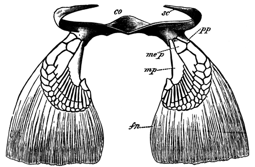
Pectoral fins and girdle of an adult of Scyllium canicula (natural size, seen from behind and above).
co. Coracoid. sc. scapula. pp. propterygium. mep. mesopterygium. mp. metapterygium. fn. part of fin supported by horny fibre.
Fig. 2.
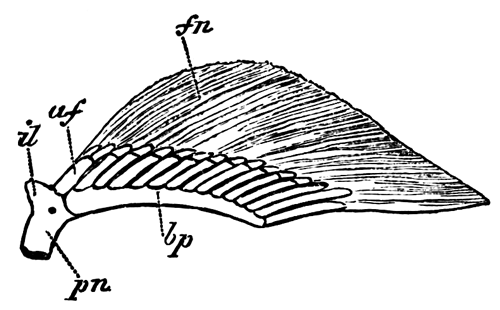
Right pelvic fin and part of pelvic girdle of an adult female of Scyllium canicula (natural size).
il. iliac process. pn. pubic process, cut across below. bp. basipterygium. af. anterior cartilaginous fin-ray articulated to pelvic girdle. fn. part of fin supported by horny fibres.
The true skeleton consists of three basal pieces articulating with the pectoral girdle; on the outer side of which there is a series of more or less segmented cartilaginous fin-rays. Of the basal cartilages one (pp) is anterior, a second (mep) is placed in the middle, and a third is posterior (mp). They have been named by Gegenbaur the propterygium, the mesopterygium, and the metapterygium; and these names are now generally adopted.
The metapterygium is by far the most important of the three, and in Scyllium canicula supports 12 or 13 rays[494]. It forms a large part of the posterior boundary of the fin, and bears rays only on its anterior border.
The mesopterygium supports 2 or 3 rays, in the basal parts of which the segmentation into distinct rays is imperfect; and the propterygium supports only a single ray.
The pelvic fins are horizontally placed, like the pectoral fins, but differ from the latter in nearly meeting each other along the median ventral line of the body. They also differ from the pectoral fins in having a relatively much broader base of attachment to the sides of the body. Their cartilaginous skeleton (woodcut, fig. 2) consists of a basal bar, placed parallel to the base of the fin, and articulated in front with the pelvic girdle.
On its outer border it articulates with a series of cartilaginous fin-rays. I shall call the basal bar the basipterygium. The rays which it bears are most of them less segmented than those of the pectoral fin, being only divided into two; and the posterior ray, which is placed in the free posterior border of the fin, continues the axis of the basipterygium. In the male it is modified in connection with the so-called clasper.
The anterior fin-ray of the pelvic fin, which is broader than the other rays, articulates directly with the pelvic girdle, instead of with the basipterygium. This ray, in the female of Scyllium canicula and in the male of Scyllium catulus (Gegenbaur), is peculiar in the fact that its distal segment is longitudinally divided into two or more pieces, instead of being single as is the case with the remaining rays. It is probably equivalent to two of the posterior rays.
Development of the paired Fins.—The first rudiments of the limbs appear in Scyllium, as in other fishes, as slight longitudinal ridge-like thickenings of the epiblast, which closely resemble the first rudiments of the unpaired fins.
These ridges are two in number on each side—an anterior immediately behind the last visceral fold, and a posterior on the level of the cloaca. In most Fishes they are in no way connected; but in some Elasmobranch embryos, more especially in that of Torpedo, they are connected together at their first development by a line of columnar epiblast cells. This connecting line of columnar epiblast, however, is a very transitory structure. The rudimentary fins soon become more prominent, consisting of a projecting ridge both of epiblast and mesoblast, at the outer edge of which is a fold of epiblast only, which soon reaches considerable dimensions. At a later stage the mesoblast penetrates into this fold, and the fin becomes a simple ridge of mesoblast covered by epiblast. The pectoral fins are at first considerably ahead of the pelvic fins in development.
The direction of the original epithelial line which connected the two fins of each side is nearly, though not quite, longitudinal, sloping somewhat obliquely ventralwards. It thus comes about that the attachment of each pair of limbs is somewhat on a slant, and that the pelvic pair nearly meet each other in the median ventral line shortly behind the anus.
The embryonic muscle-plates, as I have elsewhere shewn, grow into the bases of the fins; and the cells derived from these ingrowths, which are placed on the dorsal and ventral surfaces in immediate contact with the epiblast, probably give rise to the dorsal and ventral muscular layers of the limb, which are shewn in section in Plate 33, fig. 1, m, and in Plate 33, fig. 7, m.
The cartilaginous skeleton of the limbs is developed in the indifferent mesoblast cells between the two layers of muscles. Its early development in both the pectoral and the pelvic fins is very similar. When first visible it differs histologically from the adjacent mesoblast simply in the fact of its cells being more concentrated; while its boundary is not sharply marked.
At this stage it can only be studied by means of sections. It arises simultaneously and continuously with the pectoral and pelvic girdles, and consists, in both fins, of a bar springing at right angles from the posterior side of the pectoral or pelvic girdle, and running parallel to the long axis of the body along the base of the fin. The outer side of this bar is continued into a thin plate, which extends into the fin.
The structure of the skeleton of the fin slightly after its first differentiation will be best understood from Plate 33, fig. 1, and Plate 33, fig. 7. These figures represent transverse sections through the pelvic and pectoral fins of the same embryo on the same scale. The basal bar is seen at bp, and the plate at this stage (which is considerably later than the first differentiation) already partially segmented into rays at br. Outside the region of the cartilaginous plate is seen the fringe with the horny fibres (h.f.); and dorsally and ventrally to the cartilaginous skeleton are seen the already well-differentiated muscles (m).
The pectoral fin is shewn in horizontal section in Plate 33, fig. 6, at a somewhat earlier stage than that to which the transverse sections belong. The pectoral girdle (p.g.) is cut transversely, and is seen to be perfectly continuous with the basal bar (vp) of the fin. A similar continuity between the basal bar of the pelvic fin and the pelvic girdle is shewn in Plate 33, fig. 2, at a somewhat later stage. The plate continuous with the basal bar of the fin is at first, to a considerable extent in the pectoral, and to some extent in the pelvic fin, a continuous lamina, which subsequently segments into rays. In the parts of the plate which eventually form distinct rays, however, almost from the first the cells are more concentrated than in those parts which will form the tissue between the rays; and I am not inclined to lay any stress whatever upon the fact of the cartilaginous fin-rays being primitively part of a continuous lamina, but regard it as a secondary phenomenon, dependent on the mode of conversion of embryonic mesoblast cells into cartilage. In all cases the separation into distinct rays is to a large extent completed before the tissue of which the plates are formed is sufficiently differentiated to be called cartilage by an histologist.
The general position of the fins in relation to the body, and their relative sizes, may be gathered from Plate 33, figs. 4 and 5, which represent transverse sections of the same embryo as that from which the transverse sections shewing the fin on a larger scale were taken.
During the first stage of its development the skeleton of both fins may thus be described as consisting of a longitudinal bar running along the base of the fin, and giving off at right angles series of rays which pass into the fin. The longitudinal bar may be called the basipterygium; and it is continuous in front with the pectoral or pelvic girdle, as the case may be.
The further development of the primitive skeleton is different in the case of the two fins.
The Pelvic Fin.—The changes in the pelvic fin are comparatively slight. Plate 33, fig. 2, is a representation of the fin and its skeleton in a female of Scyllium stellare shortly after the primitive tissue is converted into cartilage, but while it is still so soft as to require the very greatest care in dissection. The fin itself forms a simple projection of the side of the body. The skeleton consists of a basipterygium (bp), continuous in front with the pelvic girdle. To the outer side of the basipterygium a series of cartilaginous fin-rays are attached—the posterior ray forming a direct prolongation of the basipterygium, while the anterior ray is united rather with the pelvic girdle than with the basipterygium. All the cartilaginous fin-rays except the first are completely continuous with the basipterygium, their structure in section being hardly different from that shewn in Plate 33, fig. 1.
The external form of the fin does not change very greatly in the course of the further development; but the hinder part of the attached border is, to some extent, separated off from the wall of the body, and becomes the posterior border of the adult fin. With the exception of a certain amount of segmentation in the rays, the character of the skeleton remains almost as in the embryo. The changes which take place are illustrated by Plate 33, fig. 3, shewing the fin of a young male of Scyllium stellare. The basipterygium has become somewhat thicker, but is still continuous in front with the pelvic girdle, and otherwise retains its earlier characters. The cartilaginous fin-rays have now become segmented off from it and from the pelvic girdle, the posterior end of the basipterygial bar being segmented off as the terminal ray.
The anterior ray is directly articulated with the pelvic girdle, and the remaining rays continue articulated with the basipterygium. Some of the latter are partially segmented.
As may be gathered by comparing the figure of the fin at the stage just described with that of the adult fin (woodcut, fig. 2), the remaining changes are very slight. The most important is the segmentation of the basipterygial bar from the pelvic girdle.
The pelvic fin thus retains in all essential points its primitive structure.
The Pectoral Fin.—The earliest stage of the pectoral fin differs, as I have shewn, from that of the pelvic fin only in minor points (Pl. 33, fig. 6). There is the same longitudinal or basipterygial bar (bp), to which the fin-rays are attached, which is continuous in front with the pectoral girdle (pg). The changes which take place in the course of the further development, however, are very much more considerable in the case of the pectoral than in that of the pelvic fin.
The most important change in the external form of the fin is caused by a reduction in the length of its attachment to the body. At first (Pl. 33, fig. 6), the base of the fin is as long as the greatest breadth of the fin; but it gradually becomes shortened by being constricted off from the body at its hinder end. In connection with this process the posterior end of the basipterygial bar is gradually rotated outwards, its anterior end remaining attached to the pectoral girdle. In this way this bar comes to form the posterior border of the skeleton of the fin (Pl. 33, figs. 8 and 9), constituting the metapterygium (mp). It becomes eventually segmented off from the pectoral girdle, simply articulating with its hinder edge.
The plate of cartilage, which is continued outwards from the basipterygium, or, as we may now call it, the metapterygium, into the fin, is not nearly so completely divided up into fin-rays as the homologous part of the pelvic fin; and this is especially the case with the basal part of the plate. This basal part becomes, in fact, at first only divided into two parts (Pl. 33, fig. 8)—a small anterior part at the front end (me.p), and a larger posterior along the base of the metapterygium (mp); and these two parts are not completely segmented from each other. The anterior part directly joins the pectoral girdle at its base, resembling in this respect the anterior fin-ray of the pelvic girdle. It constitutes the (at this stage undivided) rudiment of the mesopterygium and propterygium of Gegenbaur. It bears in my specimen of this age four fin-rays at its extremity, the anterior not being well marked. The remaining fin-rays are prolongations outwards of the edge of the plate continuous with the metapterygium. These rays are at the stage figured more or less transversely segmented; but at their outer edge they are united together by a nearly continuous rim of cartilage. The spaces between the fin-rays are relatively considerably larger than in the adult.
The further changes in the cartilages of the pectoral limb are, morphologically speaking, not important, and are easily understood by reference to Pl. 33, fig. 9 (representing the skeleton of the limb of a nearly ripe embryo). The front end of the anterior basal cartilage becomes segmented off as a propterygium (pp), bearing a single fin-ray, leaving the remainder of the cartilage as a mesopterygium (mes). The remainder of the now considerably segmented fin-rays are borne by the metapterygium.
* * * * *
General Conclusions.—From the above observations, conclusions of a positive kind may be drawn as to the primitive structure of the skeleton; and the observations have also, it appears to me, important bearings on the theories of my predecessors in this line of investigation.
The most obvious of the positive conclusions is to the effect that the embryonic skeleton of the paired fins consists of a series of parallel rays similar to those of the unpaired fins. These rays support the soft parts of the fins, which have the form of a longitudinal ridge; and they are continuous at their base with a longitudinal bar. This bar, from its position at the base of the fin, can clearly never have been a median axis with the rays on both sides. It becomes the basipterygium in the pelvic fin, which retains its embryonic structure much more completely than the pectoral fin; and the metapterygium in the pectoral fin. The metapterygium of the pectoral fin is thus clearly homologous with the basipterygium of the pelvic fin, as originally supposed by Gegenbaur, and as has since been maintained by Mivart. The propterygium and mesopterygium are obviously relatively unimportant parts of the skeleton as compared with the metapterygium.
My observations on the development of the skeleton of the fins certainly do not of themselves demonstrate that the paired fins are remnants of a once continuous lateral fin; but they support this view in that they shew the primitive skeleton of the fins to have exactly the character which might have been anticipated if the paired fins had originated from a continuous lateral fin. The longitudinal bar of the paired fins is believed by both Thacker and Mivart to be due to the coalescence of the bases of the primitively independent rays of which they believe the fin to have been originally composed. This view is probable enough in itself, and is rendered more so by the fact, pointed out by Mivart, that a longitudinal bar supporting the cartilaginous rays of unpaired fins is occasionally formed; but there is no trace in the embryo Scylliums of the bar in question being formed by the coalescence of rays, though the fact of its being perfectly continuous with the bases of the fin-rays is somewhat in favour of such coalescence.
Thacker and Mivart both hold that the pectoral and pelvic girdles are developed by ventral and dorsal growths of the anterior end of the longitudinal bar supporting the fin-rays.
There is, so far as I see, no theoretical objection to be taken to this view; and the fact of the pectoral and pelvic girdles originating continuously and long remaining united with the longitudinal bars of their respective fins is in favour of it rather than the reverse. The same may be said of the fact that the first part of each girdle to be formed is that in the neighbourhood of the longitudinal bar (basipterygium) of the fin, the dorsal and ventral prolongations being subsequent growths.
On the whole my observations do not throw much light on the theories of Thacker and Mivart as to the genesis of the skeleton of the paired fin; but, so far as they bear on the subject, they are distinctly favourable to those theories.
The main results of my observations appear to me to be decidedly adverse to the views recently put forward on the structure of the fin by Gegenbaur and Huxley, both of whom, as stated above, consider the primitive type of fin to be most nearly retained in Ceratodus, and to consist of a central multisegmented axis with numerous lateral rays.
Gegenbaur derives the Elasmobranch pectoral fin from a form which he calls the archipterygium, nearly like that of Ceratodus, with a median axis and two rows of rays—but holds that in addition to the rays attached to the median axis, which are alone found in Ceratodus, there were other rays directly articulated to the shoulder-girdle. He considers that in the Elasmobranch fin the majority of the lateral rays on the posterior (or median according to his view of the position of the limb) side have become aborted, and that the central axis is represented by the metapterygium; while the pro- and mesopterygium and their rays are, he believes, derived from those rays of the archipterygium which originally articulated directly with the shoulder-girdle.
This view appears to me to be absolutely negatived by the facts of development of the pectoral fin in Scyllium—not so much because the pectoral fin in this form is necessarily to be regarded as primitive, but because what Gegenbaur holds to be the primitive axis of the biserial fin is demonstrated to be really the base, and it is only in the adult that it is conceivable that a second set of lateral rays could have existed on the posterior side of the metapterygium. If Gegenbaur's view were correct, we should expect to find in the embryo, if anywhere, traces of the second set of lateral rays; but the fact is that, as may easily be seen by an inspection of figs. 6 and 7, such a second set of lateral rays could not possibly have existed in a type of fin like that found in the embryo. With this view of Gegenbaur's it appears to me that the theory held by this anatomist to the effect that the limbs are modified gill-arches also falls, in that his method of deriving the limbs from gill-arches ceases to be admissible, while it is not easy to see how a limb, formed on the type of the embryonic limb of Elasmobranchii, could be derived from a gill-arch with its branchial rays.
Gegenbaur's older view, that the Elasmobranch fin retains a primitive uniserial type, appears to me to be nearer the truth than his more recent view on this subject; though I hold the fundamental point established by the development of these parts in Scyllium to be that the posterior border of the adult Elasmobranch pectoral fin is the primitive base-line,i.e.line of attachment of the fin to the side of the body.
Huxley holds that the mesopterygium is the proximal piece of the axial skeleton of the limb of Ceratodus, and derives the Elasmobranch fin from that of Ceratodus by the shortening of its axis and the coalescence of some of its elements. The entirely secondary character of the mesopterygium, and its total absence in the young embryo Scyllium, appear to me as conclusive against Huxley's view as the character of the embryonic fin is against that of Gegenbaur; and I should be much more inclined to hold that the fin of Ceratodus has been derived from a fin like that of the Elasmobranchii by a series of steps similar to those which Huxley supposes to have led to the establishment of the Elasmobranch fin, but in exactly the reverse order.
There is one statement of Davidoff's which I cannot allow to pass without challenge. In comparing the skeletons of the paired and unpaired fins he is anxious to prove that the former are independent of the axial skeleton in their origin and that the latter have been segmented from the axial skeleton, and thus to shew that an homology between the two is impossible. In support of his view he states[495] that he has satisfied himself, from embryos of Acanthias and Scyllium, that the rays of the unpaired fins are undoubtedly products of the segmentation of the dorsal and ventral spinous processes.
This statement is wholly unintelligible to me. From my examination of the development of the first dorsal and the anal fins of Scyllium I find that their rays develop at a considerable distance from, and quite independently of, the neural and hæmal arches, and that they are at an early stage of development distinctly in a more advanced state of histological differentiation than the neural and hæmal arches of the same region. I have also found exactly the same in the embryos of Lepidosteus.
I have, in fact, no doubt that the skeleton of both the paired and the unpaired fins of Elasmobranchii and Lepidosteus is in its development independent of the axial skeleton. The phylogenetic mode of origin of the skeleton both of the paired and of the unpaired fins cannot, however, be made out without further investigation.
EXPLANATION OF PLATE 33.[496]
Fig. 1. Transverse section through the pelvic fin of an embryo of Scyllium belonging to stage P1, magnified 50 diameters. bp. basipterygium. br. fin ray. m. muscle. hf. horny fibres supporting the peripheral part of the fin.
Fig. 2. Pelvic fin of a very young female embryo of Scyllium stellare, magnified 16 diameters. bp. basipterygium. pu. pubic process of pelvic girdle (cut across below). il. iliac process of pelvic girdle. fo. foramen.
Fig. 3. Pelvic fin of a young male embryo of Scyllium stellare, magnified 16 diameters. bp. basipterygium. mo. process of basipterygium continued into clasper. il. iliac process of pelvic girdle. pu. pubic section of pelvic girdle.
Fig. 4. Transverse section through the ventral part of the trunk of an embryo Scyllium of stage P, in the region of the pectoral fins, to shew how the fins are attached to the body, magnified 18 diameters. br. cartilaginous fin-ray. bp. basipterygium. m. muscle of fin. mp. muscle-plate.
Fig. 5. Transverse section through the ventral part of the trunk of an embryo Scyllium of stage P, in the region of the pelvic fin, on the same scale as fig. 4. bp. basipterygium. br. cartilaginous fin-rays. m. muscle of the fins. mp. muscle-plate.
Fig. 6. Pectoral fin of an embryo of Scyllium canicula, of a stage between O and P, in longitudinal and horizontal section (the skeleton of the fin was still in the condition of embryonic cartilage), magnified 36 diameters. bp. basipterygium (eventual metapterygium). fr. cartilaginous fin-rays. pg. pectoral girdle in transverse section. fo. foramen in pectoral girdle. pe. epithelium of peritoneal cavity.
Fig. 7. Transverse section through the pectoral fin of a Scyllium embryo of stage P, magnified 50 diameters. bp. basipterygium. br. cartilaginous fin-ray. m. muscle. hf. horny fibres.
Fig. 8. Pectoral fin of an embryo of Scyllium stellare, magnified 16 diameters. mp. metapterygium (basipterygium of earlier stage). me.p. rudiment of future pro- and mesopterygium. sc. cut surface of a scapular process. cr. coracoid process. fr. foramen. hf. horny fibres.
Fig. 9. Skeleton of the pectoral fin and part of pectoral girdle of a nearly ripe embryo of Scyllium stellare, magnified 10 diameters. mp. metapterygium. mes. mesopterygium. pp. propterygium. cr. coracoid process.
XXI. On the Evolution of the Placenta, and on the possibility of employing the characters of the Placenta in the Classification of the Mammalia[497].
From Owen's observations on the Marsupials it is clear that the yolk-sack in this group plays an important (if not the most important) part, in absorbing the maternal nutriment destined for the fœtus. The fact that in Marsupials both the yolk-sack and the allantois are concerned in rendering the chorion vascular, makes it à priori probable that this was also the case in the primitive types of the Placentalia; and this deduction is supported by the fact that in the Rodentia, Insectivora, and Cheiroptera this peculiarity of the fœtal membranes is actually found. In the primitive Placentalia it is also probable that from the discoidal allantoic region of the chorion simple fœtal villi, like those of the Pig, projected into uterine crypts; but it is not certain how far the umbilical region of the chorion, which was no doubt vascular, may also have been villous. From such a primitive type of fœtal membranes divergencies in various directions have given rise to the types of fœtal membranes found at the present day.
In a general way it may be laid down that variations in any direction which tended to increase the absorbing capacities of the chorion would be advantageous. There are two obvious ways in which this might be done, viz. (1) by increasing the complexity of the fœtal villi and maternal crypts over a limited area, (2) by increasing the area of the part of the chorion covered by the placental villi. Various combinations of the two processes would also, of course, be advantageous.
The most fundamental change which has taken place in all the existing Placentalia is the exclusion of the umbilical vesicle from any important function in the nutrition of the fœtus.
The arrangement of the fœtal parts in the Rodentia, Insectivora, and Cheiroptera may be directly derived from the primitive form by supposing the villi of the discoidal placental area to have become more complex, so as to form a deciduate discoidal placenta, while the yolk-sack still plays a part, though physiologically an unimportant part, in rendering the chorion vascular.
In the Carnivora, again, we have to start from the discoidal placenta, as evinced by the fact that in the growth of the placenta the allantoic region of the placenta is at first discoidal, and only becomes zonary at a later stage. A zonary deciduate placenta indicates an increase both in area and in complexity. The relative diminution of the breadth of the placental zone in late fœtal life in the zonary placenta of the Carnivora is probably due to its being on the whole advantageous to secure the nutrition of the fœtus by insuring a more intimate relation between the fœtal and maternal parts, than by increasing their area of contact. The reason of this is not obvious, but, as shewn below, there are other cases where it is clear that a diminution in the area of the placenta has taken place, accompanied by an increase in the complexity of its villi.
The second type of differentiation from the primitive form of placenta is illustrated by the Lemuridæ, the Suidæ, and Manis. In all these cases the area of the placental villi appears to have increased so as to cover nearly the whole subzonal membrane, without the villi increasing to any great extent in complexity. From the diffused placenta covering the whole surface of the chorion, differentiations appear to have taken place in various directions. The placenta of Man and Apes, from its mode of ontogeny, is clearly derived from a diffused placenta (very probably similar to that of Lemurs) by a concentration of the fœtal villi, which are originally spread over the whole chorion, to a disk-shaped area, and by an increase in their arborescence. Thus the discoidal placenta of Man has no connexion with, and ought not to be placed in, the same class as those of the Rodentia, Cheiroptera, and Insectivora.
The polycotyledonary forms of placenta are due to similar concentrations of the fœtal villi of an originally diffused placenta.
In the Edentata we have a group with very varying types of placenta. Very probably these may all be differentiations within the group itself from a diffused placenta such as that found in Manis. The zonary placenta of Orycteropus is capable of being easily derived from that of Manis by the disappearance of the fœtal villi at the two poles of the ovum. The small size of the umbilical vesicle in Orycteropus indicates that its discoidal placenta is not, like that of the Carnivora, directly derived from a type with both allantoic and umbilical vascularization of the chorion. The discoidal and dome-shaped placentæ of the Armadillos, Myrmecophaga, and the Sloths may easily have been formed from a diffused placenta, just as the discoidal placenta of the Simiidæ and Hominidæ appears to have been formed from a diffused placenta like that of the Lemuridæ.
The presence of zonary placenta in Hyrax and Elephas does not necessarily afford any proof of affinity of these types with the Carnivora. A zonary placenta may be quite as easily derived from a diffused placenta as from a discoidal placenta; and the presence of two villous patches at the poles of the chorion in Elephas very probably indicates that its placenta has been evolved from a diffused placenta.
Although it would not be wise to attempt to found a classification upon the placental characters alone, it may be worth while to make a few suggestions as to the affinities of the orders of Mammalia indicated by the structure of the placenta. We clearly, of course, have to start with forms which could not be grouped with any of the existing orders, but which might be called the Protoplacentalia. They probably had the primitive type of placenta described above: the nearest living representatives of the group are the Rodentia, Insectivora, and Cheiroptera. Before, however, these three groups had become distinctly differentiated, there must have branched off from the primitive stock the ancestors of the Lemuridæ, the Ungulata, and the Edentata.
It is obvious on general anatomical grounds that the Monkeys and Man are to be derived from a primitive Lemurian type; and with this conclusion the form of the placenta completely tallies. The primitive Edentata and Ungulata had no doubt a diffused placenta which was probably not very different from that of the primitive Lemurs; but how far these groups arose quite independently from the primitive stock, or whether they may have had a nearer common ancestor, cannot be decided from the structure of the placenta. The Carnivora were certainly an offshoot from the primitive placental type which was quite independent of the three groups just mentioned; but the character of the placenta of the Carnivora does not indicate at what stage in the evolution of the placental Mammalia a primitive type of Carnivora was first differentiated.
No important light is thrown by the placenta on the affinities of the Proboscidea, the Cetacea, or the Sirenia; but the character of the placenta in the latter group favours the view of their being related to the Ungulata.
XXII. On the Structure and Development of Lepidosteus[498]. By F. M. Balfour and W. N. Parker.
(With Plates 34-42.)
TABLE OF CONTENTS.
PAGE
Introduction 739
General Development 740
Brain—
Adult brain 759
Development of the brain 764
Comparison of the larval and adult brain of Lepidosteus, together with some observations on the systematic value of the characters of the Ganoid brain 767
Sense Organs—
Olfactory organ 771
Anatomy of the eye ib.
Development of the eye 772
Suctorial Disc 774
Muscular System 775
Skeleton—
Vertebral column and ribs of the adult 776
Development of the vertebral column and ribs. 778
Comparison of the vertebral column of Lepidosteus with that of other forms 792
The ribs of Fishes 793
The skeleton of the ventral lobe of the tail fin, and its bearing on the nature of the tail fin of the various types of Pisces 801
Excretory and Generative Organs—
Anatomy of the excretory and generative organs of the female 810
Anatomy of the excretory and generative organs of the male 813
Development of the excretory and generative organs 815
Theoretical considerations 822
The Alimentary Canal and its Appendages—
Topographical anatomy of the alimentary canal 828
Development of the alimentary canal and its appendages 831
The Gill on the Hyoid Arch 835
The systematic position of Lepidosteus 836
List of memoirs on the Anatomy and Development of Lepidosteus 840
List of Reference Letters 841
Explanation of Plates 842
Introduction.
The following paper is the outcome of the very valuable gift of a series of embryos and larvæ of Lepidosteus by Professor Alex. Agassiz, to whom we take this opportunity of expressing our most sincere thanks. The skull of these embryos and larvæ has been studied by Professor Parker, and forms the subject of a memoir already presented to the Royal Society.
Considering that Lepidosteus is one of the most interesting of existing Ganoids, and that it is very closely related to species of Ganoids which flourished during the Triassic period, we naturally felt keenly anxious to make the most of the opportunity of working at its development offered to us by Professor Agassiz' gift. Professor Agassiz, moreover, most kindly furnished us with four examples of the adult Fish, which have enabled us to make this paper a study of the adult anatomy as well as of the development.
The first part of our paper is devoted to the segmentation, formation of the germinal layers, and general development of the embryo and larva. The next part consists of a series of sections on the organs, in which both their structure in the adult and their development are dealt with. This part is not, however, in any sense a monograph, and where already known, the anatomy is described with the greatest possible brevity. In this part of the paper considerable space is devoted to a comparison of the organs of Lepidosteus with those of other Fishes, and to a statement of the conclusions which follow from such comparison.
The last part of the paper deals with the systematic position of Lepidosteus and of the Ganoids generally.
General Development.
The spawning of Lepidosteus takes place in the neighbourhood of New York about May 20th. Agassiz (No. 1)[499] gives an account of the process from Mr S. W. Garman's notes, which we venture to quote in full.
"Black Lake is well stocked with Bill-fish. When they appear, they are said to come in countless numbers. This is only for a few days in the spring, in the spawning season, between the 15th of May and the 8th of June. During the balance of the season they are seldom seen. They remain in the deeper parts of the lake, away from the shore, and, probably, are more or less nocturnal in habits. Out of season, an occasional one is caught on a hook baited with a minnow. Commencing with the 20th of April, until the 14th of May we were unable to find the Fish, or to find persons who had seen them during this time. Then a fisherman reported having seen one rise to the surface. Later, others were seen. On the afternoon of the 18th, a few were found on the points, depositing the spawn. The temperature at the time was 68° to 69° on the shoals, while out in the lake the mercury stood at 62° to 63°. The points on which the eggs were laid were of naked granite, which had been broken by the frost and heat into angular blocks of 3 to 8 inches in diameter. The blocks were tumbled upon each other like loose heaps of brick-bats, and upon and between them the eggs were dropped. The points are the extremities of small capes that make out into the lake. The eggs were laid in water varying in depth from 2 to 14 inches. At the time of approaching the shoals, the Fish might be seen to rise quite often to the surface to take air. This they did by thrusting the bill out of the water as far as the corners of the mouth, which was then opened widely and closed with a snap. After taking the air, they seemed more able to remain at the surface. Out in the lake they are very timid, but once buried upon the shoals they become quite reckless as to what is going on about them. A few moments after being driven off, one or more of the males would return as if scouting. If frightened, he would retire for some time; then another scout would appear. If all promised well, the females, with the attendant males, would come back. Each female was accompanied by from one to four males. Most often, a male rested against each side, with their bills reaching up toward the back of her head. Closely crowded together, the little party would pass back and forth over the rocky bed they had selected, sometimes passing the same spot half-a-dozen times without dropping an egg, then suddenly would indulge in an orgasm; and, lashing and plashing the water in all directions with their convulsive movements, would scatter at the same instant the eggs and the sperm. This ended, another season of moving slowly back and forth was observed, to be in turn followed by another of excitement. The eggs were excessively sticky. To whatever they happened to touch, they stuck, and so tenaciously that it was next to impossible to release them without tearing away a portion of their envelopes. It is doubtful whether the eggs would hatch if removed. As far as could be seen at the time, upon or under the rocks to which the eggs were fastened there was an utter absence of anything that might serve as food for the young Fishes.
Other Fishes, Bull-heads, &c., are said to follow the Bill-fish to eat the spawn. It may be so. It was not verified. Certainly the points under observations were unmolested. During the afternoon of the 18th of May a few eggs were scattered on several of the beds. On the 19th there were more. With the spear and the snare, several dozens of both sexes of the Fish were taken. Taking one out did not seem greatly to startle the others. They returned very soon. The males are much smaller than the average size of the females; and, judging from those taken, would seem to have as adults greater uniformity in size. The largest taken was a female, of 4 feet 1½ inch in length. Others of 2 feet 6 inches contained ripe ova. With the 19th of May all disappeared, and for a time—the weather being meanwhile cold and stormy—there were no signs of their continued existence to be met with. Nearly two weeks later, on the 31st of May, as stated by Mr Henry J. Perry, they again came up, not in small detachments on scattered points as before, but in multitudes, on every shoal at all according with their ideas of spawning beds. They remained but two days. During the summer it happens now and then that one is seen to come up for his mouthful of air; beyond this there will be nothing to suggest the ravenous masses hidden by the darkness of the waters.
Egg membranes.—The ova of Lepidosteus are spherical bodies of about 3 millims. in diameter. They have a double investment consisting of (1) an outer covering formed of elongated, highly refractive bodies, somewhat pyriform at their outer ends (Plate 34, fig. 17, f.e.), which are probably metamorphosed follicular cells[500], and (2) of an inner membrane, divided into two zones, viz.: an outer and thicker zone, which is radially striated, and constitutes the zona radiata (z.r.), and an inner and narrow homogeneous zone (z.r´.).
Segmentation.—We have observed several stages in the segmentation, which shew that it is complete, but that it approaches the meroblastic type more nearly than in the case of any other known holoblastic ovum.
Our earliest stage shewed a vertical furrow at the upper or animal pole, extending through about one-fifth of the circumference (Plate 34, fig. 1), and in a slightly later stage we found a second similar furrow at right angles to the first (Plate 34, fig. 2). We have not been fortunate enough to observe the next phases of the segmentation, but on the second day after impregnation (Plate 34, fig. 3), the animal pole is completely divided into small segments, which form a disc, homologous to the blastoderm of meroblastic ova; while the vegetative pole, which subsequently forms a large yolk-sack, is divided by a few vertical furrows, four of which nearly meet at the pole opposite the blastoderm (Plate 34, fig. 4). The majority of the vertical furrows extend only a short way from the edge of the small spheres, and are partially intercepted by imperfect equatorial furrows.
Development of the embryo.—We have not been able to work out the stages immediately following the segmentation, owing to want of material; and in the next stage satisfactorily observed, on the third day after impregnation, the body of the embryo is distinctly differentiated. The lower pole of the ovum is then formed of a mass in which no traces of the previous segments or segmentation furrows could any longer be detected.
Some of the dates of the specimens sent to us appear to have been transposed; so that our statements as to ages must only be taken as approximately correct.
Third day after impregnation.—In this stage the embryo is about 3.5 millims. in length, and has a somewhat dumb-bell shaped outline (Plate 34, fig. 5). It consists of (1) an outer area (p.z) with some resemblance to the area pellucida of the Avian embryo, forming the parietal part of the body; and (2) a central portion consisting of the vertebral and medullary plates and the axial portions of the embryo. In hardened specimens the peripheral part forms a shallow depression surrounding the central part of the embryo.
The central part constitutes a somewhat prominent ridge, the axial part of it being the medullary plate. Along the anterior half of this part a dark line could be observed in all our specimens, which we at first imagined to be caused by a shallow groove. We have, however, failed to find in our sections a groove in this situation except in a single instance (Plate 35, fig. 20, x), and are inclined to attribute the appearance above-mentioned to the presence of somewhat irregular ridges of the outer layer of the epiblast, which have probably been artificially produced in the process of hardening.
The anterior end of the central part is slightly dilated to form the brain (b); and there is present a pair of lateral swellings near the anterior end of the brain which we believe to be the commencing optic vesicles. We could not trace any other clear indications of the differentiation of the brain into distinct lobes.
At the hinder end of the central part of the embryo a very distinct dilatation may also be observed, which is probably homologous with the tail swelling of Teleostei. Its structure is more particularly dealt with in the description of our sections of this stage.
After the removal of the egg-membranes described above we find that there remains a delicate membrane closely attached, to the epiblast. This membrane can be isolated in distinct portions, and appears to be too definite to be regarded as an artificial product.
We have been able to prepare several more or less complete series of sections of embryos of this stage (Plate 35, figs. 18-22). These sections present as a whole a most striking resemblance to those of Teleostean embryos at a corresponding stage of development.
Three germinal layers are already fully established. The epiblast (ep.) is formed of the same parts as in Teleostei, viz.:—of an outer epidermic and an inner nervous or mucous stratum. In the parietal region of the embryo these strata are each formed of a single row of cells only. The cells of both strata are somewhat flattened, but those of the epidermic stratum are decidedly the more flattened of the two.
Along the axial line there is placed, as we have stated above, the medullary plate. The epidermic stratum passes over this plate without undergoing any change of character, and the plate is entirely constituted of the nervous stratum of the epidermis.
The medullary plate has, roughly speaking, the form of a solid keel, projecting inwards towards the yolk. There is no trace, at this stage at any rate, of a medullary groove; and as, we shall afterwards shew, the central canal of the cerebro-spinal cord is formed in the middle of the solid keel. The shape of this keel varies according to the region of the body. In the head (Plate 35, fig. 18, m.c.), it is very prominent, and forming, as it does, the major part of the axial tissue of the body, impresses its own shape on the other parts of the head and gives rise to a marked ridge on the surface of the head directed towards the yolk. In the trunk (Plate 35, figs. 19, 20) the keel is much less prominent, but still projects sufficiently to give a convex form to the surface of the body turned towards the yolk.
In the head, and also near the hind end of the trunk, the nervous layer of the epiblast continuous with the keel on each side is considerably thicker than the lateral parts of the layer. The thickening of the nervous layer in the head gives rise to what has been called by Götte[501] the special sense plate, owing to its being subsequently concerned in the formation of parts of the organs of special sense. We cannot agree with Götte in regarding it as part of the brain.
In the keel itself two parts may be distinguished, viz.: a superficial part, best marked in the region of the brain, formed of more or less irregularly arranged polygonal cells, and a deeper part of horizontally placed flatter cells. The upper part is mainly concerned in the formation of the cranial nerves, and of the dorsal roots of the spinal nerves.
The mesoblast (ms.) in the trunk consists of a pair of independent plates which are continued forwards into the head, and in the prechordal region of the latter, unite below the medullary keel.
The mesoblastic plates of the trunk are imperfectly divided into vertebral and lateral regions. Neither longitudinal sections nor surface views shew at this stage any trace of a division of the mesoblast into somites. The mesoblast cells are polygonal, and no indication is as yet present of a division into splanchnic and somatic layers.
The notochord (nc.) is well established, so that its origin could not be made out. It is, however, much more sharply separated from the mesoblastic plates than from the hypoblast, though the ventral and inner corners of the mesoblastic plates which run in underneath it on either side, are often imperfectly separated from it. It is formed of polygonal cells, of which between 40 and 50 may as a rule be seen in a single section. No sheath is present around it. It has the usual extension in front.
The hypoblast (hy.) has the form of a membrane, composed of a single row of oval cells, bounding the embryo on the side adjoining the yolk.
In the region of the caudal swelling the relations of the germinal layers undergo some changes. This region may, from the analogy of other Vertebrates, be assumed to constitute the lip of the blastopore. We find accordingly that the layers become more or less fused. In the anterior part of the tail swelling, the boundary between the notochord and hypoblast becomes indistinct. A short way behind this point (Plate 35, fig. 21), the notochord unites with the medullary keel, and a neurenteric cord, homologous with the neurenteric canal of other Ichthyopsida, is thus established. In the same region the boundary between the lateral plates of mesoblast and the notochord, and further back (Plate 35, fig. 22), that between the mesoblast and the medullary keel, becomes obliterated.
Fifth day after impregnation.—Between the stage last described and the next stage of which we have specimens, a considerable progress has been made. The embryo (Plate 34, figs. 6 and 7) has grown markedly in length and embraces more than half the circumference of the ovum. Its general appearance is, however, much the same as in the earlier stage, but in the cephalic region the medullary plate is divided by constrictions into three distinct lobes, constituting the regions of the fore-brain, the mid-brain, and the hind-brain. The fore-brain (Plate 34, fig. 6, f.b.) is considerably the largest of the three lobes, and a pair of lateral projections forming the optic vesicles are decidedly more conspicuous than in the previous stage. The mid-brain (m.b.) is the smallest of the three lobes, while the hind-brain (h.b.) is decidedly longer, and passes insensibly into the spinal cord behind.
The medullary keel, though retaining to a great extent the shape it had in the last stage, is no longer completely solid. Throughout the whole region of the brain and in the anterior part of the trunk (Plate 35, figs. 23, 24, 25) a slit-like lumen has become formed. We are inclined to hold that this is due to the appearance of a space between the cells, and not, as supposed by Oellacher for Teleostei, to an actual absorption of cells, though we must admit that our sections are hardly sufficiently well preserved to be conclusive in settling this point. Various stages in its growth may be observed in different regions of the cerebro-spinal cord. When first formed, it is a very imperfectly defined cavity, and a few cells may be seen passing right across from one side of it to the other. It gradually becomes more definite, and its wall then acquires a regular outline.
The optic vesicles are now to be seen in section (Plate 35, fig. 23, op.) as flattish outgrowths of the wall of the fore-brain, into which the lumen of the third ventricle is prolonged for a short distance.
The brain has become to some extent separate from the superjacent epiblast, but the exact mode in which this is effected is not clear to us. In some sections it appears that the separation takes place in such a way that the nervous keel is only covered above by the epidermic layer of the epiblast, and that the nervous layer, subsequently interposed between the two, grows in from the two sides. Such a section is represented in Plate 35, fig. 24. Other sections again favour the view that in the isolation of the nervous keel, a superficial layer of it remains attached to the nervous layer of the epidermis at the two sides, and so, from the first, forms a continuous layer between the nervous keel and the epidermic layer of the epiblast (Plate 35, fig. 25). In the absence of a better series of sections we do not feel able to determine this point. The posterior part of the nervous keel retains the characters of the previous stage.
At the sides of the hind-brain very distinct commencements of the auditory vesicles are apparent. They form shallow pits (Plate 35, fig. 24, au.) of the thickened part of the nervous layer adjoining the brain in this region. Each pit is covered over by the epidermic layer above, which has no share in its formation.
In many parts of the lateral regions of the body the nervous layer of the epidermis is more than one cell deep.
The mesoblastic plates are now divided in the anterior part of the trunk into a somatic and a splanchnic layer (Plate 35, fig. 25, so., sp.), though no distinct cavity is as yet present between these two layers. Their vertebral extremities are somewhat wedge-shaped in section, the base of the wedge being placed at the sides of the medullary keel. The wedge-shaped portions are formed of a superficial layer of palisade-like cells and an inner kernel of polygonal cells. The superficial layer on the dorsal side is continuous with the somatic mesoblast, while the remainder pertains to the splanchnic layer.
The diameter of the notochord has diminished, and the cells have assumed a flattened form, the protoplasm being confined to an axial region. In consequence of this, the peripheral layer appears clear in transverse sections. A delicate cuticular sheath is formed around it. This sheath is probably the commencement of the permanent sheath of later stages, but at this stage it cannot be distinguished in structure from a delicate cuticle which surrounds the greater part of the medullary cord.
The hypoblast has undergone no changes of importance.
The layers at the posterior end of the embryo retain the characters of the last stage.
Sixth day after impregnation.—At this stage (Plate 34, fig. 8) the embryo is considerably more advanced than at the last stage. The trunk has decidedly increased in length, and the head forms a relatively smaller portion of the whole. The regions of the brain are more distinct. The optic vesicles (op.) have grown outwards so as to nearly reach the edges of the area which forms the parietal part of the body. The fore-brain projects slightly in front, and the mid-brain is seen as a distinct rounded prominence. Behind the latter is placed the hind-brain, which passes insensibly into the spinal cord. On either side of the mid- and hind-brain a small region is slightly marked off from the rest of the parietal part, and on this are seen two more or less transversely directed streaks, which, by comparison with the Sturgeon[502], we are inclined to regard as the two first visceral clefts (br.c.). We have, however, failed to make them out in sections, and owing to the insufficiency of our material, we have not even studied them in surface views as completely as we could have wished.
The body is now laterally compressed, and more decidedly raised from the yolk than in the previous stages. In the lateral regions of the trunk the two segmental or archinephric ducts (sg.) are visible in surface views: the front end of each is placed at the level of the hinder border of the head, and is marked by a flexure inwards towards the middle line. The remainder of each duct is straight, and extends backwards for about half the length of the embryo. The tail has much the same appearance as in the last stage.
The vertebral regions of the mesoblastic plates are now segmented for the greater part of the length of the trunk, and the somites of which they are composed (Plate 36, fig. 30, pr.) are very conspicuous in surface views.