Chapter XII · (continued 4)
Reading edition
One of the most interesting points in Klein's paper, as well as in those of Heitzmann and Eimer, is the demonstration of a connection between the reticulum of the nucleus and fibres in the body of the cell. Such a connection I have not found in ova, but may point out that it appears to exist between the sub-germinal nuclei in Elasmobranchii and the protoplasmic network in the yolk in which they lie. This point is called attention to in my Monograph on Elasmobranch Fishes, page 39[423], where it is stated that the network in favourable cases may be observed to be in connection with the nuclei just described. Its meshes are finer in the vicinity of the nuclei, and the fibres in some cases appear almost to start from them. The nuclei in the yolk are knobbed bodies divided by a sponge work of septa into a number of areas each with a nucleolar body.
EXPLANATION OF PLATES 24, 25, 26.
Plate 24.
List of Reference Letters.
dn. Modified nucleus of primitive ovum. do. Permanent ovum in the act of being formed. dv. Developing blood-vessels. dyk. Developing yolk. ep. Non-ovarian epithelium of ovarian ridge. fe. Follicular epithelium. gv. Germinal vesicle. lstr. Lymphatic region of stroma. nn. Nests of nuclei of ovarian region. o. Permanent ovum. ovr. Ovarian portion of ovarian ridge. po. Primitive ovum. pse. Pseudo-epithelium of ovarian ridge. str. Stroma ingrowths into ovarian epithelium. v. Blood-vessel. vstr. Vascular region of stroma adjoining ovarian ridge. vt. Vitelline membrane. x. Modified nucleus. yk. Yolk. zn. Zona radiata.
Fig. 1. Transverse section of the ovarian ridge of an embryo of Scy. canicula, belonging to stage P, shewing the ovarian region with thickened epithelium and numerous primitive ova. Zeiss C, ocul. 2. Picric acid.
Fig. 2. Transverse section of the ovarian ridge of an embryo of Scyllium canicula, considerably older than stage Q. Zeiss C, ocul. 2. Picric acid. Several nests, some with distinct ova, and others with the ova fused together, are present in the section (n.n.), and several examples of modified nuclei in still distinct ova are also represented. One of these is marked x. The stroma of the ovarian ridge is exceptionally scanty.
Fig. 3. Transverse section through part of the ovarian ridge, including the ovarian region of an almost ripe embryo of Scyllium canicula. Zeiss C, ocul. 2. Picric acid. Nuclear nests (n.n.), developing ova (d.o.), and ova (o.), with completely formed follicular epithelium, are now present. The ovarian region is still well separated from the subjacent stroma, and does not appear to contain any cells except those of the original germinal epithelium.
Fig. 4. Section through ovarian ridge of the same embryo as fig. 3, to illustrate the relation of the stroma (str.) and ovarian region. Zeiss a a, ocul. 2. Picric acid.
Fig. 5. Section through the ovarian ridge of an embryo of Scyllium canicula, 10 cm. long, in which the ovary was slightly less advanced than in fig. 3. To illustrate the relation of the ovarian epithelium to the subjacent vascular stroma. Zeiss A, ocul. 2. Osmic acid. y. points to a small separated portion of the germinal epithelium.
Fig. 6. Section through the ovarian ridge of an embryo of Scyllium canicula, slightly older than fig. 5. To illustrate the relation of the ovarian epithelium to the subjacent vascular[TN12] stroma. Zeiss A, ocul. 2. Osmic acid.
Fig. 7. More highly magnified portion of the same ovary as fig. 6. To illustrate the same points. Zeiss C, ocul. 2. Osmic acid.
Fig. 8. Section through the ovarian region (close to one extremity, where it is very small) from a young female of Scy. canicula. Zeiss C, ocul. 2. Picric acid. It shews the vascular ingrowths amongst the original epithelial cells of the ovarian region.
Fig. 9. Section through the ovarian region of the same embryo as fig. 8, at its point of maximum development. Zeiss A, ocul. 2. Picric acid.
Fig. 10. Section through superficial part of the ovary of an embryo, shewing the pseudo-epithelium; the cells of which are provided with tails prolonged into the general tissue of the ovary. At f.e. is seen a surface view of the follicular epithelium of an ovum. Zeiss C, ocul. 2. Picric acid.
Fig. 11. Section through part of an ovary of Scyllium canicula of stage Q, with three primitive ova, the most superficial one containing a modified nucleus.
Fig. 12. Section through part of an ovary of an example of Scyllium canicula, 8 cm. long. The section passes through a nest of ova with modified nuclei, in which the outlines of the individual ova are quite distinct. Zeiss E, ocul. 2. Picric acid.
Fig. 13. Section through part of ovary of the same embryo as in fig. 5. The section passes through a nest of nuclei, with at the least two developing ova, and also through one already formed permanent ovum. Zeiss E, ocul. 2. Osmic acid.
Figs. 14, 15, 16, 17, 18 [Figs. 17 and 18 are on Pl. 25]. Sections through parts of the ovary of the same embryo as fig. 3, with nests of nuclei and a permanent ova in the act of formation. Fig. 14 is drawn with Zeiss D D, ocul. 2. Figs. 15, 16, 17, with Zeiss E, ocul. 2. Picric acid.
Plate 25.
List of Reference Letters.
do. Permanent ovum in the act of being formed. dyk. Developing yolk. fe. Follicular epithelium. fe´. Secondary follicular epithelium. gv. Germinal vesicle. nn. Nests of nuclei of ovarian region. o. Permanent ovum. pse. Pseudo-epithelium. str. Stroma ingrowths into ovarian epithelium. vt. Vitelline membrane. x. Modified nucleus. yk. Yolk (vitellus). zn. Zona radiata.
[Figs. 17 and 18. Vide description of Plate 24.].
Fig. 19. Two nuclei from a nest which appear to be in the act of division. From ovary of the same embryo as fig. 3.
Fig. 20. Section through part of an ovary of the same embryo as fig. 6, containing a nest of nuclei. Zeiss F, ocul. 2. Osmic acid.
Fig. 21. Ovum from the ovary of a half-grown female, containing isolated deeply stained patches of developing yolk granules. Zeiss B, ocul. 2. Picric acid.
Fig. 22. Section through a small part of the ovum of an immature female of Scyllium canicula, to shew the constitution of the yolk, the follicular epithelium, and the egg membranes. Zeiss E, ocul. 2. Chromic acid.
Fig. 23. Section through part of the periphery of a nearly ripe ovum of Scy. canicula. Zeiss C, ocul. 2. It shews the remnant of the vitelline membrane (v.t.) separating the columnar but delicate cells of the follicular epithelium (f.e.) from the yolk (yk.). In the yolk are seen yolk-spherules in a protoplasmic network. The transverse markings in the yolk-spherules have been made oblique by the artist.
Fig. 24. Fully formed ovum containing a second nucleus (x), probably about to be employed as pabulum; from the same ovary as fig. 5. The follicular epithelium is much thicker on the side adjoining the stroma than on the upper side of the ovum. Zeiss F, ocul. 2. Osmic acid.
Fig. 25. A. Ovum from the same ovary as fig. 21, containing in the yolk three peculiar bodies, similar in appearance to the two small bodies in the germinal vesicle. B. Germinal vesicle of a large ovum from the same ovary, containing a body of a strikingly similar appearance to those in the body of the ovum in A. Zeiss E, ocul. 2. Picric acid.
Fig. 26. Section of the ovary of a young female of Scyllium stellare 16½ centimetres in length. The ovary is exceptional, on account of the large size of the stroma ingrowths into the epithelium. Zeiss C, ocul. 2. Osmic acid.
Fig. 27. Ovum of Scyllium canicula, 5 mm. in diameter, treated with osmic acid. The figure illustrates the development of the yolk and a peculiar mode of proliferation of the germinal spots. Zeiss A, ocul. 2.
Fig. 28. Small part of the follicular epithelium and egg membranes of a somewhat larger ovum of Scyllium canicula than fig. 22. Zeiss D D, ocul. 2.
Fig. 29. The same parts as in fig. 28, from a still larger ovum. Zeiss D D, ocul. 2.
Fig. 30. Ovum of Raja with follicular epithelium. Zeiss C, ocul. 2.
Fig. 31. Small portion of a larger ovum of Raja than fig. 30. Zeiss D D, ocul. 2.
Fig. 32. Follicular epithelium, &c., from an ovum of Raja still larger than fig. 31. Zeiss D D, ocul. 2.
Fig. 33. Surface view of follicular epithelium from an ovum of Raja of about the same age as fig. 33.
Fig. 34. Vertical section through the superficial part of an ovary of an adult Raja to shew the relation of the pseudo-epithelium to the subjacent stroma. Zeiss D D, ocul. 2.
Plate 26.
Complete List of Reference Letters.
do. Developing ovum. fc. Cells which will form the follicular epithelium, fe. Follicular epithelium. ge. Germinal epithelium. mg. Malpighian body. n. Nest of cells of the germinal epithelium. nd. Nuclei in the act of dividing. o. Permanent ovum. ov. Ovary. po. Primitive ovum. t. Tubuliferous tissue, derived from Malpighian bodies.
Fig. 35. Transverse section through the ovary of an embryo rabbit of eighteen days, hardened in osmic acid. The colours employed are intended to render clear the distinction between the germinal epithelium (ge.) and the tubuliferous tissue (t.), which has grown in from the Wolffian body, and which gives rise in the male to parts of the tubuli seminiferi. Zeiss A, ocul. 2.
Fig. 35A. Transverse section through a small part of the ovary of an embryo from the same female as fig. 35, hardened in picric acid, shewing the relation of the germinal epithelium to the subjacent tissue. Zeiss D D, ocul. 2.
Fig. 35B. Longitudinal section through part of the Wolffian body and the anterior end of the ovary of an eighteen days' embryo, to shew the derivation of tubuliferous tissue (t.) from the Malpighian bodies, close to the anterior extremity of the ovary. Zeiss A, ocul. 1.
Fig. 36. Transverse section through the ovary of an embryo rabbit of twenty-two days, hardened in osmic acid. It is coloured in the same manner as fig. 35. Zeiss A, ocul. 2.
Fig. 36A. Transverse section through a small part of the ovary of an embryo, from the same female as fig. 36, hardened in picric acid, shewing the relation of the germinal epithelium to the stroma of the ovary. Zeiss D D, ocul. 2.
Figs. 37 and 37A. The same parts of an ovary of a twenty-eight days' embryo as figs. 36 and 36A of a twenty-two days' embryo.
Fig. 38. Ovary of a rabbit five days after birth, coloured in the same manner as figs. 35, 36 and 37, but represented on a somewhat smaller scale. Picric acid.
Fig. 38A. Vertical section through a small part of the surface of the same ovary as fig. 38. Zeiss D D, ocul. 2.
Fig. 38B. Small portion of the deeper layer of the germinal epithelium of the same ovary as fig. 38. The figure shews the commencing differentiation of the cells of the germinal epithelium into true ova and follicle cells. Zeiss D D, ocul. 2.
Fig. 39A. Section through a small part of the middle region of the germinal epithelium of a rabbit seven days after birth. Zeiss D D, ocul. 2.
Fig. 39B. Section through a small part of the innermost layer of the germinal epithelium of a rabbit seven days after birth, shewing the formation of Graafian follicles. Zeiss D D, ocul. 2.
Figs. 40A and 40B. Small portions of the middle region of the germinal epithelium of a rabbit four weeks after birth. Zeiss D D, ocul. 2.
Fig. 41. Graafian follicle with two ova, about to divide into two follicles, from a rabbit six weeks after birth. Zeiss D D, ocul. 2.
XIII. On the Existence of a Head-Kidney in the Embryo Chick, and on Certain Points in the Development of the Müllerian Duct[424]. By F. M. Balfour and A. Sedgwick.
(With Plates 27 and 28.)
The following paper is divided into three sections. The first of these records the existence of certain structures in the embryo chick, which eventually become in part the abdominal opening of the Müllerian duct, and which, we believe, correspond with the head-kidney, or Vorniere of German authors. The second deals with the growth and development of the Müllerian duct. With reference to this we have come to the conclusion that the Müllerian duct does not develop entirely independently of the Wolffian duct. The third section of our paper is of a more general character, and contains a discussion of the rectifications in the views of the homologies of the parts of the excretory system in Aves, necessitated by the results of our investigations.
We have, as far as possible, avoided entering into the extended literature of the excretory system, since this has been very fully given in three general papers which have recently appeared by Semper[425], Fürbinger[426], and by one of us[427].
All recent observers, including Braun[428] for Reptilia, and Egli[429] for Mammalia, have stated that the Müllerian duct develops as a groove in the peritoneal epithelium, which is continued backward as a primitively solid rod in the space between the Wolffian duct and peritoneal epithelium.
In our preliminary account we stated[430], in accordance with the general view, that the Müllerian duct was formed as a groove, or elongated involution of the peritoneal epithelium adjoining the Wolffian duct. We have now reason to believe that this is not the case. In the earliest condition of the Müllerian duct which we have been able to observe, it consists of three successive open involutions of the peritoneal epithelium, connected together by more or less well-defined ridge-like thickenings of the epithelium. We believe, on grounds hereafter to be stated, that the whole of this formation is equivalent to the head-kidney of the Ichthyopsida. The head-kidney, as we shall continue to call it, takes its origin from the layer of thickened epithelium situated near the dorsal angle of the body-cavity, close to the Wolffian duct, which has been known since the publication of Waldeyer's important researches as the germinal epithelium. The anterior of the three open involutions or grooves is situated some little distance behind the front end of the Wolffian duct. It is simply a shallow groove in the thickest part of the germinal epithelium, and forms a corresponding projection into the adjacent stroma. In front the projection is separated by a considerable interval from the Wolffian duct; but near its hindermost part it almost comes into contact with the Wolffian duct. The groove extends in all for about five of our sections, and then terminates by its walls becoming gradually continued into a slight ridge-like thickening of the germinal epithelium. The groove arises as a simple depression in a linear area of thickened germinal epithelium. The linear area is, however, continued very considerably further forward than the groove, and sometimes exhibits a slight central depression, which might be regarded as a forward continuation of the groove. The passage from the groove to the ridge may best be conceived by supposing the groove to be suddenly filled up, so as to form a solid ridge pointing inwards towards the Wolffian duct.
The ridge succeeding the first groove is continued for about six sections, and is considerably more prominent at its posterior extremity than in front. It is replaced by groove number two, which appears as if formed by the reverse process to that by which the ridge arose, viz., by a hollowing out of the ridge on the side towards the body-cavity. The wall of the second groove is, after a few sections, continued into a second ridge or thickening of the germinal epithelium, which, however, is so faintly marked as to be hardly visible in its middle part. In its turn this ridge is replaced by the third and last groove. This vanishes after one or two sections, and behind the point of its disappearance we have failed to find any further traces of the head-kidney. The whole formation extends through about twenty-four of our sections and one and a half segments (muscle-plates).
We have represented (Plate 27, Series A, Nos. 1-10) a fairly complete series of sections through part of the head-kidney of an embryo slightly older than that last described, containing the second and third grooves and accessory parts. The connection between the grooves and the ridges is very well illustrated in Nos. 3, 4, and 5 of this series. In No. 3 we have a prominent ridge, in the interior of which there appears in No. 4 a groove, which becomes gradually wider in Nos. 5 and 6. Both the grooves and ridges are better marked in this than in the younger stage; but the chief difference between the two stages consists in the third groove no longer forming the hindermost limit of the head-kidney. Instead of this, the last groove (No. 7) terminates by the upper part of its walls becoming constricted off as a separate rod, which appears at first to contain a lumen continuous with the open groove. This rod (Nos. 7, 8, 9, 10) situated between the germinal epithelium and Wolffian duct is continued backward for some sections. It finally terminates by a pointed extremity, composed of not more than two cells abreast (Nos. 8-10).
Our third stage, sections of which are represented in series B (Plate 27), is considerably advanced beyond that last described. The most important change which has been effected concerns the ridges connecting the successive grooves. A lumen has appeared in each of these, which seems to open at both ends into the adjacent grooves. At the same time the cells, which previously constituted the ridge, have become (except where they are continuous with the walls of the grooves) partially constricted off from the germinal epithelium. The ridges, in fact, now form ducts situated in the stroma of the ovarian ridge, in the space between the Wolffian duct and the germinal epithelium. The duct continuous with the last groove is somewhat longer than before. In a general way, the head-kidney may now be described as a duct opening into the body-cavity by three groove-like apertures, and continuous behind with the rudiment of the true Müllerian duct. Although the general constitution of the head-kidney at this stage is fairly simple, there are a few features in our sections which we do not fully understand, and a few points about the organ which deserve a rather fuller description than we have given in this general sketch.
The anterior groove (Nos. 1-3, series B, Pl. 27) is at first somewhat separated from the Wolffian duct, but approaches close to it in No. 3. In Nos. 2 and 3 there appears a rod-like body on the outer side of the walls of the groove. In No. 2 this body is disconnected with the walls of the groove, and even appears as if formed by a second invagination of the germinal epithelium. In No. 3 this body becomes partially continuous with the walls of the groove, and finally in No. 4 it becomes completely continuous with the walls of the groove, and its lumen communicates freely with the groove[431].
The last trace of this body is seen on the upper wall of the groove in No. 5. We believe that the body (r1) represents the ridge between the first and second grooves of the earlier stage; so that in passing from No. 3 to No. 5 we pass from the first to the second groove. The meaning of the features of the body r1 in No. 2 we do not fully understand, but cannot regard them as purely accidental, since we have met with more or less similar features in other series of sections. The second groove becomes gradually narrower, and finally is continued into the second ridge (No. 8). The ridge contains a lumen, and is only connected with the germinal epithelium by a narrow wall of cells. A narrow passage from the body-cavity leads into that wall for a short distance in No. 8, but it is probably merely the hinder end of the groove of No. 7. The third groove appears in No. 11, and opens into the lumen of the second ridge (r2) in No. 12. In No. 13 the groove is closed, and there is present in its place a duct (r3) connected with the germinal epithelium by a wall of cells. This duct is the further development of the third ridge of the last stage; its lumen opens into the body-cavity through the third and last groove (gr3). In the next section this duct (r3) is entirely separated from the germinal epithelium, and it may be traced backwards through several sections until it terminates by a solid point, very much as in the last stage.
In the figures of this series (B) there may be noticed on the outer side of the Müllerian duct a fold of the germinal epithelium (x) forming a second groove. It is especially conspicuous in the first six sections of the series. This fold sometimes becomes much deeper, and then forms a groove, the upper end of which is close to the grooves of the head-kidney. It is very often much deeper than these are, and without careful study might easily be mistaken for one of these grooves. Fig. C, taken from a series slightly younger than B, shews this groove (x) in its most exaggerated form.
The stage we have just described is that of the fullest development of the head-kidney. In it, as in all the previous stages, there appear to be only three main openings into the body-cavity; but we have met in some of our sections with indications of the possible presence of one or two extra rudimentary grooves.
In an embryo not very much older than the one last described the atrophy of the head-kidney is nearly completed, and there is present but a single groove opening into the body-cavity.
In series D (Pl. 28) are represented a number of sections from an embryo at this stage. Nos. 1 and 2 are sections through the hind end of the single groove now present. Its walls are widely separated from the Wolffian duct in front, but approach close to it at the hinder termination of the groove (No. 2). The features of the single groove present at this stage agree closely with those of the anterior groove of the previous stages. The groove is continued into a duct—the Müllerian duct (as it may now be called, but in a previous stage the hollow ridge connecting the first and second grooves of the head-kidney)—which, after becoming nearly separated from the germinal epithelium, is again connected to it by a mass of cells at two points (Nos. 5, 6, and 8). The germinal epithelium is slightly grooved and is much reduced in thickness at these points of contact (gr2 and gr3), and we believe that they are the remnants of the posterior grooves of the head-kidney present at an earlier stage.
The Müllerian duct has by this stage grown much further backwards, but the peculiarities of this part of it are treated in a subsequent section.
We consider that, taking into account the rudiments we have just described, as well as the fact that the features of the single groove at this stage correspond with those of the anterior groove at an earlier stage, we are fully justified in concluding that the permanent abdominal opening of the Müllerian duct corresponds with the anterior of our three grooves.
Although we have, on account of their indefiniteness, avoided giving the ages of the chicks in which the successive changes of the head-kidney may be observed, we may, perhaps, state that all the changes we have described are usually completed between the 90th and 120th hour of incubation.
The Glomerulus of the Head-Kidney.
In connection with the head-kidney in Amphibians there is present, as is well known, a peculiar vascular body usually described as the glomerulus of the head-kidney. We have found in the chick a body so completely answering to this glomerulus that we have hardly any hesitation in identifying it as such.
In the chick the glomerulus is paired, and consists of a vascular outgrowth or ridge projecting into the body-cavity on each side at the root of the mesentery. It extends from the anterior end of the Wolffian body to the point where the foremost opening of the head-kidney commences. We have found it at a period slightly earlier than that of the first development of the head-kidney. It is represented in figs. E and F, Pl. 28, gl, and is seen to form a somewhat irregular projection into the body-cavity, covered by a continuation of the peritoneal epithelium, and attached by a narrow stalk to the insertion of the embryonic mesentery (me).
In the interior of this body is seen a stroma with numerous vascular channels and blood corpuscles, and a vascular connection is apparently becoming established, if it is not so already, between the glomerulus and the aorta. We have reason to think that the corpuscles and vascular channels in the glomerulus are developed in situ. The stalk connecting the glomerulus with the attachment of the mesentery varies in thickness in different sections, but we believe that the glomerulus is continued unbroken throughout the very considerable region through which it extends. This point is, however, difficult to make sure of owing to the facility with which the glomerulus breaks away.
At the stage we are describing, no true Malpighian bodies are present in the part of the Wolffian body on the same level with the anterior end of the glomerulus, but the Wolffian body merely consists of the Wolffian duct. At the level of the posterior part of the glomerulus this is no longer the case, but here a regular series of primary Malpighian bodies is present (using the term primary to denote the Malpighian bodies developed directly out of part of the primary segmental tubes), and the glomerulus of the head-kidney may frequently be seen in the same section as a Malpighian body. In most sections the two bodies appear quite disconnected, but in those sections in which the glomerulus of the Malpighian body comes into view it is seen to be derived from the same formation as the glomerulus of the head-kidney (Pl. 28, fig. F). It would seem, in fact, that the vascular tissue of the glomerulus of the head-kidney grows into the concavity of the Malpighian bodies. Owing to the stage we are now describing, in which we have found the glomerulus most fully developed, being prior to that in which the head-kidney appears, it is not possible to determine with certainty the position of the glomerulus in relation to the head-kidney. After the development of the head-kidney it is found, however, as we have already stated, that the glomerulus terminates at a point just in front of the anterior opening of the head-kidney. It is less developed than before, but is still present up to the period of the atrophy of the head-kidney. It does not apparently alter in constitution, and we have not thought it worth while giving any further representations of it during the later stages of its existence.
Summary of the development of the head-kidney and glomerulus.—The first rudiment of the head-kidney arises as three successive grooves in the thickened germinal epithelium, connected by ridges, and situated some way behind the front end of the Wolffian duct. In the next stage the three ridges connecting the grooves have become more marked, and in each of them a lumen has appeared, opening at both extremities into the adjoining grooves. Still later the ridges become more or less completely detached from the peritoneal epithelium, and the whole head-kidney then consists of a slightly convoluted duct, with, at the least, three peritoneal openings, which is posteriorly continued into the Müllerian duct. Still later the head-kidney atrophies, its two posterior openings vanishing, and its anterior opening remaining as the permanent opening of the Müllerian duct. The glomerulus arises as a vascular prominence at the root of the mesentery, slightly prior in point of time to the head-kidney, and slightly more forward than it in position. We have not traced its atrophy.
We stated in our preliminary paper that the peculiar structures we had interpreted as the head-kidney had completely escaped the attention of previous observers, though we called attention to a well-known figure of Waldeyer's (copied in the Elements of Embryology, fig. 51). In this figure a connection between the germinal epithelium and the Müllerian duct is drawn, which is probably part of the head-kidney, and may be compared with our figures (Series B, No. 8, and Series D, No. 4). Since we made the above statement, Dr Gasser has called our attention to a passage in his valuable memoir on The Development of the Allantois[432], in which certain structures are described which are, perhaps, identical with our head-kidney. The following is a translation of the passage:—
In the upper region of Müller's duct I have often observed small canals, especially in the later stages of development, which appear as a kind of doubling of the duct, and run for a short distance close to Müller's duct and in the same direction, opening, however, into the body-cavity posterior to the main duct. Further, one may often observe diverticula from the extreme anterior end of the oviduct of the bird, which form blind pouches and give one the impression of being receptacula seminis. Both these appearances can quite well be accounted for on the supposition that an abnormal communication is effected between the germinal epithelium and Müller's duct at unusual places; or else that an attempt at such a communication is made, resulting, however, only in the formation of a diverticulum of the wall of the oviduct.
The statement that these accessory canals are late in developing, prevents us from feeling quite confident that they really correspond with our head-kidney.
Before passing on to the other parts of this paper it is necessary to say a few words in justification of the comparison we have made between the modified abdominal extremity of the Müllerian duct in the chick and the head-kidney of the Ichthyopsida.
For the fullest statement of what is known with reference to the anatomy and development of the head-kidney in the lower types we may refer to Spengel and Fürbringer[433]. We propose ourselves merely giving a sufficient account of the head-kidney in Amphibia (which appears to be the type in which the head-kidney can be most advantageously compared with that in the bird) to bring out the grounds for our determination of the homologies.
The development of the head-kidney in Amphibia has been fully elucidated by the researches of W. Müller[434], Götte[435], and Fürbringer[436], while to the latter we are indebted for a knowledge of the development of the Müllerian duct in Amphibians. The first part of the urinogenital system to develop is the segmental duct (Vornieregang of Fürbringer), which is formed by a groove-like invagination of the peritoneal epithelium. It becomes constricted into a duct first of all in the middle, but soon in the posterior part also. It then forms a duct, ending in front by a groove in free communication with the body-cavity, and terminating blindly behind. The open groove in front at first deepens, and then becomes partially constricted into a duct, which elongates and becomes convoluted, but remains in communication with the body-cavity by from two to four (according to the species) separate openings. The manner in which the primitive single opening is related to the secondary openings is not fully understood. By these changes there is formed out of the primitive groove an anterior glandular body, communicating with the body-cavity by several apertures, and a posterior duct, which carries off the secretion of the gland, and which, though blind at first, eventually opens into the cloaca. In addition to these parts there is also formed on each side of the mesentery, opposite the peritoneal openings, a very vascular projection into this part of the body-cavity, which is known as the glomerulus of the head-kidney, and which very closely resembles in structure and position the body to which we have assigned the same name in the chick.
The primitive segmental duct is at first only the duct for the head-kidney, but on the formation of the posterior parts of the kidney (Wolffian body, &c.) it becomes the duct for these also.
After the Wolffian bodies have attained to a considerable development, the head-kidney undergoes atrophy, and its peritoneal openings become successively closed from before backwards. At this period the formation of the Müllerian duct takes place. It is a solid constriction of the ventral or lateral wall of the segmental duct, which subsequently becomes hollow, and acquires an opening into the body-cavity quite independent of the openings of the head-kidney.
The similarity in development and structure between the head-kidney in Amphibia and the body we have identified as such in Aves, is to our minds too striking to be denied. Both consist of two parts—(1) a somewhat convoluted longitudinal canal, with a certain number of peritoneal openings; (2) a vascular prominence at the root of the mesentery, which forms a glomerulus. As to the identity in position of the two organs we hope to deal with that more fully in speaking of the general structure of the excretory system, but may say that one of us[437] has already, on other grounds, attempted to shew that the abdominal opening of the Müllerian duct in the bird is the homologue of the abdominal opening of the segmental duct in Amphibia, Elasmobranchii, &c., and that we believe that this homology will be admitted by most anatomists. If this homology is admitted, the identity in position of this organ in Aves and Amphibia necessarily follows. The most striking difference between Aves and Amphibia in relation to these structures is the fact that in Aves the anterior pore of the head-kidney remains as the permanent opening of the Müllerian duct, while in Amphibia, the pores of the head-kidney atrophy, and an entirely fresh abdominal opening is formed for the Müllerian duct.
II.
The Growth of the Müllerian Duct.
Although a great variety of views have been expressed by different observers on the growth of the Müllerian duct, it is now fairly generally admitted that it grows in the space between a portion of the thickened germinal epithelium and the Wolffian duct, but quite independently of both of them. Both Braun and Egli, who have specially directed their attention to this point, have for Reptilia and Mammalia fully confirmed the views of previous observers. We were, nevertheless, induced, partly on account of the à priori difficulties of this view, and partly by certain peculiar appearances which we observed, to undertake the re-examination of this point, and have found ourselves unable altogether to accept the general account. We propose first describing, in as matter-of-fact a way as possible, the actual observations we have made, and then stating what conclusions we think may be drawn from these observations.
We have found it necessary to distinguish three stages in the growth of the Müllerian duct. Our first stage embraces the period prior to the disappearance of the head-kidney. At this stage the structure we have already spoken of as the rudiment of the Müllerian duct consists of a solid rod of cells, continuous with the third groove of the head-kidney. It extends through a very few sections, and terminates by a fine point of about two cells, wedged in between the Wolffian duct and germinal epithelium (described above, Nos. 7-10, series A, Plate 27).
In an embryo slightly older than the above, such as that from which series B was taken, but still belonging to our first stage, a definite lumen appears in the anterior part of the Müllerian duct, which vanishes after a few sections. The duct terminates in a point which lies in a concavity of the wall of the Wolffian duct (Plate 27, Nos. 1 and 2, series G). The limits of the Wolffian wall and the pointed termination of the Müllerian duct are in many instances quite distinct; but the outline of the Wolffian duct appears to be carried round the Müllerian duct, and in some instances the terminal point of the Müllerian duct seems almost to form an integral part of the wall of the Wolffian duct.
The second of our stages corresponds with that in which the atrophy of the head-kidney is nearly complete (series D and H, Plate 28).
The Müllerian duct has by this stage made a very marked progress in its growth towards the cloaca, and, in contradistinction to the earlier stage, a lumen is now continued close up to the terminal point of the duct. In the two or three sections before it ends it appears as a distinct oval mass of cells (No. 10, series D, and No. 1, series H), without a lumen, lying between and touching the external wall of the Wolffian duct on the one hand, and the germinal epithelium on the other. It may either lie on the ventral side of the Wolffian duct (series D), or on the outer side (series H), but in either case is opposite the maximum thickening of that part of the germinal epithelium which always accompanies the Müllerian duct in its backward growth.
In the last section in which any trace of the Müllerian duct can be made out (series D, No. 11, and series H, No. 2), it has no longer an oval, well-defined contour, but appears to have completely fused with the wall of the Wolffian duct, which is accordingly very thick, and occupies the space which in the previous section was filled by its own wall and the Müllerian duct. In the following section the thickening in the wall of the Wolffian duct has disappeared (Plate 28, series H, No. 3), and every trace of the Müllerian duct has vanished from view. The Wolffian duct is on one side in contact with the germinal epithelium.
The stage during which the condition above described lasts is not of long duration, but is soon succeeded by our third stage, in which a fresh mode of termination of the Müllerian duct is found. (Plate 28, series I.) This last stage remains up to about the close of the sixth day, beyond which our investigations do not extend.
A typical series of sections through the terminal part of the Müllerian duct at this stage presents the following features:
A few sections before its termination the Müllerian duct appears as a well-defined oval duct lying in contact with the wall of the Wolffian duct on the one hand and the germinal epithelium on the other (series I, No. 1). Gradually, however, as we pass backwards, the Müllerian duct dilates; the external wall of the Wolffian duct adjoining it becomes greatly thickened and pushed in in its middle part, so as almost to touch the opposite wall of the duct, and so form a bay in which the Müllerian duct lies (Plate 28, series I, Nos. 2 and 3). As soon as the Müllerian duct has come to lie in this bay its walls lose their previous distinctness of outline, and the cells composing them assume a curious vacuolated appearance. No well-defined line of separation can any longer be traced between the walls of the Wolffian duct and those of the Müllerian, but between the two is a narrow clear space traversed by an irregular network of fibres, in some of the meshes of which nuclei are present.
The Müllerian duct may be traced in this condition for a considerable number of sections, the peculiar features above described becoming more and more marked as its termination is approached. It continues to dilate and attains a maximum size in the section or so before it disappears. A lumen may be observed in it up to its very end, but is usually irregular in outline and frequently traversed by strands of protoplasm. The Müllerian duct finally terminates quite suddenly (Plate 28, series I, No. 4), and in the section immediately behind its termination the Wolffian duct assumes its normal appearance, and the part of its outer wall on the level of the Müllerian duct comes into contact with the germinal epithelium (Plate 28, series I, No. 5).
We have traced the growing point of the Müllerian duct with the above features till not far from the cloaca, but we have not followed the last phases of its growth and its final opening into the cloaca.
In some of our embryos we have noticed certain rather peculiar structures, an example of which is represented at y in fig. K, taken from an embryo of 123 hours, in which all traces of the head-kidney had disappeared. It consists of a cord of cells, connecting the Wolffian duct and the hind end of the abdominal opening of the Müllerian duct. At the least one similar cord was met with in the same embryo, situated just behind the abdominal opening of the Müllerian duct. We have found similar structures in other embryos of about the same age, though never so well marked as in the embryo from which fig. K is taken. We have quite failed to make out the meaning, if any, of them.
Our interpretation of the appearances we have described in connection with the growth of the Müllerian duct can be stated in a very few words. Our second stage, where the solid point of the Müllerian duct terminates by fusing with the walls of the Wolffian duct, we interpret as meaning that the Müllerian is growing backwards as a solid rod of cells, split off from the outer wall of the Wolffian duct; in the same manner, in fact, as in Amphibia and Elasmobranchii. The condition of the terminal part of the Müllerian duct during our third stage cannot, we think, be interpreted in the same way, but the peculiarities of the cells of both Müllerian and Wolffian ducts, and the indistinctness of the outlines between them, appear to indicate that the Müllerian duct grows by cells passing from the Wolffian duct to it. In fact, although in a certain sense the growth of the two ducts is independent, yet the actual cells which assist in the growth of the Müllerian duct are, we believe, derived from the walls of the Wolffian duct.
III.
General considerations.
The excretory system of a typical Vertebrate consists of the following parts:—
1. A head-kidney with the characters already described.
2. A duct for the head-kidney—the segmental duct.
3. A posterior kidney—(Wolffian body, permanent kidney, &c. The nature and relation of these parts we leave out of consideration, as they have no bearing upon our present investigations). The primitive duct for the Wolffian body is the segmental duct.
4. The segmental duct may become split into (a) a dorsal or inner duct, which serves as ureter (in the widest sense of the word); and (b) a ventral or outer duct, which has an opening into the body-cavity, and serves as the generative duct for the female, or for both sexes.
These parts exhibit considerable variations both in their structure and development, into some of which it is necessary for us to enter.
The head-kidney[438] attains to its highest development in the Marsipobranchii (Myxine, Bdellostoma). It consists of a longitudinal canal, from the ventral side of which numerous tubules pass. These tubules, after considerable subdivision, open by a large number of apertures into the pericardial cavity. From the longitudinal canal a few dorsal diverticula, provided with glomeruli, are given off. In the young the longitudinal canal is continued into the segmental duct; but this connection becomes lost in the adult. The head-kidney remains, however, through life. In Teleostei and Ganoidei (?) the head-kidney is generally believed to remain through life, as the dilated cephalic portion of the kidneys when such is present. In Petromyzon and Amphibia the head-kidney atrophies. In Elasmobranchii the head-kidney, so far as is known, is absent.
The development of the segmental duct and head-kidney (when present) is still more important for our purpose than their adult structure.
In Myxine the development of these structures is not known. In Amphibia and Teleostei it takes place upon the same type, viz. by the conversion of a groove-like invagination of the peritoneal epithelium into a canal open in front. The head-kidney is developed from the anterior end of this canal, the opening of which remains in Teleostei single and closes early in embryonic life, but becomes in Amphibia divided into two, three, or four openings. In Elasmobranchii the development is very different.
The first trace of the urinary system makes its appearance as a knob springing from the intermediate cell-mass opposite the fifth protovertebra. This knob is the rudiment of the abdominal opening of the segmental duct, and from it there grows backwards to the level of the anus a solid column of cells, which constitutes the rudiment of the segmental duct itself. The knob projects towards the epiblast, and the column connected with it lies between the mesoblast and epiblast. The knob and column do not long remain solid, but the former acquires an opening into the body-cavity continuous with a lumen, which makes its appearance in the latter.
The difference in the development of the segmental duct in the two types (Amphibia and Elasmobranchii) is very important. In the one case a continuous groove of the peritoneal epithelium becomes constricted into a canal, in the other a solid knob of cells is continued into a rod, at first solid, which grows backwards without any apparent relation to the peritoneal epithelium[439].
The abdominal aperture of the segmental duct in Elasmobranchii, in that it becomes the permanent abdominal opening of the oviduct, corresponds physiologically rather with the abdominal opening of the Müllerian duct than with that of the segmental duct of Amphibia, which, after becoming divided up to form the pores of the head-kidney, undergoes atrophy. Morphologically, however, it appears to correspond with the opening of the segmental duct in Amphibia. We shall allude to this point more than once again, and give our grounds for the above view on p. 640.
The development of the segmental duct in Elasmobranchii as a solid rod is, we hope to shew, of special importance for the elucidation of the excretory system of Aves.
The development of these parts of Petromyzon is not fully known, but from W. Müller's account (Jenaische Zeitschrift, 1875) it would seem that an anterior invagination of the peritoneal epithelium is continued backwards as a duct (segmental duct), and that the anterior opening subsequently becomes divided up into the various apertures of the head-kidney. If this account is correct, Petromyzon presents a type intermediate between Amphibia and Elasmobranchii. In certain types, viz. Marsipobranchii and Teleostei, the segmental duct becomes the duct for the posterior kidney (segmental tubes), but otherwise undergoes no further differentiation. In the majority of types, however, the case is different. In Amphibia[440], as has already been mentioned, a solid rod of cells is split off from its ventral wall, which afterwards becomes hollow, acquires an opening into the body-cavity, and forms the Müllerian duct.
In Elasmobranchii the segmental duct undergoes a more or less similar division. It becomes longitudinally split into two complete ducts in the female, and one complete duct and parts of a second in the male. The resulting ducts are (1) the Wolffian duct dorsally, which remains continuous with the excretory tubules of the kidney, and ventrally (2) the oviduct or Müllerian duct in the female, and the rudiments of this duct in the male. In the female the formation of these ducts takes place by a nearly solid rod of cells, being gradually split off from the ventral side of all but the foremost part of the original segmental duct, with the short undivided anterior part of which duct it is continuous in front. Into it a very small portion of the lumen of the original segmental duct is perhaps continued. The remainder of the segmental duct (after the loss of its anterior section and the part split off from its ventral side) forms the Wolffian duct. The process of formation of the ducts in the male chiefly differs from that in the female, in the fact of the anterior undivided part of the segmental duct, which forms the front end of the Müllerian duct, being shorter, and in the column of cells with which it is continuous being from the first incomplete.
It will be seen from the above that the Müllerian duct consists of two distinct parts—an anterior part with the abdominal opening, and a posterior part split off from the segmental duct. This double constitution of the Müllerian duct is of great importance for a proper understanding of what takes place in the Bird.
The Müllerian duct appears therefore to develop in nearly the same manner in the Amphibian and Elasmobranch type, as a solid or nearly solid rod split off from the ventral wall of the segmental duct. But there is one important difference concerning the abdominal opening of the duct. In Amphibia this is a new formation, but in Elasmobranchii it is the original opening of the segmental duct. Although we admit that in a large number of points, including the presence of a head-kidney, the urinogenital organs of Amphibia are formed on a lower type than those of the Elasmobranchii, yet it appears to us that this does not hold good for the development of the Müllerian duct.
The above description will, we trust, be sufficient to render clear our views upon the development of the excretory system in Aves.
In the bird the excretory system consists of the following parts (using the ordinary nomenclature) which are developed in the order below.
1. Wolffian duct. 2. Wolffian body. 3. Head-kidney. 4. Müllerian duct. 5. Permanent kidney and ureter.
About 2 and 5 we shall have nothing to say in the sequel.
We have already in the early part of the paper given an account of the head-kidney and Müllerian duct, but it will be necessary for us to say a few words about the development of the Wolffian duct (so called). Without entering into the somewhat extended literature on the subject, we may state that we consider that the recent paper of Dr Gasser[441] supplies us with the best extant account of the development of the Wolffian duct.
The first trace of it, which he finds, is visible in an embryo with eight protovertebræ as a slight projection from the intermediate cell mass towards the epiblast in the region of the three hindermost protovertebræ. In the next stage, with eleven protovertebræ, the solid rudiment of the duct extends from the fifth to the eleventh protovertebra, from the eighth to the eleventh protovertebra it lies between the epiblast and mesoblast, and is quite distinct from both, and Dr Gasser distinctly states that in its growth backwards from the eighth protovertebra the Wolffian duct never comes into continuity with the adjacent layers.
In the region of the fifth protovertebra, where the duct was originally continuous with the mesoblast, it has now become free, but is still attached in the region of the sixth and to the eighth protovertebra. In an embryo with fourteen protovertebræ the duct extends from the fourth to the fourteenth protovertebra, and is now free between epiblast and mesoblast for its whole extent. It is still for the most part solid though perhaps a small lumen is present in its middle part. In the succeeding stages the lumen of the duct gradually extends backwards and forwards, the duct itself also passes inwards till it acquires its final position close to the peritoneal epithelium; at the same time its hind end elongates till it comes into connection with the cloacal section of the hind-gut. It should be noted that the duct in its backward growth does not appear to come into continuity with the subjacent mesoblast, but behaves in this respect exactly as does the segmental duct in Elasmobranchii (vide note on p. 634).
The question which we propose to ourselves is the following:—What are the homologies of the parts of the Avian urinogenital system above enumerated? The Wolffian duct appears to us morphologically to correspond in part to the segmental duct[442], or what Fürbringer would call the duct of the head-kidney. This may seem a paradox, since in birds it never comes into relation with the head-kidney. Nevertheless we consider that this homology is morphologically established, for the following reasons:—
(1) That the Wolffian duct gives rise (vide supra, p. 631) to the Müllerian duct as well as to the duct of the Wolffian body. In this respect it behaves precisely as does the segmental duct of Elasmobranchii and Amphibia. That it serves as the duct for the Wolffian body, before the Müllerian duct originates from it, is also in accordance with what takes place in other types.
(2) That it develops in a strikingly similar manner to the segmental duct of Elasmobranchii.
We stated expressly that the Wolffian duct corresponded only in part to the segmental duct. It does not, in fact, in our opinion, correspond to the whole segmental duct, but to the segmental duct minus the anterior abdominal opening in Elasmobranchii, which becomes the head-kidney in other types. In fact, we suppose that the segmental duct and head-kidney, which in the Ichthyopsida develop as a single formation, develop in the Bird as two distinct structures—one of these known as the Wolffian duct, and the other the head-kidney. If our view about the head-kidney is accepted the above position will hardly require to be disputed, but we may point out that the only feature in which the Wolffian duct of the Bird differs in development from the segmental duct of Elasmobranchii is in the absence of the knob, which forms the commencement of the segmental duct, and in which the abdominal opening is formed; so that the comparison of the development of the duct in the two types confirms the view arrived at from other considerations.
The head-kidney and Müllerian duct in the Bird must be considered together. The parts which they eventually give rise to after the atrophy of the head-kidney have almost universally been regarded as equivalent to the Müllerian duct of the Ichthyopsida. By Braun[443], however, who from his researches on the Lizard satisfied himself of the entire independence of the Müllerian and Wolffian ducts in the Amniota, the Müllerian duct of these forms is regarded as a completely new structure with no genetic relations to the Müllerian duct of the Ichthyopsida. Semper[444], on the other hand, though he accepts the homology of the Müllerian duct in the Ichthyopsida and Amniota, is of opinion that the anterior part of the Müllerian duct in the Amniota is really derived from the Wolffian duct, though he apparently admits the independent growth of the posterior part of the Müllerian duct. We have been led by our observations, as well as by our theoretical deductions, to adopt a view exactly the reverse of that of Professor Semper. We believe that the anterior part of the Müllerian duct of Aves, which is at first the head-kidney, and subsequently becomes the abdominal opening of the duct, is developed from the peritoneal epithelium independently of all other parts of the excretory system; but that the posterior part of the duct is more or less completely derived from the walls of the Wolffian duct. This view is clearly in accordance with our account of the facts of development in Aves, and it fits in very well with the development of the Müllerian duct in Elasmobranchii. We have already pointed out that in Elasmobranchii the Müllerian duct is formed of two factors—(1) of the whole anterior extremity of the segmental duct, including its abdominal opening; (2) of a rod split off from the ventral side of the segmental duct. In Birds the anterior part (corresponding to factor No. 1) of the Müllerian duct has a different origin from the remainder; so that if the development of the posterior part of the duct (factor No. 2) were to proceed in the same manner in Birds and Elasmobranchii, it ought to be formed at the expense of the Wolffian (i.e. segmental) duct, though in connection anteriorly with the head-kidney. And this is what actually appears to take place.
So far the homologies of the avian excretory system are fairly clear; but there are still some points which have to be dealt with in connection with the permanent opening of the Müllerian duct, and the relatively posterior position of the head-kidney. With reference to the first of these points the facts of the case are the following:—
In Amphibia the permanent opening of the Müllerian duct is formed as an independent opening after the atrophy of the head-kidney.
In Elasmobranchii the original opening of the segmental duct forms the permanent opening of the Müllerian duct and no head-kidney appears to be formed.
In Birds the anterior of the three openings of the head-kidney remains as the permanent opening of the Müllerian duct.
With reference to the difficulties involved in there being apparently three different modes in which the permanent opening of the Müllerian duct is formed, we would suggest the following considerations:
The history of the development of the excretory system teaches us that primitively the segmental duct must have served as efferent duct both for the generative products and kidney secretion (just as the Wolffian duct still does for the testicular products and secretion of the Wolffian body in Elasmobranchii and Amphibia); and further, that at first the generative products entered the segmental duct from the abdominal cavity by one or more of the abdominal openings of the kidney (almost certainly of the head-kidney). That the generative products did not enter the segmental duct at first by an opening specially developed for them appears to us to follow from Dohrn's principle of the transmutation of function (Functionswechsel). As a consequence (by a process of natural selection) of the segmental duct having both a generative and a urinary function, a further differentiation took place, by which that duct became split into two—a ventral Müllerian duct and dorsal Wolffian duct.
The Müllerian duct without doubt was continuous with the head-kidney, and so with the abdominal opening or openings of the head-kidney which served as generative pores. At first the segmental duct was probably split longitudinally into two equal portions, but the generative function of the Müllerian duct gradually impressed itself more and more upon the embryonic development, so that, in the course of time, the Müllerian duct developed less and less at the expense of the Wolffian duct. This process appears partly to have taken place in Elasmobranchii, and still more in Amphibia; the Amphibia offering in this respect a less primitive condition than Elasmobranchii; while in Aves it has been carried even further. The abdominal opening no doubt also became specialised. At first it is quite possible that more than one abdominal pore may have served for the generative products; one of which, no doubt, eventually came to function alone. In Amphibia the specialisation of the opening appears to have gone so far that it no longer has any relation to the head-kidney, and even develops after the atrophy of the head-kidney. In Elasmobranchii, on the other hand, the functional opening appears at a period when we should expect the head-kidney to develop. This state is very possibly the result of a differentiation (along a different line to that in Amphibia) by which the head-kidney gradually ceased to become developed, but by which the primitive opening (which in the development of the head-kidney used to be divided into several pores leading into the body-cavity) remained undivided and served as the abdominal aperture of the Müllerian duct. Aves, finally, appear to have become differentiated along a third line; since in their ancestors the anterior pore of the head-kidney appears to have become specialised as the permanent opening of the Müllerian duct.
With reference to the posterior position of the head-kidney in Aves we have only to remark, that a change in position of the head-kidney might easily take place after it acquired an independent development. The fact that it is slightly behind the glomerulus would seem to indicate, on the one hand, that it has already ceased to be of any functional importance; and, on the other, that the shifting has been due to its having a connection with the Müllerian duct.
We have made a few observations on the development of the Müllerian duct in Lacerta muralis, which have unfortunately led us to no decided conclusions. In a fairly young stage in the development of the Müllerian duct (the youngest we have met with), no trace of a head-kidney could be observed, but the character of the abdominal opening of the Müllerian duct was very similar to that figured by Braun[445]. As to the backward growth of the Müllerian duct, we can only state that the solid point of the duct in the young stages is in contact with the wall of the Wolffian duct, and the relation between the two is rather like that figured by Fürbringer (Pl. 1, figs. 14-15) in Amphibia.
DESCRIPTION OF PLATES 27 AND 28.
Complete List of Reference Letters.
ao. Aorta. cv. Cardinal vein. gl. Glomerulus. gr1. First groove of head-kidney. gr2. Second groove of head-kidney. gr3. Third groove of head-kidney. ge. Germinal epithelium. mrb. Malpighian body. me. Mesentery. md. Müllerian duct. r1. First ridge of head-kidney. r2. Second ridge of head-kidney. r3. Third ridge of head-kidney. Wd. Wolffian duct. x. Fold in germinal epithelium.
Plate 27.
Series A. Sections through the head-kidney at our second stage. Zeiss 2, ocul. 3 (reduced one-third). The second and third grooves are represented with the ridge connecting them, and the rod of cells running backwards for a short distance.
No. 1. Section through the second groove.
No. 2. Section through the ridge connecting the second and third grooves.
No. 3. Section passing through the same ridge at a point nearer the third groove.
Nos. 4, 5, 6. Sections through the third groove.
No. 7. Section through the point where the third groove passes into the solid rod of cells.
No. 8. Section through the rod when quite separated from the germinal epithelium.
No. 9. Section very near the termination of the rod.
No. 10. Last section in which any trace of the rod is seen.
Series B. Sections passing through the head-kidney at our third stage. Zeiss C, ocul. 2. Our figures are representations of the following sections of the series, section 1 being the first which passes through the anterior groove of the head-kidney.
No.1 Section3.
" 2 " 4.
" 3 " 5.
" 4 " 6.
" 5 " 8.
" 6 " 10.
" 7 " 11.
" 8 " 13.
" 9 " 15.
" 10 " 16.
" 11 " 17.
" 12 " 18.
" 13 " 19.
" 14 " 20.
The Müllerian duct extends through eleven more sections.
The first groove (gr1.) extends to No. 3.
The second groove (gr2.) extends from No. 4 to No. 7.
The third groove (gr3.) extends from No. 11 to No. 13.
The first ridge (r1.) extends from No. 2 to No. 5.
The second ridge (r2.) extends from No. 8 to No. 11.
The third ridge (r3.) extends from No. 13 backwards through twelve sections, when it terminates by a pointed extremity.
Fig. C. Section through the ridge connecting the second and third grooves of the head-kidney of an embryo slightly younger than that from which Series B was taken. Zeiss C, ocul. 3 (reduced one-third).
The fold of the germinal epithelium, which gives rise to a deep groove (x.) external to the head-kidney is well marked.
Series G. Sections through the rod of cells constituting the termination of the Müllerian duct at a stage in which the head-kidney is still present. Zeiss C, ocul. 2.
Plate 28.
Series D. Sections chosen at intervals from a complete series traversing the peritoneal opening of the Müllerian duct, the remnant of the head-kidney, and the termination of the Müllerian duct. Zeiss C, ocul. 3 (reduced one-third).
Nos. 1 and 2. Sections through the persistent anterior opening of the head-kidney (abdominal opening of Müllerian duct). The approach of the Wolffian duct to the groove may be seen by a comparison of these two figures. In the sections in front of these (not figured) the two are much more widely separated than in No. 1.
No. 3. Section through the Müllerian duct, just posterior to the persistent opening.
Nos. 4 and 5. Remains of the ridges, which at an earlier stage connected the first and second grooves, are seen passing from the Müllerian duct to the peritoneal epithelium.
No. 6. Rudiment of the second groove (gr2.) of the head-kidney.
Between 6 and 7 is a considerable interval.
No. 7. All traces of this groove (gr2.) have vanished, and the Müllerian duct is quite disconnected from the epithelium.
No. 8. Rudiment of the third groove (gr3.).
No. 9. Müllerian duct quite free in the space between the peritoneal epithelium and the Wolffian duct, in which condition it extends until near its termination. Between Nos. 9 and 10 is an interval of eight sections.
No. 10. The penultimate section, in which the Müllerian duct is seen. A lumen cannot be clearly made out.
No. 11. The last section in which any trace of the Müllerian duct is visible. No line of demarcation can be seen separating the solid end of the Müllerian duct from the ventral wall of the Wolffian duct.
Figs. E. and F. Sections through the glomerulus of the head-kidney from an embryo prior to the appearance of the head-kidney. Zeiss B, ocul. 2. A comparison of the two figures shows the variation in the thickness of the stalk of the glomerulus. E. Section anterior to the foremost Malpighian body. F. Section through both the glomerulus of the head-kidney and that of a Malpighian body. The two are seen to be connected.
Series H. Consecutive sections through the hind end of the Müllerian duct, from an embryo in which the head-kidney was only represented by a rudiment. (The embryo was, perhaps, very slightly older than that from which Series D was taken.) Zeiss C, ocul. 3 (reduced one-third).
No. 1. Müllerian duct is without a lumen, and quite distinct from the Wolffian wall.
No. 2. The solid end of the Müllerian duct is no longer distinct from the internal wall of the Wolffian duct.
No. 3. All trace of the Müllerian duct has vanished.
Series I. Sections through the hinder end of the Müllerian duct from an embryo of about the middle of the sixth day. Zeiss C, ocul. 2 (reduced one-third).
No. 1. The Müllerian duct is distinct and small.
No. 2. Is posterior by twelve sections to No. 1. The Müllerian duct is dilated, and its cells are vacuolated.
No. 3. Penultimate section, in which the Müllerian duct is visible; it is separated by three sections from No. 2.
No. 4. Last section in which any trace of the Müllerian duct is visible; the lumen, which was visible in the previous section, is now absent.
No. 5. No trace of Müllerian duct. Nos. 3, 4, and 5 are consecutive sections.
Fig. K. Section through the hind end of the abdominal opening of the Müllerian duct of a chick of 123 hours. Zeiss C, ocul. 2 (reduced one-third). It illustrates the peculiar cord connecting the Müllerian and Wolffian ducts.
XIV. On the Early Development of the Lacertilia, together with some Observations on the Nature and Relations of the Primitive Streak[446].
(With Plate 29.)
Till quite recently no observations were recorded on the early developmental changes of the reptilian ovum. Not long ago Professors Kupffer and Benecke published a preliminary note on the early development of Lacerta agilis and Emys Europea[447]. I have myself also been able to make some observations on the embryo of Lacerta muralis. The number of my embryos has been somewhat limited, and most of those which I have had have been preserved in bichromate of potash, which has turned out a far from satisfactory hardening reagent. In spite of these difficulties I have been led on some points to very different results from those of the German investigators, and to results which are more in accordance with what we know of other Sauropsidan types. I commence with a short account of the results of Kupffer and Benecke.
Segmentation takes place exactly as in birds, and the resulting blastoderm, which is thickened at its edge, spreads rapidly over the yolk. Shortly before the yolk is half enclosed a small embryonic shield (area pellucida) makes its appearance in the centre of the blastoderm, which has, in the meantime, become divided into two layers. The upper of these is the epiblast, and the lower the hypoblast. The embryonic shield is mainly distinguished from the remainder of the blastoderm by the more columnar character of its constituent epiblast cells. It is somewhat pyriform in shape, the narrower end corresponding with the future posterior end of the embryo. At the narrow end an invagination takes place, which gives rise to an open sac, the blind end of which is directed forwards. The opening of this sac is regarded by the authors as the blastopore. A linear thickening of epiblast arises in front of the blastopore, along the median line of which the medullary groove soon appears. In the caudal region the medullary folds spread out and enclose between them the blastopore, behind which they soon meet again. On the conversion of the medullary groove into a closed canal the blastopore becomes obliterated. The mesoblast grows out from the lip of the blastopore as four masses. Two of these are lateral: a third is anterior and median, and, although at first independent of the epiblast, soon attaches itself to it, and forms with it a kind of axis-cord. A fourth mass applied itself to the walls of the sac formed by invagination.
With reference to the very first developmental phenomena my observations are confined to two stages during the segmentation[448]. In the earliest of these the segmentation was about half completed, in the later one it was nearly over. My observations on these stages bear out generally the statements of Kupffer and Benecke. In the second of them the blastoderm was already imperfectly divided into two layers—a superficial epiblastic layer formed of a single row of cells, and a layer below this several rows deep. Below this layer fresh segments were obviously being added to the blastoderm from the subjacent yolk.
Between the second of these blastoderms and my next stage there is a considerable gap. The medullary plate is just established, and is marked by a shallow groove which becomes deeper in front. A section through the embryo is represented in Pl. 29, Series A, fig. 1. In this figure there may be seen the thickened medullary plate with a shallow medullary groove, below which are two independent plates of mesoblast (me.p.), one on each side of the middle line, very imperfectly divided into somatopleuric and splanchnopleuric layers. Below the mesoblast is a continuous layer of hypoblast (hy.), which develops a rod-like thickening along the axial line (ch.). This rod becomes in the next stage the notochord. Although this embryo is not well preserved I feel very confident in asserting the continuity of the notochord with the hypoblast at this stage.
At the hind end of the embryo is placed a thickened ridge of tissue which continues the embryonic axis. In this ridge all the layers coalesce, and I therefore take it to be equivalent to the primitive streak of the avian blastoderm. It is somewhat triangular in shape, with the apex directed backward, the broad base placed in front.
At the junction between the primitive streak and the blastoderm is situated a passage, open at both extremities, leading from the upper surface of the blastoderm obliquely forwards to the lower.
The dorsal and anterior wall of this passage is formed of a distinct epithelial layer, continuous at its upper extremity with the epiblast, and at its lower with the notochordal plate, so that it forms a layer of cells connecting together the epiblast and hypoblast. The hinder and lower wall of the passage is formed by the cells of the primitive streak, which only assume a columnar form near the dorsal opening of the passage (vide fig. 4). This passage is clearly the blind sac of Kupffer and Benecke, who, if I am not mistaken, have overlooked its lower opening. As I hope to show in the sequel, it is also the equivalent of the neurenteric passage, which connects the neural and alimentary canals in the Ichthyopsida, and therefore represents the blastopore of Amphioxus, Amphibians, &c.
Series A, figs. 2, 3, 4, 5, illustrate the features of the passage and its relation to the embryo.
Fig. 2 passes through the ventral opening of the passage. The notochordal plate (ch´.) is vaulted over the opening, and on the left side is continuous with the mesoblast as well as the hypoblast. Figs. 3 and 4 are taken through the middle part of the passage (ne.), which is bounded above by a continuation of the notochordal plate, and below by the tissue of the primitive streak. The hypoblast (hy.), in the middle line, is imperfectly fused with the mesoblast of the primitive streak, which is now continuous across the middle line. The medullary groove has disappeared, but the medullary plate (mp.) is quite distinct.
In fig. 5 is seen the dorsal opening of the passage (ne.). If a section behind this had been figured, as is done for the next series (B), it would have passed through the primitive streak, and, as in the chick, all the layers would have been fused together. The epiblast in the primitive streak completely coalesces with the mesoblast; but the hypoblast, though attached to the other layers in the middle line, can always be traced as a distinct stratum.
Fig. B is a surface view of my next oldest embryo. The medullary groove has become much deeper, especially in front. Behind it widens out to form a space equivalent to the sinus rhomboidalis of the embryo bird. The amnion forms a small fold covering over the cephalic extremity of the embryo, which is deeply embedded in the yolk. Some somites (protovertebræ) were probably present, but this could not be made out in the opaque embryo.
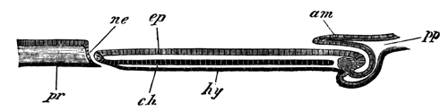
Fig. 1. Diagrammatic longitudinal section of an embryo of Lacerta. pp. body-cavity. am. Amnion. ne. Neurenteric canal. ch. Notochord. hy. Hypoblast. ep. Epiblast. pr. Primitive streak.
The woodcut (fig. 1) represents a diagrammatic longitudinal section through this embryo, and the sections belonging to Series B illustrate the features of the hind end of the embryo and of the primitive streak.
As is shown in fig. 1, the notochord (ch.) has now throughout the region of the embryo become separated from the subjacent hypoblast, and the lateral plates of mesoblast are distinctly divided into somatic and splanchnic layers. The medullary groove is continued as a deepish groove up to the opening of the neurenteric passage, which thus forms a perforation in the floor of the hinder end of the medullary groove (vide Series B, figs. 2, 3, and 4).
The passage itself is somewhat shorter than in the previous stage, and the whole of it is shown in a single section (fig. 4). This section must either have been taken somewhat obliquely, or else the passage have been exceptionally short in this embryo, since in an older embryo it could not all be seen in one section.
The front wall of the passage is continuous with the notochord, which for two sections or so in front remains attached to the hypoblast (figs. 2 and 3). Behind the perforation in the floor of the medullary groove is placed the primitive streak (fig. 5), where all the layers become fused together, as in the earlier stage. Into this part a narrow diverticulum from the end of the medullary groove is continued for a very short distance (vide fig. 5, mc.).
The general features of the stage will best be understood by an examination of the diagrammatic longitudinal section, represented in woodcut, fig. 1. In front is shown the amnion (am.), growing over the head of the embryo. The notochord (ch.) is seen as an independent cord for the greater part of the length of the embryo, but falls into the hypoblast shortly in front of the neurenteric passage. The neurenteric passage is shown at ne., and behind it is shown the primitive streak.
In a still older stage, represented in surface view on Pl. 29, fig. C, the medullary folds have nearly met above, but have not yet united. The features of the passage from the neural groove to the hypoblast are precisely the same in the embryo just described, although the lumen of the passage has become somewhat narrower. There is still a short primitive streak behind the embryo.
The neurenteric passage persists but a very short time after the complete closure of the medullary canal. It is in no way connected with the allantois, as conjectured by Kupffer and Benecke, but the allantois is formed, as I have satisfied myself by longitudinal sections of a later stage, in the manner already described by Dobrynin, Gasser, and Kölliker for the bird and mammal.
The general results of Kupffer's and Benecke's observations, with the modifications introduced by my own observations, are as follows:—After the segmentation and the formation of the embryonic shield (area pellucida) the blastoderm becomes distinctly divided into epiblast and hypoblast[449]. At the hind end of the shield a somewhat triangular primitive streak is formed by the fusion of the epiblast and hypoblast with a number of cells between them, which are probably derived from the lower rows of the segmentation cells. At the front end of the streak a passage arises, open at both extremities, leading obliquely forwards through the epiblast to the space below the hypoblast. The walls of the passage are formed of a layer of columnar cells continuous both with epiblast and hypoblast. In front of the primitive streak the body of the embryo becomes first differentiated by the formation of a medullary plate, and at the same time there grows out from the primitive streak a layer of mesoblast, which spreads out in all directions between the epiblast and hypoblast. In the axis of the embryo the mesoblast plate is stated by Kupffer and Benecke to be continuous across the middle line, but this appears very improbable. In a slightly later stage the medullary plate becomes marked by a shallow groove, and the mesoblast of the embryo is then undoubtedly constituted of two lateral plates, one on each side of the median line. In the median line the notochord arises as a ridge-like thickening of the hypoblast, which becomes very soon quite separated from the hypoblast, except at the hind end, where it is continued into the front wall of the neurenteric passage. It is interesting to notice the remarkable relation of the notochord to the walls of the neurenteric passage. More or less similar relations are also well marked in the case of the goose and the fowl (Gasser)[450], and support the conclusion deducible from the lower forms of vertebrata, that the notochord is essentially hypoblastic.
The passage at the front end of the primitive streak forms the posterior boundary of the medullary plate, though the medullary groove is not at first continued back to it. The anterior wall of this passage connects together the medullary plate and the notochordal ridge of the hypoblast. In the succeeding stages the medullary groove becomes continued back to the opening of the passage, which then becomes enclosed in the medullary folds, and forms a true neurenteric passage. It becomes narrowed as the medullary folds finally unite to form the medullary canal, and eventually disappears.
I conclude this paper with a concise statement of what appears to me the probable nature of the much-disputed organ, the primitive streak, and of the arguments in support of my view.
In a paper on the primitive streak in the Quart. Journ. of Mic. Sci., in 1873 (p. 280) [This edition, p. 45], I made the following statement with reference to this subject:—It is clear, therefore, that the primitive groove must be the rudiment of some ancestral feature.... It is just possible that it is the last trace of that involution of the epiblast by which the hypoblast is formed in most of the lower animals.
At a later period, in July, 1876, after studying the development of Elasmobranch fishes, I enlarged the hypothesis in a review of the first part of Prof. Kölliker's Entwicklungsgeschichte. The following is the passage in which I speak of it[451]:
"In treating of the exact relation of the primitive groove to the formation of the embryo, Professor Kölliker gives it as his view that though the head of the embryo is formed independently of the primitive groove, and only secondarily unites with this, yet that the remainder of the body is without doubt derived from the primitive groove. With this conclusion we cannot agree, and the very descriptions of Professor Kölliker appear to us to demonstrate the untenable nature of his results. We believe that the front end of the primitive groove at first occupies the position eventually filled by about the third pair of protovertebræ, but that as the protovertebræ are successively formed, and the body of the embryo grows in length, the primitive groove is carried further and further back, so as always to be situated immediately behind the embryo. As Professor Kölliker himself has shewn it may still be seen in this position even later than the fortieth hour of incubation.
"Throughout the whole period of its existence it retains a character which at once distinguishes it in sections from the medullary groove.
"Beneath it the epiblast and mesoblast are always fused, though they are always separate elsewhere; this fact, which was originally shewn by ourselves, has been very clearly brought out by Professor Kölliker's observations.
"The features of the primitive groove which throw special light on its meaning are the following:—
"(1) It does not enter directly into the formation of the embryo.
"(2) The epiblast and mesoblast always become fused beneath it.
"(3) It is situated immediately behind the embryo.
"Professor Kölliker does not enter into any speculations as to the meaning of the primitive groove, but the above-mentioned facts appear to us clearly to prove that the primitive groove is a rudimentary structure, the origin of which can only be completely elucidated by a knowledge of the development of the Avian ancestors.
"In comparing the blastoderm of a bird with that of any anamniotic vertebrate, we are met at the threshold of our investigations by a remarkable difference between the two. Whereas in all the lower vertebrates the embryo is situated at the edge of the blastoderm, it is in birds and mammals situated in the centre. This difference of position at once suggests the view that the primitive groove may be in some way connected with the change of position in the blastoderm which the ancestors of birds must have undergone. If we carry our investigations amongst the lower vertebrates a little further, we find that the Elasmobranch embryo occupies at first the normal position at the edge of the blastoderm, but that in the course of development the blastoderm grows round the yolk far more slowly in the region of the embryo than elsewhere. Owing to this, the embryo becomes left in a bay, the two sides of which eventually meet and coalesce in a linear fashion immediately behind the embryo, thus removing the embryo from the edge of the blastoderm and forming behind it a linear streak not unlike the primitive streak. We would suggest the hypothesis that the primitive groove is a rudiment which gives the last indication of a change made by the Avian ancestors in their position in the blastoderm, like that made by Elasmobranch embryos when removed from the edge of the blastoderm and placed in a central situation similar to that of the embryo bird. On this hypothesis the situation of the primitive groove immediately behind the embryo, as well as the fact of its not becoming converted into any embryonic organ would be explained. The central groove might probably also be viewed as the groove naturally left between the coalescing edges of the blastoderm.
Would the fusion of epiblast and mesoblast also receive its explanation on this hypothesis? We are of opinion that it would. At the edge of the blastoderm which represents the blastopore mouth of Amphioxus all the layers become fused together in the anamniotic vertebrates. So that if the primitive groove is in reality a rudiment of the coalesced edges of the blastoderm, we might naturally expect the layers to be fused there, and the difficulty presented by the present condition of the primitive groove would rather be that the hypoblast is not fused with the other layers than that the mesoblast is indissolubly united with the epiblast. The fact that the hypoblast is not fused with the other layers does not appear to us to be fatal to our hypothesis, and in Mammalia, where the primitive and medullary grooves present precisely the same relations as in birds, all three layers are, according to Hensen's account, fused together. This, however, is denied by Kölliker, who states that in Mammals, as in Birds, only the epiblast and mesoblast fuse together. Our hypothesis as to the origin of the primitive groove appears to explain in a fairly satisfactory manner all the peculiarities of this very enigmatical organ; it also relieves us from the necessity of accepting Professor Kölliker's explanation of the development of the mesoblast, though it does not, of course, render that explanation in any way untenable.
At a somewhat later period Rauber arrived at a more or less similar conclusion, which, however, he mixes up with a number of opinions from which I am compelled altogether to dissent[452].
The general correctness of my view, as explained in my second quotation, appears to me completely established by Gasser's beautiful researches on the early development of the chick and goose[453], and by my own observations just recorded on the lizard. While at the same time the parallel between the blastopore of Elasmobranchii and of the Sauropsida, is rendered more complete by the discovery of the neurenteric passage in the latter group, which was first of all made by Gasser.
The following paragraphs contain a detailed attempt to establish the above view by a careful comparison of the primitive streak and its adjuncts in the amniotic vertebrates with the blastopore in Elasmobranchii.
In Elasmobranchii the blastopore consists of the following parts:—(1), a section at the end of the medullary plate, which becomes converted into the neurenteric canal[454]; (2), a section forming what may be called the yolk blastopore, which eventually constitutes a linear streak connecting the embryo with the edge of the blastoderm (vide monograph on Elasmobranch fishes, pp. 281 and 296). In order to establish my hypothesis on the nature of the primitive streak, it is necessary to find the representatives of both these parts in the primitive streak of the amniotic vertebrates. The first section ought to appear as a passage from the neural to the enteric side of the blastoderm at the posterior end of the medullary plate. At its front edge the epiblast and hypoblast should be continuous, as they are at the hind end of the embryo in Elasmobranchii, and, finally, the passage should, on the closure of the medullary groove, become converted into the neurenteric canal. All these conditions are exactly fulfilled by the opening at the front end of the primitive streak of the lizard (vide woodcut, fig. 1, p. 647). In the chick there is at first no such opening, but, as I hope to shew in a future paper, it is replaced by the epiblast and hypoblast falling into one another at the front end of the primitive streak. At a later period, as has been shewn by Gasser[455], there is a distinct rudiment of the neurenteric canal in the chick, and a complete canal in the goose. Finally, in mammals, as has been shewn by Schäffer[456] for the guinea-pig, there is at the front end of the primitive streak a complete continuity between epiblast and hypoblast. The continuity of the epiblast and hypoblast at the hind end of the embryo in the bird and the mammal is a rudiment of the continuity of these layers at the dorsal lip of the blastopore in Elasmobranchii, Amphibia, &c. The second section of the blastopore in Elasmobranchii or yolk blastopore is, I believe, partly represented by the primitive streak. The yolk blastopore in Elasmobranchii is the part of the blastopore belonging to the yolk sac as opposed to that belonging to the embryo, and it is clear that the primitive streak cannot correspond to the whole of this, since the primitive streak is far removed from the edge of the blastoderm long before the yolk is completely enclosed. Leaving this out of consideration the primitive streak, in order that the above comparison may hold good, should satisfy the following conditions:
1. It should connect the embryo with the edge of the blastoderm.
2. It should be constituted as if formed of the fused edges of the blastoderm.
3. The epiblast of it should eventually not form part of the medullary plate of the embryo, but be folded over on to the ventral side.
The first of these conditions is only partially fulfilled, but, considering the rudimentary condition of the whole structure, no great stress can, it seems to me, be laid on this fact.
The second condition seems to me very completely satisfied. Where the two edges of the blastoderm become united we should expect to find a complete fusion of the layers such as takes place in the primitive streak; and the fact that in the primitive streak the hypoblast does not so distinctly coalesce with the mesoblast as the mesoblast with the epiblast cannot be urged as a serious argument against me.
The growth outwards of the mesoblast from the axis of the primitive streak is probably a remnant of the invagination of the hypoblast and mesoblast from the lip of the blastopore in Amphibia, &c.
The groove in the primitive streak may with great plausibility be regarded as the indication of a depression which would naturally be found along the line where the thickened edges of the blastoderm became united.
With reference to the third condition, I will make the following observations. The neurenteric canal, as it is placed at the extreme end of the embryo, must necessarily, with reference to the embryo, be the hindermost section of the blastopore, and therefore the part of the blastopore apparently behind this can only be so owing to the embryo not being folded off from the yolk sac; and as the yolk sac is in reality a specialised part of the ventral wall of the body, the yolk blastopore must also be situated on the ventral side of the embryo.
Kölliker and other distinguished embryologists have believed that the epiblast of the whole of the primitive streak became part of the neural plate. If this view were correct, which is accepted even by Rauber, the hypothesis I am attempting to establish would fall to the ground. I have, however, no doubt that these embryologists are mistaken. The very careful observations of Gasser shew that the part of the primitive streak adjoining the embryo becomes converted into the tail-swelling, and that the posterior part is folded in on the ventral side of the embryo, and, losing its characteristic structure, forms part of the ventral wall of the body. On this point my own observations confirm those of Gasser. In the lizard the early appearance of the neurenteric canal at the front end of the primitive streak clearly shews that here also the primitive streak can take no share in forming the neural plate.
The above considerations appear to me sufficient to establish my hypothesis with reference to the nature of the primitive streak, which has the merit of explaining, not only the structural peculiarities of the primitive streak, but also the otherwise inexplicable position of the embryo of the amniotic vertebrates in the centre of the blastoderm.
DESCRIPTION OF PLATE 29.
Complete List of Reference Letters.
am. Amnion. ch. Notochord. ch´. Notochordal thickening of hypoblast. ep. Epiblast. hy. Hypoblast. m.g. Medullary groove. me.p. Mesoblastic plate. ne. Neurenteric canal (blastopore). pr. Primitive streak.
Series A. Sections through an embryo shortly after the formation of the medullary groove. x 120[457].
Fig. 1. Section through the trunk of the embryo.
Figs. 2-5. Sections through the neurenteric canal.
Fig. B. Surface view of a somewhat older embryo than that from which Series A is taken. x 30.
Series B. Sections through the embryo represented in Fig. B. x 120.
Fig. 1. Section through the trunk of the embryo.
Figs. 2, 3. Sections through the hind end of the medullary groove.
Fig. 4. Section through the neurenteric canal.
Fig. 5. Section through the primitive streak.
Fig. C. Surface view of a somewhat older embryo than that represented in Fig. B. x 30.
XV. On Certain Points in the Anatomy of Peripatus Capensis[458].
The discovery by Mr Moseley[459] of a tracheal system in Peripatus must be reckoned as one of the most interesting results obtained by the naturalists of the Challenger. The discovery clearly proves that the genus Peripatus, which is widely distributed over the globe, is the persisting remnant of what was probably a large group of forms, from which the present tracheate Arthropoda are descended.
The affinities of Peripatus render any further light on its anatomy a matter of some interest; and through the kindness of Mr Moseley I have had an opportunity of making investigations on some well preserved examples of Peripatus capensis, a few of the results of which I propose to lay before the Society.
I shall confine my observations to three organs. (1) The segmental organs, (2) the nervous system, (3) the so-called fat bodies of Mr Moseley.
In all the segments of the body, with the exception of the first two or three postoral ones, there are present glandular bodies, apparently equivalent to the segmental organs of Annelids.
These organs have not completely escaped the attention of previous observers. The anterior of them were noticed by Grube[460], but their relations were not made out. By Saenger[461], as I gather from Leuckart's Bericht for the years 1868-9, these structures were also noticed, and they were interpreted as segmental organs. Their external openings were correctly identified. They are not mentioned by Moseley, and no notice of them is to be found in the text-books. The observations of Grube and Saenger seem, in fact, to have been completely forgotten.
The organs are placed at the bases of the feet in two lateral divisions of the body-cavity shut off from the main central median division of the body-cavity by longitudinal septa of transverse muscles.
Each fully developed organ consists of three parts:
(1) A dilated vesicle opening externally at the base of a foot.
(2) A coiled glandular tube connected with this and subdivided again into several minor divisions.