II. THE SEXUAL EPOCH OF THE MENACME. (continued 8)
Eden Paul translation
Incomplete development of the ovaries, with consequent defective ovulation, may result from marriage in girls who are still 473immature—a fact already known to Aristotle, who wrote, “premature marriage leads to a scanty progeny—that this is the case in man as well as the lower animals is witnessed by the weakly inhabitants of regions in which child-marriage is common.”
It is shown by statistical data that the age at which puberty occurs, the age, that is, at which the menstrual flow begins, has a relation to sterility; and the same is true as regards the age at marriage. In the former connexion, women in whom puberty is comparatively early, are less often sterile than those in whom puberty is comparatively late. Emmet, in an investigation embracing 2330 cases, showed that in our climate the average age at which the first menstruation occurred was 14.23 years, and that in the case of women who subsequently proved fertile, the first flow took place on an average 26 days earlier than in the case of women who subsequently proved barren. We also learn from Emmet’s tables that the mean duration of menstruation and the mean quantity of the flow are larger in fertile than in barren women.
As regards the influence of the age at marriage upon fertility, in women who marry between the ages of 20 and 24 years, sterility is most infrequent; it is commoner in women who marry between the ages of 14 and 20; after the age of 25, the proportion of sterile women increases with each year to which marriage is postponed.
Premature atrophy of the ovaries, with consequent incapacity for ovulation, may occur in a great variety of conditions; it has been observed in scrofula, diabetes, rickets, phthisis, and malarial cachexia; it also occurs in certain chronic intoxications, as from the long-continued use of opium or morphine, and from the abuse of alcoholic beverages. According to the observation of Burkart, Levinstein, and Erlenmeyer, morphinism is a condition which may be relied upon to bring about amenorrhœa and temporary sterility from cessation of ovulation. It has been asserted but by no means proved, that the long-continued administration of quinine hinders ovulation. As a result of various acute and chronic disorders, a simple atrophy of the ovarian follicles can be detected, dependent upon simple fatty degeneration; this has been seen by Grohe in children as a result of general atrophy, and also following caseous and suppurative diseases of the respiratory organs; by Slavjansky in children after chronic pneumonia and chronic dysentery, and in adults as a sequel of typhoid, and in one instance as a sequel of puerperal septicaemia.
Hyperplasia of the ovarian stroma, in slighter degrees of the affection, leads to menstrual disturbances, partly of nervous and partly of inflammatory nature, and in more severe degrees leads to sterility dependent upon the hindrances which the thickened tunica albuginea offers to the bursting of the mature follicles. Klebs believes 474that this anomaly is always due to a disposition acquired very early in life, and perhaps at the time when the ovaries are first developed.
Follicular cysts of the ovary, which are formed mostly at the time of puberty, and originate under the influence of menstrual congestion, from graafian follicles near to ripeness, are competent to cause sterility, owing to the pressure they exercise upon the superficially placed rudimentary follicles, leading to the atrophy of these latter. Other new-growths of the ovaries have similar effects, such as adenomata, carcinomata, dermoid cysts, cystomata, sarcomata, and fibromata. In many cases of these disorders, however, the ovarian follicles may for long periods remain unaffected; and in these instances, ovulation, menstruation, and even conception, may proceed undisturbed. Even in cases in which a neoplasm attains a great size, if it affects one ovary only, ovulation may occur normally in the other, and conception may ensue; and even in the diseased ovary, if small portions of its tissue remain unaffected, ovules may be discharged from these portions. The minutest portion of healthy ovarian tissue, though all the remainder has been destroyed by disease, may suffice to bring about conception.
Ovarian tumours appear with considerable frequency to be complicated with sterility; but in such cases the question always remains open, whether in the majority of instances the sterility is to be regarded as the cause or as the consequence of the ovarian disease. Boinet’s figures dealing with this problem are the most striking of all. He states that of 500 women with ovarian tumours, 390 were childless. But these results are challenged by other observers. Veit’s estimates, based upon a compilation of the figures of Lee, Scanzoni, and West, is that 34% of women with ovarian tumour are sterile. On the other hand, Negroni’s collection of 400 cases of ovarian tumour, including both married and unmarried, contained 43 only who had never been pregnant. Other lists show: 13 sterile women among 45 suffering from ovarian tumour (von Scanzoni); 1 sterile among 21 (Nussbaum); 8 sterile among 63 (Olshausen). Winckel, among 150 sterile married women, found 32 suffering from ovarian tumour, which in two of these cases only was bilateral. Atlee, in 15 cases of ovarian tumour, observed premature cessation of menstruation at the ages of 30, 39, 40 and 42, respectively.
Although in many cases sterility develops coincidently with the growth of an ovarian cystoma, yet in many other women such tumours have no influence in diminishing fertility. Martin in a case in which sterility existed in connexion with a unilateral ovarian cystoma, the other ovary being healthy, observed pregnancy as a sequel of the removal of the diseased ovary. In one of these cases, 475after removal of the ovarian cystoma, Martin punctured in the other ovary a dropsical follicle which had attained nearly the size of a walnut. Pregnancy in this case also followed the resumption of marital intercourse. Müller reports that in his clinique within recent years pregnancy complicated with ovarian tumour has been observed in 7 instances; in one of these cases the pregnancy occurred notwithstanding the fact that the new-growth was so large as almost to fill the abdominal cavity. Holst reports the case of a multipara 43 years of age who died in the 18th to the 20th week of pregnancy; at the post mortem examination the left ovary was found to be transformed into three cysts each the size of an apple, whilst in place of the right ovary was a medullary carcinoma the size of a man’s head; on neither side could a trace of normal ovarian tissue be detected. Spiegelberg, in a woman who died shortly after giving birth to her second child, found that both ovaries were transformed into myxo-sarcomatous tumours; in a woman aged 42, who died four weeks after her eleventh confinement, both ovaries were found to be transformed into nodular carcinomatous tumours each larger than a child’s head; in none of these ovaries was any normal stroma to be found. Ruge reports the case of a woman 36 years of age, who miscarried in the sixth month of pregnancy; she had myxo-sarcoma of both ovaries, one weighing 5620 grammes the other 480 grammes.
All these cases indicate that, notwithstanding the existence of extensive degeneration of both ovaries, some minute remaining fragment of healthy ovarian stroma is competent to produce normal mature ova—a fact which has often been proved also by microscopical examination. That under the influence of pregnancy, existing ovarian tumours often take on extremely rapid growth, is also indicated by some of the above cases.
Castration (oöphorectomy, spaying, Battey’s operation), the removal of both ovaries, naturally results in sterility. If in the literature of the subject cases are to be found in which, after this operation, not menstruation merely, but even pregnancy has occurred, this is to be explained either by the fact that in the stump there was left a fragment of the ovary, still containing tissue capable of producing mature ova; or else by the existence of a supernumerary ovary. Schatz reports the case of a woman in whom pregnancy occurred after double oöphorectomy. In the month of February, 1880, this operation was performed on a girl twenty years of age; she married in April, 1884; and in May, 1885, she was delivered of a mature female infant. The history of the case and the details of the operation showed clearly that the left ovary had been completely removed, with the outermost third of the left Fallopian tube; the right ovary was cut away in such a 476manner that a strip of tissue of at most two millimetres (one twelfth of an inch) in width was left in the body, whilst the right Fallopian tube was left intact. This case teaches us that the smallest remnant of the ovary is competent to render normal pregnancy possible; and further, that a small size of the ovary no more constitutes a hindrance to the proper reception of the ovum in the Fallopian tube, than does an abnormally large size of the ovary, or an unusual shape of this organ.
Miklucho-Mackay relates that among the indigens of Australia the removal of the ovaries is often practised, in order to create a special kind of hetairæ incapable of becoming mothers. McGillivray saw at Cape York a native girl whose ovaries had been removed because she was a congenital deaf-mute, with the object of preventing her giving birth to deaf-mute infants. In the beginning of the last century there existed in Sayn-Wittgenstein a small religious sect whose custom it was always to conclude their religious services by indiscriminate carnal union among the members of the community; when women and girls were first admitted as members of this sect, an attempt was made to render them unfitted for conception “by means of a painful and dangerous compression of the ovaries.” (Ploss.)
A transient, relative hindrance to ovulation may be brought about by various pathological states of the ovaries. Acute oophoritis usually suspends the ovarian functions; chronic oophoritis has sometimes a similar effect, not only because the profound changes that take place in the ovary hinder the formation of the ovules, but also because, as we shall later explain more fully, the expulsion of the ova and their reception by the Fallopian tubes are hindered. In severe oophoritis and perioophoritis, more especially in parenchymatous inflammation, sterility may be brought about by an absorption of the finely granular contents of the follicles, which collapse, with adhesion of their walls; when all or most of the follicles are thus affected, the ovaries become small and hard.
In perioophoritis, the exudation leads to the formation of cord-shaped or ribbon-shaped adhesions between the ovaries and the broad ligaments, the uterus, and the peritoneal folds of the neighbourhood. The ovary in such cases may also be displaced, or may undergo atrophy from pressure.
In the case of 200 sterile women, I found in 46 instances chronic oophoritis and perioophoritis. Olshausen reports that of 12 married women suffering from chronic oophoritis, five were barren, whilst of the remaining 7, three only had given birth to more than one child. Matthews Duncan, on the other hand, saw pregnancy in a case of bilateral ovarian inflammation, in which the organs were considerably enlarged.
477Further, local or general peritonitis may lead to parenchymatous inflammation of the ovaries, and this, spreading from the periphery towards the centre of the organ, attacks the follicles irrespective of their ripeness. Again, during the puerperium, the interstitial form of oophoritis is by no means rare, and this may at times lead to permanent sterility in either of two ways: it may be in consequence of the onset of a secondary parenchymatous inflammation, which destroys all the follicles; it may be because a thick and tough layer of sclerosed tissue forms around the periphery of the ovary, which mechanically prevents the maturation and rupture of the follicles. According to Slavjansky, puerperal disease is the principal cause of this form of oophoritis. Olshausen indicates as the most frequent cause of primary perioophoritis, an inflammation propagated from the Fallopian tubes, leading to the formation of masses of exudation, which envelop the ovary, and by the pressure they cause, and by interfering with the blood-supply, lead to atrophy of the gland.
Sometimes the chronic inflammatory induration by means of which the stroma of the ovary is rendered denser and firmer, is due to changes in the vessels, and depends upon valvular defects of the heart—upon venous congestion. In this way, heart disease may hinder ovulation and bring about sterility. Both syphilis and gonorrhoea may give rise to chronic inflammatory changes in the ovary, usually leading to premature contraction of the tissues and to the formation of numerous adhesions. According to Olshausen, amenorrhœa is not a common feature of ovarian disease, except in cases of defective development of these organs, of cirrhosis of the ovaries, and of bilateral new-growths. Disease affecting only a single ovary, even tumour of considerable size, rarely causes amenorrhœa until profound constitutional disturbance has ensued. An exception to this rule is found in the case of carcinomatous tumours of the ovary; these, indeed, are commonly bilateral; but even when confined to a single ovary, amenorrhœa is a comparatively early symptom. According to the same author, sterility is a common consequence of chronic oophoritis and its sequelae, and is usual also in cases of bilateral new-growths; on the other hand, tumours affecting a single ovary often fail to prevent conception even though they have attained a great size.
Syphilis in women must be regarded as a frequent cause of sterility, by interference with ovulation, but is in this regard by no means an absolute bar to the occurrence of pregnancy. According to Parent and Duchatelet, under whose observation during the space of 12 years there came annually an average number of 2625 syphilitic prostitutes, the average annual of births in these cases was 63 only. According to Marc d’Espine, 2000 prostitutes gave birth on an average to two or three children in all during a year. (That there 478are other causes besides syphilis for the remarkable infertility of women of the town, will be explained later). According to Bednar, Mayr, and others, constitutional syphilis in women invariably leads to sterility; others, as for instance Zeissl, believe that women suffering from inveterate syphilis are commonly, but not invariably, sterile; whilst according to Rosen, conception only takes place in syphilitic women in whom the disease has passed into the tertiary form. Experience shows, however, that neither early nor late forms of syphilis necessarily lead to sterility in women. It must also be pointed out, that syphilis in the male may be the cause of sterility, and must be the cause thereof when the disease is localised in the testicles, and the consequent degeneration of the glandular substance leads to the occurrence of azoospermia, more particularly when syphilitic or gummatous orchitis is bilateral. According to Lewin, we fail to find spermatozoa in 50% of men, otherwise powerful, suffering from syphilitic dyscrasia. Hanc, on the other hand, failed to find azoospermia in any one of ten men suffering from lues. In animals also syphilis is said to cause sterility.
The manner in which certain anomalies of the blood (anæmia and chlorosis), general disturbances of the nervous system, febrile states, and such constitutional disorders as scrofula, have a temporary or permanent influence in checking ovulation, is far from being understood; but the fact that ovulation is checked by such conditions, has been established beyond question by numerous observations. It is well known that severe fevers, more especially typhoid, suspend the ovarian function; that in various chronic disorders of an enfeebling nature, and notably in chlorosis, all signs of menstrual activity disappear; and that in certain nutritive disturbances, as in extreme obesity, amenorrhœa also occurs; finally, numerous cases are on record in which some sudden affection of the nervous system has instantaneously inhibited ovarian activity.
In anæmia and chlorosis, it is probable that the degree of menstrual congestion is insufficient to ensure the bursting of the graafian follicle. The sterility often observed as a sequel of typhoid, malaria, the acute exanthemata, cholera, and septicaemia, is probably due in most cases to the occurrence of parenchymatous oophoritis, with consequent destruction of the ovarian follicles. The researches of Slavjansky have shown that in acute disorders inflammatory changes often occur in the graafian follicles. When infectious disorders ran an acute course, this observer usually found that the parenchymatous inflammation of the ovary had occurred near the periphery, in the cortical layer, the destruction being limited almost exclusively to the primitive follicles; when the course of the primary disorder was more chronic, the mature or nearly mature graafian follicles were the ones destroyed. When inflammation of a follicle has led 479to its destruction, it is replaced by a linear scar. Lebedinsky found similar changes in the ovary after scarlatina—destruction of a lesser or greater number of follicles, with formation of scars. Thus, parenchymatous oophoritis as a sequel of acute diseases, may, if severe, lead to destruction of all the rudimentary follicles, with consequent sterility. In the post mortem examination of such cases, the condition of the ovaries is similar to that which is elsewhere in this work described as characteristic of these organs after the menopause: the ovary is diminished in size, its surface is furrowed, the tissue is indurated in consequence of overgrowth of fibroid tissue; often not a single follicle is to be detected on section of the organ.
Immoderate obesity is a disorder of nutrition favoring the occurrence of sterility.
In very obese women of an age which normally is the reproductive prime, amenorrhœa or scanty menstruation is a very common accompaniment. In 215 such cases which came under my own observation, amenorrhœa was present in 49, and menstruation was scanty in 116; thus in nearly three fourths of these obese women menstruation was either deficient or entirely wanting. Very remarkable also is the high percentage of sterile women among the obese. In the 215 cases already mentioned (all married women), 48 were sterile—a percentage of 21. Whilst the ordinary ratio of barren to fruitful marriages is 1 : 10 or 1 : 9, in the cases in which the wives, or both wives and husbands, are extremely obese, the ratio is according to my own observations, 1 : 5—or, if we include cases of only-child-sterility, 1 : 4.
We cannot wonder at this great frequency of sterility in obese women when we remember that, apart from the menstrual deficiencies which so commonly accompany this disorder of nutrition, obesity is apt to entail many other disorders of the reproductive organs, as for instance a morbid state of the uterine and vaginal secretions, chronic metritis, and displacements of the uterus; still, it cannot be denied, that in many instances we are unable in such obese women to detect any disorder of the reproductive organs competent to account for the sterility, and we must therefore assume that the excessive development of fat has some direct influence in preventing ovulation, or at least that it in some way exercises an unfavourable influence upon the reproductive process.
That excessive obesity hinders fertility, is shown by experience both as regards the vegetable and the animal kingdom. All animal-breeders are familiar with the fact that undue production of fat limits fertility. Thus, equally in the case of turkeys and in the case of the common fowl, if the hens are overfed and become fat, they cease to lay.
480Hippocrates already indicated obesity as a cause of sterility. Writing of the wives of the Scythians, he pointed out as a proof that their excessive obesity was the cause of the sterility from which they commonly suffered, the fact that their female slaves, who were thin, were readily impregnated by intercourse with the Scythian males. The oft repeated dwindling and disappearance of ruling families in India and in Egypt, has doubtless in part depended upon the extreme obesity of the female consorts of such rulers.
In many instances, indeed, a great accumulation of fat on the front of the abdomen and in the vulva, suffices to cause a simply mechanical hindrance to the proper performance of a fertilizing coitus. It is possible also that the phlegmatic temperament of very fat women is a contributory cause to their sterility—if indeed it is in general true that frigidity during sexual intercourse is unfavourable to conception, as is expressed by the old proverb, quo salacior mulier, eo foecundior. It is unquestionable that in very obese women sexual sensibility is commonly greatly deficient, and that their husbands often complain of their coldness and lack of passion. In several cases that have come under my observation, dyspareunia occurred in obese and sterile women.
The dependence of sterility upon obesity is often proved in the most striking manner ex juvantibus. A “cure” for the reduction of fat often results favourably in respect also of rendering the woman who undergoes it readily impregnable—a result by no means ardently desired.
It must also be pointed out that very obese women form a considerable section of those suffering from only-child sterility, and this largely in consequence of their strong predisposition towards abortion. As the impregnated uterus enlarges, the space for its accommodation is insufficient, owing to the great development of the panniculus adiposus, and thus obesity, like intra-abdominal tumour, predisposes to abortion. The excessive accumulation of fat within the abdomen, by exercising pressure upon the inferior vena cava or on its principal tributaries, hinders the venous return, and gives rise to a chronic stasis in the uterine bloodvessels, those alike of the muscle and of the mucous membrane.
Notwithstanding the fact that sterility is so common in very obese women, the fact remains that some such women are remarkably fertile, and have very large families indeed.
Towers-Smith, Duke, and Rodriguez, who have recently all been engaged in examining the relations between obesity and sterility, agree in asserting that sterility due to obesity may be cured by dietetic treatment for the relief of the primary disorder of metabolism.
Though menstruation is usually deficient or absent in obese sterile 481women, and though it is commonly supposed that amenorrhœa implies sterility, it is necessary to point out that whilst failure of menstruation is a frequent and important sign of suppression of ovulation, it by no means invariably has this significance. It is an established fact, and one borne out by my personal experience, that women who have never menstruated have nevertheless become pregnant; others, again, have become pregnant although they have ceased to menstruate for several years, and this has even occurred in women at a comparatively advanced age. Hence, from the fact that amenorrhœa exists, we cannot with certainty infer that a woman is sterile. Moreover, we must remember that physiologically amenorrhoeic women often enough conceive—during lactation. Although we hold the opinion that there is an intimate connexion between ovulation and menstruation, yet it is always possible in cases in which menstruation fails to occur, that ovulation has taken place, but that the stimulus which that process has exercised upon the reproductive organs has been insufficient to give rise to the customary flow of blood.
The following remarkable case came under my own observation: Mrs. B., 26 years of age, had lived in sterile wedlock for six years, had never menstruated, nor had she ever had any sanguineous discharge from the genitals. The body was delicately formed, the breasts were fairly well developed, the external genital organs showed no abnormality. For some weeks before consulting me, this woman, hitherto childless, and living in regular sexual intercourse with her husband, had noticed a remarkable enlargement of the abdomen. Another medical man whom she had consulted had diagnosed ovarian tumor and had urged operation. A more careful examination of the pelvis showed, however, that the woman was in the sixth month of pregnancy, a diagnosis which was duly confirmed by the delivery of a full-time child. In another of my cases, a woman married at the age of 45 years, having ceased to menstruate two years previously. She became pregnant and gave birth to a child in quite normal fashion. The following instructive case also came under my own observation: The wife of one of my colleagues, living in sterile wedlock for 17 years, extremely obese, had since puberty menstruated but scantily and with great irregularity. The menstrual interval was several months, and when the discharge did appear, it was pale in colour and small in quantity; it lasted moreover but a day or two. Last winter, the flow as usual failed to appear for several months, and since the woman had at the same time become fatter than ever, Turkish baths and energetic muscular movements were prescribed. The result of this treatment was a striking one—abortion. After 17 years of marital intercourse she had for the first time become pregnant.
482In the case of sterile women who are amenorrhoeic, even when the amenorrhœa has never been interrupted by a menstrual discharge, or when it appears entirely dependent upon obesity, it is nevertheless necessary to be extremely cautious in making a diagnosis, and above all in employing an intra-uterine sound. In such cases I have known the most eminent gynecologists unwittingly bring about abortion.
Cleveland, Godefroy, Haschek, Ritschie, Sommerus, Stark, Taylor, and Young, have all reported cases in which pregnancy occurred in women suffering from amenorrhœa; but all such cases must be regarded as quite exceptional. Szukits examined 8000 sexually mature women, and found among them fourteen only who had never menstruated. Of these, four were multiparae.
Saint Moulin reports the case of a woman 24 years of age who had never menstruated, but who none the less became pregnant and gave birth to a fine girl. One of the most striking cases of this nature is the one reported by Rodzewitsch, regarding a woman who first began to menstruate at the age of 36 years. This woman had however been married when fifteen years of age, and in the subsequent twenty-one years she gave birth to 15 children, remaining the whole time amenorrhoeic.
Puech reports the case of a woman who ceased to menstruate at the age of 40 years, and remained amenorrhoeic for the subsequent six years. Then menstruation recurred for a year, and finally ceased definitively in consequence of the occurrence of pregnancy, which terminated in the normal birth of a healthy boy. Loewy, in a woman 31 years of age, who had previously been amenorrhoeic all her life, saw menstruation appear for the first time shortly after the birth of her sixth child. Ahlfeld had under observation the case of a woman who was the mother of eight children, and had never menstruated.
Krieger reports the case observed by Mayer, of the wife of an artizan, who between the ages of 17 and 28 years had given birth to five children, and had had one abortion. After the age of 22, she had no trace of menstrual discharge, but notwithstanding this, she subsequently gave birth to three children. Krieger himself saw a woman who had had her last child at the age of 33, and in whom now, at the age of 48, menstruation had just ceased entirely. Two years later, irregular menstrual discharges recommenced; when these ceased, it appeared that the woman was once more pregnant, and she was normally delivered of a full-time girl.
Renaudin relates the case of a lady 60 years of age who gave birth to a child, menstruation having ceased 12 years earlier. Deshhayes saw the delivery of a woman 50 years of age, two years subsequent to the occurrence of a normal menopause. Capwron, 483quotes the ease of a woman who became pregnant at the age of 65 years. In this case menstruation had recurred, having ceased many years before in a normal menopause. This woman aborted at three months, and the foetus was well-formed.
In such cases of late conception, which occur after the normal cessation of menstrual activity, we cannot be certain whether we have to do with a simple persistence of ovarian activity, associated with temporary or permanent cessation of menstruation; or whether both functions, ovulation and menstruation, had ceased, and were aroused to renewed activity by some determinate cause. It is possible that in coitus we have such a stimulus, capable of reawakening the slumbering ovarian functions. That this may be the case, we are led to suppose by the fact that pregnancy at an unusually advanced age most frequently occurs as a result of marriage late in life. In Scandinavian countries, where the difficulties of providing for a family are so great that a very large number of marriages are inevitably postponed till comparatively late in life, the number of pregnancies occurring in elderly women is correspondingly large. However, pregnancy late in life occurs also in women who have married early, and the most probable assumption to account for such cases is that ovulation has occurred in the absence of menstruation.
Although by these cases the proposition is established that amenorrhœa is by no means equivalent to incapacity for ovulation, still, the former must indubitably be regarded as in general a most important indication of disturbed ovulation. When a woman attains the age of 20 years without having ever menstruated, or even having experienced menstrual molimina, we may in the great majority of such cases infer with justice that there is complete or partial failure of development of the ovaries and the reproductive apparatus generally. In some of these cases, examination discloses the fact that the uterus is in an infantile condition. When we are able to bring about the regular establishment of menstruation, we may hope also to remove the sterility dependent upon the defective ovarian functional capacity. General tonic treatment for the relief of chlorotic amenorrhœa quite as often, in the case of previously barren married women, results in the occurrence of pregnancy, as happens in cases of amenorrhœa and sterility due to obesity, when this latter condition has been relieved and menstruation has been re-established by suitable dietetic treatment. Much less often is it possible to relieve the sterility of scrofulous (tuberculous) persons, for in the majority of such cases, in consequence of the scrofulous (tuberculous) constitutional disorders, pathological changes have occurred in the ovaries already in early youth, and these it is difficult or more often impossible to remove.
484Scrofula (tuberculosis) is, according to my own experience, the constitutional disorder which of all most frequently and most seriously affects ovulation; and it appears that the ovaries are subject to changes produced by this disease similar to those which occur in other glandular organs. In cases in which no cause of the existing sterility is ascertainable, the presence of scars due to scrofulous (tuberculous) changes in the lymphatic glands may serve as an indicator to show that the capacity for ovulation has been annihilated or seriously diminished in early life by scrofulous (tuberculosis) disease.
Among the causes of sterility, these three conditions: anæmia, chlorosis, and scrofula (tuberculosis), play a leading part; indeed, their importance in this connexion has hitherto been underestimated, more especially in regard to the comparative frequency with which they cause sterility. A large part of the favourable influence in the relief of sterility in women which is exercised by the “cures” at various watering places, depends upon the amelioration which is thus effected in the aforesaid constitutional disorders.
It has been assumed that diabetes, which renders men impotent, is competent also to cause sterility in women. Hofmeier reports a case which appears decisive on this point. In a woman 20 years of age, who had menstruated regularly since she was 14 until a year previously, when the flow had ceased, he found the uterus extremely small, barely 5 cm. (2 in.) in length, extremely atrophied, the ovaries also atrophied and very small; the urine contained large quantities of sugar. Here was doubtless a case of atrophy of the reproductive organs secondary to diabetes.
In England, where the excessive use of alcohol is observed very frequently in women as well as in men, sterility has frequently been regarded as a result of chronic alcoholism. Matthews Duncan reports cases which lead to the belief that alcohol has a specifically deleterious effect upon fertility. Apart from the general or constitutional disturbances dependent upon the abuse of alcohol, this agent has in many cases a well-recognized pathogenic influence upon the female reproductive organs, the morbid condition which is most frequently and most readily assignable to this cause being chronic oophoritis. The obesity which so frequently results from alcoholic excess is a contributory cause of sterility.
Certain drugs, more especially quinine and morphine, are reputed to cause sterility. Davies, reviving an old opinion, considers that of all drugs tannin is the most effective in leading to sterility, and he considers tea-drinking as responsible for this effect.
The influence of certain cerebral affections and psychical disorders in checking ovulation has been established. Thus, de Montyel has recently shown that in families subject to hereditary mental disorders, 485there is an unusually large proportion (1 : 7) of barren marriages.
In addition, there are many influences which are known to prevent or to diminish ovulation in the case of the lower animals, and which may therefore be assumed with considerable probability to have a similar effect in women. More especially we are here concerned with external influences affecting unfavourably nutrition and innervation, and therewith also ovulation; also near kinship between the parties to the act of intercourse; and finally hereditary predisposition. In animals, captivity, exposure to cold, over-exertion, insufficient or unsuitable food, and inbreeding, have been proved to result in infertility.
Doubleday asserted that “a too abundant supply of nutriment hinders reproduction, whereas on the other hand insufficient or improper food favours reproductive activity and increases the number of the offspring.” Spencer, however, rightly points out that the infertility noticed in these circumstances is not the direct result of prosperity, but depends upon the pathological obesity which is thus engendered by overfeeding.
No less interesting are the observations that have been made regarding sterility in animals in confinement. In such animals there are wide differences. Some refuse to cohabit, or have lost sexual desire; others, again, show excessive sexual desire and cohabit too often, without any result; or even if fertilization occurs, abortion often ensues. In yet other cases, though conception follows intercourse, and the animals go on to full term before delivery, the young are still-born, or are weakly and misshapen. Caged birds often lay no eggs at all or very few; or if they do lay, they neglect their eggs; or if incubated, the eggs fail to hatch out. In France, experiments regarding this matter were made with domestic fowls. If the hens were given great freedom, 20 per cent only of the eggs remained unhatched; with less freedom, 40 per cent of the eggs were failures; whilst if the fowls were kept in a coop, 60 per cent of the eggs were unhatched.
“Convincing proofs,” writes Darwin, “have been obtained to the effect that wild animals which have recently lost their freedom have their fertility diminished to a most remarkable extent. This infertility is not dependent upon any degeneration of the reproductive organs. There are many animals of the most diverse species, which, whilst they copulate freely in confinement, fail in these circumstances to conceive; others again, even if they conceive and have living young, give birth to these in numbers which are unquestionably much smaller than would be the case were the parents in the free state.”
Interesting observations have been made by pigeon breeders. 486They state that when pigeons brought up in the same nest pair, the number of their offspring is usually very small.
The influence upon fertility of unfavourable conditions of temperature, either excessive heat or excessive cold, is very great. In the case of pigeons, for instance, if the pigeon cot is adjacent to the heated wall of a dwelling house, the pigeons sometimes begin to lay as early as January, and may have young as often as eight times in a single year. When the dovecot is cold, on the other hand, the number of broods is smaller. In general, the procreative capacity is greater in summer than in winter.
As regards inbreeding, many facts are on record showing the influence of this practice in leading to the birth of malformed offspring and to sterility. Darwin writes, “if in a pure race, characterized by a certain tendency to sterility, we allowed only brothers and sisters to pair, in a few generations the stock would become extinct.” If animals closely related by blood pair, the number of their offspring is always less than the average.
In the case of the human species, however, the influence of the marriage of near kin in diminishing fertility cannot be regarded as definitely proved.
Occasionally the incapacity for ovulation and the sterility dependent thereupon are hereditary—paradoxical as this may appear. It is necessary to assume, that just as the sperm is at times unsuited for effective fertilization, so also the ova may be in a less or greater degree insusceptible of fertilization. In the present state of our knowledge, indeed, we are not in a position to be precise as to the exact nature of such incapacity. It is possible that the enveloping membrane of the ovum varies in its resistance to penetration, as Schenk claims to have proved in respect of certain of the lower mammals. In his experiments on artificial fertilization outside the body of the mother, he ascertained that the cells derived from the discus proligerus, surrounding the ovum in immediate contact with the zona pellucida, are in some instances easily separable one from another, so that the spermatozoa can readily obtain access to the zona pellucida; whereas in other instances, in which the ovum is of the same size and apparently in the same stage of maturation as before, these cells remain closely attached each to the other, and thus prevent the passage of the spermatozoa. This condition of the ovum, so unfavourable to fertilization, may be hereditary in certain families, and its transmission may render certain members of the stock infertile. Such instances as the following from my own practice are by no means rare. Of three sisters, whose family life was intimately known to me, one had one child only, a girl, whilst the two others remained childless. The girl of the second generation married and remained childless. In England it is well established 487that when, in cases of only-child-sterility, the offspring is of the female sex, this child will probably herself be barren. Galton found that in the case of 14 heiresses (i. e. the only children of wealthy parents), all of whom were married, 8 remained absolutely barren, whilst of the others, 2 had each an only child.
It was formerly believed that when a woman gave birth to twins of opposed sexes, the female infant would prove to be barren, this barrenness being associated with defective development of her reproductive apparatus. John Hunter (Animal Economy) ascertained that in the case of twin calves of opposed sex, the genital organs of the female twin were almost invariably imperfectly developed. But the supposition that this is true also of the human species has not been confirmed by experience. I know several married women who had twin brothers, and these women have borne normal children; however, the number of their offspring is remarkably small. Simpson, in Edinburgh, recorded the results of the marriage of 113 women who had been born with twin brothers; of these, 103 had proved fruitful, and 10 (i. e., about one eleventh of the whole) barren, although of these latter women, one had been married upwards of 5 years, and the remaining 9 for periods ranging from 10 to 40 years. Simpson also gave the history of four women who were all the fruit of triple births, some of which had consisted of two boys and one girl, others of two girls and one boy. All four of these women were parous. Again, a woman who had been one of a quadruple birth (three boys and one girl), herself gave birth to triplets. A collection of all the figures accessible to me relating to this subject, indicates that about ten per cent of the women born in such circumstances prove barren—a ratio which corresponds closely with the ratio of infertility in general.
Interference with Conjugation, Conditions Preventing Access of the Spermatozoa to the Ovum.
A condition essential to fertilization is a material union between the sexual products of the male and the female respectively—the act of conjugation. Thus, all conditions which prevent the spermatozoa from obtaining access to the ova, bring about sterility.
Spermatozoon and ovum being normal, a great variety of pathological conditions may prevent the one from gaining access to the other. It is necessary for fertilization that the mature ovum should leave the ovary, enter the Fallopian tube, and there come into contact with the male sperm. Interference with any one of these essentials may lead to sterility.
Thus, the constitution of the ovum itself may be at fault; or the entrance of the ovum into the Fallopian tube may not be normally effected; defects in these earliest stages of the process of fertilization 488are precisely the commonest and the most important. The emergence of the ovum from the graafian follicle may be rendered difficult or entirely prevented by pathological states of the ovary; again, by inflammatory processes in the ovary, the tubes, or the ligaments, by developmental defects in the tube, and by obstructions in its interior, the entrance of the ovum into the tube, and its free passage along the tube may be prevented. Numerous abnormalities and diseases of the uterus may on the one hand prevent the entrance of the ovum into the uterine cavity, and on the other may prevent the upward passage of the spermatozoa to their goal. Amongst conditions competent to produce these effects we must enumerate: displacements of the uterus, structural changes in this organ and its annexa, and other congenital defects and acquired states; more particularly must be mentioned, uterus infantilis, acquired atrophy of the uterus, flexions and versions of the uterus, new-growths and inflammatory states of that organ, abnormalities in the shape or size of the cervix uteri, and, finally, all conditions of the vagina or vulva which hinder the proper performance of the act of intercourse.
In diagnosing the cause of sterility, in determining whether in any particular instance it is due to some hindrance to the indispensable conjugation between the male and female reproductive elements, we have in the first place to ascertain the presence or absence of any of the numerous conditions which interfere with the proper passage of the ovum from the ovary through the Fallopian tube to the interior of the uterus. The simpler mechanical hindrances to conception, such as displacements of the uterus, or tumours of that organ or its annexa, are easily recognized; and the same is true of atresia of the cervix uteri, and of congenital or acquired stenosis of the vagina. When obliteration or stricture of the genital tract exists, a very careful examination, visual, digital, and instrumental, must be made, rectal examination not being forgotten. Not infrequently, amenorrhœa is attributed to ovarian disease, and only subsequently on local examination is the cause ascertained to be hymeneal atresia, with haematocolpos; many a woman has believed herself to be pregnant, until examination has disclosed the fact that the hymen is still intact, and that coitus has hitherto been effected through the urethra. The importance of these stenotic conditions as causes of sterility must not, however, be overestimated, for, although they are common among the hindrances to conception, the obstacle is by no means always insuperable.
Morbid changes in the secretions of the genital passages, whereby the vitality of the spermatozoa may be destroyed before they have time to reach the ovum and effect fertilization, are hard to diagnose, for the conditions upon which such changes depend have not as yet been adequately investigated.
489
Diseases of the Ovaries and the Fallopian Tubes.
Among the conditions which, although the maturation of the ovum proceeds normally to a conclusion, may prevent conjugation between the male and female elements, we must in the first place consider an abnormal condition of the tunica albuginea of the ovary, a thickening of this membrane in consequence of inflammatory processes or of new formation of connective tissue, whereby the dehiscence of the follicle is rendered difficult or entirely prevented. Such thickenings of the ovarian envelope are the residue of perioophoritic processes.
Such a hindrance to conception may be permanent or transient, and thus the sterility dependent thereupon may be relative or absolute. Similar is the effect of inflammatory processes affecting the peritoneal investment of the uterus, the broad ligaments, and the peritoneum clothing the floor of the pelvis; these conditions, perimetritis, perisalpingitis, and pelvic peritonitis, resulting in the formation of thick and extensive pseudomembranous bands, or in less severe cases leaving merely slight adhesions and filaments, which drag the uterus and the ovaries out of place, and thus render conception difficult or impossible.
Perimetritic adhesions are apt to lead to dislocation of the tubes either forwards or backwards, and most commonly into the pouch of Douglas, thus giving rise to sterility. Rokitansky and Virchow already insisted on the great importance of perimetritic processes in causing sterility.
That congenital defects of the Fallopian tubes may lead to sterility, is indeed a possible, but certainly a rare occurrence. The defect may be unilateral or bilateral; or it may be that merely a portion of one tube may be wanting. Bilateral absence of the Fallopian tubes is usually associated with defective development of the uterus, while the ovaries may be apparently normal. Such a case is described by Foerster and Kussmaul. The vagina opened into the urethra, the uterus was not calibrated, and diverged above into two solid horns, to which the round ligaments and the ovaries were attached. A congenital cause of sterility is to be found also in atresia of the tubes, the abdominal extremities of which are closed; this condition is met with also in other mammals. It is also assumed, with less accuracy, that a supernumerary ostium tubae may lead to sterility, in consequence of the ovum, which has found its way into the normal ostium, returning into the abdominal cavity through the supernumerary orifice. An unfavourable influence upon fertility is exercised also by a form of hyperplasia of the tubes which sometimes arises in consequence of erroneous development at the time of puberty; the tubes, increasing unduly in length, become serpentine 490in form instead of being nearly straight; this tends to lead to accumulation of the secretions, and renders the passage of the ovum difficult. (Freund.) Yet another defect of development which, as Klebs has pointed out, may lead to sterility, is absence of the fimbria which normally retains the abdominal orifice of the Fallopian tube in proximity with the ovary, in which case these structures may be separated by a wide interval.
The entry of the ovum into the tube may thus be rendered difficult by abnormalities of the abdominal orifice of the tube or of the fimbriae; but still more is this the case when the mucous membrane of the tube is diseased. The fringed border of the tubal orifice has a distinct tendency to independent disease. As Klebs’s anatomicopathological studies have shown, inflammatory changes are common in this region, leading to contraction. The free margin of the tube then appears to be strictured by overgrowth of fibrous tissue on the serous surface, the opening being thus narrowed or even entirely closed, whilst the fimbriae themselves may be drawn within the aperture. In other cases, the ring of fimbriae is adherent to some neighbouring part, especially to the ovary itself, when this also is diseased. Further, on the fringed margin of the tube we see papillary growths, telangiectases, or oedema with formation of cystic cavities.
In the interior of the tubes also, pathological processes occur, catarrhal inflammations, haemorrhagic or purulent exudations, sealing up the passage completely. In some cases these exudations lead to great distension and even to rupture of the tube. Thus, among the causes of sterility must be enumerated: simple catarrh of the tube, with swelling of the mucous membrane; purulent catarrh, leading to its distension with pus—pyosalpinx; serous effusion into the tube, hydrosalpinx; and haemorrhagic effusion, haematosalpinx; further, that peculiar form of tubal inflammation, described by Chiari and Schauta under the name of salpingitis isthmica nodosa, in which hyperplasia of the muscular coat of the tube occurs at irregular intervals, so that it appears to be beset with nodes. Special mention must also be made of gonorrhoeal salpingitis, which will subsequently be described in detail.
Inflammatory states of the tube may hinder conception, either mechanically, by swelling of the mucous membrane, or by obstruction of the lumen of the tube by exudations, by injury or destruction of the ciliated epithelium, by lesion of the musculature of the tube, affecting its peristaltic movements—all these hindering or entirely preventing the passage of the ovum downwards or of the spermatozoa upwards; or, again, chemically, by the deleterious influence of many of the morbid secretions that are formed in these conditions upon the vitality of ova or spermatozoa. These inflammatory 491states of the tubes may also lead to stricture or obliteration of their abdominal extremities, or to displacement of the ostia, and thus lead to sterility; in other cases these same conditions, leading to distortion and displacement of the tube, may prevent the downward passage of the ovum while leaving possible the upward passage of the spermatozoa, and thus give rise to tubal gestation—a condition which we shall not now consider.
It must not be forgotten that tuberculosis of the genital canal attacks the tubes with especial frequency; in these organs we may find miliary tubercles, and more commonly diffuse caseous masses, completely filling the lumen of the canal. Finally we have to mention the diverse forms of saccular dilatation of the tubes (Ger. “Tubensäcke”), all of which possess the common pathological characteristics of enlargement of the tubes and their conversion into saccular cavities; the contents of these distended tubes may, however, be extremely various, and such conditions may depend upon manifold mechanical disturbances and inflammatory processes of the uterus and its annexa.
When we consider how common, during the sexual life of women, are perioophoritic inflammations, more or less intense, but often without severe symptoms (and hence apt to be overlooked); when we remember that the very process of ovulation and also the puerperal state furnish opportunities for slight or severe pelvic peritonitis to arise; and when we further take into account the frequency and importance of gonorrhoeal pelvic peritonitis—we cannot fail to admit that the results of these morbid conditions, such as adhesions between the ovary and the ostium tubae, or closure of the tube with consequent hydro- or pyosalpinx, must be reckoned among the principal causes of sterility. If the frequency and importance of these conditions is still underestimated, two reasons can be adduced for this: first, that the slighter degrees of intrapelvic inflammation often, as previously mentioned, elude diagnosis; and, secondly, that even when the treatment is expectant merely, the exudations are frequently absorbed, the adhesions give way, and the capacity for conception is gradually fully restored.
When considering the etiology of acquired sterility, especial attention must be devoted to gonorrhoeal pyosalpinx, the most important and the most dangerous of the morbid manifestations of gonorrhoeal infection in the female. Gonorrhoeal salpingitis and perisalpingitis are very serious affections, in the first place because they are apt to give rise to oophoritis and perioophoritis, as well as to pelvic peritonitis, and other local inflammatory states. The minuteness of the uterine orifice of the Fallopian tube, and the downward direction of the ciliary movement in the interior of the tube, combine to safeguard against the entrance of gonococci, but none the less they too 492often find their way up the tube, and small quantities of gonorrhoeal pus enter the pelvic cavity and give rise to inflammations, in which the ovary partakes.
According to Saenger, this gonorrhoeal disease of the uterine annexa is found with especial frequency in women either wholly sterile or affected with only-child-sterility, and is to be regarded as the cause of their infertility; “infertility is indeed the rule, fertility the exception, in all cases in which gonorrhoeal disease has passed upwards beyond the os uteri externum.” The same author maintains that, putting aside tuberculosis and actinomycosis, if, in a case of infective inflammation of the uterine annexa, septic infection can be excluded, and more especially when the disease affects both tubes, when it is reluctant to yield to treatment, and when relapses are frequent, we have no option but to believe that the affection is of gonorrhoeal origin.
In 155 cases of chronic inflammatory disease of the Fallopian tubes, von Rosthorn was able in 37 instances to prove that the affection was the direct result of gonorrhoeal infection.
Recently, however, Noble has published cases which lead us to believe that even pyosalpinx does not necessarily prevent the occurrence of pregnancy. In operating for the relief of a unilateral pyosalpinx, the uterus was opened, and a seven months’ foetus was removed. In another case, the autopsy on a woman who had succumbed to severe peritonitis arising immediately post partum, disclosed a large pyosalpinx.
Closure of the ostium may also be brought about by chronic metritis and endometritis, by chronic catarrhal states of the uterine mucous membrane, and in general by pathological changes in that membrane associated with local hyperaemia or abnormal secretions. In some cases, salpingitis with consequent sterility is the result of puerperal infection; and such a sequence of events is especially common after an abortion followed by retroflexion of the uterus, leading to elongation and kinking of the tubes.
An important hindrance to the entry of the ovum into the uterus is sometimes offered by uterine polypi or myomata; growing from the fundus, these may so fill the uterine cavity that the uterine orifices of the tubes appear to be completely occluded.
At times, also, quite small myomata, growing close to the tubes, may push these latter upwards, closing them, and thus giving rise to sterility; such myomata may also lead to saccular dilatation of the tubes, as occurred in the following case:
Mrs. S., aged 39 years, had one child when 20 years of age, but since then had been barren. For several years she had suffered from profuse menorrhagia. Owing to the enormous thickening of the abdominal wall, bimanual examination of the uterus was impossible; 493the vagina was relaxed, enlarged, and contained an excess of mucous secretion. The uterus was high up in the pelvis, anteverted, enlarged, movable, sensitive to pressure; the portio vaginalis was enlarged, soft, and excoriated; no tumour could be detected either in the uterus or in the uterine annexa. The menstrual flow recurred at intervals of from two to three weeks, lasting from one to two weeks, and being extremely profuse; menstruation was painful. Whilst the patient was under my observation an excessive menstrual haemorrhage came on quite suddenly, with slight rise of evening temperature (38.2° C.—100.8° F.), but severe general disturbance; there were paroxysms of intense abdominal pain, violent vomiting of greenish bilious masses, which after a time became haemorrhagic, the abdomen was tense and sensitive to pressure, there was cardiac weakness with general failure of strength; treatment proved unavailing, and the patient died in collapse on the third day. The autopsy disclosed: fibroma uteri submucosum, parietale, et subserosum, haematosalpinx dextra, pyosalpinx sinistra, peritonitis. The subserous myoma, of about the size of a pea, was in the middle of the fundus uteri; the submucous myoma, of about the size of a chestnut, filling the uterine cavity, sprang from the posterior wall of the body of the uterus; the intramural myoma, of about the size of a bean, was in the right wall of the corpus uteri. Both tubes were greatly elongated, exhibiting serpentine windings. The right tube was much distended, filled with sanguineous fluid; the left, partially collapsed, contained greyish-green purulent material, having an extremely offensive odour; some of this fluid had flowed through the ostium abdominale into the abdominal cavity. Death in this case ensued with great rapidity in consequence of rupture of the pyosalpinx, and evacuation of its contents into the abdominal cavity.
Cystic formations in the round ligament (hydrocele of the round ligament) sometimes lead to sterility. In the form of elongated tumours of about the size of a hen’s egg they may fill the inguinal canal, and even pass forwards into the labia majora. When as large as this, they demand operative interference. Hennig records a case in which such hydrocele of the round ligament was the cause of sterility lasting 14 years, the woman becoming pregnant after the tumour had been removed by operation. Similarly, infertility may depend upon solid tumours of the round ligaments—myomata, fibromyomata, or sarcomata.
Retro-uterine haematocele often gives rise to sterility. As a rule, prior to the formation of a blood-tumour in the pouch of Douglas, various menstrual disturbances occur, more especially menorrhagia; or it may be preceded by some puerperal disease, especially perimetritis, which by itself, indeed, seriously limits the fertility of the woman thus affected; but when haematocele is superadded, her child-bearing 494capacity is much more gravely impaired, owing to the permanent displacement of the uterus, to the perimetritic exudations, to the adhesions formed around the ovary, and to stricture or occlusion of the tubes. Still, sterility is by no means an inevitable consequence of haematocele.
By many it is assumed that in cases in which the tubes are perfectly normal, disturbances of innervation are competent to cause sterility (or tubal gestation). It is supposed that nervous influences affect the functions of the Fallopian tubes by leading to spastic contractures of the circular muscular fibres of these structures, or in other cases to paralysis; in this way nervous disorder may lead to the retention within the tube of the unfertilized (or already fertilized) ovum.
Diseases of the Uterus.
Pathological changes in the uterus may in various ways lead to sterility dependent upon prevention of conjugation (physical contact of the male and female reproductive elements). Thus, the incapacity for fertilization may, on the one hand, depend on hindrances to the passage of the ovum from the tube to the interior of the uterus; or on the other, on some abnormal condition of the vaginal portion of the cervix, whereby the passage of the spermatozoa from the vagina into the uterus is prevented; or, finally, upon displacements of the uterus or pathological structural changes in that organ, whereby the implantation of the fertilized ovum in the uterine cavity and its development therein are impeded.
The uterus may be entirely absent, but this is an extremely rare condition; much less infrequent is a rudimentary condition of that organ. In the latter case, it is either represented by a nodular rudiment, or else it is conical or bicorned; whatever its shape, it is a solid mass of muscular and connective tissue. In association with absence or a rudimentary condition of the uterus, the vagina also may be wanting, or may be represented merely by a small, blind pouch; the Fallopian tubes may in such cases either be normally developed or rudimentary. The number of instances of this kind that have been observed is very large (Kussmaul, Klebs, Cusco, Klinkosch-Hill, Cruise, Freund, Fürst, Engel, Gusserow, Nega, Kiwisch, Rokitansky, Braid, Jackson, Lucas, Duplay, Dupuytren, Renaudin, Crédé, Saexinger, and many others).
The uterus and the vagina may be absent in cases in which the vulva is developed in a perfectly normal manner, with a mons veneris projecting as usual, and covered with a proper growth of hair. Ormerod and Quain have reported cases of this kind, in which the external sexual characters were those of a fully mature, perfectly developed woman, but in whom the uterus and ovaries were entirely wanting.
495These defects of development necessarily entail complete sterility. Sometimes during life the cause of the sterility is entirely overlooked, and only discovered by chance or in post mortem examination. Although the vagina usually shares to a marked extent in the defects of the uterus, and at puberty undergoes a rudimentary development merely, the marital intercourse of such individuals commonly appears to be perfectly normal. As a result of frequently repeated and vigorous attempts at intercourse, the rudimentary vagina becomes accommodated to the needs of the case; and even when the vagina is absent, the rudimentary depression by which it is represented becomes distended into a large blind sac capable of accommodating the erect penis. In other such cases, the penis finds for itself some abnormal channel, and the husband may continue to indulge in intercourse for a long period without discovering that there is anything unusual. Sometimes it is the urethra which becomes dilated and takes on in part the function of the vagina; in other cases intercourse is effected per anum.
The following most remarkable case came under my own observation. The patient’s husband was a physician, who nevertheless was in complete ignorance of his wife’s abnormalities. The woman was 26 years of age, of medium stature, somewhat obese, breasts moderately well developed, pubic hair well grown. She stated that before marriage she had menstruated regularly, and that it was only after she had married four years previously that menstruation had ceased—statements which were unquestionably false. She consulted me on account of amenorrhœa and sterility, which her husband believed to depend upon her increasing obesity. Examination showed that the vagina admitted two fingers and was 10 cm. (4″) in length; but it was completely blind, and the mucous membrane was strikingly smooth. On bimanual examination, only a rudiment of the uterus could be detected, a mass no larger than a hazel-nut; the ovaries could not be felt.
A similar case is recorded by Heppner. A Finnish peasant woman 31 years of age consulted him on account of amenorrhœa and sterility. She had been married for 12 years, and neither before marriage nor since had menstruated or had had any periodic vicarious bleeding. The pubes and the labia majora were thinly covered with hair; the latter were very flaccid and but slightly prominent; the nymphæ hung down like an apron for as much as an inch below the genital fissure, and were very thin; the clitoris was but slightly developed. The urethral papilla was of normal size, the lacunæ around it were extremely well marked; the urethral orifice had the form of a zigzag slit. Behind this latter was an aperture environed by radiating folds, and this was the entrance to 496a blind passage about two inches in length; this aperture could not, however, be identified as the introitus vaginae, for the reason that there were no carunculæ myrtiformes, and moreover the callosity of the mucous membrane characteristic of the vaginal orifice was wanting. Behind the strongly projecting commissura labiorum, however, the fossa navicularis appears as a separate depression. The blind passage was clothed with a soft, pale-red mucous membrane, and was entirely devoid of any trace of columnæ rugarum; at the extremity of this passage there was neither scar nor induration. On rectal exploration, no trace of uterus, normal vagina, or ovaries could be felt, notwithstanding the fact that the abdominal walls were very flaccid and examination was therefore easy. The general configuration was feminine, the breasts were flabby and dependent, the waist and hips were those of a woman.
Tauffer reports the case of a woman 25 years of age, married 2½ years, absolutely amenorrhoeic; on examination she was found to have atresia vaginae with rudimentary development of the uterus. The breasts were small, the mons veneris was deficient in fat, but thickly covered with hair, the labiæ and the clitoris were normal.
R. Levi describes a case in which, in a patient 19 years of age, the uterus was wanting, though the general physical development was that of a normal woman. The breasts were well formed, and so also were the external genital organs; a blind passage 4 cm. (1.6 in.) in length, and admitting two fingers, represented the vagina. In the position normally occupied by the ovaries, were two bodies which were doubtless the rudiments of these organs. Menstrual molimina had never been experienced.
Von Hoffmann, in making a post mortem examination on an elderly married woman, found that the vagina ended blindly at a depth of 6 cm. (2.4 in.), whilst the uterus was represented merely by a pyramidally arranged bundle of fibres in the broad ligament. Lissner reports a case in which the physician was the first to draw the husband’s attention to the fact that his wife had no uterus.
Ziehl, in a married woman 57 years of age, found that the uterus was completely wanting; the vagina ended blindly half an inch from the surface; the tubes and ovaries were present. Boyd, in a married woman 72 years of age, found a blind vagina half an inch in length, and the uterus represented by a nodular rudiment on the posterior wall of the bladder.
Rare cases are also recorded in the literature of the subject, in which, notwithstanding the absence of the uterus, normal ovaries were present, and in these latter periodic ripening of the graafian follicles took place. A case of this kind was described by Burggraeve.
Complete sterility is entailed also by a persistence of the foetal 497condition of the uterus. In these cases, the uterus retains the form it possessed at the beginning of the second half of intra-uterine life. The portio vaginalis projects but slightly into the vagina, and the os uteri externum appears as a small rounded opening. The cervix is comparatively long and wide, and the folds on the mucous membrane of the cervical canal are fully formed. The body of the uterus is imperfectly developed, triangular in shape, with thin walls; it is shorter than the cervix, and its interior is marked by folds of mucous membrane converging towards the os. In these cases menstruation is absent or scanty; the other reproductive organs, including the breasts, are usually in a state of arrested development. Women with foetal uterus are capable of sexual intercourse, and carry on most of the functions of their sexual life in a manner apparently normal; they are, however, invariably sterile.
An analogous cause of sterility is presented by the condition known as uterus infantilis, in which at puberty the uterus fails to undergo the changes proper to this period, and remains in the condition characteristic of infancy. The cervix is disproportionately large, whilst the body of the uterus is cylindrical in form, and the mucous membrane lining its cavity is always smooth. The muscular substance is unduly thin. The vagina may be normal, sometimes, however, it is narrow, and the mucous membrane is less rugose than normal. Associated with an infantile condition of the uterus we find commonly, but by no means invariably, imperfect development of the external genital organs, the labia, the clitoris, and the vagina; the mons veneris is but thinly covered with hair; the breasts are small. As a rule, menstruation is entirely wanting. Occasionally the ovaries are wanting. This infantile condition of the uterus is by no means extremely rare. According to Beigel’s figures, among 155 sterile women, in four the uterus was infantile.
Among 200 cases of sterility in which it was possible for me to make a searching enquiry for the cause, I found 16 instances of infantile uterus. Neither in the general physical configuration of these women, nor in the state of their menstrual functions, was there any striking abnormality; in the condition of the external genital organs, however, in cases of defective development of the uterus and ovaries, certain striking peculiarities were, in my experience, almost invariable, and deserving therefore of close attention. The mons veneris was extremely small, sometimes completely bald, or covered very thinly with hair; and the hair when present, did not exhibit the curliness usually seen in the pubic hair of married women. On examination, the uterus, small in all its diameters from arrest of development, could in every case be detected.
How exceptional it is in adult females with well developed internal reproductive organs for the pubic hair to be scanty or completely 498wanting, has been shown by the investigation recently made by R. Bergh on this hitherto neglected subject. In 2200 individuals engaged in clandestine prostitution, he found the pubic hair extremely scanty in 148, and the genital region nearly or completely bald in 6. He states that early vigorous growth of the pubic hair is a trustworthy sign of early sexual development; but he remarks that the opinion of Aristotle that women in whom the pubic hair is slight or absent are always sterile, is erroneous.
Note.—The author’s statement regarding the extreme infrequency of absence or deficiency of the pubic hair in women with properly developed internal reproductive organs, while true of European women, does not apply to all races. In Japanese women, for instance, the pubic hair is as a rule much scantier than in European women; and baldness, complete or nearly complete, of the mons veneris is by no means uncommon. It is the exception, in Japanese prostitutes, to find a thick and vigorous growth of genital hair.—Transl.
In the Talmud, there is an interesting reference to this subject, to the effect that it may be assumed that a woman is sterile if by the 20th year of her life the pubic region be not yet covered with hair, if the breasts be not developed, if coitus be difficult, and if the tone of the voice be masculine.
Madame Boivin, Dugès, Lumpe, and Pfau, maintain that the development of the uterus from the infantile condition to that characteristic of the sexually mature virgin, often occurs very late and very slowly; and that women in whom we find the uterus in an infantile condition, may later begin to menstruate and may become pregnant. It has been suggested that in these cases there has been confusion with primary acquired atrophy of the uterus. Still, that it is necessary to be most cautious in cases of infantile uterus in asserting that a woman is permanently sterile, has recently been forcibly impressed on me by a remarkable instance. A married woman consulted me some years ago on account of amenorrhœa and sterility; examination showed clearly that the uterus was in the infantile condition, and for this reason, not I alone, but several leading gynecologists, assured her that there was no hope of her ever becoming a mother; recently, however, after ten years of sterile wedlock, she was safely delivered of a healthy child.
A sub-variety is constituted by the uterus pubescens, a uterus which indeed at puberty has undergone a certain degree of development, but has failed to attain the normal size; in such cases the menses are regular, but sometimes painful. This form of arrest of development of the uterus may occasion sterility, which, however, often proves curable when by frequent sexual intercourse and the congestion dependent thereon, the genital organs have been stimulated to the completion of the process of development; the 499muscular strength of the uterus then becomes adequate, and the dysmenorrhœic troubles disappear. In general it may be said that if the rudimentary or imperfectly developed uterus is at all competent to carry out the function of gestation, the necessary changes sometimes occur in the organ with remarkable rapidity, and result in normal pregnancy and parturition.
Uterus unicornis, when occurring alone, and not associated with other defects or errors in development, is not a cause of sterility. Women with a uterus unicornis, with or without an accessory horn, menstruate, conceive, and pass through pregnancy and parturition, in a perfectly normal manner; indeed, some women with this developmental defect have given birth to twins. The assumption that uterus unicornis predisposes to abortion does not always hold good. If, however, pregnancy occurs in a rudimentary horn, rupture of the membranes is inevitable, and the ovum or embryo passes into the abdominal cavity, with the usual accompaniment of fatal haemorrhage. The rupture commonly occurs between the third and the fourth month of foetal life (months of four weeks each).
The uterus bicornis, with which may or may not be associated duplication of the vagina, does not as a rule offer any hindrance to conception; and the same statement is true also of the uterus bilocularis or septus. Women with these defects of development may give birth to healthy children; and some such women have had twins, each foetus occupying a separate half of the uterus. Still, births in cases of double uterus and vagina are rare occurrences. Such cases have been published by Lasarewitsch, Litschkus, and Készmarsky. In very rare cases of uterus bicornis associated with double vagina, an obstacle to conception is offered by the fact that one side only of the double vagina, the larger, is utilized in sexual intercourse, and that this is a blind passage.
In cases of uterus bilocularis seu septus, the conditions as regards pregnancy and parturition are similar to those that obtain in cases of uterus bicornis. The twin uterus, uterus didelphys, the condition in which the uterus is represented by two completely separated halves, each of which has developed into an independent organ, has been observed, as P. Müller has shown, in adults as well as in infants; this condition offers no obstacle to conception, unless, indeed, as occurred in a case of Tauffer’s, the vagina is rudimentary, so that normal sexual intercourse is impossible. Satschoma reports a case of uterus didelphys in which pregnancy occurred simultaneously in both uterine cavities.
A careful distinction must be made between the congenital condition known as the infantile uterus (i. e., congenital atrophy) and acquired atrophy of the uterus, affecting the whole organ, or either 500of its segments, the body or the cervix; the latter condition may offer merely a transient and curable obstacle to conception.
Acquired primary atrophy of the uterus occurs in weakly girls who, just before the age at which the uterus normally undergoes its transformation into the adult state, have suffered from constitutional disorders, from chlorosis or anæmia, or from some other exhausting affection. The uterus is then small, limp, and flaccid, it is usually anteflexed, with a small, often insignificant portio vaginalis; the anterior lip of this structure failing to project from the vaginal fornix; the vagina is usually short and narrow. This form of atrophy of the uterus is distinguished from the foetal and from the infantile uterus more especially by the fact that no disproportion exists between body and cervix, that the muscular wall is better developed, and that the general configuration of the uterus is rather that characteristic of the normal uterus of the sexually mature woman. Persons with primary atrophy of the uterus, are, moreover, backwards in the general development of their sexual characters; the breasts are small, the pubic hair is scanty, the menstrual flow is insufficient or entirely wanting, whilst severe dysmenorrhœal manifestations are usual.
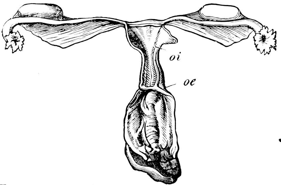
Fig. 70.—Congenital Atrophy of the Uterus (after Virchow), oi, Ostium internum; oe, Ostium Externum.
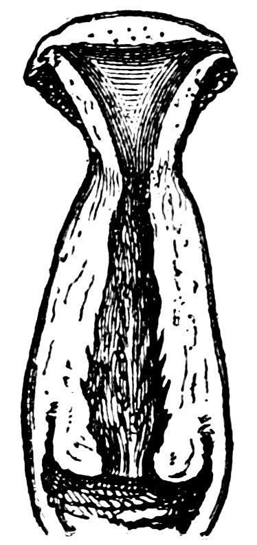
Fig. 71.
In favourable circumstances, when the constitution becomes more powerful, in these cases of primary atrophy of the uterus, improvement takes place; the uterus undergoes further development, menstruation becomes more abundant, and the woman may become pregnant. Such a favourable prognosis cannot, however, be entertained if a severe flexion of the uterus is associated with the atrophy of the organ; or if the ovaries are also atrophied.
Sterility results also from puerperal atrophy of the uterus. This condition is a sequel of severe puerperal diseases, metritis, parametritis, 501and perimetritis; sometimes, even in the absence of such inflammatory processes, it is due to puerperal hyperinvolution, occurring especially in women previously weak in constitution, and manifested by the fact that, notwithstanding the weaning of the child, the menstrual flow remains for months in abeyance. The uterus loses its firm consistency; it is sometimes shortened, sometimes of normal length, but the walls are always greatly thinned, so that, as Schroeder points out, the sound can be readily felt, through the abdominal wall. Puerperal atrophy is a curable condition, so that the sterility dependent upon this disease is not necessarily permanent. Thus, in a case of P. Müller’s, a woman in whom a twin delivery had been followed by extreme atrophy of the uterus, with well-marked symptoms both objective and subjective, became once more pregnant eighteen months after the termination of the twin pregnancy.
Other forms of atrophy of the uterus have a similar deleterious effect to that exercised by puerperal atrophy, as, for instance, atrophy from the pressure of tumours of the uterus, or of solid ovarian tumours; or, again, atrophy due to defective innervation of the pelvic organs, occurring in various forms of paralysis, and characterised by amenorrhœa and extreme smallness of the uterus. Von Scanzoni has seen several cases in which young women, previously healthy and menstruating with regularity, have been attacked by paralysis of the lower extremities, and thenceforwards have suffered from amenorrhœa and great contraction of the uterus; in some of these cases a post mortem examination was made, and disclosed the existence of true atrophy of the uterus. Jaquet saw a similar case of atrophy of the uterus in a lady who had been frightened by witnessing the storming of a barricade in front of her dwelling; she was then in her 22nd year, and had given birth to her second child 1½ years previously; thenceforwards she was completely amenorrhoeic, and her uterus measured only 3 cm. (1.2 in.) in length.
Displacements of the uterus (flexions and versions), and abnormalities in the cervix uteri, are among the conditions which lead to sterility by interfering with conjugation—by preventing the necessary physical contact between the male and the female reproductive elements. The frequency with which these diseases give rise to sterility is, however, far from being so great as is commonly asserted by those who maintain a mechanical theory of conception.
Pathological Changes in the Cervix Uteri.
In very early times, the attention of physicians was directed to abnormalities in the shape of the cervix uteri, as offering hindrances 502to the entry of the semen into the uterus. Amongst the writers of antiquity who have alluded to this matter, the names of Hippocrates and Soranus must especially be mentioned.
The normal cervix uteri (Fig. 72) has the form of a flattened ellipsoid, perforated throughout its longitudinal axis. On making a longitudinal section of the cervical canal, we see that it is dilated in the middle, and tapers towards either extremity, having thus the shape of a spindle; the internal os is, however, somewhat smaller than the external. The latter (os uteri externum, os tincæ, often referred to without qualification as “the os”), has normally the form of a transverse fissure, which, however, tends more towards the circular form, the smaller it is, and the more widely its margins are separated. In childhood, in consequence of the infolding of its margins, the external os has usually a radiated form, later it becomes rounded, and only with the attainment of sexual maturity does it assume the form of a transverse slit. This form is maintained throughout the epoch of active sexual life; but after the climacteric, owing to the separation of the margins of the orifice, it becomes once more rounded.
With regard to the greatly varying size and shape of the portio vaginalis, it may be said that in general its anterior lip appears the shorter of the two, owing to the lesser depth of the anterior vaginal fornix, but that in reality the anterior wall of the cervical canal is longer than the posterior; the actual length of the anterior lip of the portio vaginalis, measured from the summit of the anterior fornix, is from ½ to 1 cm. (0.2 to 0.4 in.), whilst the posterior lip, from the summit of the posterior fornix to the end of the lip measures 1½ cm. (0.6 in.) and upwards. The position of the cervix is such that, owing to the oblique direction of the long axis of the uterus, superadded to the absolutely greater length of the anterior lip of the cervix, the plane across the extremities of the two lips faces almost straight backwards. The axis of the portio vaginalis forms a right angle with the axis of the vagina; the cervical canal, however, is not usually straight, but has a slight S-shaped curvature. The mean length of the cervical canal in the virgin uterus is 3 cm. (1.2 in.). (Lott.)
The “ideal” form of the cervix uteri and of the os uteri externum is described by Sims in the following terms: “The vaginal portion should measure about one fifth, certainly not more than one fourth, of the entire length of the cervix uteri; that is, the anterior lip should have a length of one-fourth to one-third of an inch, and the posterior lip should be a fraction longer. The cervical canal should either be straight, or have a forwardly directed curve; the cervical axis should form a right angle with the vaginal axis; the cervix should not be markedly anteverted or retroverted.” Sims 503is of opinion that every woman whose uterus is in this condition will conceive within three or four months from the time when she first enters upon conjugal intercourse; he adds, however, the important proviso, “be it understood, that all else is in order.”
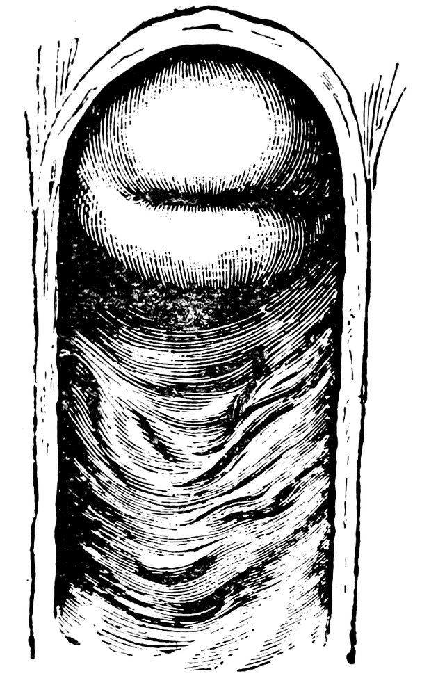
Fig. 72.—Normal Portio Vaginalis.
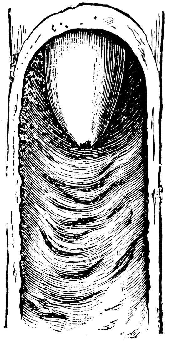
Fig. 73.—Conoidal Portio Vaginalis.
In conception, the cervix uteri subserves the important function of providing for the free passage of the spermatozoa to the interior of the uterus; and when we consider the nature of the processes of sexual intercourse and fertilization, and more especially when we bear in mind that normally the two lips of the cervix and the upper segment of the vagina form a chamber for the retention of a portion of the seminal fluid in contact with the os uteri externum, we are readily led to assume that any great abnormality, in size of the cervix (enlargement or diminution), in its shape (malformation), or in its position (displacements—flexion, version, or prolapse), or, finally, stenosis of the cervical canal,—may offer mechanical hindrances to conception. And experience shows that this assumption is justified, at any rate as regards conical elongation of the portio vaginalis (Fig. 73), as regards an apron-shaped or beak-shaped hypertrophy of the anterior lip of the cervix (Figs. 74 and 75), as regards flexion upwards of the elongated cervix, and also as regards stenosis or obliteration of the external or the internal os; although the reservation must be made that no matter how unfavourable the shape of the portio vaginalis, no matter how extensive the changes in the cervix uteri, as long as a permeable upward passage for the spermatozoa exists, conception is still possible, and in exceptional cases may occur.
504
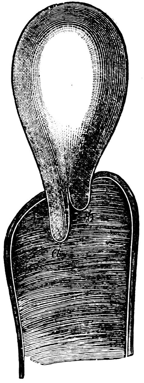
Fig. 74.—“Apron-shaped” Vaginal Portion. a. Greatly elongated anterior lip; b. Shorter posterior lip of the cervix.
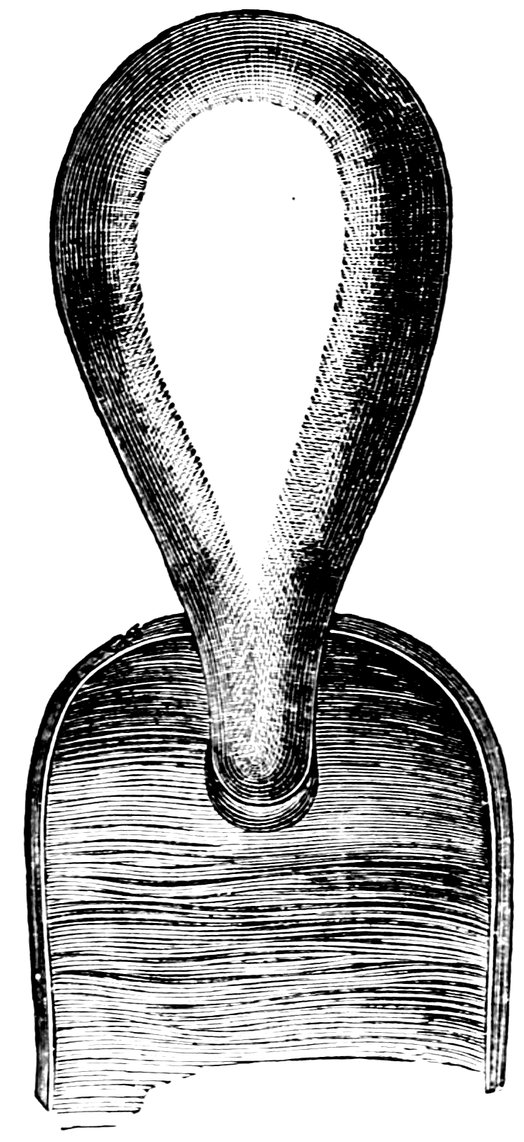
Fig. 75.—“Beak-shaped” Vaginal Portion. Posterior aspect.
When the cervix is hypertrophic and greatly enlarged, and the vaginal fornix consequently much elongated, conception is rendered difficult, for the reason that in such cases, either the semen rapidly flows out of the vagina, or else a proper juxtaposition between the penis and the external os no longer occurs, and the semen is ejaculated at some distance from the os. The change in the shape of the portio vaginalis, and also the elongation of the cervical canal, are additional obstacles to the entrance of the spermatozoa into the interior of the uterus; as regards the former condition, in nulliparae the portio vaginalis is commonly conical, or pointed, whilst the external os is very small, thus rendering the passage of the spermatozoa a difficult matter; but in parous women, it is lobulated, owing to the presence of deep fissures, whereby the penis is conducted into the vaginal fornix, and the ejaculation of the semen in this locality is facilitated. Hence, such hypertrophy of the cervix and the portio vaginalis often coincides with the occurrence of sterility. The hypertrophy is less apt to cause sterility when it is limited to one lip of the cervix, unless, indeed, the affected lip (more commonly the anterior) is so greatly enlarged that it bends over and occludes the external os, whilst conducting the penis into the fornix and away from the orifice. Cases have been known 505in which a single lip of the cervix was hypertrophied to such an extent as to protrude between the labia.
The commonest malformation of the cervix is the conical cervix, when the cervix is not merely elongated, but tapering; associated with this condition is usually found a notable diminution in size of the os uteri externum. According to Sims we find “conical cervix in 85% of all cases of natural sterility.” According to the same author, even in the absence of the conical form of cervix, “sterility is probable in cases in which the portio vaginalis projects fully half an inch into the vagina; if the cervix projects more than one inch, sterility almost inevitably results; whilst if elongation is even greater than this, so that the vaginal portion measures from one and a half to two inches, sterility is absolutely certain.”
On the other hand, congenital smallness of the portio vaginalis, the condition in which this organ appears merely as a slightly projecting nodule on the upper part of the anterior wall of the vagina, the anterior vaginal fornix being almost non-existent, and the posterior fornix very extensive—a wide cul-de-sac—is also unfavourable to conception. The probable reason is that, in consequence of this deformity, the semen, after being ejaculated into the posterior fornix, flows away down the posterior wall of the vagina, without coming into contact with the short portio vaginalis.
According to Beigel, another frequent cause of sterility is to be found in the existence of the so called “apron-shaped” portio vaginalis, the condition in which, either from congenital deformity, or else from hypertrophy or some other disease, one lip of the vaginal portion is so formed as greatly to exceed the other in length.
In consequence of hypertrophy, the portio vaginalis may assume other, very various forms; in some cases it may increase in size to such an extent that it projects into the vagina as a thick, hard ball, and thus offers a serious obstacle to the reception of the semen; or, again, in the form of the elongated, slender cervix, it may become doubled upon itself, and in this way hinder the passage of the spermatozoa (Figs. 76 and 77). Deformities of the cervix due to hypertrophy of the portio vaginalis, rarely cause congenital sterility, but more commonly the acquired form; for such hypertrophy is hardly ever congenital, occurs but rarely in virgins, and is usually met with in married women who have had difficult deliveries, and consequently have suffered from uterine disease.
Another deformity of the vaginal portion of the cervix which is important in its relations to sterility is the “snout-shaped cervix.” Here the cervix is thinnest immediately at its insertion into the vaginal fornix, and thickens gradually below, so that the organ resembles a swine’s snout in form. As a rule, this deformity is 506due to diffuse hypertrophy of the connective tissue of the cervix, the result of chronic endometritis and cervicitis.
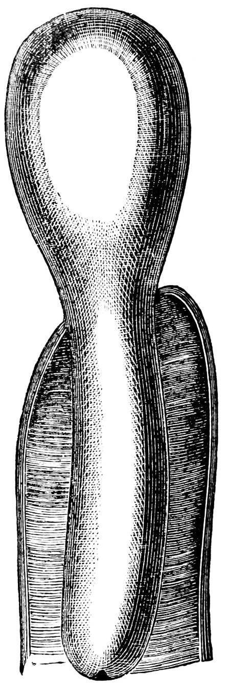
Fig. 76.—Simple Hypertrophy of the Portio Vaginalis, which projected from the Vulva.
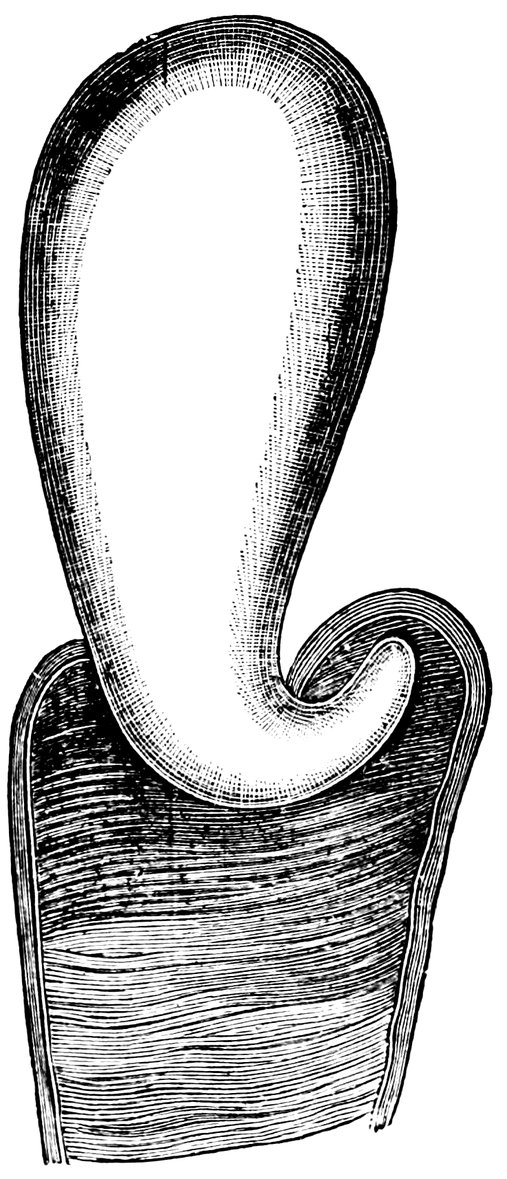
Fig. 77.—Elongated Cervix, bent upwards.
Fritsch, however, in two cases of characteristic col tapiroid, saw pregnancy occur after the relief of the previously existing uterine catarrh; in one of these cases the condition of the organs was virginal, so that it was hardly possible to believe that the patient was a multipara; even after she had had three children, the os uteri externum with difficulty admitted the passage of the uterine sound.
Pajot has devoted especial attention to the hindrances that are offered to the entrance of the spermatozoa by displacements of the cervix. In these cases, during coitus, the extremity of the glans penis is not in contact with the os uteri externum, but passes into a kind of cul-de-sac; in retroversion the posterior fornix; in anteversion, the anterior fornix; in lateral version, the lateral fornix of the side opposed to that towards which the lower extremity of the cervix points.
Complete absence of the vaginal portion of the cervix puts difficulties, though not very serious ones, in the way of conception, 507since the segment of the uterus which combines with the upper segment of the vagina to form a receptaculum seminis, is wanting. How important in predisposing to fertilization is efficient contact of the external orifice of the vaginal portion with the ejaculated semen during and immediately after intercourse, seems to be established by my own observation, that women of small stature married to men of average height exhibit much higher proportional fertility than women of average stature. In the case of these small women, the favourable circumstance is obvious, inasmuch as intimate contact is facilitated between glans penis and portio vaginalis. I have frequently heard complaints, from the husbands of such women, that a single coitus is sufficient to ensure conception; and again and again I have been informed by such women that they have had 10, 12, or 16 children. In one such instance known to me, the wife had been pregnant 23 times, and had given birth to 19 normal children. Contrariwise, women with a very long vagina, and with a high position of the portio vaginalis, do not so easily become pregnant.
Of special importance in the causation of sterility is stenosis of the cervical canal. This may be congenital, and then usually affects the whole length of the canal; or it may be acquired, being dependent upon inflammation of the mucous membrane. In these latter cases, the swollen follicles of the mucous membrane burst, and their granulating walls adhere. Other causes of acquired stenosis are trauma, severe operative procedures during parturition, puerperal inflammations, syphilitic ulceration, adhesion of the opposed granulating surfaces after operative measures (as, for instance, after severe cauterization, or after amputation of the portio vaginalis), and, in short, from scar-formations however caused.
General swelling of the tissues leading to stenosis occurs at the external os in hyperplastic uteri of virgin configuration; the small round orifice characteristic of the virgin uterus becomes narrowed, or even completely occluded, by the swelling of the tissues of the vaginal portion. True adhesion of the walls does not occur in these cases, but the minute aperture left by the swelling of the walls of the canal is plugged by the epithelium, so that a small blind depression in the centre of the portio vaginalis is all that remains of the cervical canal. Such a condition is seen with especial frequency in cases of prolapse of the vaginal portion, and is often erroneously regarded as an obliteration of the os uteri externum by epithelial adhesion (Klebs). Finally, stenosis of the cervical canal may be caused by tumours, and also by the flexions and versions of the uterus presently to be discussed.
Congenital atresia of the uterus is generally associated with other developmental anomalies of the reproductive organs. In some cases, 508all that is at fault is that the mucous covering of the vaginal portion passes uninterruptedly from one lip to the other; but in others, the cervix is unperforated throughout, and the vaginal portion is but slightly developed.
Acquired obliteration of the cervical canal may affect either the external or the internal os, with a shorter or longer portion of the rest of the canal. When very extensive necrosis of tissue has occurred, as a sequel of difficult delivery, the adhesion may include the adjoining segment of the vagina (utero-vaginal atresia).
The more marked the stenosis of the cervical canal, the smaller the passage by which the vagina communicates with the uterus, the more difficult will it be for the passage of the spermatozoa to be effected, so that of the millions of spermatozoa deposited in the neighbourhood of the os uteri, thousands will, as in normal cases, find their way to the uterine orifices of the Fallopian tubes. So much the more, then, is the contact between spermatozoon and ovum rendered difficult, and so much the more unlikely is it that conception will occur. Moreover, in consequence of the stenosis, there is retention of the cervical mucus, which becomes thick and glutinous, and offers a further obstacle to the passage of the spermatozoa. The unfavourable influence upon the possibility of conception is, finally, increased if, as is often the case, in association with the stenosis, the cervix becomes elongated and assumes a conical form (these secondary changes probably resulting from the inflammatory states of the cervix common in cases of stenosis); and an additional obstacle is offered to conception by the association with the stenosis of flexion or version of the uterus. It is in such complicated cases that we so often have the associated symptoms of dysmenorrhœa and sterility; the dysmenorrhœa being due to the fact that the menstrual discharge, if abundant, is unable to flow away with sufficient rapidity through the greatly narrowed cervical canal; exuding from the vessels of the uterine mucous membrane more rapidly than it can be discharged, it accumulates in the uterine cavity, and gives rise to painful contractions of the uterus.
Precisely what degree of narrowing of the cervical canal it is which constitutes pathological stenosis, is in practice by no means easy to define; and only in regard to extreme cases of pathological constriction can there be no possibility of dispute. In cases of congenital stenosis of the cervical canal, the diagnosis is very easy, for the os uteri externum is then always extremely small; often the aperture is no larger than a small pin’s head, a very fine probe can be passed through it with considerable difficulty and its passage is opposed all the way up to the internal os. But in cases of acquired stenosis of moderate severity, the diagnosis is often difficult. Owing to the small size of the 509orifice, and to the distensibility of the soft parts by which it is surrounded, exact measurements are impossible. When the os is with difficulty detected by the skilled finger, when the sound is not readily introduced by the experienced hand, slipping past again and again, and inserted only after repeated efforts—such an os is, as Olshausen insists, always pathological. The normal virgin os uteri permits the easy passage of a thick uterine sound with a diameter of 3 to 4 millimeters (⅛ to ⅙ in.); but there are cases in which, though a sound of this normal size can be passed, the os gives to the examining finger the sensation of being contracted. If, in such a case there is typical mechanical dysmenorrhœa with sterility, Olshausen considers that we are justified in assuming the existence of pathological stenosis of the os uteri, and in treating the case accordingly.
However, as Kehrer insists, it may be one of the greatest difficulties in diagnosis—a difficulty not always to be resolved even when all the attendant circumstances have received the fullest and most painstaking consideration—to determine whether in any individual case an anomaly of the cervix, such as stenosis of the external os or of the whole cervical canal, is or is not to be regarded as a cause of sterility. When stenosis is extreme, there need be no two opinions about the matter; the difficulty is in cases lying somewhere between a moderate degree of contraction and the lower physiological limit of smallness. Every experienced gynecologist will have seen such cases as Kehrer describes, in which before marriage the os appeared extremely small, and yet soon after marriage the woman became pregnant. For this reason we are justified, with O. Johannsen, in reverting rather to the functional than to the anatomical conception of stenosis, and in maintaining that so long as the cervical canal is sufficiently large to permit the uterine secretions to flow freely away, any stenosis that may exist is devoid of pathological significance. Only when the outlet for the uterine secretions is insufficient, so that the uterine cavity becomes distended (as manifested by an elongation of the canal in the supravaginal portion of the uterus, and by various disorders, amongst others chronic endometritis), is the stenosis with its consecutive dilatation of the uterus a serious obstacle to conception. “In such cases, the contractions of the uterus during coitus will not suffice to express the secretions it contains through the narrowed os, and the inevitable consequence of the incomplete evacuation of the uterus is that the aspiratory phase of the orgasm fails to occur.”