INFECTIOUS DISEASES OF CATTLE.
Reading edition
Revised by John R. Mohler, A. M., V. M. D.,
Chief, Bureau of Animal Industry.
GENERAL INTRODUCTION.
The importance, to the farmer and stock raiser, of a general knowledge of the nature of infectious diseases need not be insisted on, as it must be evident to all who have charge of farm animals. The growing facilities for intercourse between one section of a country and another, and between different countries, cause a wide distribution of the infectious diseases once restricted to a definite locality. Not only the animals themselves, but the cars, vessels, or other conveyances in which they are carried may become agents for the dissemination of disease. The growing tendency of specialization in agriculture, which leads to the maintenance of large herds of cattle, sheep, and hogs, makes infectious diseases more common and more dangerous. Fresh animals are being continually introduced which may be the carriers of disease from other herds, and when disease is once brought into a large herd the losses become very high, because it is difficult, if not impossible, to check it after it has once obtained a foothold.
These considerations make it plain that only by the most careful supervision by intelligent men who understand the nature of infectious diseases and their causes in a general way can these be kept away. We must likewise consider how incomplete our knowledge concerning many diseases is, and probably will be for some time to come. The suggestions and recommendations offered by investigators, therefore, may not always be correct, and may require frequent modification as our information grows more comprehensive and exact.
An infectious disease may be defined as any malady caused by the introduction, into the body, of minute organisms of a vegetable or animal nature which have the power of indefinite multiplication and of setting free certain peculiar poisons which are chiefly responsible for the morbid changes.
This definition might include diseases caused by certain animal parasites, such as trichinæ, for example, which multiply in the digestive tract, but whose progeny is limited to a single generation. By common consent the term "infectious" is restricted to those diseases caused by the invasion and multiplication of certain very minute unicellular organisms included under the general classes of bacteria and protozoa. Nearly all the diseases of cattle for which a definite cause has been traced are from bacteria. Among these are tuberculosis, anthrax, blackleg, and tetanus (or lockjaw). Some diseases, such as Texas fever and nagana, are traceable to protozoa, while others, like vactinomycosis and aspergillosis, are caused by fungi. Those diseases of which the cause is unknown or imperfectly worked out are pleuropneumonia, rinderpest, foot-and-mouth disease, rabies, cowpox, malignant catarrh, and dysentery.
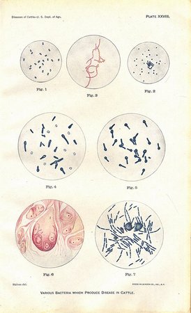
Bacteria may be defined as very minute, unicellular organisms of a plantlike character. Their form is very simple, as may be seen from an inspection of the various species depicted on Plate XXVIII. The description of these figures will be found on page 360. The magnification there given will furnish the reader some idea of their very minute size. They multiply in two ways. The bacterium elongates and then divides in the middle to form 2 daughter cells. These go through the same process at once, and thus 4 cells are produced. The division of these leads to 8, the division of 8 to 16, and so on indefinitely. The rapidity with which this multiplication takes place depends upon the nature of the bacterium. The bacillus of tuberculosis multiplies very slowly, while that of anthrax does so with great rapidity, provided both are in the most favorable condition. Another mode of reproduction, limited to certain classes of bacteria, consists in the formation of a spore within the body of the bacterium. Spore formation usually takes place when the conditions pertaining to the growth of the bacteria become unfavorable. The spores are much more resistant to destructive agents than the bacteria which produced them. The anthrax spore may live several years in a dried state, but the anthrax bacillus perishes in a few days under like conditions. This matter will be referred to again when we come to discuss the subject of disinfection.
Of the protozoa which cause disease very little is at present known. One which produces Texas fever is pictured on Plate XLV, in figs. 4 and 5. These parasites have a more complex life history than bacteria; and as they can not be grown in artificial media, their thorough investigation is at present hampered with great difficulties.
The differences in the symptoms and lesions of the various infectious diseases are due to differences in the respective organisms causing them. Similarly the great differences observed in the sources from which animals become infected and the manner in which infection takes place are due to differences in the life history of these minute organisms. Much discussion has taken place of late years concerning the precise meaning of the words "infection" and "contagion."
VARIOUS BACTERIA WHICH PRODUCE DISEASE IN CATTLE.
DESCRIPTION OF PLATE XXVIII.
The bacteria on this plate are partly from tissues, partly from cultures, and stained artificially with aniline colors (fuchsin or methylene blue). Figs 6 and 7 are copied from Fränkel and Pfeiffer's atlas. All but fig. 7 are magnified 1,000 times; fig. 7, 500 times.
Fig. 1. Bacteria from pneumonia in cattle. These are also the cause of hemorrhagic septicemia and are closely related to swine-plague bacteria. These bacteria were drawn from a piece of spleen pulp (rabbit).
Fig. 2. Micrococci (streptococcus) which produce inflammation of the lining membranes of the abdomen, thorax, heart, brain, and joints. Frequently associated with the preceding bacteria in abscesses.
Fig. 3. Micrococci (staphylococcus) which produce inflammation and suppuration; also pyemia.
Fig. 4. Bacilli of blackleg. The pale oval bodies as well as the light spots in one end of the bacilli represent spores.
Fig. 5. Bacilli which produce tetanus or lockjaw. The light spot in the enlarged end of each rod represents a spore.
Fig. 6. Bacilli of tuberculosis. Microscopic sections of a pearly nodule from the lining membrane of the chest cavity. The bacilli are stained red and appear as small straight rods within the cells of the nodule or tubercle.
Fig. 7. Bacilli of anthrax. Bacilli from the spleen of a mouse inoculated with a culture. The bacilli were obtained from the blood of a cow which died of anthrax in Mississippi. The bacilli appear as rods stained blue. The round bodies are blood corpuscles, also stained artificially.
PLATE XXVIII. VARIOUS BACTERIA WHICH PRODUCE DISEASE IN CATTLE. (Click to enlarge)
These words, however, are now wholly inadequate to express the complex processes of infection, and it may be said that each species of bacterium or protozoon has its own peculiar way of invading the animal body, differing more or less from all the rest. There are, however, a few broad distinctions which may be expressed with the help of these old terms. Infection, as laid down above, refers at present in a comprehensive way to all microorganisms capable of setting up disease in the body. Some microorganisms are transmitted directly from one animal to another, and the diseases produced may be called contagious. Among these are included pleuropneumonia, rinderpest, foot-and-mouth disease, rabies, cowpox, and tuberculosis. Again, certain organisms are perhaps never transmitted from one animal to another, but may come from the soil. Among these are tetanus, blackleg, anthrax to a large extent, and perhaps actinomycosis in part. These diseases, according to some authorities, may be called miasmatic. There is a third class of infectious diseases, the specific bacteria of which are transmitted from one animal to another, as with the contagious diseases, but the bacteria may, under certain favorable conditions, find food enough in the soil and in the surroundings of animals to multiply to some extent after they have left the sick animal and before they gain entrance into a healthy one.
This general classification is subject to change if we take other characteristics into consideration. Thus tuberculosis, because of its insidious beginning and slow course, would not by many be considered contagious in the sense that foot-and-mouth disease is; yet, in either case, the bacillus must come from preexisting disease. The disease of rabies, or hydrophobia, is not contagious in the sense that rinderpest is, because the virus of rabies must be inoculated into a wound before it can take effect; yet, in both cases, the virus passes without modification from one animal to another, though in different ways.
Again, all the diseases under the second group, which seem to come from the soil and from pastures, are in one sense contagious in that the virus may be taken from a sick animal and inoculated directly, with positive results, into a healthy animal. Other illustrations may be cited which show that these old terms are not in themselves satisfactory. There are so many conditions which enter into the process of infection that no single classification will give a sufficiently correct or comprehensive idea of it. These statements will be easily understood if the different infectious diseases in the following pages are studied with reference to the way or ways in which each disease may be contracted. Enough has been said, therefore, to show that if we wish to make ourselves acquainted with the dangers of any given disease, we must study it and not rely upon any single work to tell the whole story.
Infectious diseases have, as a general rule, a period of incubation, which comprises the time elapsing between the exposure to the infection and the actual appearance of the disease. This period varies with the malady. The most common symptom of this class of diseases is fever. The severity of the fever is measured by the temperature of the animal; this is readily and accurately ascertainable by the clinical thermometer. (See Pl. III, fig. 1.) The other symptoms are variable and depend upon the particular organ or organs most implicated. Loss of appetite, cessation of rumination and milk secretion, and general dullness are symptoms quite invariably present in most infectious diseases.
During the course of infectious diseases secondary diseases or complications may arise which are largely caused by bacteria other than those producing the original malady. These complications are often so severe as to become fatal. In general it may be stated that they are due to filthy surroundings, and hence cleanliness may become an important aid to recovery.
The treatment of infectious diseases is given under each malady so far as this is allowable or advisable. These diseases are not, as a rule, amenable to treatment. When the symptoms have once appeared the disease is liable to run its course in spite of treatment, and if it is one from which animals usually recover, all that can be done is to put them into the most favorable surroundings. Many infectious diseases lead sooner or later to death, treatment is useless so far as the sick are concerned, and it may be worse than useless for those not yet infected. All animals suffering with infectious diseases are more or less directly a menace to all others. They represent for the time being manufactories of disease germs, and they are giving them off more or less abundantly during the period of disease. They may infect others directly or they may scatter the virus about, and the surroundings may become a future source of infection for healthy animals. This leads us to the subject of prevention as the most important of all which claim our attention. In this place only a few general remarks will suffice to bring the subject before the reader.
The most important thing is to keep disease away from a herd or farm. To do this all sick or suspicious animals should be avoided. A grave form of disease may be introduced by apparently mild or trivial cases brought in from without. It is generally conceded that continual change and movement of animals are the most potent means by which infectious diseases are disseminated.
With some cattle diseases, such as anthrax, rinderpest, and pleuropneumonia, preventive inoculation is resorted to in some countries. This may be desirable when certain diseases have become established in any locality so that eradication is impossible. It should not be practiced in territories where a given disease may still be extirpated by ordinary precautions. Preventive inoculation is applicable to only a few maladies, and therefore its aid in the control of diseases is limited.
When an infectious disease has gained foothold in a herd the course to be pursued will depend upon the nature of the malady. A good rule is to kill diseased animals, especially when the disease is liable to run a chronic course, as in tuberculosis. The next important step is to separate the well from the sick by placing the former on fresh ground. This is rarely possible; hence the destruction or removal of the sick, with thorough disinfection of the infected locality, is the next thing to be done. As to the disinfectants to be used, special directions are given under the various diseases, to which the reader is referred. Here we will simply call attention briefly to the general subject.
DISINFECTION AND DISINFECTANTS.
Disinfection consists in the use of certain substances which possess the power to destroy bacteria or their spores, or both. Those which are cheapest and most available for animal diseases are ordinary freshly slaked lime or unslaked in powder, chlorid of lime, crude carbolic acid, corrosive sublimate, formaldehyde gas, formalin, and compound cresol solution.
(1) Slaked lime is perhaps the most easily procured, but its disinfecting power is limited. While it is capable of destroying all bacteria in their vegetative state, it is unable to destroy such spores as those of anthrax and blackleg. It is probable, however, that in incrusting spores it may destroy their vitality sooner or later. It is regarded as safe practice to use only spore-destroying substances for the virus of those diseases of which we have no definite knowledge. Nevertheless, in the absence of other disinfectants, lime is very useful. It may be employed as a whitewash on wood and stone and sprinkled as a dilute wash or in powder over yards, manure heaps, and over carcasses before they are buried, and over the ground on which they have lain to prevent other animals from carrying the infection away.
(2) Chlorid of lime is more efficient than simple slaked or unslaked lime, as it destroys spores. It is the ordinary bleaching powder of commerce and is quite unstable, hence old preparations, unless sealed, are of little value. A 5 per cent solution is sufficiently strong for all spore-bearing bacteria (3 ounces in 2 quarts of water). It may be efficiently applied to the walls and floor of an infected stable by mixing with limewash in the proportion of 6 ounces of the lime to each gallon of limewash. The ceilings and those portions of the walls which can not be reached should be disinfected by means of chlorin gas liberated from the chlorid of lime by crude carbolic acid. This is accomplished by making a cone of 5 or 6 pounds of chlorid of lime, in the top of which a deep crater is made for the placement of from 1 to 2 pints of crude acid. The edge of the crater is thereupon pushed into the fluid, when a lively reaction follows. The fumes of chlorin are strongly irritating to the respiratory tract and therefore all live stock should be removed before the work is started. Owing to the heat generated, it is advisable to place the lime in an iron crucible and to have nothing inflammable within a radius of 2 feet. The number and location of these cones of chlorid of lime depend on the size and structure of the building to be disinfected. As a rule, it may be stated that chlorin gas liberated from the above-sized cone will be sufficient for disinfecting 5,200 cubic feet of air space.
(3) Crude carbolic acid. The ordinary purified carbolic acid is too expensive to be used on a large scale, and the crude produce is a very good substitute. This is made more powerful by mixing with it an equal volume of commercial sulphuric acid. While the sulphuric acid is being added to the crude carbolic acid much heat is evolved, and if the glass jar in which the two are mixed is placed in cold water the resulting product is said to have a higher disinfecting power. The mixture is added to water enough to make a 5 per cent solution (about 7 ounces to 4 quarts of water). This is strong enough for all purposes. It may be kept in wood or glass, but not in metal, owing to the corroding action of the acid. It should be used freely on woodwork and on infected floors, and a force pump of the kind used by orchardists is very convenient as a means of applying the disinfectant. If the solution is warm when applied, it will penetrate the woodwork better than when cold, especially if the spraying is done during cold weather. The addition of air-slaked lime in any quantity that will dissolve in water to the above solution (say 1½ pounds of lime to 7 ounces of crude carbolic acid to each gallon of water) is preferred by many, as it makes any neglected places at once visible and leaves cleaner and better air within the buildings. In most cases in which its application becomes desirable—and this rule should apply to all disinfections—the disinfected stables, stalls, etc., should remain vacant as long as possible before cattle are again stabled therein.
(4) Mercuric chlorid, or corrosive sublimate, is a powerful disinfectant, but it is likewise very poisonous; hence its uses are limited. Cattle are especially susceptible to its action and caution must be used in its application. A solution of one-tenth of 1 per cent is usually sufficient (1 ounce to 8 gallons of water). It should not be placed in wooden pails, which would form the tannate of mercury, a weak antiseptic; nor, owing to its corrosive action, should expensive metal pails be used. Agate vessels or tin pails are to be preferred. All solutions should be labeled "poison," and to avoid accidents none should be kept on hand.
(5) Formalin and formaldehyde gas have been found very efficacious as sanitary agents. Formalin is the commercial name for the 40 per cent solution of formaldehyde gas in water, and is one of the most powerful antiseptic and disinfectants that we possess. Solutions of this strength are manufactured by different commercial houses and sold by the drug trade under the name of "formalose" and "formal." In this connection it should be mentioned that while the 40 per cent solution of formaldehyde gas and formalin are exactly the same thing, the former can be purchased at 33-1/3 to 64 per cent less than the latter. Formalin, diluted with water in the proportion of 1 pint to 30 parts of water, or 4 ounces to each gallon of water, may be applied, and it may thus be used as a wash or as a spray on all paints, metals, and woodwork, as well as on clothing and other fabrics, without injuring them. It may also be applied to floors, walls, and woodwork in whitewash by mixing 1 part to 30 parts of limewash, or 4 ounces to each gallon of limewash. Formalin has the appearance of water and in the strong solution is poisonous, but when diluted as recommended above it is not dangerous. The fumes given off by it, however, are very disagreeable and irritating to the eyes and nasal mucous membranes. One and one-half ounces of formalin added to 1 gallon of water is a valuable agent for the disinfection of the skin or septic wounds, but is somewhat painful and irritating to raw surfaces.
Formaldehyde is a gas which is soluble in 2½ parts of water (40 parts of formaldehyde gas to 100 parts of water); this solution constitutes the formalin of commerce. The use of formaldehyde gas is in most cases impracticable for stable disinfection. In case the stable is not too large and can be made almost air-tight the generation of formaldehyde gas, after removing all the animals, will be found very serviceable. It penetrates all parts of the stable—the walls, crevices, floors, ceiling—and is probably the best fumigating disinfectant that we have.
Probably one of the most simple and practical methods of liberating this gas is by means of the chemical reaction which takes place when formalin is poured upon permanganate of potassium. For each 1,000 cubic feet of air space, 16-2/3 ounces of crystallized or powdered permanganate of potassium is placed in a wide-surfaced pan; 20 ounces of formalin is then poured upon it, and the stable immediately closed for a period of 12 hours or longer. This method is efficient only when it is possible to seal tightly the place to be disinfected, and should be used only by experienced persons.
(6) Some coal-tar products are cheap, effective, and easily applied disinfectants, their action being due to the carbolic acid and creosote in their composition. They may be used in 3 to 5 per cent solution. As a rule they form a milky solution in water.
(7) Compound solution of cresol (liquor cresolis compositus), now recognized as an official preparation, is composed of equal parts of cresol and linseed-oil-potash soap. The mixture is a thick, dark, amber-colored fluid which mixes readily with water in all proportions to form a clear, soap solution. It is an efficient disinfectant in a 3 or 4 per cent solution, and in this strength it may be applied in the same manner as a 5 per cent solution of carbolic acid.
When it is desired to apply one of these above-mentioned agents to the stable or barnyard, a preliminary cleaning up of all débris and litter is advisable, together with the scraping of the floor, mangers, and walls of the stable with hoes; also the removal of all dust and filth. This should be followed by the burning of all such accumulations, inasmuch as this material likewise contains the infectious principle and is best destroyed by heat. Heat may be applied to the surface of the affected pen, byre, or barnyard by means of a cyclone burner, which consists of a tank, pump, hose, and cyclone nozzle for spraying with paraffin (gas oil). The latter is ejected in the form of spray, which when ignited gives a very hot and effective flame to be applied to the infected ground. Where such burning is impracticable the surface soil of the yard and surroundings should be removed to a depth of 5 or 6 inches and then placed in a heap and thoroughly mixed with air-slaked lime. The fresh surface of the soil thus exposed may then be sprinkled with the disinfectant.
In addition to these artificial substances there are several natural sanitary agents of great importance as destroyers of virus. These are cleanliness, ventilation, drying, and sunshine. All virus, excepting such as may live in the soil, is killed sooner or later by drying and sunshine, and the importance of these factors in the daily life of animals need not be insisted on here. Finally, all sanitary measures which contribute to the healthfulness of animal surroundings are directly or indirectly inimical to disease germs, and all carelessness in the keeping of animals may be regarded as an ally of these destructive organisms.
CONTAGIOUS PLEUROPNEUMONIA.
[Pls. XXIX-XXXII.]
Definition and history.—This disease has been eradicated from the United States, and it is not probable that it will ever be seen in this country again. As, however, much interest was manifested in regard to it for a number of years, and as our cattle are still prohibited from some foreign markets on account of its previous existence here, the subject is treated at greater length than would otherwise be necessary.
The contagious pleuropneumonia of cattle is a specific, epizootic disease which affects bovine animals, and from which other species are exempt. It is characterized, when the disease results from exposure in the usual manner, by an inflammation of the lungs and pleuræ, which is generally extensive, and which has a tendency to invade portions of these organs not primarily affected and to cause death of the diseased portion of the lung. This disease is frequently called the lung plague, which corresponds to its German name of Lungenseuche. In French it is spoken of as the péripneumonie contagieuse.
The history of the contagious pleuropneumonia of cattle can not be traced with any certainty to a period earlier than the beginning of the eighteenth century. No doubt it existed and ravaged the herds of Europe for many years and perhaps centuries before that time, but veterinary knowledge was so limited that the descriptions of the symptoms and post-mortem appearance are too vague and too limited to admit of the identification of the maladies to which they refer. It has been supposed by some writers that certain passages in the writings of Aristotle, Livy, and Virgil show the existence of pleuropneumonia at the time that their works were composed, but their references are too indefinite to be seriously accepted as indicating this rather than some other disease.
It seems quite plain that as early as 1713 and 1714 pleuropneumonia existed in Swabia and several Cantons of Switzerland. There are even clearer accounts of its prevalence in Switzerland in 1732, 1743, and 1765. In 1769 a disease called murie was investigated in Franche-Comté by Bourgelat which undoubtedly was identical with the pleuropneumonia of to-day. From that period we have frequent and well-authenticated accounts of its existence in various parts of Europe. During the period from 1790 to 1812 it was spread throughout a large portion of the Continent of Europe by the cattle driven for the subsistence of the armies, which marched and countermarched in all directions. It was generally prevalent in Italy in 1800. It appears to have been unknown, however, in the Department of the Nord, France, until 1826, but during the years from 1820 to 1840 it penetrated into most parts of that country. During the same period it was introduced into and allowed to spread over Belgium and Holland.
This contagion is said to have been carried to Ireland from Holland in 1839, and is reported as existing in England in 1842. The disease was brought to the United States at several different times. Probably its first introduction was with a diseased cow sold in Brooklyn, N. Y., in 1843. It came to New Jersey by importing affected animals in 1847. Massachusetts was infected in the same way in 1859.
South Africa was infected by a bull brought from Holland in 1854, and Australia likewise received the contagion with an English cow in 1858. It is also reported as existing in various parts of the Continent of Asia, but the time of its first appearance and the extent of its distribution are very uncertain.
Some countries, such as Norway, Sweden, and Denmark, which had been infected for only a short time, have succeeded in eradicating the disease without much difficulty by slaughtering all affected and exposed animals. Other countries long infected and in which the contagion was thoroughly established, like Australia, South Africa, Italy, France, Belgium, and parts of Germany, have labored long, in some cases making no progress and in others being only partially successful. Holland was one of the first of the thoroughly infected countries to free itself from the contagion.
In the United States, Massachusetts eradicated pleuropneumonia during the period from 1860 to 1866. New York and New Jersey made an attempt to eradicate it in 1879, but were not successful. Late in 1883 the contagion was carried to Ohio, probably by Jersey cattle purchased in the vicinity of Baltimore, Md., to which place it had extended before 1868. From the herd then infected it was spread by the sale of cattle during 1884 to a limited number of herds in Illinois, to one herd in Missouri, and to two in Kentucky. The alarm caused among the stock owners of the United States by this widespread dissemination of a disease so much dreaded led to the adoption of active measures for its control and eradication. By cooperation between the United States Department of Agriculture and the authorities of the affected States it was found possible to prevent the further spread of the contagion and to eradicate it after a few months' delay.
In 1886 pleuropneumonia was discovered in some of the large distillery stables of Chicago and among cows on neighboring lots. This led to renewed efforts for the complete extirpation of this disease from the country. Congress in 1887 enlarged the appropriation available for this purpose and gave more extended authority. During the same year the disease was stamped out of Chicago, and has not since appeared in any district west of the Allegheny Mountains.
The work of eradication was at the same time commenced in all the infected States. Before the end of the year 1889 Pennsylvania, Delaware, Maryland, the District of Columbia, and Virginia had been freed from the disease. More difficulties, however, were encountered in the States of New York and New Jersey, on account of the larger territory infected and the density of the population. The long struggle was successful, however, and the last animal in which the disease appeared in the State of New York was slaughtered early in 1891, and the last one affected in New Jersey met the same fate early in the spring of 1892.
PLATE XXIX. UPPER OR DORSAL SURFACE OF THE LUNGS OF THE OX. (ONE-TWELFTH NATURAL SIZE.) (Click to enlarge)
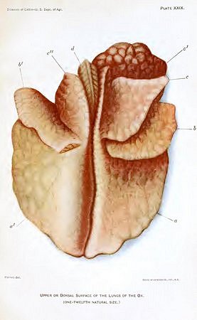
PLATE XXX. BRONCHOPNEUMONIA. (Click to enlarge)
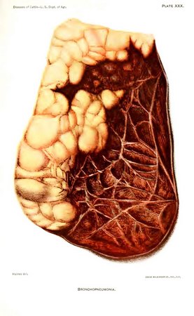
PLATE XXXI. CONTAGIOUS PLEUROPNEUMONIA. (Click to enlarge)
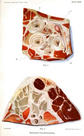
PLATE XXXII. CONTAGIOUS PLEUROPNEUMONIA. (Click to enlarge)
During these same years a supreme effort had been made to stamp out this lung plague from Great Britain. From the official reports it appears that the number of infected districts and of diseased animals had rapidly diminished, but it was not until 1898 that the infection was finally eradicated.
The other infected European countries, though they maintain a veterinary sanitary service, are not making satisfactory progress in eradicating the disease. This is owing partly to delays in carrying out the provisions of the laws and partly to mistaken ideas as to the measures which are necessary to accomplish the object. The United States was the last of the countries having old infected districts which undertook to stamp out this contagion, and, except Holland, it was the first to reach success.
The cause (etiology) of pleuropneumonia.—This is a contagious disease, and arises only by contagion from a previously affected animal; consequently it can never be seen here except as the result of importing affected animals from the Old World. When thoroughly stamped out it does not reappear; and if imported animals continue to be properly inspected and quarantined, we have every reason to believe that pleuropneumonia will never again be seen in this country.
The exact nature of the virus or contagion of lung plague has never been determined. Various investigators have from time to time claimed the discovery of the specific organism of the disease, but it was not until 1898 that Nocard and Roux, by an ingenious method of cultivation, succeeded in obtaining a very feeble growth of an exceedingly minute microorganism. With these cultures the disease was produced in cattle.
Some investigators and writers are of the opinion that the disease can be contracted only by an animal coming near enough to a living diseased one to receive the contagion directly from it. They hold that the contagion is expired with the air from the affected lungs, and that it must be almost immediately inspired by another animal in order to produce the disease. Some experimental attempts to infect animals by placing them in stables where diseased animals have been, and by placing the diseased lungs of slaughtered animals in their feeding troughs have failed, and, consequently, apparently confirm this view.
CONTAGIOUS PLEUROPNEUMONIA.
DESCRIPTION OF PLATES.
Plate XXIX. Upper or dorsal surface of the lungs of the ox, reduced to one-twelfth of the natural size: a, a', the right and left principal lobes. These are the largest and are situated posteriorly, resting upon the diaphragm; b, b', the ventral lobes, situated between the principal lobes; and c, c', c'' the most anterior, or cephalic, lobes. The right anterior is divided into two lobes (c, c'), the left is single (c''); d, trachea, or windpipe. In the majority of the lungs examined in the laboratory of the bureau which were affected with contagious pleuropneumonia the principal lobes (a, a') were primarily affected.
Plate XXX. Bronchopneumonia. The ventral or middle lobe of the right lung affected with collapse and beginning bronchopneumonia. The light yellowish portions represent healthy lung tissue; the red represents the disease. It will be noticed that the lines between the lobules are quite faint, indicating little or no inflammation of the connective tissue between the lobules. The healthy lung tissue is seen to be raised above the level of the diseased portion. In contagious pleuropneumonia the exact reverse is the case, the diseased portions being very much larger than the healthy.
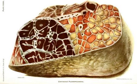
Plate XXXI. Contagious pleuropneumonia. Appearance of a cow's lung affected with contagious pleuropneumonia when sections or slices are made of it and cut surfaces examined.
Fig. 1. Transverse section through the right principal lobe in a case of acute pleuropneumonia. The area drawn includes the air tubes, veins, and arteries, and illustrates the great thickening of the interlobular connective tissue into broad whitish bands and of the walls of the air tubes, veins, and arteries: a, air tube cut obliquely; a', air tube cut directly across; b, arteries cut across; c, large vein completely occluded by a thrombus or plug formed during life. The great thickening of the walls of the artery and vein in this disease is especially brought out by stating that in the healthy lung they are so thin as to be easily overlooked.
Fig. 2. Transverse section of the principal lobe in a case of acute pleuropneumonia, illustrating the different kinds of hepatization or consolidation of the lung. These are indicated by the different colors from dark red to reddish yellow. This variation of color is regarded by some as the real marbling characteristic of pleuropneumonia, while the whitish bands penetrating the lung tissue in all directions constitute the true marbling according to other observers.
Plate XXXII. Contagious pleuropneumonia. This illustrates what are called infarctions. The right half of the figure shows nearly normal lung tissue. The left represents a blackish mass, in which the lung tissue is filled with blood and solidified. This is caused by the plugging of the vein carrying away the blood from this portion. The heart forces the blood through the artery into the tissue at considerable pressure, but owing to the fact that its return is prevented, the minute blood vessels rupture and the air vesicles become distended with blood, which coagulates and causes the firmness of the tissue.
On the other hand, it is known that the serum from affected lungs retains its virulence and may be used successfully for inoculation weeks or months after the death of the animal from which it was taken. This is particularly the case when this liquid is hermetically sealed in glass tubes. Other investigators state that they have successfully infected cattle by placing, in the nostrils, sponges or pledgets of cotton saturated with such serum. Cattle have also, according to the best evidence obtainable, been infected from the clothing of attendants, from horns used in drenching, and from smelling about wagons which have been used to transport affected carcasses. In the work of eradicating pleuropneumonia from the United States many stables were found in which the disease would appear and reappear after the slaughter of affected herds, and in spite of any precautions which were adopted. These were always old stables, with woodwork in a decaying condition and with floors underlaid with filth which could not be thoroughly removed or disinfected. In every one of these cases the destruction of the stable, the burning of the lumber of which it was constructed, the removal of the accumulations beneath the floors, and thorough disinfection, prevented the recurrence of the plague in new stables built upon the same premises. This experience conclusively shows that under certain conditions, at least, stables may retain the infection for a considerable time, and that when restocked the disease may break out again from such infection.
As a rule, however, the disease is acquired by a healthy animal being near an affected one and receiving the contagion direct. Affected animals may give off the contagion in the early stages of the disease before the symptoms are apparent to the observer; also, they may retain this infectious character, if they survive the attack, for six months and probably for a year after all symptoms of the disease have disappeared.
Incubation.—The time which elapses between exposure to the contagion of pleuropneumonia and the first appearance of the symptoms of this disease varies greatly with different individuals and with different outbreaks of the disease. Ordinarily the symptoms of disease make their appearance within three to six weeks after exposure; they may be observed, however, within two weeks or they may not become apparent until nearly or quite three months. It is this long period of incubation and the great length of time that an animal may disseminate the contagion after apparent recovery which give the plague that insidious character so often spoken of, and which greatly increase the difficulties of eradication.
Symptoms.—The symptoms are such as would be expected with inflammation of the lungs and pleuræ, but they vary considerably, according to the type which the disease manifests. If the attack is an acute one, as is frequently seen in hot weather, the symptoms appear suddenly; the breathing becomes rapid and difficult, the animal grunts or moans with each expiration, the shoulders stand out from the chest, the head is extended on the neck, the back is arched, the temperature is 104° to 107° F., the milk secretion is suspended, there is no appetite, rumination is stopped, the animal may bloat and later be affected with a severe diarrhea. Such cases are generally fatal in 7 to 20 days.
Very often the attack comes on slowly and the symptoms are much less clear. In the mildest cases there is a cough for a week or two, but no appreciable loss of appetite or elevation of temperature. The lungs are but slightly affected and recovery soon follows. Such animals may disseminate the contagion for a long time without being suspected, and for that reason are the most dangerous of all.
A more severe type of the plague is the most frequently seen. In these cases the cough is frequent, more or less painful, the back somewhat arched, and the milk secretion diminished. The prominence of these symptoms increases, the appetite is affected, the animal loses flesh, the breathing becomes more rapid, the cough more painful, pressure of the fingers between the ribs shows tenderness, the hair loses its gloss and stands erect, the skin becomes adherent, little, if any, milk is secreted, and the temperature rises, varying in different animals from 103° to 107° F. Animals thus affected may continue to grow worse and die in from three to eight weeks, or they may after a time begin to improve and make an apparent recovery. The inflammation of the lung does not, as a rule, subside and the organ return to its normal condition, as is the case in ordinary pneumonia, but with this disease the life of the affected portion of the lung is destroyed, the tissue dies, and a fibrous wall is formed around it to shut it away from the living parts. The tissue, thus encysted, gradually softens, becomes disintegrated, and breaks down into pus. The recovery, therefore, is not complete; it is only apparent and partial.
To those accustomed to examining the lungs of cattle, other and extremely important symptoms may be apparent during the course of the disease. By applying the ear over the walls of the chest an area of a certain extent may be found in which the natural breathing sound is diminished or entirely lost. This represents the diseased portion of the lungs. In other cases a loud blowing sound may be heard, quite different from any sound produced when the lung is in a healthy condition. In some cases crepitation is heard near the border line of the diseased area and friction sounds produced by the roughened pleura; these can be appreciated, however, only by those whose ears have been trained to distinguish between the different sounds which reach the ear when applied to the chest wall. By percussion—that is, by pressing the fingers of the left hand firmly against the wall of the chest and tapping upon the middle finger with the ends of the fingers of the right hand—an area of dullness may be discovered corresponding to the portion from which the respiratory murmur has disappeared. This loss of respiration detected by auscul tation, and the dullness brought out by percussion, are the most important evidences of an inflamed or consolidated lung.
Seriously affected animals remain standing if they have sufficient strength, but those which lie down always lie on the affected side.
The proportion of animals which become affected after being exposed varies according to the virulence of the outbreak, the susceptibility of the animals, and the length of time during which exposure is continued. Sometimes not more than 15, 20, or 30 per cent will contract the disease when a large herd is exposed; on the other hand, however, 80 or 90 per cent may be affected. The proportion of cases in which the disease proves fatal also varies greatly—it may not exceed 10 and it may reach 50 per cent. In general, it may be said that about 40 per cent of the exposed animals will contract the disease and about one-half of these cases will prove fatal.
Post-mortem appearances.—Owing to the complexity of the structure of the lung tissue, its ramifications of bronchial tubes and blood vessels, and its abundant supply of lymphatics, the pathological changes in pleuropneumonia are interpreted with great difficulty. Furthermore, there are certain kinds of pneumonia which present some resemblances to pleuropneumonia and which may therefore be confused with it in some of its phases.
If we kill an animal affected with acute pleuropneumonia and examine the cavity of the chest and lungs, the following appearances will be noted:
The thorax may contain more or less serum, which may be clear or clouded. There may be firm adhesions of different parts of the lungs to the chest wall, the extent of which depends on the stage and severity of the disease. The diseased lobes are unusually large and exceedingly firm to the touch. The weight of a single large lobe may reach 40 pounds. Usually only one side is affected, often but a single lobe, and this most commonly the large or principal lobe. The pleura may be covered with one or more layers of a firm, elastic, grayish membrane, which varies in thickness and which sometimes may be pulled away entirely. Sometimes it is absent. The pleura, however, is opaque and apparently very much thickened. This is owing to the diseased condition of the connective tissue beneath the pleura, as will be explained later. When an affected lobe is cut through at right angles to its long diameter, the cut surface presents a variety of interesting changes. In the first place the spaces between the small subdivisions of the lung (the lobules), which in the healthy lung are barely visible, are distended with a yellowish-white, usually quite firm, substance, which is coagulated fibrin. The cut surface thus appears divided into small fields by yellowish-white bands of varying thickness running in various directions through the lung tissue and beneath the pleura. (Pl. XXXI.) These bands may appear honeycombed and the spaces filled with yellowish fluid (serum) or they may be uniformly solid. It will also be noticed that the space immediately outside of and around the artery, vein, and air tube is similarly broadened by fibrinous deposits. Some authorities look upon these bands as constituting the so-called "marbling" of pleuropneumonia.
In addition to these changes which have taken place in the connective tissue between the lobules, the lung tissue itself may be markedly altered. Certain areas of the cut surface may be very firm in texture and of a brownish-red color. The cut surface is granular or roughened, not smooth to the eye. Other areas equally firm may be more grayish yellow and still others may be blackish. (Pl. XXXII.) Besides these areas which represent solidified (hepatized) lung tissue there may be others which approach the normal lung tissue in color, are soft, and float in water. From these a milky, purulent fluid may often be expressed. These different shades are represented in Plate XXXI, fig. 2, within a small compass. Some authorities are inclined to consider these variations in color on the same cut surface as the so-called marbling of pleuropneumonia. It matters not whether we regard the bands between the lobules or the varying shades of the lobules themselves as the marbling, provided either or both are peculiar to contagious pleuropneumonia. If we examine the blood vessels appearing on such cut surface they will usually be found plugged within the firmly hepatized regions. The artery contains a dark, soft, removable clot, the vein a grayish-pink, granular, fragile plug (thrombus), which adheres firmly to the wall of the vein, and if this is slit open, indications of a diseased condition of the inner coat will be readily detected. When large regions of the lung tissues are hepatized, the main air tube and its branches are usually filled with grayish, cylindrical branched masses of fibrin that are easily removed, as they do not adhere to the mucous membrane.
The views of pathologists differ as to the nature of the earliest changes in pleuropneumonia, and it is not within the scope of this work to present controverted or imperfectly developed theories. In the foregoing description we have taken as a type the acute pleuropneumonia in its fully developed phase, which can scarcely be mistaken for any other disease. We have seen that there is an inflammatory condition of the connective tissue between the lobules, resulting in the exudation of coagulable lymph. This inflammation is equally marked around the blood vessels and air tubes. It leads to inflammatory changes in the inner wall of the veins, and these cause the deposition of thrombi or plugs in the vessels, which prevent the return of the blood. The blood pumped into the lung tissue through the artery, but unable to get out by way of the vein, leaves the mesh-work of capillaries around the air vesicles, enters the latter, and produces the firm, hepatized condition so characteristic of this disease. If we bear in mind that the veins in different parts of the lung tissue are plugged at different times, and that, therefore, the affected regions are in different stages of disease, it will be easily understood how the different shades of color from dark red to grayish or yellowish red are produced.
The complete plugging of the veins may lead to the death of circumscribed masses of lung tissue. A line of separation forms between the living and the dead tissue and a thick cyst wall of fibrous tissue forms around the latter. The dead tissue for a time preserves the appearance of lung tissue, then undergoes disintegration and liquefaction. The softened mass is finally absorbed, and the walls of the cyst, or capsule around it, gradually collapse and form a cicatrix. This favorable termination takes place only when the dead mass is not too large. It may, however, involve over half of one of the large lobes. Under such circumstances recovery is improbable. A more favorable termination is the abundant growth of fibrous tissue around and into the hepatized masses. The formation of fibrous tissue may extend to the pleura, or lung covering, and cause firm adhesion of the lungs to the chest wall and to the pericardium, or heart case.
The same peculiar, inflammatory changes which take place between the lobules of the lung and around the bronchi and vessels may invade the pleural cavity, cause extensive membranous and spongy deposits on the pleura and firm deposits around the heart and large arteries, the gullet, and windpipe.
These are the main features of the lung disease caused by contagious pleuropneumonia. In the typical, acute cases there are a sufficient number of peculiarities to enable us to make a positive diagnosis. There are, however, many cases in which the disease is restricted to small areas, or to the interlobular tissue, or in which the changes are still imperfectly developed, or else so far advanced that doubts may arise as to the true nature of the affection. In such cases all obtainable facts, including the history of the case, the symptoms during life, and the pathological changes observed on post-mortem examination must be taken into consideration. Only one who has made a careful study of the disease is fitted to decide in such cases.
Other kinds of lung disease, because of certain features common to most lung diseases of cattle, may be confounded with pleuropneumonia. The inflammation of the connective tissue between the lobules is not infrequently observed in so-called interstitial pneumonia and may lead to the formation of whitish bands intersecting the lung tissues in various directions. On the cut surface these bands may give rise to a decidedly marbled appearance. Again, in traumatic pneumonia, caused, as its name implies, by the entrance of foreign bodies into the lung tissue, generally from the paunch, the connective tissue around the place of disease becomes inflamed and thickened, and the disease itself may simulate pleuropneumonia in its retrogressive stages when it is confined to a small portion of lung tissue. The filling up of the interlobular spaces with fibrin and connective tissue of inflammatory origin is not thus limited to pleuropneumonia, but may appear in a marked degree in other lung diseases. It must not be inferred from this statement that these interlobular changes are necessarily the same as those in pleuropneumonia, although to the naked eye they may appear the same. We simply note their presence without discussing their nature.
In general, the distinction between pleuropneumonia and bronchopneumonia is not difficult to make. In the latter disease the pneumonia generally invades certain lobes. The disease attacks the smaller lobes in their lowest portions first and gradually extends upward, i. e., toward the root of the lung or the back of the animal and backward into the large principal lobes. Again, both lungs in advanced cases are often symmetrically affected. In contagious pleuropneumonia the large principal lobe of one side is most frequently affected, and a symmetrical disease of both lungs is very rare, if, in fact, it has ever been observed. The lung tissue in bronchopneumonia is not enlarged, but rather more contracted than the normal tissue around it. This is well illustrated in Plate XXX. Normal, air-containing lobules may be scattered among and around the hepatized portion in an irregular manner. In pleuropneumonia the diseased and healthy portions are either sharply divided off, one from the other, or else they shade into each other by intermediate stages.
The hepatized lung tissue in bronchopneumonia when the cut surface is examined is visually of a more or less dark flesh color with paler grayish-yellow dots regularly interspersed, giving it a peculiar, mottled appearance. In the more advanced stages it becomes more firm, and may contain nodular and firmer masses disseminated through it. The air tubes usually contain more or less soft, creamy, or cheesy pus or a turbid fluid quite different from the loose, fibrinous casts of acute pleuropneumonia. The interlobular tissue may or may not be affected. It sometimes contains loose, fibrinous plugs, or it may be greatly distended with air, especially in the still normal portions of the lung. The pleura is seldom seriously diseased. If we contrast with these features the firm dark-red hepatizations, the plugging of the veins, the extensive interlobular deposits, and the well-marked pleuritis in pleuropneumonia, there is little chance for confusion between well-developed cases of these two lung diseases.
It should not be forgotten, however, that the lesions of the disease known as contagious pleuropneumonia may be confined to the serous membranes of the thorax, or they may be confined to the parenchyma of the lungs; they may affect a whole lobe, or only a small portion of it; they may or may not cause the so-called marbled appearance. In the same way bronchopneumonia may vary as to the parts of the lung affected, the extent of the lesions, the degree and kind of pathological changes in the interlobular tissue, the color of the lung on cross section and the amount of hepatization. In individual cases, therefore, it is often necessary to take into account the history of the animal, the course of the disease, and the communicability of the affection before a diagnosis can be made between the two diseases.
Prevention and treatment.—The prevention of pleuropneumonia, as of other contagious diseases, consists in keeping animals so that they will not be exposed to the contagion. As the disease arises only by contagion, there is no possibility of an animal becoming affected with it unless it has been exposed. If, therefore, pleuropneumonia exists in a locality the owner of healthy cattle should make every effort to keep his animals from coming near affected ones or which have been exposed. He should be equally particular not to allow persons who have been on the infected premises to visit his own pastures, stables, or cattle.
If pleuropneumonia breaks out in a herd, every animal in it should be slaughtered, the stables thoroughly cleaned and disinfected, and no other cattle allowed on the premises until a period of 90 days has elapsed.
Medical treatment of affected animals is unavailing and should not be attempted. No matter how valuable the diseased animals may have been before they contracted the disease, they should at once be destroyed and the contagion eradicated. This is the best policy for the individual as well as for the community.
The eradication of this disease by local or National Governments can be successful only when the same principles are adopted and carried out as here recommended for individual stables. It is then a difficult undertaking, simply because the contagion is generally widely disseminated before any measures are adopted, and because a great majority of cattle owners will never report the existence of the disease. Regulations must therefore be enforced which will insure the prompt discovery of every herd in which the disease appears, as well as the destruction of all diseased and exposed animals and the thorough disinfection of the premises.
To discover pleuropneumonia sufficiently early for this purpose, the district supposed to be infected should be clearly defined and inspectors should be constantly employed to inspect every herd in it at least once in two weeks, or, better, once a week. No bovine animal should be allowed to go out of the defined district alive, and all which enter it should be carefully inspected to insure their freedom from disease. As an assistance to the discovery of diseased herds, every animal which, from any cause, dies in the infected district and every animal which is slaughtered, even if apparently in good health, should be the subject of a careful post-mortem examination. Many affected herds will be found in this way.
In addition to these measures it is also necessary to guard against the removal of animals from one stable to another and the mixing of herds upon common pastures or in the public highways. The object must be to isolate every individual's cattle as completely as possible, or otherwise a single affected animal may infect a dozen or more herds. To prevent surreptitious sale or trading of cattle, each animal must in some way be numbered and recorded in the books kept by the official in charge of the district. In the work of the United States Department of Agriculture a numbered metal tag was fastened to each animal's ear and index books were so arranged that with a number given the owner could be at once ascertained, or from the owner's name the cattle for which he was responsible could be at once learned. In this way, if an animal was missing from a stable, the fact became apparent at once, or if one too many was found in a stable the number in its ear would indicate where it came from.
When pleuropneumonia is discovered by these means, the entire herd should be slaughtered as soon as the formalities of appraisement can be arranged. In country districts the carcasses should be buried, as it is generally impracticable to dispose of them in any other way. In city districts the animals may be taken to a slaughterhouse, with such precautions as are possible to prevent dissemination of the contagion. The animals should be slaughtered under the supervision of an inspector. The healthy carcasses may be utilized for food, but the blood, entrails, and all diseased carcasses should be heated to a temperature equal to that of boiling water or above, and then used for the manufacture of fertilizers.
The disinfection of premises should be thorough and should be carried out by a trained corps of men employed for the purpose. The floors of stables should be removed, the accumulations removed from beneath them, the contents of haylofts should be destroyed, and the woodwork and soil beneath the stables should be thoroughly drenched with a solution of bichlorid of mercury, 1 part to 2,000 of water. After the flooring is replaced the woodwork should be coated with limewash, containing one-fourth pound of chlorid of lime to the gallon of mixture.
Usually in these cases the owners are dependent upon their herd of cows for a living, and consequently it is difficult or impossible to hold the stables vacant for any considerable period. In a majority of instances cattle may be admitted at once to stables so disinfected, without the reappearance of the disease. Occasionally, however, it will reappear without apparent cause. For this reason the inspection and other measures must be maintained in the infected district for six months or a year after the last case of disease has been disposed of.
Many people have objected to the slaughter of diseased and exposed animals as an unscientific and expensive method of eradicating the disease. To these it may be answered that it is the only method which has ever proved successful, and that in the end it is much more economical than temporizing measures.
Inoculation has been adopted in many countries, and has undoubtedly lessened the death rate, but where this practice is allowed the disease is kept up and spreads. For this reason it should be prohibited wherever there is a possibility and disposition to eradicate the contagion.
RINDERPEST.
Rinderpest, also known as cattle plague, is an acute, infectious disease of cattle, in which the digestive organs are mainly involved. Though unknown in this country, the importance of having near at hand a few definite facts concerning this disease, should it ever reach our shores, will be at once appreciated. A knowledge of such facts may aid in an early recognition of the disease. It must not be forgotten, on the other hand, that a superficial knowledge of diseases, such as the layman may gain through reading, not infrequently leads to confounding comparatively harmless, noninfectious maladies with such as are truly dangerous (foot-and-mouth disease, rinderpest, etc), and causes temporary panics among stock owners.
According to some authorities, rinderpest has its home in the territory around the Black Sea and the Volga River in Russia; according to others, in Central Asia. Thence it has been conveyed at various times by cattle to nearly every country of Europe and Asia, where it has proved to be a veritable bovine scourge. It probably visited Europe as early as the beginning of the Christian era, and since then the migrations of the people from the Far East have from time to time introduced the disease. Especially during the eighteenth century it was more or less prevalent in Europe, owing to the frequent wars, during which herds of cattle were brought from eastern Europe and Asia to supply the demands of the armies. It prevailed in Europe during the Franco-Prussian War. At present it exists in eastern Europe and in portions of Asia and Africa.
The virus is conveyed from one country to another chiefly by means of infected cattle, although infected hides, wool, and feed may play an important part in its dissemination. The railroad facilities of the present, which furnish the means of such rapid communication, are particularly liable to aid in the spread of the disease.
In the past rinderpest has been supposed to be identical with various human diseases, among them smallpox and typhoid fever. These suppositions are unfounded, and the view of authorities to-day is that it is a disease of a peculiar kind, not identical with any other known infectious disease.
The contagion of rinderpest.—The cause of rinderpest must be looked for among microorganisms—most likely bacteria. The investigations made thus far for this causal factor have been fruitless. However, certain recent experiments would indicate that the unseen microbe is of such dimensions that it is withheld by the dense bacterial filters, but passes through the more porous ones. Formerly it was supposed by various authorities that rinderpest virus appeared spontaneously under the influence of deteriorated feed and long and exhausting drives; also during unusual meteorological conditions. This view, however, is no longer maintained. It is probable that in its home in Asia the disease is perpetuated by continual infection of fresh animals, and some authorities go even so far as to believe that the disease would be entirely stamped out, even in its native haunts, by a destruction of all sick and infected herds. However this may be, the success of such an undertaking would largely depend on the nature of the cause. If a strictly parasitic organism, like the contagion of pleuropneumonia, it might be completely extirpated in this way. If, however, the germs or bacteria may live and multiply outside of the bovine body, in the soil, water, or in some other animal, extirpation would be impossible.
The virus may be transmitted in a variety of ways, both direct and indirect, from sick to healthy animals. It is said to be present in the various excreta, such as the discharges from the nose, and the saliva, the urine, and the manure, of the diseased. For months it retains its vitality in a moist state outside the body, and the disease is reported to have developed after feeding hay a year after it had lain in an infected stable; hence manure and the fodder and bedding soiled with discharges may convey it. Persons may carry the virus on their shoes, clothing, or implements. Even small animals, such as cats and rats, which frequent barns and stables, have been looked upon as carriers of the virus.
Cattle are very susceptible to the disease, and in its virulent type all those exposed are said to become infected. Buffaloes, sheep, and goats are likewise susceptible, but in a less degree.
It is also claimed that animals after having passed through one attack successfully resist future attacks. Inoculation with virus is said to produce immunity, but in many cases the process of inoculation itself is followed by death.
Symptoms.—The symptoms of rinderpest are not very characteristic, and hence the diagnosis of a suspected case in the beginning of an invasion is attended with difficulties. Certain appearances which are characteristic of one epizootic may be absent in another. Different observers are not quite agreed as to the most constant and important.
The period of incubation, i. e., the time between the exposure to infection and the earliest outward symptoms, varies from three to nine days. The first sign is a very high fever, which may reach 107° F. The heat of the skin varies in different parts of the body, and may be felt at the base of the ears and horns. Repeated chills are frequently observed. The pulse reaches 50 to 60 beats a minute, and in very severe attacks may rise to 90 or 100.
The animal manifests great debility. The head droops and rests on some object of support. One or both ears may droop. The coat is staring and the muzzle dry. The secretion of milk diminishes very rapidly. Within twelve to twenty hours the usual quantity may have become reduced one-half or two-thirds. The back is arched, and the four limbs are brought together under the body.
As the disease progresses, symptoms with reference to the digestive and respiratory organs become prominent. The mucous membrane of the mouth and the nose, as well as that of the rectum and vagina, becomes reddened, either in patches or diffusely, and assumes a scarlet hue. The discharges, at first firm, become softer, and soon diarrhea sets in. This is said to be one of the most constant symptoms. The rectum may become everted and paralyzed, and the bowels move spontaneously. The discharges become fetid, viscid, and streaked with blood. Coughing is a common symptom, and by some is considered characteristic. It is associated with discharges from the nose and vagina and dribbling of saliva from the mouth. The eyes also are affected. There is an increased formation of a viscid secretion which flows down the face.
Another series of changes prominent in some epizootics and mild or absent in others are the ulcers, or so-called "erosions," in the mouth. These begin as red patches and streaks. The mucous membrane in such localities is converted into a grayish-white slough, which, when shed, leaves a small erosion, or ulcer. At the same time similar changes may go on in the skin of the thighs, the udder, or the scrotum, or about the vagina, which lead to small sloughs.
In severe cases, which are the most common in the susceptible cattle of western Europe, death ensues four to seven days after the first appearance of the disease, and is preceded by great emaciation and debility, fetid, purulent discharges from the nose and mouth, and the relaxed rectum and vagina.
After death, if the animal is opened and the organs carefully examined, the chief changes are found in the digestive organs. The lining membrane of the mouth and pharynx is covered with mucus, is reddened in spots, and shows superficial, yellowish-gray, cheesy patches, which represent dead tissue, and when removed expose ulcerated depressions. The same reddening in spots and the yellowish-gray, cheesy deposits or patches are found in the fourth stomach, the small intestines, and more rarely in the cecum, while the third stomach, or manyplies, is more or less impacted with dry, hard feed. Similar changes may be found on the mucous membrane of the nasal cavity, larynx, trachea, the uterus, vagina, and rectum. The lungs may be injected, edematous, or pneumonic. The heart muscle is pale and flabby, and frequently hemorrhages are observed in its internal membrane. The liver may be pale or injected with blood, and at times shows hemorrhages beneath its capsule. The bile is thin and watery in consistence. The kidneys may be inflamed or contain small hemorrhages within their substance or under the capsule. The lymphatic glands may be swollen and injected or even hemorrhagic.
Treatment.—On account of the danger of spreading the infection, neither medicinal treatment nor inoculation is permitted in European countries, with the exception of Russia, where the disease is more generally diffused. The most effective method of exterminating rinderpest in those districts in which the disease is not indigenous has been found to be the slaughter of all affected and exposed animals. Where the disease is general, successful efforts adopted for its control have followed the immunization by inoculation of the exposed animals and a strict application of appropriate sanitary measures. This protective inoculation has been practiced with very gratifying results in Russia, South Africa, and in the Philippine Islands. An active immunity is thus induced in susceptible animals which lasts until the danger from exposure to the disease is over. This immunity may be attained (1) by the inoculation of pure bile from an animal which recently died of rinderpest, (2) by the inoculation of glycerinated bile, followed by pure bile or virulent blood, or (3) by the simultaneous inoculation of strong standardized serum and virulent blood.
The latter method has been adopted by the United States Government in its endeavor to exterminate the disease in the Philippines, and to protect the cattle and carabaos against rinderpest after their importation into those islands. Owing to the existence of this and other infectious diseases in the Philippine Islands, an order has been issued by the Department of Agriculture prohibiting the landing of any live stock or animals of any kind from the Philippines at any of the ports of the United States or the dependencies thereof. This prohibition removes the greatest source of danger to which the United States is exposed as the result of its intercourse with the islands. The introduction of rinderpest from those countries from which we import animals is rendered extremely improbable, especially in live animals, owing to its short period of incubation and to the 90-day quarantine for cattle (counting from date of shipment) and 15-day (counting from date of landing) quarantine for sheep and other ruminants and swine which are at present enforced in the United States at all ports of entry.
FOOT-AND-MOUTH DISEASE.
[Pl. XXXIII.]
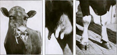
Foot-and-mouth disease, also known as aphthous fever, epizootic aphtha, and eczema contagiosa, is an acute, highly communicable disease chiefly confined to cloven-footed animals and characterized by an eruption of vesicles or blisters on the mucous membrane of the mouth and on the skin between the toes and above the hoofs. The vesicles rupture, forming erosions and ulcerations; there are also salivation, tenderness of the affected parts, loss of appetite, lameness, emaciation, and diminution in the quantity of milk secreted.
The tremendous ravages of the disease are seen in the number and variety of the species attacked. While it may be regarded as essentially a disease of cattle, hogs would seem to be as easy a prey. Almost in the same grade of receptivity are sheep and goats. Next in order of susceptibility come the buffalo, American bison, camel, chamois, llama, giraffe, and antelope. Horses, dogs, cats, and even poultry may occasionally become infected with the disease, the last three being particularly dangerous as carriers of the contagion. Man himself is not immune, and the frequency of his infection by coming in contact with diseased animals is established by numerous observations.
As with other communicable diseases, the source and origin of foot-and-mouth disease have given rise to much speculation. The disease had been known in Europe for centuries, but it was not until comparatively recent years that the erroneous conceptions of its spontaneous origin as a result of climatic and meteorological conditions, exhausting journeys, etc., were abandoned. It is now conceded that foot-and-mouth disease is propagated by a specific virus and that every outbreak starts from some preexisting outbreak.
So far investigators have been unable to identify or isolate the specific organism causing the disease, although numerous attempts have been made to cultivate and stain it by laboratory methods. Experiments have shown that the virus will pass through standard germ-proof filters, thus indicating its minute size and the reason it has not been detected by the staining methods. The contagion may be found in the serum of the vesicles on the mouth, feet, and udder; in the saliva, milk, and various secretions and excretions; also in the blood during the rise of temperature.
A wide distribution of the virus and a rapid infection of a herd is the result. Animals may be infected directly, as by licking, and in calves by sucking, or indirectly by such things as infected manure, hay, utensils, drinking troughs, railway cars, animal markets, barnyards, and pastures. Human beings may carry the virus on their shoes and clothing and transmit it on their hands when milking, since the udder is occasionally the seat of the eruption. It may also be carried by dogs, cats, rats, chickens, pigeons, etc. Milk in a raw state may also transmit the disease to animals fed with it.
The observations made by some veterinarians would lead us to suppose that the virus is quite readily destroyed. It is claimed that stables thoroughly cleaned become safe after drying for a short time; hence, litter of all kinds, such as manure or soiled hay and straw, may remain infective for a longer time because they do not dry out. Other authorities maintain that the virus is quite tenacious and may live in stables even so long as a year. They also state that animals which have passed through the disease may be a source of infection for several months after recovery.
Unlike most other infectious diseases, foot-and-mouth disease may repeatedly attack the same animals. The immunity conferred by an attack is of limited duration.
The period of incubation (that is, the time between the exposure of an animal to infection and the development of the disease) is variable, usually from three to six days. The disease may appear in 24 hours, or, in exceptional cases, not for 18 days or even longer.
Losses.—The highly contagious character of foot-and-mouth disease and its rapid spread to practically all exposed susceptible animals lead to heavy losses. Since the mortality is comparatively low, ranging from only 3 per cent or less in mild forms to 30 or 40 per cent in malignant cases, the havoc caused by the pestilence is sometimes underestimated. But there are other sources of loss which are much more important than the actual mortality. The fever and the difficulty of eating cause a rapid and extreme loss in flesh and a lessening or cessation of the milk secretion. The udders often become inflamed and ruined by the formation of abscesses, and cows affected in this way are sometimes rendered permanently valueless for milk production. The inflammation of the feet may cause the horn to drop from the toes, producing great lameness and lasting injury. Abortion is frequent, and typical lesions have been observed in the newly born at birth. Altogether these losses may amount to 20 or 30 per cent of the value of the affected animals.
In addition there are indirect losses of a commercial nature. Dairy farmers are put out of business for a time. Necessary quarantine restrictions greatly interfere with the movement of live stock and such commodities as hay, straw, hides, and farm produce. The business of the stockyards and slaughtering centers is greatly interfered with. Sometimes it is necessary to close stockyards for disinfection. The whole business of marketing, transporting, feeding, and slaughtering is interrupted and deranged. Losses of this character may reach enormous proportions.
PLATE XXXIII. FOOT-AND-MOUTH DISEASE. (Click to enlarge)
The disease in other countries.—Foot-and-mouth disease has prevailed in Europe for a great many years and has occasioned tremendous economic losses there.
In Italy, France, Switzerland, Germany, and Russia the plague has existed so long and has gained such a foothold that it is economically impossible to fight it with the American methods of slaughter and disinfection, for to do so would kill a large percentage of the live stock of those countries. In consequence, little or no progress toward eradication has been made by the authorities, though the severity of the disease in France appears to have abated somewhat in recent months.
The outbreak which appeared in Germany in 1888 increased steadily until 1892, when it diminished gradually for a few years, but the disease again reached great proportions in 1899. Thereafter it continued to exist to a greater or less extent until in 1911 it attained a virulence unequaled before. In that year 3,366,369 cattle, 1,602,927 sheep, 2,555,371 hogs, and 53,674 goats were affected. At that time the total number of cattle, sheep, swine, and goats in Germany was only 51,319,000, while there were in the United States 172,572,000, or between three and four times as many. It can readily be imagined, therefore, what it would mean to the United States if the disease were to gain the foothold here that it had in Germany, where, as these figures show, approximately one out of seven of the animals susceptible to the disease was affected.
The German Government, of course, has not left the disease to itself. It attempted to control some outbreaks by the method of slaughter, but the pestilence had gained too much headway and was too firmly established in too many portions of the country for this method to succeed, and the slaughter of the infected herds had to be abandoned. It now appears that there is no hope of getting rid of it until the virus has worn itself out. As soon as the animals' period of acquired immunity is over and favorable conditions present themselves, the contagion breaks out with renewed virulence. It has been impossible to control it by means of quarantines. One scientist has asserted that unless all the infected farms were absolutely isolated and the movement, not only of live stock but of persons, absolutely prohibited, the disease could not be stamped out. Such a quarantine is, of course, utterly impossible to enforce. In portions of Germany the farmers, realizing that the disease is inevitable, make haste to be done with it by exposing their stock deliber ately to mild cases in the hope that this will result in an immediate, mild attack and immunity for several years thereafter. Such immunity, however, is very uncertain.
Great Britain, Denmark, Norway, and Sweden, on account of their comparatively isolated positions, have been more successful in keeping out the disease. The outbreaks in those countries have been more sporadic, and by resorting to immediate slaughter the authorities have been able to stamp them out. Great Britain has applied both quarantine and slaughter for many years, and in an outbreak near Dublin in 1912 measures were adopted which were even more stringent than any that have been used in the United States. A British official (Cope) asserted in 1899 that after his country's experience with this disease it was "more dreaded by the farmers and stock raisers of Great Britain than cattle plague or pleuropneumonia, and they are now willing and ready to put up with any restrictions, of however drastic a character, considered necessary by the central department to stamp it out." The British authorities have succeeded in suppressing each outbreak, but reinfection often occurs from the neighboring continent. At the present time (April, 1922) Great Britain is having a siege of the disease, but is applying vigorous measures for its suppression.
In November, 1906, the disease reached Belgium from France, where it was quite prevalent, and by the end of the year every Province in Belgium was affected, and the Netherlands as well. Efforts to eradicate it from Belgium were unavailing. The Netherlands apparently succeeded in stamping it out for about six months, but it reappeared there.
The disease is also more or less prevalent in Central Europe, Spain, and in the Balkan countries.
Australia and New Zealand have remained free from it.
We have less accurate information regarding Asia and Africa, but the disease is known to prevail in Japan and China and in the Philippine Islands, and it is doubtful whether any considerable part of the Orient is free from it.
In South America it is reported as common in Brazil, Argentina, and Uruguay, and it probably exists in other countries.
Canada and Mexico are fortunately free from the disease.
Outbreaks in the United States.—Foot-and-mouth disease has appeared in the United States on six different occasions—1870, 1880, 1884, 1902, 1908, and 1914.
An extensive outbreak in 1870 was introduced by way of Canada, where the infection was brought by an importation of cattle from Scotland. It spread into the New England States and New York and appears to have been arrested within a few months. Its failure to spread more extensively and its early disappearance have been ascribed to favorable conditions, such as the movement of live stock from west to east, the limited trading at that period as compared with the present time, the restriction of traffic by winter weather, and the infrequency of travel which obtained at that time among people.
About 1880 two or three lots of animals affected by this disease were brought to the United States, but there was no extension from the animals originally affected.
In 1884, at Portland, Me., there was a small outbreak caused by imported cattle, and the disease spread to a few herds outside the quarantine station. Owing to the small number of animals affected and the limited area of territory covered by the disease, it was easily controlled by the ordinary measures of quarantine and disinfection.
It will be observed that in all these early outbreaks the contagion was introduced with imported animals. Since the development of a stringent system of inspection and quarantine of imported live stock, no instance of that kind has occurred. On subsequent occasions the infection has evidently been brought in with contaminated products or materials and not by means of live animals.
In November, 1902, the disease was discovered in Massachusetts and Rhode Island. The earliest cases were traced to Chelsea, Mass., near the docks, and it was suspected for a time that the infection was brought in with foreign shipping, by some such means as hay, straw, halters, ropes, hides, hair, wool, etc. Later developments, however, and especially investigations into the cause of the 1908 outbreak, led to the belief that a more probable source of the infection was cowpox vaccine virus imported from a country (probably Japan) where foot-and-mouth disease existed, the vaccine virus being contaminated with the virus of foot-and-mouth disease.
A Federal quarantine was declared by the Secretary of Agriculture on November 27, 1902, as soon as the nature of the disease was established, and steps for eradication were at once taken by the Bureau of Animal Industry of the United States Department of Agriculture in cooperation with authorities of the affected States. The methods followed consisted of inspection to trace and detect the disease, quarantine of infected premises and territory, slaughter and burial or burning of diseased and exposed animals, and disinfection of premises.
This outbreak involved Massachusetts, New Hampshire, Vermont, and Rhode Island, and was eradicated in about six months. Two hundred and forty-four herds, including 4,712 cattle, were found infected. Of these, 205 herds with 3,872 cattle, as well as 360 hogs and 220 sheep and goats, were slaughtered. The cattle infected but not slaughtered were those that either died or completely recovered before slaughtering could be carried out. The animals slaughtered were valued at $184,155.10, and the Federal Government reimbursed owners to the extent of 70 per cent, or $128,908.57. It is understood that the States paid the remainder. The total cost to the Department of Agriculture of stamping out the disease was about $300,000.
The next appearance of the foot-and-mouth disease was early in November, 1908, when it was observed in cattle near Danville, Pa. A Federal quarantine was issued November 12. The infection was traced back to the stockyards at East Buffalo, N. Y., and to Detroit, Mich. The disease appeared in the States of Michigan, New York, Pennsylvania, and Maryland. A careful and thorough investigation made by Mohler of the Bureau of Animal Industry and Rosenau of the Public Health Service demonstrated that the outbreak started from calves used to propagate vaccine virus at an establishment near Detroit, and that the source of the infection was contaminated Japanese vaccine virus.
Vigorous measures of eradication similar to those employed in 1902-3 were at once put into effect and the disease was stamped out in about five months at an expense of about $300,000 to the Department of Agriculture, and of about $113,000 to the States. The inspectors made 108,683 visits to farms, stockyards, etc., and inspected more than 1,500,000 animals (including reinspections). One hundred and fifty-seven premises were found infected, and 3,636 animals (2,025 cattle, 1,329 hogs, and 282 sheep and goats), valued at $90,033.18, were slaughtered. Owners were reimbursed for the value of their animals and property destroyed, one-third being paid by the States and two-thirds by the Federal Government.
The latest invasion was discovered in the vicinity of Niles, Mich., in October, 1914, after it had evidently been under way since August of the same year. This is the most serious and extensive outbreak ever known in this country. The disease extended to 22 States and the District of Columbia, at places ranging from the Atlantic to the Pacific coasts. The work of eradication was not completed for more than a year. The affected States were Connecticut, Delaware, Illinois, Indiana, Iowa, Kansas, Kentucky, Maryland, Massachusetts, Michigan, Minnesota, Montana, New Hampshire, New Jersey, New York, Ohio, Pennsylvania, Rhode Island, Virginia, Washington, West Virginia, and Wisconsin. Illinois had the largest infected area and the largest number of animals affected. The Union Stock Yards at Chicago became infected and were a source of dissemination of the contagion north, east, south, and west. These and other yards found infected were closed temporarily and disinfected.
The first Federal quarantine was issued October 19, 1914. A campaign to check the spread of the disease and to stamp it out was immediately begun by the United States Department of Agriculture in cooperation with the State authorities. Quarantines against the movement of animals and certain materials from the infected areas were declared, shipments were traced, rumors investigated, and thorough inspections made in an effort to discover all infected stock. As measures of eradication, diseased herds were slaughtered and buried and the premises disinfected. The owners of live stock and other property destroyed on account of the disease were reimbursed to the extent of the appraised value, half of which is paid by the Federal Government and half by the State. There were slaughtered 172,720 animals (76,575 cattle, 86,492 swine, 9,511 sheep, 133 goats, and 9 deer), in 3,482 herds. The total appraised value of these animals was more than $5,800,000. The expense to the Federal Government of eradicating this outbreak was about $4,540,000.
Symptoms.—In three to six days, or even longer, after the exposure of the animal to the infection the disease makes its appearance. It is usually first indicated by the animal suffering from a chill, quickly followed by an invasion of fever, which may cause the temperature to rise as high as 106° F. These symptoms are not always present, or may be in so slight a form as to escape notice. Following this in one or two days it will be noticed that small vesicles or blisters about the size of hempseeds or peas are making their appearance upon the mucous membranes of the mouth at the border and upper surface of the tongue near the tip, the inside of the cheeks, on the gums and the inner surface of the lips, or on the margin of the dental pad. These little blisters contain a yellowish, watery fluid and gradually become more extensive as the disease advances. Soon after the eruptions have appeared in the mouth of the animal considerable swelling, redness, and tenderness will be noticed about the feet, at the coronet, and between the digits of each foot. A day or two later eruptions similar to those within the mouth make their appearance upon these swollen regions of the foot, and at this stage it is usual to find that like lesions have made their appearance upon the perineum of the victim. In the case of milk cows the udder, and more particularly the teats, show the same vesicular eruption, but the latter as the result of milking soon become covered with reddened spots deprived of the superficial layer of skin and may develop deep, obstinate fissures.
As soon as the disease has become well established the patient evinces pain when attempting to eat; in fact, the appetite is often so seriously affected that all feed is refused, and the animal uneasily opens and shuts its mouth with a characteristic smacking sound, while strings of cohesive, ropy saliva hang suspended from the lips. With the advance of the disease the vesicles widen and extend until they may reach a diameter ranging from that of a dime to that of a silver dollar. These rupture soon after their appearance, sometimes on the first day, more rarely on the second or third day. After they have ruptured, the grayish-white membrane forming the blister may remain attached for a day or more, or disappear speedily and leave deeply reddened, sensitive spots or erosions, both within the mouth and upon the coronet and between the claws of the feet. Similar erosions, which quickly form scabs, as a rule, may be noticed in cases in which the teats of milk cows have become affected, and instances are reported in which sloughing of the tegument immediately around the lesions upon the udder has occurred. Owing to the tough, fibrous nature of the bovine skin, it is exceedingly rare for sloughing to occur upon any part of the body other than those mentioned.
The attack upon the feet of an animal is frequently manifested in all four feet at once, but one or more of the feet may entirely escape and remain unaffected throughout the course of the disease. The ulceration of the interdigital tissue may extend to the ligaments of the fetlock or produce disease of the joint or bone. As the feet become sensitive and sore the animal persistently lies down, and it has been found that bedsores develop with amazing rapidity in all such cases and wholly baffle all attempts at treatment until after the patient has regained its feet.
The disease may attack some of the internal organs before it appears upon any of the external tissues. These cases are very liable to prove quickly fatal. The animal dies from paralysis of the heart, due to the formation of poisonous principles within the system; it may suffocate by reason of the action of these same poisons upon the tissues of the lungs, or it may choke to death as a result of paralysis of the throat.
In cases of serious affection of the udder the erosions will often be found within the passages of the teats, resulting in a "caked" udder, and the same toxic poisoning which is the cause of death in the apoplectiform types just mentioned may arise from this source. In any event the milk from such cases is dangerous for use, causing fatal diarrhea in sucking calves or young pigs and serious illness in human consumers. The milk obtained from cows suffering with foot-and-mouth disease is not readily converted into either butter or cheese, but remains thick, slimy, and inert in spite of churning and attempts at curdling. Pregnant animals may abort. In pigs, sheep, and goats the lesions in the foot are most common, but both forms may be observed or only the mouth lesions.
When the disease has become fully established it will be found that the duration of the attack will vary greatly with different animals. From 10 to 20 days are usually required for the recovery of the normal appetite and spirits in mild outbreaks, while the return to a full flow of milk, in the case of milk cows, seldom occurs before the arrival of the following season.
In the malignant type of the disease it requires from three months to a year for an animal to recover. The mortality, as already stated, is usually low. The disease is more fatal in young animals that have been fed on infected milk, and produces death in from 60 to 80 per cent of these cases as a result of gastroenteritis. In the 1914 outbreak numerous new centers of infection started among hogs and calves which were fed on unpasteurized, infected milk from creameries.
Diagnosis.—The recognition of this affection should not, as a rule, be difficult, especially when the disease is known to be in the vicinity; in fact, the group of symptoms form a clinical picture too decided to be doubted. The combination of high fever, vesicular inflammation of the mouth, and hot, painful, swollen condition of the feet, followed 24 to 48 hours later by the appearance of numerous blisters varying in size from that of a pea to that of a walnut on the udder and feet and in the mouth should prevent any serious or long-continued error in the diagnosis; however, in the inoculation of calves we have a certain and final test. In 24 to 96 hours after inoculation the calves present the characteristic blisters. Such inoculation should be practiced, however, only by officials properly authorized to deal with contagious diseases.
Differential diagnosis.—The lesions of no other disease of cattle closely simulate the vesicular eruption of foot-and-mouth disease on the lining membrane of the mouth. When the blisters have ruptured, however, and the resulting lesions have become contaminated by numerous secondary forms of microorganisms, the correct recognition of the disease may be involved in considerable difficulty.
Cowpox or horsepox may be accidentally transmitted by inoculation. But the eruption of the "pox" goes on to the development of a pustule, while in foot-and-mouth disease the eruption is never more than a vesicle, even though the contained fluid may become turbid. The inoculation test in the case of cowpox does not respond with fever and eruption for at least 10 days, and often longer.
Necrotic stomatitis (sore mouth due to a germ) may be distinguished from foot-and-mouth disease by the fact that in the latter there is a rapid infection of the entire herd, including the adult cattle, as well as the infection of hogs and sheep. The characteristic lesion of foot-and-mouth disease is the appearance of blisters containing a serous fluid upon the mucous membrane of the mouth and upon the udder, teats, and feet of the affected animals. In necrotic stomatitis blisters are never formed, destruction of the tissues occurring from the beginning and being followed by the formation of yellowish, cheesy patches, principally found involving the lining membrane of the mouth, especially the tongue and cheeks.
In mycotic stomatitis (sore mouth due to a fungus or mold), portions of the lining membrane become inflamed and in a few days it changes to a croupous membrane which peels off, leaving a raw surface, while the thin skin between the toes may also be inflamed. The previous history of the case; the failure of the blisters, if any appear, to spread extensively; the absence of vesicular eruptions on other portions of the body, notably the udder and teats, and, characteristically, the hoof, together with the absence of rapid spread to practically all cattle in the herd and the complete negative character of inoculation of calves, distinguish between the local disease named and foot-and-mouth disease. Mycotic stomatitis occurs in only from 10 to 50 per cent of the animals in a herd, usually in the late summer or early fall after a dry spell, and it does not run a regular course.
The lesion, resulting from ergotism may be distinguished from those of foot-and-mouth disease by the lack of eruptions in the mouth and by the location of the disease at the tips of the ears, end of the tail, or upon the lower part of the legs, usually below the knees or hocks. The lesion of ergotism does not take the form of pustules or blisters, but manifests itself first as a swelling about the ankle, which later may slough and circumscribe the limb, forming a deep crack, extending entirely around the limb and forming a distinct line of demarcation between the healthy skin above and the diseased below. The absence of ulcerous sores on the coronet and between the claws, together with the healthy condition of the membranes of the mouth and the knowledge that the lesion upon the limb in question extends uninterruptedly around it, should point conclusively to a diagnosis of ergotism and to the exclusion of all fears of foot-and-mouth disease.