INFECTIOUS DISEASES OF CATTLE. (continued 3)
Reading edition
The plan adopted by the State and Federal authorities in eradication of the disease is known as "The Accredited-Herd Plan." Under this plan herds are tested under State and Federal supervision, the diseased animals are appraised, removed, and slaughtered under Federal inspection. Retests are then made after definite periods of time until two successive tests show all the animals to be free from the disease. At this time the herd owner is given a certificate of an accredited herd.
Details concerning the accredited-herd plan may be obtained by applying to the Chief of the Bureau of Animal Industry, Washington, D. C.
THE TUBERCULIN TESTS.
Testing animals with tuberculin is the process of introducing tuberculin into the animal and interpreting results according to well-known standards.
From the investigations and observations that have been mentioned, it may be safely concluded—
1. That the tuberculin test is a wonderfully accurate method of determining whether an animal is affected with tuberculosis.
2. That by its use the animals diseased with tuberculosis may be detected and removed from the herd, thereby eradicating the disease.
3. That it has no injurious effect upon healthy cattle.
4. That the comparatively small number of cattle which have aborted, suffered in health, or fallen off in condition after the test were either diseased before it was made or were affected by some cause other than the tuberculin.
THE SUBCUTANEOUS TEST (UNDER THE SKIN).
The most frequently used method of testing is the subcutaneous test, which consists in injecting the proper quantity of tuberculin underneath the skin into the subcutaneous tissue. If an animal is tuberculous, the action of the tuberculin causes a fever, which is indicated by a rise in temperature. This rise, under ordinary conditions, may occur any time between the eighth and twentieth hours after the tuberculin is injected, but in some cases it is desirable to measure the temperature before the eighth hour and continue to the twenty-fourth hour or longer.
The temperatures are measured at least 3 times in advance of the injection, at 2-hour intervals, to learn whether the animal is in proper condition to receive the test. The temperatures after injection are taken every 2 hours until the test is completed. The proper interpretation of the temperatures is made by the person applying the test, and a careful observance of any clinical changes is always important in determining the result. It can not be set forth too strongly that the test, including the two following methods, should be attempted only by those who are properly qualified to do the work.
THE INTRADERMIC TEST (INTO THE SKIN).
The intradermic test for detecting tuberculosis is used to a considerable extent, especially in area work and on range cattle not easily controlled. When made by those who have become skilled in its application, it is very accurate. In this test the tuberculin is injected between the layers of the skin, only a few drops being used, and it is usually applied in the region at the base of the tail, where the skin is soft and nearly hairless. The intradermic test is satisfactory also for the diagnosis of tuberculosis in swine and, when so used, the tuberculin is applied into the skin of the ear near its base.
The reaction from the intradermic test consists of a swelling at the point of injection and is observed from 72 to 150 hours after the injection. The character of the swelling varies, and a proper diagnosis of tuberculosis by this test can be made only by an experienced person.
THE OPHTHALMIC TEST (INTO THE EYE).
Still another method, known as the ophthalmic test, is used quite frequently and has been found to be of considerable value in what is known as "check" testing; that is, it is used in connection with either of the previously described methods. Sometimes a tuberculous animal that fails to react to those tests shows evidence of the disease upon the application of the ophthalmic test. The ophthalmic tuberculin is placed in one eye and the other eye is used as a check. A reaction is indicated by a characteristic discharge from the eye receiving the treatment, which may occur in from 3 to 10 hours after the application or even later. Some swelling and inflammation of the eye and lids are often noted.
TREATMENT OF TUBERCULOSIS.
Treatment of the disease is not seriously considered by any authorities at the present time.
The measures to be adopted to prevent the spreading of the disease must take into consideration not only the tubercle bacillus, but likewise all those circumstances that make cattle more susceptible to the disease, and which have already been dwelt upon. It would be useless to repeat here all that has been said above on the transmission of tubercle bacilli from one animal to another, and on the dangers of certain debilitating influences. A careful study of these influences will show how tuberculosis may, at least in some cases, be prevented. Great care should be bestowed upon the breeding, the surroundings, and the feed of the animal, so that the latter may be put into a condition to resist infection even when exposed to it. A tuberculin test should be applied to all strange cattle before they are introduced into the herd, and those which show a reaction should be refused.
A rigid exclusion of tuberculous animals is all that is necessary to prevent the appearance of the disease, provided cattle are not infected by consumptive persons and animals. The transmission of the disease from man to cattle is probably not frequent, but is regarded as a possible source of infection.
Tuberculosis in cattle must also be considered as bearing upon tuberculosis of other domesticated animals, particularly hogs. In Europe and the United States this disease is not uncommon among hogs, and appears to be on the increase. The reason for its existence may be looked for in the feeding of pigs with skim milk, buttermilk, and whey from creameries, with the offal of the abattoirs, with the household refuse generally, and behind tuberculous cattle. If tuber culosis is common among cattle, it is likely to be transmitted to hogs kept in this way.
The carcasses of animals which have died of tuberculosis should be buried deeply so that they can not be eaten by other animals. This is likewise true of all organs or tissues of slaughtered animals containing tubercles. These should never be fed to other animals, such as hogs, dogs, and cats, and should either be destroyed by fire or deeply buried.
When any of the animals in a herd of cattle show evident symptoms of tuberculosis, or when the tuberculin test proves that they are affected with this disease, the best method of procedure in most cases is to have the affected animals slaughtered and the stables disinfected. A large proportion of the animals which are slightly affected yield carcasses which are perfectly wholesome and fit for human food, but in all such cases there should be an inspection by an expert at the time of slaughter to determine which carcasses may be used and which should be destroyed.
The disinfection of stables may be accomplished by thoroughly cleaning them, scrubbing the floors with hot water, brushing down all loose dust from the walls, and tearing off all woodwork which is partly decayed. Then the whole interior of the stable should be covered with a good coat of limewash containing 1 part of formalin (which is a 40 per cent watery solution of formaldehyde) to 30 parts of the lime wash, or 4 ounces of formalin to each gallon of lime wash.
Similar precautions should be observed in removing the manure of the infected herd from the barnyard and other places accessible to cattle, since it is known that tuberculous cattle frequently eliminate large numbers of tubercle bacilli through the feces. The ground under the manure pile should then be disinfected, either by the above-mentioned formalin solution or by unslaked lime thickly sprinkled over the soil.
If all the animals which react are destroyed and the stables disinfected in this manner, the herd should remain free from the disease unless other affected animals are added to it. The introduction of the disease in this manner may be avoided by requiring a tuberculin test of all new animals admitted on the premises.
Unfortunately it is a fact that tuberculous animals which have been tested several times may become so accustomed to tuberculin that they will no longer react; consequently it is always advisable to purchase cattle from some one who is known to be reliable, as otherwise animals of this kind may be treated with tuberculin for the purpose of hiding the disease.
In the case of very valuable purebred animals and under exceptional circumstances it may be more advantageous to retain the reacting animals which are in good condition in order to breed from them and in that manner avoid the excessive loss which would follow from their immediate slaughter. This may be done if proper precautions are adopted.
The disposal of reactors depends upon the State laws and live-stock regulations of the State in which the herd belongs. If this policy is followed it should be attempted only after careful study of the plan known as the Bang method of controlling tuberculosis. The live-stock officials of the State should be frequently consulted and their advice followed; otherwise failure will surely ensue. The plan necessitates considerable trouble and is not recommended except under the circumstances mentioned.
BOVINE TUBERCULOSIS AND THE PUBLIC HEALTH.
The increasing amount of evidence pointing to the identity of human and animal tuberculosis, combined with the extraordinary mortality of human beings from this disease, often amounting to 10 to 14 per cent, has raised the question in all civilized countries as to how far animal, and especially bovine, tuberculosis is to blame for this high mortality. The medical and veterinary professions have approached this problem with equal zeal, and much has come to light within recent years which enables us to come to some conclusion. If this disease is transmitted from animals to man, how does the transmission take place? As comparatively few people come in direct contact with tuberculous cattle, it must be either through the meat, the milk, the butter, the cheese, or through all these products that the virus enters the human body. The question has thus narrowed itself down to the food products furnished by cattle.
It has become a very urgent question, especially in the poorer countries of Europe, whether all flesh from tuberculous animals is unfit for human food. It is argued there that if it can be shown that in the majority of cases of tuberculosis the bones and the muscular system are free from infection, there is no reason why the meat should not be put on sale under certain restrictions. The question may be resolved into two divisions: (1) How frequently does the disease invade those parts of the body which are used as food? (2) When the disease process is manifestly restricted to the internal organs, do tubercle bacilli circulate in the blood and lymph and can they be detected in the muscular tissue?
(1) Disease of the bones is not unknown, although very rare. According to Walley, it appears chiefly in the spongy bones of the head and backbone and in the long bones of the limbs. Occasionally the ends of the bones, where they are covered by the synovial membrane of the joints, are dotted with tubercles. The muscular system itself is very rarely the seat of tuberculous deposits, although the lymphatic glands lying near and among the muscles may be not infrequently diseased.
(2) Whether tubercle bacilli are found in muscle juice independent of any tuberculous deposits is a question which must be approached experimentally. There is on record a great variety of opinions on this matter, some authorities considering all flesh from tuberculous animals unfit for food, while others hold a contrary view. Experiments have shown that in rare cases the flesh of tuberculous cattle contains a small number of tubercle bacilli. In Germany the flesh of animals in which the disease is just beginning, or in which it is restricted to one or more related organs, is not rejected. When, however, the disease has affected the muscles or bones, or lymphatic glands situated on or between them, the flesh is condemned as unfit and dangerous. Animals are also rejected in which it is evident, from the general distribution of tubercles throughout the various organs, that the bacilli have been distributed by the blood and may have been carried into the muscular system (generalized tuberculosis).
Concerning the infectious nature of milk secreted by tuberculous cows, authorities have universally agreed that when the udder itself is in the slightest degree involved the milk possesses infectious properties, and is therefore dangerous. Tubercle bacilli in large numbers have been found in the milk and the udder under such circumstances. Unlike other affections of the udder, tuberculosis of this organ does not at once change the appearance and the quality of the milk secreted. Bang states that for at least a month after the disease has appeared the milk is normal in appearance and may be consumed and sold without arousing the suspicion of the owner. Considerable danger is, therefore, involved in this disease, and the necessity for the careful inspection of dairy cows seems more urgent than ever.
Authorities are, however, not fully agreed as to whether the milk from tuberculous cows in which the udder is apparently not invaded by the disease should be considered dangerous or not. Some incline to the belief that the milk secreted by healthy udders is never infectious, even when the lungs or other organs are affected; that, in other words, the tubercle bacilli are rarely, if ever, separated from the lesions which they produce, and that the udder itself must be diseased before tubercle bacilli can appear in the milk. Experiments made with the milk of tuberculous cows in which there were no indications of udder disease do not bear out this theory, since tubercle bacilli have been found in their milk. Some authorities still believe that the udder is diseased when the milk is infected, but that the disease escapes observation. However this may be, the fact that the udder may be diseased and the disease not recognizable simply casts suspicion upon all milk from tuberculous animals. We know that the milk of tuberculous cattle may or may not contain tubercle bacilli when the udder is apparently free from disease, but we have no rapid method of determining whether in any given case the milk contains tubercle bacilli or not. Moreover, the bacilli may be absent at one time and present at another in milk from the same cow. When we consider, therefore, the extent of tuberculosis and the hidden character of the disease, a certain degree of suspicion rests upon all milk from untested cattle. Fortunately, tubercle bacilli are readily destroyed by the temperature of boiling water, and hence both meat and milk are made safe, the former by the various processes of cooking, the latter by boiling for a few moments. It is incumbent upon all communities to have dairy cows examined and tested with tuberculin. If disease is detected, the affected animal should be killed at once or else all opportunity for the sale of such milk removed by appropriate measures. Where milk or cream is sold to the trade in large towns or cities pasteurization should be required as an additional safeguard.
Recently there has been much discussion of the question as to whether human and animal tuberculosis are identical diseases and as to the possibility of the tuberculosis of animals being transmitted to man or that of man being transmitted to animals.
The fact that tuberculous material from human subjects often failed to produce serious disease in cattle was observed by a number of the earlier investigators who experimented with such virus. It was the experiments and comparative studies of Theobald Smith, however, which attracted special attention to the difference in virulence shown by tubercle bacilli from human and bovine sources when inoculated upon cattle. Smith mentioned also certain morphological and cultural differences in bacilli from these two sources, and in the location and histology of the lesions in cattle produced by such bacilli. He did not conclude, however, that bovine bacilli could not produce disease in the human subject, but said:
It seems to me that, accepting the clinical evidence on hand, bovine tuberculosis may be transmitted to children when the body is overpowered by large numbers of bacilli, as in udder tuberculosis, or when certain unknown favorable conditions exist.
Koch, however, in his address at the British Congress on Tuberculosis, went far beyond this and maintained that "human tuberculosis differs from bovine and can not be transmitted to cattle." As to the susceptibility of man to bovine tuberculosis, he said it was not yet absolutely decided, but one was "nevertheless already at liberty to say that, if such a susceptibility really exists, the infection of human beings is but a very rare occurrence." He emphasizes this view in the following language:
I should estimate the extent of infection by the milk and flesh of tubercular cattle and the butter made of their milk as hardly greater than that of hereditary transmission, and I therefore do not deem it advisable to take any measures against it.
This conclusion was so radically different from the views of most experimenters and so out of harmony with facts which had apparently been demonstrated by others that it at once aroused opposition in the congress, followed by the adoption of dissenting resolutions, and led to numerous investigations in various countries. Koch's conclusions were based upon his failure to produce tuberculosis in cattle and other animals by inoculating them with tuberculous material of human origin and his success in causing progressive and fatal tuberculosis in the same kinds of animals when inoculated with tuberculous material of bovine origin. With such positiveness did he hold to the constant and specific difference between the human and bovine bacillus that he promulgated an experimental method of discriminating between them. Speaking of the etiology of intestinal tuberculosis in man, he said:
Hitherto nobody could decide with certainty in such a case whether the tuberculosis of the intestine was of human or of animal origin. Now we can diagnose them. All that is necessary is to cultivate in pure culture the tubercle bacilli found in the tubercular material, and to ascertain whether they belong to bovine tuberculosis by inoculating cattle with them. For this purpose I recommend subcutaneous injection, which yields quite specially characteristic and convincing results.
These important and comprehensive conclusions followed from a comparatively few experiments upon animals, and apparently no effort had been made to learn to what extent human tubercle bacilli may differ in their virulence for cattle or what grades of virulence there might be among bacilli of bovine origin. Vagedes had already shown that bacilli were sometimes present in human lesions which were as virulent as bovine bacilli, but his work was wholly ignored by Koch.
A considerable number of investigators, including Chauveau, Vagedes, Ravenel, De Schweinitz, Mohler, De Jong, Delépine, Orth, Stenström, Fibiger and Jensen, Max Wolff, Nocard, Arloing, Behring, Dean and Todd, Hamilton and Young, the German Tuberculosis Commission, and Theobald Smith, have found tubercle bacilli in the bodies of human beings who died of tuberculosis which proved to have about the same virulence for cattle as had the bacilli from bovine animals affected by the disease.
Kossel, in a preliminary report, stated that the German commission had tested 7 cultures of tuberculosis from cattle and hogs—4 from cattle and 3 from hogs. Two proved acutely fatal in cattle after eight to nine weeks; 4 likewise produced a generalized tuberculosis, but which certainly had a more chronic course, while 1 of the cultures caused only an infiltration at the point of inoculation, with some caseous foci in the adjoining prescapular gland and in one of the mediastinal glands, and there was lacking the spreading of the tuberculosis over the entire body which they were accustomed to see after the injection of cultures of bovine tuberculosis. "Hence," says Kossel, "among bovine tuberculosis bacilli there can also occur differences with regard to the virulence."
The German commission also tested 39 different freshly made cultures from tuberculous disease in man. Nineteen did not produce the slightest symptoms in cattle; with 9 others the cattle exhibited after four months very minute foci in the prescapular glands, which were mostly encapsuled and showed no inclination to progress; with 7 other cases there was somewhat more marked disease of the prescapular glands, but it did not go so far as a material spreading of the process to the adjoining glands. There were 4 cultures, however, which were more virulent and caused generalized tuberculosis in the cattle inoculated with them.
It would appear, therefore, that hereafter everyone must admit that it is impossible always to tell the source of a culture of the tubercle bacillus by its effect when it is inoculated upon cattle. One of the bovine cultures failed to produce generalized tuberculosis in cattle, and some of the human cultures did produce it in such animals. Moreover, while some of the human cultures caused no disease at all, others led to the development of minute foci in the prescapular glands, and still others to somewhat more marked disease of the glands. There were, consequently, four degrees of virulence noted in these 39 cultures of bacilli from human sources and three degrees of virulence in the 7 cultures from animal.
Now, if we accept the views of Koch as to the specific difference between human and bovine tubercle bacilli, and that the human bacilli produce only localized lesions in cattle, while bovine bacilli produce generalized lesions in them, must we not conclude that the one non-virulent bovine culture was in reality of human origin, and that the animal from which it was obtained had been infected from man? This is a logical deduction, but reverses the dictum laid down at London that human tuberculosis is not transmissible to cattle. Again, how are we to explain the human cultures of medium virulence? Are they human bacilli which, for some unknown reason, are increasing in virulence and approaching the activity of the bovine bacillus, or are they really bovine bacilli which have multiplied in the human body until their virulence has become attenuated? In whatever manner these questions are decided it would seem that the findings of the German commission, instead of supporting Koch's views that we can decide with certainty by the inoculation of cattle as to the source of any given bacillus, really show that this method of diagnosis is extremely uncertain in the present condition of our knowledge.
It is definitely admitted that 4 of the human cultures caused generalized tuberculosis in cattle; Kossel suggest, however, that it may be possible that the bacilli in cases of human tuberculosis under certain circumstances can likewise attain a very high pathogenic activity for cattle without being for that reason bovine bacilli. Undoubtedly the German commission is confronting the two horns of a dilemma, either one of which is fatal to the views of Koch as stated with such positiveness at London. If we accept this suggestion thrown out by Kossel, we must conclude that Koch was wrong in his claim that human tuberculosis can not be transmitted to cattle, and thus with one blow we destroy the entire experimental support which he had for his argument before the British Congress on Tuberculosis. If, on the other hand, we accept the conclusion which follows from the principle laid down by Koch for the discrimination between human and bovine bacilli, and which appears to be favored by Kossel, we must admit that bovine tuberculosis is an extremely important factor in the etiology of human tuberculosis. Of the 39 cases of human tuberculosis tested, 4, or more than 10 per cent, were virulent for cattle and would be classified as of bovine origin; however, these 4 cases, were all found among the 16 cases of tuberculosis in children which the commission investigated; hence it is plain that 25 per cent of the cases tested of tuberculosis in children would by Koch's method be classified as of bovine origin.
In the Bureau of Animal Industry two distinct lines of experiments have been carried on, in order that one might serve as a check against the other. There has been, however, no discrepancy in the results. De Schweinitz, in the Biochemic Division, Bureau of Animal Industry, isolated 9 cultures from human tuberculosis. Two were derived from human sputum, 3 from cases of generalized tuberculosis in adults, and 4 from cases of generalized tuberculosis in children. By comparing these cultures with a newly isolated virulent culture of bovine tuberculosis, there were found among them 2 cultures from children which were identical in their cultural and morphological characters with the bovine bacillus. These cultures also killed rabbits and guinea pigs in as short a time as did the bovine bacillus. Hogs which were inoculated subcutaneously with these 2 cultures from children died of generalized tuberculosis. Two calves weighing more than 300 pounds each were inoculated subcutaneously with these virulent human cultures, and as a result developed generalized tuberculosis. A yearling heifer inoculated with 1 of the cultures showed generalized tuberculosis when killed three months after inoculation. Both the cattle and the hogs had been tested with tuberculin and found to be free from tuberculosis before the inoculations were made. It is important to observe in this connection that 2 out of 4, or 50 per cent, of the cultures obtained from cases of generalized tuberculosis in children proved virulent for cattle.
Mohler, working in the Pathological Division, Bureau of Animal Industry, obtained 3 very virulent cultures of tubercle bacilli from the human subject. A goat inoculated subcutaneously with 1 of these cultures died in 37 days with miliary tuberculosis of the lungs involving the axillary and prescapular glands. This bacillus was obtained from the mesenteric gland of a boy. Of still greater interest is a bacillus isolated by Mohler from human sputum. A goat inoculated subcutaneously with a culture of this germ died in 95 days of pulmonary tuberculosis. A cat inoculated in the same manner died in 23 days of generalized tuberculosis. A rabbit similarly inoculated died in 59 days of pulmonary tuberculosis. Another rabbit inoculated with a bovine culture for comparison lived 10 days longer than the one inoculated with this sputum germ. Mohler also inoculated subcutaneously a 1-year-old heifer with a culture derived from the tuberculosis mesenteric gland of a boy 4 years of age. This culture was always refractory in its growth under artificial conditions, and the bacilli were short, stubby rods, corresponding in appearance to the bovine type. At the autopsy, held 127 days after the inoculation, the general condition was seen to be poor and unthrifty, and large, hard tumors were found at the points of inoculation. On the right side the swelling measured 3½ by 5 inches, and the corresponding lymph gland was 2¾ inches long by 1¾ inches in diameter. This gland contained numerous calcareous foci; one at the apex was an inch in diameter. The lesions on the left shoulder of the animal were very similar to those found on the right side, but the dimensions of the tumor were slightly less. The lungs presented an irregular mass of tuberculous nodules, and 7 or 8 grapelike nodules were seen on the parietal pleura. Bronchial and mediastinal lymph glands contained numerous tuberculous foci, and the pericardium, peritoneum, spleen, and liver also were affected.
In order to throw some light, if possible, upon the morphological constancy of the different types of tubercle bacilli, Mohler made comparative studies of bacilli from various sources, and which had been passed through various species of animals, by making the cultures upon dog serum after the method described by Theobald Smith. Some important results have been obtained. One culture of human bacilli which had morphological and cultural peculiarities similar to those of the bovine bacillus, and which produced only local lesions in cattle, was passed through a series of five cats. It was then found to be completely changed in its morphological characters, the rods being elongated, slender, more or less beaded, and entirely of the human type. Far from decreasing in virulence, however, as might be expected from its morphological appearance, this bacillus had so increased in its pathogenic activity that it produced generalized tuberculosis in a cow. This cow was inoculated subcutaneously in front of each shoulder with 2 cubic centimeters of a salt-solution emulsion of the tuberculous omentum of the last cat of the series. The cow rapidly lost flesh, had a temperature of 104° F., with the point of inoculation and adjacent glands greatly swollen. The autopsy revealed generalized tuberculosis, involving the lungs, mediastinal glands, spleen, liver, and kidneys. Tubercle bacilli of the bovine type obtained from the mesenteric glands of a sheep, hog, and cow were similarly transformed in their morphological appearance after being passed through a series of cats and recovered on dog serum. These bacilli also increased in virulence, as the last cat in the series invariably succumbed in a shorter time than the first of the series.
These experiments and observations indicate that the types of tubercle bacilli are very inconstant, and that under suitable conditions they readily change both in morphology and in virulence. A similar conclusion was reached by other investigators in working with the avian and porcine types of tubercle bacilli several years ago, and was reasonably to have been expected with the human and bovine types.
Later investigations made by Park and Krumweide, of the Research Laboratory of New York City, Novick, Richard M. Smith, Ravenel, Rosenau, Chung Yik Wang, and others tend to show the incidence of bovine infection in the human family. Chung Yik Wang stated in 1917 that studies of 281 cases of various clinical forms of tuberculosis in Edinburgh, Scotland, resulted in the isolation of the bovine tubercle bacilli in 78.4 per cent of cases under the age of 5 years, in 70.3 per cent between the ages of 5 and 16, and in 7.8 per cent over the age of 16. This investigator states that from the prophylactic point of view any measure resorted to in combating the disease should be directed not only against the human spread of infection, but also, more particularly in children's cases, against the bovine source of infection.
Ravenel, in summarizing the work of Drs. Park and Krumweide, as well as others throughout the world, gives the following results:
Of 63 children dying of tuberculosis at the babies' hospital 59 cases proved to be human infection and 4 bovine, a percentage of 6-1/3.
Of 9 children dying of tuberculosis at the foundling hospital 4 proved to have derived their infection from human sources and 5 from bovine, a percentage of 55.
Of a total of 88 children under 5 years of age who died of tuberculosis 77 proved to have derived their infection from human sources and 11 from bovine, a percentage of 12½.
Combining the cases studied in New York with those of other observers in different parts of this country and Europe, the following results are obtained:
Adults, 787 cases—777 human and 10 bovine infection.
Children, 5 to 16 years, 153 cases—117 human and 36 bovine infection.
Children under 5 years, 280 cases—215 human and 65 bovine infection.
The figures of the foundling hospital show the real danger of unprotected cows' milk.
The conclusion from these studies is inevitable, namely, that in children, in addition to the large number of deaths which occur from bovine infection, there are many cases of infection resulting in deformities, necessitating operations more or less severe in character and which frequently leave the patient disfigured permanently.
It must be plain to all, from these recent developments, that too much has been made of the slight differences in cultural characteristics, in morphology, and in virulence which have been observed in some cases in comparing the human and the bovine bacilli. The observations were interesting, and it was important that they be followed up until their significance was made entirely clear, but it was an almost unpardonable error, from a sanitary point of view, to promulgate sweeping generalizations calculated to arrest and abolish important measures for preventing human tuberculosis before the soundness of these generalizations had been established by a thorough course of experimentation.
When Koch said in the British Congress on Tuberculosis that he should estimate the extent of infection by the milk and flesh of tuberculous cattle and the butter made of their milk as hardly greater than that of hereditary transmission, and that he therefore did not deem it advisable to take any measures against it, he went far beyond what was justified by any experiments or observations which he reported, and he did a great deal of harm, which will be manifested for years to come, to those who endeavor to guard the human race from the dangers of animal tuberculosis. The researches which have been alluded to make these dangers more definite and certain than they have appeared before, and sanitarians should therefore most earnestly endeavor to counteract the erroneous and harmful impression which was made by Koch's address at London and his subsequent address at the International Conference on Tuberculosis at Berlin.
VACCINIA OR COWPOX.
Variola of cattle, more correctly vaccinia, is a contagious disease of cattle which manifests its presence through an elevation of temperature, a shrinkage in milk production, and by the appearance of characteristic, pustular eruptions, especially upon the teats and udders of dairy cows. Although this is a contagious disease, strictly speaking, it is so universally harmless and benign in its course that it is robbed of the terrors which usually accompany all spreading diseases, and is allowed to enter a herd of cattle, run its course, and disappear without exciting any particular notice.
The contagion of cowpox does not travel through the air from animal to animal, but is transfused only by actual contact of the contagious principle with the skin of some susceptible animal. It may be carried in this manner, not alone from cattle to cattle, but horses, sheep, goats, and man may readily contract the disease whenever suitable conditions attend their inoculation.
An identical disease frequently appears upon horses, attacking their heels, and thence extending upward along the leg, producing, as it progresses, inflammation and swelling of the skin, followed later by pustules, which soon rupture, discharging a sticky, disagreeable secretion. Other parts of the body are frequently affected in like manner, especially in the region of the head, where the eruptions may appear upon lips and nostrils, or upon the mucous surfaces of the nasal cavities, mouth, or eyes.
Variola of the horse is readily transmitted to cattle, if both are cared for by the same attendant, and, conversely, variola of cattle may be carried from the cow to the horse on the hands of a person who has been milking a cow affected with the disease.
The method of vaccination with material derived from the eruptions of cowpox as a safeguard against the ravages of smallpox in members of the human family is well known. The immunity which such vaccination confers upon the human subject has led many writers to assert that cowpox is simply a modified form of smallpox, whose harmless attack upon the human system is owing to a certain attenuation derived during its passages through the system of the cow or horse. The results of numerous experiments which have been carried out for the purpose of determining the relationship existing between variola of the human and bovine families seem to show, however, that although possessing many similar characteristics, they are nevertheless distinct, and that in spite of repeated inoculations from cattle to man, and vice versa, no transformation in the real character of the disease ever takes place.
Symptoms.—The disease appears in four to seven days after natural infection, or may evince itself in two or three days as the result of artificial inoculation. Young milch cows are most susceptible to an attack, but older cows, bulls, or young cattle are by no means immune. The attack causes a slight rise in temperature, which is soon followed by the appearance of reddened, inflamed areas, principally upon the teats and udder, and at times on the abdominal skin or the skin of the inner surface of the thighs. In a few cases the skin of the throat and jaws has been found similarly involved. If the affected parts are examined on the second day after the establishment of the inflammation numerous pale-red nodules will be found, which gradually expand until, within a few days, they reach a diameter of one-half inch or even larger. At this period the tops of the nodules become transformed into vesicles which are depressed in the center and contain a pale, serous fluid. They usually reach their maturity by the tenth day of the course of the disease and are then the size of a bean. From this time the contents of the vesicles become purulent, which requires about three days, when the typical pox pustule is present, consisting of a swelling with broad, reddened base, within which is an elevated, conical abscess varying from the size of a pea to that of a hazelnut.
The course of the disease after the full maturity of the pustule is rapid if outside interference has not caused a premature rupture of the small abscess at the apex of the swelling. The pustules gradually become darker colored and drier until nothing remains but a thick scab, which at last falls off, leaving only a slight, whitish scar behind. The total duration of the disease covers some 20 days in each animal, and owing to the slow spread of the infection from animal to animal, many weeks may elapse before a stable can be fully freed from it. The fallen scabs and crusts may retain their contagious properties for several days when mixed with litter and bedding upon the floor of the stable, and at any time during this period they are capable of producing new outbreaks should fresh cattle be brought into the stalls and thus come into actual contact with them. Again, the pustules may appear, one after another, on a single animal, in which case the duration of the disease is materially lengthened.
Treatment.—In herds of cattle that regularly receive careful handling, no special treatment will be found necessary beyond the application of softening and disinfecting agents to such vesicles upon the teats as may have become ruptured by the hands of the milker. Carbolized vaseline or iodoform ointment will be found well suited to this work. In more persistent cases it may be found desirable to use a milking tube in order to prevent the repeated opening of the pustules during the operation of milking. Washing the sores twice daily with a weak solution of zinc chlorid (2½ per cent solution) has been found to assist in checking the inflammation and to cleanse and heal the parts by its germicidal action. When the udder is hard, swollen, and painful, support it by a bandage and foment frequently with hot water. If calves are allowed to suckle the cows the pustules become confluent, and the ulcerations may extend up into the teat, causing garget and ruining the whole quarter of the udder.
As young cows are most susceptible to variola, the milker must exercise constant patience with these affected animals so long as their teats or udders are sore and tender, else the patient may contract vicious habits while resisting painful handling. The flow of milk is usually lessened as soon as the fever becomes established, but is again normal with the return of perfect health.
The practice of thorough cleanliness in handling or milking affected cattle may, in many instances, prevent the dissemination of the trouble among the healthy portion of the herd, but even the greatest care may prove insufficient to check the spread until it has attacked each animal of the herd in turn.
ACTINOMYCOSIS (LUMPY JAW)
[Pls. XXXIX-XLI.]
Actinomycosis, also known as lumpy jaw, big jaw, wooden tongue, etc., is a chronic infectious disease characterized by the formation of peculiar tumors in various regions of the body, more particularly the head, and is due to the specific action of a certain fungus-like germ (actinomyces). This fungus is an organism which occurs in the tissues in the form of rosettes, and it has therefore been termed the "ray fungus." The disease is not directly transmitted from one animal to another, but it seems apparent that the fungus is conveyed into the tissues by various feedstuffs through slight wounds of the mucous membrane of the mouth, decayed teeth, or during the shedding of milk teeth. The ray fungus is found in nature vegetated on grasses, on the awns of barley, the spears of oats, and on other grains. Quantities of the fungi have been found between the vegetable fibers of barley which had penetrated the gums of cattle and on the awns of grain embedded in the tongues of cows.
Although actinomycotic tumors on cattle had been the object of study for many years, it was not until 1877 that the constant presence of actinomyces was pointed out by Bollinger, of Munich, and since that time considered the cause. This fungus was observed in these tumors as early as 1860 by Rivolta, and by others subsequently, without having been suspected as causing them.
Since Bollinger's publication much work has been done, many observations made, and many hitherto obscure disease processes brought into relation with this fungus. Furthermore, a similar disease in man was first definitely shown to be associated with the same fungus in 1878 by Israel, and in the following year Ponfick pointed out that the disease described by Bollinger in animals and that found by Israel in man were due to the same cause; that is, that the fungi described by these observers were one and the same.
The tumors and abscesses wherever they may be are all found to be the same in origin by the presence of the actinomyces fungus. When they are incised, a very close scrutiny with the naked eye, or, at most, a hand lens, will reveal the presence of minute grains which vary from a pale-yellow to a sulphur-yellow color. They may be very abundant or so few as to be overlooked. They are embedded in the soft tissue composing the tumor or in the pus of the abscess. With a needle they are easily lifted out from the tissue, and then they appear as roundish masses about one-half millimeter (1/50 inch) in diameter. To anyone familiar with the use of a microscope the recognition of these grains or particles without any previous preparation is a comparatively easy task.
When examined in the fresh condition under a microscope magnifying up to 250 diameters the general structure is made out without much difficulty. These grains consist of collections of minute, roundish masses. Their outer surface is made up of club-shaped bodies all radiating from the center of the mass (see Pl. XXXIX, fig. 2), somewhat like a rosette. If the fungus is crushed, the interior is found made up of bundles of very fine filaments, which are probably continuous into the club-shaped bodies. The addition of a dilute solution of caustic soda or potash greatly aids the examination, as it removes the layer of cells adhering to the fungus, which obscures the structure. Now and then these grains are found to be in a calcified condition. The exterior is incrusted in lime salts, which are dissolved by adding some weak dilute acid, like acetic acid. Only by this procedure can the fungus be definitely recognized when in a mummified condition.
These are the bodies whose presence causes sufficient irritation in the tissues into which they find their way to set up inflammatory growths. These growths increase as the fungus continues to multiply until they reach enormous dimensions, if the affected animal is permitted to live long enough. The true nature of this parasite is not yet definitely settled, although many excellent observers have occupied themselves with it. According to earlier observers, it is a true fungus. Later ones are inclined to place it among the higher bacteria. Present knowledge concerning the actinomyces growth indicates that it should be classified with the higher bacteria or trichomycetes.
Whatever the situation of the disease caused by actinomyces may be, its nature is fundamentally the same and peculiar to the fungus. The pathological details which make this statement clear can not be entered upon in this place, nor would they be of any practical value to the farmer. We will simply dwell upon a few obvious characters.
The consistency of the tumor varies in different situations according to the quantity of fibrous or connective tissue present. When very little of this is present the tumor is of a very soft consistency. As the quantity of connective tissue is increased the tumor is firmer and of a more honeycombed appearance. The individual actinomyces colonies are lodged in the spaces or interstices formed by the meshwork of the connective tissue. There they are surrounded by a mantle of cellular elements which fill up the spaces. By scraping the cut surface of such a tumor these cell masses inclosing the fungi come away, and the latter may be seen as pale-yellow or sulphur-yellow specks, as described above.
Location of the disease.—In cattle the disease process may be located both externally, where it is readily detected, and in internal organs. Its preferred seat is on the bones of the lower and upper jaws, in the parotid salivary gland in the angle of the jaw, and in the region of the throat. It may also appear under the skin in different parts of the body. Internally it may attack the tongue and appear in the form of a tumor in the mouth, pharynx, or larynx. It may cause extensive disease of the lungs, more rarely of the digestive tract.
It appears, furthermore, that in certain districts or countries the disease seems by preference to attack certain parts. Thus in England actinomycosis of the tongue is most prevalent. In Denmark the soft parts of the head are most prone to disease, while in Russia the lips are the usual seat. In certain parts of Germany actinomycotic tumors are most frequently encountered in the throat region and in the jawbones.
A description of actinomycosis of the jaw (lumpy jaw) and of the tongue has already been given in a previous chapter, and hence they will be dealt with here only very briefly. When the disease attacks the soft parts of the head a rather firm swelling appears, in which are formed one or more smaller projecting tumors, varying from the size of a nut to that of an egg. These push their way outward and finally break through the skin as small, reddish, funguslike bodies covered with thin sloughs. Or the original swelling, in place of enlarging in the manner described, may become transformed into an abscess which finally bursts to discharge creamy pus. The abscess cavity, however, does not disappear, but is soon filled with fungus-like growths, which force their way outward through the opening.
When the tumors are situated within the cavity of the pharynx they have broken through from some gland, perhaps beneath the mucous membrane, where the disease first appeared, and hang or project into the cavity of the pharynx, either as pendulous masses with slender stems or as tumors with broad bases. Their position may be such as to interfere with swallowing and with breathing. In either case serious symptoms will soon appear.
The invasion of the bones of the jaws by actinomycosis must be regarded as one of the most serious forms of the disease. (Pls. XXXIX, XL.) It may start in the marrow of the bone and by a slow extension gradually undermine the entire thickness of the bone itself. The growth may continue outward, and after working its way through muscle and skin finally break through and appear externally as stinking fungoid growths. The growth may at the same time work its way inward and appear in the mouth. The disease may also begin in the periosteum, or covering of the bone, and destroy the bone from without inward.
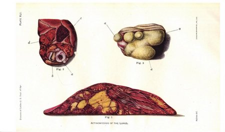
Actinomycosis of the lungs is occasionally observed, and it is not improbable that at times it has been mistaken for tuberculosis. The actinomyces grains are, however, easily observed if the diseased tissue is carefully examined. The changes in the lungs as they appear to the naked eye vary considerably from case to case. Thus in one animal the lungs were affected as in ordinary bronchopneumonia as to the location, extent, and appearance of the disease process. The affected lobes had a dark-red flesh appearance, with yellowish areas sprinkled in here and there. (See Pl. XLI, figs. 1, 2.) These latter areas were the seat of multiplication of the actinomyces fungus. In another case, of which only a small portion of the lungs was sent to the laboratory, they were completely transformed into a uniformly grayish mass, very soft and pulpy to the touch, and appearing like very soft and moist dough. (Pl. XLI, fig. 3.) The actinomyces grains were exceedingly abundant in this tissue, and appeared when the tissue was incised as minute sulphur-yellow grains, densely sprinkled through the tissue, which readily came away and adhered to the knife blade. In still another case a portion of the lung tissue was converted into large, soft masses from 1 to 3 inches in diameter, each partly inclosed in very dense connective tissue. These soft, grayish-yellow masses likewise resembled moist dough in their consistency, and the actinomyces grains, though neither very distinct nor at all abundant, were easily fished out and identified as such. A portion of this growth, which was as large as a child's head, was converted into an abscess filled with creamy semiliquid pus.
This case differed from the preceding in that all appearance of lung tissue had disappeared from the diseased mass. Only on the exterior the lung tissue could be recognized, although even there it had been largely converted into very dense, whitish connective tissue inclosing the fungoid growth. In the other case the external form of the lung and the shape and outline of the lobules were preserved, but the lung tissue itself was not recognizable as such. In the case first mentioned the changes were still less marked, and actinomycosis would not have been suspected by a simple inspection. These few illustrations suffice to show that actinomycosis of the lungs may appear under quite different forms, and that the nature of the disease can be accurately determined only by finding the fungus itself. Rarely actinomycosis attacks the body externally in places other than the head and neck. Crookshank describes the case of a bull in which the flank was attacked and subsequently the scrotum became diseased. A large portion of the skin of the flank was destroyed and covered with a leathery crust. When this was pulled away the pus beneath it showed the actinomyces grains to the naked eye.
Actinomycosis may also involve the udder, the spermatic cord of castrated animals, the vagina, and, when it becomes generalized, the brain, liver, spleen, and muscular tissue.
Actinomycosis may in some cases be confounded with tuberculosis. The diagnosis does not offer any difficulties, since the presence of the actinomyces fungus at once removes any existing doubts. As has already been intimated, these grains, simulating sulphur balls, are visible to the naked eye, and their nature is readily determined with the aid of a microscope.
The course of the disease is quite slow. As the tumors grow they may interfere with the natural functions of the body. According to their situation, mastication, rumination, or breathing may be interfered with, and in this way the animal may become emaciated. Actinomycosis of the jawbones leads to destruction of the teeth and impedes the movements necessary to chewing the feed. Similarly, when the disease attacks the soft parts of the head obstructions may arise in the mouth by an inward growth of the tumor. If tumors exist in the pharynx they may partially obstruct the movements necessary to breathing, or close the air passages and cause partial suffocation. Actinomycosis of the tongue, in interfering with the many and varied movements of this important organ, is also a serious matter. There is no reason to suppose that the localized disease interferes with the general health in any other way than indirectly until internal organs, such as the lungs, become involved.
A very small proportion of the cases may recover spontaneously, the tumors being encysted or undergoing calcification. In most cases the disease yields readily to proper treatment, and about 75 per cent of the affected animals may be cured.
Prevention.—The question as to how and where animals take this disease is one concerning which we are still in the stage of conjecture, because so far we possess very little information concerning the life history of the actinomyces itself. The quite unanimous view of all observers is that animals become infected from the feed. The fungus is lodged upon the plants and in some way enters the tissues of the head, the lungs, and the digestive tract, where it sets up its peculiar activity. It is likewise generally believed that the fungus is, as it were, inoculated into the affected part. This inoculation is performed by the sharp and pointed parts of plants which penetrate the mucous membrane and carry the fungus with them. The disease is therefore inoculable rather than contagious. The mere presence of the diseased animal will not give rise to disease in healthy animals unless the actinomyces grains pass directly from the diseased into some wound or abrasion of the healthy or else drop upon the feed which is consumed by the healthy. Not only are these views deducible from clinical observation, but they have been proved by the positive inoculation of calves and smaller animals with actinomyces. The danger therefore of the presence of actinomyces for healthy animals is a limited one. Nevertheless an animal affected with this disease should not be allowed to go at large or run with other animals. If the fungus is being scattered by discharging growths we certainly can not state at this stage of our knowledge that other animals may not be infected by such distribution, and we must assume, until more positive information is at hand, that this actually occurs.
It is, however, the opinion of the majority of authorities that when actinomycosis appears among a large number of animals they all contract it in the same way from the feed. Much speculation has therefore arisen whether any particular plant or group of plants is the source of the infection and whether any special condition of the soil favors it. Very little positive information is at hand on these questions. It would be very desirable for those who live in localities where this disease is prevalent to make statistical and other observations on the occurrence of the disease with reference to the season of the year, the kind of feed, the nature of the soil (whether swampy or dry, recently reclaimed, or cultivated for a long time) upon which the animals are pastured or upon which the feed is grown.
It is highly probable that such investigations will lead to an understanding of the source of the fungus and the means for checking the spread of the disease itself. Veterinarian Jensen, of Denmark, made some observations upon an extensive outbreak of actinomycosis a number of years ago which led him to infer that the animals were inoculated by eating barley straw harvested from pieces of ground just reclaimed from the sea. While the animals remained unaffected so long as they pastured on this ground or ate the hay obtained from it, they became diseased after eating the straw of cereals from the same territory. Others have found that cattle grazing upon low pastures along the banks of streams and subject to inundations are more prone to the disease. It has also been observed that feed gathered from such grounds, even after prolonged drying, may give rise to the disease. Actinomycosis is not infrequent in cattle in the Southwest and is generally supposed to be the result of eating the prickly fruit of the cactus plant, causing wounds of the mucous membrane and subsequent infection with the parasite. Much additional information of a similar kind must be forthcoming before the source and manner of infection in this disease and its dependence upon external conditions will be known. It is not at all improbable that they may vary considerably from place to place.
Treatment.—Until recently this has been almost entirely surgical. When the tumors are external and attached to soft parts only, an early removal may lead to recovery. This, of course, can be undertaken only by a trained veterinarian, especially as the various parts of the head and neck contain important vessels, nerves, and ducts which should be injured as little as possible in any operation. Unless the tumor is completely removed it will reappear. Disease of the jawbones is at best a very serious matter and treatment is liable to be of no avail.
In March, 1892, an important contribution to our knowledge of this subject was made by Nocard, of the Alfort Veterinary School, in a communication to the French Central Society of Veterinary Medicine. He showed clearly that the actinomycosis of the tongue, a disease which appears to be quite common in Germany, and is there known as "wooden tongue," could be quickly and permanently cured by the administration of iodid of potassium. Nocard calls attention to the success of Thomassen, of Utrecht, who recommended this treatment so long ago as 1885, and who has since treated more than 80 cases, all of which have been cured. A French veterinarian, Godbille, has used the same remedy in a number of cases of actinomycosis in the tongue, all of which have been cured. Nocard also gives details of a case which was cured by himself.
All the cases referred to were of actinomycosis of the tongue, and no one appears to have attempted the cure of actinomycosis of the jaw until it was undertaken by Nörgaard, of the Bureau of Animal Industry. In April, 1892, he selected a young steer in fair condition which had a tumor on the jaw measuring 15½ inches in circumference and from which a discharge had already been established. This animal was treated with iodid of potassium, and the result was a complete cure.
The iodid of potassium is given in doses of 1½ and 2½ drams once a day, dissolved in water, and administered as a drench. The dose should vary somewhat with the size of the animal and with the effects that are produced. If the dose is sufficiently large signs of iodism appear in the course of a week or 10 days. The skin becomes scurfy, there is weeping from the eyes, catarrh of the nose, and loss of appetite. When these symptoms appear the medicine may be suspended for a few days and afterward resumed in the same dose. The cure requires from three to six weeks' treatment. Some animals, generally the ones which show no signs of iodism, do not improve under treatment with iodid of potassium.
If there is no sign of improvement after the animals have been treated four or five weeks, and the medicine has been given in as large doses as appear desirable, it is an indication that the particular animal is not susceptible to the curative effects of the drug, and the treatment may therefore be abandoned.
It is not, however, advisable to administer iodid of potassium to milch cows, as it will considerably reduce the milk secretion or stop it altogether. Furthermore, a great part of the drug is excreted through the milk, making the milk unfit for use. It should not be given to animals in advanced pregnancy, as there is danger of producing abortion.
The best results are obtained by pushing the drug until its effect is seen. The many tests to which this treatment has been subjected have proved, with few exceptions, its specific curative value. In addition to this the tumor should be painted externally with either the tincture of iodin or Lugol's solution, or the drug should be injected subcutaneously into the tumor.
Godbille has given as much as 4 drams of potassium iodid in one day to a steer, decreasing the dose one-fourth dram each day until the dose was 1¼ drams, which was maintained until the twelfth day of treatment, when the animal appeared to be entirely cured.
Nocard gave the first day 1½ drams in one dose to a cow; the second and succeeding days a dose of 1 dram in the morning and evening, in each case before feeding. This treatment was continued for 10 days, when the animal was cured.
Actinomycosis and the public health.—The interest which is shown concerning this cattle disease is largely due to the fact that the same disease attacks human beings. Its slow progress, its tendency to remain restricted to certain localities, and the absence of any directly contagious properties have thus far not aroused any anxiety in other countries as to its influence on the cattle industry, not even to the point of placing it among the infectious diseases of which statistics are annually published. Its possible bearing on public health has, however, given the disease a place in the public mind which it hardly deserves.
It has already been stated that the actinomyces fungus found in human disease is considered by authorities the same as that occurring in bovine affections. It is therefore of interest to conclude this article with a brief discussion of the disease in man and its relation to actinomycosis in cattle.
In man the location of the disease process corresponds fairly well to that in cattle. The majority of cases which have been reported in different parts of the world—and they are now rather numerous—indicate disease of the face. The skin, tongue, or the jawbones may become affected, and by a very slow process it may extend downward upon the neck and even into the cavity of the chest. In many cases the teeth have been found in a state of more or less advanced decay and ulceration. In a few cases disease of the lungs was observed without coexisting disease of the bones or soft parts of the head. In such cases the fungus must have been inhaled. The disease of the lungs after a time extends upon the chest wall, where it may corrode the ribs and work its way through the muscles and the skin. An abscess is thus formed discharging pus containing actinomyces grains. Disease of the digestive organs caused by this fungus has also been observed in a few instances.
Granting the identity of the disease in man and cattle, the question has been raised whether cattle are responsible for its occurrence in man. Any transmission of the infectious agent may be conceived of as taking place during the life of the animal and from the meat after slaughter. That human beings have contracted actinomycosis by coming in contact with diseased cattle is not shown by the cases that have been reported, for the occupations of most of the patients did not bring them into any relation whatever with cattle. While the possibility of such direct transmission is not denied, nevertheless it must be considered as extremely remote. Practically the same position is maintained at present by most authorities as regards the transmission of the disease to man by eating meat. Israel, who has studied this question carefully, found the disease in Jews who never ate pork,[5] and who likewise were protected from bovine actinomycosis by the rigorous meat inspection practiced by that race. Furthermore, it must be borne in mind that actinomycosis is a local disease, causing great destruction of tissue where the fungus multiplies, but which very rarely becomes generally disseminated over the body from the original disease focus. The fungus is found only in places where the disease process is manifest to the eye or becomes so in a very short time after the lodgment of the fungus. Only the greatest negligence would allow the actually diseased parts to be sold and consumed. Finally, this parasite, like all others, would be destroyed in the process of cooking. Most authorities thus do not believe that actinomycosis in man is directly traceable to the disease in animals, but are of the opinion that both man and animals are infected from a third source, which has already been discussed above. How far these views may be modified by further and more telling investigations of the parasite fungus itself no one can predict. There are still wide gaps in our knowledge, and the presentation above simply summarizes the prevailing views, from which there are dissenters, of course. An attempt to give the views of both sides on this question would necessitate the summarizing and impartial discussion of all the experiments thus far made—a task entirely beyond the scope of the present work.
Whether an animal affected with actinomycosis should be used for human food after all diseased organs and tissues have been thoroughly removed is a question the answer to which depends on a variety of circumstances. Among these may be mentioned the thoroughness of the meat inspection itself, the extent of the disease, and the general condition of the animal affected.
The Federal meat-inspection regulations require that carcasses of animals showing generalized actinomycosis shall be condemned. If carcasses are in a well-nourished condition, showing uncomplicated localized lesions of actinomycosis, they may be passed after the infected organs or parts have been removed and condemned. When the disease of the jaw is slight, strictly localized, and without pus formation, fistulous tracts, or lymph-gland involvement, the tongue, if free from disease, may be passed. The heads affected with actinomycosis (lumpy jaw), including the tongue, shall be condemned, except that when the lesions in the jaw are strictly localized and slight in extent, the tongue may be passed, if free from disease.
ANTHRAX.
Anthrax or charbon may be defined as an infectious disease which is caused by specific bacteria, known as anthrax bacilli, and which is more or less restricted by conditions of soil and moisture to definite geographical localities. While it is chiefly limited to cattle and sheep, it may be transmitted to goats, horses, cats, and certain kinds of game. Smaller animals, such as mice, rabbits, and guinea pigs, speedily succumb to inoculation. Dogs and hogs are slightly susceptible, while fowls are practically immune. The variety of domesticated animals which it may attack renders it one of the most dreaded scourges of animal life. It may even attack man. Of this more will be stated further on.
ACTINOMYCOSIS (LUMPY JAW).
DESCRIPTION OF PLATES.
Plate XXXIX. Actinomycosis. (From Jöhne's Encyklopädie d. gesammt. Thierheilkunde.)
Fig. 1. Actinomycosis of the jaw. The lower jawbone has been extensively eaten away by the disease.
Fig. 2. Actinomyces fungus from a tumor of the jawbone in cattle, magnified 550 times.
Plate XL. Actinomycosis of the jaw. (Reduced one-half. From Jöhne's Encyklopädie d. gesammt. Thierheilkunde.) The lower jaw is sawed through transversely, i.e., from right to left, and shows the disease within the jawbone itself; a, within the mouth, showing the papillæ on the mucous membrane of the cheek; b, front view of a molar tooth; c, the skin covering the lower surface of the jawbone; d, the jawbone hollowed out and enlarged by the formation of cavities within it, which are filled with the soft growth of the actinomycotic tumor. The section makes it appear as if the bone were broken into fragments and these forced apart; e, a portion of the tumor which has broken through the bone and the skin and appears as a tumor on the cheek. The little roundish masses represent the granulomata (minute tumors) in which the fungus vegetates.
Plate XLI. Actinomycosis of the lungs.
Fig. 1. Transverse section of the ventral lobe of the right lung, from a case studied in the laboratory. The yellowish dots represent the places where the actinomyces fungus is lodged. The larger yellowish patches are produced by the confluence of a number of isolated centers. The entire lobe is of a dark flesh-red color, due to collapse and bronchopneumonia.
Fig. 2. The cut surface of a portion of the principal lobe of the same lung, showing the recent invasion of antinomycosis from the other lobe: a, large air tube; b, artery; c, a pneumatic lobule; d, lobule containing minute yellowish dots. In these the actinomyces fungus is lodged.
Fig. 3. Cut surface of a small portion of another lung, showing a few lobules, a. The fungus is sprinkled throughout the lung tissue in the form of yellowish grains, as shown in the illustration. The pleural covering of the lung tissue is shown in profile above.
PLATE XXXIX. ACTINOMYCOSIS. (Click to enlarge)
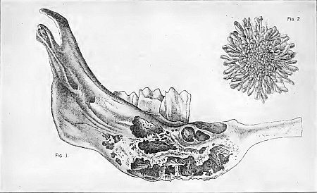
PLATE XL. ACTINOMYCOSIS OF THE JAW. (Click to enlarge)
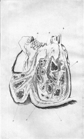
PLATE XLI. ACTINOMYCOSIS OF THE LUNGS. (Click to enlarge)
Cause.—The cause of anthrax is a microscopic organism known as the anthrax bacillus. (See Pl. XXVIII, fig. 7.) In form it is cylindrical or rodlike, measuring 1/5000 to 1/2500 inch in length and 1/25000 inch in diameter. Like all bacteria, these rodlike bodies have the power of indefinite multiplication, and in the bodies of infected animals they produce death by rapidly increasing in numbers and producing substances which poison the body. In the blood they multiply in number by becoming elongated and then dividing into two, each new organism continuing the same process indefinitely. Outside the body, however, they multiply in a different way when under conditions unfavorable to growth. Oval bodies, which are called spores, appear within the rods, and remain alive and capable of germination after years of drying. They also resist heat to a remarkable degree, so that boiling water is necessary to destroy them. The bacilli themselves, on the other hand, show only very little resistance to heat and drying. It has long been known that the anthrax virus thrives best under certain conditions of the soil and on territories subject to floods and inundations. The particular kinds of soil upon which the disease is observed are black, loose, warm, humous soils; also those containing lime, marl, and clay, finally peaty, swampy soils resting upon strata which hold the water, or, in other words, are impervious. Hence fields containing stagnant pools may be the source of infection. The infection may be limited to certain farms, or even to restricted areas on such farms. Even in the Alps, more than 3,000 feet above sea level, where such conditions prevail in secluded valleys, anthrax persists among herds.
Aside from these limitations to specific conditions of the soil, anthrax is a disease of world-wide distribution. It exists in most countries of Europe, in Asia, Africa, Australia, and in our own country in the lower Mississippi Valley, the Gulf States, and in some of the Eastern and Western States. It seems to be gradually spreading in this country and every year occurs in new districts.
Meteorological conditions also have an important share in determining the severity of the disease. On those tracts subject to inundations in spring a very hot, dry summer is liable to cause a severe outbreak. The relation which the bacillus bears to these conditions is not positively known. It may be that during and immediately after inundations or in stagnant water the bacilli find nourishment enough in the water here and there to multiply and produce an abundant crop of spores, which are subsequently carried, in a dry condition, by the winds during the period of drought and disseminated over the vegetation. Animals feeding upon this vegetation may contract the disease if the spores germinate in the body.
Another source of the virus, and one regarded by many authorities as perhaps the most important, is the body of an animal which has died of anthrax. It will be remembered that in such bodies the anthrax bacilli are present in great numbers, and wherever blood or other body fluids are exposed to the air on the surface of the carcass there the formation of spores will go on with great rapidity in the warm season of the year. It will thus be readily understood how this disease may become stationary in a given locality and appear year after year and even grow in severity if the carcasses of animals which have succumbed to it are not properly disposed of. These carcasses should be buried deeply, so that spore formation may be prevented and no animal have access to them. By exercising this precaution the disease will not be disseminated by flies and other insect pests.
We have thus two agents at work in maintaining the disease in any locality—the soil and meteorological conditions, and the carcasses of animals that have died of the disease. Besides these dangers, which are of immediate consequence to cattle on pastures, the virus may be carried from place to place in hides, hair, wool, hoofs, and horns, and it may be stored in the hay or other fodder from the infected fields and cause an outbreak among stabled animals feeding upon it in winter. In this manner the affection has been introduced into far-distant localities.
How cattle are infected.—We have seen above that the spores of the anthrax bacilli, which in their functions correspond to the seeds of higher plants and which are the elements that longest resist the unfavorable conditions in the soil, air, and water, are the chief agents of infection. They may be taken into the body with the feed and produce disease which begins in the intestinal tract, or they may come in contact with scratches, bites, or other wounds of the skin, mouth, and tongue, and produce in these situations swellings or carbuncles. From such swellings the bacilli penetrate into the blood and produce a general disease.
It has likewise been asserted that the disease may be transmitted by various kinds of insects which carry the bacilli from the sick and inoculate the healthy as they pierce the skin. When infection of the blood takes place from the intestines the carbuncles may be absent. It has already been stated that since anthrax spores live for several years, the disease may be contracted in winter from feed gathered on permanently infected fields.
The disease may appear sporadically, i. e., only one or several animals may be infected while the rest of the herd remain well, or it may appear as an epizootic attacking a large number at about the same time.
Symptoms.—The symptoms in cattle vary considerably, according as the disease begins in the skin, in the lungs, or in the intestines. They depend also on the severity of the attack. Thus we may have what is called anthrax peracutus or apoplectiform, when the animal dies very suddenly as if from apoplexy. Such cases usually occur in the beginning of an outbreak. The animal, without having shown any signs of disease, suddenly drops in the pasture and dies in convulsions, or one apparently well at night is found dead in the morning.
The second type (anthrax acutis), without any external swellings, is the one most commonly observed in cattle. The disease begins with a high fever. The temperature may reach 106° to 107° F. The pulse beats from 80 to 100 a minute. Feeding and rumination are suspended. Chills and muscular tremors may appear and the skin show uneven temperature. The ears and base of the horns are cold, the coat staring. The animals are dull and stupid and manifest great weakness.
To these symptoms others are added in the course of the disease. The dullness may give way to great uneasiness, champing of the jaws, spasms of the limbs, kicking and pawing the ground. The breathing may become labored. The nostrils then dilate, the mouth is open, the head raised, and all muscles of the chest are strained during breathing, while the visible mucous membranes (nose, mouth, rectum, and vagina) become bluish. If the disease has started in the bowels, there is much pain, as shown by the moaning of the animal; the discharges, at first firm, become softer and covered with serum, mucus, and blood.
As the disease approaches the fatal termination the weakness of the animal increases. It leans against supports or lies down. Blood vessels may rupture and give rise to spots of blood on the various mucous membranes and bloody discharges from nose, mouth, rectum, and vagina. The urine not infrequently contains blood (red water), and death ensues within one or two days.
A third type of the disease (anthrax subacutus), which is rarely observed, includes those cases in which the disease is more prolonged. It may last from three to seven days and terminate fatally or end in recovery. In this type, the symptoms are practically as described in the acute form, only less marked.
In connection with these types of intestinal anthrax, swellings may appear in different parts of the body under the skin, or the disease may start from such a swelling, caused by the inoculation of anthrax spores in one of the several different ways already described. If the disease begins in the skin, it agrees in general with the subacute form in prolonged duration, and it may occasionally terminate in recovery if the swellings are thoroughly incised and treated.
Lesions.—These swellings appear as edemas and carbuncles. The former are doughy tumors of a more or less flattish form passing gradually into the surrounding healthy tissue. As a rule, they are situated beneath the skin in the fatty layer, and the skin itself is at first of healthy appearance, so that they are often overlooked, especially when covered with a good coat of hair. When they are cut open they are found to consist of a peculiar, jellylike mass of a yellowish color and more or less stained with blood. The carbuncles are firm, hot, tender swellings, which later become cool and painless and undergo mortification. The edemas and carbuncles may also appear in the mouth, pharynx, larynx, in the tongue, and in the rectum.
The bodies of cattle which have died of anthrax soon lose their rigidity and become bloated, because decomposition sets in very rapidly. From the mouth, nose, and anus bloodstained fluid flows in small quantities. When such carcasses are opened and examined it is found that nearly all organs are sprinkled with spots of blood or extravasations of various sizes. The spleen is enlarged from two to five times, the pulp blackish and soft and occasionally disintegrated. The blood is of tarry consistency, not firmly coagulated, and blackish in color. In the abdomen, the thoracic cavity, and in the pericardium, or bag surrounding the heart, more or less blood-stained fluid is present. In addition to these characteristic signs, the carbuncles and swellings under the skin, already described, will aid in determining the true nature of the disease. The most reliable method of diagnosis is the examination of the blood and tissues for anthrax bacilli, which requires a trained bacteriologist. The cases of fatal anthrax number from 70 to 90 per cent, and are usually more numerous at the first outbreak of the disease.
Differential diagnosis.—The diagnosis from blackleg may be made by noting the subcutaneous swellings which appear upon the patient. Those of blackleg are found to crackle under pressure with the finger, owing to the presence of gas within the tissues, while the tumors of anthrax, being caused by the pressure of serum, are entirely free from this quality and have a somewhat doughy consistence. The tumors of blackleg are usually on the shoulder or thigh and are not found so frequently about the neck and side of the body as are the swellings of anthrax. The blood of animals dead of blackleg is normal, and the spleen does not appear swollen or darkened, as in those affected with anthrax. The chief differences between anthrax and Texas fever are that the course of the former is more acute and the blood of the animal is dark and of a tarlike consistence, while in the latter it is thinner than normal. The presence of Texas-fever ticks on the cattle would also lead one to suspect that disease in regions where cattle are not immune from it.
Treatment.—In cases which originate from external wounds, the swellings should be opened freely by long incisions with a sharp knife and washed several times daily with carbolic-acid solution (1 ounce to a quart of water). Care should be taken to disinfect thoroughly any fluid discharge that may follow the incision. When suppuration has set in the treatment recommended in the chapter on wounds should be carried out.
In the treatment of animals showing symptoms of anthrax, the serum recommended under the next heading of "Prevention" should be administered in large doses. Animals showing only a high temperature with no other symptoms of the disease should be given from 30 to 50 cubic centimeters of the serum, but if the gravity of the disease is pronounced 100 cubic centimeters should be administered. In most instances a drop in temperature may be observed and a diminishing of the severity of the symptoms. At times, however, a relapse occurs about the second or third day following the serum injection, when it becomes necessary to administer another dose of serum. It has been proved that animals affected with anthrax may recover after injections of potent serum.
Prevention.—In this disease prevention is the most important subject demanding consideration. The various means to be suggested may be brought under two heads: (1) The surroundings of the animal, and (2) preventive inoculation.
(1) Surroundings.—What has already been stated of those conditions of the pastures which are favorable to anthrax, after a little thought, will suggest to most minds some of the preventive measures which may be of service in reducing losses in anthrax localities. All that conduces to a better state of the soil should be attempted. The State or Nation, by appropriate engineering, should do its share in preventing frequent inundations. If pools of stagnant water exist in the pastures, or if any particular portions are known by experience to give rise to anthrax, they should be fenced off. Efforts should likewise be made toward the proper draining of swamp lands frequented by cattle. Sometimes it has been found desirable to abandon for a season any infected or dangerous pastures. This remedy can not be carried out by most farmers, and it is liable to extend the infected territory. In some instances withdrawal of cattle from pastures entirely and feeding them in stables is said to have reduced the losses.
It is of the utmost importance that carcasses of animals which have died of anthrax be properly disposed of, as every portion of such animal contains the bacilli, ready to form spores when exposed to the air. Perhaps the simplest means is to bury the carcasses deep, where they can not be exposed by dogs or wild animals. It may be necessary to bury them on the pasture, but it is better to remove them to places not frequented by susceptible animals and to a point where drainage from the graves can not infect any water supply.
If they are moved some distance it must be borne in mind that the ground and all objects which have come in contact with the carcass should be disinfected. This is best accomplished with chlorid of lime. For washing utensils, etc., a 5 per cent solution may be prepared by adding 3 ounces to 2 quarts of water. This should be prepared fresh from the powder, and it is but little trouble to have a small tin measure of known capacity to dip out the powder, to be added to the water whenever necessary. The carcass and the ground should be sprinkled with powdered chlorid, or, if this is not at hand, an abundance of ordinary, unslaked lime should be used in its place.
The removal of carcasses to rendering establishments is always fraught with danger, unless those who handle them are thoroughly aware of the danger of scattering the virus by careless handling in wagons that are not tight. As a rule, the persons in charge of such transfer have no training for this important work, so that deep burial is to be preferred. Burning large carcasses is not always feasible; it is, however, the most certain means of destroying infectious material of any kind, and should be resorted to whenever practicable and economical. All carcasses, whether buried, rendered, or burned, should be disposed of unopened. When stables have become infected they should be thoroughly cleaned out, and the solution of chlorid of lime freely applied on floors and woodwork. The feed should be carefully protected from contamination with the manure or other discharges from the sick.
(2) Preventive inoculation.—One of the most important discoveries in connection with the disease was made by Louis Pasteur in 1881, and consisted in the new principle of producing immunity by the inoculation of weakened cultures of the bacillus causing the disease. This method has been quite extensively adopted in France, and to some extent in other European countries, and in the United States. The fluid used for inoculation consists of bouillon in which modified anthrax bacilli have multiplied and are present in large numbers. The bacilli have been modified by heat so that to a certain degree they have lost their original virulence. Two vaccines are prepared. The first or weaker, for the first inoculation, is obtained by subjecting the bacilli to the attenuating effects of heat for a longer period of time than in the case of the second, or stronger vaccine, for a second inoculation some 12 days later.
There are several difficulties inherent in the practical application of Pasteur's vaccine. Among them may be mentioned the variable degree of attenuation of different tubes of the vaccine and the varying susceptibility of the animals to be inoculated. The use of this vaccine is increasing, nevertheless, and has reduced the mortality in the affected districts from an average of 10 per cent in the case of sheep, to less than 1 per cent, and from 5 per cent with cattle, to less than one-half of 1 per cent.
It is very important to call attention to the possibility of distributing anthrax by this method of protective inoculation, as the bacilli themselves are present in the culture liquid. It is true that they have been modified and weakened by the process adopted by Pasteur, but it is not impossible for such modified virus to regain its original virulence after it has been scattered broadcast by the inoculation of large herds. It is obviously unsafe to have such vaccine injected by a layman; instead, it should be handled only by a competent veterinarian.
There are other disadvantages in this method of vaccination, and they all must be given due consideration. The unstable keeping quality of the Pasteur vaccine is a very important factor to be considered. Experience in this line has proved that Pasteur vaccine may deteriorate within a very short time after its preparation, and in repeated instances it has proved inert within three months of its preparation. When exposed to warm temperature and light, it deteriorates very rapidly; and when it is considered that the products of manufacturers may be stored under unfavorable conditions in branch houses and on the shelves of rural drug stores, the loss of potency can be readily explained. These deficiencies have been recognized by many investigators, and because of the superior keeping qualities particular attention has been directed toward the preparation of a spore vaccine by Zenkowsky of Russia, Detre of Hungary, and Nitta of Japan. For the purpose of producing a spore vaccine it is desirable to use a peptone-free agar medium, and after inoculation with an attenuated culture of the anthrax bacillus, it is allowed to grow at a temperature of 37° C. for 4 to 7 days. By this time an abundance of spores will have formed. The growth is then collected in sterile flasks and heated to a temperature of 60° C. for one-half hour to destroy the vegetative forms of the organism. If it is desired to use for vaccination one million spores, it is advisable to dilute the vaccine to a quantity of which 1 cubic centimeter would contain this number. Of such a vaccine 1 cubic centimeter would constitute the dose for cattle and horses. In all forms of vaccination against anthrax in sheep the greatest care must be exercised, as these animals are very susceptible to the disease, and at times vaccines which have no ill effects on cattle will prove fatal to sheep. Therefore the dose of the spore vaccine for sheep should not be more than one-fourth of that given to cattle.
Sclavo, Sobernheim, and others have established that injections of increasing quantities of virulent cultures into immune animals produced a serum which has great protective value against anthrax. Such protective serum may be produced in the various susceptible animals.
For immunization purposes it is advisable to use the simultaneous method; that is, both the spore vaccine and the anthrax serum should be injected. It is desirable to divide the herd to be treated into groups of ten or twelve and inject, first, each animal of the group with the serum, following this with the injection of the spore vaccine. The serum should be injected on one side, either on the neck or back of the shoulder, and the spore vaccine on the other side, injections being made subcutaneously. In herds in which the disease has already made its appearance it is necessary to take the temperatures of all the animals and to subject to the simultaneous vaccination only those that show no rise in temperature. All others should be given the serum-alone treatment in doses varying in accordance with the severity of the symptoms manifested by the individual animals. If the examination reveals a considerable number of infections, it is advisable to use the serum alone for all the animals, and in three or four weeks to revaccinate by the simultaneous method. The dosage should depend on the potency of the serum, serum of a high potency naturally being most desirable. Thus serum in 10 cubic centimeter doses for large animals, and 3 to 5 cubic centimeter doses for smaller ones, has been found to be effective in producing a temporary immunity.
As anthrax is entirely different from blackleg, vaccine for the latter does not act as a preventive against the former.
ANTHRAX IN MAN (MALIGNANT PUSTULE, OR CARBUNCLE).
Anthrax may be transmitted to man in handling the carcasses and hides of animals which have succumbed to the disease. The infection usually takes place through some abrasion or slight wound of the skin into which the anthrax spores, or bacilli, find their way. The point of inoculation appears at first as a dark point or patch, compared by some writers to the sting of a flea. After a few hours this is changed into a reddened pimple, which bears on its summit, usually around a hair, a yellowish blister, or vesicle, which later on becomes red or bluish in color. The burning sensation in this stage is very great. Later this pimple enlarges, its center becomes dry, gangrenous, and is surrounded by an elevated, discolored swelling. The center becomes drier and more leatherlike, and sinks in as the whole increases in size. The skin around this swelling or carbuncle is stained yellow or bluish, and is not infrequently swollen and doughy to the touch. The carbuncle itself rarely grows larger than a pea or a small nut, and is but slightly painful.
Anthrax swellings or edemas, already described as occurring in cattle, may also be found in man, and they are at times so extensive as to produce distortion in the appearance of the part of the body on which they are found. The color of the skin over these swellings varies according to the situation and thickness of the skin and the stage of the disease, and may be white, red, bluish, or blackish.
As sooner or later these carbuncles and swellings may lead to an infection of the entire body, and thus be fatal, surgical assistance should at once be called if there is well-grounded suspicion that any swellings resembling those described above have been caused by inoculation with anthrax virus. Inasmuch as physicians differ as to medicinal treatment of such accidents in man, it would be out of place to make any suggestions in this connection.
Extensive data are available, however, on the effectiveness of anthrax serum for the treatment of the disease in man. It is recommended that from 30 to 40 cubic centimeters of serum be injected in three or four different places. Should no improvement follow in 24 hours additional injections of 20 to 30 cubic centimeters should be administered.
In most instances the results are favorable, and this treatment is acknowledged to be superior to any other mode of treatment known for the disease.
To show that the transmission of anthrax to man is not so very uncommon, we take the following figures from the 1890 report of the German Government: The attention of the authorities was brought to 111 cases, of which 11 terminated fatally. The largest number of inoculations were caused by the slaughtering, opening, and skinning of animals affected with anthrax; hence, the butchers suffered most extensively. Of the 111 thus affected, 36 belonged to this craft. Infected shaving brushes also are very dangerous.
In addition to anthrax of the skin (known as malignant pustule), human beings are subject, though very rarely, to the disease of the lungs and the digestive organs. In the former case the spores are inhaled by workmen in establishments in which wool, hides, and rags are worked over, and it is therefore known as woolsorter's disease. In the latter case the disease is contracted by eating the flesh of diseased animals which has not been thoroughly cooked. These forms of the disease are more fatal than those in which the disease starts from the skin.
BLACKLEG.
[Pl. XLII.]
Blackleg, black quarter, quarter ill, symptomatic anthrax, charbon symptomatique of the French, Rauschbrand of the Germans, is a rapidly fatal, infectious disease of young cattle, associated with external swellings which emit a crackling sound when handled. This disease was formerly regarded identical with anthrax, but investigations by various scientists in recent times have definitely proved the entire dissimilarity of the two affections, both from a clinical and a causal standpoint. The disease is produced by a specific bacillus, readily distinguishable from that causing anthrax. (Pl. XXVIII, fig. 4.) Cattle between 6 months and 2 years of age are the most susceptible. Sucking calves under 6 months are rarely attacked, nor are they so susceptible to inoculation as older animals. Cattle more than 2 years of age may become affected, but such cases are infrequent. Sheep and goats may also contract the disease, but man, horses, hogs, dogs, cats, and fowls appear to be immune.
Like anthrax, blackleg is more or less restricted to definite localities. There are certain pastures upon which the disease regularly appears in the summer and fall of the year. As to any peculiarities of the soil nothing is definitely known. Some authors are inclined to regard moist, undrained, and swampy pastures favorable to this disease, but these theories will hardly hold, as it is found in all kinds of soil, in all altitudes, at all seasons of the year, and under various climatic conditions. It occurs in this country from the Atlantic to the Pacific and from Mexico to Canada, but it is more prevalent in the Western and Southwestern States. In Europe it exists in France, various parts of Germany, in Belgium, Norway, Denmark, Italy, and in the Alps of Switzerland. In Africa it occurs in Algeria and to some extent in Natal and bordering countries. In South America it prevails quite extensively throughout Argentina. Cattle in Cuba and Australia also suffer.