DISEASES OF THE HEART, BLOOD VESSELS, AND LYMPHATICS.
Reading edition
By W. H. Harbaugh, V. S.
[Revised by Leonard Pearson, B. S., V. M. D.]
THE CIRCULATORY SYSTEM.
In cattle, as in human beings, the heart, blood vessels, and lymphatics may be described as the circulatory apparatus.
The heart is in the thoracic cavity (chest). It is conical in form, with the base or large part uppermost, while the apex, or point, rests just above the sternum (breastbone). It is situated between the right and left lungs, the apex inclining to the left, and owing to this the heart beats are best felt on the left side of the chest, behind the elbow. The heart may be considered as a hollow muscle, containing four compartments, two on each side. The upper compartments are called auricles and the lower ones ventricles. The right auricle and ventricle are completely separated from the left auricle and ventricle by a thick septum or wall, so that there is no communication between the right and left sides of the organ.
At the bottom of each auricle is the auriculo-ventricular opening, each provided with a valve to close it when the heart contracts to force the blood into the arteries. In the interval between the contractions these valves hang down into the ventricles.
The muscular tissue of the heart belongs to that class known as involuntary, because its action is not controlled by the will.
The cavities of the heart are lined by a serous membrane, called the endocardium, which may be considered as a continuation of the veins and the arteries, forming their internal lining. The walls of the ventricles are thicker than those of the auricles, and the walls of the left ventricle are much thicker than those of the right.
The heart is enveloped by a fibrous sac (or bag), called the pericardium, which assumes much of the general shape of the outer surface of the heart.
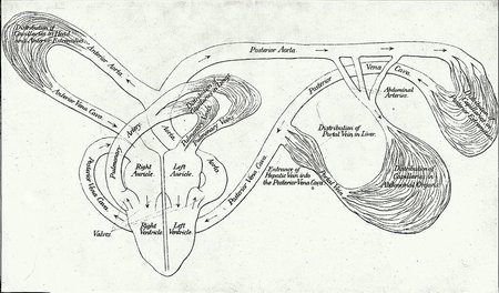
The action of the heart is similar to that of a pump and its function is to keep the blood in circulation. The auricles may be considered as the reservoirs or receivers of the blood and the ventricles as the pump chambers. During the interval between contractions, the heart being in momentary repose, the blood pours into the auricles from the veins; the auriculo-ventricular orifices being widely open, the ventricles also receive blood; the auricles contract and the ventricles are filled; contraction of the ventricles follows; the auriculo-ventricular valves are forced up by the pressure of the blood and close the auriculo-ventricular openings and prevent the return of blood into the auricles; the contraction of the ventricles forces the blood from the right ventricle into the lungs through the pulmonary artery and its branches, and from the left ventricle into the aorta, thence through the arteries to all parts of the body. After the contraction of the ventricles the heart is again in momentary repose and is being filled with blood, while the valves in the aorta and pulmonary artery close to prevent the return of blood into the ventricles. (See Pl. VII.)
The average weight of the heart of an ox is said to be from 3½ to 5 pounds; but, of course, owing to the many breeds and sizes of cattle, it must vary in different animals.
The vessels that convey the blood from the heart to all parts of the body are called arteries; those which return the blood to the heart are called veins. The arteries divide and subdivide (like the branches of a tree), become smaller and smaller, and ultimately ramify into every part of the body. Between the ultimate ramifications of the arteries and the beginning of the veins there is an intermediate system of very minute vessels called capillaries, which connect the arterial with the venous system of the circulation. The walls of the arteries are possessed of a certain amount of rigidity, sufficient to keep the tubes open when they are empty.
The blood leaves the left ventricle through a single vessel, the common aorta, consisting of the anterior and posterior aortas, which give off the large arteries.
The veins take the blood from the capillaries in all parts of the body. They begin in very small tubes, which unite to become larger in size and less in number as they approach the heart.
In its course an artery is usually accompanied with a vein and in many situations with a nerve. The more important arteries are placed deep within the body; when they are superficial, however, they are generally found where least exposed to injury, as, for example, on the inner side of the legs. Arteries are less numerous than veins, and their total capacity is much less than that of the veins. A great number of veins are in the tissue immediately beneath the skin and do not generally accompany arteries.
The blood, throughout its course in the heart, arteries, capillaries, and veins, is inclosed within these vessels. Except where the large lymphatics empty into the venous blood, there is no opening into the course of the blood.
All the arteries except the pulmonary and its branches carry bright-red blood, and all the veins, except the pulmonary veins, carry dark-red blood. The impure dark-red blood is collected from the capillary vessels and carried to the right auricle by the veins; it passes down into the right ventricle, and thence into the pulmonary artery and through its branches to the capillaries of the lungs, where the carbonic-acid gas and other impurities are given up to the air in the air cells of the lungs (through the thin walls between the capillaries and the air cells), and where it also absorbs from the air the oxygen gas necessary to sustain life. This gas changes it to the bright-red, pure blood. It passes from the capillaries to the branches of the pulmonary veins, which convey it to the left auricle of the heart; it then passes through the auriculo-ventricular opening into the left ventricle, the contraction of which forces it through the common aorta into the posterior and anterior aortas, and through all the arteries of the body into the capillaries, where it parts with its oxygen and nutritive elements and where it absorbs carbonic-acid gas and becomes dark colored. (See theoretical diagram of the circulation, Pl. VII.)
The branches of certain arteries in different parts unite again after subdividing. This reuniting is called anastomosing, and assures a quota of blood to a part if one of the anastomosing arteries should be tied in case of hemorrhage, or should be destroyed by accident or operation.
THE BLOOD.
The various kinds of food, after being digested in the alimentary canal, are absorbed and carried into the blood by the lymphatics, and by the blood to the places where nutrition is required. The blood takes from all parts of the body all that is no longer required, and carries it to the different organs through which it is eliminated from the body. It contains within itself all the elements which nourish the body.
The blood may be considered as a fluid holding in solution certain inorganic elements and having certain bodies suspended in it. To facilitate description, the blood may be considered as made up of the corpuscles and the liquor sanguinis. The corpuscles are of two kinds, the red and the white, the red being the more numerous. The color of the blood is caused by the coloring matter in the red corpuscles, which are the oxygen carriers. Both kinds are very minute bodies, which require the aid of the microscope to recognize them. The liquor sanguinis is composed of water containing in solution salts, albumen, and the elements of fibrin.
The lymphatics, or absorbents, are the vessels which carry the lymph and chyle in the blood. They begin as capillaries in all parts of the body, gradually uniting to form larger trunks. Placed along the course of the lymphatic vessels are glands, in some situations collected into groups; for example, in the groin. These glands are often involved in inflammation arising from the absorption of deleterious matter.
Absorption is the function of the lymphatics. The liquor sanguinis passes from the blood capillaries to supply nutrition to the tissues. All of the liquor sanguinis that is not required is absorbed by the lymphatic vessels and conveyed back to the blood by the lymphatic ducts. The lymphatics which proceed from the intestines convey the chyle into the blood during digestion. As a rule, the lymphatic vessels follow the course of the veins. All the absorbent vessels convey their contents to the thoracic duct and right great lymphatic vein, which empty into the anterior vena cava, where the lymph and chyle mix with the venous blood, and thus maintain the supply of nutritive elements in the blood.
THE PULSE.
As fully explained, the heart pumps the blood throughout the arterial system. The arteries are always full and each contraction of the ventricle pumps more blood into them; this distends their elastic walls and sends along them a wave which gradually becomes less perceptible as it nears the very small arteries. This wave constitutes the pulse, and is lost before the capillaries are reached. The sensation or impression given to the finger when placed upon the artery shows the force exerted by the heart and some important facts concerning the condition of the circulation. In adult cattle the average number of pulsations in a minute is from 50 to 60. The pulse is faster than normal after exercise, excitement, on hot days, from pain, and as a result of fullness of the stomach. In old animals it is slower than in the young and in males slightly slower than in females. In fevers and inflammations and in local diseases of the heart the pulse rate is increased. If the rate is greater than 100 or 110 to the minute the outlook for recovery is not good.
Other variations of the pulse are known as infrequent pulse, which means that the number of pulsations in a given time is less than normal. The irregular or the intermittent pulse is when the pulsations do not follow in regular order. The large pulse and the small pulse refer to the volume of the pulse, which may be larger or smaller than usual. The strong pulse and the feeble pulse refer to the strength or weakness of the pulsation. The pulse is said to be hard when the vessel feels hard and incompressible, the soft pulse being the opposite. By dicrotic pulse is meant that kind of pulsation which makes each beat seem double, and therefore it is generally called the double pulse.
The venous or "jugular pulse" is the pulsation so frequently observed in the jugular vein of cattle and is particularly noticeable while they are ruminating—"chewing the cud." It is not always associated with disease, but may be a symptom of some disease of the heart; in such cases the jugular pulse is continuous.
The place selected for feeling the pulse in cattle is where the submaxillary artery winds around the lower jawbones, just at the lower edge of the flat muscle on the side of the cheek; or, if the cow is lying down, the metacarpal artery on the back part of the fore fetlock is very convenient for the purpose.
THE EXAMINATION OF THE HEART.
Corresponding to the beats of the heart two sounds are emitted which are of a definite type in healthy animals. The first is produced by the contraction of the heart and the flow of blood out of it; the second is caused by the rebound of blood in the aorta and the closure of the valves that prevent it from flowing backward into the heart, whence it came. The first sound is the longer and louder of the two, though of low pitch. The second sound is sharper and shorter, and is not always easy to hear. There is a brief interval between them.
To distinguish these sounds, the ear is placed against the left side of the chest, a little above the point where the elbow rests when the animal is standing in a natural position, and about opposite the sixth rib. Both heart sounds are reduced in intensity when the animal is weak or when the heart is forced away from the chest wall by collections of fluid or by tubercular or other growths. Nonrhythmical heart sound is often caused by pericarditis or by disease of the valves. It may also be attributable to overfilling of the heart upon the right side, as occurs in severe congestion of the lungs and in some febrile diseases.
In pericarditis, sounds like scraping, rubbing, or splashing may be heard, entirely apart from the two normal sounds above described.
The impulse of the heart, as felt by placing the hand against the chest, is of some consequence in reaching a conclusion in respect to disease of the heart; but it must be remembered that the impulse may be very much increased by diseases other than those of the heart, as, for example, inflammation of various organs, severe pains, etc. The impulse may be increased also (when disease does not exist) by work, exercise, fright, or any cause of excitement, or, in general, by anything that causes acceleration of the pulse.
The impulse of the heart may be felt and the sounds may be heard fairly well in lean cattle, but in fat ones it is difficult and often impossible to detect either impulse or sound with any degree of satisfaction.
PALPITATION.
When the impulse of the heart is excessive—that is, when it beats more or less tumultuously—the familiar expression "palpitation of the heart" is applied; by many it is called "thumps." The hand or ear placed against the chest easily detects the unnatural beating. In some cases it is so violent that the motion may be seen at a distance. Palpitation is but a symptom, and in many instances not connected with disease of the structure of the heart or its membranes. A badly frightened animal may have palpitation. When it comes on suddenly and soon passes away, it depends on some cause other than diseases of the heart; when it is gradually manifested, however, and becomes constant, although more pronounced at one time than another, heart disease may be suspected, especially if other symptoms of heart disease are present.
INJURY TO THE HEART BY FOREIGN BODIES.
Cattle are addicted to the habit of chewing and swallowing many objects not intended as articles of food. Every veterinarian of experience has met with instances to remind him of this, and it is well known to butchers. Among the great variety of things that have thus found their way into the stomachs of cattle the following have been noticed: Finger rings, knitting needles, old shoes, table knives, wood, pieces of leather, pieces of wire, buttons, hairpins, brushes, nails, coins, etc. The more sharply pointed objects sometimes penetrate the wall of the stomach, in some cases causing gastric irritation enough to produce indigestion, gradually work their way through the diaphragm toward the heart, pierce the pericardium (bag inclosing the heart), wound the heart, and thus prove fatal to the animal. Cases are recorded in which the foreign body has actually worked its way into one of the cavities of the heart. Instances are known, however, in which the object took a different course, and finally worked its way toward the surface and was extracted from the wall of the chest. While it is possible that the foreign body may pierce the wall at different parts of the alimentary canal, as it frequently does that of the rumen (paunch), it is thought that in most cases it passes through the wall of the reticulum (smaller honeycombed compartment, or second stomach) and is drawn toward the heart by the suctionlike action of the chest. Post-mortem examinations have demonstrated the course it pursued, as adhesions and other results of the inflammation it caused were plainly to be seen. All manner of symptoms may precede those showing involvement of the heart, depending upon the location of the foreign body and the extent of inflammation caused by it. Severe indigestion may occur; stiffness and difficulty in moving about owing to the prods of the sharp body following muscular contraction; pain on pressure over the front, lower, or right side of the abdomen; coughing and difficult, quick breathing. In most cases the foreign body does not penetrate to the heart, nor even to the pericardium.
Symptoms.—The symptoms are as follows: The animal is disinclined to move actively, the step is restricted and cautious, sudden motion causes grunting, the attitude is constrained, the feet are drawn somewhat together, the back is arched, the face has an anxious expression. If the disease is of several days' standing, there is likely to be soft swelling (edema) beneath the neck, in the dewlap, and under the chest, between the fore legs. Breathing is short and difficult; it may clearly be painful. The pulse is rapid, 80 to 120 per minute. The muscles quiver as though the animal were cold. Rumination and appetite are depressed or checked. The dung is hard, and to void it appears to cause pain. These symptoms usually develop gradually, and, of course, they vary considerably in different animals, depending upon the size and location of the foreign body and the irritation it causes.
As a matter of course, in such cases treatment is useless, but when it is possible to diagnose correctly the animal could be turned over to the butcher before the flesh becomes unfit for use; that is, before there is more than a little suppuration and before there is fever. Knowing that cattle are prone to swallow such objects, ordinary care may be taken in keeping their surroundings as free of them as possible.
PERICARDITIS.
Inflammation of the pericardium (heart bag) is often associated with pneumonia and pleurisy, rheumatism, and other constitutional diseases, or with an injury. It also occurs as an independent affection, owing to causes similar to those of other chest affections, as exposure to cold or dampness and changes of the weather.
Symptoms.—It may be ushered in with a chill, followed by fever, of more or less severity; the animal stands still and dull, with head hanging low, and anxiety expressed in its countenance. The pulse may be large, perhaps hard; there is also a venous pulse. The hand against the chest will feel the beating of the heart, which is often irregular, sometimes violent, and in other instances weak, depending in part upon the quantity of fluid that has transuded into the pericardial sac. The legs are cold, the breathing quickened and usually abdominal; if the left side of the chest is pressed on or struck, the animal evinces pain. There may be spasms of the muscles in the region of the breast, neck, or hind legs. After a variable time swelling may also appear in the legs and under the chest and brisket.
In those animals in which the heart sounds may be heard somewhat distinctly, the ear applied against the chest will detect a to-and-fro friction sound, corresponding to the beats of the heart. This sound is produced by the rubbing of the internal surface of the heart bag against the external surface of the heart. During the first stages of the inflammation these surfaces are dry, and the rubbing of one against the other during the contraction and relaxation of the heart produces this sound. The dry stage is followed by the exudation of fluid into the heart sac, and the friction is not heard until the fluid is absorbed sufficiently to allow the surfaces to come in contact again. But during the time the friction sound is lost a sound which has been called a "churning noise" may take its place.
The friction sound of pericarditis can not be mistaken for the friction sound of pleurisy if the examination is a careful one, because in the heart affection the sound is made in connection with the heart beats, while in the pleuritic affection the sound is synchronous with each respiration or breath of air taken in and expelled from the lungs.
Treatment.—When pericarditis is complicated with rheumatism or other diseases the latter must be treated as directed in the description of them. The animal must be kept in a quiet, comfortable place, where it will be free from excitement. Warm clothing should be applied to the body, and the legs should be hand-rubbed until the circulation in them is reestablished, and then they should be snugly bandaged. The food should be nutritive and in moderate quantity. Bleeding should not be performed unless the case is in the hands of an expert.
At the beginning give as a purgative Epsom salt—1 pound to an average-sized cow—dissolved in about a quart of warm water and administered as a drench. When there is much pain 2 ounces of laudanum, diluted with a pint of water, may be given every three hours until the animal is better. Do not give the laudanum unless demanded by the severity of the pain, as it tends to constipate. Give one-half ounce of nitrate of potassium (saltpeter), dissolved in drinking water, four or five times a day. After the attack has abated mustard mixed with water may be rubbed well over the left side of the chest to stimulate the absorption of the fluid within the pericardium. The other medicines may be discontinued and the following administered: Sulphate of iron, 2 ounces; powdered gentian, 6 ounces; mix and make 8 powders. Give one powder every day at noon, mixed with feed, if the animal will eat it, or shaken up with water in a bottle as a drench. Also the following: Iodid of potassium, 2 ounces; nitrate of potassium, 8 ounces; mix and make 16 powders. Give one in drinking water or in drench every morning and evening. The last two prescriptions may be continued for several weeks if necessary.
In extreme cases tapping the pericardium with a trocar and cannula to draw off the fluid is resorted to, but the operation requires exact anatomical knowledge.
After death from pericarditis there is always more or less fluid found in the pericardium; the surfaces are rough and covered with a yellow-colored exudate. There are also in many cases adhesions to a greater or less extent between the heart and pericardium.
MYOCARDITIS.
Inflammation of the muscular structure of the heart occurs in limited, circumscribed areas, as evidenced by post-mortem examination, and it is probably always somewhat involved in connection with pericarditis and endocarditis. It may readily be inferred that if the whole organ were inflamed death would ensue immediately. Usually myocarditis results from the preexistence of blood poisoning or of some infectious febrile disease.
Symptoms.—The chief symptoms are those of heart weakness. The heart beat is fast, weak, and often irregular. Respiration is difficult and rapid. There is great general weakness and depression. Death comes suddenly.
Treatment.—Treatment consists in supporting the animal by the use of stimulants, such as ammonia, coffee, digitalis, camphor, etc. Complete quiet must be provided, and the general care should be as in pericarditis.
ENDOCARDITIS.
When the membrane which lines the cavities of the heart—the endocardium— suffers inflammation, the disease is called endocarditis. The cause is another disease, during which substances that irritate the lining of the heart are produced and admitted into the circulation. These substances are usually living organisms, or it is possible that in some cases they are chemical irritants. Endocarditis occurs as a complication of or sequel to pneumonia, blood poisoning, inflammation of the womb, rheumatism, or severe wounds or abscesses. The symptoms are much the same as those of pericarditis, and it is difficult to discriminate between the two affections. There is a jugular pulse, the legs may become dropsical, and there is a tendency to faint if the head is elevated suddenly. The bellowslike sound is more distinct than it is in pericarditis. It is the most fatal of heart diseases, because of the liability of the formation of clots, which may adhere to the valves, change in the structure of the valves, and often a complication with an abnormal condition of the blood. Clots may be formed in the heart, and, being carried to other parts, prove fatal by interrupting the circulation in some vital organ.
Treatment similar to that advised for myocarditis may be followed in this disease.
VALVES OF THE HEART.
The valves are subject to abnormal growths and structural changes in chronic endocarditis or as a result of acute endocarditis. Sometimes valves are torn by sudden, extreme muscular effort or a congenital abnormality. Cases are also reported in which they have been found ruptured.
Symptoms.—The general symptoms are those of heart weakness, accompanied with edema and congestion of the lungs.
Treatment.—Relief is sometimes afforded, but usually only temporarily, by the use of stimulants, especially digitalis.
RUPTURE OF THE HEART.
Sudden effort, blows, or disease may lead to rupture of the heart of the ox. The first cause does not operate so often in cattle as in horses. Tuberculosis or ulceration from other causes, such as a foreign body, is the most common source of this accident. Rupture is shown by sudden fainting, followed very shortly by death.
HYPERTROPHY AND DILATATION OF THE HEART.
This is an enlargement of the heart, and may consist of the thickening of the walls alone, or at the same time the cavities may be either enlarged or diminished. Dilatation of the cavities has been noticed as existing independently of thickened walls. In hypertrophy the sounds of the heart are loud and pronounced, may be heard on both sides of the chest distinctly, and palpitation occurs to a greater or less extent. Fortunately both conditions are very rare in cattle.
ATROPHY.
Atrophy is the technical term for wasting of the muscular tissue. Atrophy of the heart is very rare among cattle, and is usually a result of other diseases.
FATTY DEGENERATION OF THE HEART.
This condition of the heart is met with in some very fat cattle, but it must be understood that the accumulation of fat around the heart is not referred to by this designation. In fatty degeneration the elements of the muscular tissue are replaced by fatty or oily granules. The muscle becomes weak, the heart contractions are insufficient, and heart weakness is shown by general weakness, shortness of breath, and weak, rapid pulse.
CYANOSIS.
Owing to the most prominent symptoms, cyanosis is also called "blue disease," and is seen occasionally in new-born calves. It is recognized by the blue color of the mucous membrane (easily seen by looking within the mouth and nostrils), the coldness of the surface of the body, and rapid, labored breathing. It is caused by non-closure of the foramen ovale, connecting the right with the left side of the heart, and the consequent mixing of the venous with the arterial blood. Calves so affected live but a short time.
MISPLACEMENT OF THE HEART.
Cases are recorded in which the heart has been found out of its natural position, sometimes even outside the chest. This is a congenital condition for which there is no remedy. A heifer calf with the heart entirely outside the thoracic cavity and beneath the skin in the lower part of the neck was kept for two years at the veterinary hospital of the University of Pennsylvania, during which time it grew to be a well-developed cow.
WOUNDS OF ARTERIES AND VEINS.
When a blood vessel is opened a glance will tell whether it is an artery or a vein by simply remembering that bright-red blood comes from arteries and dark-red from veins. When a vein or a very small artery is severed the blood flows from the vessel in a continuous and even stream, but when one of the larger arteries is severed the blood comes in intermitting jets or spurts corresponding to the beats of the heart. It is well to call attention to the fact that the dark-red blood which flows or oozes from a wound soon becomes bright red, because it gives up its carbonic-acid gas to the air, and absorbs oxygen gas from the air, which is exactly the change it undergoes in the capillaries of the lungs.
The general treatment of wounds will be found in another section; here it is necessary only to refer briefly to some of the most practical methods used to arrest hemorrhages, as instances occur in which an animal may lose much strength from the loss of blood, or even bleed to death unless action is prompt.
BLEEDING (HEMORRHAGE).
The severity of a hemorrhage depends upon the size of the vessel from which the blood escapes, though it may be stated that it is more serious when arteries are severed. If the wound in an artery is in the direction of its length, the blood escapes more freely than if the vessel is completely severed, because in the latter instance the severed ends retract, curl in, and may aid very much in arresting the flow. When the blood merely oozes from the wound, and even when it flows in a small stream, the forming of the clot arrests the hemorrhage in a comparatively short time.
Slight hemorrhages may be checked by the continuous application of cold water, ice, or snow, to the wound, as cold causes contraction of the small vessels. Water from a hose may be thrown on a wound, or dashed on it from the hand or a cup, or folds of cotton cloths may be held on it and kept wet. Ice or snow may be held against the wound, or they may be put into a bag and conveniently secured in position.
Hot water of an average temperature of 115° to 120° F. injected into the vagina or womb is often efficient in arresting hemorrhages from those organs. Tow, raw cotton, lint, or sponges may be forced into a wound and held or bound there with bandages. This is an excellent method of checking the flow of blood until the arrival of an expert. If the flow persists, these articles may be saturated with tincture of iron, but it is not advisable to use it unless necessary, as it is a caustic and retards healing by causing a slough. In cases of necessity, the articles may be saturated with vinegar, or tannic acid or alum dissolved in water may be used instead. Whatever article is used should be left in the wound sufficiently long to make sure that its removal will not be followed by a renewal of the hemorrhage. Sometimes it must remain there one or two days.
An iron heated white and then pressed on the bleeding vessel for three or four seconds is occasionally used. It should not be applied longer, or else the charred tissue will come away with the iron and thus defeat the purpose of its application.
Compression may be applied in different ways, but only the most convenient will be mentioned. To many wounds bandages may easily be applied. The bandages may be made of linen, muslin, etc., sufficiently wide and long, according to the nature of the wound and the region to be bandaged. Bed sheets torn in strips the full length make excellent bandages for this purpose. Cotton batting, tow, or a piece of sponge may be placed on the wound and firmly bound there with the bandages.
Many cases require ligating, which is almost entirely confined to arteries. A ligature is a piece of thread or string tied around the vessel. Veins are not ligated unless very large (and even then only when other means are not available) on account of the danger of causing phlebitis, or inflammation of a vein. The ligature is tied around the end of the artery, but in some instances this is difficult and it is necessary to include some of the adjacent tissue, although care should be taken not to include a nerve. To apply a ligature, it is necessary to have artery forceps (tweezers or small pincers may suffice) by which to draw out the artery in order to tie the string around it. To grasp the vessel it may be necessary to sponge the blood from the wound so that the end will be exposed. In case the end of the bleeding artery has retracted, a sharp-pointed hook, called a tenaculum, is used to draw it out far enough to tie. The ligature should be drawn tightly, so that the middle and internal coats will be cut through.
Another method of checking hemorrhage is called torsion. It consists in catching the end of the bleeding vessel, drawing it out a little, and then twisting it around a few times with the forceps, which lacerates the internal coats so that a check is effected. This is very effectual in small vessels, and is to be preferred to ligatures, because it leaves no foreign body in the wound. A needle or pin may be stuck through the edges of the wound and a string passed around between the free ends and the skin (Pl. XXVII, fig. 10), or it may be passed around in the form of a figure 8, as is often done in the operation of bleeding from the jugular vein.
ANEURISM.
A circumscribed dilatation of an artery, constituting a tumor which pulsates synchronously with the beats of the heart, is called aneurism. It is caused by disease and rupture of one or two of the arterial coats. The true aneurism communicates with the interior of the artery and contains coagulated blood. It is so deeply seated in cattle that treatment is out of the question. Such abnormalities are ascribable to severe exertion, to old age, to fatty or calcareous degeneration, or to parasites in the blood vessels. Death is sudden when caused by the rupture of an aneurism of a large artery, owing to internal hemorrhage. Sometimes spontaneous recovery occurs. As a rule no symptoms are caused in cattle by the presence of deep-seated aneurisms, and their presence is not known until after death.
A false aneurism results from blood escaping from a wounded artery into the adjacent tissue, where it clots, and the wound, remaining open in the artery, causes pulsation in the tumor.
THROMBOSIS (OBSTRUCTION) OF THE ARTERIES.
Arteries become obstructed as a result of wounds and other injuries to them, as those caused by the formation of an abscess or the extension of inflammation from surrounding structures to the coats of an artery. Arteries are also obstructed by the breaking off of particles of a plug or clot, partly obstructing the aorta or other large artery. These small pieces (emboli) are floated to an artery that is too small to permit them to pass and are there securely held, producing obstruction. These obstructions are shown by loss of power in the muscles supplied by the obstructed artery and by excitation of the heart and by respiration after exercise. The loss of power may not come into evidence until after exercise.
Symptoms.—While standing still or when walking slowly the animal may appear to be normal, but after more active exercise a group of muscles, a leg, or both hind legs, may be handled with difficulty, causing lameness, and later there is practically a local paralysis. These symptoms disappear with rest. In some cases the collateral circulation develops in time, so that the parts receive sufficient blood and the symptoms disappear.
INFLAMMATION OF VEINS (PHLEBITIS).
When bleeding is performed without proper care or with an unclean lancet, inflammation of the vein may result, or it may be caused by the animal rubbing the wound against some object. When inflammation follows the operation, the coats of the vein become so much enlarged that the vessel may be felt hard and knotted beneath the skin, and pressure produces pain. A thin, watery discharge, tinged with blood, issues from the wound. The blood becomes coagulated in the vessel. In inflammation of the jugular the coagulation extends from the wound upward to the first large branch. Abscesses may form along the course of the vein. The inflammation is followed by obliteration of that part in which coagulation exists. This is of small import, as cattle have an accessory jugular vein which gradually enlarges and accommodates itself to the increased quantity of blood it must carry.
Treatment.—The treatment for inflammation of the vein is to clip the hair from along the course of the affected vessel and apply a blister, the cerate of cantharides. Abscesses should be opened as soon as they form, because there is a possibility of the pus getting into the circulation.
In the operation of bleeding the instruments should be clean and free from rust. If the skin is not sufficiently opened, or when closing the wound the skin is drawn out too much, blood may accumulate in the tissue, and if it does it should be removed by pressing absorbent cotton or a sponge on the part. Care should also be used in opening the vein, so that the instrument may not pass entirely through both sides of the vein and open the artery beneath it.
DISEASES OF THE HEART, BLOOD VESSELS, AND LYMPHATICS.
DESCRIPTION OF PLATE.
PLATE VII:
Diagram illustrating the circulation of the blood. The arrows indicate the direction in which the blood flows. The valves of the heart, situated between the right auricle and ventricle, and left auricle and ventricle, and between the ventricles and large arteries, are represented by curved lines. These valves are intended to prevent the flow of blood in a direction contrary to that indicated by the arrows.
Plate VII. DIAGRAM OF THE CIRCULATION OF THE BLOOD. (Click to enlarge)