INFECTIOUS DISEASES OF CATTLE. (continued 4)
Reading edition
Cause.—The cause of the disease is a bacillus resembling in some minor respects the anthrax bacillus and differing but little from it in size. It also possesses the power of forming within itself a spore. In Plate XXVIII, figure 4, this is represented as an uncolored spot located in one end of the rod, which is enlarged so that the rod itself appears more or less club-shaped. What has already been stated concerning the significance of the spore of the anthrax bacillus applies equally well to these bodies. They resist destructive agents for a considerable time, and may still produce disease when inoculated after several years of drying. This fact may account for the occasional appearance of blackleg in stables. In order to meet the requirements for the development of the spores, which takes place only in the absence of the atmosphere, it is necessary that the wound be very small and deep enough to penetrate the subcutaneous tissue.
Several observers have found this organism in the mud of swamps. By placing a little of the mud under the skin, the disease has been produced.
Since the disease may be produced by placing under the skin material containing the specific bacilli and spores, it has been assumed that cattle contract the disease through wounds, principally of the skin, or very rarely of the mouth, tongue, and throat. Slight wounds into which the virus may find access may be caused by barbed wire, stubbles, thorns, briers, grass burs, and sharp or pointed parts of feed. Infection by way of digestive tract is also probable.
Symptoms and lesions.—The symptoms of blackleg may be either of a general or of a local nature, though more frequently of the latter. The general symptoms are very much like those belonging to other acute infectious or bacterial diseases. They begin, from one to three days after the infection has taken place, with loss of appetite and of rumination, with dullness and debility, and a high fever. The temperature may rise to 107° F. To these may be added lameness or stiffness of one or more limbs, due to the tumor or swelling quite invariably accompanying the disease. After a period of disease lasting from one to three days the affected animal almost always succumbs. Death is preceded by increasing weakness, difficult breathing, and occasional attacks of violent convulsions.
The most important characteristic of this disease is the appearance of a tumor or swelling under the skin a few hours after the setting in of the constitutional symptoms described above. In some cases it may appear first. This tumor may be on the thighs (hence "blackleg," "black quarter"), the neck, the shoulder, the breast, the flanks, or the rump; never below the carpal (or knee) and the hock joint. It more rarely appears in the throat and at the base of the tongue. The tumor, at first small and painful, spreads very rapidly both in depth and extent. When it is stroked or handled a peculiar crackling sound is heard under the skin; this is due to a collection of gas formed by the bacilli as they multiply. At this stage the skin becomes dry, parchment-like, and cool to the touch in the center of the tumor. If the swelling is cut into, a frothy, dark-red, rather disagreeable-smelling fluid is discharged. The animal manifests little or no pain during the operation.
As it is frequently desirable to know whether the disease is anthrax or blackleg, a few of the most obvious post-mortem changes may here be cited. The characteristic tumor with its crackling sound when stroked has already been described. If after the death of the animal it is more thoroughly examined, it will be noted that the tissues under the skin are infiltrated with blood and yellowish, jellylike material and gas bubbles. The muscular tissue beneath the swelling may be brownish or black, shading into dark red. (Pl. XLII.) It is soft, easily torn and broken up. The muscle tissue is distended with numerous smaller or larger gas-filled cavities, often to such extent as to produce a resemblance to lung tissue. Upon incision it does not collapse perceptibly, as the gas cavities are not connected with one another.
In the abdomen and the thorax bloodstained fluid is not infrequently found, together with bloodstaining of the lining membrane of these cavities. Blood spots (or ecchymoses) are also found on the heart and lungs. The liver is congested, but the spleen is always normal in appearance.
Differential diagnosis.—Among the features of this disease which distinguish it from anthrax may be mentioned the unchanged spleen and the ready clotting of the blood. It will be remembered that in anthrax the spleen (milt) is very much enlarged, the blood tarry, coagulating feebly. The anthrax carbuncles and swellings differ from the blackleg swellings in not containing gas, in being hard and solid, and in causing death less rapidly.
It is difficult to distinguish between the swellings of blackleg and malignant edema, as they resemble each other very closely and both are distended with gas. Malignant edema, however, generally starts from a wound of considerable size; it usually follows surgical operations, and seldom results from the small abrasions and pricks to which animals are subjected in pastures. Inoculation experiments on guinea pigs, rabbits, and chickens will generally disclose the differences between the three diseases above, as all these species are killed by the germ of malignant edema, only the first two species by the anthrax bacillus, while the guinea pigs alone will succumb to the blackleg infection. Hemorrhagic septicemia may be differentiated from blackleg by its affecting cattle of all ages, by the location of the swelling usually about the region of the throat, neck, and dewlap, by the soft, doughy character of the swellings without the presence of gas bubbles, and finally by the characteristic hemorrhages widely distributed throughout the body. Other means of diagnosis, which have reference to the specific bacilli, to the inoculable character of the virus upon small animals, and which are of decisive and final importance, can be utilized only by the trained bacteriologist and veterinarian.
Treatment.—In this disease remedies have thus far proved unavailing. Some writers recommend the use of certain drugs, which seem to have been beneficial in a few cases, but a thorough trial has shown them to be valueless. Others advise that the swelling be opened by deep and long incisions and a strong disinfectant, such as a 5 per cent solution of carbolic acid, applied to the exposed parts; but this procedure can not be too strongly condemned. As nearly all those attacked die, in spite of every kind of treatment, and in view of the fact that when these tumors are opened the germs of the disease are scattered over the stables or pastures, thus becoming a source of danger to other cattle, it is obvious that such measures do more harm than good and should be put aside as dangerous. Bleeding, nerving, roweling, or setoning have likewise some adherents, but the evidence indicates that they have neither curative nor preventive value and therefore should be discarded for the method of vaccination which has been thoroughly tried and proved to be efficacious.
Prevention.—The various means suggested under "Anthrax" to prevent the spread or recurrence of this disease are equally applicable to blackleg, and hence do not need to be repeated here in full. They consist in the removal of well animals from the infected pasture to a noninfected field, the draining of the swampy ground, the burial or burning of the carcasses to prevent the dissemination of the germs over vast areas through the agency of dogs, wolves, buzzards, or crows, the disinfection of the stables and the ground where the animals lay at the time of death, and, if possible, the destruction of the germs on the infected pastures. One of the most effective methods for freeing an infected pasture from blackleg is to allow the grass to grow high, and when sufficiently dry to burn it off. One burning off is not sufficient to redeem an infected pasture, but the process should be repeated several years in succession. This method, however, is in many instances impracticable, as few cattle owners can afford to do it, and the only means left for the protection of the animals is vaccination.
Immunization by vaccination.—Three French veterinarians, Arloing, Cornevin, and Thomas, were the first to discover that cattle may be protected against blackleg by inoculation with virulent material obtained from animals which have died of this disease. Later they devised a method of inoculation with the attenuated or weakened blackleg spores which produced immunity from natural or artificial inoculation of virulent blackleg germs. Their method has undergone various modifications both in regard to the manufacture of the vaccine and in the mode of its application. Kitt, a German scientist, modified the method so that but one inoculation of the vaccine was required instead of two, as was the case with that made by the French investigators. The vaccine formerly prepared and distributed by the Bureau of Animal Industry combined the principle of Arloing, Cornevin, and Thomas, and the modification of Kitt.
By vaccination we understand the injection of a minute amount of attenuated—that is, artificially weakened—blackleg virus into the system. This virus is obtained from animals which have died from blackleg, by securing the affected muscles, cutting them into strips, and drying them in the air. When they are perfectly dry they are pulverized and mixed with water to form a paste, smeared in a thin layer on flat dishes, placed in an oven, and heated for six hours at a temperature close to that of boiling water. The paste is then transformed into a hard crust, which is pulverized and sifted and distributed in packages containing either 10 or 25 doses. This constitutes the vaccine, the strength of which is thoroughly tested on experiment animals before it is distributed among the cattle owners. This vaccine, which is in the form of a brownish, dry powder, is mixed with definite quantities of sterile water, filtered, and by means of a hypodermic syringe the filtrate injected under the skin in front of the shoulder of the animal. The inoculation is usually followed by insignificant symptoms. In a few cases there is a slight rise of temperature, and by close observation a minute swelling may be noted at the point of inoculation. The immunity conferred in this way may last for 18 months, but animals vaccinated before they are 6 months old and those in badly infected districts should be revaccinated before the following blackleg season.
The effect of the vaccine prepared by this bureau in preventing outbreaks of the disease and in immediately abating outbreaks already in progress was highly satisfactory, and it is not to be doubted that thousands of young cattle were saved to the stock owners during the 25 years in which the vaccine was distributed.[6] More than 47,000,000 doses were sent out during this period, and from reports received it is safe to conclude that more than 40,000,000 were actually injected, whereby the percentage of loss from blackleg has been reduced from 10 per cent, which annually occurred before using, to less than one-half of 1 per cent per annum. With these figures before us it is plain that the general introduction of preventive vaccination must be of material benefit to the cattle raisers in the infected districts. Moreover, there is every reason to believe that with the continued use of blackleg vaccine in all districts where the disease is known to occur, and an earnest effort on the part of the stock owners to prevent the reinfection of their pastures by following the directions given, blackleg may be kept in check and gradually eradicated.
Immunization against blackleg is now frequently accomplished by the use of the so-called blackleg aggressin and blackleg filtrates.
NECROTIC STOMATITIS (CALF DIPHTHERIA).
[Pl. XLIII.]
Necrotic stomatitis is an acute, specific, highly contagious inflammation of the mouth occurring in young cattle, and characterized locally by the formation of ulcers and caseo-necrotic patches and by constitutional symptoms, chiefly toxic.
This disease has also been termed calf diphtheria, gangrenous stomatitis, ulcerative stomatitis, malignant stomatitis, tubercular stomatitis, and diphtheritic patches of the oral mucous membrane.
History.—During the last few years farmers and cattlemen in this country, especially in Colorado, Texas, and South Dakota, have increasingly noted the occurrence of enzootics of "sore mouth" among the young animals of their herds. Instead of healing, like the usual forms, of themselves, these cases, if untreated, die. Careful study of some of them has resulted in their identification with cases reported in 1877 by Dammann, from the shore of the Baltic; in 1878 by Blazekowic, in Slavonia; in 1879 by Vollers, in Holstein; in 1880 by Lenglen, in France; in 1881 by Macgillivray, in England; and in 1884 by Löffler, who isolated and described the microorganism which produces the disease. Bang obtained this organism from the diphtheritic lesions of calves in 1890, and Kitt likewise recovered the bacillus from similar lesions of the larynx and pharynx of calves and pigs in 1893.
PLATE XLII. SECTION OF MUSCLE FROM A BLACKLEG SWELLING. A. Gas Bubbles. B. Cavities due to Gas Formation. (Click to enlarge)
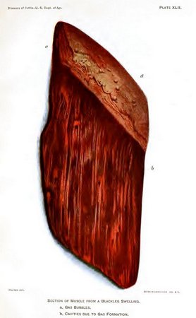
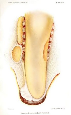
PLATE XLIII. NECROTIC STOMATITIS (CALF DIPTHERIA). (Click to enlarge)
Etiology.—The cause of necrotic stomatitis, as demonstrated by Löffler and since confirmed by other investigators, is Bacillus necrophorus, often spoken of as the bacillus of necrosis. This organism varies in form from a coccoid rod to long, wavy filaments, which may reach a length of 100µ; the width varies from 0.75µ to 1µ. Hence it is described as polymorphic. It does not stain by Gram, but takes the ordinary anilin dyes, often presenting, especially the longer forms, a beaded appearance. A characteristic of the organism, of great moment when we come to treatment, is that it grows only in the absence of oxygen, from which fact it is described as an obligate anærobe.
Very few organisms exhibit a wider range of pathogenesis. According to clinical observation to the present time, Bacillus necrophorus is pathogenic for cattle, horses, hogs, sheep, reindeer, kangaroos, antelope, and rabbits. Experimentally it has been proved pathogenic for rabbits and white mice. The dog, cat, guinea pig, pigeon, and chicken appear to be absolutely immune. It is not pathogenic for man.
The importance of this bacillus is far beyond even its relation to necrotic stomatitis. Besides this disease it has been demonstrated as the causative factor in foot rot, multiple liver abscesses, disseminated liver necrosis, embolic necrosis of the lungs, necrosis of the heart, in cattle; gangrenous pox of the teats, diphtheria of the uterus and vagina, in cows; diphtheritic inflammation of the small intestine of calves. Among horses it is the agent in the production of necrotic malanders, quittor, and diphtheritic inflammation of the large intestine. In hogs it has caused necrotic or diphtheritic processes in the mucous membrane of the mouth, necrosis of the anterior wall of the nasal septum, and pulmonary and intestinal necrosis, accompanying hog cholera. Abscesses of the liver, gangrenous processes of the lips and nose, and gangrenous affections of the hoof have all been caused in sheep by this organism.
Pathology.—The principal lesions in necrotic stomatitis occur in the mucous membrane of the mouth and pharynx. The alterations may extend to the nasal cavities, the larynx, the trachea, the lung, the esophagus, the intestines, and to the hoof. The oral surfaces affected are, in the order of frequency, tongue, cheeks, hard palate, gums, lips, and pharynx. In the majority of cases the primary infection seems to occur in the tongue. (Pl. XLIII.)
Infection takes place by inoculation. Some abrasion or break in the continuity of the mucous membrane of the mouth occurs. Very likely the origin may be connected with the eruption of the first teeth after birth, or, in animals somewhat older, the entrance of a sharp-pointed particle of feed. Gaining an entrance at this point, the bacilli begin to multiply. During their development they elaborate a toxin, or poisonous substance, which causes the death, or necrosis, of the epithelial, or superficial, layer of the mucous membrane and also of the white blood cells which have sallied forth through the vessel walls to the defense of the tissues against the bacillary attack. This destruction of the surface epithelium seems to be the essential factor in the production of the caseous patch, often called the false membrane. From the connective-tissue framework below is poured forth an inflammatory exudate highly albuminous or rich in fibrin-forming elements. When this exudate and the necrosed cellular elements come in contact, the latter furnish a fibrin ferment which transforms the exduate into a fibrinous mass. This process is known as coagulation necrosis, and the resulting fibroid mass, containing in its meshes the necrosed and degenerated epithelium and leucocytes, constitutes the diphtheritic or false membrane. Did the process cease at this point it would be properly called a diphtheritic inflammation, but it does not. A caseating ferment is supplied by the bacilli, and this, acting upon the fibroid patch, transforms it into a dry, finely granular, yellowish mass of tissue detritus resembling cheese.
Frequently this caseous inflammation results in the formation of one or more ulcers with thickened, slightly reddened borders, surmounted by several layers of this necrosed tissue. The floor of the ulcer is formed by a grayish-yellow, corroded surface, under which the tissue is transformed into a dry, friable, or firm cheesy mass. In the tongue this may progress to two fingers' thickness into the muscular portion; in the cheek it may form an external opening, permitting fluids to escape from the mouth; upon the palate it frequently reaches and includes the bone in its destructive course; upon the gums it has produced necrosis of the tooth sockets, causing loss of the teeth. In the advanced forms, caseous foci may be seen in the lung and in the liver and necrotic patches observed on the mucous membrane of the gastrointestinal tract.
Symptoms.—Necrotic stomatitis is both a local and a systemic affection. Primarily it is local. The local lesion is the caseo-necrotic patch or ulcer developed as a result of the multiplication of the bacilli at the point of inoculation. The general affection is an intoxication, or poisoning, of the whole system produced by a soluble toxin elaborated by the bacilli.
The stage of incubation is from three to five days. The first symptoms noted are a disinclination to take nourishment, some drooling from the mouth, and an examination of the mouth will show on some portion of its mucous membrane a circumscribed area of infiltration and redness, possibly an erosion. The latter gradually extends in size and depth, forming a sharply circumscribed area of necrotic inflammation. It may measure anywhere from the size of a 5-cent piece to that of a silver dollar or even larger. It has the appearance of a corroded surface, under which the mucous membrane or muscular tissue seems transformed into a dry, friable, or firm cheesy mass. It is grayish yellow in color and is bordered by a zone of thickened tissue slightly reddened and somewhat granulated. The necrotic tissue is very adherent and can be only partially peeled off. It is homogeneous, cheesy, and may extend two fingers' depth into the tissues beneath. The general symptoms are languor, weakness, and slight fever. In spite of plenty of good feed the calf is seen to be failing. It stops sucking, or, if older, altogether refuses to eat. The temperature at this time may be from 104° to 107° F. The slobber becomes profuse, swallowing very difficult, opening of the mouth quite painful, and a most offensive odor is exhaled. The tongue is swollen and its motion greatly impaired. Sometimes the mouth is kept open, permitting the tumefied tongue to protrude. One or more of the above symptoms direct the attention to the mouth as the seat of disease; or, having noticed the debility and disinclination to eat, an examination of the animal may show a lump under the neck or swelling of the throat or head. The following extract from a letter is characteristic:
I noticed my calves beginning to fail about the first week in December, but could not account for it, as they were getting plenty of grain and hay. My attention was first attracted by a swelling under the neck of one of the calves. I cast the animal and found that it was feed that had collected and the animal couldn't swallow it. I removed it, and in so doing noticed a large ulcer on the tongue and a very offensive odor. This was the first knowledge I had of anything being wrong with the calves' mouths. They may have been sick for some time before this.
Out of a herd of 100 belonging to this man, 70 were affected, and the letter emphasizes the insidious character of the onset.
The general affection at this time manifests itself by dejectedness, extreme weakness, and emaciation, constant lying down, with stiffness and marked difficulty in standing.
The disease frequently extends to the nasal cavities, producing a thin, yellowish, or greenish-yellow, sticky discharge which adheres closely to the borders of the nostrils. Their edges also show caseous patches similar to those in the mouth. Sometimes the nasal passage is obstructed by great masses of the necrosed exudate, thus causing extreme difficulty in breathing. When the caseous process involves the larynx and trachea there result cough, wheezing, and dyspnea, together with a yellowish mucopurulent expectoration.
When life is prolonged three or four weeks, caseous foci may be established in the lung, giving rise to all the signs of a bronchopneumonia. Many of these cases are associated with a fibrinous pleurisy. The invasion of the gastrointestinal tract is announced by diarrheal symptoms. This disease principally attacks sucklings not more than 6 weeks of age, but calves 8 and 10 months old are frequently affected, and several cases in adult cattle have been reported to this office.
In its very acute form many of the cases run their course in from five to eight days. In these the local lesions are not strongly marked, and death seems due to acute intoxication. In other enzootics the majority of the affected animals live from three to five weeks. These are cases that occasionally present the pulmonary and intestinal symptoms, and sometimes develop also caseo-necrotic lesions in the liver.
Ordinarily cases show no tendency to spontaneous cure. Left to themselves they die. On the contrary, if taken in hand early, the disease is readily amenable to treatment. In the latter event the prospects of recovery are excellent.
Differential diagnosis.—Necrotic stomatitis may be differentiated from foot-and-mouth disease by the fact that in the latter there is a rapid infection of the entire herd, including the adult cattle, as well as the infection of hogs and sheep. The characteristic lesion of foot-and-mouth disease is the appearance of vesicles containing a serous fluid upon the mucous membrane of the mouth and upon the udder, teats, and feet of the affected animals. In necrotic stomatitis vesicles are never formed, necrosis occurring from the beginning and followed by the formation of yellowish, cheesy patches, principally found in the mouth. Mycotic stomatitis occurs in only a few animals of the herd, chiefly the adult cattle, and the lesions produced consist of an inflammation of the mouth and lips and of the skin between the toes, followed in a few days by small irregular ulcers in the mouth. This disease appears sporadically, usually in the early fall after a dry summer, does not run a regular course, and can not be inoculated.
Prevention.—Prophylaxis should be carried out along three lines:
(1) Separation of the sick from the healthy animals.
(2) Close scrutiny and thorough disinfection once or twice daily for five days of the mouths and nasal passages of those animals that have been exposed.
(3) Complete disinfection of all stalls and sheds.
The disease appears to break out in winter and hold over to spring. It is conceivable that exposure to cold might so disturb the normal circulation of the oral tissues as to make the mucous membrane an excellent location for the causative factor of the disease. There is another possibility, however, which bears on the third line of prophylaxis. The so-called diphtheritic inflammations of the vagina and uterus in cows are caused by the same organism that induces necrotic stomatitis. A European writer has recently pointed out the almost constant relation of such attacks to previous occurrences of foul foot or foot rot in the same or other cattle on the place.
In all likelihood, in such cases, the stalls and sheds are the harborers of this germ. It is possible that many of these outbreaks have some relation to preceding cases of the above-mentioned diseases and the greater use in winter of the stalls and sheds, thus harboring the Bacillus necrophorus.
Treatment.—The treatment consists almost solely in careful and extensive cleansing and disinfection of the mouth and other affected surfaces. The mucous membrane of the mouth should be copiously irrigated with a 4 per cent solution of boracic acid in warm water at least twice daily. As exposure to oxygen kills the bacilli, one need have no fear about disturbing or tearing off the caseous patches or necrotic tissue during irrigation. The irrigation of the sores should then be followed by the application with a brush or rag on a stick of a paste made with 1 part of salicylic acid and 10 parts of water, or the affected areas may be painted with Lugol's solution of iodin (iodin, 1; potassium iodid, 5; water, 200). Frequent injections of 1 per cent carbolic-acid solution into the mouth make an excellent treatment. The internal administration of 2 grams of salicylic acid and 3 grams of chlorate of potassium three times a day has also proved to be very beneficial when accompanied with local antiseptic treatment.
MALIGNANT CATARRH.
Malignant catarrh, or infectious catarrhal fever, is an acute infectious disease of cattle preeminently involving the respiratory and digestive tracts, although the sinuses of the head, the eyes, and the urinary and sexual organs are very frequently affected. It is relatively rare in this country, being more common on the continent of Europe. Outbreaks have occurred, however, in Minnesota, New York, and New Jersey. So far the causal agent of the disease has never been isolated, and inoculation experiments with the view of artificially reproducing the disease have proved negative in every case. In spite of the foregoing statements the consensus of opinion of eminent investigators points to malignant catarrh as being of specific origin; that is, due to some form of microorganism the contagious character of which is poorly developed. This accounts for the slow transmissibility of the disease from one animal to another. In fact, malignant catarrh is a type of that class of affections scientifically known as miasmatic diseases; that is, they remain stationary in stables with damp floors, low ceilings, poor ventilation, and bad sanitary conditions in general. Such places furnish a favorable seat of propagation for the infective material, and it will remain active for a long time, causing the loss of a few animals each year. One European veterinarian reports an instance in which the disease remained for 25 years on the same farm, attacking in all 225 animals, with a mortality of about 98 per cent.
The disease is most common in late winter and early spring, at all altitudes, and has a special preference for young, well-nourished cattle, although older animals are not immune. The time between the entrance of the infective principle into the body of the animal and the appearance of the first symptoms is relatively very long, averaging, according to German investigators, from 20 to 30 days. Fortunately, it is not a disease which spreads to any great extent or which causes severe losses, and hence legislative enactments do not seem to be necessary for its restriction.
Symptoms.—These are extremely variable according to the point of localization of the lesions. It is usually ushered in with a chill, followed by a marked rise of temperature (104° to 107° F.). The head droops, the skin is hot and dry, and the coat staring. Quivering of the muscles in various parts of the body is frequently observed. Marked dullness of the animal, passing, according to some observers, into an almost stupefied condition later on, is quite common. The secretion of milk stops in the beginning of the disease, and loss of flesh, invariably associated with the disease, is extremely marked and rapid. The lesions of the eyes may best be likened to moon blindness (periodic ophthalmia) in horses.
There is first an abundant secretion of tears, which run down the face. The lids are swollen and inflamed, and indeed this may be so marked as to cause involuntary eversion, exposing the reddened conjunctiva to view. Sunlight is painful, as is shown by the fact that the animal keeps the eyes continuously closed. This inflammation may extend to the cornea, causing it to assume a slightly clouded appearance in mild cases or a chalky whiteness in more severe affection. Cases of ulceration of the cornea followed by perforation and subsequent escape of the aqueous humor, leading to shrinking of the eyeball and permanent loss of sight, have been recorded, but these are relatively rare, although slight inflammation of the deeper structures of the eye (iris) are more frequent. In mild cases this inflammation may undergo complete resolution, but more frequently permanent cloudliness of the cornea, either diffuse or in spots (leucoma), is the result. The mucous membrane of the mouth, nose, sinuses of the head, throat, and lower respiratory passages are also involved. It is first catarrhal in character, but soon a false or diphtheritic membrane is formed, with the production of shallow ulcers. There is dribbling of saliva from the mouth and discharge from the nose, at first watery, becoming thicker and mixed with blood and small masses of cast-off croupous membrane, causing a very fetid odor. These croupous areas when they form in the throat, larynx, or windpipe, may lead to narrowing of the passages, with consequent difficult breathing and even suffocation. Various respiratory murmurs may also be heard, caused by the to-and-fro movement of mucus and inflammatory deposits along the air passages. There is also inflammation of the horn core with consequent loosening of the horn shell, and the horns are thus readily knocked off by the uneasy, blind sufferer. The animal may refuse all feed from the time of the initial rise of temperature, or in less severe cases, and especially when the lesions of the digestive tract are not so marked, the appetite may remain until the disease is well advanced. Constipation is quite common at the commencement of the attack, followed by diarrhea and severe straining, the evacuations becoming very soft, fetid, and streaked with blood. Cases of the evacuation of desquamated patches of diphtheritic membrane from the intestinal mucosa 6 to 9 feet in length have been reported. The kidneys and bladder are usually inflamed, the urine being voided with difficulty and the animal evincing signs of pain. Inflammatory elements, as albumen, casts, etc., may be seen on examination of the urine. In cows the mucous membrane of the vestibule is congested, swollen, and may contain ulcers and an excessive quantity of mucus. Abortion during advanced pregnancy is not infrequent, following a severe attack. In connection with these various symptoms there may be much uneasiness on the part of the animal, leading in some cases to madness and furious delirium, in others to spasms and convulsions or paralysis. A vesicular eruption of the skin may occur, seen principally between the toes and on the inside of the flank and in the armpits, with subsequent loss of hair and epidermis.
Like other infectious diseases, malignant catarrh pursues a longer or shorter course in accordance with the severity of the attack. In acute cases death is said to take place three to seven days after the appearance of symptoms. Recovery, if it occurs, may take three or four weeks. According to statistics, from 50 to 90 per cent of the affected animals die.
If animals which have died of this disease are examined, in addition to the changes of the mucous membrane of mouth and nasal cavities referred to above, shallow ulcers in these situations will be found occasionally. These necrotic processes may pass beneath the mucous membrane and even involve the underlying bony structure. In severe cases membranous (croupous) deposits are found in the throat. Similar deposits have been found upon the mucous membrane of the fourth stomach and intestine, which is always inflamed. There is more or less inflammation of the membranes of the brain, kidneys, and liver, and some fatty degeneration of the voluntary muscles. In countries where rinderpest occasionally appears it may be difficult to distinguish between it and malignant catarrh, owing to a general similarity of the symptoms. The principal points to be observed in differentiating between the two are the very slight transmissibility of the latter as compared with the intense contagiousness of the former, and the tendency of malignant catarrh to run a more chronic course than rinderpest, which usually results fatally in a very few days. Only a trained veterinarian who takes into consideration all the different symptoms and lesions of both diseases should decide in such cases.
Treatment.—There is no specific treatment for this affection. Copious blood letting in the earliest stages has been highly recommended, however, as this has a tendency to deplete the system and lessen the exudation of inflammatory products. Antiseptic washes, such as 4 per cent boric-acid solution to the eyes and Dobell's solution applied to the nose and mouth with ice poultices over the crest of the head and frontal region, have also proved efficacious. Calomel should also be given in 1-dram doses twice a day for three days, and in severe cases, involving the respiratory tract, a powder containing ferrous sulphate, quinin, and subnitrate of bismuth, given twice a day, will be found beneficial. At the same time it must be remembered that much greater success is to be looked for in the preventive treatment. This consists in the removal of the healthy from the infected animals (not vice versa) and thorough cleaning and disinfecting of the contaminated stables. If the floors are low and damp, they should be raised and made dry. If this can not be done, place a layer of cement under the stable floor to prevent water from entering from below. The stable should be well ventilated and the soil in the pastures thoroughly drained. If this is carefully carried out, the contagion should be destroyed and the danger of the reappearance of the disease in a great measure lessened.
MALIGNANT EDEMA.
Malignant edema, also termed gangrenous septicemia, is an acute, inflammatory disease of domestic and wild animals, resulting from the introduction of a specific organism into the deep connective tissues of a susceptible animal and proving fatal in many instances within 24 to 48 hours. The disease may be inoculated from one animal to another, but only by inserting the virus deeply below the skin. It is infrequently met with in cattle, but may follow operating wounds, as roweling, castration, and phlebotomy, which have become infected with septic matter, soil, or unclean instruments. In the pathological laboratory of the Bureau of Animal Industry the organism has also been obtained from the infected muscles of a calf that was supposed to have died of blackleg, and, as a result, all blackleg virus is thoroughly tested before it is made into blackleg vaccine in order to exclude the malignant-edema organism. The essential cause of malignant edema is a long, slender, motile, spore-bearing bacillus, resembling the bacillus of blackleg, and which can develop only in the absence of the atmosphere. Unlike the bacilli of anthrax and blackleg, which are confined to certain districts, this organism is widely distributed and found in ordinary garden soil, foul water, and in the normal intestinal tract of the herbivora. It may be brought to the surface of the soil by growing plants, rains, winds, or burrowing insects and rodents. In animals that have succumbed to the disease the germ is confined to the seat of infection, but a few hours after death it may migrate through the blood channels to other parts of the body. The bacillus may attack man, horses, asses, goats, sheep, pigs, cats, dogs, and poultry. Adult cattle, although refractory to experimental inoculation, suffer from natural infection, while calves are susceptible to both these methods of exposure. (Kitt.) The introduction of the bacillus into abrasions of the skin and superficial sores rarely does any harm, because the germ is quickly destroyed by contact with air. If, however, the organisms are inserted deeply into the subcutaneous tissues of susceptible animals, they quickly develop, producing a soluble poison, which is the fatal agent.
In lamb-shearing season, or after docking or castration, the mortality is higher among these animals because of wounds inflicted at such times. The application of antiseptics to wounds thus made will reduce the percentage of deaths to a minimum.
Symptoms.—Usually the first symptoms are overlooked. In the early stages the animal appears listless, disinclined to move about, and lies down in shady and quiet places. If forced to move about, the hind legs are drawn forward with a peculiar, stiff, dragging movement, and there may be slight muscular trembling over all the body, which becomes more intense as the disease progresses. When driven, the animal shows signs of fatigue, ultimately dropping to the ground completely exhausted. Breathing becomes fast and painful, with frequent spasmodic jerks.
The pulse is quick and weak and the temperature is 106° to 107° F. An edematous, doughy, and painful swelling appears at the point of infection. This tumefaction spreads more and more, and crackles on pressure. In case of an open wound, a fetid liquid and frothy discharge is observed. The center of the swelling may appear soft and jellylike, while the margin is tense, hot, and painful. The symptoms increase rapidly, resulting in coma and death.
Lesions.—After death the fat and subcutaneous tissues surrounding the infected area are infiltrated with a yellow gelatinous material containing an orange-colored foam, due to the presence of gas bubbles.
The muscles at this point are friable, spongy, and of a uniform brownish tint, dissociated by gas and with a blood-tinged exudate. This gangrenous tissue, when present before death, can be removed without pain to the animal. The intestines are generally normal, but, together with the peritoneum, they may be inflamed, and the lungs are usually the seat of an edema. The spleen, liver, and kidneys retain their normal appearance, in marked contrast with anthrax.
Differential diagnosis.—Unlike blackleg, this disease never appears as an epizootic but in isolated cases. It may also be differentiated from the former by the history of a recent parturition or surgical operation, by the presence of an external injury at the site of the swelling accompanied with a fetid liquid discharge, and the gangrenous appearance of the tumefaction. Man is susceptible to malignant edema, but not to blackleg. Malignant edema may also be easily differentiated from anthrax in that the blood and spleen are normal in appearance, while in the latter disease the blood is dark and of a tarlike consistency, and the spleen appears swollen, injected, and softened. The local tumor in malignant edema contains gas bubbles, which are absent in anthrax swellings. Inoculation experiments of guinea pigs, rabbits, and chickens will also disclose the differences among the above-mentioned three diseases, since all these species are killed by the germ of malignant edema, only the first two species by the anthrax bacillus, while the guinea pig alone will succumb to the blackleg infection.
Treatment.—Treatment is chiefly surgical and consists in laying the infected areas wide open by free incision, followed by a liberal application of a 30 per cent solution of hydrogen dioxid and subsequently a 5 per cent solution of carbolic acid. Usually the disease when observed has advanced to such an extent that medicinal interference is without avail. Preventive treatment is by far the most desirable, and consists, essentially, in a thorough disinfection of all accidental and surgical wounds, the cleansing of the skin, and the exclusion of soil, filth, and bacteria during surgical operations of any nature. Sheds, barns, and stables should receive a thorough application of quicklime or crude carbolic acid wash after all rubbish has been removed and burned. All dead animals should be burned or deeply buried and covered well with quicklime.
SOUTHERN CATTLE FEVER (TEXAS FEVER, TICK FEVER).
[Pls. XLIV-XLIX.]
This disease, which is more commonly known as Texas fever, and sometimes as splenetic fever, is a specific fever communicated by cattle which have recently been moved northward from the infected district; it is also contracted by cattle taken into the infected district from other parts of the world. It is characterized by the peculiarity among animal diseases that the animals which disseminate the infection are apparently in good health, while those which sicken and die from it do not, as a rule, infect others.
It is accompanied with high fever, greatly enlarged spleen, destruction of the red blood corpuscles, escape of the coloring matter of the blood through the kidneys, giving the urine a deep-red color, with a yellowness of the mucous membranes and fat, which is seen more especially in fat cattle, by a rapid loss of strength, and with fatal results in a large proportion of cases.
This disease has various names in different sections of the country where it frequently appears. It is often called Spanish fever, acclimation fever, red water, black water, distemper, murrain, dry murrain, yellow murrain, bloody murrain, Australian tick fever, and tristeza of South America.
The earliest accounts we have of this disease date back to 1814, when it was stated by Dr. James Mease, before the Philadelphia Society for Promoting Agriculture, that the cattle from a certain district in South Carolina so certainly disease all others with which they mix in their progress to the North that they are prohibited by the people of Virginia from passing through the State; that these cattle infect others while they themselves are in perfect health, and that cattle from Europe or the interior taken to the vicinity of the sea are attacked by a disease that generally proves fatal. Similar observations have been made in regard to a district in the southern part of the United States.
The northern limits of this area are changed yearly as a result of the dissemination or eradication of the cattle tick along the border, but the infected area has gradually decreased, owing to the successful endeavors pushed forward to eliminate the ticks.
It was the frequent and severe losses following the driving of cattle from the infected district in Texas into and across the Western States and Territories which led to the disease being denominated Texas fever. It is now known, however, that the infection is not peculiar to Texas or even to the United States, but that it also exists in southern Europe, Central and South America, Australia, South Africa, and the West Indies.
When cattle from other sections of the country are taken into the infected district they contract this disease usually during the first summer, and if they are adult animals, particularly milch cows or fat cattle, nearly all die. Calves are much more likely to survive. The disease is one from which immunity is acquired, and therefore calves which recover are not again attacked, as a rule, even after they become adult.
When the infection is disseminated beyond the permanently infected district, the roads, pastures, pens, and other inclosures are dangerous for susceptible animals until freezing weather. The infection then disappears, and cattle may be driven over the grounds or kept in the inclosures the succeeding summer and the disease will not reappear. There are some exceptions to this rule in the section just north of the boundary line of the infected district. In this locality the infection sometimes resists the winters, especially if they are mild.
In regard to the manner in which the disease is communicated, experience shows that this does not occur by animals coming near or in contact with one another. It is an indirect infection. The cattle from the infected district first infect the pastures, roads, pens, cars, etc., whence the susceptible cattle obtain the virus secondhand. Usually animals do not contract the disease when separated from infected pastures by a fence. If, however, there is any drainage or washing by rains across the line of fence this rule does not hold good.
The investigations made by the Bureau of Animal Industry demonstrate that the ticks which adhere to cattle from the infected district are the only known means of conveying the infection to susceptible cattle. The infection is not spread by the saliva, the urine, or the manure of cattle from the infected district. In studying the causation and prevention of this disease, attention must therefore be largely given to the tick, and it now seems apparent that if cattle could be freed from this parasite when leaving the infected district they would not be able to spread the malady. The discovery of the connection of the ticks with the production of the disease has played a very important part in determining the methods that should be adopted in preventing its spread. It established an essential point and indicated many lines of investigation which have yielded and are still likely to yield very important results.
Nature of the disease.—Texas fever is caused by an organism which lives within the red blood corpuscles and breaks them up. It is therefore simply a blood disease. The organism does not belong to the bacteria but to the protozoa. It is not, in other words, a microscopic plant, but it belongs to the lowest forms of the animal kingdom. This very minute organism multiplies very rapidly in the body of the infected animal, and in acute cases causes an enormous destruction of red corpuscles in a few days. How it gets into the red corpuscle it is not possible to state, but it appears that it enters as an exceedingly minute body, probably endowed with motion, and only after it has succeeded in entering the corpuscle does it begin to enlarge. Plate XLV, figure 4, illustrates an early stage of this blood parasite. The red corpuscle contains a very minute, roundish body which is stained blue to bring it into view. The body is, as a rule, situated near the edge of the corpuscle. Figure 5 illustrates an older stage in the growth of the parasite, in fact the largest which has thus far been detected. It will be noticed that there are usually two bodies in a corpuscle. These bodies are in general pear-shaped. The narrow ends are always toward each other when two are present in the same corpuscle. If we bear in mind that the average diameter of the red blood corpuscles of cattle is from 1/4000 to 1/5000 inch, the size of the contained parasite may be at once appreciated by a glance at the figures referred to.
The various disease processes which go on in Texas fever, and which we may observe by examining the organs after death, all result from the destruction of the red corpuscles; this destruction may be extremely rapid or slow. When it is rapid we have the acute, usually fatal, type of Texas fever, which is always witnessed in the height of the Texas-fever season, that is, during the latter weeks of August and the early weeks of September. When the destruction of corpuscles is slower, a mild, usually nonfatal, type of the disease is called forth, which is only witnessed late in autumn or more rarely in July and the early part of August. Cases of the mild type occurring thus early usually become acute later and terminate fatally.
The acute disease is fatal in most cases, and the fatality is due not so much to the loss of blood corpuscles as to the difficulty which the organs have in getting rid of the waste products arising from this wholesale destruction. How great this may be a simple calculation will serve to illustrate. In a steer weighing 1,000 pounds, the blood in its body weighs about 50 pounds, if we assume that the blood represents one-twentieth of the weight of the body, which is a rather low estimate. According to experimental determination at the bureau station, which consists in counting the number of blood corpuscles in a given quantity of blood from day to day in such an animal, the corpuscles contained in from 5 to 10 pounds of blood may be destroyed within 24 hours. The remains of these corpuscles and the coloring matter in them must be either converted into bile or excreted unchanged. The result of this effort on the part of the liver causes extensive disease of this organ. The bile secreted by the liver cells contains so much solid material that it stagnates in the finest bile canals and chokes these up completely. This in turn interferes with the nutrition of the liver cells and they undergo fatty degeneration and perish. The functions of the liver are thereby completely suspended and death is the result. This enormous destruction of corpuscles takes place to a large extent in the kidneys, where a great number of corpuscles containing the parasites are always found in acute cases. This accounts largely for the blood-colored urine, or red water, which is such a characteristic feature of Texas fever. The corpuscles themselves are not found in the urine; it is the red coloring matter, or hemoglobin, which leaves them when they break up and pass into the urine.
Symptoms.—After a period of exposure to infected soil, which may vary from 13 to 90 days, and which will be more fully discussed under the subject of cattle ticks as bearers of the Texas-fever parasite, the disease first shows itself in dullness, loss of appetite, and a tendency to leave the herd and stand or lie down alone. A few days before these symptoms appear the presence of a high fever may be detected by the clinical thermometer. The temperature rises from a normal of 101° to 103° F. to 106° and 107° F. There seems to be little or no change in temperature until recovery or death ensues. The period of high temperature or fever varies considerably. As it indicates the intensity of the disease process going on within, the higher it is the more rapid the fatal end. When it does not rise above 104° F. the disease is milder and more prolonged.
The bowels are mostly constipated during the fever; toward the end the feces may become softer and rather deeply tinged with bile. The urine shows nothing abnormal during the course of the disease until near the fatal termination, when it may be deeply stained with the coloring matter of the blood. (Hemoglobinuria; see Pl. XLV, fig. 3.) Although this symptom is occasionally observed in animals which recover, yet it may generally be regarded as an indication of approaching death. The pulse and respiration are usually much more rapid than during health.
Other symptoms in addition to those mentioned have been described by observers, but they do not seem to be constant, and only those described above are nearly always present. As the end approaches emaciation becomes very marked, the blood is very thin and watery, and the closing of any wound of the skin by clots is retarded. The animal manifests increasing stupor and may lie down much of the time. Signs of delirium have been observed in some cases. Death occurs most frequently in the night.
The duration of the disease is very variable. Death may ensue in from three days to several weeks after the beginning of the fever. Those that recover ultimately do so very slowly, owing to the great poverty of the blood in red corpuscles. The flesh is regained but very gradually, and the animal may be subjected to a second, though mild, attack later on in the autumn, which pushes the full recovery onward to the beginning of winter.
In the mild type of the disease, which occurs in October and November, symptoms of disease are well-nigh absent. There is little if any fever, and if it were not for loss of flesh and more or less dullness the disease may pass unnoticed, as it undoubtedly does in a majority of cases. If, however, the blood corpuscles are counted from time to time a gradually diminishing number will be found, and after several weeks only about one-fifth or one-sixth of the normal number are present. It is indeed surprising how little impression upon the animal this very impoverished condition of the blood appears to make. It is probable, however, that if two animals kept under the same conditions, one healthy and the other at the end of one of these mild attacks, are weighed, the difference would be plainly shown.
Pathological changes observable after death.—In the preceding pages some of these have already been referred to in describing the nature of the disease. It is very important at times to determine whether a certain disease is Texas fever or some other disease, like anthrax, for example. This fact can, as a rule, be determined at once by a thorough microscopic examination of the blood. The necessary apparatus and the requisite qualifications for this task leave this method entirely in the hands of experts. There are, however, a considerable number of changes caused by this disease which may be detected by the naked eye when the body has been opened. Put together they make a mistake quite impossible. The presence of small ticks on the skin of the escutcheon, the thighs, and the udder is a very important sign in herds north of the Texas-fever line, as it indicates that they have been brought in some manner from the South and have carried the disease with them, as will be explained later. Another very important sign is the thin, watery condition of the blood, either just before death or when the fever has been present for four or five days. A little incision into the skin will enable any one to determine this point. Frequently the skin is so poor in blood that it may require several incisions to draw a drop or more.
The changes in the internal organs, as found on post-mortem examinations, are briefly as follows: The spleen, or milt, is much larger than in healthy animals. It may weigh three or four times as much. When it is incised the contents or pulp is blackish (see Pl. XLIV, fig. 1), and may even well out as a disintegrated mass. The markings of the healthy spleen (fig. 2) are all effaced by the enormous number of blood corpuscles which have collected in it, and to which the enlargement is attributable. Next to the spleen the liver will arouse our attention. (See Pl. XLV, fig. 2.) It is larger than in the healthy state, has lost its natural brownish color (fig. 1), and now has on the surface a paler, yellowish hue. When it is incised this yellowish tinge, or mahogany color, as it has been called by some, is still more prominent. This is owing to the large quantity of bile in the finest bile capillaries, and as these are not uniformly filled with it the cut surface has a more or less mottled appearance. This bile injection causes in many cases a fatty degeneration of the liver cells, which makes the organ appear still lighter in color.
In all cases the gall bladder should be examined. This is distended with bile, which holds in suspension a large number of yellow flakes, so that when it is poured into a tall bottle to settle fully one-half or more of the column of fluid will be occupied by a layer of flakes. If mucus is present at the same time, the bile may become so viscid that when it is poured from one glass to another it forms long bands. The bile in health is a limpid fluid, containing no solid particles.
If the animal during life has not been observed to pass urine colored with blood or red water, the bladder should be opened. This quite invariably, in acute cases, contains urine which varies in color from a deep port wine to a light claret. In many cases the color is so dense that light will not pass through even a thin layer. (Pl. XLV, fig. 3.) The kidneys are always found congested in the acute attack. The disease exerts but little effect on the stomach and intestines beyond more or less reddening of the mucous membrane; hence an examination of them may be safely omitted. The lungs are, as a rule, not diseased. The heart usually shows patches of blood extravasation on the inside (left ventricle) and less markedly on the outer surface.
We have observed jaundice of the various tissues but very rarely. It has been observed by some quite regularly, however.
During the hot season about 90 per cent of the susceptible mature animals from a noninfected district die, but later, in the cool weather, the disease assumes a milder type, with a consequent decrease in the number of deaths.
The cattle tick, Margaropus annulatus, as the carrier of Texas fever. (Pls. XLVI, XLVII, and XLVIII.) —The cattle tick is, as its name indicates, a parasite of cattle in the southern part of the United States. It belongs to the group of Arthropoda and to the genus Margaropus (or Boophilus), which is included in the order Acarina. Its life history is quite simple and easily traced from one generation to another. It is essentially a parasite, attaching itself to the skin (Pl. XLVIII) and drawing the blood of its host. It is unable to come to maturity and reproduce its kind unless it becomes attached to the skin of cattle, whence it may obtain its food.
The eggs laid on the ground after the female has dropped from the host begin to develop at once. When the embryo is fully formed within the shell it ruptures this and gains its freedom. The time required from the laying of the eggs to their hatching varies considerably, according to the temperature. In the laboratory in the heat of midsummer this was accomplished in about 13 days. In the late fall, under the same conditions, it required from four to six weeks. The larva after emerging from the egg is very minute, six-legged, and is just visible to the naked eye. (Pl. XLVI, fig. 3.) If these larvæ are kept on a layer of moist sand or earth in a covered dish, they may remain alive for months, but there is no appreciable increase in size. So soon, however, as they are placed upon cattle growth begins.
On pastures these little creatures soon find their way on to cattle. They attach themselves by preference to the tender skin on the escutcheon, the inside of the thighs, and on the base of the udder. Yet when they are very numerous they may be found in small numbers on various parts of the body, such as the neck, the chest, and the ears. (Pl. XLVIII and Pl. XLIX, fig. 1.)
The changes which they undergo during their parasitic existence were first studied by Dr. Cooper Curtice, of the Bureau of Animal Industry, in 1889. The young tick molts within a week, and the second or nymphal stage of the parasite's life is thus ushered in. After this change it has four pairs of legs. Within another week another molt takes place by which the tick passes from the nymphal to the sexual, or adult, stage. Impregnation now takes place, and, with the development of the ova in the body, the tick takes an increased quantity of blood, so that in a few days it becomes very much larger. That the rapid growth is due to the blood taken in may be easily proved by crushing one. The intestine is distended with a thick, tarry mass composed of partly digested blood. When the female has reached a certain stage of maturity she drops to the ground and begins to lay a large number of eggs, which hatch in the time given above.
The life of the cattle tick is thus spent largely on cattle, and although the young, or larvæ may live for a long time on the ground in the summer season, they can not mature except as parasites on cattle and horses. We have purposely omitted various details of the life history, including that of the male, as they are not necessary to an understanding of our present subject—Texas fever. How this is transmitted we will proceed to consider. Before the enforcement of the Federal quarantine southern cattle sent north during the spring and summer months carried on their bodies large numbers of the cattle ticks, which, when matured, would drop off and lay their eggs in the northern pastures. After hatching, the young ticks would soon get upon any northern cattle which happened to be on the pasture. So soon as they attached themselves to the skin they inoculated the cattle, and Texas fever would break out a week or more thereafter. For many years there had been a growing suspicion that the cattle tick was in some way concerned in the spread of Texas fever, and the facts which supported this supposition finally became so numerous and convincing that a series of experiments was inaugurated by the Bureau of Animal Industry which served to show that the tick is abundantly able to carry the disease to a herd of healthy cattle, and, in fact, is probably the only agent concerned in the transmission of the disease from southern cattle to susceptible northern animals.
Injurious effects of cattle ticks.—Unfortunately many cattle owners who have always been accustomed to see both ticks and ticky cattle on their farms are not inclined to attach much importance to these parasites, and, as a rule, through lack of appreciation of their damaging effects, placidly consider them as of little consequence. That ticks may be detrimental to their hosts in several ways has probably not suggested itself to these stockmen, who are most vitally affected, and it therefore seems necessary to emphasize the fact that, in addition to their relation to Texas fever, they may also be injurious to cattle as external parasites. While the power of transmitting Texas fever is undoubtedly the most dangerous property possessed by the cattle tick and is the principal cause for adopting stringent measures looking to its complete eradication, nevertheless there still remain other good reasons for the accomplishment of this achievement. These secondary objections to the presence of ticks on cattle consists in the physical harm they do to the host aside from the production of the specific disease of Texas fever. True, a few parasites may remain on cattle indefinitely without causing any noticeable effect, but it is not uncommon to notice bovine animals on pastures with their hides heavily infested with these pests. In such cases it can readily be seen that the continuous sucking of blood causes more or less impoverishment of the circulation. The animal must therefore be fed more in order to meet the demands of the parasites in addition to the ordinary needs of the host. If the ticks are removed from the body, the bites inflicted are often distinguished by small, inflamed or reddened areas somewhat swollen, with perforations of the skin which may allow the entrance of various kinds of disease germs, and showing that more or less irritation of the hide is produced by these parasites. This condition, together with the loss of blood, frequently induces an irritable state and evidence of uneasiness commonly known as "tick worry," which results in the loss of energy and other derangements of the animal's health. It may in some cases, especially in hot weather, become so pronounced that the animal will lose flesh in spite of good pasturing, thereby reducing the vitality and rendering it more susceptible to the inroads of disease. Moreover, if the infestation of ticks is not controlled, the cattle may be so reduced in condition that growth is retarded, and, in the case of young animals, they may never become fully developed, but remain thin, weak, and stunted—a condition that has been termed "tick poverty"—and easily succumb to other diseases as a result of lowered vitality. In milch cows this debilitating influence of the numerous ticks is shown in a greatly reduced milk supply. This should not appear strange when it is considered that some animals harbor several thousand of the bloodsucking parasites. If these parasites are crushed, it will be found that their intestines are completely filled with a dark, thick mass of blood abstracted from the animal host and containing nutriment that should go to the formation of milk, flesh, and the laying on of fat. In some rare cases the large number of bites on a limited area of skin may be followed by infection with pus-producing organisms, giving rise to small abscesses which may terminate in ulcers. The discharge from these sores, or in some cases the mere oozing of blood serum through the incision made by the mouth parts of the ticks, keeps the hair moist and matted together, and the laying and hatching of fly eggs in these areas give rise to infestation with destructive maggots, causing ulcers and other complications that require medical treatment. These statements regarding the secondary injurious effects of cattle ticks also apply to those ticks which have been previously spoken of as harmless so far as Texas fever is concerned, and, in fact, to all external parasites. Therefore, it is just as important to eradicate the cattle ticks for reasons other than those associated with Texas fever as it is to exterminate lice, fleas, and other vermin. Furthermore, cattle ticks, aside from the losses sustained by their purely parasitic effects, are the greatest menace to the profitable raising and feeding of cattle in the South, because they are an obstacle to cattle traffic between the infected and noninfected districts.
Loss occasioned by cattle ticks.—The economic aspect of the tick problem is unquestionably of the greatest practical interest, since the fundamental importance of all the other questions which surround it depends upon the actual money value involved. A careful and conservative estimate made in 1916 placed the annual loss caused by the ticks in the United States at $40,000,000, and indicated that the ticks also lowered the assets of the South by an additional $33,000,000. The principal items in these losses are set forth below.
It is well known that those animals coming from an infected district and sold in the "southern pens" of northern stockyards bring about one-half a cent less per pound than the quoted market price. The handicap that is placed on the southern cattle raiser as a result of this decrease in value of his stock will average at this figure $3 per head, allowing an individual weight of 600 pounds for all classes of animals. This decreased value reacts and fixes the valuation of all cattle which remain in the infected territory, thereby reducing the assets of the cattle industry of that section. In addition there is a very great loss from the decrease in flesh and lack of development of southern cattle occasioned by the parasitic life of the ticks from without and by the blood-destroying and enervating properties of the protozoan parasites from within.
The presence of the tick among the cattle of the South not only lessens the value of the cattle on the hoof but causes the gradings of hides that have been infested with ticks as No. 4 quality. The same hide, if free from tick marks, would grade No. 2. The difference in price between these two grades of hides is 3 cents a pound. As the hide of a southern steer weighs about 42 pounds, the presence of the tick in the hide causes a loss in the hide alone of more than $1.26 a hide. It has been shown that the cost of tick eradication is only about 50 cents a head, so that if the counties make a systematic campaign to eradicate the tick, the increase in value of the hide alone would pay for the cost of tick eradication and leave the farmer a net profit of about 76 cents a hide.
The shrinkage in the milk production of cattle harboring many ticks will average 1 quart a day, which in the aggregate is a heavy loss. The damage resulting to the southern purchaser of northern purebred or high-grade cattle is another item of no small moment. About 10 per cent of all such cattle taken into the South die of Texas fever, even after they are immunized by blood inoculations, and about 60 per cent of them succumb to Texas fever when not so treated. As they are usually very expensive animals and of a highly valued strain of blood, the loss in certain cases is excessive and in others almost irreparable, owing to the possible extinction of some particular type especially selected for the improvement of the herd.
Another instance in which it is difficult to figure the injury done by the ticks is in the case of death of nonimmune cattle in the tick-free pastures of the South. Such animals are as susceptible to Texas fever as nonimmune northern cattle, and inasmuch as there is in many States only one out of every four farms infested with ticks, the cattle on the remaining farms will in many cases contract Texas fever when exposed to the fever tick. These losses can scarcely be computed, as the death rate depends so much on the season of the year when exposure occurs and on the age of the animal affected. However, the deaths among such cattle are considerable, although this fact is little appreciated or understood by many outside the infected area.
On rare occasions a small outbreak of Texas fever occurs north of the quarantine line as a result of improperly disinfected cars, of unscrupulous dealers breaking the quarantine regulations, or of some accidental condition. Such damage, however, is slight, but should be considered in summing up the loss occasioned by the fever tick.
The advertisement which a breeder obtains and the sales which are made by having his stock in the show ring are usually lost to the cattle raiser in the infected area who aspires to display his animals in the North, as they are barred from most of these exhibitions. On the other hand, the southern farmer is not given an opportunity to see and be stimulated by the fine specimens of northern cattle which might be shown at southern stock exhibits, for the reason that the danger of contracting Texas fever is too patent to warrant such exposure. A heavy expense is incurred by the Government and the States in enforcing the regulations that apply to the quarantine line.
Another loss which is indirectly sustained by the southern cattle industry through increased freight rates is the cost, to the railroad companies, of cleaning and disinfecting the cars that carry cattle and in providing separate pens for them at various places.
These statements are sufficient to indicate that the loss to the quarantined section from the cattle tick is something enormous. Such a series of encumbrances as those recorded could be carried by the cattle industry of no other section of the country than the South, whose excellent pastures, rich soil, and salubrious climate are the only reasons for its ability to overcome such obstacles in meeting the competition of the West; and it is the inherent capacity of the South for greatly increasing its herds and enlarging its pasture lands that makes the actual loss even secondary to the potential loss from restrictions necessitated by the presence of the cattle tick. This potential loss may be described as the difference between the value of the cattle industry of the South to-day and the extent to which this industry would be increased if farmers and ranchmen were assured that their lands and cattle would not become infested with fever ticks. Could this assurance be given, the beneficial effects would extend over the entire country, because the market of the northern breeder would thereby become greatly extended.
These appalling losses and annual sacrifices of the cattle raisers of the infected district can be entirely effaced, and this at a small proportionate cost; for, with enthusiastic stockmen, satisfactory State legislation, sufficient money, and a corps of trained inspectors, the cattle tick may be exterminated, and every dollar expended in this work will be returned many times during each succeeding year.
The so-called period of incubation.—After the young ticks have attached themselves to cattle the fever appears about 10 days thereafter in midsummer. When the weather is cool, as in autumn, this period may be a little longer. The actual period of incubation may be shorter, for if blood from a case of Texas fever is injected into the blood vessels of healthy cattle the fever may appear within five days. When cattle graze upon pastures over which southern cattle have passed, the time when the disease appears varies within wide limits. When the animals have been put upon pastures immediately after southern cattle have infected them with ticks, it may take from 30 to 60 days, or even longer, before the disease appears. This will be readily understood when we recall the life history of ticks. The southern cattle leave only matured ticks which have dropped from them. These must lay their eggs and the latter must be hatched before any ticks can get upon native cattle. The shortest period is thus not less than 30 days if we include 10 days for the period of incubation after the young ticks have attached themselves to native cattle. When the infection of pastures with ticks has taken place early in the season, or when it is cold, the period is much longer, because it takes longer for the eggs to hatch.
If native cattle are placed upon pastures which have been infected with ticks some time before, the disease will appear so much sooner, because the young ticks may be already hatched and attack the cattle at once. It will be evident, therefore, that the length of time between the exposure of native cattle on infected fields and the appearance of the disease depends on the date of original infection, and on the weather, whether cold or hot. When native cattle are placed upon fields on which young ticks are already present, they will show the fever in 13 to 15 days if the season is hot.
The fever appears before the ticks have matured. In fact, they are still small enough to be overlooked. In any case very careful search should be made for them in those places which they prefer—the thighs, escutcheon, and udder. After the acute stage of the fever has passed the ticks begin to swell up and show very plainly. (Pl. XLVI, figs. 6 and 7.)
Prevention.—It is generally accepted that if southern cattle are entirely free from that species of tick known as Margaropus annulatus they can be allowed to mingle with the most susceptible animals without danger. Furthermore, it has been learned from the study of the life history of the cattle tick and by observation that this tick infests pastures only transiently, never permanently, and will not mature except upon cattle or equines, that its extermination is possible, and that the disease it causes may be prevented. Therefore the various methods with these results in view should be directed toward the destruction of ticks on cattle as well as their eradication from the pastures.
METHODS OF ERADICATING THE TICKS.[7]
In undertaking measures for eradicating the tick it is evident that the pest may be attacked in two locations, namely, on the pasture and on the cattle.
In freeing pastures the method followed may be either a direct or an indirect one. The former consists in excluding all cattle, horses, and mules from pastures until all the ticks have died from starvation. The latter consists in permitting the cattle and other animals to continue on the infested pasture and treating them at regular intervals with agents destructive to ticks and thus preventing engorged females from dropping and reinfesting the pasture. The larvæ on the pasture, or those which hatch from eggs laid by females already there, will all eventually meet death. Such of these as get upon the cattle from time to time will be destroyed by the treatment, while those which fail to find a host will starve in the pasture.
Animals may be freed of ticks in two ways. They may be treated with an agent that will destroy all the ticks present, or they may be rotated at proper intervals on tick-free fields until all the ticks have dropped. The method most generally used is dipping the cattle in a solution of arsenic. The pasture-rotation method is not only more complicated, but the necessary tick-free fields are seldom available.
DIPPING.
The dipping vat is the best and cheapest means of applying the tick-destroying solution. The great advantage of dipping over spraying and applying remedies by hand lies in the fact that thoroughness of the treatment is practically assured.
When eradication is undertaken, all the cattle, and also the horses and mules if they harbor ticks, are treated regularly every two weeks during the part of the year that the temperature is favorable to treatment, until the ticks have disappeared. The purpose of the treatment is to destroy all ticks that get on the animals before they have had a chance to mature and drop, thus preventing them from reinfesting the pasture, farm, or range. If the treatment used were absolutely effective in destroying each and every tick on the animals treated there would be no renewal of the infestation after the treatment is begun. The cattle would act simply as collectors of ticks which would be destroyed regularly by the treatment applied every two weeks. It is probable, however, that in most instances, either because of the lack of efficiency of the dip or imperfect application, or because of failure to dip all cattle systematically, some ticks escape treatment and reproduce, thus prolonging the time that otherwise would be required for eradication.
If ticks apparently disappear from the cattle after they have been under treatment for some time, the dipping should not be discontinued until a number of careful inspections show that the cattle are free of ticks. If ticks continue on cattle until cold weather and then finally disappear it should be borne in mind that in all probability eradication has not been accomplished and that there may be engorged females, unhatched eggs, and inactive seed ticks on the farm or range, and that even if the cattle should remain free of ticks during the winter they may become reinfested the following spring. In any case in which ticks disappear from the cattle and treatment is discontinued, the cattle should be watched very carefully for ticks until ample time has elapsed to leave no doubt that eradication has been accomplished.
As a general rule it has been found that if dipping is begun in March and systematically and thoroughly done, all cattle being dipped every 14 days until November, complete eradication will be secured. In dipping, each animal should be completely covered by the dip. To prevent any animals from going through the vat without becoming wet all over, a man, provided with a forked stick, should be stationed at the middle of the vat to shove under those that have not been completely submerged.
Dipping is the only really satisfactory method of treating animals for ticks. In cases of emergency, however, or where there are not cattle enough within a radius of several miles to warrant the construction of a vat in which all the cattle of the community may be dipped, spraying may be advisable. In spraying animals the work should be done with great thoroughness and every portion of the body treated. An animal can not be sprayed properly unless it is tied or otherwise held, nor can good results be obtained unless the hair and skin are thoroughly wetted.
Preparation and use of arsenical dips.[8]—After experimenting for many years to discover a practical method for dipping cattle to destroy ticks without injury to the cattle, the Bureau of Animal Industry has developed a very satisfactory arsenical dip. Two formulas are given for homemade dips, one known as the "S-B" (self-boiled) and the other as the boiled dip. The former is the one usually employed.
The S-B dip.—The formula calls for two stock solutions, arsenic stock and tar stock, which must not be mixed except in the diluted dipping bath.
Arsenic stock requires the following materials ready to hand before starting:
Pounds.
Caustic soda 4
White arsenic 10
Sal-soda crystals 10
There should be also some means for heating the solution in case, as sometimes happens because of impure materials, lack of skill, or some unforeseen circumstance, the heat created by mixing the materials should be insufficient to dissolve all the arsenic.
In a 5-gallon kettle or metal[9] pail place the 4 pounds of caustic soda, add 1 gallon of cold water, and stir with a stick until the caustic soda is practically all dissolved. Without delay begin adding the white arsenic, in portions of a pound or two at a time, as fast as it can be dissolved without causing the solution to boil, stirring all the time. If the liquid begins to boil, stop stirring and let it cool slightly before adding more arsenic. The secret of success is to work the arsenic in fast enough to keep the solution very hot—nearly but not quite at the boiling point. The result should be a clear solution, except for dirt. If the liquid persistently remains muddy or milky, it may be because the operation has been conducted so fast that much water has been boiled out and sodium arsenite is beginning to crystallize, so add another gallon of water and stir. If the solution does not then clear up, the caustic soda must have been very low grade, and the undissolved substance must be arsenic. In that case, put the kettle over the fire, heat nearly, but not quite, to boiling, and stir. As soon as the solution of arsenic is complete, dilute to about 4 gallons, add the sodium carbonate, and stir until dissolved.
Cautions.—It is necessary to avoid splashing. Hence never work hurriedly; stir deliberately and regularly; do not dump in the arsenic and sal soda, but carefully slide them in from a grocer's scoop held close to the side of the pail and to the surface of the liquid. Perform the whole operation in a well-ventilated place and avoid inhaling steam.
After the solution has become cold add water to make it to exactly 5 gallons,[10] mix well, let settle, and draw off into containers which can be tightly corked or otherwise closed. Jugs or demijohns are best, but tin cans will serve if occasionally inspected for leaks which may occur after a time through the action of the solution upon the solder of the can.
Tar stock is prepared thus: In a large metal pail dissolve three-fourths of a pound of caustic soda in 1 quart of water, add 1 gallon of pine tar, and stir thoroughly with a wooden paddle until the mixture, which at first looks streaked and muddy, brightens to a uniform, thick fluid somewhat resembling molasses. Test it by letting about a teaspoonful drip from the paddle into a glass of water (a glass fruit jar or a wide-mouth bottle will do) and stirring thoroughly with a sliver of wood. It should mix perfectly with the water. Globules of tar which can be seen by looking at the glass from underneath and which can not be blended with the water by repeated stirring indicate that more caustic-soda solution is needed. In that case make up more caustic-soda solution of the same strength and add it, not more than a pint at a time, with thorough stirring, until the desired effect is produced.
If an appropriate glass vessel for making the test is not at hand, take a little of the mixture between the fingers, then dip the fingers under water and try to rub off the tar. It should leave the fingers perfectly clean after a little rubbing with water. If an oily coating remains, more caustic-soda solution is needed. Such an extra addition of caustic soda will be required only in case of a very low-grade chemical or a very highly acid tar. The tar stock should be kept in closed containers, such as a pail with a friction top.
The quantity of S-B arsenic stock or of tar stock made in one operation can be varied as desired, provided the above-given proportions of the ingredients are adhered to. But one should attempt to work the S-B formula on a larger scale only after skill and experience have been acquired.
The boiled dip is less convenient than the S-B dip, but the final composition and effect of dipping baths prepared from the two are the same.
To make a 500-gallon bath provide:
Sal-soda crystals 24 pounds.
White arsenic 8 pounds.
Pine tar 1 gallon.
Put 25 gallons of water into a kettle or tank of from 40 to 50 gallons' capacity, heat to boiling, and add the sal soda. When this has dissolved add the white arsenic, then boil and stir for 15 minutes or longer, until the white arsenic has entirely disappeared. If intended for immediate use cool to 140° F. (by addition of cold water if desired), then pour in the pine tar in a thin stream while constantly and vigorously stirring the solution. Immediately empty the liquid into the dipping vat, which has already been three-fourths filled with water, and stir thoroughly.
All the utensils must be free from greasy or oily matter which would coat the arsenic and hinder its solution. The operation of boiling requires constant attention to avoid loss by foaming. Hard water may be used, but in that case considerable undissolved material, which, however, does not contain any arsenic, may be left after boiling.
For a stock solution to be kept on hand and used when needed add no tar, but after the solution has become cold make it up to 25 gallons, stir well, let settle, and draw off into containers which can be well closed. In this case the tar stock previously described is also required.
Diluting the dip.—First run water into the vat about three-fourths up to the dipping line, at which its capacity must be known. If tar stock is to be used the necessary amount will be one-third of a gallon for every 100 gallons of vat capacity. Measure it out, mix it with 2 or 3 times its volume of water and pour it along the surface of the water in the vat, stirring a little.
Every 100 gallons of standard-strength bath calls for 1-3/5 pounds white arsenic, which quantity is contained in four-fifths of a gallon of S-B stock or in 5 gallons of boiled stock. From these figures the quantity of arsenic or stock needed to charge the vat may be calculated. Or one may base the calculation on the following facts:
One pound of white arsenic will make 62½ gallons of bath.
One gallon of S-B stock will make 125 gallons of bath.
One gallon of boiled arsenic stock will make 20 gallons of bath.
All solutions of arsenic are considerably heavier than water and if carelessly put into the vat they may plunge to the bottom and be difficult to mix. Therefore always pour the arsenic stock or a proprietary dip in a thin stream evenly along the vat except at the shallow exit end. Another precaution to be taken in handling proprietary dips is never to mix them first with small quantities of water, which may "break" them. Pour them directly into the water in the vat.
Finally, add water up to the dipping line and stir well. An excellent way to stir is by a pail tied to a rope. Sink it at the entrance end of the vat and haul it along the bottom to the exit. Then raise it, throw it back to the entrance end, and haul through again, repeating as many times as necessary but always hauling through in the same direction.
The standard-strength bath prepared as above contains practically 0.19 per cent arsenious oxid when fresh. After use oxidation may set in and weaken it, but it will not need to be strengthened so long as it tests not less than 0.175 per cent arsenious oxid.
To make up small quantities for spraying, to each 5 gallons of water measured out add first 2 fluid ounces (4 tablespoonfuls) of tar stock, and then 5-1/8 fluid ounces of S-B stock or 2-1/8 pints of boiled arsenic stock.
The standard strength of bath should be adhered to so far as possible because its effectiveness against ticks will effect eradication in the least time and with fewest dippings. But if time is not pressing it is sometimes best to begin with a lower strength, say 0.14 or 0.15 per cent, and gradually work up to full strength as the cattle become accustomed to the treatment. This is certainly a wise method for the individual cattle owner who is outside the area of cooperative work and who lacks aid and advice from experts. Weather conditions also need to be considered. Hot or moist weather is more trying to the cattle than cool or dry weather. The longer the time needed for the cattle to dry off after dipping, which of course primarily depends on the proportion of moisture in the air, the more liable they are to show blistering or other injury through the continued absorption of arsenic by the skin. The combination of heat and moisture is particularly bad, and under such conditions it may be desirable, unless other conditions prohibit, to use the bath somewhat weaker than standard strength. The following table shows the quantities of arsenic and stock solutions contained in 100 gallons of bath of different strengths, so that the quantities necessary to charge a vat of any size at any strength can be found by simple multiplication.
Composition of dipping baths
.
Actual arsenious oxid. Per 100 gallons of bath. Actual arsenious oxid. Per 100 gallons of bath.
White arsenic. S-B stock. Boiled stock. White arsenic. S-B stock. Boiled stock.
0.05 0.42 0.21 1.3 .15 1.25 .63 3.9
.06 .50 .25 1.6 .16 1.33 .67 4.2
.07 .58 .29 1.8 .17 1.41 .71 4.4
.08 .66 .33 2.1 .18 1.49 .75 4.7
.09 .75 .38 2.3 .19 1.58 .79 4.9
.10 .83 .42 2.6 .20 1.66 .83 ..........
.11 .91 .46 2.8 .21 1.74 .87 ..........
.12 1.00 .50 3.1 .22 1.83 .92 ..........
.13 1.08 .54 3.4 .23 1.91 .96 ..........
.14 1.16 .58 3.6 .24 2.00 1.00 ..........
As dipping goes on the bath naturally needs replenishing, and its strength probably needs correction from time to time. Full directions on these points may be found in Farmers' Bulletin 1057.
Prepared dips.—Proprietary arsenical cattle dips appear now to have passed the experimental stage and to have become established as reliable and useful products. At any rate this can be said of the brands which have received permission for use in official dipping in place of the homemade dip. The formulas and standard samples of all such brands are in possession of the Bureau of Animal Industry and the manufacturers are required to guarantee that their products as placed on the market will be kept up to standard and that all requirements of the bureau will be observed. Like the homemade dip they all contain sodium arsenite as the active tick-killing agent. They do not all contain pine tar, because that substance is difficult to blend into a highly concentrated product, but they all contain some other substance or mixture of substances of such character and in such quantity as field trials have proved will produce the same effects.
They are not regarded as any more effective or any milder on the cattle than properly prepared homemade dips. None the less they are undoubtedly safer for general use because they offer decidedly fewer opportunities for making mistakes in the quantities used or in the operations gone through and also fewer chances for accidental poisoning or other injury from the handling of powerful chemicals. Whether their higher cost is sufficiently outweighed by these considerations is necessarily a matter for individual decision.
Precautions in the use of arsenic and arsenical dips.—The fact that arsenic is a violent poison is what renders it valuable, for the fever tick is hard to kill. But, like a keen-edged tool, it may be decidedly dangerous if ignorantly or carelessly handled. Three possibilities of danger must be kept constantly in mind; danger to oneself, danger to other persons, danger to animals.
The dry, powdered white arsenic should be kept in a tightly covered pail, plainly labeled. Paper bags are unsafe because they easily burst, and arsenic so scattered about looks harmless enough. In weighing or otherwise handling the arsenic avoid raising dust or breathing it in, if raised, and keep it off the skin and clothing. In mixing or boiling stock solutions work only in a well-ventilated place, and on the windward side of the kettle so that steam arising from it will not be inhaled.
The stock solutions are in some respects more dangerous than the original substance because the arsenic in them is already in solution and can act very quickly. If any gets on the skin or clothing it must be washed off without delay. Cattle must be kept away from such solutions or from anything that has been in contact with them, for cattle craving salt have been poisoned by licking the outside of leaky barrels and by licking the earth around dipping vats where a little concentrate had been carelessly spilled in charging the vat. All such poisoned earth must be removed, buried, and replaced by fresh.
The diluted bath is naturally much less dangerous, but no chances can be taken with it. No puddles from which animals may drink should be allowed to accumulate. The persons who do the dipping should not allow the skin or clothing to be wet by the dip any more or any longer than absolutely necessary. When spraying, the operator should see to it that neither he nor the animals inhale any of the spray.
When a vat is to be emptied the approved practice is to run the waste bath into a pit properly guarded by a fence, where it will gradually seep away under the surface and do no harm, provided only that seepage can not be carried to a well, stream, or spring from which any person or domestic animal may drink.
The symptoms of arsenical poisoning are rather variable and also depend on the size of the dose and the method of administration. If an animal sickens or dies shortly after dipping it by no means follows that arsenical poisoning or any other effect of the dipping is the cause. Very few cattle relative to the total number dipped have suffered undoubted arsenical poisoning and in most of the cases the cause could be traced to somebody's error or carelessness.