INFECTIOUS DISEASES OF CATTLE. (continued 2)
Reading edition
In foul foot or ground itch of cattle, the inflammation of the skin and toes is general and not in certain spots, as in foot-and-mouth disease. The mouth remains unaffected, and the presence of the disease may be traced to filth and poor drainage.
The severer forms of the disease might be confounded with certain general diseases. If gastrointestinal symptoms predominate, acute gastric catarrh or inflammation of the intestines might be thought of. Involvement of the lungs may lead to a diagnosis of acute congestion of the lungs or pneumonia. The distinction is apparent in these diseases by the lack of vesicular eruption on the mucous membrane or skin, and also by lack of evidences of infection in the herd or neighboring animals.
Prevention and eradication.—The measures to be adopted to prevent the spread of the affection must take into consideration the highly infectious nature of the disease, its ease of dissemination, and the liability of the virus to live for long periods outside the body of an animal. Great care should therefore be observed in keeping healthy animals unexposed to the contagion. When an outbreak occurs in a community the owner should make every effort to keep other animals from coming in contact with his diseased cattle. This especially applies to dogs, cats, goats, and poultry, which usually have access to the stables and barnyards and in this way furnish excellent means for disseminating the infectious principle. He should be equally particular in prohibiting any person from coming onto his premises, especially an attendant or owner or other person in any way connected with cattle. Such a herd may be placed under quarantine, with an inspector appointed to keep the premises under constant surveillance.
This method of quarantine alone, while very satisfactory in many instances, is rather tardy in obtaining the desired result. The experience of European Governments already mentioned shows that eradication by this method alone, when the disease has obtained a foothold, is practically impossible. For this reason, when the disease breaks out in a country like the United States, where the contagion is likely to spread rapidly by means of infected cars, manure, hay, and other feed, and where the results of its obtaining a firm foothold would be so disastrous, it seems that this method of temporizing is rather tedious, and more radical steps are required in order to suppress and eradicate completely the infection in the quickest and most thorough manner possible.
It would therefore appear better, after judicious appraisement, to concentrate the expense incident to the extermination of foot-and-mouth disease by purchasing and slaughtering all affected and exposed cattle. The carcasses of these animals should be totally destroyed, preferably by cremation, or otherwise by burying them in a hole 6 feet deep and covering them with air-slaked lime. The infected stable should be disinfected by thoroughly cleaning it, scrubbing the floor with hot water, brushing down all loose dust from the walls, and tearing off all woodwork which is partly decayed. Then the whole interior of the stable should be disinfected with one of the following substances:
A 5 per cent solution of pure carbolic acid.
Chlorid of lime, U. S. P. strength (30 per cent available chlorin), 1 pound to 3 gallons of water.
Formaldehyde, 1 quart 40 per cent solution to 5 gallons of water.
A 3 per cent solution of cresol compound, U. S. P., or accepted substitute therefor, containing at least 50 per cent cresylic acid.
All stable utensils should be thoroughly cleaned and disinfected by the application of a solution of one of the above-named disinfectants. The manure should be burned or disinfected and spread over ground (other than meadow land) that is to be turned under. No other cattle should be purchased for at least sixty days after the complete disinfection of the premises.
The success in eradicating the disease by combined quarantine, slaughter, and disinfection, as practiced in the United States, Denmark, Great Britain, and a few other countries, demonstrates in a striking manner the efficacy of slaughtering and the futility of relying upon quarantine alone to stamp out the disease.
Inoculation has been adopted in some countries in order to have the disease spread quickly through the herds, and while this practice has undoubted value where the disease is indigenous, it is not desirable in this country and should not be adopted.
As a rule medicinal treatment with a view of curing affected animals is not to be recommended under conditions prevailing in the United States, where the disease has not become established, and the first object is to stamp it out as quickly as possible. Even though most animals would recover, with or without treatment, it would be practically impossible, while they were being held for recovery, to prevent the spread of the infection to others. The disease would be liable to spread faster than it could be cured. As already pointed out, it has been found impossible to prevent absolutely the spread of the contagion by the strictest quarantine alone, under the usual farm conditions. In addition, the affected animals that have passed through the disease may become a source of further infection as virus carriers for weeks and months after they have apparently recovered, and are susceptible of reinfection, as one attack does not confer permanent immunity.
Foot-and-mouth disease in man.—Foot-and-mouth disease is primarily and principally a disease of cattle; secondarily and casually, a disease of man. It is transmissible to man through the eating or drinking of raw milk, buttermilk, butter, cheese, and whey from animals suffering from foot-and-mouth disease. It is also transmitted directly, though more rarely, from the salivary secretions or other infected material which may gain entrance through the mucous membrane of the mouth. It is doubtful whether the disease can be transmitted to man by cutaneous or subcutaneous inoculation, though it is probable that the infection may be communicated if the virus directly enters the blood through wounds of any kind. Children are not infrequently infected by drinking unboiled milk during the periods in which the disease is prevalent in the neighborhood, while persons in charge of diseased animals may become infected through contact with the diseased parts or by milking, slaughtering, or caring for the animals.
The symptoms in man resemble those observed in animals. There is fever, sometimes vomiting, painful swallowing, heat and dryness of the mouth, followed by an eruption of vesicles on the mucous membrane of the mouth, and very rarely by similar ones on the fingers. The vesicles appear on the lips, gums, cheek, and edge of the tongue, and are about the size of a pea. The vesicles soon rupture, leaving a small erosion which is soon covered by a thin crust under which the new formation of epithelium proceeds rapidly. The skin eruption mostly appears on the hands, tips of the fingers, base of the nails, and more seldom on the toes and other parts of the body. Besides these local changes, during the course of the disease headache, pain in the limbs, vertigo, abdominal cramps, vomiting, diarrhea, and weakness are occasionally observed. The disease is seldom fatal, usually appearing in a very mild form except in weakened children, in whom an accompanying intestinal catarrh may lead to a fatal termination.
Veterinarians who have had considerable experience with the disease among animals regard the human affection as by no means uncommon in countries where foot-and-mouth disease prevails, but the disturbance of health is usually too slight to come to the notice of the family physician.
But few outbreaks of the disease in man have occurred in the United States, and therefore cases of its transmission to man in this country are rather rare. Dr. James Law reports having observed the disease in man from drinking infected milk during the epizootic of 1870 in the Eastern States, but the outbreaks of 1880 and 1884 affected such a small number of animals and were so quickly suppressed that no instance of its transmission to man was recorded. A few cases have been reported by Brush accompanying the New England outbreak of 1902. Similar reports have been likewise received concerning the appearance of vesicular eruptions in the mouths of children during the 1908 and 1914 outbreaks, and the history of these cases incriminates the milk supply.
Experiments by Loeffler and Froesch, as well as recent experiments which have been made in Denmark and Germany, indicate that the infection is comparatively easy to destroy by heat or the usual antiseptics. Milk pasteurized at a temperature of 60° C. for 20 minutes is safe so far as infection by foot-and-mouth disease is concerned.
SEPTICEMIA AND PYEMIA.
These two names are applied to diseased conditions which are so nearly alike in their symptoms that it is sometimes difficult to distinguish the one from the other. Indeed, the name pyosepticemia, or septicopyemia, is often applied when it is impossible to make a distinction between septicemia and pyemia or where each is equally responsible for the diseased condition. The name septicemia is derived from two Greek words meaning "poison" and "blood," and signifies that the germ lives in the blood, hence the use of the term "blood poisoning" for this disease. Pyemia is likewise derived from two Greek words, meaning "pus" and "blood," and is that form of septicemia caused by pus-producing organisms and characterized by secondary abscesses.
Causes.—Neither of these diseases is brought about, strictly speaking, by any specific organism; hence neither can be looked upon as a specific disease. The organisms most frequently found in cases of septicemia are, on the whole, the same as those of pyemia, and may be pus cocci, the bacillus coli, or other pus-producing organisms. These organisms are often found as secondary invaders in other diseases, such as advanced cases of tuberculosis, in which cases they are responsible for the formation of pus.
Aside from the causative organism, or, in other words, the active cause, there are many secondary causes. The most important of these in pyemia is a break in continuity of the protective covering, as a wound, which affords an entrance into the tissues for the organisms. Among the different varieties of wounds may be mentioned cuts, bruises, punctures, burns, chemical or frozen wounds, and compound fractures of bones. Injuries received during parturition, stoppage of the milk ducts, and infection of the umbilicus in the newly born are also frequent causes of pyemia. Septicemia usually follows surgical wounds, local suppuration, enteritis, bronchitis—in fact, wherever there is a local lesion of any kind permitting germs to enter the blood. Septicemia was formerly applied to designate the condition in which the organisms were localized, but in which their toxins were diffused in the blood. Pyemia was made to represent that condition when the organisms were localized, but in which the pus was transported by the blood. These terms now are applied to conditions in which both the organisms and their toxins, or the pus, are present in the blood. The term septicemia is indicated when intoxication is the more pronounced symptom and pyemia if pus formation and metastatic or secondary abscess formation are observed.
Symptoms.—The symptoms of both diseases include primarily a high fever (104° to 107° F.). Coupled with this there is disinclination to move, the animal is depressed and not cognizant of its surroundings. The pulse is rapid, small, and feeble, respiration increased, mucous membrane injected, swollen, and of a yellowish tinge. Appetite is lost and death follows in the case of septicemia in from two to four days. In pyemia the symptoms come on more slowly and are not so intense as in septicemia, while the course of the disease is longer, lasting from six days to four weeks. The mortality is not so great as in septicemia, but the period of convalescence is always long.
Lesions.—Septicemia is characterized by the destructive changes in the blood, which is chocolate colored, noncoagulable, and swarms with bacteria. The lining membranes of the heart are studded with red spots, often running together to form a large hemorrhagic area. The lungs, liver, and kidneys may also show these hemorrhages. The spleen is enlarged and full of black blood. The cadaver decomposes very rapidly and in some cases forms great quantities of fetid gas. In pyemia, in addition to these lesions, abscesses are formed in the various organs throughout the body. If the disease develops slowly a post-mortem examination shows the abscesses to be the chief alterations. The pus content is usually greenish, stained with blood, and contains strings of fibrous tissue and necrosed matter.
Treatment.—Treatment is almost futile in advanced cases of either disease. Septicemia is usually fatal and pyemia frequently so. Prevention and the immediate treatment of local infections are the surest means of combating them. For local treatment of wounds the usual antiseptics are indicated, such as 3 per cent compound cresol or carbolic acid, or one one-thousandth bichlorid-of-mercury solution. For pyemia, where the abscesses are near the skin, they should be opened and treated antiseptically by injecting any of the previously mentioned germicides. General and heart stimulants are indicated, such as a drench containing digitalis 2 drams and alcohol 2 ounces. Quinin and calomel in repeated small doses of one-half dram each three times a day are sometimes beneficial. Camphor in the form of oil of camphor (camphor dissolved in 10 parts of sweet oil) is a good stimulant and has some antiseptic properties, which make it a valuable drug in combating these diseases when it is given in doses of 2 drams three times daily.
HEMORRHAGIC SEPTICEMIA.
Hemorrhagic septicemia is a name applied to a highly fatal, infectious disease existing in various species of domestic and wild animals, from a microorganism having definite biological characters and possessing the properties of producing clearly defined and characteristic lesions.
This causal agent, Bacterium bovisepticum, belongs to the same group of cocco-bacilli as those causing chicken cholera, swine plague, and rabbit septicemia, and may be described as an ovoid, nonmotile, polar-staining bacterium with rounded ends, 1/38000 of an inch wide by 1/20000 of an inch long, sometimes seen in pairs and sometimes in chains.
Various names have been applied to this disease, and though the causative agent and the distinctive lesions are well known, it is more than likely that the affection is seldom recognized. It was described by Bollinger in 1878, and named Wild und Rinderseuche, from its having affected deer, wild boars, cattle, and horses in an epizootic which swept over Germany at that time. Before this, however, several epizootics of what was evidently the same disease had been well described, notably that which occurred in England in 1854. Since then it has occurred in epizootic and enzootic forms in many sections of Europe, Asia, Africa, and America. In this country the disease has been observed in Texas, Tennessee, New York, Minnesota, Pennsylvania, District of Columbia, South Dakota, and Wisconsin. Other names given to it are game and cattle disease, buffalo disease, barbone, pasteurellosis bovina, ghotwa, and infectious pneumoenteritis.
In earlier times it was evidently confounded with gloss anthrax, and even now it is probably mistaken in a great many instances for anthrax, blackleg, cornstalk disease, and cerebrospinal meningitis.
The disease is essentially a septicemia, or blood poisoning, and the microbic invasion occurs from inoculation probably either through abrasions of the skin or by injury to the mucous membranes from coarse fodder, etc. Moore and Smith have found in the mouths and nasal cavities of healthy animals, including cattle, bacteria belonging to this group; but these organisms proved to be nonpathogenic. As is well known, however, many pathogenic germs at times exist in a saprophytic state, and it is not hard to conceive how a microbe may cease such existence and assume parasitic or pathogenic properties when the surroundings are eminently favorable. This may be a connecting link in the etiology of sporadic outbreaks of the disease in which all other hypotheses as to its genesis seem untenable. The disease seems to occur most frequently in swampy or mucky localities or in pastures receiving the overflow from infected fields. It is said to occur usually in the spring of the year, when the melting snows and rains bring to the surface the subterranean waters from rich soils containing nitrogenous materials in which the bacteria have been existing. In a great many instances there does not seem to be any plausible explanation for an outbreak of the disease and one can only surmise as to its origin.
Symptoms.—Three forms of the disease are recognized, based upon the distribution of the lesions—the superficial, or cutaneous, the pectoral, or thoracic, and the intestinal form. The last is a usual accompaniment of the other two and may be mild or severe. Naturally the symptoms vary according to the violence of the attack and to the particular form of disease with which the animal is affected. In the superficial, or cutaneous, form the presence of a swollen tongue, throat, and dewlap, or even of the lower portion of the legs, gives us a clew to the trouble. An entire loss of appetite occurs, and in milk cows there is a diminution of the milk secretion. The temperature may be only slightly elevated, but it is usually very high. Salivation is set up by the inflammation of the mouth and pharynx. Unsuccessful efforts at eating and swallowing are made. There may be difficulty in breathing, depending on the amount of involvement of the larynx, trachea, bronchi, or lungs. There may be a blood-stained discharge from the nostrils, and the mucous membrane thereof will often show punctiform hemorrhages. The pulmonary form shows the same symptoms as croupous pneumonia, with a frequent suffocative cough and oppressed breathing, or dyspnea. When the intestines are involved the patient strains to defecate, and passes shreds of intestinal mucus along with blood-stained feces. The urine also may be tinged with blood. Finally a severe diarrhea takes place, the animal becomes correspondingly weak, and death takes place in 24 to 36 hours. Cases may die in as short a period as six to eight hours, while in the pectoral form of the disease the animal may linger six or eight days. Cases have been reported which became chronic and in which death did not take place for a month or more. In some of the cases running an acute course, symptoms of toxemia are present; there is a lack of sensation of the skin, staggering gait, trembling, eyes fixed, neck at times bent to one side, and the eyes showing a wild expression. At times the animals appear as if in pain and look around at the flanks. In the pectoral form they may stand with the forelegs wide apart in evident effort to breathe more freely. Sometimes there is a champing of the jaws and a very free flow of glairy saliva dropping from the mouth.
The prognosis is decidedly unfavorable and 80 to 90 per cent of the cases result fatally.
Lesions.—The characteristic lesions of hemorrhagic septicemia consist of hemorrhagic areas in the subcutaneous, subserous, and muscular tissues, the lymph glands, and the viscera; in fact, they are distributed more or less widely throughout the body and vary in size from a mere speck to the diameter of a half dollar or even larger. The superficial form presents itself first as a doughy tumefaction of the skin about the region of the throat, neck, dewlap, or legs, which pits on pressure. This tumefaction consists essentially of a cerogelatinous exudate into the subcutaneous and intermuscular tissues.
Bloody extravasations may take place in subcutaneous tissues in various places, but they are usually seen about the lower portion of the neck. The mucous membranes and submucous tissues of the mouth, tongue, pharynx, and larynx become involved in the process and are greatly thickened, inflamed, and infiltrated with serum. The mucous membrane becomes reddish purple, and that of the nostrils may in addition show hemorrhagic spots on its surface. The lymphatic glands in this region are also swollen and infiltrated with bloody serum. The salivary glands are pale and dry. The pectoral type, though at times existing alone, may coexist with the cutaneous form. The inflammatory edema of the mouth extends to the mucous membrane of the trachea and bronchi, producing an extensive thickening and a yellowish infiltration. The lung shows interstitial thickening from the outpouring of serum into its meshes. It may become pneumonic.
The diaphragm, heart sac, and heart walls show numerous hemorrhagic points and larger bloody extravasations. Sometimes there is a serous pleurisy, with more or less fibrinous exudate. In the intestinal form the submucous and subperitoneal tissues show alterations from a few hemorrhagic spots to large bloody suffusions, or even gelatinous infiltrations. This latter is seen about the region of the pancreas and in the folds of the mesentery. There is a severe hemorrhagic inflammation of the intestines and a staining of the intestinal contents with blood. The muscular system throughout shows hemorrhagic areas. The abdominal viscera, liver, spleen, and kidneys often present hemorrhagic lesions.
Differential diagnosis.—Anthrax, which presents superficial swellings, like hemorrhagic septicemia, may be distinguished from that affection on post-mortem examination by the enlargement and engorgement of the spleen, the contents of which are soft and tarry. The blood of anthrax animals is very dark, and does not become light red on exposure to air, nor does it coagulate, while in hemorrhagic septicemia the blood is normal in appearance and coagulates. The detection of the anthrax bacillus in the blood would be final.
In blackleg the animals affected are usually under 2 years of age. The swellings are quite evident, and usually occur on the legs, above the knees or hocks, and are distended with gas, which crackles, or crepitates, when pressed upon. If one of these tumors is opened, a bloody serum will exude, and the gas gives off the odor of rancid butter. The internal hemorrhages are not general, although they may occur. A microscopic examination of the juices from the tumefaction will show the blackleg bacillus.
In cerebrospinal meningitis the causative agent is unknown, but probably exists in the feed. It may occur in any locality and at any season of the year. There are no local swellings, and cattle are not frequently affected.
Cornstalk disease may be differentiated from this affection from the fact that it always occurs after the cattle are turned into a cornstalk field, by its sudden onset, the absence of any characteristic symptoms or post-mortem lesions, and the failure to find the causative agent in the blood.
In making a post-mortem examination of animals affected with hemorrhagic septicemia, it would be well to examine the articular surfaces of the long bones, as it has been reported that they are frequently ulcerated. This should apply especially to those cases that have shown lameness.
Treatment.—Treatment is absolutely useless, so far as we know at present, and for all practical purposes prophylaxis alone should be relied upon. The same sanitary precautions, such as isolation, disinfection, and burial or burning of all dead carcasses, should be observed as for anthrax and other highly infectious diseases. All the premises, barns, stalls, litter, and stable utensils should be thoroughly disinfected. Separate the apparently well animals from the sick by placing them in a separate lot.
Experiments by the Bureau of Animal Industry toward protective inoculation of the exposed cattle on infected premises have been made and the results have been so satisfactory that several commercial houses handling biological products are manufacturing a vaccine for hemorrhagic septicemia in accordance with the Government's experiments on this subject. The method of preparing the vaccine is similar to that recommended by Lignières. It consists in growing the cultures of the organism of the disease at 42 to 43° C. and preparing from them growing at this temperature two different strengths of vaccine. The weaker vaccine, which is used for the first injection, is grown for five days at this temperature, whereas the stronger vaccine, for the second injection, is grown for only two days. These vaccines are used with an interval of 10 days between the injections, the dose being 1 cubic centimeter at each injection. The effect of this vaccine in abating outbreaks already in progress has been highly satisfactory and it is plain that the general introduction of preventive vaccination for hemorrhagic septicemia must be of material benefit to the cattle raisers in the infected districts.
VESICULAR ERUPTION OF THE GENITAL ORGANS.
This contagious disease is called coital exanthema or vesicular exanthema, and is more or less prevalent on the Continent. It has also been observed in the breeding districts of the United States. It is the subject of legislation in Germany, and governmental statistics are published annually concerning its distribution in the Empire. According to the reports from Hungary 492 head of cattle were attacked during 1898, 587 in 1899, and 207 in 1900.
A similar or perhaps identical disease of horses has the same distribution and is transmissible from horses to cattle and vice versa.
The disease may be defined as a highly contagious eruption situated upon the external genital organs of both sexes and accompanied with little or no general disturbance of health. The contagion, the nature of which remains still unknown, is transmitted mainly during copulation. The bull may have the disease and convey it to all the cows with which he comes in contact, or he may become infected by one cow, and, although not showing the disease, he may, during copulation, transmit it for several days after to all other cows. Simple contact between one cow and another may convey the disease, or the sponges used in cleaning the diseased may carry the virus to the healthy. It has also been conveyed to healthy cows by these animals lying with their hind quarters against infected wooden troughs.
Symptoms.—The period between the infection and the appearance of symptoms is somewhat variable. It is usually given as three to six days. It may be briefer or much longer. In cows the mucous membrane of the vagina and the vulva become swollen, inflamed, very tender, and covered with dark-red spots. The secretion is very abundant and consists at first largely of serum and mucus resembling the white of an egg. Small vesicles then appear, which rapidly burst and are converted into excoriations or deeper ulcerations. The secretion becomes more purulent and is apt to dry in crusts about the root of the tail. The eruption is accompanied with much itching and difficulty in urinating. The walk may be stiff and awkward. In bulls the eruption is situated on the prepuce and the end of the penis, and consists of pimples, vesicles, and ulcers, as in cows. It is accompanied with a little purulent discharge from the prepuce, itching, and difficulty in urinating. In severe cases the inflammation and swelling may extend backward to the scrotum and forward upon the abdomen.
The disease lasts from one to four weeks and always terminates in recovery. The acute stage lasts only four or five days, while the complete healing of the inflammation is slow. The eruption is usually accompanied with very little general disturbance. If the pain and irritation are severe, there may be some light loss of appetite and diminished milk secretion in cows. The disease rarely causes abortion. Chronic catarrh of the vagina and permanent sterility frequently follow as sequelæ.
Treatment need not be resorted to excepting in severe cases. The secretion and exudation should be washed off and a mild antiseptic applied, such as a 1 per cent solution of carbolic acid (1 ounce to 3 quarts of water) or 2 per cent solution of cresol compound in water. Care must be taken not to carry the disease from the sick to the well by sponges, etc., which have come in contact with the affected organs. These should be destroyed. To prevent the spread of the disease the infected animals should be kept isolated until they have recovered.
RABIES OF CATTLE.
Rabies is a disease preeminently affecting the canine race, although all warm-blooded animals, including man, are susceptible to the malady, which is always communicated through bites from a preceding case. It has required many years of patient, scientific research to lead the ablest investigators to a clear comprehension of the cause, nature, and characteristics of this affection. It was known and described several centuries prior to the beginning of the Christian era, and from the earliest dawn of history it has been feared and dreaded. Its terrible manifestations have always been surrounded with an atmosphere of awe and mystery, and it is not surprising that myths, fallacies, and misconceptions in regard to it have been common and widely accepted. As the investigations by which we have come to a tolerably clear understanding of the facts concerning rabies have been comparatively recent, and for the most part, have appeared in scientific periodicals, fallacies in regard to the disease continue to have a strong hold upon the public mind. For instance, it is still a widely prevalent belief that if persons or animals are bitten by a dog they are liable to become rabid if the dog should contract the disease at any future time. There is no foundation for this impression, and it would be a great comfort to many people who are now and then bitten by animals if the fallacy of this idea were known. All experience, both scientific and practical, goes to show that rabies is transmitted only by animals that are actually diseased at the time the bite is inflicted. Rabies is an infectious disease involving the nervous system and characterized by extreme excitability and other nervous disorders and always terminating in death. The contagion of this disease has never been isolated, but the fact that it is caused by a specific organism principally found in the nervous system is indisputable. For instance, if an emulsion of the brain of a rabid animal is filtered through a germ-proof filter, the filtrate will be harmless. This fact indicates that the infectious principle is not in solution, but is an organism withheld from the filtrate by the filter. This contagion can be propagated only in the body of an animal. It is transmitted naturally from one animal to another solely by bites, and the old idea of spontaneous appearance of the disease is absolutely fallacious. It may be produced artificially by inoculating susceptible animals with an emulsion of the brain or spinal cord, as well as the saliva, milk, and other secretions of the affected animal. The blood, on the contrary, seems to be free from the infectious principle. The saliva contains the virus, which, under natural conditions, is introduced into or under the skin on the tooth of the rabid animal. The disease is widespread, being found in many countries of Europe, Asia, and Africa, and in certain sections of the United States.
Owing to the rigid quarantine regulations enforced against dogs imported into Australia, that country remains absolutely free from the disease. Following the canine race, cattle seem to be the most frequently affected, probably because rabid dogs, next to their morbid desire to attack other members of their own race, have a better opportunity to bite grazing cattle than any other species of animal. The relative frequency of rabies in these two species of animals is indicated by the carefully compiled statistics of the German Empire, which shows that 904 dogs and 223 cows died of rabies in 1898, while in 1899 there were 911 cases in dogs and 171 in cattle. The latter receive bites most frequently on the hind legs and in the hips and about the lower jaw. These places are most accessible to dogs, owing to the habit of cattle to drive their tormentors away by lowering their heads and using their horns. Every animal bitten does not necessarily develop the disease, but the per cent of fatalities has been variously estimated, and averages from 25 to 30. This, however, depends on the location and size of the wound as well as the amount of hemorrhage produced, and various other conditions. In general, the nearer the bite is located to the central nervous system and the deeper the wound inflicted, the greater the danger of a fatal result. In cases in which the hemorrhage resulting from the bite is profuse, there is a possibility that the virus will be washed out of the wound and thus obviate the danger of subsequent appearance of the disease.
The virus after being deposited in the wound remains latent for an extremely variable period of time, which also depends on the size and depth of the wound as well as its location and the amount of the virulent saliva introduced. Experiments have proved that the virus follows the course of the nerves to the spinal cord and along the latter to the brain before the symptoms appear. Gerlach, having collected the statistics from 133 cases, has found this time, known as the period of incubation, to vary from 14 to 285 days. The great majority of cases, however, contract the disease in one to three months after the bite has been inflicted.
Symptoms.—As in dogs, both furious and dumb rabies are met with, the former being more common in cattle. A sharp line of distinction, however, can not be drawn between these two forms of the disease, as the furious form usually merges into the dumb, from the paralysis which appears prior to death. The typical cases of dumb rabies are those in which the paralysis appears at the beginning of the attack and remains until death. The disease first manifests itself by a loss of appetite and rumination, stopping of the secretion of milk, great restlessness, anxiety, manifestation of fear, and change in the disposition of the animal. This preliminary stage is followed in a day or two by the stage of excitation, or madness, which is indicated by increasing restlessness, loud roaring at times with a peculiar change in the sound of the voice, violent butting with the horns and pawing the ground with the feet, with an insane tendency to attack other animals, although the desire to bite is not so marked in cattle as in the canine race. A constant symptom is the increased secretion of saliva with a consequent frothing at the mouth, or the secretion may hang from the lips in long strings. Constipation is marked, and there is manifested a continual, although unsuccessful, desire to defecate. Spasms of the muscles in different parts of the body are also seen at intervals. About the fourth day the animal usually becomes quieter and the walk is stiff, unsteady, and swaying, showing that the final paralysis is coming on. This is called the paralytic stage. The loss of flesh is extremely rapid, and even during the short course of the disease the animal becomes exceedingly emaciated. The temperature is never elevated, it usually remaining about normal or even subnormal. Finally, there is complete paralysis of the hind quarters, the animal being unable to rise, and but for irregular convulsive movements lies in a comatose condition and dies usually from the fourth to the sixth day after the appearance of the first symptom.
Anatomy.—If animals which have succumbed to rabies are examined post mortem, very slight evidence of disease will be found in any of the organs, and, indeed, the absence of any specific lesions may be considered as characteristic. The blood is dark and imperfectly coagulated. The throat is frequently reddened, and there may be small spots of extravasated blood in the intestines. The stomachs are usually empty. In the spleen there may be hemorrhagic enlargements (infarcts). The cadavers rapidly undergo decomposition.
Differential diagnosis.—It is not an easy matter to decide definitely that a given animal has rabies, since the symptoms given above belong in part to a variety of other diseases, among which may be mentioned the excitement seen in young animals following close confinement, certain vegetable and mineral poisons, acute enteritis, and alterations of the central nervous system in cattle, the most common of which is tuberculosis of the brain and its covering membranes. The post-mortem lesions, however, should assist in making a correct diagnosis. Tetanus may readily be differentiated from rabies by the persistence of muscular cramps, especially of the face and abdomen, which cause these muscles to become set and as hard as wood. In tetanus there is also an absence of a depraved appetite or of a willful propensity to hurt other animals or to damage the surroundings. The cow remains quiet and the general muscular contraction gives her a rigid appearance. There is an absence of paralysis which marks the advanced stage of rabies. The form of dumb rabies in dogs is characterized by the paralysis and pendency of the lower jaw, while in tetanus the jaws are locked. This locking of the jaws in cattle renders the animal incapable of bellowing, as in rabies. Finally, tetanus may be distinguished from rabies by the fact that the central nervous system does not contain the infectious principle, while in rabies the inoculation of test rabbits with the brain or cord of a rabid animal will produce the disease with characteristic symptoms after an interval of 15 to 20 days. This period of incubation is much longer than in tetanus, since the inoculation of rabbits with tetanus cultures invariably results in death after a short period and usually within three days. The positive evidence that a rabid dog has been near cattle would greatly assist in making a decision in doubtful cases.
The disease in dogs is pretty well recognized by most people, but in case a suspected dog is killed it is desirable to open the animal and examine the contents of the stomach. While feed is absent, a variety of odd things may be present which the abnormally changed appetite of the rabid dog has induced it to swallow. Among such things may be straws, sticks, glass, rags, earth, pieces of leather, and whatever the animal may have encountered small enough to be swallowed. This miscellaneous collection in the stomach of dogs, together with absence of feed, is regarded by authorities as a very valuable sign, and in case of doubt may be made use of by laymen. In important cases, however, the head of the dog, cow, or other suspected animal should be removed and sent to the nearest biological laboratory, where a positive diagnosis can be made within 36 hours by the histological examination of the plexiform nerve ganglia, and within two or three weeks by the intracerebral inoculation of rabbits with an emulsion of the brain of the suspected animal.
Treatment.—This is useless after the first appearance of symptoms. When, however, a wound inflicted by a rabid animal can be discovered, it should be immediately cauterized or even completely extirpated, care being taken to cut entirely around the wound in the healthy tissues. For cauterizing the wound, fuming nitric acid, the hot iron, and 10 per cent solution of zinc chlorid are the most efficacious. To afford an absolute protection, this should be done within a few moments after the bite has been inflicted, although even as late as a few hours it has been known to thwart the development of the disease.
Pasteur originated and perfected a system of preventive inoculation against this disease which has greatly reduced the mortality in human subjects. Its application to animals, however, is difficult and requires considerable time and expense. A method of vaccination applicable to animals, consisting of a single injection of a suspension of "fixed" rabies virus, is now being quite extensively employed by veterinarians. Sanitary regulations which seek to control effectively the disease by exterminating it among dogs are most likely to prove successful. The measures which are adopted to this end can not be discussed in this place, but it is a striking fact that where the muzzling of all dogs has been rigidly enforced, as in England and in certain German districts, the disease has been practically stamped out.
TUBERCULOSIS.
[Pls. XXXIV-XXXVIII.]
Tuberculosis is an infectious and communicable disease characterized in its early stages by the formation, in various organs of the body, of minute nodules or tubercles, which contain Mycobacterium tuberculosis, the cause of the disease.
The disease, in its various manifestations, has been known for many centuries, and legislative enactments having reference to the destruction of affected animals and forbidding the use of the flesh date far back into the Middle Ages. The opinions entertained regarding the nature and the cause of the malady varied much in different periods and very markedly influenced the laws and regulations in vogue. Thus, in the sixteenth century, the disease was considered identical with syphilis in man. In consequence of this belief very stringent laws were enacted, which made the destruction of tuberculous cattle compulsory. In the eighteenth century this erroneous conception of the nature of the disease was abandoned and all restrictions against the use of meat were removed. Since that time, however, its communicable nature has been established by many investigators, and the tide of opinion has again turned in favor of repressing the disease and prohibiting the sale of contaminated products.
Occurrence.—The statistics concerning tuberculosis show that it is a disease prevalent in all civilized countries. In some countries, such as the northern part of Norway and Sweden, on the steppes of eastern Europe and Russia, in Sicily and Iceland, and in Algiers, it is said to be quite rare.
The returns from testing British cattle with tuberculin, supplied by the Royal Veterinary College, as stated in March, 1900, showed that among 15,392 animals tested 4,105, or 26 per cent, reacted.
During the slaughter of cattle for pleuropneumonia careful examinations of the carcasses were made for tuberculosis. Of 300 head killed near Edinburgh 120, or 40 per cent, were tuberculous. Of 4,160 killed in England 20 per cent were tuberculous. Of one of these lots of cattle (451 animals) the president of the Lancashire Farmers' Association testified that they were fairly representative cattle—cows, heifers, and growing stock—a thoroughly mixed lot; 20 per cent of them had tuberculosis.
Of 398 bovine animals taken haphazard in the city of Manchester, 120, or 30 per cent, were tuberculous. Among them were 168 cows, 69, or 41 per cent, being tuberculous, and 2 having diseased udders.
The result of testing the Queen's herd at Windsor was that 36 out of 40, or 90 per cent, were found tuberculous.
The investigations made by the British Dairy Farmers' Association deserve particular attention, coming as they do directly from a cattle owners' organization. The council of this association "resolved to submit the general consideration of the question to a committee, with a view to some more definite understanding as to the possible extent to which tuberculosis exists in dairy cattle." The secretary was instructed to write to a number of dairy farmers being members of the association, asking their cooperation and the use of their herds for the application of the tests. Of the herds offered, 9 were selected, containing 461 cows and 12 bulls, and 188 of these animals reacted, being 40.8 per cent. There were among these cattle 335 Shorthorns, of which 119, or 35 per cent, reacted; 67 crossbreds, of which 28, or 42 per cent, reacted; 47 Ayrshires, of which 37, or 80 per cent, reacted.
Another experiment of much interest is that of the Cheshire County Council. The technical instruction committee set aside £250 to be used by a joint committee from the agricultural and horticultural schools and Worleson Dairy Institute for applying the tuberculin test to their herds. The tests were made February 15, 1899. The results were: Worleson herd of 54 animals, 16 diseased, or 29.6 per cent; agricultural school herd of 17 animals, 4 diseased, or 23.5 per cent. The Worleson herd consisted of Shorthorn cows. In each herd the purebred Shorthorn bull was tuberculous. The results of the tuberculin test were confirmed by the slaughter of the animals and examination of the carcasses.
Sir T. D. G. Carmichael, member of Parliament for Midlothian, gave evidence before the royal commission that his Polled Angus herd was tested in the spring of 1895. "The results of the test were fearfully unexpected and alarming." Of 30 tested 13 showed decided reaction—43 per cent. Again, he speaks of having 41 animals tested the same spring and 16 reacted—39.5 per cent.
Of 80 Shorthorn cattle intended for export which were tested 34 reacted, or 42 per cent.
Of a herd of 25 British Shorthorns recently tested in quarantine 40 per cent were found tuberculous.
The addition of these animals above referred to gives 20,930 head examined and 5,441, or 26 per cent, pronounced tuberculous. And these herds were not selected because they were supposed to be tuberculous, but represent the general cattle stock of the country. These animals included at least 470 head of Shorthorns, of which 170, or 34 per cent, were tuberculous.
To these facts may be added the evidence of Prof. Bang that in the first half of the nineteenth century tuberculosis was brought to Denmark by cattle from Switzerland, Schleswig, and England, and that the same thing is now going on in Sweden and Norway, particularly through English cattle. Also the evidence of M. Sivori, chief of section at the ministry of agriculture, Argentina, who has investigated tuberculosis in that country and who says that "30 or 40 years ago tuberculosis was unknown in Argentine cattle, and it is still unknown among the native (criollo) cattle. Its appearance dates from the introduction of pure breeding animals. Statistics prove that tuberculosis is observed among the grades— above all among those of the Durham and less among the Hereford."
Moreover, the reports of the royal commission of Victoria, Australia, and of the New Zealand department of agriculture show a large proportion of tuberculous cattle in those colonies, where the disease was almost certainly carried by British cattle.
In the same manner that tuberculosis has been carried from Great Britain to Denmark, Sweden, Norway, Argentina, and Australia, it has also been taken to Canada. In one herd of imported cattle slaughtered in the Canadian quarantine station, 13 of the 14 animals were found tuberculous. One of the largest Shorthorn herds in Canada was some time ago tested because an animal from it was condemned when offered for shipment to the United States. This herd was found to be very badly affected, and an effort is being made to eradicate the disease by the Bang method. A Canadian official publication says of another Shorthorn herd, which at one time had a very high reputation, that when an investigation in regard to tuberculosis was recently made the disease was found among ordinary cattle wherever animals from this herd had been introduced, and that this herd, which had been looked upon as one of the greatest benefits to the farming community, was really a danger, because it disseminated tuberculosis among the farmers' herds. Still another well-known herd recently attracted attention because four animals from it offered for export to the United States were all tuberculous.
From December 23, 1900, to February 19, 1901, the period that the department inspector tested all Canadian cattle intended for shipment to the United States, 140 purebred Shorthorns and 3 Shorthorn grades were tested, and of the total number 26, or 18 per cent, reacted. During the first month that this inspection was enforced, and when it may be assumed that the condition of the cattle most nearly represented what it had previously been, 74 cattle were offered for importation, and 18, or 24.3 per cent, were found tuberculous.
In justice to Shorthorn cattle it should be said in this connection that they are probably no more susceptible to tuberculosis than are other breeds, but the disease has been allowed to spread in certain herds and families to such an extent as to give a wrong impression concerning the breed as a whole.
The slaughterhouse statistics of Prussia show 14.6 per cent of the cattle and 2.14 per cent of the hogs to be tuberculous. In Saxony the percentage is 29.13 with cattle and 3.10 with hogs. In the city of Leipzig the figures are 36.4 for cattle and 2.17 for hogs. (Siedamgrotzky.) Of 20,850 animals in Belgium tested with tuberculin in 1896, 48.88 per cent reacted. (Stubbe.) Of 25,439 tested in Denmark from 1893 to 1895, 49.3 per cent reacted; and of 67,263 tested from 1896 to 1898, 32.8 per cent reacted. (Bang.)
Figures available in the United States allow us to make a reliable estimate of the extent of tuberculosis of cattle. The following summary is presented:
Statistics of tests for tuberculosis in the United States, 1917 to March 1, 1922
.
State. Number tested. Number tuberculous. Per cent tuberculous.
Alabama 68,772 1,071 1.6
Arkansas 5,917 98 1.7
Colorado 1,959 76 3.9
Connecticut 29,286 4,695 16.0
Delaware 19,003 2,132 11.2
Florida 56,533 1,438 2.5
Georgia 46,522 998 2.1
Idaho 57,731 1,052 1.8
Illinois 92,781 6,112 6.6
Indiana 142,833 3,991 2.8
Iowa 158,514 9,958 6.3
Kansas 64,341 1,796 2.8
Kentucky 66,839 1,492 2.2
Louisiana 36,391 981 2.7
Maine 67,406 1,792 2.7
Maryland 65,888 5,491 8.3
Massachusetts 26,297 2,371 9.0
Michigan 163,323 5,361 3.3
Minnesota 240,888 7,555 3.1
Mississippi 99,245 503 .5
Missouri 196,208 2,587 1.3
Montana 165,640 3,346 2.0
Nebraska 125,162 3,947 3.2
Nevada 29,541 1,042 3.5
New Hampshire 16,623 1,697 10.2
New Jersey 32,184 2,542 7.9
New Mexico 3,897 39 1.0
New York 167,852 23,071 13.7
North Carolina 64,008 1,098 1.7
North Dakota 139,501 4,142 3.0
Ohio 97,612 4,470 4.6
Oklahoma 67,522 2,453 3.6
Oregon 123,792 2,581 2.1
Pennsylvania 102,465 6,322 6.2
Rhode Island 3,458 338 9.8
South Carolina 41,868 740 1.8
South Dakota 43,433 2,353 5.4
Tennessee 63,631 956 1.5
Texas 61,956 1,256 2.0
Utah 59,711 586 1.0
Vermont 160,361 11,486 7.2
Virginia 135,677 3,881 2.9
Washington 154,292 3,864 2.5
West Virginia 36,603 798 2.2
Wisconsin 285,269 8,166 2.9
Wyoming 22,811 322 1.4
Reports of tuberculin tests made on 400,000 cattle in the United States during the years 1893 to 1908 by Federal, State, and other officers with tuberculin prepared by the Bureau of Animal Industry show 37,008 reactions, or 9.25 per cent. These were mostly dairy cattle, and in some cases herds were suspected of being diseased.
Later reports of tuberculin tests made in the United States from July 1, 1917, to March 1, 1922, on 3,911,546 cattle by State, county, and Federal officers engaged in cooperative tuberculosis eradication work showed 153,046 reactions, or 3.9 per cent.
All cattle in the District of Columbia, numbering 1,701, were tested with tuberculin in 1909-10, and 18.87 per cent reacted. In 1909-11 herds in Maryland and Virginia supplying milk to the District of Columbia were tested, with 19.03 and 15.38 per cent of reactions, respectively, among 4,501 cattle.
All cattle in the District of Columbia were tuberculin tested in 1920-21, numbering 1,313, and 5 animals reacted, or 0.4 per cent, demonstrating that tuberculosis may be eradicated from all the herds in a circumscribed area.
The beef cattle of the United States show a much smaller proportion of the disease than dairy cattle, though the percentage of cattle found tuberculous in the Government meat-inspection service has increased considerably in recent years. This increase is due partly, but not wholly, to more stringent inspection. Of 7,781,030 adult cattle slaughtered under Federal inspection during the fiscal year ended June 30, 1911, 76,448 were found tuberculous, a percentage of 0.98.
From the statistics above referred to, and other data, it appears that in the more densely populated areas of Europe and America from 5 to 50 per cent of the dairy cattle are more or less affected with tuberculosis, while the proportion of beef cattle affected is distinctly less, ranging from 0.14 to 30 per cent. This difference is due to a number of causes. Beef cattle average younger when slaughtered. They are not so frequently stabled, and are for that reason less liable to infection, and as the males constitute a large proportion of this class of animals the effect of milk secretion in lowering the vital forces is not so apparent. In the United States it has been estimated that about 10 per cent of the dairy cattle are tuberculous, while only about 2 per cent of the beef cattle are so infected.
Cause and nature of the disease.—The cause of tuberculosis is the tubercle bacillus, which gains entrance to the body, lodges somewhere in the tissues, and begins to grow and multiply at that point. As this bacillus vegetates and increases in numbers it excretes substances which act as irritants and poisons and which lead to the formation of a small nodule, called a tubercle, at the point of irritation. As the bacilli are disseminated through the animal body they affect many parts and cause the formation of an enormous number of tubercles. By the union of such tubercles, masses of tubercular material are formed, which in some cases are of great size. The disease is called tuberculosis because it is characterized by the formation of these peculiar nodules, and the bacillus which causes the disease is for the same reason known technically as the Mycobacterium tuberculosis.
There are undoubtedly predisposing conditions which contribute toward the development of the disease; some of these are found in the animal body and others in the environment. An enfeebled condition caused by insufficient feed, exposure to great extremes of atmospheric temperature and insanitary surroundings, or the drain occasioned by heavy production of milk, appear to aid the development of the bacillus, and there is also a special individual susceptibility in some cases which may be otherwise described as an inability of the animal tissues to resist and destroy the bacilli when they have penetrated to the inner recesses of the body.
Among the conditions of environment which aid the development of tuberculosis may be mentioned stabling with lack of ventilation, damp buildings, the keeping of many animals together, drafts of air which cause colds and catarrhs, and, in general, everything which prevents the animals from developing and maintaining the highest condition of health. None of these conditions of body or environment are sufficient to cause the disease, however, unless the animals are exposed to the Mycobacterium tuberculosis and it penetrates the tissues of their bodies.
The ways in which the tubercle bacilli find their way into the body may be considered under four heads: (1) By inhalation into the lungs; (2) by taking into the digestive tract in the milk of tuberculous cows or with other contaminated feed; (3) during coition when the sexual organs are tuberculous; (4) from the tuberculous mother to the fetus in the uterus. The bacilli can reach the lungs by inhalation only when the bacilli are thoroughly dried and pulverized and in condition to be carried by currents of air.
It is well known that the bacilli withstand drying for months before they lose their power of producing disease. They leave the bodies of diseased animals in several ways. There may be a little discharge occasionally coughed up as a spray from the diseased lungs, or this material may be swallowed and the bacilli carried off with the excrement, or milk may be spilt, or there may be a discharge from the vagina when the genital organs are tuberculous. There may also be ulcers of the intestines, from which many bacilli escape with the feces. The bacilli from these sources may become dried and pulverized and carried in the air of the stable and into the lungs of still healthy cattle, where the disease then develops.
The disease of the stomach, intestines, and mesenteric glands is very probably the result of feed infection. Tubercle bacilli may have been scattered upon the feed by diseased animals, but the most common source of such infection is the milk of tuberculous cows. Calves may become infected in this way. The disease may remain latent until the animal becomes older. The not-infrequent occur rence of tuberculosis of the uterus and ovaries makes it probable that the disease may be transmitted by a diseased bull or carried by a healthy one from a diseased cow to a number of healthy cows.
The source of infection is always some previous case of the disease, for the disease can never rise spontaneously; hence, in those stables in which there is frequent change of cattle the introduction of tuberculosis by cattle coming from other infected stables is the most frequent source of infection. Since the bacilli when dried can be carried by the air, it is not necessary that healthy animals come in direct contact with cases of disease to become infected. In general, the greatest number of cases occur in the immediate environment of cities, where there are not only abundant opportunities for infection, owing to the frequent introduction of new animals into herds, but where the sanitary conditions may be regarded as the poorest.
The bacillus of tuberculosis was discovered by Robert Koch in 1882. It is a slender, rodlike body (see Pl. XXVIII, fig. 6) from one-third to two-thirds the diameter of a red blood corpuscle in length. As already explained, when the bacillus has become lodged in any organ or tissue it begins to multiply, and thereby causes an irritation in the tissue around it, which leads to the formation of the so-called tubercle. The tubercle, when it has reached its full growth, is a little nodule about the size of a millet seed. It is composed of several kinds of tissue cells. Soon a change takes place within the tubercle. Disintegration begins, and a soft, cheesy substance is formed in the center, which may contain particles of lime salts. When these tubercles continue to form in large numbers they run together, forming masses of various sizes. The disintegration which attacks them leads to the formation of large cheesy masses of a yellowish color, containing more or less of lime salts in the form of gritty particles. These large tuberculous masses are surrounded by or embedded in layers of fibrous tissue which in some cases becomes very dense and thick.
The disease is thus a development of these tubercles in one or more organs of the body. The distribution and number of the tubercles determine the course of the disease.
In a large number of cases the changes are limited to the lungs and the serous membranes[4] of the thorax and abdomen. Pathologists have been in the habit of calling the lung disease tuberculosis and the disease of the serous membranes "pearly disease." Statistics have shown that in about one-half of the cases both lungs and serous membranes are diseased, in one-third only the lungs, and in one-fifth only the serous membranes. At the same time the lymphatic glands near the diseased organs are usually involved. Other organs, such as the liver, not infrequently contain tubercles. Though the disease may remain restricted to a single organ, it now and then is found generalized, affecting all organs of the body.
In the lungs (Pl. XXXIV) the changes observed vary according to the age and intensity of the disease process. They usually begin with the appearance of very minute tubercles. These may appear in large numbers on the surface of the lungs or within the lung tissue. Later the contents become cheesy and partly calcified. When these tubercles are sufficiently numerous to become confluent, large masses may be formed, which undergo the same retrogressive changes of caseation and calcification. In addition to the formation of tubercles in the lung tissue, certain other changes take place. There is usually bronchitis with abundant catarrhal secretion; this plugs up the smaller air tubes, and the lung tissue supplied with air by the tubes collapses. Subsequently it becomes filled with yellowish, cheesy matter, which greatly distends the small air tubes and air vesicles (bronchopneumonia). The connective tissue between the lung lobules, around the tubercles, and around the air tubes becomes thickened and indurated. In the larynx and the bronchi tubercles may vegetate upon the mucous membrane, and ulcers may result from their breaking down. The inflammatory irritation which the growth of the tubercles on the surface of the lungs arouses gives rise to adhesion of the lungs to the ribs and diaphragm. This adhesion is sometimes so firm and extensive that the lungs appear grown to the chest wall.
When, therefore, the lungs in advanced stages of the disease are cut open we observe large yellowish masses, from one-quarter to three-quarters of an inch in diameter, of a cheesy texture, in which calcified, gritty particles are embedded and which are surrounded by very firm connective tissue. The neighboring lung tissue, when collapsed and involved in bronchopneumonia, has the color and consistency of pale-red flesh. The air tubes, large and small, stand out prominently on the cut surface. They are distended with a pasty, yellowish, cheesy mass, surrounded and enveloped in thick mucus, and their walls greatly thickened. The larger bronchi may be sacculated, owing to the distention produced by the cheesy contents.
The disease usually attacks the bronchial glands, which are situated on the trachea and bronchial tubes at the bifurcation. The changes in the glands are the same as those going on in the lung tissue, and they frequently reach an enormous size.
The tubercle formation on the serous membranes covering the lungs and chest wall (Pl. XXXVII, fig. 2), which may go on at the same time with the lung disease or independent of it, has been called "pearly disease," on account of the peculiar appearance of the tubercles. These begin as very minute, grayish nodules, which give the originally smooth, lustrous membrane a roughened appearance. These minute tubercles enlarge, become confluent, and project above the surface of the membrane as wartlike masses, attaining the size of peas. In this stage their attachment to the membrane is by means of delicate fibers. The attachment is loose, so that the tubercle hangs by a short pedicle or neck and may be moved slightly to and fro. Large masses are frequently formed by a coalescence of many tubercles and the secondary formation of the same. These may be found on the lungs, the ribs, and the diaphragm. These tubercles likewise undergo degenerative changes. The center partly softens and partly calcifies into a grayish mortarlike mass, and is gritty. Associated with the formation of tubercles on the pleura, those glands situated back of the center of the lungs between the two main lobes (posterior mediastinal) become greatly enlarged and the center cheesy. (Pl. XXXVI, fig. 1.) They may compress the esophagus and interfere with swallowing. The size attained by these tumors and new growths is well illustrated by the fact that, taken together, they not infrequently weigh from 60 to 80 pounds. The bronchial glands, which in the healthy state are not so large as horse-chestnuts, have been found to attain a weight of more than 10 pounds.
In the abdominal cavity tubercles may be found, both in the organs and on the serous membranes covering them. They are situated usually on the omentum, or caul (see Pl. XXXVI, fig. 2), the diaphragm, and the walls of the abdomen. In the liver large and small tubercular masses are occasionally encountered. (See Pl. XXXV.) The mesenteric glands are occasionally enlarged and tuberculous; likewise the glands near the liver. Tubercles may also develop in the spleen, the kidneys, the uterus and ovaries, and the testicles.
Tubercular affection of the intestines seems to be quite rare, although ulcers of the large intestines have been observed. Nodules may also form under the serous covering of the intestines.
The brain and spinal cord are occasionally found tuberculous. Of 40 cases, Semmer found tuberculosis of the brain in 4. It is not improbable that, owing to the infrequency of exposing the brain and spinal cord, tuberculosis may have escaped the attention of pathologists, and it may be that it is not so uncommon as is generally supposed. The tubercles occur on the membranes of the brain as well as in the substance of the brain itself. They project into the ventricles as masses, varying in size from a pinhead to a hen's egg. They finally lead to various inflammatory changes. Jöhne has observed numerous small tubercles on the membranes of the spinal cord.
Very rarely tuberculous lesions have been observed in the bones and muscles of the body. Not so rare, however, is the affection of the lymphatic glands embedded in the muscular tissue, and those which can be felt beneath the skin. These are situated at the joints, under the jaw, and along the neck.
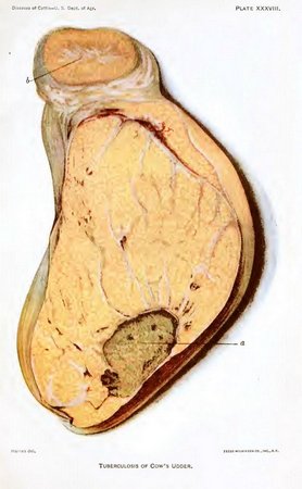
Of late tubercular disease of the udder in cows (Pl. XXXVIII) has received considerable attention from sanitarians, owing to the infection of the milk with the virus of tuberculosis. According to those who have given this subject special attention, the udder becomes swollen uniformly and quite firm. This swelling, which is painless, frequently attacks but one quarter, more rarely two, these being usually the hind quarters. The larger milk ducts contain yellowish, cheesy particles, in which are many tubercle bacilli. Later larger nodules can be felt within the udder, which undergo the various changes to which tubercles are subject. The udder may grow very hard to the touch and become very large, weighing in some cases up to 40 pounds. The milk, at first normal, becomes thin and watery after a month or so, and is mixed with flakes and tubercle bacilli.
As regards the frequency of the tuberculous processes in the different organs, the following carefully compiled statistics of the disease in Bavaria and Baden may serve as a guide:
Bavaria: Per cent.
Tuberculosis of lungs and serous membranes 41
Tuberculosis of lungs alone 33
Tuberculosis of serous membranes alone (pearly disease) 17
Tuberculosis of other organs 8
Baden:
Tuberculosis of lungs alone 21
Tuberculosis of serous membranes alone 28
Both combined 39
Generalized tuberculosis 9
Tuberculosis of the sexual organs alone 3
Symptoms.—The beginning of the disease usually passes unnoticed, inasmuch as it is very slow and insidious and rarely accompanied with fever. When the lungs are involved a dull, short cough is noticed, which may later on become prolonged, convulsive, and very troublesome to the animal. The cough is more frequent in the morning after movement and drinking. The breathing varies. Only when much of the lung tissue is diseased is it labored and accompanied with active movements of the chest and nostrils. Discharge from the nose is rare or absent. At times, however, when the tubercles have broken down and cavities containing cheesy masses have formed in the lung tissue, or when the air tubes have become filled with cheesy and mucous masses, coughing will dislodge them and cause their discharge. In advanced stages the breath may have a disagreeable odor. Pressure on the chest wall may give rise to pain.
The general effect on the body is at first slight. In fact, animals may remain in good flesh for a considerable time. Invariably as the disease progresses loss of flesh and appetite and paleness of the mucous membranes become manifest. These symptoms are accompanied with a gradual diminution of the milk secretion. The debilitated condition of the animal is also manifested by a staring coat and a tough, dry, harsh skin (hidebound). Digestive disturbances are indicated by tympanites, or distention of the rumen by gas, colic, and diarrhea, alternating with constipation. The animal generally dies from exhaustion after a period of sickness which may last months or even years.
PLATE XXXIV. TUBERCULOSIS OF THE LUNGS OF CATTLE. (Click to enlarge)
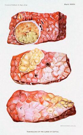
PLATE XXXV. TUBERCULOSIS OF THE LIVER. (Click to enlarge)
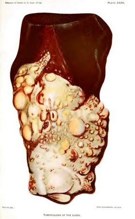
PLATE XXXVI. TUBERCULOSIS OF LYMPH GLAND AND OF OMENTUM (CAUL). (Click to enlarge)
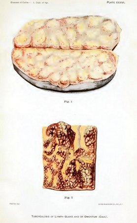
PLATE XXXVII. Fig. 1.—Tuberculosis of Sirloin and Porterhouse Cuts of Beef. Fig. 2.—Tuberculosis of Pleura of a Cow, So-called "Pearly Disease." (Click to enlarge)
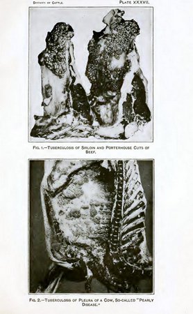
PLATE XXXVIII. TUBERCULOSIS OF COW'S UDDER. (Click to enlarge)
TUBERCULOSIS.
DESCRIPTION OF PLATES.
Plate XXXIV. Tuberculosis of the lungs of cattle. The upper figure represents a large cheesy mass, surrounded by a capsule of connective tissue, the whole embedded in healthy lung tissue. The lower figure illustrates in section a mass of tubercles which have undergone cheesy degeneration, and some of which are surrounded by dense connective tissue.
Plate XXXV. Tuberculosis of the liver. A large portion of the lobe represented in the Plate has undergone tuberculous changes. Numerous nodules are shown in various stages of the disease, the majority of which, however, contain the yellowish, partly cheesy, partly gritty areas characteristic of advanced tuberculous degeneration. This large mass involves the surface of the liver, and also extends into the liver substance.
Plate XXXVI. Tuberculosis of lymph gland and of omentum (caul).
Fig. 1. A lymph gland from the region of the thorax behind or above the esophagus, or gullet (posterior, or dorsal, mediastinum). The gland is shown cut through and laid open. It is very much enlarged, and the yellowish cheesy masses which represent tissue undergoing tuberculous changes are well shown on the cut surface.
Fig. 2. Omentum, or caul, resting upon the paunch. The reddish nodules with which the membrane is beset are tubercles, the product of the disease.
Plate XXXVII.
Fig. 1. Tuberculosis of the sirloin and porterhouse cuts of beef. The grapelike tuberculous growths are mainly restricted to the lining membrane of the abdomen.
Fig. 2. Tuberculosis of the pleura of a cow, so-called "pearly disease." Notice the grapelike clusters of tubercular nodules scattered over the lining membrane of the chest (pleura).
Plate XXXVIII. Tuberculosis of cow's udder. The udder was uniformly swollen and quite firm. Small cheesy foci and yellowish lines of tuberculous material follow the course of the milk ducts. The mucous membrane of the milk cistern (a) is ulcerated and covered with yellowish cheesy particles. The supramammary lymphatic gland (b) is greatly enlarged and contains many miliary tubercular foci.
Tuberculosis in the abdominal organs is often signalized by abortion and by abnormal sexual manifestations. When the brain is involved, the disease may cause convulsions, unconsciousness, paralysis, as well as peculiar movements in a circle, oblique position of the head, etc. Lydtin quotes the following description of the disease as taken from a Swiss sanitary order:
A dry, short, interrupted, hoarse cough, which the sick animals manifest, especially in the morning at feeding time, still more after somewhat violent exertion. At first these animals may be full blooded and lay on a considerable amount of fat when well fed. As the disease progresses they grow thin and show more and more those appearances which indicate diseased nutrition, such as a staring, lusterless, disheveled coat; dirty, tense skin, which appears very pale in those regions free from hair. The temperature of the skin is below normal. The loss of fat causes sinking of the eyes in their sockets. They appear swimming in water, and their expression is weak. The cough is more frequent, but never or very rarely accompanied with discharge. The body continues to emaciate, even with plenty of food and a good appetite, so that the quantity of milk is small. At times in the early stages of the disease, still more in the later stages, the diseased animals manifest considerable tenderness when pressure is applied to the front or the sides of the chest by coughing, moaning, etc. Often symptoms are wanting in spite of the existence of the disease.
Lydtin also quotes at length a description of the abnormal sexual desire occasionally observed among cows when affected with this disease.
Diagnosis.—A disease so varied in its attack upon the different organs of the body and in the extent of the disease process must necessarily lead to mistakes when diagnosis is attempted by ordinary means of examination. It has been confounded with the later stages of pleuropneumonia, with parasitic diseases of the brain, the lungs, the intestines, and with actinomycosis. A careful examination of the lungs by auscultation and percussion enables the expert to locate large tuberculous masses, owing to dullness, loss of respiratory murmur, and abnormal sounds, such as blowing, whistling, and creaking. The majority of cases of tuberculosis in cattle, however, including many in which the lungs are quite seriously involved, can not be detected in this manner.
THE TUBERCULIN TEST.
The tuberculin test, which is marvelously accurate in its indications, has been almost universally adopted for the detection of tuberculosis. Tuberculin is a drug prepared by sterilizing, filtering, and concentrating the liquids in which the tubercle bacillus has been allowed to vegetate. It contains the cooked products of the growth of these bacilli, but no living bacilli; consequently, when this substance is injected under the skin of an animal it is absolutely unable to produce the disease, cause abortion, or otherwise injure the animal. In case the injected animal is normal there is no more effect upon the system than would be expected from the injection of sterile water; however, if the animal is tuberculous, a decided rise of temperature will follow the use of tuberculin by the subcutaneous method. This substance, discovered by Koch, has the effect, when injected into the tissues of a tuberculous animal, of causing a decided rise of temperature or other manifestations while it has no such effect upon animals free from the disease. The value of tuberculin for this purpose was tested during the years 1890 and 1891 by Guttman, Roeckl and Schütz, Bang and Salomonsen, Lydtin, Jöhne and Siedamgrotzky, Nocard, and many others. It was at once recognized as a most remarkable and accurate method of detecting tuberculosis even in the early stages and when the disease had yet made but little progress. It is now quite generally employed.
The tuberculin test came into existence through the most careful and thorough scientific experimentation.
As a result of its use an accurate diagnosis may be established in more than 90 per cent of the cases tested. The relatively few failures in diagnoses are included among two classes of cattle. The first class contains those that are tuberculous, but which do not react either because of the slight effect of an ordinary-sized dose of tuberculin on an advanced case of the disease with so much natural tuberculin already in the system, or on account of a recent previous test with tuberculin which produces a tolerance to this material, lasting for about six weeks. The second class includes those that are not tuberculous, but which show indications of a reaction as a result of (a) advanced pregnancy, (b) the excitement of œstrum, (c) concurrent diseases, as inflammation of the lungs, intestines, uterus, udder, or other parts, abortion, retention of afterbirth, indigestion, etc., (d) inclosure in a hot, stuffy stable, especially in summer, or exposure to cold drafts or rains, (e) any change in the method of feeding, watering, or stabling of the animal during the test. Notwithstanding all these possibilities of error, the results of thousands of tests show that in less than 3 per cent of the cases tested do these failures actually occur. In the first class the chances of error are decidedly reduced by the skilled veterinarian by making careful physical examination and diagnosing clinically these advanced cases, and by the injection of double or triple doses into all recently tested cattle, with the taking of the after-temperature, beginning two hours following the injection and continuing hourly for 20 hours.
It is therefore apparent that tuberculin should be applied only by or under the direction of a competent veterinarian, capable not only of injecting the tuberculin but also of interpreting the results, and particularly of picking out all clinical cases by physical examination. The latter observation is extremely important and should always be made on every animal tested.
In the second class, where the temperature test is used, errors are avoided by eliminating from the test those cases that are nearing parturition or are in heat or show evidence of the previously mentioned diseases or exhibit temperatures sufficiently high to make them unreliable for use as normal. Where other methods of test are used these conditions do not have an important bearing on the results. In addition, a satisfactory tuberculin must be used; also an accurate thermometer and a reliable syringe, in order that a sufficient dose of tuberculin may be given. Finally, the number of apparent errors of the tuberculin test will be greatly diminished if a careful post-mortem examination is made, giving especial attention to the lymph glands. This low percentage of failures being the case, cattle owners should welcome the tuberculin test, not only for their own interest but for the welfare of the public as well. Where this method of diagnosing the disease has been adopted tuberculosis is gradually being eradicated. Without its use the disease can not be controlled and the owner is confronted with serious and continuous losses; with its use the disease can be eradicated from the herd, a clean herd established in a few years without very serious loss or hardship, and the danger of its spread to man removed. Tuberculin may therefore be considered a most beneficial discovery for the stock raiser.
Law has clearly stated the question when he says—
Many stock owners still entertain an ignorant and unwarranted dread of the tuberculin test. It is true that when recklessly used by ignorant and careless people it may be made a root of evil, yet as employed by the intelligent and careful expert it is not only perfectly safe, but it is the only known means of ascertaining approximately the actual number affected in a given herd. In most infected herds living under what are in other respects good hygienic conditions two-thirds or three-fourths are not to be detected without its aid, so that in clearing a herd from tuberculosis and placing both herd and products above suspicion the test becomes essential. * * * In skilled hands the tuberculin test will show at least nine-tenths of all cases of tuberculosis when other methods of diagnosis will not detect one-tenth.
Probably the most popular objection to tuberculin is that it is too searching, since it discovers cases in which the lesions are small and obscure. While this fact is admitted, it should also be remembered that such a small lesion to-day may break down and become widely disseminated in a relatively short period. Therefore any cow affected with tuberculosis, even to a slight degree, must be considered as dangerous not only to the other animals in the herd but also to the consumer of her products.
In 1898 Bang, of Copenhagen, one of the highest European authorities, in his paper presented to the Congress for the Study of Human and Animal Tuberculosis, at Paris, said:
Numerous tests made in almost every civilized country have demonstrated that in the majority of cases tuberculin is an excellent means for diagnosing the existence or nonexistence of the disease, but giving us no positive information as to the extent to which the disease has progressed. When tuberculin produces a typical reaction we may be almost sure that there exists in the body of the animal a tubercular process. The cases in which a careful examiner has not succeeded in finding it are very rare, and I am led to believe that when, notwithstanding all the pains taken, it has escaped discovery, the reason is that it is located in a portion of the body that is particularly inaccessible. Nevertheless, it is not to be denied that a fever, entirely accidental and of short duration, may in some rare cases have simulated a reaction. However this may be, the error committed in wrongly condemning an occasional animal for tuberculosis is of no practical consequence.
A worse aspect of the case is that there are some diseased animals in which tuberculin fails to discover the existence of tuberculosis. In most of these, no doubt, the deposits are old, insignificant, and generally calcified, or they are cases where the disease is arrested and perhaps in process of recovery, and which are possibly incapable of disseminating the contagion. But it is known that there are cases, not altogether rare, where tuberculin fails to cause a reaction in a highly tuberculous animal, and consequently one in which the disease exists in an extremely contagious form. For this reason a clinical examination should always be made of an animal which does not give a reaction but which shows symptoms indicating that, notwithstanding the test, it may suffer from tuberculosis.
Nocard, of Paris, wrote also in 1898 as follows:
The degree of certainty of the indications furnished may be stated in precise terms. The observation of a clear reaction to tuberculin is unequivocal; the animal is tuberculous. The pretended errors imputed to the method are explained by the extreme sensitiveness of the reagent, which is capable of detecting the smallest lesion. It often requires prolonged and minute researches in the depths of all the tissues to discover the few miliary centers, the presence of which has been revealed. The reaction is absolutely specific. In those cases where it is observed with animals which show lesions of another disease (actinomycosis, hydatid disease, verminous bronchitis, distomatosis), it may be affirmed that there exists, in addition to these conspicuous changes, a tuberculous center which alone has provoked the reaction.
The failure to react does not necessarily imply absence of tuberculosis. Such failures of tuberculin are very exceptional. They are seen most frequently with animals affected with tuberculosis in a very advanced stage and made evident by plain external signs. Sometimes, also, there are found at the post-mortem examination of animals which have not reacted small fibrous or calcified lesions in such a condition that one is tempted to believe them cured. Whether sterile or not, these lesions have no tendency to increase, and they are not very dangerous from the point of view of contagion.
These opinions of two eminent authorities, living in different countries, after long experience of their own and after studying the results of the many tests made in different parts of the world, should have great weight. They are essentially the same throughout.
In 1897 Voges compiled statistics of tuberculin tests, the accuracy of which had been determined by post-mortem examination. Of 7,327 animals tested, it appeared that errors had been made with 204, or 2.78 per cent. In the work of the Pennsylvania Live Stock Sanitary Board post-mortem examinations were made on about 4,400 reacting cattle and the disease was found in all but 8 of those which had given characteristic reactions.
The results of a much larger number of tests might be compiled at this time, but they would not materially change the average of those already mentioned. It is plain that tuberculin is a remarkably accurate test of tuberculosis, that the animals which react may be safely considered as tuberculous, and that when a careful clinical examination is practiced in addition to the test there are few animals in a dangerous condition which escape detection.
The first questions asked by those who oppose the adoption of the tuberculin tests are: Is this test infallible? and, if it is not infallible, why should it be forced upon the cattle owners of the country?
In answer to these questions it may be said that tuberculin is not absolutely infallible, and yet it is by far the best method of diagnosing tuberculosis that has been discovered. It is much better than any test known for pleuropneumonia when that disease was eradicated.
Practically all the animals that react are affected with tuberculosis and should be separated from the herd, not only in the interest of the public, but in the interest of the owner of the herd. The best authorities admit, after studying many thousands of tests, that there are few, if any, mistakes made in condemning cattle which show a typical tuberculin reaction. The errors are principally in the other direction—that is, some tuberculous animals are not discovered by the tuberculin test, but as the most dangerous of these may be picked out by ordinary clinical examination this fault of tuberculin is not so serious as it at first sight appears. This being the case, it should not be necessary to force the tuberculin test upon owners. They should be anxious to adopt it in their own interests and for the protection of their patrons. There is to-day no greater danger to the cattle and hog industries than that which confronts them in the form of tuberculosis, a disease already widespread and rapidly extending. Furthermore, in view of the results revealed by numerous tests covering vast numbers of animals, tuberculin must be considered as harmless for healthy animals. It has also been clearly demonstrated that tuberculin interferes in no way with the milking function in healthy cattle; neither in the quantity of milk nor in butter-fat value has any variation been detected. The conclusions of some of the best authorities on the subject of its harmlessness to healthy animals are given below.
Nocard and Leclainche state:
Direct experiments and observations collected by thousands show that the tuberculin injections have no unfavorable effect. With healthy animals the system is indifferent to the inoculation; with tuberculous animals it causes slight changes which are not at all serious.
Bang has written as follows on this question:
We will now consider the following question, a very important one, in the application of tuberculin, viz: Can the reaction produce a worse condition in tuberculous animals than before existed? Hess emphatically states that it can, and on this account he earnestly warns against its application. My attention has been directed to this question from the beginning. In my first publication on tuberculin injection I reported two cases in which acute miliary tuberculosis was proved in two high-grade tuberculous cows several weeks after the tuberculin injection. I then stated my suspicion that perhaps the tuberculin injection had some connection with this, just as is often supposed to be the case in human practice. With my present very large amount of material for observation at hand I may express the following opinion: Such an acute development of tuberculosis as a result of tuberculin injection is to be feared only exceptionally, and then in cases of advanced tuberculosis. It must not be forgotten that acute miliary tuberculosis by no means rarely accompanies an advanced tuberculosis of long standing. It is therefore impossible to offer strict proof of the causal connection with the injection, and only oft-repeated observation could make this probable. In support of my view I offer the following: In the course of the last three years I have made careful post-mortem examinations of 83 tuberculous animals, which have been removed from my experiment farm, Thurebylille. Among these were 18 (or, strictly speaking, 23) high-grade tuberculous animals. I have been able to prove miliary tuberculosis in only 4 of these. Among the others, which showed less developed tuberculosis, I have never found miliary tuberculosis, and with very many I have never found any sign of a more rapid development of the process. On the contrary, it has been proved that the disease was restricted locally, often for years, in spite of yearly repeated injections. Dissections were made at very different periods after the injections—in 17 cases from 4 to 12 days after the last test. In all of these cases earlier tests had been made months or years before. In 28 cases the injection took place from 19 days to 2 months before the butchering; in 3 of these cases earlier injections had been made. In 38 cases from two and one-half months to one year intervened between the last injection and the dissection. Dissection gives the best explanation of this question, but a clinical observation, continued for years, of a herd tested with tuberculin can render very essential aid. If Hess's opinion is correct, it is to be assumed that tuberculosis must take an unusually vicious course in such herds, but this I have been unable to prove. At Thurebylille there has existed for three years a reacting division, consisting originally of 131 head and now 69. Although these animals are yearly tested, and although most of them react every year, the division certainly appears to be made up of healthy animals, and the farm inspector has expressed the decided opinion that the tuberculosis in this division is no more developed than at the beginning of the experiment. The testimony of many owners of large herds of cattle which have long ago been injected is to the same effect. I will adduce statements from several. A farm tenant whose cattle were injected 20 months previously, when 82 per cent of the grown animals reacted, wrote me recently as follows: "Only 2 cows from the division of 100 head had been sold as decidedly tuberculous. The majority appeared afterwards, just as before, entirely healthy. The fat animals which had been slaughtered had been pronounced healthy by the butchers." Another farm tenant with a herd injected in 1894 had not been obliged to remove a single animal from the tuberculosis division, numbering 70 head. A large farm owner in Jutland stated in September that he had traced no undesirable result from the injection. His herd of 350 had been injected in February and about 75 per cent reacted. Similar answers have been given by other owners and veterinarians.
A veterinarian who had injected 600 animals, among them a herd of a large farm, 18 months previously, expressed the belief that the injection had produced in no single case an unusually rapid or vicious course of tuberculosis. In spite of a demand made months ago, I have received thus far no report from any veterinarian of an undesirable result.
On a large farm, on which before the injection tuberculosis had appeared in a vicious form, the owner had the impression that the severe cases had afterwards become more numerous. He had, however, not suffered severe losses, and 8 months later the large reacting division by no means made a bad impression. Finally, it is to be noticed that tuberculin has been employed on a large scale in Denmark for years, and still the demand from farmers constantly increases. This could certainly not be the case if the injections were generally followed by bad results.
Paige said, after the tests of the herd of the Massachusetts Agricultural College, that "its use is not followed by any ill effects of a serious or permanent nature."
Lamson, of the New Hampshire College Agricultural Experiment Station, said: "There is abundant testimony that its use is not in any way injurious to a healthy animal."
Conn, who made a special study of the present attitude of European science toward tuberculosis in cattle, reached the following conclusions:
It has been, from the first, thought by some that the use of tuberculin produces a direct injury upon the inoculated animals. This, however, is undoubtedly a mistake, and there is no longer any belief anywhere on the part of scientists that the injury thus produced is worthy of note. In the first place, the idea that it may produce the disease in a perfectly healthy animal by the inoculation is absolutely fallacious. The tuberculin does not contain the tubercle bacillus, and it is absolutely certain that it is impossible to produce a case of tuberculosis in an animal unless the tubercle bacilli are present. The use of tuberculin, therefore, certainly can never produce the disease in the inoculated animal.
It has been more widely believed, however, that the inoculation of an animal with this material has a tendency to stimulate an incipient case of tuberculosis. It has been thought that an animal with a very slight case of the disease may, after inoculation, show a very rapid extension of this disease and be speedily brought to a condition where it is beyond any use. The reasons given for this have been the apparent activity of the tuberculosis infection in animals that have been slaughtered shortly after inoculation. This has been claimed, not only by agriculturists who have not understood the subject well, but also by veterinarians and bacteriologists. But here, too, we must recognize that the claim has been disproved, and that there is now a practical unanimity of opinion on the part of all who are best calculated to judge that such an injurious effect does not occur. Even those who have been most pronounced in the claim that there is injury thus resulting from tuberculin have, little by little, modified their claim, until at the present time they say either that the injury which they formerly claimed does not occur or that the stimulus of the disease is so slight that it should be absolutely neglected in view of the great value which may arise from the use of tuberculin. Apart from two or three who hold this very moderate opinion, all bacteriologists and veterinarians unite in agreeing that there is no evidence for believing that any injury results. In Denmark, especially, many hundreds of thousands of animals have been inoculated, and the veterinarians say there is absolutely no reason in all their experience for believing that the tuberculin inoculation is followed by any injurious results.
In 1898 tuberculosis was found in the large Shorthorn herd belonging to W. C. Edwards, of Canada, who with commendable promptness and public spirit had his animals tested, and at once proceeded to separate the diseased from the healthy animals. They were all finely bred animals, and of the very class which we have been told are most susceptible to the injurious effects of tuberculin. After using this test regularly for two years, Mr. Edwards wrote as follows:
I have seen nothing to lead me to believe that the tuberculin test had any injurious influence on the course of the disease. It is by no means our opinion that the disease has been stimulated or aggravated by the application of the tuberculin test. All animals that we have tested two or three times continue as hale and hearty as they were previously, and not one animal in our herds has broken down or failed in any way since we began testing.
Mr. Edwards, in December, 1901, verbally stated that his views as to the harmlessness of tuberculin remained unchanged, and that he had not seen the least ill effect in any of his cattle from its use.
Those who have had most experience with tuberculin have failed to observe any injurious effects following its use upon healthy cattle. With tuberculous cattle it produces a fever of short duration, and in the great majority of cases all derangement of the system which it causes disappears within 48 hours after the tuberculin is administered. There appear to have been a very few cases in which the disease was aggravated, and a greater number in which it was benefited by the injection of tuberculin. The cases of abortion following the tuberculin test have not been numerous, even when cows were tested within a few weeks of the normal time of calving. The few cases of this kind which have occurred may be explained by the fact that abortion in cattle is a very common occurrence, and that it would inevitably happen sometimes after the tuberculin test as a mere coincidence and without any relation between the test and the loss of the calf. The cases of abortion which have been cited appear to be no more numerous than might be expected to have occurred among the same number of cattle within the same period if the test had not been applied.
At the present time there is ample evidence to show that tuberculin is the most reliable means of detecting tuberculosis in the living animal and that its use is not attended by any harmful aftereffects.
An act of Congress was approved July 24, 1919, for the purpose of controlling and eradicating tuberculosis of animals. The official means of detecting tuberculosis in the living animal is the tuberculin test, which may be applied by three different methods—the subcutaneous, the intradermic, and the ophthalmic. It is not necessary to discuss here the details of these three methods, which are made use of in the work of eradication of tuberculosis.