Front matter (continued 7)
Reading edition
A more direct factor, which not only contributed to the establishment of a section on the healing arts, but also had a greater effect upon the Smithsonian Institution than any other event since its founding, was the 1876 centennial exhibition in Philadelphia.
This magnificent international fair commemorated the hundredth anniversary of the adoption of the Declaration of Independence. The finest exhibits of 30 foreign countries and various States of the Union participating in the fair were finally donated to the Smithsonian Institution as the official depository of historical and archeological objects for this country. As a result, the Institution’s collections increased to an extent far beyond the capacity of the first Smithsonian building. This led to the erection of the National Museum, known for the last two decades and until date of publication as the Arts and Industries building, which was completed on March 4, 1881, and was used that evening for the inaugural reception of incoming President James A. Garfield.
Section of Materia Medica (1881-1898)
Throughout the 19th century, the study of materia medica (dealing with the nature and properties of drugs of various kinds and origins, their collection and mode of administration for the treatment of diseases, and the medicinal utilization of animal products) held an increasingly important place among the medical sciences. In the United States, as in other civilized countries, this topic was greatly emphasized in the curriculum of almost every school teaching the health professions. Today, the subject matter contained in this branch of science is taught under the heading of several specialized fields, such as pharmacology, pharmacognosy, and drug analysis of various types. However, when the decision was made in 1881 to promote greater knowledge and interest in the healing arts by creating a section devoted to such pursuits in the U.S. National Museum, the title of Section of Materia Medica was adopted. Added to this, was the fact that the bulk of the first collections received in the Section was a great variety of crude drugs, which constituted much of the material then taught in the academic courses of materia medica.
The new Section was included in the Department of Arts and Industries, then under the curatorship of Assistant Director G. Brown Goode. From its beginning and for two decades, however, the Section of Materia Medica was sponsored and supervised by the U.S. Navy in cooperation with the Smithsonian Institution. For this reason, the Navy decided not to establish a similar bureau for a health museum as did the Army in starting the Medical Museum (of the Armed Forces Institute of Pathology) in 1862 through the efforts of Dr. William Alexander Hammond. The Smithsonian did, however, provide a clerk to relieve the curator of much of the routine work. The Section’s early vigorous activities were the result of the ingenuity of the first honorary curator, Dr. James Milton Flint (1838-1919), an Assistant Surgeon of the U.S. Navy. From the establishment of the Section, in 1881, to 1912, Dr. Flint was curator during separate periods for a total of nearly 25 years. For three of his tenures (1881-1884; 1887-1891; 1895-1900), he was detailed to the Smithsonian Institution by the Surgeon General of the U.S. Navy. During the interim periods, other naval doctors were detailed as curators. Finally, in 1900, Dr. Flint retired from the Navy with the rank of Rear Admiral and volunteered to continue his services to the National Museum. The proposal was gladly accepted and he continued as a curator until his retirement from the Smithsonian Institution in 1912.
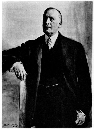
Figure 3.—Rear Admiral James M. Flint, U.S. Navy surgeon and first honorary curator of the Section of Materia Medica. (Photo courtesy of the Library of Congress.)
The Section commenced with a wealth of material. After the close of the 1876 centennial exhibition, its materia medica collection had been stored with the other collections in a warehouse, awaiting an appropriation by Congress for transfer and installation. This collection was gradually brought into the new National Museum after that building’s completion in 1881. Many other materia medica specimens were transferred from the Department of Agriculture. In addition to these large collections of crude drugs, generous contributions came from several prominent pharmaceutical firms such as Parke, Davis & Company of Detroit, Michigan; Wallace Brothers of Statesville, North Carolina; and Schieffelin and Company of New York City. These manufacturing houses are mentioned here because they and their agents abroad were the first to take interest and donate to the Section, complete assortments of contemporary remedial agents then in common use throughout the United States and Europe, besides many hundreds of “rare and curious drugs.” Thus, in spite of difficulties encountered from bringing several collections into the building at one time, the materia medica exhibition got off to a good start.
It was Dr. Flint, the first curator, who stated in 1883 that remedial agents used by a nation or a community are as indicative of the degree of their cultural development and standard of living as is the nature of their food, the character of their dwellings, and their social and religious traditions. Therefore, he felt that collections of drugs and medical, surgical and pharmaceutical instruments and appliances should not be thought of or designed as instructive to the specialist only, but should also possess a general interest for the public. Because of these objectives, Dr. Flint added, this section was conceived as a departmental division for the collecting and exhibiting of objects related to medicine, surgery, pharmacology, hygiene, and all material related to the health field at large. [4]
During his first term of curatorship (1881-1884), Dr. Flint devoted much of his time to sorting, examining, identifying, and classifying the materia medica specimens. [5] In 1881, he issued a memorandum of instructions to be followed by collectors of drugs and urged them to give detailed and accurate information regarding acquired specimens so that they might be “more than mere museum curiosities.” In addition, in 1883, he prepared a brief manual of classification of the materia medica collection in the Museum as well as a useful, detailed catalog of informational labels of the individual objects on exhibition. The unpublished catalog is still the property of the Smithsonian Institution Archives, Division of Medical Sciences’ Library.
It was Dr. Flint’s ambition to obtain a comprehensive, worldwide collection of all substances used as remedies. Then, in order to identify drugs from foreign countries, he tried to collect illustrated works on medical botany and printed pharmacopoeias of all nations having them. He rightly defined an official pharmacopoeia as “a book containing directions for the identification and preparation of medicines prepared and issued with the sanction of a government or organized and authorized medical and pharmaceutical societies. Its purpose is to establish uniformity in the nomenclature of remedies and in the character and potency of the pharmaceutical preparations. It is enacted by legislation, and thus becomes binding on all who prepare drugs or sell them for medication.” By soliciting the help of various American consuls and Navy officers abroad, about 16 such official pharmacopoeias were collected, making an almost complete international representation of all available, official, drug standards. With these sources of information, Dr. Flint compiled and arranged an international list of materia medica specimens, indicating the authorized preparations of each. By so doing, the first curator of this Section took the initiative at least in proposing and, to some extent acting, on the preparation of an international pharmacopoeia of drugs used in existing authorized formularies giving “official synonyms, and tables showing the constituents and comparative strength of all preparations.” [6] This undertaking is of special importance in the history of American pharmacy, since it was probably the first attempt of its kind in the United States.[7] In addition, colored plates and photographs of medicinal plants were collected, forming the nucleus of the Division’s current collection of pictorial and photographic material related to the history of the health field.
Dr. Flint also put on exhibition 630 Chinese materia medica specimens from the 1876 Philadelphia centennial. These had been collected originally by the Chinese Imperial Customs Commission for the centennial and were subsequently given to this country.
In 1881, the numbered objects in the Section’s register amounted to 1,574 entries. In the following year, 1,590 more specimens were added, most of them drugs in their crude state. By the end of 1883, the total collection had reached 4,037, out of which 3,240 individual drugs in good condition were classified and put on display. Of these, about 500 specimens with beautiful illustrations of parts of their original plants had been mounted for exhibition. The drug exhibitions also included materials transferred from the Department of Agriculture in 1881, which originally had been brought from Central America and South America for the 1876 centennial exhibition, a variety of opium specimens from Turkey, and a number of rare drugs listed in the official formulary which were acquired from the Museum of Karachi in what was then India.
Dr. Flint commented in the Smithsonian Annual Report for 1883 that the collection of cinchona barks was especially complete. It was comprised of specimens of nearly all the natural cinchona barks of South America and every known variety of the cultivated product from the British government plantations in India. In addition, there were specimens from Java, Ceylon, Mexico, and Jamaica. The Indian and Jamaican barks were accompanied by herbarium specimens of the leaf and flower (and, in some cases, the fruit) of each variety of tree from which the bark was obtained.[8]
In an attempt to protect specimens liable to attack by insects, a small piece of blotting paper moistened with chloroform was inserted underneath the stopper in each bottle. Later on, bichloride of mercury was found to be a better insecticide.
These early collections of the Section were brought into admirable condition and received compliments for their organization and completeness. In the Smithsonian Annual Report for 1883, the collections were praised as “superior to any other in the United States and scarcely excelled by any in Europe.”
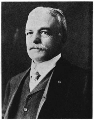
Figure 4.—Dr. Henry Gustav Beyer, the second honorary curator of the Section of Materia Medica (1884-1887). (Photo courtesy of American Physiological Society.)
In spite of the apparent emphasis on the displaying of drugs, the first curator of the Section had envisioned that the exhibits eventually would embrace the entire field of the healing arts. In the Smithsonian Annual Report for 1883, Dr. Flint noted that “in the establishment of a museum designed to illustrate man and his environment, it is proper that the materials and methods used for the prevention and cure of disease should have a place.” However, his plans were temporarily interrupted when his first term as honorary curator ended in 1884.
On June 4, 1884, Dr. Henry Gustav Beyer was detailed by the Department of the Navy to become the second honorary curator of the Section of Materia Medica. As a young man, Dr. Beyer (1850-1918) had come from Saxony, Germany, to the United States and, in due course, became a naturalized citizen. He was graduated from the Bellevue Hospital Medical College of New York City in 1876.
Because of his interest in physiological experimental research, Dr. Beyer enrolled at the Johns Hopkins University, where he was awarded a Ph. D. degree in 1887. Unlike his predecessor, Dr. Beyer was primarily interested in carrying on research on the physiological action of certain drugs and in pharmacology. This was evident from the original scientific papers mentioned in the Smithsonian Annual Reports and published by him during the period of his curatorship from 1884 to 1887.
Despite the pressure of his postgraduate studies at Johns Hopkins University, Dr. Beyer helped in arranging and classifying the materia medica collection without trying to extend materially the scope of the Section.
After the term of Dr. Beyer expired in 1887, Dr. Flint returned to take charge of the Section. Surprisingly, at this time, it seems that he showed less enthusiasm and devotion to the work of the Museum which he had previously served so well. It could have been a disappointment resulting from a lack of evidence of any real progress in the Section since he had left it three years before. Whatever the reasons may have been, the Smithsonian Annual Reports show that only a few hundred specimens were added to the materia medica collections between 1887 and 1890, bringing the total to 5,915 preserved in good condition. Further curtailment of the Section’s activities began in November 1891 when Dr. Flint was again transferred to other duties for the U.S. Navy. From November 1891 to May 24, 1895, curatorship of the Section was charged to five physicians of the U.S. Navy: Drs. John C. Boyd (from November 1891 to April 6, 1892); William S. Dixon (April 1892 to January 5, 1893); C. H. White (January 1893 to July 15, 1893); C. U. Gravatt (July 1893 to January 22, 1894); R. A. Marmion (January 22, 1894 to June 15, 1894); and to Medical Inspector Daniel McMurtrie (June 1894 to May 24, 1895). During this interim of nearly three and a half years, there were neither literary contributions nor additions made to the collections of the Section that were of any significance. The reason is obvious, for all of these curators averaged less than seven months of service which is not enough time, even for a well-trained individual, to accomplish very much in a museum. Therefore, it is easy to imagine that when the Secretary of the Navy detailed Dr. Flint for a third time to take charge of the Section, he was rather discouraged. Nevertheless, at the Cotton States and International Exposition in Atlanta, Georgia, from September 18 to December 31, 1895, the materia medica was represented by two displays: one on mineral waters and amounts of solid constituents in pure state; and another showing the quantities of minerals after analysis of the composition of the human body.
A similar project was undertaken in 1897 at the Tennessee Centennial Exposition (May 1 to October 31) in Nashville, where there were two displays of materia medica. One showed several kinds of the cinchona barks and the medicinal preparations made from them, and another containing the commercial varieties of the alkaloids of opium.
At this time, Dr. Flint’s attention turned to a new phase of medical exhibition. He felt the need for a program of exhibits on the practice and the historical development of the healing arts. A change of the Section’s name was deemed necessary and, thus, in 1898 the more comprehensive title of Division of Medicine was adopted.
Division of Medicine (1898-1939)
The statement by L. Emmett Holt of the Rockefeller Institute for Medical Research, that before 1906, the Smithsonian Institution was never a beneficiary to medicine in any form,[9] is not entirely applicable. The previous discussion has clearly shown that the U.S. National Museum’s cooperation with the Navy contributed materially towards encouraging and promoting medical knowledge. Furthermore, Dr. Flint tried to bring many of his plans for this medical division of the Museum to a practical fulfillment. He devised a program for presenting medical history in a way which would be of interest both to the public and to the profession. In order to best illustrate the history of the healing art, he divided his subject matter into five provisional classifications according to the Report upon the Condition and Progress of the U.S. National Museum during 1898:
1. Magical medicine including exorcism, amulets, talismans, fetishes and incantation;
2. Psychical medicine including faith cures, and hypnotism;
3. Physical and external medicine including baths, exercise, electricity, massage, surgery, cautery, and blood-letting;
4. Internal medicine including medications and treatment used by the ancient Egyptians, Greeks, Hindus, Arabians, and Chinese; and
5. Preventive medicine including beverages, food, soil, clothing and habitation.
It is certainly to Dr. Flint’s credit that from its early conception, first as Section of Materia Medica and thereafter as Division of Medicine, he planned for an all-embracing exhibition and reference collection of the medical sciences. Until the end of the 19th century and the early years of the 20th century, crude drugs as well as primitive and magic medicine held a more prominent place than medical instruments in the exhibits and collections. In 1905, Flint issued his last, known, literary contribution, “Directions for Collecting Information and Objects Illustrating the History of Medicine,” in Part S of Bulletin of the U.S. National Museum, no. 39. The emphasis he put upon this shows Dr. Flint’s interest in collecting medical and pharmaceutical objects and equipment of historical value. Consequently, he arranged new exhibits including one on American Indian medicine. A medical historian, Fielding H. Garrison, inspected these about 1910 and, in his “An Introduction to the History of Medicine,” wrote of their novelty and appeal. “In the interesting exhibit of folk medicine in the National Museum at Washington,” he commented, “a buckeye or horse chestnut (Aesculus flavus), an Irish potato, a rabbit’s foot, a leather strap previously worn by a horse, and a carbon from an arc light are shown as sovereign charms against rheumatism. Other amulets in the Washington exhibit,” he added, “are the patella of a sheep and a ring made out of a coffin nail (dug out of a graveyard) for cramps and epilepsy, a peony root to be carried in the pocket against insanity, and rare and precious stones for all and sundry diseases.” It had been Dr. Flint’s intention, besides presenting an educational display on the history of the medical arts, to warn the public against the perils of quackery and the faults of folk medicine, as well as to expose evils in drug adulteration. Today, we can see actual fulfillment of these intentions in the present exhibit at the medical gallery which has been executed recently on the basis of scientific, historical research.
After Dr. Flint’s retirement from the Smithsonian Institution in 1912, there was no replacement for over five years. Therefore, the Division of Medicine was placed, for administrative purposes, under the supervision of the curator of the newly reestablished (1912) Division of Textiles, Frederick L. Lewton. During these years, he fought against the dispersal of the medical and materia medica collections. Thus, for lack of a curator of its own, almost all new activities in the Division of Medicine were curtailed until 1917.
On January 31, 1917, Lewton addressed members of the American Pharmaceutical Association inviting them to cooperate in gathering up and preserving at the National Museum the “many unique and irreplaceable objects” connected with the early history of pharmacy in this country which could still be saved.[10] Then, on March 14, 1917, an examination was announced by the Civil Service (held May 2) for an assistant curator for the Division of Medicine, and the position was filled by Joseph Donner on August 16, 1917. Donner was the first full-time employee paid by the Smithsonian Institution for the curatorship of this Division. He held the post until January 31, 1918, when he was inducted into the Sanitary Corps of the United States Army. No significant activities in the Division of Medicine were reported during these few months.
Mr. Donner was followed by a second, full-time, museum officer who promoted a great amount of good will towards the Division during his curatorship of a little over 30 years. Dr. Charles Whitebread (1877-1963), the first pharmacist to head the Division, joined the Smithsonian in 1918 and remained until his retirement in 1948, the longest service, thus far, of any individual in the Division.
Dr. Whitebread received his degree of Doctor of Pharmacy from the School of Pharmacy at George Washington University in Washington, D.C., in 1911. He entered government service late in 1915, but it was not until April 2, 1918, that he agreed to become assistant curator of the Division of Medicine.
Curator Whitebread’s first year was an active and challenging one, for in this new position he began to develop a deep interest in the history of the healing arts. He made a number of important acquisitions, most of them pertaining to pharmaceutical products, synthetic chemicals and crude drugs. He found that many specimens from the older drug collections had deteriorated to such an extent as to be worthless, and he began replacing them with freshly marketed drugs.
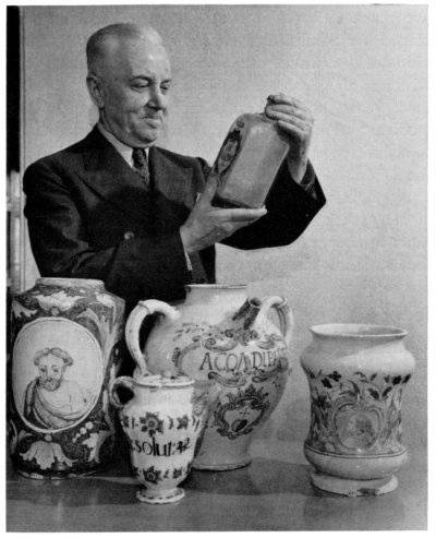
Figure 5.—Curator Charles Whitebread inspecting, with admiration, five drug containers from the Squibb collection (1945). (Photo courtesy of the American Pharmaceutical Association.)
Plans were completed for the opening of new medical exhibits and adopting, with some modifications and additions, earlier classifications set by Dr. Flint. Dr. Whitebread grouped these into the following classes: the evaluation of the healing arts; a picture display of medical men prominent in American history;[11] a materia medica display including the history of pharmacy; and an exhibition on Sanitation and Public Hygiene[12] which was later to evolve into the Hall of Health.
In 1920, Dr. Whitebread added a number of specimens of medical-dosage forms and pharmaceutical preparations to the Division’s collections. He also acquired other gifts to complete existing exhibits illustrating the basic principles of the various schools of medicine, such as homeopathy and osteopathy—their methods, tools, and ways of thought.
In 1921, a tablet machine by the Arthur Colton Company of Detroit, Michigan, was acquired, and an exhibit illustrating vaccine and serum therapy was installed in the medical gallery. This was followed, in 1922, by a collection arranged to tell the story of the prevention and cure of specific diseases by means of biological remedies.
During the following two years, two more exhibits related to hospital supplies and sanitation were added to the rapidly developing Hall of Health exhibition which was opened in 1924. A third exhibit in 1925 consisted of 96 mounted color transparencies illustrating services provided by hospitals to promote public health. Plans for the further development of the Hall of Health continued during 1926, and contacts were made with organizations interested in the educational aspects of the healing arts. As a result, several new exhibits were added. In 1926, the American Optometric Association helped in the installation of an exhibit on conservation of vision or the care of the eyes under the slogan “Save your vision,” as a phase of health work. Other exhibits in the Hall at this time were: what parasites are; water pollution and how to obtain pure water; waste disposal; ventilation and healthy housing, and the importance of recreation; purification of milk and how to obtain pure milk; transmission of diseases by insects and animals; how life begins; prenatal and postnatal care and preschool care; duties of the public health nurse; and social, oral and mental hygiene.
With the acquiring of more medical appliances and the widening of the scope of the exhibits, more and more space was needed, and attention was turned to the area of the medical gallery which had been occupied by the materia medica collection for almost four decades. To gain more exhibit space, it was decided that the greater part of the crude drugs should be removed from the exhibits and be kept as a reference collection and for research.[13]
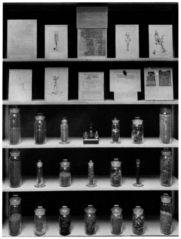
Figure 6.—Exhibit on Egyptian and Hebrew medicine, installed about 1924, which was illustrated by graphs and drugs mentioned in extant records of this ancient period. (Smithsonian photo 30796-C.)
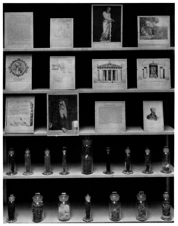
Figure 7.—Exhibit on medical history during the Greco-Roman period. (Smithsonian photo 30796-D.)
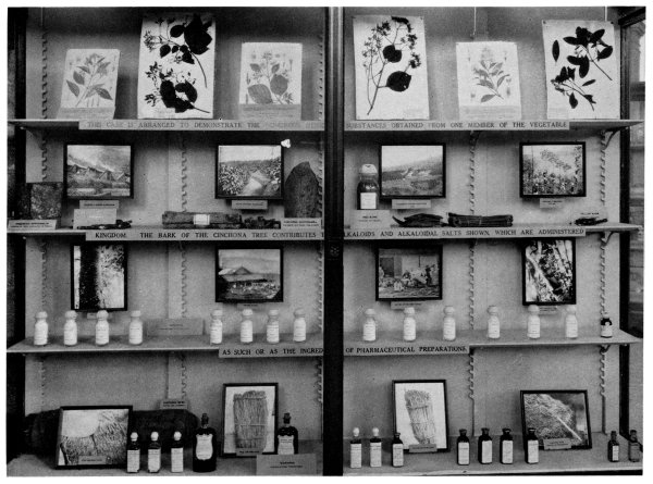
Figure 8.—Exhibit on remedies derived from drugs of vegetable origin, displayed about mid-1930’s. (Smithsonian photo 30439.)
In 1926, original patent models including those related to pharmacy, medicine, and dentistry, were transferred from the U.S. Patent Office to the National Museum. These patent models, together with other apothecary tools and the machines used in drug production took up most of the available space. This unfortunate situation led Dr. Whitebread to turn down significant medical and pharmaceutical collections offered the Museum between 1927 and 1930. Since the patent models were devised for inventions designed to simplify the practice of the health professions, three cases of these models were displayed in the medical gallery in the early 1930’s. Other exhibits shown during this decade included the deception of folk medicine with warnings against superstitions, and an exhibition on osteopathy,[14] as well as dioramas on the manufacture of medicines and their use in scientific medical treatment.
In the meantime, Dr. Whitebread was an active contributor to the literature of the health field in various periodicals, as well as in pamphlets issued by the Museum and other governmental agencies (see bibliography). His literary contributions, guided by the exhibits he designed and the collections he acquired, were focused on the Division’s collections, such as primitive and psychic medicine and warnings against reliance on magic and superstitions in treatment, medical oddities, and the utilization of drugs of animal origin, both past and present.
Division of Medicine and Public Health (1939-1957)
After taking charge of the Division of Medicine in 1918, Dr. Whitebread gave special attention to public health displays. His activities in this area were accelerated after 1924 when the health exhibit at the Smithsonian Institution was inaugurated. As the exhibits in this field increased, the Division, in 1939, took the more comprehensive title of Division of Medicine and Public Health. Also, in 1939, Dr. Whitebread was promoted to the rank of associate curator.
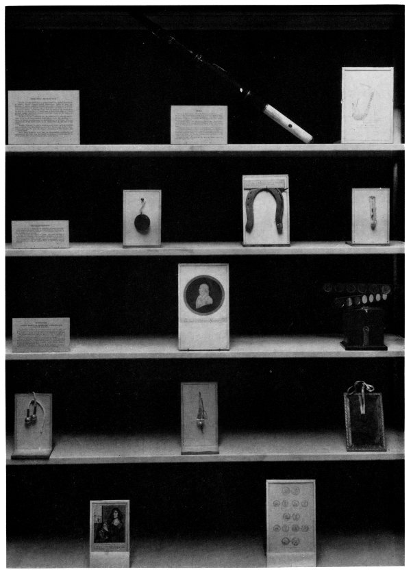
Figure 9.—Exhibit on methods of treatment of diseases through mental impressions and psychic conditions as displayed about 1925. (Smithsonian photo 30796-B.)
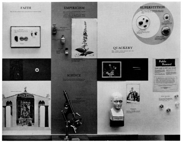
Figure 10.—An exhibit on superstitions, empiricism, magic, and faith healing in the light of scientific medicine, completed in 1962, is in sharp contrast with that shown in figure 9.
He continued his efforts to collect more specimens of interest to medical history and to contribute to the literature. Among exhibited specimens in 1941 were a powder paper-crimping machine, a portable drug crusher, an odd device for spreading plaster on cloth, a pill-coating apparatus, various suppository molds, a lozenge cutter, and an ingenious Seidlitz powder machine. The derivation of medicinal drugs from animal, vegetable, and mineral sources was also depicted, as were synthetic materials and their intermediates. Basic prescription materials were displayed, and rows of glass-enclosed cases held samples of crude botanical drugs from almost every part of the globe with explanatory cards giving brief, concise descriptions. The exhibition provided medical and pharmaceutical students about to take state-board examinations, the opportunity to study the subject in detail, especially the enormous collection of materia medica samples.[15] Also in 1941, Eli Lilly and Company donated an exhibit on the medical treatment of various types of anemia. In the same year, a diorama including a hypochlorinator for purification of water on a farm was installed in the gallery. In 1942, the first Emerson iron lung (developed in 1931 by John Haven Emerson) for artificial respiration was acquired by the Division. The Division acquired, in 1944, the first portable x-ray machine known to have been operated successfully on the battlefield, as well as other x-ray equipment and early medicine chests.
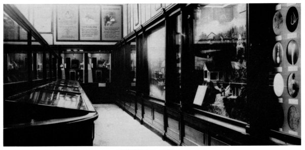
Figure 11.—Old public health exhibition installed in the gallery about 1924. (Smithsonian photo 19952.)
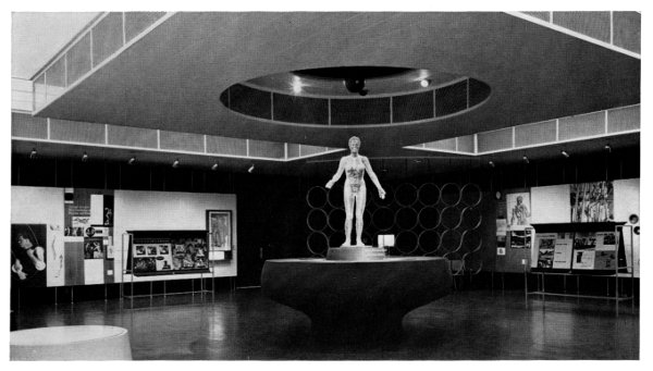
Figure 12.—The Hall of Health, reestablished and opened in November 1957. (Smithsonian photo 44931.)
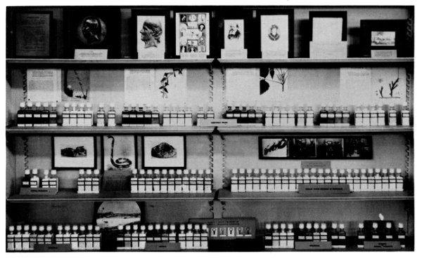
Figure 13.—Early exhibit on homeopathy showing its history, methods and remedies which was installed about 1929. (Smithsonian photo 27049.)
Without a doubt, the most outstanding accession in the field of pharmaceutical history during Dr. Whitebread’s years of service was the acquisition of the E. R. Squibb and Sons old apothecary shop. Most of the baroque fixtures, including the stained-glass windows with Hessian-Nassau coats of arms and wrought-iron frames, were part of the mid-18th-century cathedral pharmacy “Münster Apotheke” in Freiburg im Breisgau, Germany. It was offered for sale in September 1930 by Dr. Jo Mayer of Wiesbaden, Germany, who was an enthusiastic collector of antiques, especially those related to the health professions. Earlier that year, a historian of pharmacy and chemistry, Fritz Ferchl of Mittenwald, Germany, had published a series of scholarly and informative articles on the Meyer collection in which the outstanding specimens were beautifully portrayed and thoroughly described (see bibliography).
As a result of Dr. Mayer’s efforts to sell his collection, the impact of Ferchl’s illustrated articles, and the uniqueness of the collection, E. R. Squibb and Sons purchased it in 1932 and brought it to the United States “with the thought that it would provide for American pharmacy, its teachers and students, a museum illuminating the history, growth, and development of pharmacy, its interesting background and struggle through the ages.” It was displayed at the Century of Progress exposition held in Chicago during 1933 and 1934; subsequently, it was assembled in the Squibb Building in New York City as a private museum where, for about 10 years, it was visited by many interested in pharmacy, ceramics, and art. Charles H. LaWall, who was originally engaged to prepare a descriptive catalog on the exhibit, gave it the title “The Squibb Ancient Pharmacy.”
Late in 1943, E. R. Squibb and Sons offered the collection as a gift to the American Pharmaceutical Association if the latter would provide museum space for it. The offer was accepted, but the Association finally found it difficult to spare the needed space for the collection and decided to take up the matter with the U.S. National Museum.
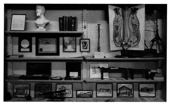
Figure 14.—This early exhibit on osteopathy was renovated several times prior to the early 1940’s. (Smithsonian photo 19250.)
At this point, it should be stated that since 1883 the members of the American Pharmaceutical Association have been keenly interested in having the National Museum serve as the custodian for all collected objects and records of historical interest to pharmacy. In 1944, the Association officially offered to deposit on permanent loan, the Squibb’s pharmacy collection in the Smithsonian Institution with the understanding that a suitable place would be provided for prompt and permanent display. The offer was accepted, and during April and May of 1945, the entire collection was transferred to the Smithsonian Institution, and construction to recreate the original two rooms for the old, 18th-century, European “Apotheke” was underway.
By August 1946, the exhibit was completed. In the large room where the pharmacist met his customers, the shelves were filled with 15th- to 19th-century, European pharmaceutical antiques. These included Renaissance mortars; 16th- and 17th-century nested weights; beautiful Italian, French, Swiss, and German majolica and faience drug jars; Dutch and English delft; drug containers made of flint or opal glass with fused-enamel labels with alchemical symbols; rare, 16th-century, wooden drug containers, each with the coat of arms of the city in which each was made; and two glass-topped, display tables contained franchises issued and signed by Popes or state rulers, medical edicts, dispensatories, herbals, pharmacopoeias, and pharmaceutical utensils.
On the walls in the small laboratory room, which also had been used as a workshop and a study, were a stuffed crocodile, shark’s head, tortoise, fish, and salamander, parts of which were utilized as remedial agents. Their presence provided tangible evidence that the pharmacy dispensed genuine drugs and not substitutes.
The pharmaceutical profession in this country hailed the outstanding exhibition, and the November 1946 issue of the Journal of the American Pharmaceutical Association, Practical Pharmacy Edition, devoted its front cover to depicting one corner of the study and laboratory room of the shop.[16] Also, in a letter dated January 2, 1947, addressed to Dr. Alexander Wetmore, then Secretary of the Smithsonian Institution, Dr. Robert P. Fischelis, the secretary of the American Pharmaceutical Association, considered the completion of the deposited exhibition a triumph and “as one of the highlights of the accomplishments of the Association in 1946.”
From 1946 to 1948, the Division’s collection was further enriched with a number of historical specimens, among which was a “grosse Flamme” x-ray machine with induction-coil tube and stand developed by Albert B. Koett. It is one of the earliest American-made machines of its kind, producing a 12-inch spark, the largest usable at that time with 180,000-volt capacity, and a forerunner of later autotransformers. Other accessions included two 19th-century drug mills, an electric belt used in quackery, two medicine chests, three sets of Hessian crucibles used in a pioneer drugstore in Colorado, a drunkometer, mineral ores, and purely produced chemical elements.
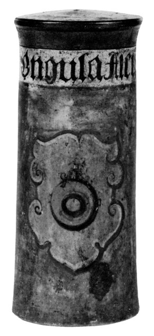
Figure 15.—Late 16th-century, wooden drug container with coat-of-arms, in the Squibb collection. The inscription Ungula Alcis (the hoof of the elk) suggests a superstitious attitude in medical practice and the wide use of animal organs in medical treatment. (Courtesy of the American Pharmaceutical Association.)
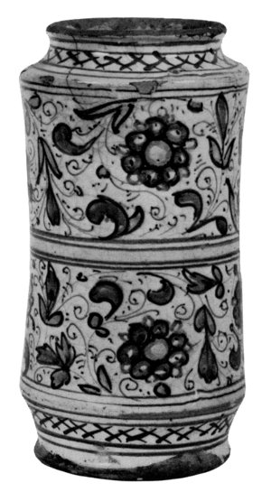
Figure 16.—A Rare, Antwerp, 16th-century drug jar in the Squibb collection deposited by the American Pharmaceutical Association.
In the spring of 1948, Associate Curator Whitebread retired after 30 years of service with the U.S. National Museum. He was a pioneer in the field of health museums and during his curatorship had developed a moribund section into a Division of field-wide importance. Dr. Whitebread was succeeded by George S. Thomas, also a pharmacist, who served as associate curator from August 1948 until early 1952.
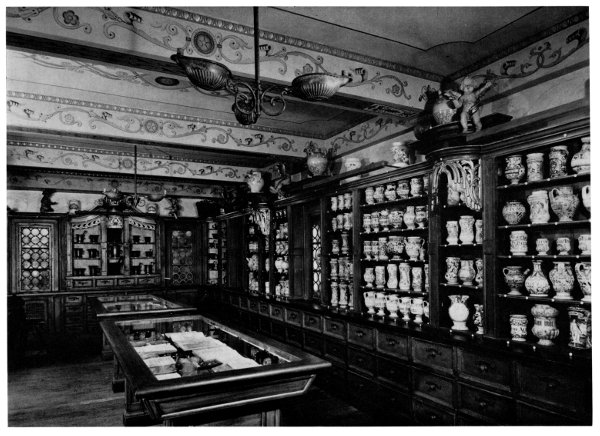
Figure 17.—The Apothecary Shop as seen in the Arts and Industries building (1946-1964). (Courtesy of the American Pharmaceutical Association.)
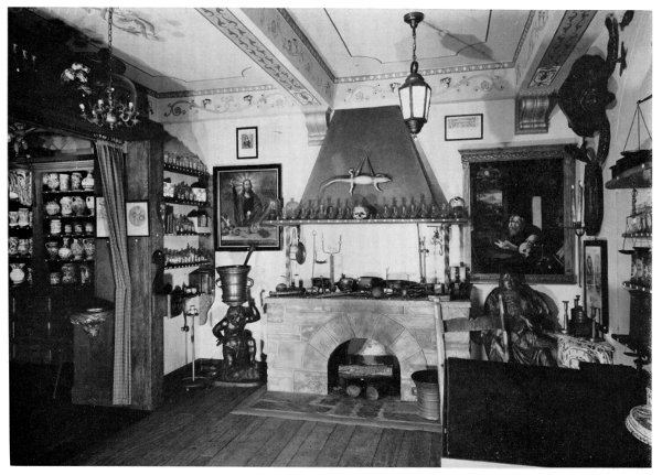
Figure 18.—View of the laboratory and study room of the apothecary shop. On the left, the German-Swiss bronze mortar and pestle (1686) sign and above it an 18th-century German painting on canvas of Christ, “the apothecary of the soul.” The drug containers represent “the fruits of the spirit,” faith, patience, charity, etc., and the scales represent justice. Underneath is the verse from Matthew, 11:28, “Come unto me, all ye that labour and are heavy laden, and I will give you rest.” (Courtesy of the American Pharmaceutical Association.)
During his almost-three and a half years of service, Thomas acquired hearing-aid appliances from which he designed an exhibit on the development of these aids, surgical sutures, early samples of Aureomycin, and a static-electricity machine made by Henkel about 1840. He also published three short articles under the title, “Now and Then,” in the National Capital Pharmacist (1950), no. 1, pp. 8-9; no. 2, pp. 18-19, 29; and no. 3, pp. 15-16. In early 1952, Dr. Arthur O. Morton presented to the Division, a Swiss-made keratometer which he had purchased in 1907, and it is believed to be one of the first used in the United States to measure the curves of the cornea.
The achievements of the Division reached their highest point, thus far, in significantly increasing the national collection, as well as in contributing to the scientific, historical, and professional literature, under the curatorships of George B. Griffenhagen (December 8, 1952, to June 27, 1959) and John B. Blake (July 1, 1957, to September 2, 1961). Their reorganization of exhibits and collections, their competence and industry, fulfilled the hopes, plans, and purposes laid down by earlier curators for the Division.
Immediately after assuming the responsibilities of the Division and throughout 1953, Mr. Griffenhagen (M.S. in pharmaceutical chemistry from the University of Southern California) undertook to develop the collections still further. He increased the emphasis not only on historical pharmacy, but also on medicine, surgery, and dentistry. He also renovated the exhibits in the medical gallery.
In 1954, several antibiotics were donated to the Division including a mold of Penicillium notatum prepared and presented to the Smithsonian Institution by Sir Alexander Fleming (1881-1955), the discoverer of penicillium (1929), and a few Petri dishes used by botanist Benjamin M. Daggar who, while working for Lederle Laboratories, developed Aureomycin (chlortetracycline) in 1948. The Forest D. Dodrill—G.M.R. mechanical heart (1952), the first machine reported to be used successfully for the complete bypass of one side of the human heart during a surgical operation, [17] was presented to the Smithsonian Institution.
The following year, 1955, the Division acquired one of the earliest Einthoven string galvanometers (named after the Dutch physiologist Willem Einthoven, 1860-1927) made in the United States in 1914 by Charles F. Hindle for an electrocardiograph. Also added to the Division’s collections was the electrocardiograph used by Dr. Frank E. Wilson of the United States, a pioneer educator in this field. Two temporary exhibits on allergy and surgical dressings were installed in the gallery. In the same year, Curator Griffenhagen published Early American Pharmacies, a catalog on 28 pharmacy restorations in this country.
In 1956, among many publications of interest in the fields of medical and pharmaceutical history, was Curator Griffenhagen’s Pharmacy Museum, with a foreword by Laurence V. Coleman, who termed it a useful catalog and “a good reflection of the history of the museum movement at large.” A third x-ray tube of Wilhelm Konrad Roentgen (1845-1922) was added to the collection in 1957 as well as a complete set of hospital-ward fixtures of about 1900 from the Massachusetts General Hospital, rare patent medicines, 18th-century microscopes, and a 13th-century mortar and pestle made in Persia.
In 1957, Mr. Griffenhagen published a series of illustrated articles in the Journal of the American Pharmaceutical Association, Practical Pharmacy Edition, which were later reprinted by the Association in a booklet entitled, Tools of the Apothecary. In it, he described several pharmaceutical specimens in the collection and their place in history.
Division of Medical Sciences (1957 to Present)
The U.S. National Museum was reorganized on July 1, 1957, into two units, the Natural History Museum and the Museum of History and Technology. At the same time, and in view of the widening scope of the Division, its more scientifically based planning, and the constantly increasing collection with equal emphasis on all branches of the healing arts, the Division’s title was changed to the Division of Medical Sciences—the title it still bears in 1964. With the reorganization, the Department of Engineering and Industries, under which the Division fell administratively, was renamed the Department of Science and Technology of the Museum of History and Technology. It was also the first time since its establishment in 1881 that the Division had two curators, for on July 1, 1957, Dr. John B. Blake joined the staff.
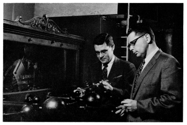
Figure 19.—Curators John B. Blake and George Griffenhagen examine the newly acquired (1957) electromagnetic, Morton-Wimshurst-Holz Influence Machine. It was manufactured by the Bowen Company of Providence, Rhode Island (1889). With the discovery of x-ray, it was used for making x-ray photographs until early in the 20th century.
As a result of these changes, the Division was subdivided into a Section of Pharmaceutical History and Health and a Section of Medical and Dental History. The former was planned to encompass the collections of materia medica, pharmaceutical equipment, and all material related to the history of pharmacy, toxicology, pharmacology, and biochemistry, as well as the Hall of Health which was opened November 2, 1957, and which emphasizes man’s progressing knowledge of his body and the functions of its major organs. [18] The latter Section was planned to include all that belongs to the development of surgery, medicine, dentistry, and nursing, especially in relation to hospitals.
In October 1957, the Division acquired a collection of rare, ceramic, drug jars which included two, 13th-century, North Syrian and Persian, albarello-shaped, majolica jars; a 15th-century, Hispano-Moresque drug container; and a 16th-century, Italian faience, dragon-spout ewer. During the following two years, Curator Griffenhagen periodically toured museums and medical and pharmaceutical institutions in this country, South America, and Europe gathering specimens and information for the Division and for publication, respectively. However, on June 27, 1959, he resigned his curatorship to join the staff of the American Pharmaceutical Association in Washington, D.C. Dr. Blake became the curator in charge of the Division and Mr. Griffenhagen was succeeded on September 24, 1959, by the author of this paper as associate curator in charge of the Section of Pharmaceutical History and Health.
Dr. Blake, as curator of the Section of Medical and Dental History, acquired a large number of valuable and varied specimens for the Division’s collections. They included optometric refracting instruments, an early 1920’s General Electric, portable, x-ray machine, the Charles A. Lindbergh and Alexis Carrel pump (designed in 1935 to perfuse life-sustaining fluids to the organs of the body), the Sewell heart pump (1950) to control delivery of air pressure and suction to the pumping mechanism, and a large and valuable collection of dental equipment formerly at the universities of Pennsylvania and Illinois. Dr. Blake wrote the explanatory material and supervised the design and production of the majority of exhibits in the renovated hall of medical and dental history. He also contributed several scholarly articles and a book (see bibliography) on the history of the healing arts and public health in particular. He resigned on September 2, 1961, to join the staff of the National Library of Medicine as chief of the History of Medicine Division, and was succeeded by the author as curator of the Division. From the summer of 1962 to April 1964, the Division benefited from the expert advice of Dr. Alfred R. Henderson as consultant in the preparation and designing of the surgical and medical exhibits of the Museum of History and Technology.
During the period from 1961 to May 1964, the Division’s collections expanded greatly through its medical, dental, and pharmaceutical acquisitions. Specimens of antiques acquired from 1961 through 1963 numbered up to 1,539 and included gifts from leading institutions and individual philanthropists. The scope of these gifts and acquisitions ranges from electronic resuscitators, microscopes, x-ray equipment, and spectacles, to patent medicines, amulets, apothecary tools, dental instruments, and office material of practitioners.
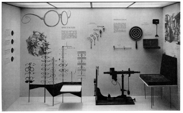
Figure 20.—Exhibit on spectacles, lorgnettes, optometers, and refraction, completed in 1960. It features a cross section of the Division’s large collection of eyeglasses. (Smithsonian photo 47943-D.)
In the last decade, the interest in the national endeavor for promoting research and scholarship in the history of medicine has increased greatly. It was most appropriate, therefore, for the Smithsonian Institution to play host on May 2 for two sessions of the 37th annual meeting of the American Association for the History of Medicine held in the Washington, D.C., area from April 30 through May 2, 1964. In welcoming the members to the morning session in the auditorium of the new Museum of History and Technology, Frank A. Taylor, director of the United States National Museum, expressed the feeling that the meeting of the Association was, in a sense, a dedication of the new auditorium and an opportunity for the Smithsonian to reaffirm its deep interest and commitment in fostering research and furthering the appreciation of scholarly endeavor in the history of the healing arts.
A New Dimension For the Healing Arts
“One day the United States will have a National Museum of science, engineering, and industry, as most large nations have.” This was the prediction made in 1946 by the director of the U.S. National Museum, Mr. Frank A. Taylor, then curator of the Division of Engineering. [19] It was in 1963, that the new $36,000,000 building of the Museum of History and Technology was completed, and opened to the public in 1964. The offices of the Division of Medical Sciences as well as the reference and study collections were moved to the fifth floor of the new building. The exhibits, however, will be displayed in the gallery at the southwest corner of the first floor. These exhibits, it is hoped, will show a new dimension and an unprecedented approach in displaying the development of the healing arts throughout the ages and the instruments and equipment associated with health professions. They also present the expanding objectives and plans of the Division’s growth as an integral part of the Smithsonian Institution. Conveniently, the exhibits form four, closely connected halls in one large gallery which will be open to the public in the summers of 1965 to 1966.
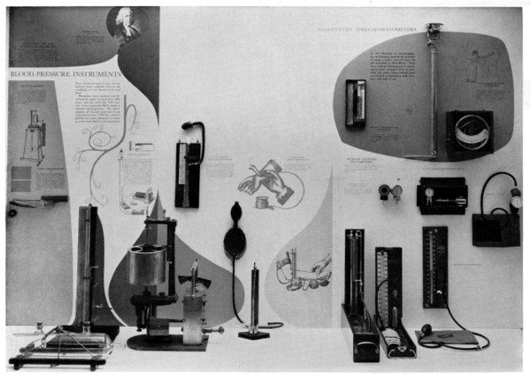
Figure 21.—Exhibit on the development of blood-pressure instruments and the early 20th-century sphygmomanometers which was completed in 1960. (Smithsonian photo 47943-M.)
1. The Hall of Health displays models and graphic and historical exhibit materials to demonstrate the function of the various healthy organs of the human body. The main topics emphasized are: embryology and childbirth; tooth structure; the heart and blood circulation; respiration; the endocrine glands; kidneys and the urinary-excretory system; the brain and the nervous system; the ear; and vision and the use of eyeglasses.
The most appreciated exhibit of all in this Hall is the “transparent woman” figure which rotates, automatically, every 15 minutes with a recorded message describing the function of each major organ of the body at the same time that the organ is electronically lighted, so that the viewer can see its place in the body.
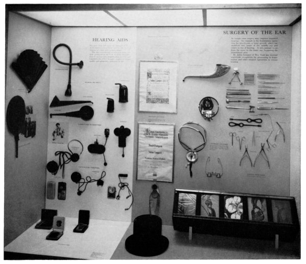
Figure 22.—Hearing-aid exhibit designed in 1962. It includes otologist Julius Lempert’s personal memorabilia and original surgical instruments used in the fenestration operation for restoring hearing. (Smithsonian photo 49345-C.)
2. The Hall of Medicine and Dentistry will depict the history of these two sciences with exhibits of the equipment used through the centuries. In the medical field, early trephining and other surgical instruments will be displayed along with a diorama of an 1805 surgical operation performed by Dr. Philip Syng Physick in the amphitheater of the Pennsylvania Hospital. Diagnostic instruments such as stethoscopes, endoscopes, speculums, and blood-pressure measuring devices will be exhibited with a series of microscopes illustrating the development of these instruments. Exhibits of original galvanometers and other apparatus will trace the development of cardiography. The early use of anesthesia will be shown by apparatus of William Morton and Crawford W. Long, American pioneers in this field. The development of the devices of modern medicine and surgery will be shown by exhibits of the iron lung and x-ray tubes, including a tube used by W. K. Roentgen. Medicine chests and surgical kits of different periods will graphically summarize the state of medical science in the period each represents.
Exhibits on the development of dentistry and dental surgery will display examples of tooth-filling and extracting tools, drilling apparatus from the early hand and foot engines to the first ultrasonic cutting instrument (1954), and the original contra-angle, hydraulic and air-turbine handpiece model [20] which revolutionized the field of instrumentation for dental surgery (with speeds of 200,000 to 400,000 rpm). This hydraulic turbine of Dr. Robert J. Nelson and associates of the National Bureau of Standards set the design pattern for the remarkable and successful high-speed, air-turbine handpiece developed by Paul H. Tanner and Oscar P. Nagel of the U.S. Naval Dental School in 1956. Also underway is the reconstruction of the offices of famous dentists such as G. V. Black and the father of American orthodontia, Edward H. Angle, using their original equipment and instruments. In addition, an exhibit is planned to include x-ray tubes and the electric dental engine, the first to be operated in a human mouth by the pioneer dentist on dental skiagraphy, Charles E. Kells (1856-1928). [21]
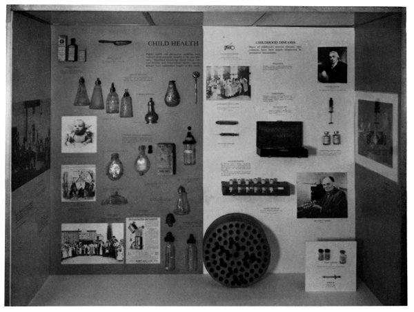
Figure 23.—Exhibit on nursing bottles and measures to promote child health to counteract the once-common diseases of childhood. This display was completed in 1962. (Smithsonian photo 49345-G.)
3. The Hall of Pharmaceutical History will feature exhibits on the reconstruction of two pharmacy shops: an 18th-century apothecary shop, originally from Germany, with a very elegant collection of drug jars, decorated medicinal bottles, balances, mortars and pestles, and other tools and documents pertaining to the apothecary art, and a late 19th-century American drugstore with shelves filled with patent medicines and drug containers of various sizes and shapes. The window will also feature symbols of pharmacy and beautiful show globes. Displays will show the development of antibiotics and the early tools used in the manufacture of the so-called “miracle drugs,” including a mold from Sir Alexander Fleming, the discoverer of penicillin. In addition, a platform will be reconstructed to display a variety of pharmaceutical apparatus used in the preparation and manufacture of drugs, such as tablet and capsule machines and drug mills and percolators. Recently, with the assistance of Professor Glenn Sonnedecker, the Division acquired a fine collection of pharmaceutical equipment and devices from the School of Pharmacy of the University of Wisconsin.
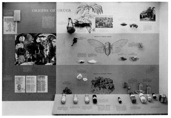
Figure 24.—The origins of drugs from the three natural kingdoms, drug synthesis, and the increase in the manufacture of vitamins. This display was completed in 1962 and is now on display at the Museum of History and Technology. (Smithsonian photo P6316.)
Since the Division houses the largest collection of materia medica in the country, a representative cross section of crude drugs will be displayed in alphabetical order as well as a display illustrating the role of cinchona and antimalarial drugs in the fight against disease. An exhibit will portray the “origin of drugs” from the three natural kingdoms, animal, vegetable, and mineral, together with synthetic drugs including the manufacture of vitamins.
Plans are being made for an elaborate exhibit of weights and balances used in many countries throughout the centuries, their impact on accuracy of dosage and weighing of drugs, and their use in the apothecary art.
The Division will also display pictorial and printed materials, as well as artifacts from all periods and all countries. These collections are intended to help in presenting a more complete picture of the story of the medical sciences for educational purposes and research, and to increase man’s knowledge in fighting disease and promoting health.
Thus, from a few hundred specimens of crude drugs in the Section of Materia Medica of 83 years ago, there has developed a Museum Division today which embraces the evolution of the health professions through the ages. This Division now has the largest collection in the Western Hemisphere of historical objects which are related to the healing arts. The reference collections are available to the researcher and scholar, and the exhibits are intended for pleasure and educational purposes in these fields. The plans for expansion have no limitation as we keep pace with man’s progress in the medical sciences and continue to collect materials that contributed to the historical development in the fight against diseases and the attempts to secure better health for everyone.
BIBLIOGRAPHY
The Annual report of the Board of Regents of the Smithsonian Institution from 1872 to date and the Proceedings of the United States National Museum from 1881 to date were used extensively as sources in this survey. In the latter, see in particular, the year 1881, pp. 545-546; 1882, pp. 1-2; and 1884, pp. 431-475.
Atkinson, William B. The physicians and surgeons of the United States. Philadelphia, 1878. [On Dr. Toner.]
Blake, John B. Dental history and the Smithsonian Institution. Journal of the American College of Dentists (1961), vol. 28, pp. 125-127.
—— Public health in the town of Boston, 1630-1822. Cambridge, Mass.: Harvard University Press, 1959.
[Braisted, William C.] The biography of Dr. Beyer. Page 94 in Dictionary of American medical biography, by Howard A. Kelly and Walter L. Burrage; New York: D. Appleton and Co., 1928.
Clark, Leila F. The library of the Smithsonian Institution. Science (1946), vol. 104, p. 143.
Coleman, Laurence Vail. The museum in America: A critical study. 3 vols. Baltimore: Waverly Press, 1939. [Printed for the American Association of Museums, Washington, D.C.] See vol. 1, pp. 3, 11-12, 32-33, 143-146, 222, 318; vol. 3, p. 471.
Daukes, S. H. The medical museum: modern developments, organization and technical methods based on a new system of visual teaching. London: Wellcome Foundation Ltd., 1929.
Dodrill, Forest D., and others. Pulmonary volvuloplasty under direct vision using the mechanical heart for a complete bypass of the right heart in a patient with congenital pulmonary stenosis. Journal of Thoracic Surgery (1953), vol. 26, pp. 584-595.
—— Temporary mechanical substitution for the left ventricle in man. Journal of the American Medical Association (1952), vol. 150, pp. 642-644.
Dunglison, Robley. A dictionary of medical science. Rev. ed. Pp. 629-630. Philadelphia: Lea, 1874.
Edwards, J. J., and Edwards, M. J. Medical museum technology. London: Oxford University Press, 1959. [See in particular, pp. 33-62, 142-159.]
Ferchl, Fritz. Die Mörser der Sammlung Jo Mayer—Wiesbaden; Libri rari et curiosi der Sammlung Dr. Jo Mayer—Wiesbaden; Bildnisse und Bilder der Sammlung Jo Mayer—Wiesbaden; Kuriositäten und Antiquitäten der Sammlung Jo Mayer—Wiesbaden; and Gläser, Majoliken und Faensen der Sammlung Jo Mayer—Wiesbaden. Pharmazeutische Zeitung (Berlin, 1930), vol. 75: January 4, no. 2, pp. 19-24; February 15, no. 14, pp. 219-223; March 8, no. 20, pp. 309-314; April 19, no. 32, pp. 487-489; and June 21, no. 50, pp. 735-740.
Flint, James M. Classification and arrangement of the materia medica collection. Proceedings of the United States National Museum (1881), vol. 4, app. no. 6.
—— Classification of the materia medica collection of the United States National Museum, and catalogue of specimens. Proceedings of the United States National Museum (1883), vol. 6, app. 19, pp. 431-475.
—— Directions for collecting information and objects illustrating the history of medicine. Part S of Bulletin of the United States National Museum (1905). No. 39.
—— Memoranda for collectors of drugs for the materia medica section of the National Museum. Proceedings of the United States National Museum (1881), vol. 4, app. 8.
Foley, Matthew O. Smithsonian Institution devotes much space to hospital exhibit. Hospital Management (April 1929), pp. 271-287.
Galdston, Iago. Research in the United States. Ciba Symposia (June-July 1946), vol. 8, nos. 3 and 4, p. 366.
Garrison, Fielding H. An introduction to the history of medicine. 2d ed. p. 38. Philadelphia: Saunders, 1917.
Gebhard, Bruno. From medicine show to health museum. Ciba Symposia (March 1947), vol. 8, no. 12, p. 579.
Goode, George Brown. The Smithsonian Institution (1846-1896): The history of its first half century. Pp. 325-329, 362-363. Washington, 1897.
Griffenhagen, George. Pharmacy museums. Madison, Wis.: American Institute of the History of Pharmacy, 1956.
—— and Hughes, Calvin H. The history of the mechanical heart. Annual report of the Board of Regents of the Smithsonian Institution for the year ended June 30, 1955 (Washington, 1956), pp. 339-356.
Hamarneh, Sami. At the Smithsonian … exhibits on pharmaceutical dosage forms. Journal of the American Pharmaceutical Association (1962), new ser., vol. 2, pp. 478-479.
—— For the collector, facts and artifacts. Pharmacy in History (1961), vol. 6, p. 48.
—— Historical and educational exhibits on dentistry at the Smithsonian Institution. Journal of the American-Dental Association (July 1962), vol. 65, pp. 111-114.
—— New dental exhibits at the Smithsonian Institution. Journal of the American Dental Association (May 1963), vol. 66, pp. 676-678.
Haynes, William. Out of alchemy into chemistry. The Scientific Monthly (November 1952), vol. 75, p. 268.
Holt, L. Emmett. A sketch of the development of the Rockefeller Institute for Medical Research. Science (July 6, 1906), new ser., vol. 24, no. 601, p. 1.
Howell, William H. The American Physiological Society during its first twenty-five years. Pp. 21-22 [biography of Dr. Beyer] in History of the American Physiological Society semicentennial, 1881-1937; Baltimore, 1938.
[Klein, Allen.] He directs pharmacy exhibits at the Smithsonian Institution. Modern Pharmacy (July 1941), vol. 25, pp. 20-21.
LaWall, Charles H. Ancient pharmacy on display. Pacific Drug Review (1933), vol. 45, p. 18.
—— The curious lore of drugs and medicines. Garden City, N.Y.: Garden City Publishing Co., Inc., 1927. [See p. 453 on Division of Medical Sciences’ collection.]
Lewton, Frederick L. A national pharmaceutical collection. Journal of the American Pharmaceutical Association (1919), vol. 8, pp. 45-46.
—— The opportunity for developing historical pharmacy collections at the National Museum. Journal of the American Pharmaceutical Association (1917), vol. 6, pp. 259-262.
Long, Esmond R. The Army Medical Museum. Military Medicine (May 1963), vol. 128, pp. 367-369.
Monell, S. H. “Dental Skiagraphy” (pp. 313-336 in A system in x-ray methods and medical uses of light hot-air, vibration and high-frequency currents by Monell; New York: Pelton, 1902).
Murray, David. Museums, their history and their use. Glasgow: MacLehose, 1904. [See vol. 1, pp. 13-77.]
Nelson, Robert J.; Pelander, Carl E.; and Kumpula, John W. Hydraulic turbine, contra-angle handpiece. Journal of the American Dental Association (September 1953), vol. 47, pp. 324-329.
Official Catalogue of the Cotton States and International Exposition: Atlanta, Georgia, September 18 to December 31, 1895. Atlanta: Claflin and Mellichamp, 1895. [See p. 204.]
Packard, Frances R. History of medicine in the United States. New York, 1931. [See vol. 1, pp. 5-6, 37-51, 168-176, 602-607 on Dr. Toner.]
Pickard, Madge E. Government and science in the United States: Historical background. Journal of the History of Medicine and Allied Sciences (1946), vol. 1, nos. 2 and 3, pp. 265-266, 289, 446-447, 478.
Purtle, Helen R. Notes on the Medical Museum of the Armed Forces Institute of Pathology. Bulletin of the Medical Library Association (1956), vol. 44, no. 3, pp. 300-305.
Rathbun, Richard. A descriptive account of the building recently erected for the Departments of Natural History of the United States National Museum. (U.S. National Museum Bulletin 80.) Washington, 1913. [See pp. 7-15.]
Rhees, William J. The Smithsonian Institution; documents relative to its origin and history, 1835-1899. 2 vols. (Smithsonian Miscellaneous Collections: vol. 42, 1835-1881; vol. 43, 1881-1899.) Washington, 1901.
Shufeldt, R. W. Suggestions for a national museum of medicine. Medical Record (March 22, 1919), pp. 4-5. [Also reprinted, 1919, by William Wood and Co., New York.]
Sigerist, Henry E. Primitive and archaic medicine. (Vol. 1 of A history of medicine, by Sigerist.) New York: Oxford University Press, 1951. [See pp. 525-531.]
Silver, Edwin H. Description of the exhibit on conservation of vision placed in the United States Museum at Washington, D.C. The Optical Journal and Review of Optometry (February 3, 1927), vol. 59, no. 5, pp. 39-40.
[Sonnedecker, Glenn.] Apothecary shop nears completion. Journal of the American Pharmaceutical Association, Practical Pharmacy Edition (1946), vol. 7, pp. 157.
—— Dr. Charles Whitebread, pharmacist and museum curator. Journal of the American Pharmaceutical Association, Practical Pharmacy Edition (1946), vol. 7, p. 203.
—— Old apothecary shop. Journal of the American Pharmaceutical Association, Practical Pharmacy Edition (1945), vol. 6, pp. 184-187.
—— Old apothecary shop opened. Journal of the American Pharmaceutical Association, Practical Pharmacy Edition (1946), vol. 7, p. 427.
Taylor, Frank A. A national museum of science, engineering and industry. The Scientific Monthly (1946), vol. 63, pp. 359.
—— The background of the Smithsonian’s Museum of Engineering and Industries. Science (1946), vol. 104, no. 2693, pp. 130-132.
Toner Lectures:
1. J. J. Woodward. On the structure of cancerous tumors and the mode in which adjacent parts are invaded. No. 266 in Smithsonian Miscellaneous Collections, vol. 15; Washington, 1878. [Lecture given on March 28, 1873.]
2. C. E. Brown-Séquard. Dual character of the brain. No. 291 in Smithsonian Miscellaneous Collections, vol. 15; Washington, 1878. [Lecture given on April 22, 1874.]
3. J. M. Da Costa. On strain and over-action of the heart. No. 279 in Smithsonian Miscellaneous Collections, vol. 15; Washington, 1878. [Lecture given on May 14, 1874.]
4. H. C. Wood. A study of the nature and mechanism of fever. No. 282 in Smithsonian Miscellaneous Collections, vol. 15; Washington, 1878. [Lecture given on January 20, 1875.]
5. William W. Keen. On the surgical complications and sequels of the continued fevers. No. 300 in Smithsonian Miscellaneous Collections, vol. 15; Washington, 1878. [Lecture given on February 17, 1876.]
6. William Adams. Subcutaneous surgery: Its principles, and its recent extension in practice. No. 302 in Smithsonian Miscellaneous Collections, vol. 15; Washington, 1878. [Lecture given on September 13, 1876.]
7. Edward O. Shakespeare. The nature of reparatory inflammation in arteries after ligatures, acupressure, and torsion. No. 321 in Smithsonian Miscellaneous Collections, vol. 16; Washington, 1880. [Lecture given on June 27, 1878.]
8. George E. Waring. Suggestions for the sanitary drainage of Washington City. No. 349 in Smithsonian Miscellaneous Collections, vol. 26; Washington, 1883. [Lecture given on May 26, 1880.]
9. Charles K. Mills. Mental over-work and premature disease among public and professional men. No. 594 in Smithsonian Miscellaneous Collections, vol. 34; Washington, 1893. [Lecture given on March 19, 1884.]
10. Harrison Allen. A clinical study of the skull. No. 708 in Smithsonian Miscellaneous Collections, vol. 34; Washington, 1893. [Lecture given on May 29, 1889.]
True, Webster P. The Smithsonian Institution. (Vol. 1 of the Smithsonian Scientific Series.) Washington, 1929.
Urdang, George, and Nitardy, F. W. The Squibb ancient pharmacy. New York, 1940. [Out of print, but remaining catalogs were given to the Division of Medicine to “be reserved for pharmaceutical educators, foreign dignitaries, pharmacists of national and international reputation, and pharmaceutical historians,” according to a letter from Mr. Nitardy in 1945.]
Whitebread, Charles. Animal pharmaceuticals of the past and present. Journal of the American Pharmaceutical Association (1933), vol. 22, pp. 431-437.
—— An old apothecary shop of 1750. National Capital Pharmacist (September 1946), vol. 8, pp. 11-13, 35.
—— Early American pharmaceutical inventions. Journal of the American Pharmaceutical Association (1937), vol. 26, pp. 918-928.
—— Handbook of the health exhibits of the United States National Museum. Baltimore: Lord Baltimore Press [1924].
—— Health superstitions. Journal of the American Pharmaceutical Association, Practical Pharmacy Edition (1942), vol. 3, pp. 268-274.
—— Medicine making as depicted by museum dioramas. Journal of the American Pharmaceutical Association (January 1936), vol. 25, pp. 40-46.
—— Superstition, credulity and skepticism. Journal of the American Pharmaceutical Association (1933), vol. 22, pp. 1140-1145.
—— The Indian medical exhibit of the Division of Medicine in the United States National Museum. Article 10 in vol. 67 of Proceedings of the U.S. National Museum; Washington, 1926.
—— The magic, psychic, ancient Egyptian, Greek, and Roman medical collections of the Division of Medicine in the United States National Museum. Article 15 in vol. 65 of Proceedings of the U.S. National Museum; Washington, 1925.
—— The odd origin of medical discoveries. Journal of the American Pharmaceutical Association, Practical Pharmacy Edition (1943), vol. 4, p. 321.
—— The United States National Museum pharmaceutical collection, its aims, problems, and accomplishments. Journal of the American Pharmaceutical Association (1930), vol. 19, pp. 1125-1126.
Winters, S. R. Magic medicine. Hygeia (July 1937), vol. 15, pp. 630-633.
Paper 43 - Transcriber's Note
P. 277 ‘the basis of scientific, historical’—was ‘the bases of scientific, historical’
Contributions from The Museum of History and Technology: Paper 44 Development of Gravity Pendulums in the 19th Century
Victor F. Lenzen and Robert P. Multhauf
GALILEO, HUYGENS, AND NEWTON 304
FIGURE OF THE EARTH 306
EARLY TYPES OF PENDULUMS 309
KATER’S CONVERTIBLE AND INVARIABLE PENDULUMS 314
REPSOLD-BESSEL REVERSIBLE PENDULUM 320
PEIRCE AND DEFFORGES INVARIABLE, REVERSIBLE PENDULUMS 327
VON STERNECK AND MENDENHALL PENDULUMS 331
ABSOLUTE VALUE OF GRAVITY AT POTSDAM 338
APPLICATION OF GRAVITY SURVEYS 342
SUMMARY 346
Victor F. Lenzen and Robert P. Multhauf
DEVELOPMENT OF GRAVITY PENDULUMS IN THE 19th CENTURY
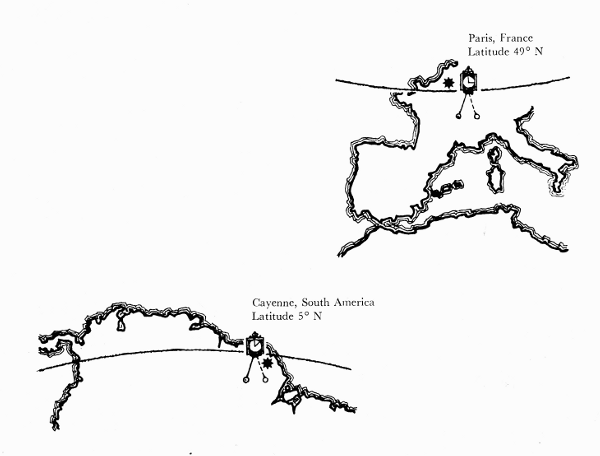
Figure 1.—A study of the figure of the earth was one of the earliest projects of the French Academy of Sciences. In order to test the effect of the earth’s rotation on its gravitational force, the Academy in 1672 sent Jean Richer to the equatorial island of Cayenne to compare the rate of a clock which was known to have kept accurate time in Paris. Richer found that the clock lost 2 minutes and 28 seconds at Cayenne, indicating a substantial decrease in the force of gravity on the pendulum. Subsequent pendulum experiments revealed that the period of a pendulum varied not only with the latitude but also regionally, under the influence of topographical features such as mountains. It became clear that the measurement of gravity should be made a part of the work of the geodetic surveyor.
The history of gravity pendulums dates back to the time of Galileo. After the discovery of the variation of the force of gravity over the surface of the earth, gravity measurement became a major concern of physics and geodesy. This article traces the history of the development of instruments for this purpose.
THE AUTHORS: Victor F. Lenzen is Professor of Physics, Emeritus, at the University of California at Berkeley and Robert P. Multhauf is Chairman of the Department of Science and Technology in the Smithsonian Institution’s Museum of History and Technology.
The intensity of gravity, or the acceleration of a freely falling body, is an important physical quantity for the several physical sciences. The intensity of gravity determines the weight of a standard pound or kilogram as a standard or unit of force. In physical experiments, the force on a body may be measured by determining the weight of a known mass which serves to establish equilibrium against it. Thus, in the absolute determination of the ampere with a current balance, the force between two coils carrying current is balanced by the earth’s gravitational force upon a body of determinable mass. The intensity of gravity enters into determinations of the size of the earth from the angular velocity of the moon, its distance from the earth, and Newton’s inverse square law of gravitation and the laws of motion. Prediction of the motion of an artificial satellite requires an accurate knowledge of gravity for this astronomical problem.
The gravity field of the earth also provides data for a determination of the figure of the earth, or geoid, but for this problem of geodesy relative values of gravity are sufficient. If g is the intensity of gravity at some reference station, and Δg is the difference between intensities at two stations, the values of gravity in geodetic calculations enter as ratios (Δg)/g over the surface of the earth. Gravimetric investigations in conjunction with other forms of geophysical investigation, such as seismology, furnish data to test hypotheses concerning the internal structure of the earth.
Whether the intensity of gravity is sought in absolute or relative measure, the most widely used instrument for its determination since the creation of classical mechanics has been the pendulum. In recent decades, there have been invented gravity meters based upon the principle of the spring, and these instruments have made possible the rapid determination of relative values of gravity to a high degree of accuracy. The gravity meter, however, must be calibrated at stations where the absolute value of gravity has been determined by other means if absolute values are sought. For absolute determinations of gravity, the pendulum historically has been the principal instrument employed. Although alternative methods of determining absolute values of gravity are now in use, the pendulum retains its value for absolute determinations, and even retains it for relative determinations, as is exemplified by the Cambridge Pendulum Apparatus and that of the Dominion Observatory at Ottawa, Ontario.
The pendulums employed for absolute or relative determinations of gravity have been of two basic types. The first form of pendulum used as a physical instrument consisted of a weight suspended by a fiber, cord, or fine wire, the upper end of which was attached to a fixed support. Such a pendulum may be called a “simple” pendulum; the enclosure of the word simple by quotation marks is to indicate that such a pendulum is an approximation to a simple, or mathematical pendulum, a conceptual object which consists of a mass-point suspended by a weightless inextensible cord. If l is the length of the simple pendulum, the time of swing (half-period in the sense of physics) for vibrations of infinitely small amplitude, as derived from Newton’s laws of motion and the hypothesis that weight is proportional to mass, is T = π√(l/g).
The second form of pendulum is the compound, or physical, pendulum. It consists of an extended solid body which vibrates about a fixed axis under the action of the weight of the body. A compound pendulum may be constituted to oscillate about one axis only, in which case it is nonreversible and applicable only for relative measurements. Or a compound pendulum may be constituted to oscillate about two axes, in which case it is reversible (or “convertible”) and may be used to determine absolute values of gravity. Capt. Henry Kater, F.R.S., during the years 1817-1818 was the first to design, construct, and use a compound pendulum for the absolute determination of gravity. He constructed a convertible pendulum with two knife edges and with it determined the absolute value of gravity at the house of Henry Browne, F.R.S., in Portland Place, London. He then constructed a similar compound pendulum with only one knife edge, and swung it to determine relative values of gravity at a number of stations in the British Isles. The 19th century witnessed the development of the theory and practice of observations with pendulums for the determination of absolute and relative values of gravity.
Galileo, Huygens, and Newton
The pendulum has been both an objective and an instrument of physical investigation since the foundations of classical mechanics were fashioned in the 17th century. [1] It is tradition that the youthful Galileo discovered that the period of oscillation of a pendulum is constant by observations of the swings of the great lamp suspended from the ceiling in the cathedral of Pisa. [2] The lamp was only a rough approximation to a simple pendulum, but Galileo later performed more accurate experiments with a “simple” pendulum which consisted of a heavy ball suspended by a cord. In an experiment designed to confirm his laws of falling bodies, Galileo lifted the ball to the level of a given altitude and released it. The ball ascended to the same level on the other side of the vertical equilibrium position and thereby confirmed a prediction from the laws. Galileo also discovered that the period of vibration of a “simple” pendulum varies as the square root of its length, a result which is expressed by the formula for the time of swing of the ideal simple pendulum. He also used a pendulum to measure lapse of time, and he designed a pendulum clock. Galileo’s experimental results are important historically, but have required correction in the light of subsequent measurements of greater precision.
Mersenne in 1644 made the first determination of the length of the seconds pendulum, [3] that is, the length of a simple pendulum that beats seconds (half-period in the sense of physics). Subsequently, he proposed the problem to determine the length of the simple pendulum equivalent in period to a given compound pendulum. This problem was solved by Huygens, who in his famous work Horologium oscillatorium … (1673) set forth the theory of the compound pendulum. [4]
Huygens derived a theorem which has provided the basis for the employment of the reversible compound pendulum for the absolute determination of the intensity of gravity. The theorem is that a given compound pendulum possesses conjugate points on opposite sides of the center of gravity; about these points, the periods of oscillation are the same. For each of these points as center of suspension the other point is the center of oscillation, and the distance between them is the length of the equivalent simple pendulum. Earlier, in 1657, Huygens independently had invented and patented the pendulum clock, which rapidly came into use for the measurement of time. Huygens also created the theory of centripetal force which made it possible to calculate the effect of the rotation of the earth upon the observed value of gravity.
The theory of the gravity field of the earth was founded upon the laws of motion and the law of gravitation by Isaac Newton in his famous Principia (1687). It follows from the Newtonian theory of gravitation that the acceleration of gravity as determined on the surface of the earth is the resultant of two factors: the principal factor is the gravitational attraction of the earth upon bodies, and the subsidiary factor is the effect of the rotation of the earth. A body at rest on the surface of the earth requires some of the gravitational attraction for the centripetal acceleration of the body as it is carried in a circle with constant speed by the rotation of the earth about its axis. If the rotating earth is used as a frame of reference, the effect of the rotation is expressed as a centrifugal force which acts to diminish the observed intensity of gravity.
Glossary of Gravity Terminology
ABSOLUTE GRAVITY: the value of the acceleration of gravity, also expressed by the length of the seconds pendulum.
RELATIVE GRAVITY: the value of the acceleration of gravity relative to the value at some standard point.
SIMPLE PENDULUM: see theoretical pendulum.
THEORETICAL PENDULUM: a heavy bob (point-mass) at the end of a weightless rod.
SECONDS PENDULUM: a theoretical or simple pendulum of such length that its time of swing (half-period) is one second. (This length is about one meter.)
GRAVITY PENDULUM: a precisely made pendulum used for the measurement of gravity.
COMPOUND PENDULUM: a pendulum in which the supporting rod is not weightless; in other words, any actual pendulum.
CONVERTIBLE PENDULUM: a compound pendulum having knife edges at different distances from the center of gravity. Huygens demonstrated (1673) that if such a pendulum were to swing with equal periods from either knife edge, the distance between those knife edges would be equal to the length of a theoretical or simple pendulum of the same period.
REVERSIBLE PENDULUM: a convertible pendulum which is also symmetrical in form.
INVARIABLE PENDULUM: a compound pendulum with only one knife edge, used for relative measurement of gravity.
From Newton’s laws of motion and the hypothesis that weight is proportional to mass, the formula for the half-period of a simple pendulum is given by T = π√(l/g). If a simple pendulum beats seconds, 1 = π√(λ/g), where λ is the length of the seconds pendulum. From T = π√(l/g) and 1 = π√(λ/g), it follows that λ = l/T2. Then g = π2λ. Thus, the intensity of gravity can be expressed in terms of the length of the seconds pendulum, as well as by the acceleration of a freely falling body. During the 19th century, gravity usually was expressed in terms of the length of the seconds pendulum, but present practice is to express gravity in terms of g, for which the unit is the gal, or one centimeter per second per second.
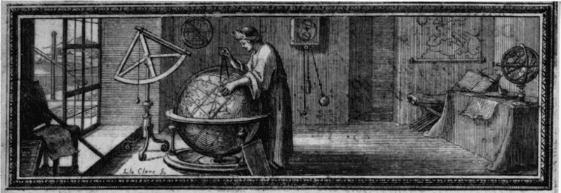
Figure 2.—This drawing, from Richer’s Observations astronomiques et physiques faites en l’isle de Caïenne (Paris, 1679), shows most of the astronomical instruments used by Richer, namely, one of the two pendulum clocks made by Thuret, the 20-foot and the 5-foot telescopes and the large quadrant. The figure may be intended as a portrait of Richer. This drawing was done by Sebastian Le Clerc, a young illustrator who made many illustrations of the early work of the Paris Academy.
Figure of the Earth
A principal contribution of the pendulum as a physical instrument has been the determination of the figure of the earth. [5] That the earth is spherical in form was accepted doctrine among the ancient Greeks. Pythagoras is said to have been the first to describe the earth as a sphere, and this view was adopted by Eudoxus and Aristotle.
The Alexandrian scientist Eratosthenes made the first estimate of the diameter and circumference of a supposedly spherical earth by an astronomical-geodetic method. He measured the angle between the directions of the rays of the sun at Alexandria and Syene (Aswan), Egypt, and estimated the distance between these places from the length of time required by a caravan of camels to travel between them. From the central angle corresponding to the arc on the surface, he calculated the radius and hence the circumference of the earth. A second measurement was undertaken by Posidonius, who measured the altitudes of stars at Alexandria and Rhodes and estimated the distance between them from the time required to sail from one place to the other.
With the decline of classical antiquity, the doctrine of the spherical shape of the earth was lost, and only one investigation, that by the Arabs under Calif Al-Mamun in A.D. 827, is recorded until the 16th century. In 1525, the French mathematician Fernel measured the length of a degree of latitude between Paris and Amiens by the revolutions of the wheels of his carriage, the circumference of which he had determined. In England, Norwood in 1635 measured the length of an arc between London and York with a chain. An important forward step in geodesy was the measurement of distance by triangulation, first by Tycho Brahe, in Denmark, and later, in 1615, by Willebrord Snell, in Holland.
Of historic importance, was the use of telescopes in the triangulation for the measurement of a degree of arc by the Abbé Jean Picard in 1669. [6] He had been commissioned by the newly established Academy of Sciences to measure an arc corresponding to an angle of 1°, 22′, 55″ of the meridian between Amiens and Malvoisine, near Paris. Picard proposed to the Academy the measurement of the meridian of Paris through all of France, and this project was supported by Colbert, who obtained the approval of the King. In 1684, Giovanni-Domenico Cassini and De la Hire commenced a trigonometrical measure of an arc south of Paris; subsequently, Jacques Cassini, the son of Giovanni-Domenico, added the arc to the north of Paris. The project was completed in 1718. The length of a degree of arc south of Paris was found to be greater than the length north of Paris. From the difference, 57,097 toises [7] minus 56,960 toises, it was concluded that the polar diameter of the earth is larger than the equatorial diameter, i.e., that the earth is a prolate spheroid (fig. 3).
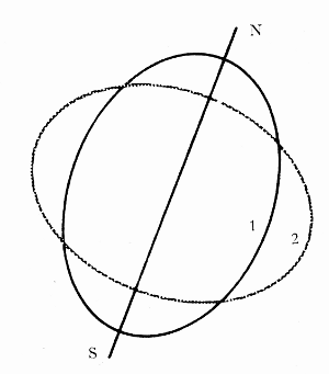
Figure 3.—Measurements of the length of a degree of latitude which were completed in different parts of France in 1669 and 1718 gave differing results which suggested that the shape of the earth is not a sphere but a prolate spheroid (1). But Richer’s pendulum observation of 1672, as explained by Huygens and Newton, indicated that its shape is that of an oblate spheroid (2). The disagreement is reflected in this drawing. In the 1730’s it was resolved in favor of the latter view by two French geodetic expeditions for the measurement of degrees of latitude in the equatorial and polar regions (Ecuador—then part of Peru—and Lapland).
Meanwhile, Richer in 1672 had been sent to Cayenne, French Guiana, to make astronomical observations and to measure the length of the seconds pendulum. [8] He took with him a pendulum clock which had been adjusted to keep accurate time in Paris. At Cayenne, however, Richer found that the clock was retarded by 2 minutes and 28 seconds per day (fig. 1). He also fitted up a “simple” pendulum to vibrate in seconds and measured the length of this seconds pendulum several times every week for 10 months. Upon his return to Paris, he found that the length of the “simple” pendulum which beat seconds at Cayenne was 11/4 Paris lines [9] shorter than the length of the seconds pendulum at Paris. Huygens explained the reduction in the length of the seconds pendulum—and, therefore, the lesser intensity of gravity at the equator with respect to the value at Paris—in terms of his theory of centripetal force as applied to the rotation of the earth and pendulum. [10]
A more complete theory was given by Newton in the Principia. [11] Newton showed that if the earth is assumed to be a homogeneous, mutually gravitating fluid globe, its rotation will result in a bulging at the equator. The earth will then have the form of an oblate spheroid, and the intensity of gravity as a form of universal gravitation will vary with position on the surface of the earth. Newton took into account gravitational attraction and centrifugal action, and he calculated the ratio of the axes of the spheroid to be 230:229. He calculated and prepared a table of the lengths of a degree of latitude and of the seconds pendulum for every 5° of latitude from the equator to the pole. A discrepancy between his predicted length of the seconds pendulum at the equator and Richer’s measured length was explained by Newton in terms of the expansion of the scale with higher temperatures near the equator.
Newton’s theory that the earth is an oblate spheroid was confirmed by the measurements of Richer, but was rejected by the Paris Academy of Sciences, for it contradicted the results of the Cassinis, father and son, whose measurements of arcs to the south and north of Paris had led to the conclusion that the earth is a prolate spheroid. Thus, a controversy arose between the English scientists and the Paris Academy. The conflict was finally resolved by the results of expeditions sent by the Academy to Peru and Sweden. The first expedition, under Bouguer, La Condamine, and Godin in 1735, went to a region in Peru, and, with the help of the Spaniard Ullo, measured a meridian arc of about 3°7′ near Quito, now in Ecuador. [12] The second expedition, with Maupertuis and Clairaut in 1736, went to Lapland within the Arctic Circle and measured an arc of about 1° in length. [13] The northern arc of 1° was found to be longer than the Peruvian arc of 1°, and thus it was confirmed that the earth is an oblate spheroid, that is, flattened at the poles, as predicted by the theory of Newton.
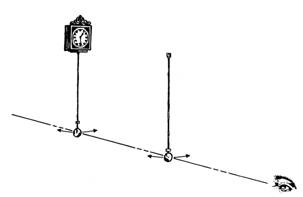
Figure 4.—The direct use of a clock to measure the force of gravity was found to be limited in accuracy by the necessary mechanical connection of the pendulum to the clock, and by the unavoidable difference between the characteristics of a clock pendulum and those of a theoretical (usually called “simple”) pendulum, in which the mass is concentrated in the bob, and the supporting rod is weightless.
After 1735, the clock was used only to time the swing of a detached pendulum, by the method of “coincidences.” In this method, invented by J. J. Mairan, the length of the detached pendulum is first accurately measured, and the clock is corrected by astronomical observation. The detached pendulum is then swung before the clock pendulum as shown here. The two pendulums swing more or less out of phase, coming into coincidence each time one has gained a vibration. By counting the number of coincidences over several hours, the period of the detached pendulum can be very accurately determined. The length and period of the detached pendulum are the data required for the calculation of the force of gravity.
The period from Eratosthenes to Picard has been called the spherical era of geodesy; the period from Picard to the end of the 19th century has been called the ellipsoidal period. During the latter period the earth was conceived to be an ellipsoid, and the determination of its ellipticity, that is, the difference of equatorial radius and polar radius divided by the equatorial radius, became an important geodetic problem. A significant contribution to the solution of this problem was made by determinations of gravity by the pendulum.
An epoch-making work during the ellipsoidal era of geodesy was Clairaut’s treatise, Théorie de la figure de la terre. [14] On the hypothesis that the earth is a spheroid of equilibrium, that is, such that a layer of water would spread all over it, and that the internal density varies so that layers of equal density are coaxial spheroids, Clairaut derived a historic theorem: If γE, γP are the values of gravity at the equator and pole, respectively, and c the centrifugal force at the equator divided by γE, then the ellipticity α = (5/2)c - (γP - γE)/γE.
Laplace showed that the surfaces of equal density might have any nearly spherical form, and Stokes showed that it is unnecessary to assume any law of density as long as the external surface is a spheroid of equilibrium. [15] It follows from Clairaut’s theorem that if the earth is an oblate spheroid, its ellipticity can be determined from relative values of gravity and the absolute value at the equator involved in c. Observations with nonreversible, invariable compound pendulums have contributed to the application of Clairaut’s theorem in its original and contemporary extended form for the determination of the figure and gravity field of the earth.
Early Types of Pendulums
The pendulum employed in observations of gravity prior to the 19th century usually consisted of a small weight suspended by a filament (figs. 4-6). The pioneer experimenters with “simple” pendulums changed the length of the suspension until the pendulum beat seconds. Picard in 1669 determined the length of the seconds pendulum at Paris with a “simple” pendulum which consisted of a copper ball an inch in diameter suspended by a fiber of pite from jaws (pite was a preparation of the leaf of a species of aloe and was not affected appreciably by moisture).
A celebrated set of experiments with a “simple” pendulum was conducted by Bouguer [16] in 1737 in the Andes, as part of the expedition to measure the Peruvian arc. The bob of the pendulum was a double truncated cone, and the length was measured from the jaw suspension to the center of oscillation of the thread and bob. Bouguer allowed for change of length of his measuring rod with temperature and also for the buoyancy of the air. He determined the time of swing by an elementary form of the method of coincidences. The thread of the pendulum was swung in front of a scale and Bouguer observed how long it took the pendulum to lose a number of vibrations on the seconds clock. For this purpose, he noted the time when the beat of the clock was heard and, simultaneously, the thread moved past the center of the scale. A historic aspect of Bouguer’s method was that he employed an “invariable” pendulum, that is, the length was maintained the same at the various stations of observation, a procedure that has been described as having been invented by Bouguer.
Since T = π√(l/g), it follows that T12/T22 = g2/g1. Thus, if the absolute value of gravity is known at one station, the value at any other station can be determined from the ratio of the squares of times of swing of an invariable pendulum at the two stations. From the above equation, if T1 is the time of swing at a station where the intensity of gravity is g, and T2 is the time at a station where the intensity is g + Δg, then (Δg)/g = (T12/T22) - 1.
Bouguer’s investigations with his invariable pendulum yielded methods for the determination of the internal structure of the earth. On the Peruvian expedition, he determined the length of the seconds pendulum at three stations, including one at Quito, at varying distances above sea level. If values of gravity at stations of different elevation are to be compared, they must be reduced to the same level, usually to sea level. Since gravity decreases with height above sea level in accordance with the law of gravitation, a free-air reduction must be applied to values of gravity determined above the level of the sea. Bouguer originated the additional reduction for the increase in gravity on a mountain or plateau caused by the attraction of the matter in a plate. From the relative values of gravity at elevated stations in Peru and at sea level, Bouguer calculated that the mean density of the earth was 4.7 times greater than that of the cordilleras. [17] For greater accuracy in the study of the internal structure of the earth, in the 19th century the Bouguer plate reduction came to be supplemented by corrections for irregularities of terrain and by different types of isostatic reduction.
La Condamine, who like Bouguer was a member of the Peruvian expedition, conducted his own pendulum experiments (fig. 4). He experimented in 1735 at Santo Domingo en route to South America, [18] then at various stations in South America, and again at Paris upon his return to France. His pendulum consisted of a copper ball suspended by a thread of pite. For experimentation the length initially was about 12 feet, and the time of swing 2 seconds, but then the length was reduced to about 3 feet with time of swing 1 second. Earlier, when it was believed that gravity was constant over the earth, Picard and others had proposed that the length of the seconds pendulum be chosen as the standard. La Condamine in 1747 revived the proposal in the form that the length of the seconds pendulum at the equator be adopted as the standard of length. Subsequently, he investigated the expansion of a toise of iron from the variation in the period of his pendulum. In 1755, he observed the pendulum at Rome with Boscovich. La Condamine’s pendulum was used by other observers and finally was lost at sea on an expedition around the world. The knowledge of the pendulum acquired by the end of the 18th century was summarized in 1785 in a memoir by Boscovich. [19]
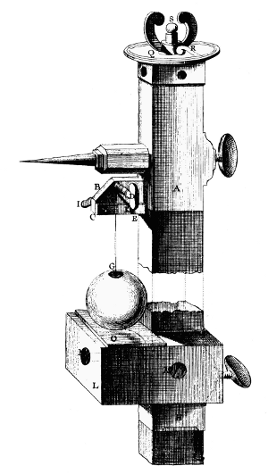
Figure 5.—An apparatus for the practice measurement of the length of the pendulum devised on the basis of a series of preliminary experiments by C. M. de la Condamine who, in the course of the French geodetic expedition to Peru in 1735, devoted a 3-month sojourn on the island of Santo Domingo to pendulum observations by Mairan’s Method. In this arrangement, shown here, a vertical rod of ironwood is used both as the scale and as the support for the apparatus, having at its top the brass pendulum support (A) and, below, a horizontal mirror (O) which serves to align the apparatus vertically through visual observation of the reflection of the pointer projecting from A. The pendulum, about 37 inches long, consists of a thread of pite (a humidity-resistant, natural fiber) and a copper ball of about 6 ounces. Its exact length is determined by adjusting the micrometer (S) so that the ball nearly touches the mirror. It will be noted that the clock pendulum would be obscured by the scale. La Condamine seems to have determined the times of coincidence by visual observation of the occasions on which “the pendulums swing parallel.” (Portion of plate 1, Mémoires publiés par la Société française de Physique, vol. 4.)
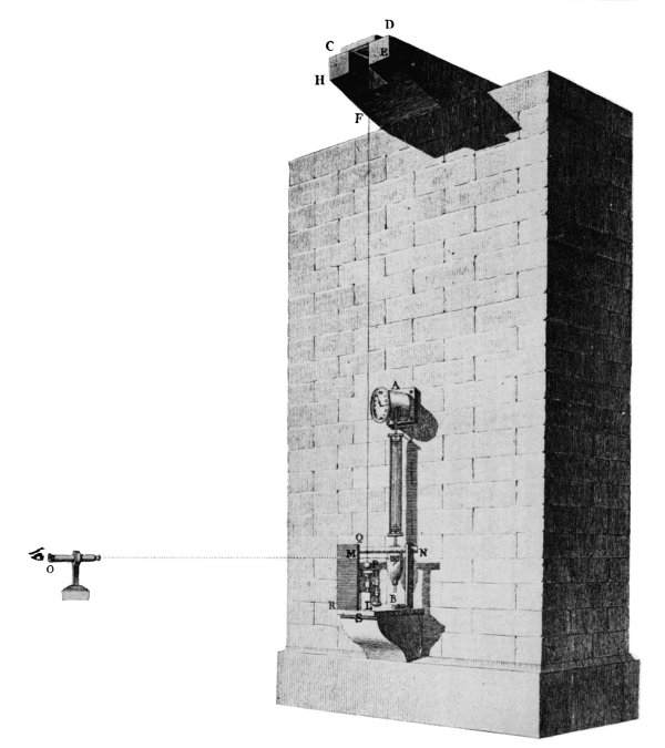
Figure 6.—The result of early pendulum experiments was often expressed in terms of the length of a pendulum which would have a period of one second and was called “the seconds pendulum.” In 1792, J. C. Borda and J. D. Cassini determined the length of the seconds pendulum at Paris with this apparatus. The pendulum consists of a platinum ball about 11/2 inches in diameter, suspended by a fine iron wire. The length, about 12 feet, was such that its period would be nearly twice as long as that of the pendulum of the clock (A). The interval between coincidences was determined by observing, through the telescope at the left, the times when the two pendulums emerge together from behind the screen (M). The exact length of the pendulum was measured by a platinum scale (not shown) equipped with a vernier and an auxiliary copper scale for temperature correction.
When, at the end of the 18th century, the French revolutionary government established the metric system of weights and measures, the length of the seconds pendulum at Paris was considered, but not adopted, as the unit of length. (Plate 2, Mémoires publiés par la Société française de Physique, vol. 4.)
The practice with the “simple” pendulum on the part of Picard, Bouguer, La Condamine and others in France culminated in the work of Borda and Cassini in 1792 at the observatory in Paris [20] (fig. 6). The experiments were undertaken to determine whether or not the length of the seconds pendulum should be adopted as the standard of length by the new government of France. The bob consisted of a platinum ball 161/6 Paris lines in diameter, and 9,911 grains (slightly more than 17 ounces) in weight. The bob was held to a brass cup covering about one-fifth of its surface by the interposition of a small quantity of grease. The cup with ball was hung by a fine iron wire about 12 Paris feet long. The upper end of the wire was attached to a cylinder which was part of a wedge-shaped knife edge, on the upper surface of which was a stem on which a small adjustable weight was held by a screw thread. The knife edge rested on a steel plate. The weight on the knife-edge apparatus was adjusted so that the apparatus would vibrate with the same period as the pendulum. Thus, the mass of the suspending apparatus could be neglected in the theory of motion of the pendulum about the knife edge.
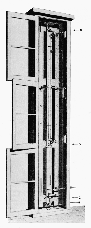
Figure 7.—Results of experiments in the determination of the length of the seconds pendulum at Königsberg by a new method were reported by F. W. Bessel in 1826 and published in 1828. With this apparatus, he obtained two sets of data from the same pendulum, by using two different points of suspension. The pendulum was about 10 feet long. The distance between the two points of suspension (a and b) was 1 toise (about six feet). A micrometric balance (c) below the bob was used to determine the increase in length due to the weight of the bob. He projected the image of the clock pendulum (not shown) onto the gravity pendulum by means of a lens, thus placing the clock some distance away and eliminating the disturbing effect of its motion. (Portion of plate 6, Mémoires publiés par la Société française de Physique, vol. 4.)
In the earlier suspension from jaws there was uncertainty as to the point about which the pendulum oscillated. Borda and Cassini hung their pendulum in front of a seconds clock and determined the time of swing by the method of coincidences. The times on the clock were observed when the clock gained or lost one complete vibration (two swings) on the pendulum. Suppose that the wire pendulum makes n swings while the clock makes 2n + 2. If the clock beats seconds exactly, the time of one complete vibration is 2 seconds, and the time of swing of the wire pendulum is T = (2n + 2)/n = 2(1 + 1/n). An error in the time caused by uncertainty in determining the coincidence of clock and wire pendulum is reduced by employing a long interval of observation 2n. The whole apparatus was enclosed in a box, in order to exclude disturbances from currents of air. Corrections were made for buoyancy, for amplitude of swing and for variations in length of the wire with temperature. The final result was that the length of the seconds pendulum at the observatory in Paris was determined to be 440.5593 Paris lines, or 993.53 mm., reduced to sea level 993.85 mm. Some years later the methods of Borda were used by other French investigators, among whom was Biot who used the platinum ball of Borda suspended by a copper wire 60 cm. long.
Another historic “simple” pendulum was the one swung by Bessel (fig. 7) for the determination of gravity at Königsberg 1825-1827. [21] The pendulum consisted of a ball of brass, copper, or ivory that was suspended by a fine wire, the upper end of which was wrapped and unwrapped on a horizontal cylinder as support. The pendulum was swung first from one point and then from another, exactly a “toise de Peru” [22] higher up, the bob being at the same level in each case (fig. 7). Bessel found the period of vibration of the pendulum by the method of coincidences; and in order to avoid disturbances from the comparison clock, it was placed at some distance from the pendulum under observation.
Bessel’s experiments were significant in view of the care with which he determined the corrections. He corrected for the stiffness of the wire and for the lack of rigidity of connection between the bob and wire. The necessity for the latter correction had been pointed out by Laplace, who showed that through the circumstance that the pull of the wire is now on one side and now on the other side of the center of gravity, the bob acquires angular momentum about its center of gravity, which cannot be accounted for if the line of the wire, and therefore the force that it exerts, always passed through the center. In addition to a correction for buoyancy of the air considered by his predecessors, Bessel also took account of the inertia of the air set in motion by the pendulum.