VOLUME XVII SLICE II Luray Cavern to Mackinac Island (continued 4)
Reading edition
Comedies.—In 1632 Edmund Blount published “Six Court Comedies,” including Endymion (1591), Sappho and Phao (1584), Alexander and Campaspe (1584), Midas (1592), Mother Bombie (1594) and Gallathea (1592). To these should be added the Woman in the Moone (Lyly’s earliest play, to judge from a passage in the prologue and therefore earlier than 1584, the date of Alexander and Campaspe), and Love’s Metamorphosis, first printed in 1601. Of these, all but the last are in prose. A Warning for Faire Women (1599) and The Maid’s Metamorphosis (1600) have been attributed to Lyly, but on altogether insufficient grounds. The first editions of all these plays were issued between 1584 and 1601, and the majority of them between 1584 and 1592, in what were Lyly’s most successful and popular years. His importance as a dramatist has been very differently estimated. Lyly’s dialogue is still a long way removed from the dialogue of Shakespeare. But at the same time it is a great advance in rapidity and resource upon anything which had gone before it; it represents an important step in English dramatic art. His nimbleness, and the wit which struggles with his pedantry, found their full development in the dialogue of Twelfth Night and Much Ado about Nothing, just as “Marlowe’s mighty line” led up to and was eclipsed by the majesty and music of Shakespearian passion. One or two of the songs introduced into his plays are justly famous and show a real lyrical gift. Nor in estimating his dramatic position and his effect upon his time must it be forgotten that his classical and mythological plots, flavourless and dull as they would be to a modern audience, were charged with interest to those courtly hearers who saw in Midas Philip II., Elizabeth in Cynthia and perhaps Leicester’s unwelcome marriage with Lady Sheffield in the love affair between Endymion and Tellus which brings the former under Cynthia’s displeasure. As a matter of fact his reputation and popularity as a play-writer were considerable. Gabriel Harvey dreaded lest Lyly should make a play upon their quarrel; Meres, as is well known, places him among “the best for comedy”; and Ben Jonson names him among those foremost rivals who were “outshone” and outsung by Shakespeare.
Euphues.—It was not, however, as a dramatist, but as the author of Euphues, that Lyly made most mark upon the Elizabethan world. His plays amused the court circle, but the “new English” of his novel threatened to permanently change the course of English style. The plot of Euphues is extremely simple. The hero, whose name may very possibly have been suggested by a passage in Ascham’s Schoolmaster, is introduced to us as still in bondage to the follies of youth, “preferring fancy before friends, and this present humour before honour to come.” His travels bring him to Naples, where he falls in love with Lucilla, the governor’s light-minded daughter. Lucilla is already pledged to Euphues’s friend Philautus, but Euphues’s passion betrays his friendship, and the old lover finds himself thrown over by both friend and mistress. Euphues himself, however, is very soon forsaken for a more attractive suitor. He and Philautus make up their quarrel, and Euphues writes his friend “a cooling card,” to be “applied to all lovers,” which is so severe upon the fair sex that Lyly feels it necessary to balance it by a sort of apology addressed “to the grave matrons and honest maidens of Italy.” Euphues then leaves Naples for his native Athens, where he gives himself up to study, of which the first fruits are two long treatises—the first, “Euphues and his Ephoebus,” a disquisition on the art of education addressed to parents, and the second, “Euphues and Atheos,” a discussion of the first principles of religion. The remainder of the book is filled up with correspondence between Euphues and his friends. We have letters from Euphues to Philautus on the death of Lucilla, to another friend on the death of his daughter, to one Botonio “to take his exile patiently,” and to the youth Alcius, remonstrating with him on his bad behaviour at the university. Finally a pair of letters, the first from Livia “at the emperour’s court to Euphues at Athens,” answered by “Euphues to Livia,” wind up the first part, and announce to us Euphues’s intention of visiting England. An address from Lyly to Lord Delawarr is affixed, to which was added in the second edition “An Address to the Gentlemen Scholars of England.”
Euphues and his England is rather longer than the first part. Euphues and Philautus travel from Naples to England. They arrive at Dover, halt for the night at Fidus’s house at Canterbury, and then proceed to London, where they make acquaintance with Surius, a young English gentleman of great birth and noble blood; Psellus, an Italian nobleman reputed “great in magick”; Martius, an elderly Englishman; Camilla, a beautiful English girl of insignificant family; Lady Flavia and her niece Fraunces. After endless correspondence and conversation on all kinds of topics, Euphues is recalled to Athens, and from there corresponds with his friends. “Euphues’ Glasse for Europe” is a flattering description of England sent to Livia at Naples. It is the most interesting portion of the book, and throws light upon one or two points of Lyly’s own biography. The author naturally seized the opportunity for paying his inevitable tribute to the queen, and pays it in his most exalted style. “O fortunate England that hath such a queene, ungratefull if thou praye not for hir, wicked if thou do not love hir, miserable if thou lose hir!”—and so on. The book ends with Philautus’s announcement of his marriage to Fraunces, upon which Euphues sends characteristic congratulations and retires, “tormented in body and grieved in mind,” to the Mount of Silexedra, “where I leave him to his musing or Muses.”
Such is a brief outline of the book which for a time set the fashion for English prose. Two editions of each part appeared within the first year after publication, and thirteen editions of both are enumerated up to 1636, after which, with the exception of a modernized version in 1718, Euphues was never reprinted until 1868, when Dr Arber took it in hand. The reasons for its popularity are not far to seek. As far as matter was concerned it fell in with all the prevailing literary fashions. Its long disquisitions on love, religion, exile, women or education, on court life and country pleasures, handled all the most favourite topics in the secularized speculation of the time; its foreign background and travel talk pleased a society of which Lyly himself said “trafic and travel hath woven the nature of all nations into ours and made this land like arras full of device which was broadcloth full of workmanship”; and, although Lyly steered clear in it of the worst classical pedantries of the day, the book was more than sufficiently steeped in classical learning, and based upon classical material, to attract a literary circle which was nothing if not humanist. A large proportion of its matter indeed was drawn from classical sources. The general tone of sententious moralizing may be traced to Plutarch, from whom the treatise on education, “Euphues and his Ephoebus,” and that on exile, “Letter to Botonio to take his exile patiently,” are literally translated, as well as a number of other shorter passages either taken direct from the Latin versions or from some of the numerous English translations of Plutarch then current. The innumerable illustrations based upon a kind of pseudo natural history are largely taken from Pliny, while the mythology is that of Virgil and Ovid.
It was not the matter of Euphues, however, so much as the style which made it famous (see Euphuism). The source of Lyly’s peculiar style has been traced by Dr Landmann (Der Euphuismus, sein Wesen, seine Quelle, seine Geschichte, &c. Giessen, 1881) to the influence of Don Antonio de Guevara, whose Libro Aureo de Marco Aurelio (1529)—a sort of historical romance based upon Plutarch and upon Marcus Aurelius’s Meditations, the object of which was to produce a “mirror for princes,” of the kind so popular throughout the Renaissance—became almost immediately popular in England. The first edition, or rather a French version of it, was translated into English by Lord Berners in 1531, and published in 1534. Before 1560 twelve editions of Lord Berners’s translation had been printed, and before 1578 six different translators of this and later works of Guevara had appeared. The translation, however, which had most influence upon English literature was that by North, the well-known translator of Plutarch, in 1557, called The Dial for Princes, Compiled by the Reverend Father in God Don Antony of Guevara, Byshop of Guadix, &c., Englished out of the Frenche by Th. North. The sententious and antithetical style of the Dial for Princes is substantially that of Euphues, though Guevara on the whole handles it better than his imitator, and has many passages of real force and dignity. The general plan of the two books is also much the same. In both the biography is merely a peg on which to hang moral disquisitions and treatises. The use made of letters is the same in both. Even the names of some of the characters are similar. Thus Guevara’s Lucilla is the flighty daughter of Marcus Aurelius. Lyly’s Lucilla is the flighty daughter of Ferardo, governor of Naples; Guevara’s Livia is a lady at the court of Marcus Aurelius, Lyly’s Livia is a lady at the court “of the emperor,” of whom no further description is given. The 9th, 10th, 11th and 12th chapters of the Dial for Princes suggested the discussion between Euphues and Atheos. The letter from Euphues to Alcius is substantially the same in subject and treatment as that from Marcus Aurelius to his nephew Epesipo. Both Guevara and Lyly translated Plutarch’s work De educatione liberorum, Lyly, however, keeping closer than the Spanish author to the original. The use made by Lyly of the university of Athens was an anachronism in a novel intended to describe his own time. He borrowed it, however, from Guevara, in whose book a university of Athens was of course entirely in place. The “cooling card for all fond lovers” and the address to the ladies and gentlemen of Italy have their counterparts among the miscellaneous letters by Guevara affixed by North to the Dial for Princes; and other instances of Lyly’s use of these letters, and of two other treatises by Guevara on court and country life, could be pointed out.
Lyly was not the first to appropriate and develop the Guevaristic style. The earliest book in which it was fully adopted was A petite Pallace of Pettie his Pleasure, by George Pettie, which appeared in 1576, a production so closely akin to Euphues in tone and style that it is difficult to believe it was not by Lyly. Lyly, however, carried the style to its highest point, and made it the dominant literary fashion. His principal followers in it were Greene, Lodge and Nash, his principal opponent Sir Philip Sidney; the Arcadia in fact supplanted Euphues, and the Euphuistic taste proper may be said to have died out about 1590 after a reign of some twelve years. According to Landmann, Shakespeare’s Love’s Labour Lost is a caricature of the Italianate and pedantic fashions of the day, not of the peculiar style of Euphues. The only certain allusion in Shakespeare to the characteristics of Lyly’s famous book is to be found in Henry IV., where Falstaff, playing the part of the king, says to Prince Hal, “Harry, I do not only marvel where thou spendest thy time, but also how thou art accompanied; for, though the camomile the more it is trodden on the faster it grows, yet youth the more it is wasted the sooner it wears.” Here the pompous antithesis is evidently meant to caricature the peculiar Euphuistic sentence of court parlance.
(M. A. W.)
See Lyly’s Complete Works, ed. R. W. Bond (3 vols., 1902); Euphues, from early editions, by Edward Arber (1868); A. W. Ward, English Dramatic Literature, i. 151; J. P. Collier, History of Dramatis Poetry, iii. 172; “John Lilly and Shakespeare,” by C. C. Hense in the Jahrbuch der deutschen Shakesp. Gesellschaft, vols. vii. and viii. (1872, 1873); F. W. Fairholt, Dramatic Works of John Lilly (2 vols., 1858); Shakespeare’s Euphuism, by W. L. Rushton; H. Morley, “Euphuism” in the Quarterly Review (1861); R. W. Bond, “John Lyly, Novelist and Dramatist,” in the Quarterly Review (Jan. 1896); J. A. Symonds, Shakespeare’s Predecessors (1883); J. D. Wilson, John Lyly (Cambridge, 1905); A. Ainger, “Euphuism,” in Lectures and Essays (1905); and Albert Feuillerat, John Lyly. Contribution à l’histoire de la Renaissance en Angleterre (1910).
1 The evidence for his authorship may be found in Gabriel Harvey’s Pierce’s Supererogation (written November 1589, published 1593), in Nash’s Have with you to Saffron Walden (1596), and in various allusions in Lyly’s own plays. See Fairholt’s Dramatic Works of John Lilly, i. 20.

LYME REGIS, a market town and municipal borough and watering-place in the western parliamentary division of Dorsetshire, England, 151 m. W.S.W. of London by the London & South Western railway, the terminus of a light railway from Axminster. Pop. (1901) 2095. It is situated at the mouth of a narrow combe or valley opening upon a fine precipitous coast-line; there is a sandy shore affording excellent bathing, and the country inland is beautiful. The church of St Michael and All Angels is mainly Perpendicular, but the tower (formerly central) and the portion west of it are Norman. A guildhall and assembly rooms are the chief public buildings. The principal industries are stone-quarrying and the manufacture of cement. There is a curved pier of ancient foundation known as the Cobb. The harbour, with a small coasting trade, is under the authority of the corporation. The borough is under a mayor, 4 aldermen and 12 councillors. Area, 1237 acres.
No evidence of settlement on the site of Lyme Regis exists before that afforded by a grant, dated 774, purporting to be by Cynewulf, king of the West-Saxons, of land here to the church of Sherborne, and a similar grant by King Æthelstan to the church of Glastonbury. In 1086 three manors of Lyme are mentioned: that belonging to Sherborne abbey, which was granted at the dissolution to Thomas Goodwin, who alienated it in the following year; that belonging to Glastonbury, which seems to have passed into lay lands during the middle ages, and that belonging to William Belet. The last was acquired by the family of Bayeux, from whom it passed by marriage to Elias de Rabayne, whose nephew, Peter Baudrat, surrendered it to the crown in 1315-1316 when the king became lord of one moiety of the borough, henceforth known as Lyme Regis. Lyme ranked as a port in 1234, and Edward I. in 1284 granted to the town a charter making it a free borough, with a merchant gild, and in the same year the mayor and bailiffs are mentioned. In the following January the bailiffs were given freedom from pleading without the borough, freedom from toll and privileges implying considerable foreign trade; the importance of the port is also evident from the demand of two ships for the king’s service in 1311. In 1332-1333 Edward III. granted Lyme to the burgesses at a fee-farm of 32 marks; on the petition of the inhabitants, who were impoverished by tempests and high tides, this was reduced to 100 shillings in 1410 and to 5 marks in 1481. In 1591 Elizabeth incorporated Lyme, and further charters were obtained from James I., Charles II. and William III. Lyme returned two members to parliament from 1295 to 1832 when the representation was reduced to one. The borough was disfranchised in 1867. The fairs granted in 1553 for the 1st of February and the 20th of September are now held on altered dates. Trade with France in wine and cloth was carried on as early as 1284, but was probably much increased on the erection of the Cobb, first mentioned in 1328 as built of timber and rock. Its medieval importance as the only shelter between Portland Roads and the river Exe caused the burgesses to receive grants of quayage for its maintenance in 1335 and many subsequent years, while its convenience probably did much to bring upon Lyme the unsuccessful siege by Prince Maurice in 1644. In 1685 Lyme was the scene of the landing of James, duke of Monmouth, in his attempt upon the throne.
LYMINGTON, a municipal borough and seaport in the New Forest parliamentary division of Hampshire, England, 98 m. S.W. from London by the London & South Western railway. Pop. (1901) 4165. It lies on the estuary of the Lymington, which opens into the Solent. The church of St Thomas à Becket is an irregular structure, dating from the reign of Henry VI., but frequently restored. There is some coasting trade, and yacht-building is carried on. Regular passenger steamers serve Yarmouth in the Isle of Wight. In summer the town is frequented for sea-bathing. It is governed by a mayor, 4 aldermen and 12 councillors. Area, 1515 acres.
There was a Roman camp near Lymington (Lentune, Lementon), and Roman relics have been found, but there is no evidence that a town existed here until after the Conquest. Lymington dates its importance from the grant of the town to Richard de Redvers, earl of Devon, in the reign of Henry I. No charter has been found, but a judgment given under a writ of quo warranto in 1578 confirms to the burgesses freedom from toll, passage and pontage, the tolls and stallage of the quay and the right to hold two fairs—privileges which they claimed under charters of Baldwin de Redvers and Isabel de Fortibus, countess of Albemarle, in the 13th century, and Edward Courtenay, earl of Devon, in 1405. The town was governed by the mayor and burgesses until the corporation was reformed in 1835. A writ for the election of a member to parliament was issued in the reign of Edward III., but no return was made. From 1585 two members were regularly returned; the number was reduced to one in 1867, and in 1885 the representation was merged in that of the county. Fairs on the 13th and 14th of May and the 2nd and 3rd of October, dating from the 13th century, are still held. The Saturday market probably dates from the same century. Lymington was made a port in the reign of Henry I., and its large shipping trade led to frequent disputes with Southampton as to the levying of duties. The case was tried in 1329 and decided against Lymington, but in 1750 the judgment was reversed, and since then the petty customs have been regularly paid. From an early date and for many centuries salt was the staple manufacture of Lymington. The rise of the mineral saltworks of Cheshire led to its decline in the 18th century, and later the renewed importance of Southampton completed its decay.
See E. King, Borough and Parish of Lymington (London, 1879).
LYMPH and LYMPH FORMATION. Lying close to the blood-vessels of a limb or organ a further set of vessels may be observed. They are very pale in colour, often almost transparent and very thin-walled. Hence they are frequently difficult to find and dissect. These are the lymphatic vessels, and they are found to be returning a fluid from the tissues to the bloodstream. When traced back to the tissues they are seen to divide and ultimately to form minute anastomosing tubules, the lymph capillaries. The capillaries finally terminate in the spaces between the structures of the tissue, but whether their free ends are closed or are in open communication with the tissue spaces is still undecided. The study of their development shows that they grow into the tissue as a closed system of minute tubes, which indicates that in all probability they remain permanently closed. If we trace the lymphatic vessels towards the thorax we find that in some part of their course they terminate in structures known as lymphatic glands. From these again fresh lymphatic vessels arise which carry the fluid towards the main lymph-vessel, the thoracic duct. This runs up the posterior wall of the thorax close to the aorta, and finally opens into the junction of the internal jugular and left subclavian veins. The lymph-vessels from the right side of the head and neck and from the right arm open, however, into the right subclavian vein (see Lymphatic System below).
Chemical Constitution of Lymph.—The lymph collected from the thoracic duct during hunger is almost water clear and yellowish in colour. Its specific gravity varies from 1015 to 1025. It tastes salt and has a faint odour. It is alkaline in reaction, but is much less alkaline than blood-serum. Like blood it clots, but clots badly, only forming a soft clot which quickly contracts. The lymph collected from a lymphatic before it has passed through a lymph gland contains a few leucocytes, and though the number of lymphocytes is greater in the lymph after it has flowed through a gland it is never very great. In normal states there are no red blood corpuscles.
The total solids amount to 3.6 to 5.7%, the variations depending upon the amount of protein present. The lymph during hunger contains only a minute quantity of fat. Sugar (dextrose) is present in the same concentration as in the blood. The inorganic constituents are the same as in blood, but apparently the amounts of Ca, Mg and P2O5 are rather less than in serum. Urea is present to the same amount as in blood. If the lymph be collected after a meal, one important alteration is to be found. It now contains an abundance of fat in a very fine state of subdivision, if fat be present in the food. The concentrations of protein and dextrose are not altered during the absorption of these substances.
The Significance of Lymph.—In considering the significance and use of lymph we must note in the first place that it forms an alternative medium for the removal of water, dissolved materials, formed elements or particles away from the tissues. All materials supplied to a tissue are brought to it by the blood, and are discharged from the blood through the capillary wall. They thus come to lie in the tissue spaces between the cells, and from this supply of material in a dissolved state the cells take up the food they require. In the opposite direction the cell discharges its waste products into this same tissue fluid. The removal of material from the tissue fluid may be effected either by its being absorbed through the capillary wall into the bloodstream, or by sending it into the lymphatic vessels and thus away from the tissue. From this point of view the lymphatics may be looked upon in a sense as a drainage system of the tissues. Again, besides discharging fluid and dissolved material into the tissue spaces, the blood may also discharge leucocytes, and under many conditions this emigration of leucocytes may be very extensive. These also may leave the tissue space by the path of the lymph channels. Moreover, the tissues are at any time liable to be injured, and the injury as well as damaging many cells may cause rupture of capillaries (as in bruising) with escape of red blood-cells into the tissue spaces. If this occurs we know that the damaged cells are destroyed and their débris removed either by digestion by leucocytes or by disintegration and solution. The damage of a tissue also commonly involves an infection of the damaged area with living micro-organisms, and these are at once admitted to the tissue spaces. Hence we see that the lymphatics may be provided as channels by which a variety of substances can be removed from the tissue spaces. The question at once arises, is the lymph channel at all times open to receive the materials present in the tissue space? If such be the case, lymph is simply tissue fluid, and anything that modifies the constitution or amount of the tissue fluid should in like proportion lead to a variation in the amount and constitution of the lymph. But if the lymph capillary is a closed tubule at its commencement this does not follow.
From these considerations we see that in the first instance the whole problem of lymph formation is intimately bound up with the study of the interchanges of material between the blood and the various tissue cells. The exchange of material between blood and tissue cell may possibly be determined in one or both of two ways. Either it may result from changes taking place within the tissue cell, or the tissue cell remaining passive material may be sent to or withdrawn from it owing to a change occurring either in the composition of the blood or to a change in the circulation through the tissue. Let us take first the results following increased activity of a tissue. We know that increased activity of a tissue means increased chemical change within the tissue and the production of new chemical bodies of small molecular size (e.g. water, carbonic acid, &c.). The production of these metabolites means the destruction of some of the tissue substance, and to make good this loss the tissue must take a further amount of material from the blood. We know that this takes place, and moreover that the waste products resulting from activity are ultimately removed. The question then becomes: When does this restoration take place, and what is the intermediate state of the tissue? We know that increased activity is always accompanied by an increase in the blood-supply, indicating a greater supply of nutritive material, though it may be that, the increased supply required at the actual time of activity is oxygen only. Simultaneously the opportunity for a more rapid removal of the waste products is provided. We have to inquire then: Does this increased vascularity necessarily mean an increased outpouring of water and dissolved material into the tissues, for this might follow directly from the greater filling of the capillaries, or from the increased attracting power of the tissues to water (osmotic effect) due to the sudden production of substances of small molecular size within the tissue? The other possibility is that the increased volume of blood sent to the tissue is for the sole purpose of giving it a more rapid supply of oxygen, and that the ordinary normal blood-supply would amply suffice for renewing the chemical material used up during activity. Tissues undoubtedly vary among themselves in the amount of water and other materials they take from the blood when thrown into activity, and their behaviour in this respect depends upon the work they are called upon to perform. We must discriminate between the substance required by and consumed by the tissue, the chemical food which on combustion yields the energy by which the tissue performs work, and, on the other hand, the substance taken from the blood and either with or without further elaboration discharged from the tissue (as, for instance, in the process of secretion). The tissue contains in itself a store of food amply sufficient to enable it to continue working for a long time after its blood-supply has been stopped, and everything indicates that the supply of chemical energy to the tissue may be slow or even withheld for a considerable time. Hence we are led to conclude that the increased flow of blood sent to a tissue when it is thrown into activity is first and foremost to give that tissue an increased oxygen supply; secondly, to remove waste carbonic acid; thirdly, and only in the case of some tissues, to provide water salts and other materials for the outpouring of a secretion, as an instance of which we may take the kidney as a type. Hence there is no need to suppose that an extensive accumulation of fluid and dissolved substances takes place within a tissue when it becomes active. This must be an accumulation which would lead to an engorgement of the tissue spaces and then to a discharge of fluid along the lymph channels. To enable us to determine the various points just raised we must know whether an increased blood-supply to a tissue necessarily means an increased exudation of fluid into the tissue spaces, and moreover we must study the exchange of fluid between a tissue and the blood under as varied a series of conditions as possible, subsequently examining whether exchange of fluid and other substances between the tissue and the blood necessarily determines quantitatively the amount of lymph flowing from the tissue. Hence we will first study the exchanges between the blood and a tissue, and then turn our attention to the lymph-flow from the tissues.
The Exchanges of Fluids and dissolved Substances between the Blood and the Tissues.—Numerous experiments have been performed in studying the conditions under which fluid passes into the tissues and tissue spaces—or in the reverse direction into the blood. We may group them into (1) conditions during which the total volume of circulating fluid is increased or decreased; (2) conditions in which the character of the blood is altered, e.g. it is made more watery or its saline concentration is altered; (3) conditions in which the blood-supply to the part is altered; (4) conditions in which the physical character of the capillary wall is altered.
1. The total volume of blood in an animal has been increased among other ways by the transfusion of the blood of one animal directly into the veins of a second of the same species. It is found that within a very short time a large percentage of the plasma has been discharged from the blood-vessels. It has been sent into the tissues, notably the muscles, and it may be noted in passing without producing any increase in the lymph-flow from these vessels. An analogous experiment, but one which avoids the fallacy introduced by injecting a second animal’s blood, has been performed by driving all the blood out of one hind limb by applying a rubber bandage tightly round it from the foot upwards. This increases the volume of blood circulating in the rest of the body, and again a rapid disappearance of the fluid part of the blood from the vessels was observed—the fluid being mainly sent into the muscles, as was indicated by showing that the specific gravity of the muscles fell during the experiment. The experiments converse to these have also been studied. Bleeding is very rapidly followed by a large inflow of fluid into the circulating blood—this fluid being derived from all the tissues, and especially again from the muscles. Or again, when the bandage from the limb in the above-cited experiment was removed, the total capacity of the circulatory system was thereby suddenly increased, and it was found that the total volume of blood increased correspondingly, the increased volume of fluid being drawn from the tissues and especially again from the muscles. The rapidity with which this movement of fluid into or out of the blood takes place is very striking. The explanation usually offered is that the movement is effected by changes in the capillary pressure due to the alteration in the volume of blood circulating. While this seems feasible when the volume of blood is increased, it does not offer a satisfactory explanation of the rapid movement of fluid from the tissues when the volume of the blood is decreased. One must therefore look for yet further factors in this instance.
2. Let us next turn attention to the second of our three main variations, viz. that in which the composition of the blood is altered. It has long been known that the injection of water, or of solutions of soluble bodies such as salts, urea, sugar, &c., leads to a very rapid exchange of water and salts between the blood and the tissues. Thus if a solution less concentrated than the blood be injected, the blood is thereby diluted, but with very great rapidity water leaves the blood and is taken up by the tissues. Again, if a strong sugar or salt solution be injected, the first effect is a big discharge of water from the tissues into the blood and the movement of fluid is effected with great rapidity. In these instances a new physical factor is brought into play, viz. that of osmosis. When a solution of lower osmotic pressure than the blood is injected the osmotic pressure of the blood falls temporarily below that of the tissues, and water is therefore attracted to the tissues. The converse is the case when a solution of osmotic pressure higher than the blood is injected. This at first sight seems to be an all-sufficient explanation of the results recorded, but difficulties arise when we find that the tissues are not equally active in producing the effects. Thus it is found that the muscles and skin act as the chief water depot, while such tissues as the liver, intestines or pancreas take a relatively small share in the exchange. Again, when a strong sodium chloride solution is injected a considerable part of the sodium chloride is soon found to have left the blood, and it has been shown that the chloride depot is not identical with the water depot. The lung, for instance, is found to take up relatively far more of the salt than other tissues. Simultaneously with the passage of the salt into the tissue an exchange of water from the tissue into the blood can be observed, both processes being carried out very rapidly. The result is that the blood very quickly returns to a state in which its osmotic pressure is only slightly raised; the tissue, on the other hand, loses water and gains salt, and its osmotic pressure and specific gravity therefore rises. Again, the tissues do not participate equally in producing the final result, nor is the tissue which gives up the largest amount of water necessarily that which gains the largest amount of salt. The results following the injection of solutions of other bodies of small molecular size, e.g. urea or sugar, are quite analogous to those above described in the case of the non-toxic salt solutions. Hence we see that the rate of exchange of fluid and dissolved substance between a tissue and the blood can be extremely rapid and that the exchange can take place in either direction. We may also conclude that the main cause of the exchange, and possibly the only one, is the osmotic action set up by the solution injected, and that muscle tissue is particularly active in the process.
Seeing that a very considerable amount of water or of dissolved substance can be taken up from the blood into a tissue, the question next arises: Where is this material held, in the tissue cell or in the tissue space? Immediately the water or salt leaves the blood it reaches the tissue space, but unless the process be extreme in amount it probably passes at once into the tissue cell itself and is stored there. If the process is excessive oedema is set up and fluid accumulates in the tissue space.
These, taken quite briefly, are some of the more important conditions under which fluid exchanges, take place. They are selected here because of the extent and rapidity of the changes effected.
3. The third factor which may bring about a change in the amount of fluid sent to a tissue is a variation in the capillary pressure. A rise in capillary pressure will, if filtration can occur through the capillary wall, cause an increased exudation of fluid from the blood. Thus the rise in general blood-pressure following the injection of a salt solution could cause an increased filtration into the tissues. Or again, the hydraemia following a salt injection would favour an increased exudation because the blood would be more readily filtrable. We, however, know very little of the effect of changes in capillary pressure upon movement of fluid into the tissue spaces and tissues, most of such observations being confined to a study of their effect upon lymph-flow. We will therefore return to them in this connexion.
4. The remaining factor to be mentioned is a change in the character of the capillary wall. It is well known that many poisons can excite an increased exudation from the blood and the tissue may become oedematous. Of such bodies we may mention cantharidin and the lymphogogues of Class I (see later). A like change is also probably the cause of the oedema of nephritis and of heart disease. It has also been suggested that the capillaries of different organs show varying degrees of permeability, a suggestion to which we will return later.
Lymph Formation.—There are two theories current at the present day offering explanations of the manner in which lymph is formed. The first, which owes its inception to Ludwig, explains lymph formation upon physical grounds. Thus according to this theory the lymphatics are open capillary vessels at their origin in the tissues along which the tissue fluid is driven. The tissue fluid is discharged from the blood by filtration, and therefore its amount varies directly with the capillary pressure. The amount of fluid movement also is further determined by osmotic actions and by the permeability of the capillary wall.
The second theory first actively enunciated by Heidenhain regards lymph formation as a secretory process of the capillary wall, i.e. one in the discharge of which these cells perform work and are not merely passive as in the former theory. As we shall see, it is now probable that neither theory is completely correct.
In considering lymph formation we have to examine both the total amount of lymph formed in the body and the variations in amount leaving each separate organ under different conditions. In most investigations the lymph was collected from the thoracic duct, i.e. it was the lymph returned from all parts of the body with the exception of the right arm and right side of the head and neck. The collection of the lymph from organs is much more difficult to effect, and hence has not, to the present, been so extensively studied. We will consider first variations in the amount of the thoracic duct lymph. Lymph is always flowing along the thoracic duct, and if the body is at rest, it has been shown that this lymph is coming practically entirely from the intestines and liver, chiefly, moreover, from the liver. The variations in the amount flowing under various conditions has been extensively studied. We will discuss them under the following headings: Changes brought about (a) by altered circulatory conditions, (b) by the injection of various substances, and (c) as a result of throwing an organ into activity.
Ligature of the portal vein leads to an increased flow of duct lymph. Ligature of the inferior vena cava above the diaphragm also leads to a large increase in the flow of duct lymph. Ligature of the aorta may result in either an increased or decreased flow of direct lymph. One explanation of these results has been offered from a study of the changes in capillary pressure set up in the main organs involved. Thus, after ligature of the portal vein the capillary pressure in the intestines rises, and it was proved that the increase in thoracic duct lymph came from the intestines. Ligaturing the inferior vena cava causes a big rise in the pressure in the liver capillaries, the intestinal capillary pressure remaining practically unaltered. Here it was proved that the increase in lymph-flow came from the liver and was more copious in amount than in the former instance. A further difference is that this lymph is more concentrated, a feature which always characterizes liver lymph. Ligature of the aorta may or may not cause a rise in the liver capillary pressure, and it has been shown that if the pressure rises there is an increased lymph-flow from the liver and conversely. The increase of lymph comes entirely in this instance also from the liver. It is in fact but a special instance of the former experiment. From these results it has been argued that lymph formation is simply a filtration fundamentally, and the lymph-flow is determined mainly by the capillary pressure. Variations in the quantity of lymph issuing from different organs have been on this theory ascribed to differences in the permeability of the capillaries of the organs. Thus as liver lymph is richest in protein content and is produced in greatest amount, it has been concluded that the liver capillaries possess the highest permeability. The intestines stand next in producing a concentrated lymph, and their capillaries are therefore assumed to stand second as regards permeability. Lastly, the lymph coming from limbs and other organs is much poorer in solids and much less copious in amount. Hence it is argued that their capillaries show the least permeability. It is, however, very unsafe to compare the liver capillaries with those of other organs, since they are not in reality capillaries but rather venous sinuses, and their relation to the liver cells is characteristically different from that of ordinary capillaries. If an animal is at rest, no lymph flows from the hind limbs. To obtain a sample of limb lymph it is necessary to massage the limb. If, however, the veins to the limb be ligatured, we obtain a flow of lymph. The ligature of course causes a rise of the capillary pressure, and it has been argued that this rise of pressure starts a filtration through the capillary wall and hence a flow of lymph. But the stoppage of the blood-flow also damages the capillary wall and tissue cells by asphyxiation, and the resulting lymph-flow is in all probability the resultant of many complex processes. This case is analogous to the production of oedema in cases of heart disease where the circulation is feeble and the oxygen supply to the parts deficient. The results of these experiments form the main evidence in support of the filtration theory of lymph formation. They were first systematically studied by Heidenhain, to whom we owe so much of our knowledge of lymph formation. He did not, however, conclude that they established the filtration theory.
In continuing his observations Heidenhain next studied the results following the injection of a number of substances into the blood. He found many which on injection gave rise to an increased lymph-flow from the thoracic duct, and arranged them in two classes. As instances of lymphogogues of the first class we may mention extract of mussels, leech extract, peptone, extract of crayfish muscle, extract of strawberries, of raspberries and many other like substances. Lymphogogues of the second class comprise neutral salt solutions, urea, sugar, &c. Considering the latter class first we may take as a type a solution of sodium chloride. Injection of such a solution causes a large increase in the lymph-flow, and it has been proved that the lymph comes from the liver and intestines only—chiefly from the former. It is especially to be noted that there is no lymph-flow from the limbs, and the same is true for all lymphogogues of this class. As indicated above, the injection of a saline solution leads to a large and rapidly effected transport of fluid from the blood into muscle tissue, but though there is this large increase in tissue fluid, no lymph flows from the tissue. This result very powerfully disfavours the filtration theory of lymph formation. It practically refutes the idea that lymph formation is solely dependent upon such processes as filtration, osmosis and capillary permeability only. It brings out quite clearly that the exchange of fluid and dissolved salts, &c., between the blood and a tissue, and the flow of lymph from that tissue, are two separate and distinct processes, and especially that the first does not determine the second. Also it is to be noted that the injection of a strong salt solution also excites a flow of duct lymph, again arising from the liver and intestines, but none from the limbs. In this instance, as previously stated, the muscles of the limbs are losing water, and so presumably are the liver and intestinal cells. This independence of tissue-blood exchange and lymph-flow is distinctly in favour of the view, which is rapidly gaining ground from histological observations, that in all instances the lymphatics commence in a tissue as closed capillary vessels.
Turning, in the next place, to the lymphogogues of the first class, it has been proved that the origin of this increase of flow is again from the liver. Very many of the substances of this class are bodies which may when taken cause urticarial (nettle-rash) eruptions, a state which is generally regarded as being due to an action upon the capillary endothelium. Their action as lymphogogues is also generally ascribed to an effect upon the capillary wall rendering it according to some more permeable, according to others leading to a direct secretory action on the part of the endothelium. We also know that many of the bodies of this class act upon the liver in other directions than in exciting an increased lymph production. Thus they may cause an increase in bile secretion, or, as in the case of peptone, the liver cells may be excited to produce a new chemical material, in this instance an antithrombin.
We have now to consider the effect of throwing an organ into activity upon the lymph-flow from the organ. In all cases in which it has been examined it is found that increased activity is accompanied by increased lymph-flow. Thus, to take the instance of the submaxillary gland, which at rest does not discharge any lymph, stimulation of the chorda tympani is followed by a flow of lymph accompanying the flow of saliva simultaneously excited. The stimulation of the nerve also produces dilatation of the blood-vessels and therefore a rise in capillary pressure. But that this vascular change is not the factor determining the lymph-flow is proved by the administration of a small dose of atropine, which arrests the secretion without influencing the vascular reaction following chorda stimulation. After the atropine no lymph-flow occurs on stimulating the nerve. Many other instances of a similar kind might be adduced. Thus, we have seen that peptone specifically excites the liver cells and also causes an increased lymph-flow from the liver; or, as a last instance, the injection of bile salt excites a flow of bile and also excites a flow of lymph from the liver. The supporters of the filtration theory have argued that as activity of a tissue is necessarily accompanied by the discharge of metabolites from the active tissue cells, and as these are of small molecular size, they must set up an osmotic effect. Water is therefore drawn into the tissue spaces, and this rise in fluid content results mechanically in a flow of lymph from the organ. The lymph simply drains away along the open lymphatics. This argument, however, loses all its force when we recall the fact that we may set up an enormous flow of fluid and salt into a tissue and its tissue spaces without causing the least flow of lymph. Further, there is no reason to suppose that the metabolites discharged from a tissue during activity are produced in large quantities. The chief metabolite is undoubtedly carbonic acid, and this diffuses very rapidly and is quickly carried away by the blood. If, moreover, as is probably the case, the lymphatics commence as closed capillaries, we have a further difficulty in explaining how the fluid is driven through the lymphatic wall. Either we must imagine the wall to be porous or there must be a greater pressure outside than inside, and it is very difficult to conceive how this is possible. As a general conclusion, then, it seems much more probable that we are here dealing with a secretory process, and that the active tissue produces some substance or substances—it may be carbonic acid—which throws the lymphatic capillary cells into activity.
To sum up in a few words the present state of our knowledge as to lymph formation we may say that the exchange of water and salts between the blood and the tissues is probably entirely determined by processes of filtration and osmosis. Further, that the physical condition of the capillary cells is frequently altered by many chemical substances, and that in consequence it may permit exudation into the tissue spaces much more freely. In the next place, the flow of lymph from a tissue is not solely determined by the amount of the tissue fluids. The lymph capillaries start as closed tubules, and the endothelial walls of these tubules play an active part (secretory) in determining when water and other substances shall be admitted into the capillary and further determine the quantity of such discharge. Apparently, too, these cells are specifically excited when the tissue is thrown into activity, the exciting substance being a metabolite from the active tissue. Leucocytes also are capable of passing through or between the endothelial cells of the lymph capillary.
(T. G. Br.)
LYMPHATIC SYSTEM. In anatomy, the lymphatic system (Lat. lympha, clear water) comprises the lymphoid or adenoid tissue so plentifully distributed about the body, especially in the course of the alimentary canal (see Connective Tissues), lymph spaces, lymphatic vessels of which the lacteals are modifications, lymphatic glands, haemolymph glands, and the thoracic and right lymphatic ducts by which the lymph (q.v.) finally reaches the veins.
Lymph spaces are mere spaces in the connective tissue, which usually have no special lining, though sometimes there is a layer of endothelial cells like those of the lymphatic and blood vessels. Most of these spaces are very small, but sometimes, as in the case of the sub-epicranial space of the scalp, the capsule of Tenon in the orbit, and the retropharyngeal space in the neck, they are large and are adaptations to allow free movement. Opening from these spaces, and also communicating with the serous membranes by small openings called stomata,1 are the lymph capillaries (see Vascular System), which converge to the lymphatic vessels. These resemble veins in having an internal layer of endothelium, a middle unstriped muscular coat, and an external coat of fibrous tissue, though in the smaller vessels the middle coat is wanting. They have numerous endothelial valves, formed of two crescentic segments allowing the lymph to pass toward the root of the neck. When the vessels are engorged these valves are marked by a constriction, and so the lymphatics have a beaded appearance. The vessels divide and anastomose very freely, and for this reason they do not, like the veins, increase in calibre as they approach their destination. It is usual to divide the lymphatic vessels into a superficial and a deep set; speaking generally, the superficial ones are found near the course of the superficial veins, while the deeper ones accompany the arteries. Probably any single drop of lymph passes sooner or later through one or more lymphatic glands, and so those vessels which are approaching a gland are called afferent, while those leaving are spoken of as efferent lymphatics. The lacteals are special lymphatic vessels which carry the chyle from the intestine; they begin in lymphatic spaces in the villi and round the solitary and agminated glands, and pass into the mesentery, where they come in contact with a large number of mesenteric glands before reaching the receptaculum chyli.
The lymphatic glands are pink bodies situated in the course of the lymphatic vessels, to which they act as filters. They are generally oval in shape and about the size of a bean, but sometimes, especially in the groin, they form irregular flattened masses 2 in. long, while, at other times, they are so small as almost to escape notice. They are usually found in groups.
Each gland has a fibrous capsule from which trabeculae pass toward the centre, where they break up and interlace, forming a network, and in this way a cortical and medullary region for each gland is distinguished; the intervals are nearly filled by lymphoid tissue, but close to the trabeculae is a lymph path or sinus, which is only crossed by the reticular stroma of the lymphoid tissue, and this probably acts as a mechanical sieve, entangling foreign particles; as an example of this the bronchial glands are black from carbon strained off in its passage from the lungs, while the axillary glands in a tattooed arm are blue. The blood-vessels enter at one spot, the hilum, and are distributed along the trabeculae. In addition to their function as filters the lymphatic glands are probably one of the sources from which the leucocytes are derived.
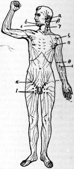
Fig. 1.—Superficial Lymphatic Vessels and Glands.
α, Preauricular. β, Mastoid. γ, Superficial cervical. δ, Submaxillary. ε, Submental. ζ, Infraclavicular. η, Anterior axillary. θ, Supratrochlear. ι, Antecubital. κ, Inguinal. λ, Superficial femoral.
The exact position of the various groups of glands is very important from a medical point of view, but here it is only possible to give a brief sketch which will be helped by reference to the accompanying diagram. In the head are found occipital and mastoid glands (fig. 1, β), which drain the back of the scalp; internal maxillary glands, in the zygomatic fossa, draining the orbit, palate, nose and membranes of the brain; preauricular glands (fig. 1, α), embedded in the parotid, draining the side of the scalp, pinna, tympanum and lower eyelid; and buccal glands, draining the cheek region. In the neck are the superficial cervical glands (fig. 1, γ), along the course of the external jugular vein, draining the surface of the neck; the submaxillary glands (fig. 1, δ), lying just above the salivary gland of the same name and draining the front of the face and scalp; the submental glands (fig. 1, ε), beneath the chin, draining the lower lip, as well as sometimes the upper, and the front of the tongue; the retropharyngeal glands, draining the naso-pharynx and tympanum; the pretracheal glands, draining the trachea and lower part of the thyroid body; and the deep cervical glands, which are by far the most important and form a great mass close to the internal jugular vein; they receive afferent vessels from most of the glands already mentioned and so are liable to be affected in any trouble of the head or neck, especially of the deeper parts. Into them the lymphatics of the brain pass directly. The lower part of this mass is sometimes distinguished as a separate group called the supra-clavicular glands, which drain the back of the neck and receive afferents from the occipital and axillary glands. The efferents from the deep cervical glands join to form a common vessel known as the jugular lymphatic trunk, and this usually opens into the thoracic duct on the left side and the right lymphatic duct on the right.
In the thorax are found intercostal glands (fig. 2, I.), near the vertebral column draining the back of the thoracic walls and pleura; internal mammary glands, draining the front of the same parts as well as the inner part of the breast and the upper part of the abdominal wall; diaphragmatic glands, draining that structure and the convex surface of the liver; anterior, middle, posterior and superior mediastinal glands, draining the contents of those cavities. The bronchial glands, draining the lungs, have already been referred to.
In the abdomen and pelvis the glands are usually grouped round the large arteries and are divided into visceral and parietal. Among the visceral are the gastric glands, draining the stomach (these are divided into coronary, subpyloric and retropyloric groups); the splenic glands at the hilum of the spleen, draining that organ, the tail of the pancreas and the fundus of the stomach; the hepatic glands in the small omentum, draining the lower surface and deep parts of the liver; the pancreatic glands, behind the lesser sac of the peritoneum, draining the head and body of the pancreas, the superior mesenteric glands; from one to two hundred in number, lying in the mesentery and receiving the lacteals; the ileo-caecal glands, draining the caecum, one of which is known as the appendicular gland and drains the vermiform appendix and right ovary; the colic glands along the right and middle colic arteries, draining the ascending and transverse colon; the inferior mesenteric glands in the course of that artery, draining the descending iliac and pelvic colons; the rectal glands, behind the rectum, draining its upper part.
Among the parietal glands are the external iliac glands, divided into a lateral and mesial set (see fig. 2, E.I.), and receiving the inguinal efferent vessels and lymphatics from the bladder, prostate, cervix uteri, upper part of the vagina, glans penis vel clitoridis and urethra. The supra and infra-umbilical glands receive the deep lymphatics of the abdominal wall, the former communicating with the liver, the latter with the bladder. From the latter, vessels pass to the epigastric gland lying in front of the termination of the external iliac artery. The internal iliac glands (fig. 2, I. I.) are situated close to the branches of this artery and drain the rectum, vagina, prostate, urethra, buttock and perinaeum. Common iliac glands (fig. 2, C.I.) lie around that artery and receive afferents from the external and internal iliac glands as well as a few from the pelvic viscera.2 The aortic glands are grouped all round the length of the aorta, and are divided into pre-, retro- and lateral aortic groups (fig. 2 P.A. and L), all of which communicate freely. The upper preaortic glands are massed round the coeliac axis, and receive afferents from the gastric, hepatic, splenic and pancreatic glands; they are known as coeliac glands. The lateral aortic glands drain the kidney, adrenal, testis, ovary, fundus of uterus and lateral abdominal walls. In the upper extremity a few small glands are sometimes found near the deep arteries of the forearm. At the bend of the elbow are the ante-cubital glands (fig. 1 λ) and just above the internal condyle, one or two supra-trochlear glands (fig. 1, θ). The axillary glands (fig. 1, η) are perhaps the most practically important in the body. They are divided into four sets: (1) external, along the axillary vessels, draining the greater part of the arm; (2) anterior, behind the lower border of the pectoralis major muscle, draining the surface of the thorax including the breast and upper part of the abdomen; (3) posterior along the subscapular artery, draining the back and side of the trunk as low as the umbilical zone; (4) superior or infra-clavicular glands (fig. 1, ζ), receiving the efferents of the former groups as well as lymphatics accompanying the cephalic vein. In the lower limb all the superficial lymphatics pass up to the groin, where there are two sets of glands arranged like a T. The superficial femoral glands (fig. 1, λ) are the vertical ones, and are grouped round the internal saphenous vein; they are very large, drain the surface of the leg, and are usually in two parallel rows. The inguinal glands form the cross bar of the T (fig. 1, κ), and drain part of the buttock, the surface of the abdomen below the umbilicus and the surface of the genital organs. The deep lymphatics of the leg drain into the anterior tibial gland on that artery, the popliteal glands in that space, and the deep femoral glands surrounding the common femoral vein.
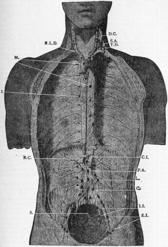
From A. M. Paterson, Cunningham’s Text-book of Anatomy.
Fig. 2.—Deep Lymphatic Glands and Vessels of the Thorax and Abdomen (diagrammatic). Afferent vessels are represented by continuous lines and efferent and interglandular vessels by dotted lines.
C. Common iliac glands. C.I. Common intestinal trunk. D.C. Deep cervical glands. E.I. External iliac glands. I. Intercostal glands and vessels. I.I. Internal iliac glands. L. Lateral aortic glands. M. Mediastinal glands and vessels. P.A. Pre-aortic glands and vessels. R.C. Receptaculum chylii. R.L.D. Right lymphatic duct. S. Sacral glands. S.A. Scalenus anticus muscle. T.D. Thoracic duct.
The thoracic duct begins as an irregular dilatation known as the receptaculum chyli, opposite the first and second lumbar vertebrae, which receives all the abdominal lymphatics as well as those of the lower intercostal spaces. The duct runs up on the right of the aorta through the posterior mediastinum and then traverses the superior mediastinum to the left of the oesophagus. At the root of the neck it receives the lymphatics of the left arm and left side of the neck and opens into the beginning of the left innominate vein, usually by more than one opening.
The right lymphatic duct collects the lymphatics from the right side of the neck and thorax, the right arm, right lung, right side of the heart and upper surface of the liver; it is often represented by several ducts which open separately into the right innominate vein.
Haemolymph glands are structures which have only been noticed since 1884. They differ from lymphatic glands in their much greater vascularity. They assist the spleen in the destruction of red blood corpuscles, and probably explain or help to explain the fact that the spleen can be removed without ill effects. In man they extend along the vertebral column from the coeliac axis to the pelvis, but are specially numerous close to the renal arteries.
T. Lewis suggests that lymphatic and haemolymph glands should be classified in the following way:—
Haemolymph Glands. Haemal glands. Simple.
Specialized (Spleen)
Haemal lymphatic glands. 1. Blood and lymph sinuses separate.
2. Blood lymph sinuses.
3. Other combined forms.
Lymphatic glands.
Details and references will be found in papers by T. Lewis, J. Anat. & Phys. vol. xxxviii. p. 312; W. B. Drummond, Journ. Anat. and Phys. vol. xxxiv. p. 198; A. S. Warthin, Journ. Med. Research, 1901, p. 3, and H. Dayton, Am. Journ. of Med. Sciences, 1904, p. 448. For further details of man’s lymphatic system see The Lymphatics by Delamere, Poirier and Cuneo, translated by C. H. Leaf (London, 1903).
Embryology.—The lymphatic vessels are possibly developed by the hollowing out of mesenchyme cells in the same way that the arteries are; these cells subsequently coalesce and form tubes (see Vascular System). There is, however, a good deal of evidence to show that they are originally offshoots of the venous system, and that their permanent openings into the veins are either their primary points of communication or are secondarily acquired. The lymphatic and haemolymph glands are probably formed by the proliferation of lymphocytes around networks of lymphatic vessels; the dividing lymphocytes form the lymphoid tissue, and eventually the network breaks up to form distinct glands into which blood vessels penetrate. If the blood vessels enlarge more than the lymphatic, haemolymph glands result, but if the lymphatic vessels become predominant ordinary lymphatic glands are formed. At an early stage in the embryo pig two thoracic ducts are formed, one on either side of the aorta, and the incomplete fusion of these may account for the division often found in man’s duct. In the embryo pig too there have been found two pairs of lymph hearts for a short period.
See A. S. Warthin, Journ. Med. Research, vol. vii. p. 435; F. R. Sabin, Am. Journ. of Anat. i., 1902; and, for literature, Development of the Human Body, by J. P. McMurrich (London, 1906), and Quain’s Anatomy (vol. i., London, 1908).
Comparative Anatomy.—A lymphatic system is recognized in all the Craniata, and in the lower forms (fishes and Amphibia) it consists chiefly of lymph spaces and sinuses in communication with the coelom. In fishes, for instance, there is a large subvertebral lymph sinus surrounding the aorta and another within the spinal canal. In Amphibia the subvertebral sinus is also found, and in the Anura (frogs and toads) there is a great subcutaneous lymph sinus. Lymph hearts are muscular dilatations of vessels and are found in fishes, amphibians, reptiles and bird embryos, and drive the lymph into the veins; they are not known in adult mammals.
In birds the thoracic duct is first recognized, and opens into both right and left precaval veins, as it always does in some mammals. In birds, however, some of the lymphatics open into the sacral veins, and it is doubtful whether true lymphatic glands ever occur. In birds and mammals lymphatic vessels become more definite and numerous and are provided with valves.
Haemolymph glands are present in mammals and birds, but have not been seen lower in the scale, though S. Vincent and S. Harrison point out the resemblance of the structure of the head kidney of certain Teleostean fishes to them (Journ. Anat. and Phys. vol. xxxi. p. 176).
For further details see Comparative Anat. of Vertebrates, by R. Wiedersheim (London, 1907).
(F. G. P.)
Diseases of the Lymphatic System and Ductless Glands.
Lymphadenitis or inflammatory infection of the lymphatic glands, is a condition characterized by hyperaemia of and exudation into the gland, which becomes reader, firmer and larger than usual. Three varieties may be distinguished: simple, suppurative and tuberculous. The cause is always the absorption of some toxic or infective material from the periphery. This may take place in several of the acute infectious diseases, notably in scarlet fever, mumps, diphtheria and German measles, or may be the result of poisoned wounds. The lymphatic glands are also affected in constitutional diseases such as syphilis. Simple lymphadenitis usually subsides of its own accord, but if toxins are produced in the inflamed area the enlargement is obvious and painful, while if pyogenic organisms are absorbed the inflammation progresses to suppuration.
Tuberculous lymphadenitis (scrofula) is due to the infection of the lymph glands by Koch’s tubercle bacillus. This was formerly known as “King’s Evil,” as it was believed that the touch of the royal hand had power to cure it. It occurs most commonly in children and young adults whose surroundings are unhealthy, and who are liable to develop tuberculous disease from want of sufficient food and fresh air. Some local focus of irritation is usually present. The ways in which the tubercle bacillus enters the body are much disputed, but catarrh of the mucous membranes is regarded as a predisposing factor, and the tonsils as a probable channel of infection. Any lymphoid tissue in the body may be the seat of tuberculous disease, but the glands of the neck are the most commonly involved. The course of the disease is slow and may extend over a period of years. The earliest manifestation is an enlargement of the gland. It is possible in this stage for spontaneous healing to take place, but usually the disease progresses to caseation, in which tuberculous nodules are found diffused throughout the gland. Occasionally this stage may end in calcification of the caseous matter, the gland shrinking and becoming hard; but frequently suppuration follows from liquefaction of the caseating material. Foci of pus occur throughout the gland, causing destruction of the tissue, so that the gland may become a single abscess cavity. If left to itself the abscess sooner or later bursts at one or several points, leaving ulcerated openings through which a variable amount of pus escapes. Temporary healing may take place, to be again followed by further breaking down of the gland. This condition, if untreated, may persist for years and may finally give rise to a general tuberculosis. The treatment consists mainly in improving the general health with good diet, fresh air (particularly sea air), cod-liver oil and iron, and the removal of all sources of local irritation such as enlarged tonsils, adenoids, &c. Vaccination with tuberculin (TR) may be useful. Suppuration and extension of the disease require operative measures, and removal of the glands en masse can now be done through so small an opening as to leave only a very slight scar.
In Tabes mesenterica (tuberculosis of the mesenteric glands), usually occurring in children, the glands of the mesentery and retroperitonaeum become enlarged, and either caseate or occasionally suppurate. The disease may be primary or may be secondary to tuberculous disease of the intestines or to pulmonary phthisis. The patients are pale, wasted and anaemic, and the abdomen may be enormously enlarged. There is usually moderate fever, and thin watery diarrhoea. The caseating glands may liquefy and give rise to an inflammatory attack which may simulate appendicitis. Limited masses are amenable to surgical treatment and may be removed, while in the earlier stages constitutional treatment gives good results. Tuberculous peritonitis frequently supervenes on this condition.
Lymphadenoma (Hodgkin’s Disease), a disease which was first fully described by Hodgkin in 1832, is characterized by a progressive enlargement of the lymphatic glands all over the body, and generally starts in the glands of the neck. The majority of cases occur in young adults, and preponderate in the male sex. The first symptom is usually enlargement of a gland in the neck, with generally progressive growth of the glands in the submaxillary region and axilla. The inguinal glands are early involved, and after a time the internal lymph glands follow. The enlargements are at first painless, but in the later stages symptoms are caused by pressure on the surrounding organs, and when the disease starts in the deeper structures the first symptoms may be pain in the chest and cough, pain in the abdomen, pain and oedema in the legs. The glands may increase until they are as large as eggs, and later may become firmly adherent one to another, forming large lobulated tumours. Increase of growth in this manner in the neck may cause obstructive dyspnoea and even death. In the majority of cases the spleen enlarges, and in rare instances lymphoid tumours may be found on its surface. Anaemia is common and is secondary in character; slight irregular fever is present, and soon a great and progressive emaciation takes place. The cases are of two types, the acute cases in which the enlargements take place rapidly and death may occur in two to three months, and the chronic cases in which the disease may remain apparently stationary. In acute lymphadenoma the prognosis is very unfavourable. Recovery sometimes takes place in the chronic type of the disease. Early surgical intervention has in some cases been followed by success. The application of X-rays is a valuable method of treatment, superficial glands undergoing a rapid diminution in size. Of drugs arsenic is of the most service, and mercurial inunction has been recommended by Dreschfeld. Organic extracts have of late been used in the treatment of lymphadenoma.
Glandular Fever is an acute infectious fever, generally occurring in epidemics, and was first described by E. Pfeiffer in 1889. It usually affects children and has a tendency to run through all the children of a family. The incubation period is said to be about 7 days. The onset is sudden, with pain in the neck and limbs, headache, vomiting, difficulty in swallowing and high temperature. On the second day, or sometimes on the first, swelling of the cervical glands is noticed, and later the posterior cervical, axillary and inguinal glands become enlarged and tender. In about half the cases the spleen and liver are enlarged and there is abdominal tenderness. West found the mesenteric nodes enlarged in 37 cases. Nephritis is an occasional complication, and constipation is very usual. The disease tends to subside of itself, and the fever usually disappears after a few days; the glandular swellings may, however, persist from one to three weeks. Considerable anaemia has been noticed to follow the illness. Rest in bed while the glands are enlarged, and cod-liver oil and iron to meet the anaemia, are the usual treatment.
Status lymphaticus (lymphatism) is a condition found in children and some adults, characterized by an enlargement of the lymphoid tissues throughout the body and more particularly by enlargement of the thymus gland. There is a special lowering of the patient’s powers of resistance, and it has been said to account for a number of cases of sudden death. In all cases of status lymphaticus the thymus has been found enlarged. At birth the gland (according to Bovaird and Nicoll) weighs about 6 grammes, and does not increase after birth. In lymphatism it may weigh from 10 to 50 grammes. The clinical features are indefinite, and the condition frequently passes unrecognized during life. In most cases there is no hint of danger until the fatal syncope sets in, which may be after any slight exertion or shock, the patient becoming suddenly faint, gasping and cyanosed, and the heart stopping altogether before the respirations have ceased. The most trifling causes have brought on fatal issues, such as a wet pack (Escherich) or a hypodermic injection, or even a sudden plunge into water though the head is not immersed. The greater number of deaths occur during the administration of anaesthetics, which seem peculiarly dangerous to these subjects. When an attack of syncope takes place no treatment is of any avail.
Virchow, West and Goodhardt have described a form of asthma in adults which they ascribe to a hypertrophied thymus gland and term “thymic asthma.”
Diseases of the Spleen.—Physiological variations and abnormalities and absence of the spleen are so rare as to require no comment. The most usual pathological condition which gives rise to symptoms is that of wandering spleen, which may or may not be secondary to a wandering left kidney. It may produce symptoms of dragging and discomfort, dyspepsia, vomiting and abdominal pain, and sometimes jaundice (Treves), or the pedicle may become twisted, producing extremely severe symptoms. The treatment is entirely surgical. Abscess in the spleen occasionally occurs, usually in association with infective endocarditis or with general pyaemia. The spleen may be the seat of primary new growths, but these are rare, and only in a small portion of cases does it share in the metastatic reproduction of carcinoma. Infection of the spleen plays a prominent part in many diseases, such as malaria, typhoid fever, lymphadenoma and leucaemia.
Diseases of the thyroid gland (see Goitre) and Addison’s disease (of the suprarenal glands) are treated separately.
(H. L. H.)
1 It has recently been stated that stomata do not exist in the peritoneum.
2 For further details of the pelvic glands see “Seventh Report of the Committee of Collective Investigation,” Journ. Anat. and Phys. xxxii. 164.
LYNCH, PATRICIO (1825-1886) Chilean naval officer, was born in Valparaiso on the 18th of December 1825, his father being a wealthy Irish merchant resident in Chile, and his mother, Carmen Solo de Saldiva, a descendant of one of the best-known families in the country. Entering the navy in 1837, he took part in the operations which led to the fall of the dictator, Santa Cruz. Next, he sought a wider field, and saw active service in the China War on board the British frigate “Calliope.” He was mentioned in despatches for bravery, and received the grade of midshipman in the British service. Returning to Chile in 1847 he became lieutenant, and seven years later he received the command of a frigate, but was deprived of his command for refusing to receive on board his ship political suspects under arrest. The Spanish War saw him again employed, and he was successively maritime prefect of Valparaiso, colonel of National Guards, and, finally, captain and minister of marine in 1872. In the Chile-Peruvian War a brilliant and destructive naval raid, led by him, was followed by the final campaign of Chorrillos and Miraflores (1880), in which he led at first a brigade (as colonel) and afterwards a division under Baquedano. His services at the battle of Chorrillos led to his appointment to command the Army of Occupation in Peru. This difficult post he filled with success, but his action in putting the Peruvian president, Garcia Calderon, under arrest excited considerable comment. His last act was to invest Iglesias with supreme power in Peru, and he returned to his own country in 1883. Promoted rear-admiral, he served as Chilean Minister at Madrid for two years, and died at sea in 1886. Lynch is remembered as one of the foremost of Chile’s naval heroes.
LYNCHBURG, a city of Campbell county, Virginia, U.S.A., on the James river, about 125 m. W. by S. of Richmond. Pop. (1900) 18,891, of whom 8254 were negroes; (1910) 29,494. It is served by the Southern, the Chesapeake & Ohio and the Norfolk & Western railways. Its terraced hills command fine views of mountain, valley and river scenery, extending westward to the noble Peaks of Otter and lesser spurs of the Blue Ridge about 20 m. distant. On an elevation between Rivermont Avenue and the James river are the buildings of Randolph-Macon Woman’s college (opened in 1893), which is conducted by a self-perpetuating board under the auspices of the Methodist Episcopal Church, South, and is one of the Randolph-Macon system of colleges and academies (see Ashland, Va.). In Lynchburg, too, are the Virginia Christian college (co-educational, 1903), and the Virginia collegiate and industrial school for negroes. The city has a public library, well-equipped hospitals, public parks and the Rivermont Viaduct, 1100 ft. long and 140 ft. high. Lynchburg is the see of a Protestant Episcopal bishop. Tobacco of a superior quality and large quantities of coal, iron ore and granite are produced in the neighbourhood. Good water power is furnished by the James river, and Lynchburg is one of the principal manufacturing cities of the state. The boot and shoe industry was established in 1900, and is much the most important. In 1905 the city was the largest southern manufacturer of these articles and one of the largest distributors in the country. The factory products increased in value from $2,993,551 in 1900 to $4,905,435 in 1905, or 65.9%.
Lynchburg, named in honour of John Lynch, who inherited a large tract of land here and in 1757 established a ferry across the James, was established as a village by Act of Assembly in 1786, was incorporated as a town in 1805, and became a city in 1852. During the Civil War it was an important base of supplies for the Confederates; on the 16th of June 1864 it was invested by Major-General David Hunter (1802-1886), but three days later he was driven away by General Jubal A. Early. In 1908 the city’s corporate limits were extended.
LYNCH LAW, a term loosely applied to various forms of executing rough popular justice, or what is thought to be justice, for the punishment of offenders by a summary procedure, ignoring, or even contrary to, the strict forms of law. The word lynching “originally signified a whipping for reformatory purposes with more or less disregard for its legality” (Cutler), or the infliction of minor punishments without recourse to law; but during and after the Reconstruction Period in the United States, it came to mean, generally, the summary infliction of capital punishment. Lynch law is frequently prevalent in sparsely settled or frontier districts where government is weak and officers of the law too few and too powerless to enforce law and preserve order. The practice has been common in all countries when unsettled frontier conditions existed, or in periods of threatened anarchy. In what are considered civilized countries it is now found mainly in Russia, south-eastern Europe and in America, but it is essentially and almost peculiarly an American institution. The origin of the name is obscure; different writers have attempted to trace it to Ireland, to England, to South Carolina, to Pennsylvania and to Virginia. It is certain that the name was first used in America, but it is not certain whether it came from Lynch’s Creek, South Carolina, where summary justice was administered to outlaws, or from Virginia and Pennsylvania, where men named Lynch were noted for dealing out summary punishment to offenders.1 In Europe early examples of a similar phenomenon are found in the proceedings of the Vehmgerichte in medieval Germany, and of Lydford law, gibbet law or Halifax law, Cowper justice and Jeddart justice in the thinly settled and border districts of Great Britain; and since the term “lynch law” came into colloquial use, it is loosely employed to cover any case in which a portion of the community takes the execution of its ideas of justice into its own hands, irrespective of the legal authorities.
In America during the 18th and 19th centuries the population expanded westward faster than well-developed civil institutions could follow, and on the western frontier were always desperadoes who lived by preying on the better classes. To suppress these desperadoes, in the absence of strong legal institutions, resort was continually made to lynch law. There was little necessity for it until the settlement crossed the Alleghany Mountains, but the following instances of lynching in the East may be mentioned: (1) the mistreatment of Indians in New England and the Middle Colonies in disregard of laws protecting them; (2) the custom found in various colonies of administering summary justice to wife-beaters, idlers and other obnoxious persons; (3) the acts of the Regulators of North Carolina, 1767-1771; (4) the popular tribunals of the Revolutionary period, when the disaffection toward Great Britain weakened the authority of the civil governments and the war replaced them by popular governments, at a time when the hostilities between “Patriots” and “Tories” were an incentive to extra-legal violence. In the South, lynching methods were long employed in dealing with agitators, white and black, who were charged with endeavouring to excite the slaves to insurrection or to crime against their masters, and in dealing with anti-slavery agitators generally.
In the West, from the Alleghanies to the Golden Gate, the pioneer settlers resorted to popular justice to get rid of bands of outlaws, and to regulate society during that period when laws were weak or confused, when the laws made in the East did not suit western conditions, and when courts and officials were scarce and distant. The Watauga settlements and the “State” of Franklin furnished examples of lynch law procedure almost reduced to organization. Men trained in the rough school of the wilderness came to have more regard for quick, ready-made, personal justice than for abstract justice and statutes; they were educated to defend themselves, to look to no law for protection or regulation; consequently they became impatient of legal forms and lawyers’ technicalities; an appeal to statute law was looked upon with suspicion, and, if some personal matter was involved, was likely to result in deadly private feuds. Thus were formed the habits of thought and action of the western pioneers. Lynch law, not civil law, cleared the western forests, valleys and mountain passes of horse and cattle thieves, and other robbers and outlaws, gamblers and murderers. This was especially true of California and the states of the far West. H. H. Bancroft, the historian of Popular Tribunals, wrote in 1887 that “thus far in the history of these Pacific States far more has been done toward righting wrongs and administering justice outside the pale of law than within it.” However, the lack of regard for law fostered by the conditions described led to a survival of the lynching habit after the necessity for it passed away. In parts of the Southern states, where the whites are few and greatly outnumbered by the blacks, certain of the conditions of the West have prevailed, and since emancipation released the blacks from restraint many of the latter have been lawless and turbulent. The Reconstruction, by giving to the blacks temporary political supremacy, increased the friction between the races, and greatly deepened prejudice. The numerous protective societies of whites, 1865-1876, culminating in the Ku Klux movement, may be described as an application of lynch law. With the increase of negro crimes came an increase of lynchings, due to prejudice, to the fact that for some time after Reconstruction the governments were relatively weak, especially in the districts where the blacks outnumber the whites, to the fact that negroes nearly always shield criminals of their own race against the whites, and to the frequent occurrence of the crime of rape by negro men upon white women.
Since 1882 the Chicago Tribune has collected statistics of lynching, and some interesting facts may be deduced from these tables.2 During the twenty-two years from 1882 to 1903 inclusive, the total number of persons lynched in the United States was 3337, the number decreasing during the last decade; of these 2385 were in the South and 752 in the North; of those lynched in the East and West 602 were white and 75 black, and of those in the South 567 were white and 1985 black.3 Lynchings occur mostly during periods of idleness of the lower classes; in the summer more are lynched for crimes against the person and in the winter (in the West) for crimes against property; the principal causes of lynching in the South are murder and rape, in the North and West, murder and offences against property; more blacks than whites were lynched between 1882 and 1903, the numbers being 2060 negroes, of whom 40 were women, and 1169 whites, of whom 23 were women; of the 707 blacks lynched for rape 675 were in the South; 783 blacks were lynched for murder, and 753 of these were in the South; most of the lynchings of whites were in the West; the lynching of negroes increased somewhat outside of the South and decreased somewhat in the South. Lynching decreases and disappears in a community as the population grows denser and civil institutions grow stronger; as better communications and good police make it harder to commit crime; and as public sentiment is educated to demand legal rather than illegal and irregular infliction of punishment for even the most horrible of crimes.
See James E. Cutler, Lynch Law (New York, 1905), an admirable and unbiased discussion of the subject; H. H. Bancroft, Popular Tribunals (2 vols., San Francisco, 1887); C. H. Shinn, Mining Camps: A Study in American Frontier Government (New York, 1885); and J. C. Lester and D. L. Wilson, Ku Klux Klan (New York, 1905).
(W. L. F.)
1 The usual explanation is that the name was derived from Charles Lynch (1736-1796), a justice of the peace in Virginia after 1774, who in 1780, toward the close of the War of Independence, greatly exceeded his powers in the punishment of Tories or Loyalists detected in a conspiracy in the neighbourhood of his home in Bedford county, Va. Lynch was a man of influence in his community, was for many years a member of the Virginia legislature, was a member of the famous Virginia Convention of 1776 and was later (in 1781) an officer in the American army. See an article, “The Real Judge Lynch,” in the Atlantic Monthly, vol. lxxxviii. (Boston, 1901).
2 They have been corrected and somewhat modified by Dr. J. E. Cutler, from whose book the figures above have been taken. Lynching as used in this connexion applies exclusively to the illegal infliction of capital punishment.
3 For present purposes the former slave states (of 1860) constitute the South; the West is composed of the territory west of the Mississippi river, excluding Missouri, Arkansas, Louisiana, Texas and Oklahoma; the East includes those states east of the Mississippi river not included in the Southern group; the East and the West make up the North as here used—that is, the former free states of 1860.
LYNDHURST, JOHN SINGLETON COPLEY, Baron (1772-1863), lord chancellor of England, was born at Boston, Massachusetts, in 1772. He was the son of John Singleton Copley, the painter. He was educated at a private school and Cambridge university, where he was second wrangler and fellow of Trinity. Called to the bar at Lincoln’s Inn in 1804, he gained a considerable practice. In 1817 he was one of the counsel for Dr J. Watson, tried for his share in the Spa Fields riot. On this occasion Copley so distinguished himself as to attract the attention of Castlereagh and other Tory leaders, under whose patronage he entered parliament as member for Yarmouth in the Isle of Wight. He afterwards sat for Ashburton, 1818-1826, and for Cambridge university 1826-1827. He was solicitor-general in 1819, attorney-general in 1824, master of the rolls in 1826 and lord chancellor in 1827, with the title of Lord Lyndhurst. Before being taken up by the Tories, Copley was a man of the most advanced views, a republican and Jacobin; and his accession to the Tories excited a good deal of comment, which he bore with the greatest good humour. He gave a brilliant and eloquent but by no means rancorous support to all the reactionary measures of his chief. The same year that he became solicitor-general he married the beautiful and clever widow of Lieut.-Colonel Charles Thomas of the Coldstream Guards, and began to take a conspicuous place in society, in which his noble figure, his ready wit and his never-failing bonhomie made him a distinguished favourite.
As solicitor-general he took a prominent part in the trial of Queen Caroline. To the great Liberal measures which marked the end of the reign of George IV. and the beginning of that of William IV. he gave a vigorous opposition. He was lord chief baron of the exchequer from 1831 to 1834. During the Melbourne administration from 1835 to 1841 he figured conspicuously as an obstructionist in the House of Lords. In these years it was a frequent practice with him, before each prorogation of parliament, to entertain the House with a “review of the session,” in which he mercilessly attacked the Whig government. His former adversary Lord Brougham, disgusted at his treatment by the Whig leaders, soon became his most powerful ally in opposition; and the two dominated the House of Lords. Throughout all the Tory governments from 1827 Lyndhurst held the chancellorship (1827-1830 and 1834-1835); and in the Peel administration (1841-1846) he resumed that office for the last time. As Peel never had much confidence in Lyndhurst, the latter did not exert so great an influence in the cabinet as his position and experience entitled him to do. But he continued a loyal member of the party. As in regard to Catholic emancipation, so in the agitation against the corn laws, he opposed reform till his chief gave the signal for concession, and then he cheerfully obeyed. After 1846 and the disintegration of the Tory party consequent on Peel’s adoption of free trade, Lord Lyndhurst was not so assiduous in his attendance in parliament. Yet he continued to an extreme old age to take a lively interest in public affairs, and occasionally to astonish the country by the power and brilliancy of his speeches. That which he made in the House of Lords on the 19th of June 1854, on the war with Russia, made a sensation in Europe; and throughout the Crimean War he was a strong advocate of the energetic prosecution of hostilities. In 1859 he denounced with his old energy the restless ambition of Napoleon III. When released from office he came forward somewhat as the advocate of liberal measures. His first wife had died in 1834, and in August 1837 he had married Georgina, daughter of Lewis Goldsmith. She was a Jewess; and it was therefore natural that he strenuously supported the admission of Jews into parliament. He also advocated women’s rights in questions of divorce. At the age of eighty-four he passed the autumn at Dieppe, “helping to fly paper kites, and amusing himself by turns with the writings of the Greek and Latin fathers on divorce and the amorous novels of Eugene Sue.” His last speech, marked by “his wonted brilliancy and vigour,” was delivered in the House of Lords at the age of eighty-nine. He died in London on the 12th of October 1863. He left no male issue and the title became extinct.
See Lives of the Lord Chancellors of England, vol. viii. (Lords Lyndhurst and Brougham), by Lord Campbell (1869). Campbell was a personal friend, but a political opponent. Brougham’s Memoirs; Greville Memoirs; Life of Lord Lyndhurst (1883) by Sir Theodore Martin; J. B. Atlay, The Victorian Chancellors (1906).
LYNDSAY, SIR DAVID (c. 1490-c. 1555), Scottish poet, was the son of David Lyndsay of the Mount, near Cupar-Fife, and of Garmylton, near Haddington. His place of birth and his school are undetermined. It is probable that his college life was spent at St Andrews university, on the books of which appears an entry “Da Lindesay” for the session 1508-1509. He was engaged at court, first as an equerry, then as an “usher” to the young Prince James, afterwards James V. In 1522 he married Janet Douglas, a court seamstress, and seven years later was appointed Lyon King of Arms, and knighted. He was several times engaged in diplomatic business (twice on embassies abroad—to the Netherlands and France), and he was, in virtue of his heraldic office, a general master of ceremonies. After the death of James V., in 1542, he continued to sit in parliament as commissioner for Cupar-Fife; and in 1548 he was member of a mission to Denmark which obtained certain privileges for Scottish merchants. There is reason to believe that he died in or about 1555.
Most of Lyndsay’s literary work, by which he secured great reputation in his own day and by which he still lives, was written during the period of prosperity at court. In this respect he is unlike his predecessor Gavin Douglas (q.v.), who forsook literature when he became a politician. The explanation of the difference is partly to be found in the fact that Lyndsay’s muse was more occasional and satirical, and that the time was suitable to the exercise of his special gifts. It is more difficult to explain how he enjoyed a freedom of speech which is without parallel even in more secure times. He chastised all classes, from his royal master to the most simple. There is no evidence that he abjured Catholicism; yet his leading purpose was the exposure of its errors and abuses. His aid was readily accepted by the reforming party, and by their use of his work he shared with their leaders throughout many generations a reputation which is almost exclusively political and ecclesiastical.
Lyndsay’s longer poems are The Dreme (1134 lines), The Testament and Complaynt of the Papynago (1190 lines), The Testament of Squyer Meldrum (1859 lines), Ane Dialog betwix Experience and ane Courteour of the Miserabyll Estait of the World (6333 lines), and Ane Pleasant Satyre of the Thrie Estaitis (over 4000 lines). These represent, with reasonable completeness, the range of Lyndsay’s literary talent. No single poem can give him a chief place, though here and there, especially in the last, he gives hints of the highest competence. Yet the corporate effect of these pieces is to secure for him the allowance of more than mere intellectual vigour and common sense. There is in his craftsmanship, in his readiness to apply the traditional methods to contemporary requirements, something of that accomplishment which makes even the second-rate man of letters interesting.
Lyndsay, the last of the Makars, is not behind his fellow-poets in acknowledgment to Chaucer. As piously as they, he reproduces the master’s forms; but in him the sentiment and outlook have suffered change. His nearest approach to Chaucer is in The Testament of Squyer Meldrum, which recalls the sketch of the “young squire”; but the reminiscence is verbal rather than spiritual. Elsewhere his memory serves him less happily, as when he describes the array of the lamented Queen Magdalene in the words which Chaucer had applied to the eyes of his wanton Friar. So too, in the Dreme, the allegorical tradition survives only in the form. “Remembrance” conducts the poet over the old-world itinerary, but only to lead him to speculation on Scotland’s woes and to an “Exhortatioun to the Kingis Grace” to bring relief. The tenor is well expressed in the motto from the Vulgate—“Prophetias nolite spernere. Omnia autem probate: quod bonum est tenete.” This didactic habit is freely exercised in the long Dialog (sometimes called the Monarche), a universal history of the medieval type, in which the falls of princes by corruption supply an object lesson to the unreformed church of his day. The Satyre is more direct in its attack on ecclesiastical abuse; and its dramatic form permits more lively treatment. This piece is of great historical interest, being the only extant example of a complete Scottish morality. It is in respect of literary quality Lyndsay’s best work, and in dramatic construction and delineation of character it holds a high place in this genre. The farcical interludes (in places too coarse for modern taste) supply many touches of genuine comedy; and throughout the play there are passages, as in the speeches of Veritie in the First Part and of Dame Chastitie in the “Interlude of the Sowtar and the Taylor,” in which word and line are happily conceived. The Testament of the Papyngo (popinjay), drawn in the familiar medieval manner, is another tract for the time, full of admonition to court and clergy. Of his shorter pieces, The Complaynt and Publict Confessions of the Kingis Auld Hound, callit Bagsche, directit to Bawtie, the Kingis best belovit Dog, and his companyeonis, and the Answer to the Kingis Flyting have a like pulpit resonance. The former is interesting as a forerunner of Burns’s device in the “Twa Dogs.” The Deploratioun of the Deith of Queen Magdalene is in the extravagant style of commemoration illustrated in Dunbar’s Elegy on the Lord Aubigny. The Justing betwix James Watsoun and Jhone Barbour is a contribution to the popular taste for boisterous fun, in spirit, if not in form, akin to the Christis Kirk on the Grene series; and indirectly, with Dunbar’s Turnament and Of ane Blak-Moir, a burlesque of the courtly tourney. Lyndsay approaches Dunbar in his satire The Supplicatioun in contemptioun of syde taillis (“wide” trains of the ladies), which recalls the older poet’s realistic lines on the filthy condition of the city streets. In Lyndsay’s Descriptioun of Pedder Coffeis (pedlars) we have an early example of the studies in vulgar life which are so plentiful in later Scottish literature. In Kitteis Confessioun he returns, but in more sprightly mood, to his attack on the church.