VOLUME XVI SLICE I L to Lamellibranchia (continued 12)
Reading edition
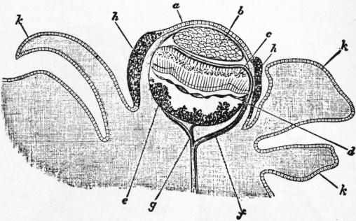
Fig. 21.—Pallial Eye of Spondylus. (From Hickson.)
a, Prae-corneal epithelium. b, Cellular lens. c, Retinal body. d, Tapetum. e, Pigment. f, Retinal nerve. g, Complementary nerve. h, Epithelial cells filled with pigment. k, Tentacle.
The sense-organs of Anodonta other than the osphradia consist of a pair of otocysts attached to the pedal ganglia (fig. 1 (6), ay). The otocysts of Cyclas are peculiarly favourable for study on account of the transparency of the small foot in which they lie, and may be taken as typical of those of Lamellibranchs generally. The structure of one is exhibited in fig. 20. A single otolith is present as in the veliger embryos of Opisthobranchia. In Filibranchia and many Protobranchia the otocyst (or statocyst) contains numerous particles (otoconia). The organs are developed as invaginations of the epidermis of the foot, and in the majority of the Protobranchia the orifice of invagination remains open throughout life; this is also the case in Mytilus including the common mussel.
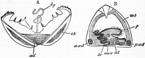
Fig. 22.—Two Stages in the Development of Anodonta. (From Balfour.) Both figures represent the glochidium stage.
A, When free swimming, shows the two dentigerous valves widely open. B, A later stage, after fixture to the fin of a fish. sh, Shell. ad, Adductor muscle. s, Teeth of the shell. by, Byssus. a.ad, Anterior adductor. p.ad, Posterior adductor. mt, Mantle-flap. f, Foot. br, Branchial filaments. au.v, Otocyst. al, Alimentary canal.
Anodonta has no eyes of any sort, and the tentacles on the mantle edge are limited to its posterior border. This deficiency is very usual in the class; at the same time, many Lamellibranchs have tentacles on the edge of the mantle supplied by a pair of large well-developed nerves, which are given off from the cerebro-pleural ganglion-pair, and very frequently some of these tentacles have undergone a special metamorphosis converting them into highly-organized eyes. Such eyes on the mantle-edge are found in Pecten, Spondylus, Lima, Pinna, Pectunculus, Modiola, Cardium, Tellina, Mactra, Venus, Solen, Pholas and Galeomma. They are totally distinct from the cephalic eyes of typical Mollusca, and have a different structure and historical development. They have originated not as pits but as tentacles. They agree with the dorsal eyes of Oncidium (Pulmonata) in the curious fact that the optic nerve penetrates the capsule of the eye and passes in front of the retinal body (fig. 21), so that its fibres join the anterior faces of the nerve-end cells as in Vertebrates, instead of their posterior faces as in the cephalic eyes of Mollusca and Arthropoda; moreover, the lens is not a cuticular product but a cellular structure, which, again, is a feature of agreement with the Vertebrate eye. It must, however, be distinctly borne in mind that there is a fundamental difference between the eye of Vertebrates and of all other groups in the fact that in the Vertebrata the retinal body is itself a part of the central nervous system, and not a separate modification of the epidermis—myelonic as opposed to epidermic. The structure of the reputed eyes of several of the above-named genera has not been carefully examined. In Pecten and Spondylus, however, they have been fully studied (see fig. 21, and explanation). Rudimentary cephalic eyes occur in the Mytilidae and in Avicula at the base of the first filament of the inner gill, each consisting of a pigmented epithelial fossa containing a cuticular lens. In the Arcidae the pallial eyes are compound or faceted somewhat like those of Arthropods.
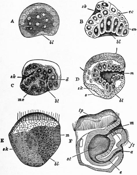
Fig. 23.—Development of the Oyster, Ostrea edulis. (Modified from Horst.)
A, Blastula stage (one-cell-layered sac), with commencing invagination of the wall of the sac at bl, the blastopore. B, Optical section of a somewhat later stage, in which a second invagination has begun—namely, that of the shell-gland sk. bl, Blastopore. en, Invaginated endoderm (wall of the future arch-enteron). ec, Ectoderm. C, Similar optical section at a little later stage. The invagination connected with the blastopore is now more contracted, d; and cells, me, forming the mesoblast from which the cœlom and muscular and skeleto-trophic tissues develop, are separated. D, Similar section of a later stage. The blastopore, bl, has closed; the anus will subsequently perforate the corresponding area. A new aperture, m, the mouth, has eaten its way into the invaginated endodermal sac, and the cells pushed in with it constitute the stomodaeum. The shell-gland, sk, is flattened out, and a delicate shell, s, appears on its surface. The ciliated velar ring is cut in the section, as shown by the two projecting cilia on the upper part of the figure. The embryo is now a Trochosphere. E, Surface view of an embryo at a period almost identical with that of D. F, Later embryo seen as a transparent object. m, Mouth. ft, Foot. a, Anus. e, Intestine. st, Stomach. tp, Velar area of the prostomium. The extent of the shell and commencing upgrowth of the mantle-skirt is indicated by a line forming a curve from a to F.
N.B.—In this development, as in that of Pisidium (fig. 25), no part of the blastopore persists either as mouth or as anus, but the aperture closes—the pedicle of invagination, or narrow neck of the invaginated arch-enteron, becoming the intestine. The mouth and the anus are formed as independent in-pushings, the mouth with stomodaeum first, and the short anal proctodaeum much later. This interpretation of the appearances is contrary to that of Horst, from whom our drawings of the oyster’s development are taken. The account given by the American William K. Brooks differs greatly as to matter of fact from that of Horst, and appears to be erroneous in some respects.
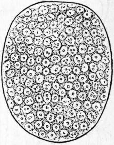
Fig. 24.—Embryo of Pisidium pusillum in the diblastula stage, surface view (after Lankester). The embryo has increased in size by accumulation of liquid between the outer and the invaginated cells. The blastopore has closed.
Generative Organs.—The gonads of Anodonta are placed in distinct male and female individuals. In some Lamellibranchs—for instance, the European Oyster and the Pisidium pusillum—the sexes are united in the same individual; but here, as in most hermaphrodite animals, the two sexual elements are not ripe in the same individual at the same moment. It has been conclusively shown that the Ostrea edulis does not fertilize itself. The American Oyster (O. virginiana) and the Portuguese Oyster (O. angulata) have the sexes separate, and fertilization is effected in the open water after the discharge of the ova and the spermatozoa from the females and males respectively. In the Ostrea edulis fertilization of the eggs is effected at the moment of their escape from the uro-genital groove, or even before, by means of spermatozoa drawn into the sub-pallial chamber by the incurrent ciliary stream, and the embryos pass through the early stages of development whilst entangled between the gill-lamellae of the female parent (fig. 23). In Anodonta the eggs pass into the space between the two lamellae of the outer gill-plate, and are there fertilized, and advance whilst still in this position to the glochidium phase of development (fig. 22). They may be found here in thousands in the summer and autumn months. The gonads themselves are extremely simple arborescent glands which open to the exterior by two simple ducts, one right and one left, continuous with the tubular branches of the gonads. In the most primitive Lamellibranchs there is no separate generative aperture but the gonads discharge into the renal cavity, as in Patella among Gastropods. This is the case in the Protobranchia, e.g. Solenomya, in which the gonad opens into the reno-pericardial duct. But the generative products do not pass through the whole length of the renal tube: there is a direct opening from the pericardial end of the tube to the distal end, and the ova or sperms pass through this. In Arca, in Anomiidae and in Pectinidae the gonad opens into the external part of the renal tube. The next stage of modification is seen in Ostraea, Cyclas and some Lucinidae, in which the generative and renal ducts open into a cloacal slit on the surface of the body. In Mytilus the two apertures are on a common papilla, in other cases the two apertures are as in Anodonta. The Anatinacea and Poromya among the Septibranchia are, however, peculiar in having two genital apertures on each side, one male and one female. These forms are hermaphrodite, with an ovary and testis completely separate from each other on each side of the body, each having its own duct and aperture.
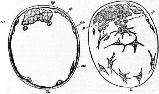
Fig. 25.—B, Same embryo as fig. 24, in optical median section, showing the invaginated cells hy which form the arch-enteron, and the mesoblastic cells me which are budded off from the surface of the mass hy, and apply themselves to the inner surface of the epiblastic cell-layer ep. C. The same embryo focused so as to show the mesoblastic cells which immediately underlie the outer cell-layer.
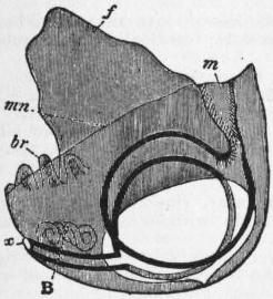
Fig.26.—Diagram of Embryo of Pisidium. The unshaded area gives the position of the shell-valve. (After Lankester.)
m, Mouth. x, Anus. f, Foot. br, Branchial filaments. mn, Margin of the mantle-skirt. B, Organ of Bojanus.
The development of Anodonta is remarkable for the curious larval form known as glochidium (fig. 22). The glochidium quits the gill-pouch of its parent and swims by alternate opening and shutting of the valves of its shell, as do adult Pecten and Lima, trailing at the same time a long byssus thread. This byssus is not homologous with that of other Lamellibranchs, but originates from a single glandular epithelial cell embedded in the tissues on the dorsal anterior side of the adductor muscle. By this it is brought into contact with the fin of a fish, such as perch, stickleback or others, and effects a hold thereon by means of the toothed edge of its shells. Here it becomes encysted, and is nourished by the exudations of the fish. It remains in this condition for a period of two to six weeks, and during this time the permanent organs are developed from the cells of two symmetrical cavities behind the adductor muscle. The early larva of Anodonta is not unlike the trochosphere of other Lamellibranchs, but the mouth is wanting. The glochidium is formed by the precocious development of the anterior adductor and the retardation of all the other organs except the shell. Other Lamellibranchs exhibit either a trochosphere larva which becomes a veliger differing only from the Gastropod’s and Pteropod’s veliger in having bilateral shell-calcifications instead of a single central one; or, like Anodonta, they may develop within the gill-plates of the mother, though without presenting such a specialized larva as the glochidium. An example of the former is seen in the development of the European oyster, to the figure of which and its explanation the reader is specially referred (fig. 23). An example of the latter is seen in a common little fresh-water bivalve, the Pisidium pusillum, which has been studied by Lankester. The gastrula is formed in this case by invagination. The embryonic cells continue to divide, and form an oval vesicle containing liquid (fig. 24); within this, at one pole, is seen the mass of invaginated cells (fig. 25, hy). These invaginated cells are the arch-enteron; they proliferate and give off branching cells, which apply themselves (fig. 25, C) to the inner face of the vesicle, thus forming the mesoblast. The outer single layer of cells which constitutes the surface of the vesicle is the ectoderm or epiblast. The little mass of hypoblast or enteric cell-mass now enlarges, but remains connected with the cicatrix of the blastopore or orifice of invagination by a stalk, the rectal peduncle. The enteron itself becomes bilobed and is joined by a new invagination, that of the mouth and stomodaeum. The mesoblast multiplies its cells, which become partly muscular and partly skeleto-trophic. Centro-dorsally now appears the embyronic shell-gland. The pharynx or stomodaeum is still small, the foot not yet prominent. A later stage is seen in fig. 26, where the pharynx is widely open and the foot prominent. No ciliated velum or pre-oral (cephalic) lobe ever develops. The shell-gland disappears, the mantle-skirt is raised as a ridge, the paired shell-valves are secreted, the anus opens by a proctodaeal ingrowth into the rectal peduncle, and the rudiments of the gills (br) and of the renal organs (B) appear (fig. 26, lateral view), and thus the chief organs and general form of the adult are acquired. Later changes consist in the growth of the shell-valves over the whole area of the mantle-flaps, and in the multiplication of the gill-filaments and their consolidation to form gill-plates. It is important to note that the gill-filaments are formed one by one posteriorly. The labial tentacles are formed late. In the allied genus Cyclas, a byssus gland is formed in the foot and subsequently disappears, but no such gland occurs in Pisidium.
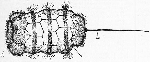
After Drew, in Lankester’s Treatise on Zoology. (A. & C. Black.)
Fig. 27.—Surface view of a forty-five hour embryo of Yoldia limatula. a.c, Apical cilia. bl, Blastopore. x, Depression where the cells that form the cerebral ganglia come to the surface.
An extraordinary modification of the veliger occurs in the development of Nucula and Yoldia and probably other members of the same families. After the formation of the gastrula by epibole the larva becomes enclosed by an ectodermic test covering the whole of the original surface of the body, including the shell-gland, and leaving only a small opening at the posterior end in which the stomodaeum and proctodaeum are formed. In Yoldia and Nucula proxima the test consists of five rows of flattened cells, the three median rows bearing circlets of long cilia. At the anterior end of the test is the apical plate from the centre of which projects a long flagellum as in many other Lamellibranch larvae. In Nucula delphinodonta the test is uniformly covered with short cilia, and there is no flagellum. When the larval development is completed the test is cast off, its cells breaking apart and falling to pieces leaving the young animal with a well-developed shell exposed and the internal organs in an advanced state. The test is really a ciliated velum developed in the normal position at the apical pole but reflected backwards in such a way as to cover the original ectoderm except at the posterior end. In Yoldia and Nucula proxima the ova are set free in the water and the test-larvae are free-swimming, but in Nucula delphinodonta the female forms a thin-walled egg-case of mucus attached to the posterior end of the shell and in communication with the pallial chamber; in this case the eggs develop and the test-larva is enclosed. A similar modification of the velum occurs in Dentalium and in Myzomenia among the Amphineura.
Classification of Lamellibranchia
The classification originally based on the structure of the gills by P. Pelseneer included five orders, viz.: the Protobranchia in which the gill-filaments are flattened and not reflected; the Filibranchia in which the filaments are long and reflected, with non-vascular junctions; the Pseudolamellibranchia in which the gill-lamellae are vertically folded, the inter-filamentar and interlamellar junctions being vascular or non-vascular; the Eulamellibranchia in which the inter-filamentar and interlamellar junctions are vascular; and lastly the Septibranchia in which the gills are reduced to a horizontal partition. The Pseudolamellibranchia included the oyster, scallop and their allies which formerly constituted the order Monomyaria, having only a single large adductor muscle or in addition a very small anterior adductor. The researches of W. G. Ridewood have shown that in gill-structure the Pectinacea agree with the Filibranchia and the Ostraeacea with the Eulamellibranchia, and accordingly the order Pseudolamellibranchia is now suppressed and its members divided between the two other orders mentioned. The four orders now retained exhibit successive stages in the modification of the ctenidia by reflection and concrescence of the filament, but other organs, such as the heart, adductors, renal organs, may not show corresponding stages. On the contrary considerable differences in these organs may occur within any single order. The Protobranchia, however, possess several primitive characters besides that of the branchiae. In them the foot has a flat ventral surface used for creeping, as in Gastropods, the byssus gland is but slightly developed, the pleural ganglia are distinct, there is a relic of the pharyngeal cavity, in some forms with a pair of glandular sacs, the gonads retain their primitive connexion with the renal cavities, and the otocysts are open.
Order I. Protobranchia
In addition to the characters given above, it may be noted that the mantle is provided with a hypobranchial gland on the outer side of each gill, the auricles are muscular, the kidneys are glandular through their whole length, the sexes are separate.
Fam. 1. Solenomyidae.—One row of branchial filaments is directed dorsally, the other ventrally; the mantle has a long postero-ventral suture and a single posterior aperture; the labial palps of each side are fused together; shell elongate; hinge without teeth; periostracum thick. Solenomya.
Fam. 2. Nuculidae.—Labial palps free, very broad, and provided with a posterior appendage; branchial filaments transverse; shell has an angular dorsal border; mantle open along its whole border. Nucula. Acila. Pronucula.
Fam. 3. Ledidae.—Like the Nuculidae, but mantle has two posterior sutures and two united siphons. Leda. Yoldia. Malletia.
Fam. 4. Ctenodontidae.—Extinct; Silurian.
The fossil group Palaeoconcha is connected with the Protobranchia through the Solenomyidae. It contains the following extinct families.
Fam. 1. Praecardiidae.—Shell equivalve with hinge dentition as in Arca. Praecardium; Silurian and Devonian.
Fam. 2. Antipleuridae.—Shell inequivalve. Antipleura; Silurian.
Fam. 3. Cardiolidae.—Shell equivalve and ventricose; hinge without teeth. Cardiola; Silurian and Devonian.
Fam. 4. Grammysiidae.—Shell thin, equivalve, oval or elongate; hinge without teeth. Grammysia; Silurian and Devonian. Protomya; Devonian. Cardiomorpha; Silurian to Carboniferous.
Fam. 5. Vlastidae.—Shell very inequivalve; hinge without teeth. Vlasta; Silurian.
Fam. 6. Solenopsidae.—Shell equivalve, greatly elongated, umbones very far forward. Solenopsis; Devonian to Trias.
Order II. Filibranchia
Gill-filament ventrally directed and reflected, connected by ciliated junctions. Foot generally provided with a highly developed byssogenous apparatus.
Sub-order I.—Anomiacea.
Very asymmetrical, with a single large posterior adductor. The heart is not contained in the pericardium, lies dorsad of the rectum and gives off a single aorta anteriorly. The reflected borders of the inner gill-plates of either side are fused together in the middle line. The gonads open into the kidneys and the right gonad extends into the mantle. Shell thin; animal fixed.
Fam. 1. Anomiidae.—Foot small; inferior (right) valve of adult perforated to allow passage of the byssus. Anomia; byssus large and calcified; British. Placuna; byssus atrophied in adult. Hypotrema. Carolia. Ephippium. Placunanomia.
Sub-order II.—Arcacea.
Symmetrical; mantle open throughout its extent; generally with well developed anterior and posterior adductors. The heart lies in the pericardium and gives off two aortae. Gills without interlamellar junctions. Renal and genital apertures separate.
Fam. 1. Arcidae.—Borders of the mantle bear compound pallial eyes. The labial palps are direct continuations of the lips. Hinge pliodont, that is to say, it has numerous teeth on either side of the umbones and the teeth are perpendicular to the edge. Arca; foot byssiferous; British. Pectunculus; foot without byssus; British. Scaphula; freshwater; India. Argina. Bathyarca. Barbatia. Senilia. Anadara. Adacnarca.
Fam. 2. Parallelodontidae.—Shell as in Arca, but the posterior hinge teeth elongated and parallel to the cardinal border. Cucullaea; recent and fossil from the Jurassic. All the other genera are fossil: Parallelodon; Devonian to Tertiary. Carbonaria; Carboniferous, &c.
Fam. 3. Limopsidae.—Shell orbicular, hinge curved, ligament longer transversely than antero-posteriorly; foot elongate, pointed anteriorly and posteriorly. Limopsis. Trinacria; Tertiary.
Fam. 4. Philobryidae.—Shell thin, very inequilateral, anterior part atrophied, umbones projecting. Philobrya.
Fam. 5. Cyrtodontidae.—Extinct; shell equivalve and inequilateral, short, convex. Cyrtodonta; Silurian and Devonian. Cypricardites, Silurian. Vanuxemia; Silurian.
Fam. 6. Trigoniidae.—Shell thick; foot elongated, pointed in front and behind, ventral border sharp; byssus absent. Trigonia; shell sub-triangular, umbones directed backwards. This genus was very abundant in the Secondary epoch, especially in Jurassic seas. There are six living species, all in Australian seas. Living specimens were first discovered in 1827. Schizodus; Permian. Myophoria; Trias.
Fam. 7. Lyrodesmidae.—Extinct; shell inequilateral, posterior side shorter; hinge short, teeth in form of a fan. Lyrodesma; Silurian.
Sub-order III.—Mytilacea.
Symmetrical, the anterior adductor small or absent. Heart gives off only an anterior aorta. Surface of gills smooth, gill-filaments all similar, with interlamellar junctions. Gonads generally extend into mantle and open at sides of kidneys. Foot linguiform and byssiferous.
Fam. 1. Mytilidae.—Shell inequilateral, anterior end short; hinge without teeth; ligament external. Mantle has a posterior suture. Cephalic eyes present. Mytilus; British. Modiola; British. Lithodomus. Modiolaria; British. Crenella. Stavelia. Dacrydium. Myrina. Idas. Septifer.
Fam. 2. Modiolopsidae.—Extinct; Silurian to Cretaceous; adductor muscles sub-equal. Modiolopsis.—Modiomorpha. Myoconcha.
Fam. 3. Pernidae.—Shell very inequilateral; ligament subdivided; mantle open throughout; anterior adductor absent. Perna. Crenatula; inhabits sponges. Bakewellia. Gervilleia; Trias to Eocene. Odontoperna; Trias. Inoceramus; Jurassic to Cretaceous.
Sub-order IV.—Pectinacea.
Monomyarian, with open mantle. Gills folded and the filaments at summits and bases of the folds are different from the others. Gonads contained in the visceral mass and generally open into renal cavities. Foot usually rudimentary.
Fam. 1. Vulsellidae.—Shell high; hinge toothless; foot without byssus. Vulsella.
Fam. 2. Aviculidae.—Shell very inequilateral; cardinal border straight with two auriculae, the posterior the longer. Foot with a very stout byssus. Gills fused to the mantle. Avicula; British. Meleagrina. Pearls are obtained from a species of this genus in the Persian Gulf, Indian Ocean, &c. Malleus. Several extinct genera.
Fam. 3. Prasinidae.—Shell inequilateral, with anterior umbones and prominent anterior auricula; cardinal border arched. Prasina.
Fam. 4. Pterineidae.—Extinct; Palaeozoic.
Fam. 5. Lunulicardiidae.—Extinct; Silurian and Devonian.
Fam. 6. Conocardiidae.—Extinct; Silurian to Carboniferous.
Fam. 7. Ambonychiidae.—Extinct; Silurian and Devonian. The last two families are dimyarian, with small anterior adductor.
Fam. 8. Myalinidae.—Extinct; Silurian to Cretaceous; adductors sub-equal.
Fam. 9. Amussiidae.—Shell orbicular, smooth externally with radiating costae internally. Gills without interlamellar junctions. Amussium.
Fam. 10. Spondylidae.—Shell very inequivalve, fixed by the right valve which is the larger. No byssus. Spondylus; shell with spiny ribs, adherent by the spines. Plicatula.
Fam. 11. Pectinidae.—Shell with radiating ribs; dorsal border with two auriculae. Foot byssiferous. Mantle borders with well developed eyes. Pecten; shell orbicular, with equal auriculae; without a byssal sinus; British. Chlamys; anterior auricula the larger and with a byssal sinus; British. Pedum. Hinnites. Pseudamussium. Camptonectes. Hyalopecten; abyssal.
Sub-order V.—Dimyacea.
Dimyarian, with orbicular and almost equilateral shell; adherent; hinge without teeth and ligament internal. Gills with free non-reflected filaments.
Fam. Dimyidae.—Characters of the sub-order. Dimya; recent in abyssal depths and fossil since the Jurassic.
Order III. Eulamellibranchia
Edges of the mantle generally united by one or two sutures. Two adductors usually present. Branchial filaments united by vascular inter-filamentar junctions and vascular interlamellar junctions; the latter contain the afferent vessels. The gonads always have their own proper external apertures.
Sub-order I.—Ostraeacea.
Monomyarian or with a very small anterior adductor. Mantle open; foot rather small; branchiae folded; shell inequivalve.
Fam. 1. Limidae.—Shell with auriculae. Foot digitiform, with byssus. Borders of mantle with long and numerous tentacles. Gills not united with mantle. Lima; members of this genus form a nest by means of the byssus, or swim by clapping the valves of the shell together. Limaea.
Fam. 2. Ostraeidae.—Foot much reduced and without byssus. Heart usually on the ventral side of the rectum. Gills fused to the mantle. Shell irregular, fixed in the young by the left and larger valve. Ostraea; foot absent in the adult; edible and cultivated; some species, as the British O. edulis, are hermaphrodite.
Fam. 3. Eligmidae.—Extinct; Jurassic.
Fam. 4. Pinnidae.—Shell elongated, truncated and gaping posteriorly. Dimyarian, with a very small anterior adductor. Foot with byssus. Pinna; British. Cyrtopinna. Aviculopinna; fossil, Carboniferous and Permian. Pinnigena; Jurassic and Cretaceous. Atrina; fossil and recent, from Carboniferous to present day.
Sub-order II.—Submytilacea.
Mantle only slightly closed; usually there is only a single suture. Siphons absent or very short. Gills smooth. Nearly always dimyarian. Shell equivalve, with an external ligament.
Fam. 1. Dreissensiidae.—Shell elongated; hinge without teeth; summits of valves with an internal septum. Siphons short. Dreissensia; lives in fresh water, but originated from the Caspian Sea; introduced into England about 1824.
Fam. 2. Modiolarcidae.—Foot with a plantar surface; the two branchial plates serve as incubatory pouches. Modiolarca.
Fam. 3. Astartidae.—Shell concentrically striated; foot elongate, without byssus. Astarte; British. Woodia. Opis; Secondary. Prosocoelus; Devonian.
Fam. 4. Crassatellidae.—Shell thick, with concentric striae, ligament external; foot short. Crassatella. Cuna.
Fam. 5. Carditidae.—Shell thick, with radiating costae; foot carinated, often byssiferous. Cardita. Thecalia. Milneria. Venericardia.
Fam. 6. Condylocardiidae.—Like Carditidae, but with an external ligament. Condylocardia. Carditella. Carditopsis.
Fam. 7. Cyprinidae.—Mantle open in front, with two pallial sutures; external gill-plates smaller than the internal. Cyprina; British. Cypricardia. Pleurophorus; Devonian to Trias. Anisocardia; Jurassic to Tertiary. Veniella; Cretaceous to Tertiary.
Fam. 8. Isocardiidae.—Mantle largely closed, pedal orifice small; gill-plates of equal size; shell globular, with prominent and coiled umbones. Isocardia; British.
Fam. 9. Callocardiidae.—Siphons present; external gill-plate smaller than the internal; umbones not prominent. Callocardia; abyssal.
Fam. 10. Lucinidae.—Labial palps very small; gills without an external plate. Lucina; British. Montacuta; British. Cryptodon.
Fam. 11. Corbidae.—Shell thick, with denticulated borders; anal aperture with valve but no siphon; foot elongated and pointed. Corbis. Gonodon; Trias and Jurassic. Mutiella; Upper Cretaceous.
Fam. 12. Ungulinidae.—Foot greatly elongated, vermiform, ending in a glandular enlargement. Ungulina. Diplodonta; British. Axinus; British.
Fam. 13. Cyrenellidae.—Two elongated, united, non-retractile siphons; freshwater. Cyrenella. Joanisiella.
Fam. 14. Tancrediidae.—Shell elongate, sub-triangular. Extinct. Tancredia; Trias to Cretaceous. Meekia; Cretaceous.
Fam. 15. Unicardiidae.—Shell sub-orbicular, nearly equilateral, with concentric striae. Extinct, Carboniferous to Cretaceous. Unicardium. Scaldia. Pseudedmondia.
Fam. 16. Leptonidae.—Shell thin; no siphons; foot long and byssiferous; marine; hermaphrodite and incubatory. Kellya; British. Lepton; commensal with the Crustacean Gebia; British. Erycina; Tertiary. Pythina. Scacchia. Sportella. Cyamium.
Fam. 17. Galeommidae.—Mantle reflected over shell; shell thin, gaping; adductors much reduced. Galeomma; British. Scintilla. Hindsiella. Ephippodonta; commensal with shrimp Axius. The three following genera with an internal shell probably belong to this family:—Chlamydoconcha. Scioberetia; commensal with a Spatangid. Entovalva; parasitic in Synapta.
Fam. 18. Kellyellidae.—Shell ovoid; anal aperture with very short siphon; foot elongated. Kellyella. Turtonia; British. Allopagus; Eocene. Lutetia; Eocene.
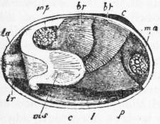
Fig. 28.—Lateral view of a Mactra, the right valve of the shell and right mantle-flap removed, and the siphons retracted. (From Gegenbaur.)
br, br′, Outer and inner gill-plates. t, Labial tentacle. ta, tr, Upper and lower siphons. ms, Siphonal muscle of the mantle-flap. ma, Anterior adductor muscle. mp, Posterior adductor muscle. p, Foot. c, Umbo.
Fam. 19. Cyrenidae.—Two siphons, more or less united, with papillose orifices; pallial line with a sinus; freshwater. Cyrena. Corbicula. Batissa. Velorita. Galatea. Fischeria.
Fam. 20. Cycladidae.—One siphon or two free siphons with simple orifices; pallial line simple; hermaphrodite, embryos incubated in external gill-plate; freshwater, Cyclas; British. Pisidium; British.
Fam. 21. Rangiidae.—Two short siphons, shell with prominent umbones and internal ligament. Rangia; brackish water, Florida.
Fam. 22. Cardiniidae.—Shell elongated, inequilateral. Extinct. Cardinia; Trias and Jurassic. Anthracosia; Carboniferous and Permian. Anoplophora; Trias. Pachycardia; Trias.
Fam. 23. Megalodontidae.—Shell inequilateral, thick; posterior adductor impression on a myophorous apophysis. Extinct. Megalodon; Devonian to Jurassic. Pachyrisma; Trias and Jurassic. Durga; Jurassic. Dicerocardium; Jurassic.
Fam. 24. Unionidae.—Shell equilateral; mantle with a single pallial suture and no siphons; freshwater; larva a glochidium. Unio; British. Anodonta; British. Pseudodon. Quadrula. Arconaia. Monocondylea. Solenaia. Mycetopus.
Fam. 25. Mutelidae.—Differs from Unionidae in having two pallial sutures; freshwater. Muleta. Pliodon. Spatha. Iridina. Hyria. Castalia. Aplodon. Plagiodon.
Fam. 26. Aetheriidae.—Shell irregular, generally fixed in the adult; foot absent; freshwater. Aetheria. Mulleria. Bartlettia.
Sub-order III.—Tellinacea.
Mantle not extensively closed; two pallial sutures and two well-developed siphons. Gills smooth. Foot compressed and elongated. Labial palps very large. Dimyarian; pallial line with a deep sinus.
Fam. 1. Tellinidae.—External gill-plate directed upwards; siphons separate and elongated; foot with byssus; palps very large; ligament external. Tellina; British. Gastrana; British. Capsa. Macoma.
Fam. 2. Scrobiculariidae.—External gill-plates directed upwards; siphons separate and excessively long; foot without byssus. Scrobicularia; estuarine; British. Syndosmya; British. Cumingia.
Fam. 3. Donacidae.—External gill-plate directed ventrally; siphons separate, of moderate length, anal siphon the longer. Donax; British. Iphigeneia.
Fam. 4. Mesodesmatidae.—External gill-plate directed ventrally; siphons separate and equal. Mesodesma. Ervilia; British.
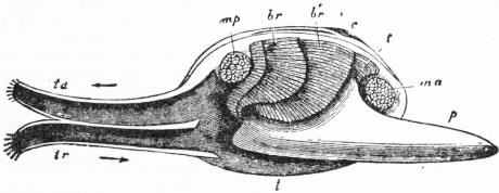
Fig. 29.—The same animal as fig. 28, with its foot and siphons expanded. Letters as in fig. 28. (From Gegenbaur.)
Fam. 5. Cardiliidae.—Shell very high and short; dimyarian; posterior adductor impression on a prominent apophysis. Cardilia.
Fam. 6. Mactridae.—External gill-plate directed ventrally; siphons united, invested by a chitinous sheath; foot long, bent at an angle, without byssus. Mactra; British (figs. 28, 29). Mulinia. Harvella. Raeta. Eastonia. Heterocardia. Vanganella.
Sub-order IV.—Veneracea.
Two pallial sutures, siphons somewhat elongated and partially or wholly united. Gills slightly folded. A bulb on the posterior aorta. Ligament external.
Fam. 1. Veneridae.—Foot well developed; pallial sinus shallow or absent. Venus; British. Dosinia; British. Tapes; British. Cyclina. Lucinopsis; British. Meretrix. Circe; British. Venerupis.
Fam. 2. Petricolidae.—Boring forms with a reduced foot; shell elongated, with deep pallial sinus. Petricola. P. pholadiformis, originally an inhabitant of the coast of the United States, has been acclimatized for some years in the North Sea.
Fam. 3. Glaucomyidae.—Siphons very long and united; foot small; shell thin, with deep pallial sinus; fresh or brackish water. Glaucomya. Tanysiphon.
Sub-order V.—Cardiacea.
Two pallial sutures. Siphons generally short. Foot cylindrical, more or less elongated, byssogenous. Gills much folded. Shell equivalve, with radiating costae and external ligament.
Fam. 1. Cardiidae.—Mantle slightly closed; siphons very short, surrounded by papillae which often bear eyes; foot very long, geniculated; pallial line without sinus; two adductors, Cardium; British. Pseudo-kellya. Byssocardium; Eocene. Lithocardium; Eocene.
Fam. 2. Limnocardiidae.—Siphons very long, united throughout; shell gaping; two adductors; brackish waters. Limnocardium; Caspian Sea and fossil from the Tertiary. Archicardium; Tertiary.
Fam. 3. Tridacnidae.—Mantle closed to a considerable extent; apertures distant from each other; no siphons; a single adductor; shell thick. Tridacna. Hippopus.
Sub-order VI.—Chamacea.
Asymmetrical, inequivalve, fixed, with extensive pallial sutures; no siphons. Two adductors. Foot reduced and without byssus. Shell thick, without pallial sinus.
Fam. 1. Chamidae.—Shell with sub-equal valves and prominent umbones more or less spirally coiled; ligament external. Chama. Diceras; Jurassic. Requienia; Cretaceous. Matheronia; Cretaceous.
Fam. 2. Caprinidae.—Shell inequivalve; fixed valve spiral or conical; free valve coiled or spiral; Cretaceous. Caprina. Caprotina. Caprinula, &c.
Fam. 3. Monopleuridae.—Shell very inequivalve; fixed valve conical or spiral; free valve operculiform; Cretaceous. Monopleuron. Baylea. The two following families, together known as Rudistae, are closely allied to the preceding; they are extinct marine forms from Secondary deposits. They were fixed by the conical elongated right valve; the free left valve is not spiral, and is furnished with prominent apophyses to which the adductors were attached.
Fam. 4. Radiolitidae.—Shell conical or biconvex, without canals in the external layer. Radiolites. Biradiolites.
Fam. 5. Hippuritidae.—Fixed valve long, cylindro-conical, with three longitudinal furrows which correspond internally to two pillars for support of the siphons. Hippurites. Arnaudia.
Sub-order VII.—Myacea.
Mantle closed to a considerable extent; siphons well developed; gills much folded and frequently prolonged into the branchial siphon. Foot compressed and generally byssiferous. Shell gaping, with a pallial sinus.
Fam. 1. Psammobiidae.—Siphons very long and quite separate; foot large; shell oval, elongated, ligament external. Psammobia; British. Sanguinolaria. Asaphis. Elizia. Solenotellina.
Fam. 2. Myidae.—Siphons united for the greater part of their length, and with a circlet of tentacles near their extremities; foot reduced; shell gaping; ligament internal. Mya; British. Sphenia; British. Tugonia. Platyodon. Cryptomya.
Fam. 3. Corbulidae.—Shell sub-trigonal, inequivalve; pallial sinus shallow; siphons short, united, completely retractile; foot large, pointed, often byssiferous. Corbulomya. Paramya. Erodona and Himella are fluviatile forms from South America.
Fam. 4. Lutrariidae.—Mantle extensively closed; a fourth pallial aperture behind the foot; siphons long and united; shell elongated, a spoon-shaped projection for the ligament on each valve. Lutraria; British. Tresus. Standella.
Fam. 5. Solenidae.—Elongated burrowing forms; foot cylindrical, powerful, without byssus; shell long, truncated and gaping at each end. Solenocurtus; British. Tagelus; estuarine. Ceratisolen; British. Cultellus; British. Siliqua. Solen; British. Ensis; British.
Fam. 6. Saxicavidae.—Mantle extensively closed, with a small pedal orifice; siphons long, united, covered by a chitinous sheath; gills prolonged into the branchial siphon; foot small; shell gaping. Saxicava; British. Glycimeris. Cyrtodaria.
Fam. 7. Gastrochaenidae.—Shell thin, gaping widely at the posterior end; anterior adductor much reduced; mantle extensively closed; siphons long, united. Gastrochaena; British. Fistulana.
Sub-order VIII.—Adesmacea.
Ligament wanting; shell gaping, with a styloid apophysis in the umbonal cavities. Gills prolonged into the branchial siphon. Mantle largely closed, siphons long, united. Foot short, truncated, discoid, without byssus.
Fam. 1. Pholadidae.—Shell containing all the organs; heart traversed by the rectum; two aortae. Shell with a pallial sinus; dorsal region protected by accessory plates. Pholas; British. Pholadidea; British. Jouannetia. Xylophaga; British. Martesia.
Fam. 2. Teredinidae.—Shell globular, covering only a small portion of the vermiform body; heart on ventral side of rectum; a single aorta; siphons long, united and furnished with two posterior calcareous “pallets.” Teredo; British. Xylotrya.
Sub-order IX.—Anatinacea.
Hermaphrodite, the ovaries and testes distinct, with separate apertures. Foot rather small. Mantle frequently presents a fourth orifice. External gill-plate directed dorsally and without reflected lamella. Hinge without teeth.
Fam. 1. Thracidae.—Mantle with a fourth aperture; siphons long, quite separate, completely retractile and invertible. Thracia; British. Asthenothaerus.
Fam. 2. Periplomidae.—Siphons separate, naked, completely retractile but not invertible. Periploma. Cochlodesma. Tyleria.
Fam. 3. Anatinidae.—Siphons long, united, covered by a chitinous sheath, not completely retractile. Anatina. Plectomya; Jurassic and Cretaceous.
Fam. 4. Pholadomyidae.—Mantle with fourth aperture; siphons very long, completely united, naked, incompletely retractile; foot small, with posterior appendage. Pholadomya.
Fam. 5. Arcomyidae.—Extinct; Secondary and Tertiary. Arcomya. Goniomya.
Fam. 6. Pholadellidae.—Extinct; Palaeozoic. Pholadella. Phytimya. Allorisma.
Fam. 7. Pleuromyidae.—Extinct; Secondary. Pleuromya. Gresslya. Ceromya.
Fam. 8. Pandoridae.—Shell thin, inequivalve, free; ligament internal; siphons very short. Pandora; British. Coelodon. Clidiophora.
Fam. 9. Myochamidae.—Shell very inequivalve, solid, with a pallial sinus; siphons short; foot small. Myochama. Myodora.
Fam. 10. Chamostraeidae.—A fourth pallial aperture present; pedal aperture small; siphons very short and separate; shell fixed by the right valve, irregular. Chamostraea.
Fam. 11. Clavagellidae.—Pedal aperture very small, foot rudimentary; valves continued backwards into a calcareous tube secreted by the siphons. Clavagella. Brechites (Aspergillum).
Fam. 12. Lyonsiidae.—Foot byssiferous; siphons short, invertible. Lyonsia; British. Entodesma. Mytilimeria.
Fam. 13. Verticordiidae.—Siphons short, gills papillose; foot small; shell globular. Many species abyssal. Verlicordia. Euciroa. Lyonsiella. Halicardia.
Order IV. Septibranchia
Gills have lost their respiratory function, and are transformed into a muscular septum on each side between mantle and foot. All marine, live at considerable depths, and are carnivorous.
Fam. 1. Poromyidae.—Siphons short and separate; branchial siphon with a large valve; branchial septum bears two groups of orifices on either side; hermaphrodite. Poromya; British. Dermatomya. Liopistha; Cretaceous.
Fam. 2. Cetoconchidae.—Branchial septum with three groups of orifices on each side; siphons short, separate, branchial siphon with a valve. Cetoconcha (Silenia).
Fam. 3. Cuspidariidae.—Branchial septum with four or five pairs of very narrow symmetrical orifices; siphons long, united, their extremities surrounded by tentacles; sexes separate. Cuspidaria; British.
Authorities.—T. Barrois, “Le Stylet crystallin des Lamellibranches,” Revue biol. Nord France, i. (1890); Jameson, “On the Origin of Pearls,” Proc. Zool. Soc. (London, 1902); R. H. Peck, “The Minute Structure of the Gills of Lamellibranch Mollusca,” Quart. Journ. Micr. Sci. xvii. (1877); W. G. Ridewood, “On the Structure of the Gills of the Lamellibranchia,” Phil. Trans. B. cxcv. (1903); K. Mitsukuri, “On the Structure and Significance of some aberrant forms of Lamellibranchiate Gills,” Quart. Journ. Micr. Sci. xxi. (1881); A. H. Cooke, “Molluscs,” Cambridge Natural History, vol. iii.; Paul Pelseneer, “Mollusca,” Treatise on Zoology, edited by E. Ray Lankester, pt. v.
(E. R. L.; J. T. C.)
