VOLUME XIV SLICE III Ichthyology to Independence (continued 2)
Reading edition
The kidney tubules subserve the excretory function in two different ways. The wall of the tubule, bathed in blood from the posterior cardinal vein, serves to extract nitrogenous products of excretion from the blood and pass them into the lumen of the tubule. The open ciliated funnel or nephrostome at the coelomic end of the tubule serves for the passage outwards of coelomic fluid to flush the cavity of the tubule. The secretory activity of the coelomic lining is specially concentrated in certain limited areas in the neighbourhood of the nephrostomes, each such area ensheathing a rounded mass depending into the coelom and formed of a blood-vessel coiled into a kind of skein—a glomerulus. In the case of the pronephros the glomeruli are as a rule fused together into a single glomus. In the mesonephros they remain separate and in this case the portion of coelom surrounding the glomerulus tends to be nipped off from the general coelom—to form a Malpighian body. The separation may be incomplete—the Malpighian coelom remaining in connexion with the general coelom by a narrow peritoneal canal. The splanchnocoelic end of this is usually ciliated and is termed a peritoneal funnel: it is frequently confused with the nephrostome.
Mesonephros.—The kidney of the adult fish is usually a compact gland extending over a considerable distance in an anteroposterior direction and lying immediately dorsal to the coelomic cavity.
Peritoneal funnels are present in the adult of certain Selachians (e.g. Acanthias, Squatina), though apparently in at least some of these forms they no longer communicate with the Malpighian bodies or tubules. The kidneys of the two sides become fused together posteriorly in Protopterus and in some Teleosts. The mesonephric ducts undergo fusion posteriorly in many cases to form a median urinary or urinogenital sinus. In the Selachians this median sinus is prolonged forwards into a pair of horn-like continuations—the sperm sacs. In Dipnoans the sinus becomes greatly dilated and forms a large, rounded, dorsally placed cloacal caecum. In Actinopterygians a urinary bladder of similar morphological import is commonly present.
Gonads.—The portion of coelomic lining which gives rise to the reproductive cells retains its primitive relations most nearly in the female, where, as a rule, the genital cells are still shed into the splanchnocoele. Only in Teleostomes (Lepidosteus and most Teleosts) the modification occurs that the ovary is shut off from the splanchnocoele as a closed cavity continuous with its duct.
In a few Teleosts (Salmonidae, Muraenidae, Cobitis) the ovary is not a closed sac, its eggs being shed into the coelom as in other groups.
The appearance of the ovary naturally varies greatly with the character of the eggs.
The portion of coelomic lining which gives rise to the male genital cells (testis) is in nearly, if not quite, all cases, shut off from the splanchnocoele. The testes are commonly elongated in form. In Dipneusti20 (Lepidosiren and Protopterus) the hinder portion of the elongated testis has lost its sperm-producing function, though the spermatozoa produced in the anterior portion have to traverse it in order to reach the kidney. In Polypterus21 the testis is continued backwards as a “testis ridge,” which appears to correspond with the posterior vesicular region of the testis in Lepidosiren and Protopterus. Here also the spermatozoa pass back through the cavities of the testis ridge to reach the kidney duct. In the young Teleost22 the rudiment of the duct forms a backward continuation of the testis containing a network of cavities and opening as a rule posteriorly into the kidney duct. It is difficult to avoid the conclusion that the testis duct of the Teleost is for the most part the equivalent morphologically of the posterior vesicular region of the testis of Polypterus and the Dipneusti.
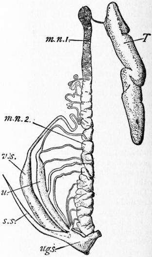
From Arch. zool, expérimentale, by permission of Schleicher Frères.
Fig. 11.—Urino-Genital Organs of the right side in a male Scyllium. (After Borcea.)
m.n. 1, Anterior (genital) portion of mesonephros with its coiled duct. m.n. 2, Posterior (renal) portion of mesonephros. s.s, Sperm sac. T, Testis. u, “Ureter” formed by fusion of collecting tubes of renal portion of mesonephros. u.g.s, Urino-genital sinus; v.s, Vesicula seminalis.
Relations of Renal and Reproductive Organs. (1) Female.—In the Selachians and Dipnoans the oviduct is of the type (Müllerian duct) present in the higher vertebrates and apparently representing a split-off portion of the archinephric duct. At its anterior end is a wide funnel-like coelomic opening. Its walls are glandular and secrete accessory coverings for the eggs. In the great majority of Teleosts and in Lepidosteus the oviduct possesses no coelomic funnel, its walls being in structural continuity with the wall of the ovary. In most of the more primitive Teleostomes (Crossopterygians, sturgeons, Amia) the oviduct has at its front end an open coelomic funnel, and it is difficult to find adequate reason for refusing to regard such oviducts as true Müllerian ducts. On this interpretation the condition characteristic of Teleosts would be due to the lips of the oviduct becoming fused with the ovarian wall, and the duct itself would be a Müllerian duct as elsewhere.
A departure from the normal arrangement is found in those Teleosts which shed their eggs into the splanchnocoele, e.g. amongst Salmonidae, the smelt (Osmerus) and capelin (Mallotus) possess a pair of oviducts resembling Müllerian ducts while the salmon possesses merely a pair of genital pores opening together behind the anus. It seems most probable that the latter condition has been derived from the former by reduction of the Müllerian ducts, though it has been argued that the converse process has taken place. The genital pores mentioned must not be confused with the abdominal pores, which in many adult fishes, particularly in those without open peritoneal funnels, lead from coelom directly to the exterior in the region of the cloacal opening. These appear to be recent developments, and to have nothing to do morphologically with the genitourinary system.23
(2) Male.—It seems that primitively the male reproductive elements like the female were shed into the coelom and passed thence through the nephridial tubules. In correlation probably with the greatly reduced size of these elements they are commonly no longer shed into the splanchnocoele, but are conveyed from the testis through covered-in canals to the Malpighian bodies or kidney tubules. The system of covered-in canals forms the testicular network, the individual canals being termed vasa efferentia. In all probability the series of vasa efferentia was originally spread over the whole length of the elongated testis (cf. Lepidosteus), but in existing fishes the series is as a rule restricted to a comparatively short anteroposterior extent. In Selachians the vasa efferentia are restricted to the anterior end of testis and kidney, and are connected by a longitudinal canal ending blindly in front and behind. The number of vasa efferentia varies and in the rays (Raia, Torpedo) may be reduced to a single one opening directly into the front end of the mesonephric duct. The anterior portion of the mesonephros is much reduced in size in correlation with the fact that it has lost its renal function. The hinder part, which is the functional kidney, is considerably enlarged. The primary tubules of this region of the kidney have undergone a modification of high morphological interest. Their distal portions have become much elongated, they are more or less fused, and their openings into the mesonephric duct have undergone backward migration until they open together either into the mesonephric duct at its posterior end or into the urinogenital sinus independently of the mesonephric duct. The mesonephric duct is now connected only with the anterior part of the kidney, and serves merely as a vas deferens or sperm duct. In correlation with this it is somewhat enlarged, especially in its posterior portion, to form a vesicula seminalis.
The morphological interest of these features lies in the fact that they represent a stage in evolution which carried a little farther would lead to a complete separation of the definitive kidney (metanephros) from the purely genital anterior section of the mesonephros (epididymis), as occurs so characteristically in the Amniota.
Dipneusti.—In Lepidosiren24 a small number (about half a dozen) of vasa efferentia occur towards the hind end of the vesicular part of the testis and open into Malpighian bodies. In Protopterus the vasa efferentia are reduced to a single one on each side at the extreme hind end of the testis.
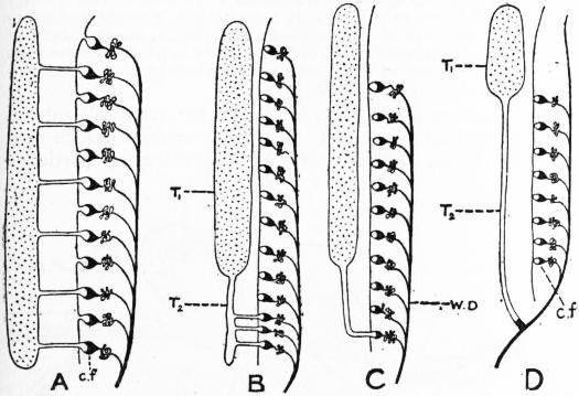
Graham Kerr, Proc. Zool. Soc. London.
Fig. 12.—Diagram illustrating Connexion between Kidney and Testis in Various Groups of Fishes.
A, Distributed condition of vasa efferentia (Acipenser, Lepidosteus). B, Vasa efferentia reduced to a few at the hind end (Lepidosiren). C, Reduction of vasa efferentia to a single one posteriorly (Protopterus). D, Direct communication between testis and kidney duct (Polypterus, Teleosts). c.f, Nephrostome leading from Malpighian coelom into kidney tubule. T1, Functional region of testis. T2, Vesicular region of testis. WD, Mesonephric duct.
Teleostomi.—In the actinopterygian Ganoids a well-developed testicular network is present; e.g. in Lepidosteus25 numerous vasa efferentia arise from the testis along nearly its whole length and pass to a longitudinal canal lying on the surface of the kidney, from which in turn transverse canals lead to the Malpighian bodies. (In the case of Amia they open into the tubules or even directly into the mesonephric duct.) In the Teleosts and in Polypterus there is no obvious connexion between testis and kidney, the wall of the testis being continuous with that of its duct, much as is the case with the ovary and its duct in the female. In all probability this peculiar condition is to be explained26 by the reduction of the testicular network to a single vas efferens (much as in Protopterus or as in Raia and various anurous Amphibians at the front end of the series) which has come to open directly into the mesonephric duct (cf. fig. 12).
Organs of the Mesenchyme.—In vertebrates as in all other Metazoa, except the very lowest, there are numerous cell elements which no longer form part of the regularly arranged epithelial layers, but which take part in the formation of the packing tissue of the body. Much of this forms the various kinds of connective tissue which fill up many of the spaces between the various epithelial layers; other and very important parts of the general mesenchyme become specialized in two definite directions and give rise to two special systems of organs. One of these is characterized by the fact that the intercellular substance or matrix assumes a more or less rigid character—it may be infiltrated with salts of lime—giving rise to the supporting tissues of the skeletal system. The other is characterized by the intercellular matrix becoming fluid, and by the cell elements losing their connexion with one another and forming the characteristic fluid tissue, the blood, which with its well-marked containing walls forms the blood vascular system.
Skeletal System.—The skeletal system may be considered under three headings—(1) the chordal skeleton, (2) the cartilaginous skeleton and (3) the osseous skeleton.
1. Chordal Skeleton.—The most ancient element of the skeleton appears to be the notochord—a cylindrical rod composed of highly vacuolated cells lying ventral to the central nervous system and dorsal to the gut. Except in Amphioxus—where the condition may probably be secondary, due to degenerative shortening of the central nervous system—the notochord extends from a point just behind the infundibulum of the brain (see below) to nearly the tip of the tail. In ontogeny the notochord is a derivative of the dorsal wall of the archenteron. The outer layer of cells, which are commonly less vacuolated and form a “chordal epithelium,” soon secretes a thin cuticle which ensheaths the notochord and is known as the primary sheath. Within this there is formed later a secondary sheath, like the primary, cuticular in nature. This secondary sheath attains a considerable thickness and plays an important part in strengthening the notochord. The notochord with its sheaths is in existing fishes essentially the skeleton of early life (embryonic or larval). In the adult it may, in the more primitive forms (Cyclostomata, Dipneusti), persist as an important part of the skeleton, but as a rule it merely forms the foundation on which the cartilaginous or bony vertebral column is laid down.
2. Cartilaginous or Chondral Skeleton.—(A) Vertebral column.27 In the embryonic connective tissue or mesenchyme lying just outside the primary sheath of the notochord there are developed a dorsal and a ventral series of paired nodules of cartilage known as arcualia (fig. 13, d.a, v.a). The dorsal arcualia are commonly prolonged upwards by supradorsal cartilages which complete the neural arches and serve to protect the spinal cord. The ventral arcualia become, in the tail region only, also incorporated in complete arches—the haemal arches. In correlation with the flattening of the body of the fish from side to side the arches are commonly prolonged into elongated neural or haemal spines.
The relations of the arcualia to the segmentation of the body, as shown by myotomes and spinal nerves, is somewhat obscure. The mesenchyme in which they arise is segmental in origin (sclerotom), which suggests that they too may have been primitively segmental, but in existing fishes there are commonly two sets of arcualia to each body segment.
In gnathostomatous fishes the arcualia play a most important part in that cartilaginous tissue derived from them comes into special relationships with the notochord and gives rise to the vertebral column which functionally replaces this notochord in most of the fishes. This replacement occurs according to two different methods, giving rise to the different types of vertebral column known as chordacentrous and arcicentrous.
(a) Chordacentrous type. An incipient stage in the evolution of a chordacentrous vertebral column occurs in the Dipneusti, where cartilage cells from the arcualia become amoeboid and migrate into the substance of the secondary sheath, boring their way through the primary sheath (fig. 13, C). They wander throughout the whole extent of the secondary sheath, colonizing it as it were, and settle down as typical stationary cartilage cells. The secondary sheath is thus converted into a cylinder of cartilage. In Selachians exactly the same thing takes place, but in recent forms development goes a step further, as the cartilage cylinder becomes broken into a series of segments, known as vertebral centra. The wall of each segment becomes much thickened in the middle so that the notochord becomes constricted within each centrum and the space occupied by it is shaped like the cavity of a dice-box. When free from notochord and surrounding tissues such a cartilaginous centrum presents a deep conical cavity at each end (amphicoelous).
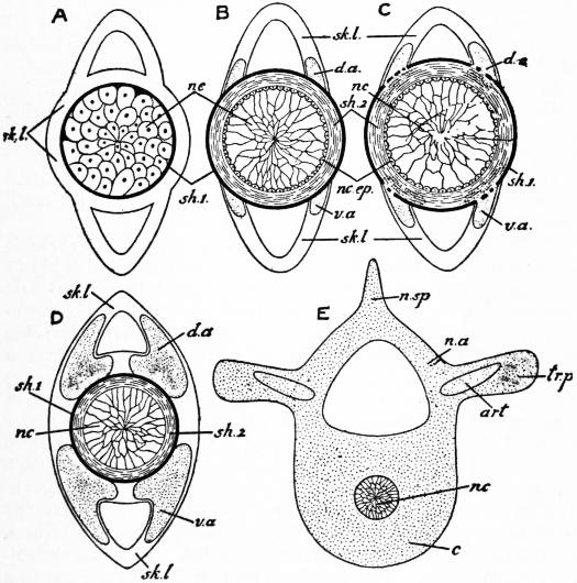
From Wiedersheim, Grundriss der vergleichenden Anatomie, by permission of Gustav Fischer.
Fig. 13.—Diagrammatic transverse sections to illustrate the morphology of the vertebral column.
A, Primitive conditions as seen in any young embryo. B, Condition as it occurs in Cyclostomata, sturgeons, embryos of bony Actinopterygians. C, Condition found in Selachians and Dipnoans. D and E, Illustrating the developmental process in bony Actinopterygians and higher vertebrates. c, Centrum. d.a, Dorsal arcualia. n.a, Neural arch. nc, Notochord. nc.ep, Chordal epithelium. n.sp, Neural spine. sh.1, Primary sheath. sh.2, Secondary sheath. sk.l, Connective tissue. tr.p, Transverse process. v.a, Ventral arcualia.
A secondary modification of the centrum consists in the calcification of certain zones of the cartilaginous matrix. The precise arrangement of these calcified zones varies in different families and affords characters which are of taxonomic importance in palaeontology where only skeletal structures are available (see Selachians).
(b) Arcicentrous type. Already in the Selachians the vertebral column is to a certain extent strengthened by the broadening of the basis of the arcualia so as partially to surround the centra. In the Teleostomes, with the exceptions of those ganoids mentioned, the expanded bases of the arcualia undergo complete fusion to form cartilaginous centra which, unlike the chordacentrous centra, lie outside the primary sheath (figs. 13, D and E). In these forms no invasion of the secondary sheath by cartilage cells takes place. The composition of the groups of arcualia which give rise to the individual centrum is different in different groups. The end result is an amphicoelous or biconcave centrum in general appearance much like that of the Selachian.
In Lepidosteus the spaces between adjacent centra become filled by a secondary development of intervertebral cartilage which then splits in such a way that the definitive vertebrae are opisthocoelous, i.e. concave behind, convex in front.
Ribs.—In the Crossopterygians a double set of “ribs” is present on each side of the vertebral column, a ventral set lying immediately outside the splanchnocoelic lining and apparently serially homologous with the haemal arches of the caudal region, and a second set passing outwards in the thickness of the body wall at a more dorsal level. In the Teleostomes and Dipnoans only the first type is present; in the Selachians only the second. It would appear that it is the latter which is homologous with the ribs of vertebrates above fishes.
Median Fin Skeleton.—the foundation of the skeleton of the median fins consists of a series of rod-like elements, the radialia, each of which frequently is segmented into three portions. In a few cases the radialia correspond segmentally with the neural and haemal arches (living Dipnoans, Pleuracanthus tail region) and this suggests that they represent morphologically prolongations of the neural and haemal spines. That this is so is rendered probable by the fact that we must regard the evolution of the system of median fins as commencing with a simple flattening of the posterior part of the body. It is only natural to suppose that the edges of the flattened region would be at first supported merely by prolongations of the already existing spinous processes. In the Cyclostomes (where they are branched) and in the Selachians, the radialia form the main supports of the fin, though already in the latter they are reinforced by a new set of fin rays apparently related morphologically to the osseous or placoid skeleton (see below).
The series of radialia tends to undergo the same process of local concentration which characterizes the fin-fold as a whole. In its extreme form this leads to complete fusion of the basal portions of a number of radialia (dorsal fins of Holoptychius and various Selachians, and anal fin of Pleuracanthus). In view of the identity in function it is not surprising that a remarkable resemblance exists between the mechanical arrangements (of skeleton, muscles, &c.), of the paired and unpaired fins. The resemblance to paired fins becomes very striking in some of the cases where the basal fusion mentioned above takes place (Pleuracanthus).
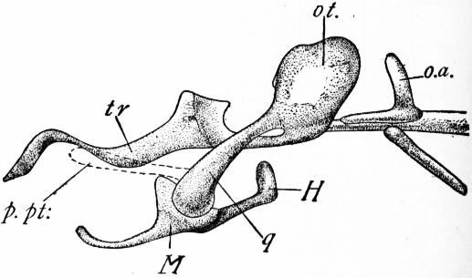
Trans. Roy. Soc. Edinburgh.
Fig. 14.—Chondrocranium of a young Lepidosiren, showing the suspension of the lower jaw by the upper portion of the mandibular arch. (After Agar.)
H, Hyoid arch. M, Mandibular arch. o.a, Occipital arch. ot, Auditory capsule. q, Quadrate = upper end of mandibular arch. tr, Trabecula.
The palatopterygoid bar (p.pt) is represented by a faint vestige which disappears before the stage figured.
(B) Chondrocranium28.—In front of the vertebral column lies the cartilaginous trough, the chondrocranium, which protects the brain. This consists of a praechordal portion—developed out of a pair of lateral cartilaginous rods—the trabeculae cranii—and a parachordal portion lying on either side of the anterior end of the notochord. This arises in development from a cartilaginous rod (parachordal cartilage) lying on each side of the notochord and possibly representing a fused row of dorsal arcualia. The originally separate parachordals and trabeculae become connected to form a trough-like, primitive cranium, complete or nearly so laterally and ventrally but open dorsally. With the primitive cranium there are also connected cartilaginous capsules developed round the olfactory and auditory organs. There also become fused with the hinder end of the cranium a varying number of originally distinct neural arches.
After W. K. Parker, Trans. Zool. Soc. London.
After Gegenbaur, Untersuchungen zur verg. Anat. der Wirbeltiere, by permission of Wilhelm Engelmann.
After Hubrecht, Brown’s Tierreich, by permission of Gustav Fischer.
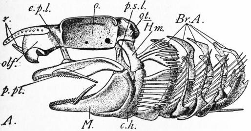
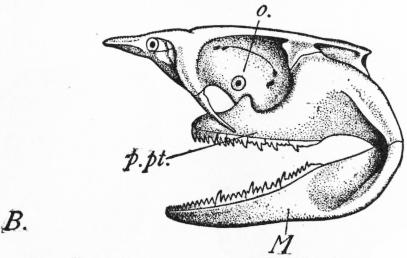
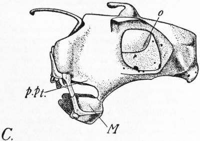
Fig. 15.—Chondrocranium, &c. of Scyllium (A), Notidanus cinereus (B) and Chimaera (C).
Br.A, Branchial arches. c.h, Ceratohyal. e.p.l, Ethmopalatine ligament. Hm, Hyomandibular. M, Meckel’s cartilage. o, Orbit. olf, Olfactory capsule. ot, Auditory capsule. p.pt, Palato-pterygoid bar. p.s.l, Prespiracular ligament. r, Rostrum.
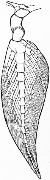
Fig. 16.—Fore-limb of Ceratodus.
(C) Visceral Arches.—The skeleton of the visceral arches consists essentially of a series of half-hoops of cartilage, each divided in the adult into a number of segments and connected with its fellow by a median ventral cartilage. The skeleton of arches I. and II. (mandibular and hyoidean) undergoes modifications of special interest (figs. 14 and 15). The lower portion of the mandibular arch becomes greatly thickened to support the lower or hinder edge of the mouth. It forms the primitive lower jaw or “Meckel’s cartilage.” Dorsal to this an outgrowth arises from the anterior face of the arch which supports the upper or anterior margin of the mouth: it is the primitive upper jaw or palato-pterygoquadrate cartilage. The portion of the arch dorsal to the palato-pterygo-quadrate outgrowth may form the suspensorial apparatus of the lower jaw, being fused with the cranium at its upper end. This relatively primitive con-arrangement (protostylic, as it may be termed) occurs in Dipneusti among fishes (cf. fig. 14). More usually this dorsal part of the mandibular arch becomes reduced, its place being occupied by a ligament (pre-spiracular) uniting the jaw apparatus to the chondrocranium, the upper jaw being also attached to the chondrocranium by the ethmopalatine ligament situated more anteriorly. The main attachment, however, of the jaws to the chondrocranium in such a case, as holds for the majority of fishes, is through the enlarged dorsal segment of the hyoid arch (hyomandibular) which articulates at its dorsal end with the chondrocranium, while its ventral end is attached to the hinge region of the jaw by stout ligamentous bands. A skull in which the jaws are suspended in this manner is termed a hyostylic skull (e.g. Scyllium in fig. 15).
In Notidanus (fig. 15, B) there is a large direct articulation of the upper jaw to the chondrocranium in addition to the indirect one through the hyomandibular: such a skull is amphistylic. In Heterodontus the upper jaw is firmly bound to the cranium throughout its length, while in Holocephali (fig. 15, C) complete fusion has taken place, so that the lower jaw appears to articulate directly with the cranium (“auto stylic” condition). In Dipneusti29 (Lepidosiren and Protopterus) the cartilaginous upper jaw never develops (except in its hinder quadrate portion) beyond the condition of a faint rudiment, owing doubtless to its being replaced functionally by precociously developed bone.
(D) Appendicular Skeleton.—The skeleton of the free part of the limb is attached to the limb girdle which lies embedded in the musculature of the body. Each limb girdle is probably to be looked upon as consisting, like the skeleton of the visceral arches, of a pair of lateral half-hoops of cartilage. While in Pleuracanthus the lateral halves are distinct (and segmented like the branchial arches), in living Selachians generally the two halves are completely fused ventrally with one another. The part of the girdle lying dorsal to the articulation of the limb is termed scapular in the case of the pectoral limb, iliac in the case of the pelvic, while the ventral portions are known respectively as coracoid and ischio-pubic.
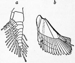
Fig. 17.—a, Skeleton of pectoral limb of Pleuracanthus. (From Gegenbauer, after Frisch.) b, Skeleton of pectoral limb of Acanthias. (After Gegenbauer.)
In most Teleostomes the primitive pelvic girdle does not develop; in the Dipneusti it is represented by a median unpaired cartilage.
The skeleton of the free limb is probably seen in its most archaic form amongst existing fishes in the biserial archipterygium of Ceratodus (fig. 16). This is indicated by the relative predominance of this type of fin amongst the geologically more ancient fishes. The biserial archipterygium consists of a segmented axial rod, bearing a praeaxial and a postaxial series of jointed rays.
In Protopterus and Lepidosiren the limbs are reduced and the lateral rays have less (Protopterus) or more (Lepidosiren) completely disappeared.
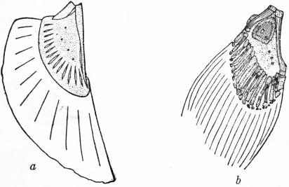
From Budgett, Trans. Zool. Soc. London, xvi, part vii. From Wiedersheim’s Verg. Anat. der Wirbeltiere, by permission of Gustav Fischer.
Fig. 18.—Skeleton of Pectoral Limb of Polypterus. a, 30 mm. larva. b, Adult.

From Wiesdersheim’s Verg. Anat. der Wirbeltiere, by permission of Gustav Fischer.
Fig. 19.—Skeleton of Pectoral Fin of Amia.
In such an archaic Selachian as Pleuracanthus the fin is clearly of the biserial archipterygial type, but the lateral rays are reduced (pectoral) or absent (pelvic) (fig. 17, a) on one side of the axis. In a typical adult Selachian the pectoral fin skeleton has little apparent resemblance to the biserial archipterygium—the numerous outwardly directed rays springing from a series of large basal cartilages (pro-, meso- and metapterygium). The condition in the young (e.g. fig. 17, b, Acanthias) hints strongly, however, at the possibility of the fin skeleton being really a modified biserial archipterygium, and that the basal cartilages represent the greatly enlarged axis which has become fixed back along the side of the body. In Crossopterygians (Polypterus) the highly peculiar fin skeleton (fig. 18) while still in the embryonic cartilaginous stage is clearly referable to a similar condition. In the Actinopterygians—with the increased development of dermal fin rays—there comes about reduction of the primitive limb skeleton. The axis becomes particularly reduced, and the fin comes to be attached directly to the pectoral girdle by a number of basal pieces (Teleosts) probably representing vestigial rays (cf. fig. 19).
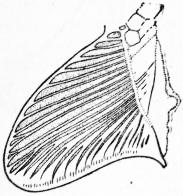
Views on the general morphology of the fin skeleton are strongly affected by the view held as to the mode of evolution of the fins. By upholders of the lateral fold hypothesis the type of fin skeleton described for Cladoselache30 is regarded as particularly primitive. It is, however, by no means clear that the obscure basal structures figured (Fig. 20) in this fin do not really represent the pressed back axis as in Pleuracanthus.
The pelvic fin skeleton, while built obviously on the same plan as the pectoral, is liable to much modification and frequently degeneration.
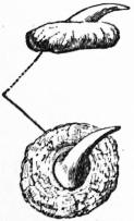
From Bashford Dean, Mem. N.Y. Acad. of Science. Fig. 21.—Placoid elements of a male Thorn-back, Raia clavata.
Fig. 20.—Skeleton of Pectoral Fin of Cladoselache.
Osseous or Bony Skeleton.—The most ancient type of bony skeleton appears to be represented in the placoid elements such as are seen in the skin of the Selachian (fig. 21). Each placoid element consists of a spine with a broadly expanded base embedded in the dermis. The base is composed of bone: the spine of the somewhat modified bone known as dentine. Ensheathing the tip of the spine is a layer of extremely hard enamel formed by the inner surface of the ectoderm which originally covered it. Such typical placoid scales are well seen on any ordinary skate. In the groups of fishes above the Selachians, the coating of placoid elements shows various modifications. The spines disappear, though they may be present for a time in early development. The bony basal plates tend to undergo fusion—in certain cases they form a continuous bony cuirass (various Siluroids, trunk-fishes) formed of large plates jointed together at their edges. More usually the plates are small and regular in size. In Crossopterygians and Lepidosteus and in many extinct forms the scales are of the ganoid type, being rhomboidal and having their outer layer composed of hard glistening ganoine. In other Teleostomes the scales are as a rule thin, rounded and overlapping—the so-called cycloid type (fig. 22, A); where the posterior edge shows toothlike projections the scale is termed ctenoid (fig. 22, B). In various Teleosts the scales are vestigial (eel); in others (as in most electric fishes) they have completely disappeared.
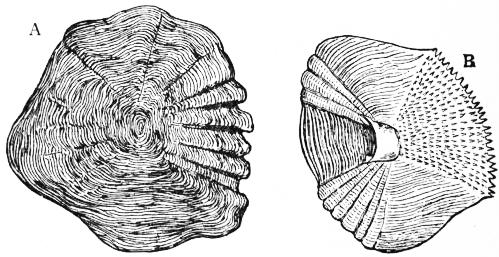
Fig. 22.—A, Cycloid Scale of Scopelus resplendens (magn.). B, Ctenoid Scale of Lethrinus (magn.).
Teeth.—Certain of the placoid elements belonging to that part of the skin which gives rise to the lining of the stomodaeum have their spines enlarged or otherwise modified to form teeth. In the majority of fishes these remain simple, conical structures: in some of the larger sharks (Carcharodon) they become flattened into trenchant blades with serrated edges: in certain rays (Myliobatis) they form a pavement of flattened plates suited for crushing molluscan shells. In the young Neoceratodus31 there are numerous small conical teeth, the bases of which become connected by a kind of spongework of bony trabeculae. As development goes on a large basal mass is formed which becomes the functional tooth plate of the adult, the original separate denticles disappearing completely. In the other two surviving Dipnoans, similar large teeth exist, though here there is no longer trace in ontogeny of their formation by the basal fusion of originally separate denticles. In the Selachians the bony skeleton is restricted to the placoid elements. In the Teleostomes and the Dipnoans the original cartilaginous skeleton becomes to a great extent unsheathed or replaced by bony tissue. It seems highly probable that the more deeply seated osseous elements occurring in these as in the higher groups arose in the course of evolution by the spreading inwards of bony trabeculae from the bases of the placoid elements. Such a method has been demonstrated as occurring in individual development in the case of certain of the more superficially placed bones.32
The placoid element with its cap of enamel secreted by the ectoderm is probably originally derived from a local thickening of the basement membrane which with the external cuticle may be looked on as the most ancient skeletal structure in the Metazoa. The basal plate appears to have been a later development than the spine; in the palaeozoic Coelolepidae33 the basal plate is apparently not yet developed.
Only a brief summary can be given here of the leading features in the osteology of fishes. Care must be taken not to assume that bony elements bearing the same name in fishes and in other groups, or even in the various sub-divisions of the fishes, are necessarily strictly homologous. In all probability bony elements occupying similar positions and described by the same anatomical name have been evolved independently from the ancestral covering of placoid elements.
From Parker & Haswell’s Text-book of Zoology, by permission of Messrs. Macmillan & Co., Ltd.
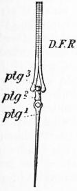
Fig. 23.—One of the radialia of the salmon, consisting of three segments, ptg¹, ptg², ptg³, and supporting a dermal fin ray. D.F.R.
Teleostei.—It will be convenient to take as the basis of our description the bony skeleton of such a Teleostean fish as the salmon. In the vertebral column all the cartilaginous elements are replaced by bone. The haemal spines of the turned-up tip of the tail are flattened (hypural bones) and serve to support the caudal fin rays.
In Argyropelecus and in one or two deep-sea forms the vertebral column remains cartilaginous.
Apart from the ossification of the radialia which takes place in the adults of bony fishes there exist special supporting structures in the fins (paired as well as median) of all the gnathostomatous fishes and apparently in nature independent of the cartilaginous skeleton. These are known as dermal fin-rays.34 Morphologically they are probably to be looked on (like placoid elements) as local exaggerations of the basement membrane.
In their detailed characters two main types of dermal fin-ray may be recognized. The first of these are horny unjointed rays and occur in the fins of Selachians and at the edge of the fins of Teleostomes (well seen in the small posterior dorsal or “adipose” fin, particularly in Siluroids). The second type of dermal fin-ray is originally arranged in pairs and forms the main supports of the fin in the adult Teleost (fig. 23). The members of each pair are in close contact except proximally where they separate and embrace the tip of one of the radialia. The fin-rays of this second type are frequently branched and jointed: in other cases they form unbranched rigid spines.
In the angler or fishing-frog (Lophius) the anterior rays of the dorsal fin become greatly elongated to form small fishing-rods, from which depend bait-like lures for the attraction of its prey.
From Wiedersheim, Verg. Anat. der Wirbeltiere, by permission of Gustav Fischer.
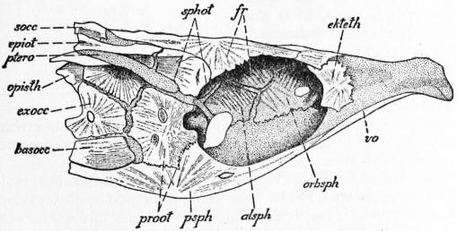
Fig. 24.—Chondrocranium of Salmon, seen from the right side.
alsph, Alisphenoid. basocc, Basioccipital. ekteth, Lateral ethmoid. epiot, Epiotic. exocc, Exoccipital. fr, Frontal. opisth, Opisthotic. orbsph, Orbitosphenoid. proot, Prootic. psph, Parasphenoid. ptero, Pterotic. socc, Supra occipital. sphot, Sphenotic. vo, Vomer.
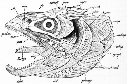
In the skull of the adult salmon it is seen that certain parts of the chondrocranium (fig. 24) have been replaced by bone (“cartilage bones”) while other more superficially placed bones (“membrane bones”) cover its surface (fig. 25). Of cartilage bones four are developed round the foramen magnum—the basioccipital, supraoccipital and two exoccipitals. In front of the basioccipital is the basisphenoid with an alisphenoid on each side. The region (presphenoidal) immediately in front of the basisphenoid is unossified, but on each side of it an orbitosphenoid is developed, the two orbitosphenoids being closely approximated in the mesial plane and to a certain extent fused, forming the upper part of the interorbital septum. In the anterior or ethmoidal portion of the cranium the only cartilage bones are a pair of lateral ethmoids lying at the anterior boundary of the orbit. A series of five distinct elements are ossified in the wall of the auditory or otic capsule, the prootic and opisthotic more ventrally, and the sphenotic, pterotic and epiotic more dorsally. The roof of the cranium is covered in by the following dermal bones—parietals (on each side of the supraoccipital), frontals, dermal ethmoid and small nasals, one over each olfactory organ. The floor of the cranium on its oral aspect is ensheathed by the large parasphenoid and the smaller vomer in front of and overlapping it. The cartilaginous lower jaw is ossified posteriorly to form the articular (fig. 25) with a small membrane bone, the angular, ventral to it, but the main part of the jaw is replaced functionally by a large membrane bone which ensheaths it—the dentary—evolved in all probability by the spreading outwards of bony tissue from the bases of the placoid elements (teeth) which it bears. The original upper jaw (palatopterygoid bar) is replaced by a chain of bones—palatine in front, then pterygoid and mesopterygoid, and posteriorly metapterygoid and quadrate, the latter giving articulation to the articular bone of the lower jaw. These representatives of the palatopterygoid bar no longer form the functional upper jaw. This function is performed by membrane bones which have appeared external to the palatopterygoid bar—the premaxilla and maxilla—which carry teeth—and the small scale-like jugal behind them. The quadrate is suspended from the skull as in the Selachians (hyostylic skull) by the upper portion of the hyoid arch—here represented by two bones—the hyomandibular and symplectic. The ventral portion of the hyoid arch is also represented by a chain of bones (stylohyal, epihyal, ceratohyal, hypohyal and the ventral unpaired basihyal), as is also each of the five branchial arches behind it. In addition to the bony elements belonging to the hyoid arch proper a series of membrane bones support the opercular flap. Ventrally there project backwards from the ceratohyal a series of ten overlapping branchiostegal rays, while more dorsally are the broader interopercular, subopercular and opercular.
From Wiedersheim, Verg. Anat. der Wirbeltiere, by permission of Gustav Fischer.
Fig. 25.—Complete Skull of Salmon from left side.
art, Articular. branchiost, Branchiostegal. dent, Dentary. epiot, Epiotic. eth, Dermal ethmoid. fr, Frontal. hyom, Hyomandibular. intop, Interopercular. Jug, Jugal. mpt, Mesopterygoid. mtpt, Metapterygoid. mx, Maxilla. nas, Nasal. op, Opercular. pal, Palatine. par, Parietal. pmx, Premaxilla. preop, Preopercular. pt, Pterygoid. pter, Pterotic. Quad, Quadrate. socc, Supraoccipital. sphot, Sphenotic. subop, Subopercular. sympl, Symplectic. Zunge, Tongue.
In addition to the bones already enumerated there is present a ring of circumorbital bones, a preopercular, behind and external to the hyomandibular and quadrate, and squamosal, external to the hinder end of the auditory capsule.
In the salmon, pike, and various other Teleosts, extensive regions of the chondrocranium persist in the adult, while in others (e.g. the cod) the replacement by bone is practically complete. Bony elements may be developed in addition to those noticed in the salmon.
In the sturgeon the chondrocranium is ensheathed by numerous membrane bones, but cartilage bones are absent. In the Crossopterygians35 the chondrocranium persists to a great extent in the adult, but portions of it are replaced by cartilage bones—the most interesting being a large sphenethmoid like that of the frog. Numerous membrane bones cover the chondrocranium externally. In the Dipneusti36 the chondrocranium is strengthened in the adult by numerous bones. One of the most characteristic is the great palatopterygoid bone which develops very early by the spreading of ossification backwards from the tooth bases, and whose early development probably accounts for the non-development of the palatopterygoid cartilage.
Appendicular Skeleton.—The primitive pectoral girdle, which in the Dipneusti is strengthened by a sheath of bone, becomes in the Teleostomes reduced in size (small scapula and coracoid bones) and replaced functionally by a secondary shoulder girdle formed of superficially placed membrane bones (supraclavicular and cleithrum or “clavicle,” with, in addition in certain cases, an infraclavicular and one or two postclavicular elements), and connected at its dorsal end with the skull by a post-temporal bone.
The pelvic girdle is in Teleostomes completely absent as a rule.
The skeleton of the free limb undergoes ossification to a less or greater extent in the Teleostomes.
In Polypterus the pectoral fin (fig. 18, B) shows three ossifications in the basal part of the fin—pro-, meso- and metapterygium. Of these the metapterygium probably represents the ossified skeletal axis: while the propterygium and also the numerous diverging radials probably represent the lateral rays of one side of the archipterygium.
In the Teleostomes the place of the pelvic girdle is taken functionally by an element apparently formed by the fusion of the basal portions of several radials.
Vascular System.—The main components of the blood vascular system in the lower vertebrates are the following: (1) a single or double dorsal aorta lying between the enteron and notochord; (2) a ventral vessel lying beneath the enteron; and (3) a series of paired hoop-like aortic arches connecting dorsal and ventral vessels round the sides of the pharynx. The blood-stream passes forwards towards the head in the ventral vessel, dorsalwards through the aortic arches, and tailwards in the dorsal aorta.
The dorsal aorta is single throughout the greater part of its extent, but for a greater or less extent at its anterior end (circulus cephalicus) it consists of two paired aortic roots. It is impossible to say whether the paired or the unpaired condition is the more primitive, general morphological conditions being in favour of the latter, while embryological evidence rather supports the former. The dorsal aorta, which receives its highly oxygenated blood from the aortic arches, is the main artery for the distribution of this oxygenated blood. Anteriorly the aortic roots are continued forwards as the dorsal carotid arteries to supply the head region. A series of paired, segmentally-arranged arteries pass from the dorsal aorta to supply the muscular body wall, and the branches which supply the pectoral and pelvic fins (subclavian or brachial artery, and iliac artery) are probably specially enlarged members of this series of segmental vessels. Besides these paired vessels a varying number of unpaired branches pass from dorsal aorta to the wall of the alimentary canal with its glandular diverticula (coeliac, mesenteric, rectal).
The ventral vessel undergoes complicated changes and is represented in the adults of existing fishes by a series of important structures. Its post-anal portion comes with the atrophy of the post-anal gut to lie close under the caudal portion of the dorsal aorta and is known as the caudal vein. This assumes a secondary connexion with, and drains its blood into, the posterior cardinal veins (see below). In the region between cloaca and liver the ventral vessel becomes much branched or even reticular and—serving serving to convey the food-laden blood from the wall of the enteron to the capillary network of the liver—is known as the hepatic portal vein. The short section in front of the liver is known as the hepatic vein and this conveys the blood, which has been treated by the liver, into a section of the ventral vessel, which has become highly muscular and is rhythmically contractile. This enlarged muscular portion, in which the contractility—probably once common to the main vessels throughout their extent—has become concentrated, serves as a pump and is known as the heart. Finally the precardiac section of the ventral vessel—the ventral aorta—conveys the blood from heart to aortic arches.
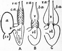
From Boas, Lehrbuch der Zoologie, by permission of Gustav Fischer.
Fig. 26.—Diagram to illustrate the condition of the Conus in an Elasmobranch (A), Amia (B) and a typical Teleost (C).
a, Atrium. b.a, Bulbus aortae. c.a, Conus arteriosus. s.v, Sinus venosus. v,v′, Valves. v.a, Ventral aorta. vt, Ventricle.
In addition to the vessels mentioned a large paired vein is developed in close relation to the renal organ which it serves to drain. This is the posterior cardinal. An anterior prolongation (anterior cardinal) serves to drain the blood from the head region. From the point of junction of anterior and posterior cardinal a large transverse vessel leads to the heart (ductus Cuvieri).
Heart.—Originally a simple tube curved into a somewhat S-shape, the heart, by enlargements, constrictions and fusions of its parts, becomes converted into the complex, compact heart of the adult. In this we recognize the following portions—(1) Sinus venosus, (2) Atrium, (3) Ventricle. A fourth chamber, the conus arteriosus, the enlarged and contractile hinder end of the ventral aorta, is also physiologically a part of the heart. The sinus venosus receives the blood from the great veins (ductus Cuvieri and hepatic veins). It—like the atrium which it enters by an opening guarded by two lateral valves—has thin though contractile walls. The atrium is as a rule single, but in the Dipnoans, in correlation with the importance of their pulmonary breathing, it is incompletely divided into a right and a left auricle. In Neoceratodus the incomplete division is effected by the presence of a longitudinal shelf projecting into the atrial cavity from its posterior wall. The opening of the sinus venosus is to the right of this shell, that of the pulmonary vein to the left. In Prototerus and Lepidosiren a nearly complete septum is formed by the fusion of trabeculae, there being only a minute opening in it posteriorly. The atrium opens by a wide opening guarded by two or more flap valves provided with chordae tendineae into the ventricle.
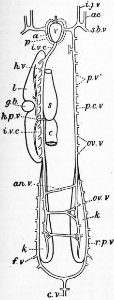
After Newton Parker, from Trans. of the Royal Irish Academy, vol. xxx.
Fig. 27.—Venous System of Protopterus, as seen from ventral side.
a, Atrium. ac, Anterior cardinal. an.v, Anastomotic vein. c, Intestine. c.v, Caudal vein. f.v, Femoral vein. g.b, Gall-bladder. h.v, Hepatic vein. i.j.v, Inferior jugular vein. i.v.c, Posterior vena cava. k, Kidney. l, Liver. ov.v, Ovarian veins. p, Pericardium. p.c.v, Left posterior cardinal. p.v′, Parietal veins. r.p.v, Renal portal. s, Stomach. s.b.v, Subclavian.
The ventricle, in correspondence with it being the main pumping apparatus, has its walls much thickened by the development of muscular trabeculae which, in the lower forms separated by wide spaces in which most of the blood is contained, become in the Teleostomes so enlarged as to give the wall a compact character, the spaces being reduced to small scattered openings on its inner surface. In the Dipnoans the ventricle, like the atrium, is incompletely divided into a right and left ventricle. In Ceratodus this is effected by an extension of the interauricular shelf into the ventricle. In Lepidosiren the separation of the two ventricles is complete but for a small perforation anteriorly, the heart in this respect showing a closer approximation to the condition in the higher vertebrates than is found in any Amphibians or in any reptiles except the Crocodilia. The conus arteriosus is of interest from the valvular arrangements in its interior to prevent regurgitation of blood from ventral aorta into ventricle. In their simplest condition, as seen e.g. in an embryonic Selachian, these arrangements consist of three, four or more prominent longitudinal ridges projecting into the lumen of the conus, and serving to obliterate the lumen when jammed together by the systole of the conus. As development goes on each of these ridges becomes segmented into a row of pocket valves with their openings directed anteriorly so that regurgitation causes them to open out and occlude the lumen by their free edges meeting. Amongst the Teleostomes the lower ganoids show a similar development of longitudinal rows of valves in the conus. In Amia (fig. 26, B), however, the conus is shortened and the number of valves in each longitudinal row is much reduced. This leads to the condition found in the Teleosts (fig. 26, O), where practically all trace of the conus has disappeared, a single circle of valves representing a last survivor of each row (save in a few exceptional cases, e.g. Albula, Tarpen, Osteoglossum, where two valves of each row are present).
In Front of the conus vestige of the Teleost there is present a thick walled bulbus aortae differing from the conus in not being rhythmically contractile, its walls being on the contrary richly provided with elastic tissue.
The Dipnoans37 show an important advance on the conus as in atrium and ventricle. The conus has a characteristic spiral twist. Within it in Neoceratodus are a number of longitudinal rows of pocket valves. One of these rows is marked out by the very large size of its valves and by the fact that they are not distinct from one another but even in the adult form a continuous, spirally-running, longitudinal fold. This ridge projecting into the lumen of the conus divides it incompletely into two channels, the one beginning (i.e. at its hinder end) on the left side and ending in front ventrally, the other beginning on the right and ending dorsally. In Protopterus a similar condition occurs, only in the front end of the conus a second spiral fold is present opposite the first and, meeting this, completes the division of the conus cavity into two separate parts. The rows of pocket valves which do not enter into the formation of the spiral folds are here greatly reduced.
These arrangements in the conus of the Dipnoans are of the highest morphological interest, pointing in an unmistakable way towards the condition found in the higher lung-breathing vertebrates. Of the two cavities into which the conus is partially divided in the Dipneusti the one which begins posteriorly on the right receives the (venous) blood from the right side of the heart, and ending up anteriorly dorsal to the other cavity communicates only with aortic arches V. and VI. In the higher vertebrates this cavity has become completely split off to form the root of the pulmonary arteries, and a result of aortic arch V. receiving its blood along with the functionally much more important VI. (the pulmonary arch) from this special part of the conus has been the almost complete disappearance of this arch (V.) in all the higher vertebrates.
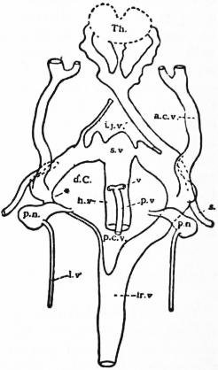
Fig. 28.—Venous System of Polypterus 30 mm. larva (dorsal view).
a.c.v, Anterior cardinal vein. d.C, Ductus Cuvieri. h.v, Hepatic vein. i.j.v, Inferior jugular vein. ir.v, Inter-renal vein. l.v, Lateral cutaneous vein. p.c.v, Posterior cardinal vein. p.n, Pronephros. p.v, Pulmonary vein. s, Subclavian vein. s.v, Sinus venosus. th, Thyroid. v, Vein from pharyngeal wall. * Anterior portion of left posterior cardinal vein.
Arterial System.—There are normally six aortic arches laid down corresponding with the visceral arches, the first (mandibular) and second (hyoidean) undergoing atrophy to a less or greater extent in post-embryonic life. Where an external gill is present the aortic arch loops out into this, a kind of short-circuiting of the blood-stream taking place as the external gill atrophies. As the walls of the clefts assume their respiratory function the aortic arch becomes broken into a network of capillaries in its respiratory portion, and there is now distinguished a ventral afferent and a dorsal efferent portion of each arch. Complicated developmental changes, into which it is unnecessary to enter,38 may lead to each efferent vessel draining the two sides of a single cleft instead of the adjacent walls of two clefts as it does primitively. In the Crossopterygians and Dipnoans as in the higher vertebrates the sixth aortic arch gives off the pulmonary artery to the lung. Among the Actinopterygians this, probably primitive, blood-supply to the lung (swimbladder) persists only in Amia.
Venous System.—The most interesting variations from the general plan outlined have to do with the arrangements of the posterior cardinals. In the Selachians these are in their anterior portion wide and sinuslike, while in the region of the kidney they become broken into a sinusoidal network supplied by the postrenal portion now known as the renal portal vein. In the Teleostomes the chief noteworthy feature is the tendency to asymmetry, the right posterior cardinal being frequently considerably larger than the left and connected with it by transverse anastomotic vessels, the result being that most of the blood from the two kidneys passes forwards by the right posterior cardinal. The Dipnoans (fig. 27) show a similar asymmetry, but here the anterior end of the right posterior cardinal disappears, being replaced functionally by a new vessel which conveys the blood from the right posterior cardinal direct to the sinus venosus instead of to the outer end of the ductus Cuvieri. This new vessel is the posterior vena cava which thus in the series of vertebrates appears for the first time in the Dipneusti.
Pulmonary Veins.—In Polypterus (fig. 28) the blood is drained from the lungs by a pulmonary vein on each side which unites in front with its fellow and opens into the great hepatic vein behind the heart. In the Dipnoans the conjoined pulmonary veins open directly into the left section of the atrium as in higher forms. In the Actinopterygians with their specialized air-bladder the blood passes to the heart via posterior cardinals, or hepatic portal, or—a probably more primitive condition—directly into the left ductus Cuvieri (Amia).
Lymphatics.—More or less irregular lymphatic spaces occur in the fishes as elsewhere and, as in the Amphibia, localized muscular developments are present forming lymph hearts.
Central Nervous System.—The neural tube shows in very early stages an anterior dilated portion which forms the rudiment of the brain in contradistinction to the hinder, narrower part which forms the spinal cord. This enlargement of the brain is correlated with the increasing predominance of the nerve centres at the anterior end of the body which tend to assume more and more complete control over those lying behind.
Spinal Cord.—A remarkable peculiarity occurs in the sun fishes (Molidae), where the body is greatly shortened and where the spinal cord undergoes a corresponding abbreviation so as to be actually shorter than the brain.
Brain.—It is customary to divide the brain into three main regions, fore-, mid-, and hind-brain, as in the most familiar vertebrates there is frequently seen in the embryo a division of the primitive brain dilatation into three vesicles lying one behind the other. A consideration of the development of the brain in the various main groups of vertebrates shows that these divisions are not of equal importance. In those archaic groups where the egg is not encumbered by the presence of a large mass of yolk it is usual for the brain to show in its early stages a division into two main regions which we may term the primitive fore-brain or cerebrum and the primitive hind-brain or rhombencephalon. Only later does the hinder part of the primitive fore-brain become marked off as mid-brain. In the fully developed brain it is customary to recognize the series of regions indicated below, though the boundaries between these regions are not mathematical lines or surfaces any more than are any other biological boundaries:—
Rhombencephalon (Hind-brain) Myelencephalon (Medulla oblongata).
Metencephalon (Cerebellum).
Cerebrum (Primitive Fore-brain) Mesencephalon (Mid-brain).
Thalamencephalon (Diencephalon).
[Hemispheres (Telencephalon).]
The myelencephalon or medulla oblongata calls for no special remark, except that in the case of Torpedo there is a special upward bulging of its floor on each side of the middle line forming the electric lobe and containing the nucleus of origin of the nerves to the electric organ.
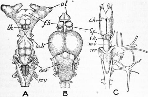
A and B from Wiedersheim, by permission of Gustav Fischer.
Fig. 29.—Brain of Scyllium (A), Salmo (B) and Lepidosiren (C). The three figures are not drawn to the same scale.
cer, Cerebellum. c.h, Cerebral hemisphere. th, Thalamencephalon. f.b, Primitive fore-brain (in B the line points to the thickened wall of the fore-brain, the so-called “basal ganglia”). G.p, Pineal body. m.b, Roof of mid-brain, optic lobes, tectum opticum. o.l, Olfactory lobe. IV.v, Fourth ventricle.
The cerebellum occurs in its simplest form in lampreys and Dipnoans (fig. 29, C), where it forms a simple band-like thickening of the anterior end of the roof of the hind-brain. In Selachians it is very large and bulges upwards, forming a conspicuous organ in a dorsal view of the brain (fig. 29, A). In Teleosts (fig. 29, B) the cerebellum is also large. It projects back as a great tongue-like structure over the roof of the fourth ventricle, while in front it dips downwards and projects under the roof of the mid-brain forming a highly characteristic valvula cerebelli. A valvula cerebelli occurs also in ganoids, while in the Crossopterygians a similar extension of the cerebellum projects backwards into the IV. ventricle or cavity of the hind-brain (fig. 30).
The mesencephalon is a conspicuous structure in the fishes from its greatly developed roof (tectum opticum) which receives the end pencils of the optic nerve. Normally it projects upwards as a pair of large optic lobes, but in the Dipnoans (fig. 29, C) the lateral thickening is not sufficiently great to cause obvious lateral swellings in external view.
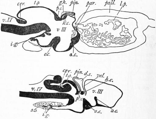
Fig. 30.—Median Longitudinal Section through the brain of Lepidosiren and Polypterus. In the upper figure (Lepidosiren) the habenular ganglion and hemisphere are shown in outline though not actually present in a median section.
a.c, Anterior commissure. cer, Cerebellum. d.s, Dorsal sac. g.h, Habenular ganglion. h.c, Habenular commissure. i.g, Infundibular gland. l.p, Lateral plexus. o.c, Optic chiasma. pall, Pallium. par, Paraphysis. pin, Pineal body. p.c, Posterior commissure. s.v, Saccus vasculosus. t.o, Tectum opticum. v.III, Third ventricle. v.IV, Fourth ventricle. vel, Velum transversum.
The thalamencephalon is one of the most interesting parts of the brain from its remarkable uniformity throughout the Vertebrata. Even in Amphioxus the appearance of a sagittal section strongly suggests vestiges of a once present thalamencephalon.39 The roof—like that of the myelencephalon—remains to a great extent membranous, forming with the closely applied pia mater a vascular roof to the III. ventricle. Frequently a transverse fold of the roof dips down into the III. ventricle forming the velum transversum (fig. 30).
The side walls of the thalamencephalon are greatly thickened forming the thalamus (epithalamus and hypothalamus), while a ganglionic thickening of the roof posteriorly on each side forms the ganglia habenulae which receive olfactory fibres from the base of the hemisphere. The habenular ganglia are unusually large in the lampreys and are here strongly asymmetrical, the right being the larger.
The floor of the thalamencephalon projects downwards and backwards as the infundibulum. The side walls of this are thickened to form characteristic lobi inferiores, while the blind end develops glandular outgrowths (infundibular gland, fig. 30) overlaid by a rich development of blood sinuses and forming with them the saccus vasculosus. The optic chiasma, where present, is involved in the floor of the thalamencephalon and forms a large, upwardly-projecting ridge. Farther forwards on the floor or anterior wall is the anterior commissure (see below).
Passing forwards from the mid-brain (cf. fig. 30) a series of interesting structures are found connected with the roof of the primitive fore-brain, viz.—posterior commissure (intercalary region), pineal organ, habenular commissure with anterior parietal organ, dorsal sac (= pineal cushion), velum transversum, paraphysis. The posterior commissure is situated in the boundary between thalamencephalon and mid-brain. It is formed of fibres connecting up the right and left sides of the tectum opticum (?). The habenular or superior commissure situated farther forwards connects the two ganglia habenulae. In the immediate neighbourhood of these ganglia there project upwards two diverticula of the brain-roof known as the pineal organ and the parapineal (or anterior parietal) organ. The special interest of these organs40 lies in the fact that in certain vertebrates one (parapineal in Sphenodon and in lizards) or both (Petromyzon) exhibit histological features which show that they must be looked on as visual organs or eyes. In gnathostomatous fishes they do not show any definite eye-like structure, but in certain cases (Polyodon, Callichthys, &c.) the bony plates of the skull-roof are discontinuous over the pineal organ forming a definite parietal foramen such as exists in lizards where the eye-like structure is distinct. It is also usual to find in the epithelial wall of the pineal organ columnar cells which show club-shaped ends projecting into the lumen (exactly as in the young visual cells of the retina41) and are prolonged into a root-like process at the other end. Definite nerve fibres pass down from these parietal organs to the brain. It is stated that the fibres from the pineal organ pass into the posterior commissure, those of the parapineal organ into the habenular commissure.
The facts mentioned render it difficult to avoid the conclusion that these organs either have been sensory or are sensory. Possibly they represent the degenerate and altered vestiges of eye-like organs present in archaic vertebrates, or it may be that they represent the remains of organs not eye-like in function but which for some other reason lay close under the surface of the body. It would seem natural that a diverticulum of brain-tissue exposed to the influence of light-rays should exhibit the same reaction as is shown frequently elsewhere in the animal kingdom and tend to assume secondarily the characters of a visual organ. The presence of the rod-like features in the epithelial cells is perhaps in favour of the latter view. In evolution we should expect these to appear before the camera-like structure of a highly developed eye, while in the process of degeneration we should expect these fine histological characters to go first.
Selachians.—No parapineal organ is present. The pineal body (except in Torpedo where it is absent) is in the form of a long slender tube ending in front in a dilated bulb lying near the front end of the brain in close contact with, or enclosed in, a definite foramen in the cranial roof.
Holocephali and Crossopterygii.—Here also the pineal body is long and tubular: at its origin it passes dorsalwards or slightly backwards behind the large dorsal sac.
Actinopterygian Ganoids resemble Selachians on the whole. In Amia a parapineal organ is present, and it is said to lie towards the left side and to be connected by a thick nerve with the left habenular ganglion (cf. Petromyzon, article Cyclostomata). This is adduced to support the view that the pineal and parapineal bodies represent originally paired structures.
Teleostei.—A parapineal rudiment appears in the embryo of some forms, but in the adult only the pineal organ is known to exist. This is usually short and club-shaped, its terminal part with much folded wall and glandular in character. In a few cases a parietal foramen occurs (Callichthys, Loricaria, &c.).
Dipneusti.—The pineal organ is short and simple. No parapineal organ is developed.
The dorsal sac is formed by that part of the roof of the thalamencephalon lying between the habenular commissure and the region of the velum. In some cases a longitudinal groove is present in which the pineal organ lies (Dipneusti). In the Crossopterygians the dorsal sac is particularly large and was formerly mistaken for the pineal organ.
The velum transversum is a transverse, inwardly-projecting fold of the roof of the primitive fore-brain in front of the dorsal sac. To those morphologists who regard the hemisphere region or telencephalon as a primitively unpaired structure the velum is an important landmark indicating the posterior limit of the telencephalon. Those who hold the view taken in this article that the hemispheres are to be regarded as paired outpushings of the side wall of the primitive fore-brain attribute less morphological importance to the velum. Physiologically the velum is frequently important from the plexus of blood-vessels which passes with it into the III. ventricle.
In Petromyzon and Chimaera the velum is not developed. In Dipnoans there are present in its place paired transverse folds which are probably merely extensions backwards of the lateral plexuses.
The Paraphysis is a projection from the roof of the primitive fore-brain near its anterior end. It is well seen in Dipnoans42 (Lepidosiren and Protopterus) where in the larva (exactly as in the urodele larva) it forms a blindly ending tube sloping upwards and forwards between the two hemispheres. In the adult it becomes mixed with the two lateral plexuses and is liable to be confused with them. In the other groups—except the Teleosts where it is small (Anguilla) or absent (most Teleosts)—the paraphysis is by no means such a definite structure, but generally there is present a more or less branched and divided diverticulum of the brain wall, frequently glandular, which is homologized with the paraphysis. The morphological significance of the paraphysis is uncertain. It may represent the remains of an ancient sense organ, or it may simply represent the last connexion between the brain and the external ectoderm from which it was derived.
An important derivative of the primitive fore-brain is seen in the pair of cerebral hemispheres which in the higher vertebrates become of such relatively gigantic dimensions. The hemispheres appear to be primitively associated with the special sense of smell, and they are prolonged anteriorly into a pair of olfactory lobes which come into close relation with the olfactory organ. From a consideration of their adult relations and of their development—particularly in those groups where there is no disturbing factor in the shape of a large yolk sac—it seems probable that the hemispheres are primitively paired outpushings of the lateral wall of the primitive fore-brain43—in order to give increased space for the increased mass of nervous matter associated with the olfactory sense. They are most highly developed in the Dipneusti amongst fishes. They are there (cf. fig. 29, C) of relatively enormous size with thick nervous floor (corpus striatum) and side walls and roof (pallium) surrounding a central cavity (lateral ventricle) which opens into the third ventricle. At the posterior end of the hemisphere a small area of its wall remains thin and membranous, and this becomes pushed into the lateral ventricle by an ingrowth of blood-vessel to form the huge lateral plexus (= plexus hemisphaerium). In this great size of the hemispheres44 and also in the presence of a rudimentary cortex in the Dipnoi we see, as in many other features in these fishes, a distinct foreshadowing of conditions occurring in the higher groups of vertebrates. The Cyclostomes possess a distinct though small pair of hemispheres. In the Selachians the relatively archaic Notidanidae45 possess a pair of thick-walled hemispheres, but in the majority of the members of the group the paired condition is obscured (fig. 29, A).
In the Teleostomes the mass of nervous matter which in other groups forms the hemispheres does not undergo any pushing outwards except as regards the small olfactory lobes. On the contrary, it remains as a great thickening of the lateral wall of the thalamencephalon (the so-called basal ganglia), additional space for which, however, may be obtained by a considerable increase in length of the fore-brain region (cf. fig. 30, A) or by actual involution into the third ventricle (Polypterus).46 The great nervous thickenings of the thalamencephalic wall bulge into its cavity and are covered over by the thin epithelial roof of the thalamencephalon which is as a consequence liable to be confused with the pallium or roof of the hemispheres with which it has nothing to do: the homologue of the pallium as of other parts of the hemisphere is contained within the lateral thickening of the thelamencephalic wall, not in its membranous roof.47
Associated with the parts of the fore-brain devoted to the sense of smell (especially the corpora striata) is the important system of bridging fibres forming the anterior commissure which lies near the anterior end of the floor, or in the front wall, of the primitive fore-brain. It is of great interest to note the appearance in the Dipnoans (Lepidosiren and Protopterus) of a corpus callosum (cf. fig. 30 B) lying dorsal to the anterior commissure and composed of fibres connected with the pallial region of the two hemispheres.
Sense Organs.—The olfactory organs are of special interest in the Selachians, where each remains through life as a widely-open, saccular involution of the ectoderm which may be prolonged backwards to the margin of the buccal cavity by an open oronasal groove, thus retaining a condition familiar in the embryo of the higher vertebrates. In Dipnoans the olfactory organ communicates with the roof of the buccal cavity by definite posterior nares as in the higher forms—the communicating passage being doubtless the morphological equivalent of the oronasal groove, although there is no direct embryological evidence for this. In the Teleostomes the olfactory organ varies from a condition of great complexity in the Crossopterygians down to a condition of almost complete atrophy in certain Teleosts (Plectognathi).48
The eyes are usually of large size. The lens is large and spherical and in the case of most Teleostomes accommodation for distant vision is effected by the lens being pulled bodily nearer the retina. This movement is brought about by the contraction of smooth muscle fibres contained in the processus falciformis, a projection from the choroid which terminates in contact with the lens in a swelling, the campanula Halleri. In Amia and in Teleosts a network of capillaries forming the so-called choroid gland surrounds the optic nerve just outside the retina. As a rule the eyes of fishes have a silvery, shining appearance due to the deposition of shining flakes of guanin in the outer layer of the choroid (Argentea) or, in the case of Selachians, in the inner layers (tapetum). Fishes which inhabit dark recesses, e.g. of caves or of the deep sea, show an enlargement, or, more frequently, a reduction, of the eyes. Certain deep-sea Teleosts possess remarkable telescopic eyes with a curious asymmetrical development of the retina.49
The otocyst or auditory organ agrees in its main features with that of other vertebrates. In Selachians the otocyst remains in the adult open to the exterior by the ductus endolymphaticus. In Squatina50 this is unusually wide and correlated; with this the calcareous otoconia are replaced by sand-grains from the exterior. In Dipnoans (Lepidosiren and Protopterus) curious outgrowths arise from the ductus endolymphaticus and come to overlie the roof of the fourth ventricle, recalling the somewhat similar condition met with in certain Amphibians.
In various Teleosts the swimbladder enters into intimate relations with the otocyst. In the simplest condition these relations consist in the prolongation forwards of the swimbladder as a blindly ending tube on either side, the blind end coming into direct contact either with the wall of the otocyst itself or with the fluid surrounding it (perilymph) through a gap in the rigid periotic capsule. A wave of compression causing a slight inward movement of the swimbladder wall will bring about a greatly magnified movement of that part of the wall which is not in relation with the external medium, viz. the part in relation with the interior of the auditory capsule. In this way the perception of delicate sound waves may be rendered much more perfect. In the Ostariophysi (Sagemehl), including the Cyprinidae, the Siluridae, the Characinidae and the Gymnotidae, a physiologically similar connexion between swimbladder and otocyst is brought about by the intervention of a chain of auditory ossicles (Weberian ossicles) formed by modification of the anterior vertebrae.51
Lateral Line Organs.52—Epidermal sense buds are scattered about in the ectoderm of fishes. A special arrangement of these in lines along the sides of the body and on the head region form the highly characteristic sense organs of the lateral line system. In Lepidosiren these organs retain their superficial position; in other fishes they become sunk beneath the surface into a groove, which may remain open (some Selachians), but as a rule becomes closed into a tubular channel with openings at intervals. It has been suggested that the function of this system of sense organs is connected with the perception of vibratory disturbances of comparatively large wave length in the surrounding medium.
Peripheral Nerves.—In the Cyclostomes the dorsal afferent and ventral efferent nerves are still, as in Amphioxus, independent, but in the gnathostomatous fishes they are, as in the higher vertebrates, combined together into typical spinal nerves.
As regards the cranial nerves the chief peculiarities of fishes relate to (1) the persistence of the branchial clefts and (2) the presence of an elaborate system of cutaneous sense organs supplied by a group of nerves (lateralis) connected with a centre in the brain which develops in continuity with that which receives the auditory nerve. These points may be exemplified by the arrangements in Selachians (see fig. 31). I., II., III., IV. and VI. call for no special remark.
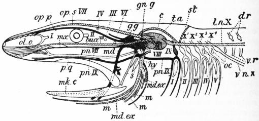
From Bridge, Cambridge Natural History, vol. vii. “Fishes” (by permission of Macmillan & Co., Ltd.). After Wiedersheim, Grundriss der vergleichenden Anatomie (by permission of Gustav Fischer).
Fig. 31.—Diagram of Cranial nerves of a Fish. Cranial nerves and branchial clefts are numbered with Roman figures. Trigeminus black; Facialis dotted; Lateralis oblique shading; Glossopharyngeal cross-hatched; Vagus white.
bucc, Buccal. c, Commissure between pre- and postauditory parts of lateralis system. d.r, Dorsal roots of spinal nerves. g.g, Gasserian ganglion. gn.g, (Geniculate) ganglion of VII. hy, Hyomandibular. l.n.X, Lateralis vagi. m, Motor branches of hy. md, Mandibular. md.ex, External mandibular. mk.c, Meckel’s cartilage. mx, Maxillary. oc, Occipitospinal. ol.o, Olfactory organ. op.p, Ophthalmicus profundus. op.s, Ophthalmicus superficialis. pn, Palatine. pq., Palatopterygo-quadrate cartilage. s, Spiracle. st, Supra-temporal branch of lateralis system. t.a, Lateralis centre in brain. v.n, Visceral nerve. v.r, Ventral roots.
Trigeminus (V.).—The ophthalmicus profundus branch (op.p.)—which probably is morphologically a distinct cranial nerve—passes forwards along the roof of the orbit to the skin of the snout. As it passes through the orbit it gives off the long ciliary nerves to the eyeball, and is connected with the small ciliary ganglion (also connected with III.) which in turn gives off the short ciliary nerves to the eyeball. The ophthalmicus superficialis (cut short in the figure) branch passes from the root ganglion of V. (Gasserian ganglion), and passes also over the orbit to the skin of the snout. It lies close to, or completely fused with, the corresponding branch of the lateralis system.
The main trunk of V. branches over the edge of the mouth into the maxillary (mx.) and mandibular (md.) divisions, the former, like the two branches already mentioned, purely sensory, the latter mixed—supplying the muscles of mastication as well as the teeth of the lower jaw and the lining of the buccal floor.
The main trunk of the Facialis (VII.) bifurcates over the spiracle into a pre-spiracular portion—the main portion of which passes to the mucous membrane of the palate as the palatine (pnVII.)—and a postspiracular portion, the hyomandibular (hy.) trunk which supplies the muscles of the hyoid arch and also sends a few sensory fibres to the lining of the spiracle, the floor of mouth and pharynx and the skin of the lower jaw. Combined with the main trunk of the facial are branches belonging to the lateralis system.
Lateralis Group of Nerves.—The lateralis group of nerves are charged with the innervation of the system of cutaneous sense organs and are all connected with the same central region in the medulla. A special sensory area of the ectoderm becomes involuted below the surface to form the otocyst, and the nerve fibres belonging to this form the auditory nerve (VIII.). Other portions of the lateralis group become mixed up with various other cranial nerves as follows:
(a) Facial portion.
(1) Ophthalmicus superficialis (op.s.VII.): passes to lining of nose or to the lateral line organs of the dorsal part of snout.
(2) Buccal (bucc.VII): lies close to maxillary division of V. and passes to the sensory canals of the lower side of the snout.
(3) External mandibular (md.ex.): lies in close association with the mandibular division of V., supplies the sensory canals of the lower jaw and hyoid region.
Lateralis vagi (l.n.X.) becomes closely associated with the vagus. It supplies the lateral line organs of the trunk.
In the lamprey and in Dipnoans the lateralis vagi loses its superficial position in the adult and comes into close relation with the notochord.
In Actinopterygians and at least some Selachians a lateralis set of fibres is associated with IX., and in the former fishes a conspicuous trunk of lateralis fibres passes to some or all (Gadus) of the fins. This has been called the lateralis accessorius and is apparently connected with V., VII., IX., X. and certain spinal nerves.53
Vagus Group (IX., X., XI.).—The glossopharyngeus (IX.) forks over the first branchial cleft (pretrematic and post-trematic branches) and also gives off a palatine branch (pn.IX.). In some cases (various Selachians, Ganoids and Teleosts) it would seem that IX. includes a few fibres of the lateralis group.
Vagus (X.) is shown by its multiple roots arising from the medulla and also by the character of its peripheral distribution to be a compound structure formed by the fusion of a number of originally distinct nerves. It consists of (1) a number of branchial branches (X.¹ X.² &c.), one of which forks over each gill cleft behind the hyobranchial and which may (Selachians) arise by separate roots from the medulla; (2) an intestinal branch (v.n.X.) arising behind the last branchial and innervating the wall of the oesophagus and stomach and it may be even the intestine throughout the greater part of its length (Myxine).
The accessorius (XI.) is not in fishes separated as a distinct nerve from the vagus.
With increased development of the brain its hinder portion, giving rise to the vagus system, has apparently come to encroach on the anterior portion of the spinal cord, with the result that a number of spinal nerves have become reduced to a less or more vestigial condition. The dorsal roots of these nerves disappear entirely in the adult, but the ventral roots persist and are to be seen arising ventrally to the vagus roots. They supply certain muscles of the pectoral fins and of the visceral arches and are known as spino-occipital nerves.54
These nerves are divisible into an anterior more ancient set—the occipital nerves—and a posterior set of more recent origin—(occipito-spinal nerves). In Selachians 1-5 pairs of occipital nerves alone are recognizable: in Dipnoans 2-3 pairs of occipital and 2-3 pairs of occipito-spinal: in Ganoids 1-2 pairs occipital and 1-5 pairs occipito-spinal; in Teleosts finally the occipital nerves have entirely disappeared while there are 2 pairs of occipito-spinal. In Cyclostomes no special spino-occipital nerves have been described.
The fibres corresponding with those of the Hypoglossus (XII.) of higher vertebrates spring from the anterior spinal nerves, which are here, as indeed in Amphibia, still free from the cranium.
Sympathetic.—The sympathetic portion of the nervous system does not in fishes attain the same degree of differentiation as in the higher groups. In Cyclostomes it is apparently represented by a fine plexus with small ganglia found in the neighbourhood of the dorsal aorta and on the surface of the heart and receiving branches from the spinal nerves. In Selachians also a plexus occurs in the neighbourhood of the cardinal veins and extends over the viscera: it receives visceral branches from the anterior spinal nerves. In Teleosts the plexus has become condensed to form a definite sympathetic trunk on each side, extending forwards into the head and communicating with the ganglia of certain of the cranial nerves.
(J. G. K.)
V. Distribution in Time and Space
The origin of Vertebrates, and how far back in time they extend, is unknown. The earliest fishes were in all probability devoid of hard parts and traces of their existence can scarcely be expected to be found. The hypothesis that they may be derived from the early Crustaceans, or Arachnids, is chiefly based on the somewhat striking resemblance which the mailed fishes of the Silurian period (Ostracodermi) bear to the Arthropods of that remote time, a resemblance, however, very superficial and regarded by most morphologists as an interesting example of mimetic resemblance—whatever this term may be taken to mean. The minute denticles known as conodonts, which first appear in the Ordovician, were once looked upon as teeth of Cyclostomes, but their histological structure does not afford any support to the identification and they are now generally dismissed altogether from the Vertebrates. As a compensation the Lower Silurian of Russia has yielded small teeth or spines which seem to have really belonged to fishes, although their exact affinities are not known (Palaeodus and Archodus of J. V. Rohon).
It is not until we reach the Upper Silurian that satisfactory remains of unquestionable fishes are found, and here they suddenly appear in a considerable variety of forms, very unlike modern fishes in every respect, but so highly developed as to convince us that we have to search in much earlier formations for their ancestors. These Upper Silurian fishes are the Coelolepidae, the Ateleaspidae, the Birkeniidae, the Pteraspidae, the Tremataspidae and the Cephalaspidae, all referred to the Ostracophori. The three last types persist in the Devonian, in the middle of which period the Osteolepid Crossopterygii, the Dipneusti and the Arthrodira suddenly appear. The most primitive Selachian (Cladoselache), the Acanthodian Selachians (Diplacanthidae), the Chimaerids (Ptyctodus), and the Palaeoniscid ganoids (Chirolepis) appear in the Upper Devonian, along with the problematic Palaeospondylus.
In the Carboniferous period, the Ostracophori and Arthrodira have disappeared, the Crossopterygii and Dipneusti are still abundant, and the Selachians (Pleuracanthus, Acanthodians, truesharks) and Chondrostean ganoids (Palaeoniscidae and Platysomidae) are predominant. In the Upper Permian the Holostean ganoids (Acanthophorus) make their appearance, and the group becomes dominant in the Jurassic and the Lower Cretaceous. In the Trias, the Crossopterygii and Dipneusti dwindle in variety and the Ceratodontidae appear; the Chondrostean and Holostean ganoids are about equally represented, and are supplemented in the Jurassic by the first, annectant representatives of the Teleostei (Pholidophoridae, Leptolepidae). In the latter period, the Holostean ganoids are predominant, and with them we find numerous Cestraciont sharks, some primitive skates (Squatinidae and Rhinobatidae), Chimaerids and numerous Coelacanthid crossopterygians.
The fish-fauna of the Lower Cretaceous is similar to that of the Jurassic, whilst that of the Chalk and other Upper Cretaceous formations is quite modern in aspect, with only a slight admixture of Coelacanthid crossopterygians and Holostean ganoids, the Teleosteans being abundantly represented by Elopidae, Albulidae, Halosauridae, Scopelidae and Berycidae, many being close allies of the present inhabitants of the deep sea. At this period the spiny-rayed Teleosteans, dominant in the seas of the present day, made their first appearance.
With the Eocene, the fish-fauna has assumed the essential character which it now bears. A few Pycnodonts survive as the last representatives of typically Mesozoic ganoids, whilst in the marine deposits of Monte Bolca (Upper Eocene) the principal families of living marine fishes are represented by genera identical with or more or less closely allied to those still existing; it is highly remarkable that forms so highly specialized as the sucking-fish or remoras, the flat-fish (Pleuronectidae), the Pediculati, the Plectognaths, &c., were in existence, whilst in the freshwater deposits of North America Osteoglossidae and Cichlidae were already represented. Very little is known of the freshwater fishes of the early Tertiaries. What has been preserved of them from the Oligocene and Miocene shows that they differed very slightly from their modern representatives. We may conclude that from early Tertiary times fishes were practically as they are at present. The great hiatus in our knowledge lies in the period between the Cretaceous and the Eocene.
At the present day the Teleosteans are in immense preponderance, Selachians are still well represented, the Chondrostean ganoids are confined to the rivers and lakes of the temperate zone of the northern hemisphere (Acipenseridae, Polyodontidae), the Holostean ganoids are reduced to a few species (Lepidosteus, Amia) dwelling in the fresh waters of North America, Mexico and Cuba, the Crossopterygians are represented by the isolated group Polypteridae, widely different from any of the known fossil forms, with about ten species inhabiting the rivers and lakes of Africa, whilst the Dipneusti linger in Australia (Neoceratodus), in South America (Lepidosiren), and in tropical Africa (Protopterus). The imperfections of the geological record preclude any attempt to deal with the distribution in space as regards extinct forms, but several types, at present very restricted in their habitat, once had a very wide distribution. The Ceratodontidae, for instance, of which only one species is now living, confined to the rivers of Queensland, has left remains in Triassic, Rhaetic, Jurassic and Cretaceous rocks of Europe, North America, Patagonia, North and South Africa, India and Australia; the Amiidae and Lepidosteidae were abundant in Europe in Eocene and Miocene times; the Osteoglossidae, now living in Africa, S.E. Asia and South America, occurred in North America and Europe in the Eocene.
In treating of the geographical distribution of modern fishes, it is necessary to distinguish between fresh-water and marine forms. It is, however, not easy to draw a line between these categories, as a large number of forms are able to accommodate themselves to either fresh or salt water, whilst some periodically migrate from the one into the other. On the whole, fishes may be roughly divided into the following categories:—
I. Marine fishes. A. shore-fishes; B. pelagic fishes; C. deep-sea fishes.
II. Brackish-water fishes.
III. Fresh-water fishes.
IV. Migratory fishes. A. anadromous (ascending fresh waters to spawn); B. catadromous (descending to the sea to spawn).
About two-thirds of the known recent fishes are marine. Such are nearly all the Selachians, and, among the Teleosteans, all the Heteromi, Pediculati and the great majority of Apodes, Thoracostei, Percesoces, Anacanthini, Acanthopterygii and Plectognathi. All the Crossopterygii, Dipneusti, Opisthomi, Symbranchii, and nearly all the Ganoidei and Ostariophysi are confined to fresh-water.
The three categories of marine fishes have thus been defined by Günther:—
“1. Shore Fishes—that is, fishes which chiefly inhabit parts of the sea in the immediate neighbourhood of land either actually raised above, or at least but little submerged below, the surface of the water. They do not descend to any great depth,—very few to 300 fathoms, and the majority live close to the surface. The distribution of these fishes is determined, not only by the temperature of the surface water, but also by the nature of the adjacent land and its animal and vegetable products,—some being confined to flat coasts with soft or sandy bottoms, others to rocky and fissured coasts, others to living coral formations. If it were not for the frequent mechanical and involuntary removals to which these fishes are exposed, their distribution within certain limits, as it no doubt originally existed, would resemble still more that of freshwater fishes than we find it actually does at the present period.
2. Pelagic Fishes—that is, fishes which inhabit the surface and uppermost strata of the open ocean, and approach the shores only accidentally or occasionally (in search of prey), or periodically (for the purpose of spawning). The majority spawn in the open sea, their ova and young being always found at a great distance from the shore. With regard to their distribution, they are still subject to the influences of light and the temperature of the surface water; but they are independent of the variable local conditions which tie the shore fish to its original home, and therefore roam freely over a space which would take a freshwater or shore fish thousands of years to cover in its gradual dispersal. Such as are devoid of rapidity of motion are dispersed over similarly large areas by the oceanic currents, more slowly than the strong swimmers, but not less surely. An accurate definition, therefore, of their distribution within certain areas equivalent to the terrestrial regions is much less feasible than in the case of shore fishes.
3. Deep-Sea Fishes—that is, fishes which inhabit such depths of the ocean that they are but little or not at all influenced by light or the surface temperature, and which, by their organization, are prevented from reaching the surface stratum in a healthy condition. Living almost under identical tellurian conditions, the same type, the same species, may inhabit an abyssal depth under the equator as well as one near the arctic or antarctic circle; and all that we know of these fishes points to the conclusion that no separate horizontal regions can be distinguished in the abyssal fauna, and that no division into bathymetrical strata can be attempted on the base of generic much less of family characters.”
A division of the world into regions according to the distribution of the shore-fishes is a much more difficult task than that of tracing continental areas. It is possible perhaps to distinguish four great divisions: the Arctic region, the Atlantic region, the Indo-Pacific region and the Antarctic region. The second and third may be again subdivided into three zones: Northern, Tropical and Southern. This appears to be a more satisfactory arrangement than that which has been proposed into three zones primarily, each again subdivided according to the different oceans. Perhaps a better division is that adopted by D. S. Jordan, who arranges the littoral fishes according to coast lines; we then have an East Atlantic area, a West Atlantic, an East Pacific and a West Pacific, the latter including the coasts of the Indian Ocean. The tropical zone, whatever be the ocean, is that in which fishes flourish in greatest abundance and where, especially about coral-reefs, they show the greatest variety of bizarre forms and the most gorgeous coloration. The fish-fauna of the Indo-Pacific is much richer than that of the Atlantic, both as regards genera and species.
As regards the Arctic and Antarctic regions, the continuity or circumpolar distribution of the shore fishes is well established. The former is chiefly characterized by its Cottids, Cyclopterids, Zoarcids and Gadids, the latter by its Nototheniids. The theory of bipolarity receives no support from the study of the fishes.
Pelagic fishes, among which we find the largest Selachians and Teleosteans, are far less limited in their distribution, which, for many species, is nearly world-wide. Some are dependent upon currents, but the great majority being rapid swimmers able to continue their course for weeks, apparently without the necessity of rest (many sharks, scombrids, sword-fishes), pass from one ocean into the other. Most numerous between the tropics, many of these fishes occasionally wander far north and south of their habitual range, and there are few genera that are at all limited in their distribution.
Deep-sea fishes, of which between seven hundred and eight hundred species are known, belong to the most diverse groups and quite a number of families are exclusively bathybial (Chlamydoselachidae, Stomiatidae, Alepocephalidae, Nemichthyidae, Synaphobranchidae, Saccopharyngidae, Cetomimidae, Halosauridae, Lipogenyidae, Notacanthidae, Chiasmodontidae, Icosteidae, Muraenolepididae, Macruridae, Anomalopidae, Podatelidae, Trachypteridae, Lophotidae, Ceratiidae, Gigantactinidae). But they are all comparatively slight modifications of the forms living on the surface of the sea or in the shallow parts, from which they may be regarded as derived. In no instance do these types show a structure which may be termed archaic when compared with their surface allies. That these fishes are localized in their vertical distribution, between the 100-fathoms line, often taken as the arbitrary limit of the bathybial fauna, and the depth of 2750 fathoms, the lowest point whence fishes have been procured, there is little doubt. But our knowledge is still too fragmentary to allow of any general conclusions, and the same applies to the horizontal distribution. Yet the same species may occur at most distant points; as these fishes dwell beyond the influence of the sun’s rays, they are not affected by temperature, and living in the Arctic zone or under the equator makes little difference to them. A great deal of evidence has been accumulated to show the gradual transition of the surface into the bathybial forms; a large number of surface fishes have been met with in deep water (from 100 to 500 fathoms), and these animals afford no support to Alexander Agassiz’s supposition of the existence of an azoic zone between the 200-fathoms line and the bottom.
Brackish-water fishes occur also in salt and fresh water, in some localities at least, and belong to various groups of Teleosteans. Sticklebacks, gobies, grey mullets, blennies are among the best-known examples. The facility with which they accommodate themselves to changes in the medium in which they live has enabled them to spread readily over very large areas. The three-spined stickleback, for instance, occurs over nearly the whole of the cold and temperate parts of the northern hemisphere, whilst a grey mullet (Mugil capito) ranges without any appreciable difference in form from Scandinavia and the United States along all the Atlantic coasts to the Cape of Good Hope and Brazil. It would be hardly possible to base zoo-geographical divisions on the distribution of such forms.
The fresh-water fishes, however, invite to such attempts. How greatly their distribution differs from that of terrestrial animals has long ago been emphasized. The key to their mode of dispersal is, with few exceptions, to be found in the hydrography of the continents, latitude and climate, excepting of course very great altitudes, being inconsiderable factors, the fish-fauna of a country deriving its character from the headwaters of the river-system which flows through it. The lower Nile, for instance, is inhabited by fishes bearing a close resemblance to, or even specifically identical with, those of tropical Africa, thus strikingly contrasting with the land-fauna of its banks. The knowledge of the river-systems is, however, not sufficient for tracing areas of distribution, for we must bear in mind the movements which have taken place on the surface of the earth, owing to which present conditions may not have existed within comparatively recent times, geologically speaking; and this is where the systematic study of the aquatic animals affords scope for conclusions having a direct bearing on the physical geography of the near past. It is not possible here to enter into the discussion of the many problems which the distribution of fresh-water fishes involves; we limit ourselves to an indication of the principal regions into which the world may be divided from this point of view. The main divisions proposed by Günther in the 9th edition of the Encyclopædia Britannica still appear the most satisfactory. They are as follows:—
I. The Northern Zone or Holarctic Region.—Characterized by Acipenseridae. Few Siluridae. Numerous Cyprinidae, Salmonidae, Esocidae, Percidae.
1. Europaeo-Asiatic or Palaearctic Region. Characterized by absence of osseous Ganoidei; Cobitinae and Barbus numerous.
2. North American or Nearctic Region. Characterized by osseous Ganoidei and abundance of Catostominae; but no Cobitinae or Barbus.
II. The Equatorial Zone.—Characterized by the development of Siluridae.
A. Cyprinoid Division. Characterized by presence of Cyprinidae, Mastacembelidae. Anabantidae, Ophiocephalidae.
1. Indian Region. Characterized by absence of Dipneusti, Polypteridae, Mormyridae and Characinidae. Cobitinae numerous.
2. African Region. Characterized by presence of Dipneusti, Polypterid and Mormyrid; Cichlid and Characinid numerous.
B. Acyprinoid Division. Characterized by absence of Cyprinidae and the other families mentioned above.
1. Tropical American or Neotropical Region. Characterized by presence of Dipneusti; Cichlidae and Characinidae numerous; Gymnotidae and Loricariidae.
2. Tropical Pacific Region. Includes the Australian as well as the Polynesian Region. Characterized by presence of Dipneusti. Cichlidae and Characinidae absent.
III. The Southern Zone.—Characterized by absence of Cyprinidae and scarcity of Siluridae. Haplochitonidae and Galaxiidae represent the Salmonids and Esoces of the northern zone. One region only.
1. Antarctic Region. Characterized by the small number of species; the fishes of
(a) The Tasmanian subregion;
(b) The New Zealand subregion; and
(c) The Patagonian or Fuegian subregion being almost identical.
Although, as expressed in the above synopsis, the resemblance between the Indian and African regions is far greater than exists between them and the other regions of the equatorial zone, attention must be drawn to the marked affinity which some of the fishes of tropical Africa show to those of South America (Lepidosirenidae, Characinidae, Cichlidae, Nandidae), an affinity which favours the supposition of a connexion between these two parts of the world in early Tertiary times.
The boundaries of Günther’s regions may thus be traced, beginning with the equatorial zone, this being the richest.
Equatorial Zone.—Roughly speaking, the borders of this zoological zone coincide with the geographical limits of the tropics of Cancer and Capricorn; its characteristic forms, however, extend in undulating lines several degrees both northwards and southwards. Commencing from the west coast of Africa, the desert of the Sahara forms a boundary between the equatorial and northern zones; as the boundary approaches the Nile, it makes a sudden sweep towards the north as far as northern Syria, crosses through Persia and Afghanistan to the southern ranges of the Himalayas, and follows the course of the Yang-tse-Kiang, which receives its contingent of equatorial fishes through its southern tributaries. Its continuation through the North Pacific may be indicated by the tropic, which strikes the coast of Mexico at the southern end of the Gulf of California. Equatorial types of South America are known to extend so far northwards; and, by following the same line, the West India Islands are naturally included in this zone.
Towards the south the equatorial zone embraces the whole of Africa and Madagascar, and seems to extend still farther south in Australia, its boundary probably following the southern coast of that continent; the detailed distribution of the freshwater fishes of south-western Australia has been little studied, but the tropical fishes of that region follow the principal watercourse, the Murray river, far towards the south and probably to its mouth. The boundary-line then stretches to the north of Tasmania and New Zealand, coinciding with the tropic until it strikes the western slope of the Andes, on the South American continent, where it again bends southward to embrace the system of the Rio de la Plata.
The four regions into which the equatorial zone is divided arrange themselves into two well-marked divisions, one of which is characterized by the presence of Cyprinid fishes, combined with the development of Labyrinthic Percesoces (Anabantidae and Ophiocephalidae) and Mastacembelids, whilst in the other these types are absent. The boundary between the Cyprinoid and Acyprinoid division seems to follow the now exploded Wallace’s line—a line drawn from the south of the Philippines between Borneo and Celebes, and farther south between Bali and Lombok. Borneo abounds in Cyprinids; from the Philippine Islands a few only are known, and in Bali two species have been found; but none are known from Celebes or Lombok, or from islands situated farther east.
The Indian region comprises Asia south of the Himalayas and the Yang-tse-Kiang, and includes the islands to the west of Celebes and Lombok. Towards the north-east the island of Formosa, which also by other parts of its fauna shows the characters of the equatorial zone, has received some characteristic Japanese freshwater fishes. Within the geographical boundaries of China the freshwater fishes of the tropics pass gradually into those of the northern zone, both being separated by a broad, debateable ground. The affluents of the great river traversing this district are more numerous from the south than from the north, and carry the southern fishes far into the temperate zone. Scarcely better defined is the boundary of this region towards the north-west, in which fishes were very poorly represented by types common to India and Africa.
The African region comprises the whole of Africa south of the Sahara. It might have been conjectured that the more temperate climate of its southern extremity would have been accompanied by a conspicuous difference in the fish fauna. But this is not the case; the difference between the tropical and southern parts of Africa consists simply in the gradual disappearance of specifically tropical forms, whilst Silurids, Cyprinids and even Anabas penetrate to its southern coast; no new form, except a Galaxias at the Cape of Good Hope, has entered to impart to South Africa a character distinct from the central portion of the continent. In the north-east the African fauna passes the isthmus of Suez and penetrates into Syria; the system of the Jordan presents so many African types that it has to be included in a description of the African region as well as of the Europaeo-Asiatic.