VOLUME XIII SLICE II Hearing to Helmond
Reading edition
Articles in This Slice
HEARING HEIDEGGER, JOHANN HEINRICH
HEARN, LAFCADIO HEIDELBERG (town of Germany)
HEARNE, SAMUEL HEIDELBERG (town of Transvaal)
HEARNE, THOMAS HEIDELBERG CATECHISM, THE
HEARSE HEIDELOFF, KARL ALEXANDER VON
HEART HEIDENHEIM
HEART-BURIAL HEIFER
HEARTH HEIGEL, KARL AUGUST VON
HEARTS HEIJERMANS, HERMANN
HEAT HEILBRONN
HEATH, BENJAMIN HEILIGENSTADT
HEATH, NICHOLAS HEILSBERG
HEATH, WILLIAM HEILSBRONN
HEATH HEIM, ALBERT VON ST GALLEN
HEATHCOAT, JOHN HEIM, FRANÇOIS JOSEPH
HEATHCOTE, SIR GILBERT HEIMDAL
HEATHEN HEINE, HEINRICH
HEATHFIELD, GEORGE AUGUSTUS ELIOTT HEINECCIUS, JOHANN GOTTLIEB
HEATING HEINECKEN, CHRISTIAN HEINRICH
HEAVEN HEINICKE, SAMUEL
HEBBEL, CHRISTIAN FRIEDRICH HEINSE, JOHANN JAKOB WILHELM
HEBBURN HEINSIUS, DANIEL
HEBDEN BRIDGE HEINSIUS, NIKOLAES
HEBE HEIR
HEBEL, JOHANN PETER HEIRLOOM
HEBER, REGINALD HEJAZ
HEBER, RICHARD HEJIRA
HEBERDEN, WILLIAM HEL
HÉBERT, EDMOND HELDENBUCH, DAS
HÉBERT, JACQUES RENÉ HELDER
HEBREW LANGUAGE HELEN
HEBREW LITERATURE HELENA, ST
HEBREW RELIGION HELENA (Arkansas, U.S.A.)
HEBREWS, EPISTLE TO THE HELENA (Montana, U.S.A.)
HEBRIDES, THE HELENSBURGH
HEBRON HELENUS
HECATAEUS OF ABDERA HELGAUD
HECATAEUS OF MILETUS HELGESEN, POVL
HECATE HELIACAL
HECATOMB HELIAND
HECATO OF RHODES HELICON (mountain range)
HECKER, FRIEDRICH FRANZ KARL HELICON (contrabass tuba)
HECKER, ISAAC THOMAS HELIGOLAND
HECKMONDWIKE HELIOCENTRIC
HECTOR HELIODORUS
HECUBA HELIOGABALUS (ELAGABALUS)
HEDA, WILLEM CLAASZ HELIOGRAPH
HEDDLE, MATTHEW FORSTER HELIOMETER
HEDGEHOG HELIOPOLIS
HEDGES AND FENCES HELIOSTAT
HEDON HELIOTROPE
HEDONISM HELIOZOA
HEEL HELIUM
HEEM, JAN DAVIDSZ VAN HELIX
HEEMSKERK, JOHAN VAN HELL
HEEMSKERK, MARTIN JACOBSZ HELLANICUS
HEER, OSWALD HELLEBORE
HEEREN, ARNOLD HERMANN LUDWIG HELLENISM
HEFELE, KARL JOSEF VON HELLER, STEPHEN
HEGEL, GEORG WILHELM FRIEDRICH HELLESPONT
HEGEMON OF THASOS HELLEVOETSLUIS
HEGEMONY HELLÍN
HEGESIAS OF MAGNESIA HELLO, ERNEST
HEGESIPPUS (Athenian orator) HELMERS, JAN FREDERIK
HEGESIPPUS (early Christian writer) HELMERSEN, GREGOR VON
HEGESIPPUS (author of Jewish War) HELMET
HEGIUS [VON HEEK], ALEXANDER HELMHOLTZ, HERMANN LUDWIG FERDINAND VON
HEIBERG, JOHAN LUDVIG HELMOLD (historian)
HEIDE HELMOND (town in Holland)
HEARING (formed from the verb “to hear,” O. Eng. hyran, heran, &c., a common Teutonic verb; cf. Ger. hören, Dutch hooren, &c.; the O. Teut. form is seen in Goth. hausjan; the initial h makes any connexion with “ear,” Lat. audire, or Gr. ἀκούειν very doubtful), in physiology, the function of the ear (q.v.), and the general term for the sense or special sensation, the cause of which is an excitation of the auditory nerves by the vibrations of sonorous bodies. The anatomy of the ear is described in the separate article on that organ. A description of sonorous vibrations is given in the article Sound; here we shall consider the transmission of such vibrations from the external ear to the auditory nerve, and the physiological characters of auditory sensation.
1. Transmission in External Ear.—The external ear consists of the pinna, or auricle, and the external auditory meatus, or canal, at the bottom of which we find the membrana tympani, or drum head. In many animals the auricle is trumpet-shaped, and, being freely movable by muscles, serves to collect sonorous waves coming from various directions. The auricle of the human ear presents many irregularities of surface. If these irregularities are abolished by filling them up with a soft material such as wax or oil, leaving the entrance to the canal free, experiment shows that the intensity of sounds is weakened, and that there is more difficulty in judging of their direction. When waves of sound strike the auricle, they are partly reflected outwards, while the remainder, impinging at various angles, undergo a number of reflections so as to be directed into the auditory canal. Vibrations are transmitted along the auditory canal, partly by the air it contains and partly by its walls, to the membrana tympani. The absence of the auricle, as the result of accident or injury, does not cause diminution of hearing. In the auditory canal waves of sound are reflected from side to side until they reach the membrana tympani. From the obliquity in position and peculiar curvature of this membrane, most of the waves strike it nearly perpendicularly, and in the most advantageous direction.
2. Transmission in Middle Ear.—The middle ear is a small cavity, the walls of which are rigid with the exception of the portions consisting of the membrana tympani, and the membrane of the round window and of the apparatus filling the oval window. This cavity communicates with the pharynx by the Eustachian tube, which forms an air-tube between the pharynx and the tympanum for the purpose of regulating pressure on the membrana tympani. During rest the tube is open, but it is closed during the act of deglutition. As this action is frequently taking place, not only when food or drink is introduced, but when saliva is swallowed, it is evident that the pressure of the air in the tympanum will be kept in a state of equilibrium with that of the external air on the outer surface of the membrana tympani, and that thus the membrana tympani will be rendered independent of variations of atmospheric pressure such as occur when we descend in a diving bell or ascend in a balloon. By a forcible expiration, the oral and nasal cavities being closed, air may be driven into the tympanum, while a forcible inspiration (Valsalva’s experiment) will draw air from that cavity. In the first case, the membrana tympani will bulge outwards, in the second case inwards, and in both, from excessive stretching of the membrane, there will be partial deafness, especially for sounds of high pitch. Permanent occlusion of the tube is one of the most common causes of deafness.
The membrana tympani is capable of being set into vibration by a sound of any pitch included in the range of perceptible sounds. It responds exactly as to number of vibrations (pitch), intensity of vibrations (intensity), and complexity of vibration (quality or timbre). Consequently we can hear a sound of any given pitch, of a certain intensity, and in its own specific timbre or quality. Generally speaking, very high tones are heard more easily than low tones of the same intensity. As the membrana tympani is not only fixed by its margin to a ring or tube of bone, but is also adherent to the handle of the malleus, which follows its movements, its vibrations meet with considerable resistance. This diminishes the intensity of its vibrations, and prevents also the continued vibration of the membrane after an external pressure has ceased, so that a sound is not heard much longer than its physical cause lasts. The tension of the membrane may be affected (1) by differences of pressure on the two surfaces of the membrana tympani, as may occur during forcible expiration or inspiration, and (2) by muscular action, due to contraction of the tensor tympani muscle. This small muscle arises from the apex of the petrous temporal and the cartilage of the Eustachian tube, enters the tympanum at its anterior wall, and is inserted into the malleus near its root. The handle of the malleus is inserted between the layers of the membrana tympani, and, as the malleus and incus move round an axis passing through the neck of the malleus from before backwards, the action of the muscle is to pull the membrana tympani inwards towards the tympanic cavity in the form of a cone, the meridians of which are not straight but curved, with convexity outwards. When the muscle contracts, the handle of the malleus is drawn still farther inwards, and thus a greater tension of the tympanic membrane is produced. On relaxation of the muscle, the membrane returns to its position of equilibrium by its elasticity and by the elasticity of the chain of bones. This power of varying the tension of the membrane is an accommodating mechanism for receiving and transmitting sounds of different pitch. With different degrees of tension it will respond more readily to sounds of different pitch. Thus, when the membrane is tense, it will readily respond to high sounds, while relaxation will be the condition most adapted for low tones. In addition, increased tension of the membrane, by increasing the resistance, will diminish the intensity of vibrations. This is especially the case for sounds of low pitch.
The vibrations of the membrana tympani are transmitted to the internal ear partly by the air which the middle ear or tympanum contains, and partly by the chain of bones, consisting of the malleus, incus and stapes. Of these, transmission by the chain of bones is by far the most important. In birds and in the amphibia, this chain is represented by a single rod-like ossicle, the columella, but in man the two membranes—the membrana tympani and the membrane filling the fenestra ovalis—are connected by a compound lever consisting of three bones, namely, the malleus, or hammer, inserted into the membrana tympani, the incus, or anvil, and the stapes, or stirrup, the base of which is attached to a membrane covering the oval window. It must also be noted that in the transmission of vibrations of the membrana tympani to the fluid in the labyrinth or internal ear, through the oval window, the chain of ossicles vibrates as a whole and acts efficiently, although its length may be only a fraction of the wave-length of the sound transmitted. The chain is a lever in which the handle of the malleus forms the long arm, the fulcrum is where the short process of the incus abuts against the wall of the tympanum, while the long process of the incus, carrying the stapes, forms the short arm. The mechanism is a lever of the second order. Measurements show that the ratio of the lengths of the two arms is as 1.5 : 1; the ratio of the resulting force at the stapes is therefore as 1 : 1.5; while the amplitudes of the movements at the tip of the handle of the malleus and the stapes is as 1.5 : 1. Hence, while there is a diminution in amplitude there is a gain in power, and thus the pressures are conveyed with great efficiency from the membrana tympani to the labyrinth, while the amplitude of the oscillation is diminished so as to be adapted to the small capacity of the labyrinth. As the drum-head is nearly twenty times greater in area than the membrane covering the oval window, with which the base of the stapes is connected, the energy of the movements of the membrana tympani is concentrated on an area twenty times smaller; hence the pressure is increased thirtyfold (1.5 × 20) when it acts at the base of the stapes. Experiments on the human ear have shown that the movement of greatest amplitude was at the tip of the handle of the malleus, 0.76 mm.; the movement of the tip of the long arm process of the incus was 0.21 mm.; while the greatest amplitude at the base of the stapes was only .0714 mm. Other observations have shown the movements at the stapes to have a still smaller amplitude, varying from 0.001 to 0.032 mm. With tones of feeble intensity the movements must be almost infinitesimal. There may also be very minute transverse movements at the base of the stapes.
3. Transmission in the Internal Ear.—The internal ear is composed of the labyrinth, formed of the vestibule or central part, the semicircular canals, and the cochlea, each of which consists of an osseous and a membranous portion. The osseous labyrinth may be regarded as an osseous mould in the petrous portion of the temporal bone, lined by tesselated endothelium, and containing a small quantity of fluid called the perilymph. In this mould, partially surrounded by, and to some extent floating in, this fluid, there is the membranous labyrinth, in certain parts of which we find the terminal apparatus in connexion with the auditory nerve, immersed in another fluid called the endolymph. The membranous labyrinth consists of a vestibular portion formed by two small sac-like dilatations, called the saccule and the utricle, the latter of which communicates with the semicircular canals by five openings. Each canal consists of a tube, bulging out at each extremity so as to form the so-called ampulla, in which, on a projecting ridge, called the crista acustica, there are cells bearing long auditory hairs, which are the peripheral end-organs of the vestibular branches of the auditory nerve. The cochlear division of the membranous labyrinth consists of the ductus cochlearis, a tube of triangular form fitting in between the two cavities in the cochlea, called the scala vestibuli, because it commences in the vestibule, and the scala tympani, because it ends in the tympanum, at the round window. These two scalae communicate at the apex of the cochlea. The roof of the ductus cochlearis is formed by a thin membrane called the membrane of Reissner, while its floor consists of the basilar membrane, on which we find the remarkable organ of Corti, which constitutes the terminal organ of the cochlear division of the auditory nerve. It is sufficient to state here that this organ consists essentially of an arrangement of epithelial cells bearing hairs which are in communication with the terminal filaments of this portion of the auditory nerve, and that groups of these hairs pass through holes in a closely investing membrane, membrana reticularis, which may act as a damping apparatus, so as quickly to stop their movements. The ductus cochlearis and the two scalae are filled with fluid. Sonorous vibrations may reach the fluid in the labyrinth by three different ways—(1) by the osseous walls of the labyrinth, (2) by the air in the tympanum and the round window, and (3) by the base of the stapes inserted into the oval window.
When the head is plunged into water, or brought into direct contact with any vibrating body, vibrations must be transmitted directly. Vibrations of the air in the mouth and in the nasal passages are also communicated directly to the walls of the cranium, and thus pass to the labyrinth. In like manner, we may experience auditive sensations, such as blowing, rubbing and hissing sounds, due to muscular contraction or to the passage of blood in vessels close to the auditory organ. It is doubtful whether any vibrations are communicated to the fluid in the labyrinth by the round window. Vibrations which cause hearing are communicated by the chain of bones. When the base of the stirrup is pushed into the oval window, the pressure in the labyrinth increases, and, as the only mobile part of the wall of the labyrinth is the membrane covering the round window, this membrane is forced outwards; when the base of the stirrup moves outwards a reverse action takes place. Thus the fluid of the labyrinth receives a series of pulses isochronous with the movements of the base of the stirrup, and these pulses affect the terminal apparatus in connexion with the auditory nerve.
The sacs of the internal ear, known as the utricle and saccule, receive the impulses of the base of the stapes. They are organs connected with the perception of sounds as sounds, without reference to pitch or quality. For the analysis of tone a cochlea is necessary. Even in mammals all the parts of the ear may be destroyed or affected by disease, except these sacs, without causing complete deafness.
It has been suggested by Lee (Amer. Jour. of Physiol. vol. i. No. 1, p. 128) that in fishes the sac has nothing to do with hearing, but serves for the perception of movements, such as those of rotation and translation through space, movements much coarser than those that form the physical basis of sound. He considers, also, that as fishes, with few exceptions, are dumb, they are also deaf. In the fish there are peculiar organs along the lateral line which are known to be connected with the perception of movements of the body as a whole, and Beard (Zool. Anz. Leipzig, 1884, Bd. vii. S. 140) has attempted to trace a phylogenetic connexion between the sacs of the internal ear and the organs in the lateral line. According to this view, when animals became air-breathers, a part of the ear (the papilla acustica basilaris) was gradually evolved for the perception of delicate vibrations of sound. (See Equilibrium.)
It is by means of the cochlea that we discriminate pitch, hear beats, and are affected by quality of tone.
Since the size of the membranous labyrinth is so small, measuring, in man, not more than ½ in. in length by 1⁄8 in. in diameter at its widest part, and since it is a chamber consisting partly of conduits of very irregular form, it is impossible to state accurately the course of vibrations transmitted to it by impulses communicated from the base of the stirrup. In the cochlea vibrations must pass from the saccule along the scala vestibuli to the apex, thus affecting the membrane of Reissner, which forms its roof; then, passing through the opening at the apex (the helicotrema), they must descend by the scala tympani to the round window, and affect in their passage the membrana basilaris, on which the organ of Corti is situated. From the round window impulses must be reflected backwards, but how they affect the advancing impulses is not known. But the problem is even more complex when we take into account the fact that impulses are transmitted simultaneously to the utricle and to the semicircular canals communicating with it by five openings. The mode of action of these vibrations or impulses upon the nervous terminations is still unknown; but to appreciate critically the hypothesis which has been advanced to explain it, it is necessary, in the first place, to refer to some of the general characters of auditory sensation.
4. General Characters of Auditory Sensations.—Certain conditions are necessary for excitation of the auditory nerve sufficient to produce a sensation. In the first place, the vibrations must have a certain amplitude and energy; if too feeble, no impression will be produced.
Various physicists have attempted to measure the sensitiveness of the ear by estimating the amplitude of the molecular movements necessary to call forth the feeblest audible sound. Thus A. Töpler and L. Boltzmann, on data founded on experiments with organ pipes, state that the ear is affected by vibrations of molecules of the air not more in amplitude than .0004 mm. at the ear, or 0.1 of the wave-length of green light, and that the energy of such a vibration on the drum-head is not more than 1⁄543 billionth kilog., or 1⁄17th of that produced upon an equal surface of the retina by a single candle at the same distance (Ann. d. Phys. u. Chem., Leipzig. 1870, Bd. cxli. S. 321). Lord Rayleigh, by two other methods, arrived at the conclusion “that the streams of energy required to influence the eye and ear are of the same order of magnitude.” He estimated the amplitude of the movement of the aërial particles, with a sound just audible, as less than the ten-millionth of a centimetre, and the energy emitted when the sound was first becoming audible, at 42.1 ergs per second. He also states that in considering the amplitude or condensation in progressive aërial waves, at a distance of 27.4 metres from a tuning-fork, the maximum condensation was = 6.0 × 10⁻⁹ cm., a result showing “that the ear is able to recognize the addition or subtraction of densities far less than those to be found in our highest vacua” (Proc. Roy. Soc., 1877, vol. xxvi. p. 248; Lond. Edin. and Dub. Phil. Mag., 1894, vol. xxxviii. p. 366).
In the next place, vibrations must have a certain duration to be perceived; and lastly, to excite a sensation of a continuous musical sound, a certain number of impulses must occur in a given interval of time. The lower limit is about 30, and the upper about 30,000 vibrations per second. Below 30, the individual impulses may be observed, and above 30,000 few ears can detect any sound at all. The extreme upper limit is not more than 35,000 vibrations per second. Auditory sensations are of two kinds—noises and musical sounds. Noises are caused by impulses which are not regular in intensity or duration, or are not periodic, or they may be caused by a series of musical sounds occurring instantaneously so as to produce discords, as when we place our hand at random on the keyboard of a piano. Musical tones are produced by periodic and regular vibrations. In musical sounds three characters are prominent—intensity, pitch and quality. Intensity depends on the amplitude of the vibration, and a greater or lesser amplitude of the vibration will cause a corresponding movement of the transmitting apparatus, and a corresponding intensity of excitation of the terminal apparatus. Pitch, as a sensation, depends on the length of time in which a single vibration is executed, or, in other words, the number of vibrations in a given interval of time. The ear is capable of appreciating the relative pitch or height of a sound as compared with another, although it may not ascertain precisely the absolute pitch of a sound. What we call an acute or high tone is produced by a large number of vibrations, while a grave or low tone is caused by few. The musical tones which can be used with advantage range between 40 and 4000 vibrations per second, extending thus from 6 to 7 octaves. According to E. H. Weber, practised musicians can perceive a difference of pitch amounting to only the 1⁄64th of a semitone, but this is far beyond average attainment. In a few individuals, and especially in early life, there may be an appreciation of absolute pitch. Quality or timbre (or Klang) is that peculiar characteristic of a musical sound by which we may identify it as proceeding from a particular instrument or from a particular human voice. It depends on the fact that many waves of sound that reach the ear are compound wave systems, built up of constituent waves, each of which is capable of exciting a sensation of a simple tone if it be singled out and reinforced by a resonator (see Sound), and which may sometimes be heard without a resonator, after special practice and tuition. Thus it appears that the ear must have some arrangement by which it resolves every wave system, however complex, into simple pendular vibrations. When we listen to a sound of any quality we recognize that it is of a certain pitch. This depends on the number of vibrations of one tone, predominant in intensity over the others, called the fundamental or ground tone, or first partial tone. The quality, or timbre, depends on the number and intensity of other tones added to it. These are termed harmonic or partial tones, and they are related to the first partial or fundamental tone in a very simple manner, being multiples of the fundamental tone: thus—
Fundamental Tone Upper Partials or Harmonics.
Notes do1 do2 sol2 do3 mi3 sol3 si♭3 do4 re4 mi4
Partial tones 1 2 3 4 5 6 7 8 9 10
Number of vibrations 33 66 99 132 165 198 231 264 297 330
When a simple tone, or one free from partials, is heard, it gives rise to a simple, soft, somewhat insipid sensation, as may be obtained by blowing across the mouth of an open bottle or by a tuning-fork. The lower partials added to the fundamental tone give softness combined with richness; while the higher, especially if they be very high, produce a brilliant and thrilling effect, as is caused by the brass instruments of an orchestra. Such being the facts, how may they be explained physiologically?
Little is yet known regarding the mode of action of the vibrations of the fluid in the labyrinth upon the terminal apparatus connected with the auditory nerve. There can be no doubt that it is a mechanical action, a communication of impulses to delicate hair-like processes, by the movements of which the nervous filaments are irritated. In the human ear it has been estimated that there are about 3000 small arches formed by the rods of Corti. Each arch rests on the basilar membrane, and supports rows of cells having minute hair-like processes. It would appear also that the filaments of the auditory nerve terminate in the basilar membrane, and possibly they may be connected with the hair-cells. At one time it was supposed by Helmholtz that these fibres of Corti were elastic and that they were tuned for particular sounds, so as to form a regular series corresponding to all the tones audible to the human ear. Thus 2800 fibres distributed over the tones of seven octaves would give 400 fibres for each octave, or nearly 33 for a semitone. Helmholtz put forward the hypothesis that, when a pendular vibration reaches the ear, it excites by sympathetic vibration the fibre of Corti which is tuned for its proper number of vibrations. If, then, different fibres are tuned to tones of different pitch, it is evident that we have here a mechanism which, by exciting different nerve fibres, will give rise to sensations of pitch. When the vibration is not simple but compound, in consequence of the blending of vibrations corresponding to various harmonics or partial tones, the ear has the power of resolving this compound vibration into its elements. It can only do so by different fibres responding to the constituent vibrations of the sound—one for the fundamental tone being stronger, and giving the sensation of a particular pitch to the sound, and the others, corresponding to the upper partial tones, being weaker, and causing undefined sensations, which are so blended together in consciousness as to terminate in a complex sensation of a tone of a certain quality or timbre. It would appear at first sight that 33 fibres of Corti for a semitone are not sufficient to enable us to detect all the gradations of pitch in that interval, since, as has been stated above, trained musicians may distinguish a difference of 1⁄64th of a semitone. To meet this difficulty, Helmholtz stated that if a sound is produced, the pitch of which may be supposed to come between two adjacent fibres of Corti, both of these will be set into sympathetic vibration, but the one which comes nearest to the pitch of the sound will vibrate with greater intensity than the other, and that consequently the pitch of that sound would be thus appreciated. These theoretical views of Helmholtz have derived much support from experiments of V. Hensen, who observed that certain hairs on the antennae of Mysis, a Crustacean, when seen with a low microscopic power, vibrated with certain tones produced by a keyed horn. It was seen that certain tones of the horn set some hairs into strong vibration, and other tones other hairs. Each hair responded also to several tones of the horn. Thus one hair responded strongly to d♯ and d′♯, more weakly to g, and very weakly to G. It was probably tuned to some pitch between d″ and d″♯. (Studien über das Gehörorgan der Decapoden, Leipzig, 1863.)
Histological researches have led to a modification of this hypothesis. It has been found that the rods or arches of Corti are stiff structures, not adapted for vibrating, but apparently constituting a support for the hair-cells. It is also known that there are no rods of Corti in the cochlea of birds, which are capable nevertheless of appreciating pitch. Hensen and Helmholtz suggested the view that not only may the segments of the membrana basilaris be stretched more in the radial than in the longitudinal direction, but different segments may be stretched radially with different degrees of tension so as to resemble a series of tense strings of gradually increasing length. Each string would then respond to a vibration of a particular pitch communicated to it by the hair-cells. The exact mechanism of the hair-cells and of the membrana reticularis, which looks like a damping apparatus, is unknown.
5. Physiological Characters of Auditory Sensation.—Under ordinary circumstances auditory sensations are referred to the outer world. When we hear a sound, we associate it with some external cause, and it appears to originate in a particular place or to come in a particular direction. This feeling of exteriority of sound seems to require transmission through the membrana tympani. Sounds which are sent through the walls of the cranium, as when the head is immersed in, and the external auditory canals are filled with, water, appear to originate in the body itself.
An auditory sensation lasts a short time after the cessation of the exciting cause, so that a number of separate vibrations, each capable of exciting a distinct sensation if heard alone, may succeed each other so rapidly that they are fused into a single sensation. If we listen to the puffs of a syren, or to vibrating tongues of low pitch, the single sensation is usually produced by about 30 or 35 vibrations per second; but when we listen to beats of considerable intensity, produced by two adjacent tones of sufficiently high pitch, the ear may follow as many as 132 intermissions per second.
The sensibility of the ear for sounds of different pitch is not the same. It is more sensitive for acute than for grave sounds, and it is probable that the maximum degree of acuteness is for sounds produced by about 3000 vibrations per second, that is near fa5♯. Sensibility as to pitch varies much with the individual. Thus some musicians may detect a difference of 1⁄1000th of the total number of vibrations, while other persons may have difficulty in appreciating a semitone.
6. Analytical Power of the Ear.—When we listen to a compound tone, we have the power of picking out these partials from the general mass of sound. It is known that the frequencies of the partials as compared with that of the fundamental tone are simple multiples of the frequency of the fundamental, and also that physically the waves of the partials so blend with each other as to produce waves of very complicated forms. Yet the ear, or the ear and the brain together, can resolve this complicated wave-form into its constituents, and this is done more easily if we listen to the sound with resonators, the pitch of which corresponds, or nearly corresponds, to the frequencies of the partials. Much discussion has taken place as to how the ear accomplishes this analysis. All are agreed that there is a complicated apparatus in the cochlea which may serve this purpose; but while some are of opinion that this structure is sufficient, others hold that the analysis takes place in the brain. When a complicated wave falls on the drum-head, it must move out and in in a way corresponding to the variations of pressure, and these variations will, in a single vibration, depend on the greater or less degree of complexity of the wave. Thus a single tone will cause a movement like that of a pendulum, a simple pendular vibration, while a complex tone, although occurring in the same duration of time, will cause the drum-head to move out and in in a much more complicated manner. The complex movement will be conveyed to the base of the stapes, thence to the vestibule, and thence to the cochlea, in which we find the ductus cochlearis containing the organ of Corti. It is to be noted also that the parts in the cochlea are so small as to constitute only a fraction of the wave-length of most tones audible to the human ear. Now it is evident that the cochlea must act either as a whole, all the nerve fibres being affected by any variations of pressure, or the nerve fibres may have a selective action, each fibre being excited by a wave of a definite period, or there may exist small vibratile bodies between the nerve filaments and the pressures sent into the organ. The last hypothesis gives the most rational explanation of the phenomena, and on it is founded a theory generally accepted and associated with the names of Thomas Young and Hermann Helmholtz. It may be shortly stated as follows:—
“(1) In the cochlea there are vibrators, tuned to frequencies within the limits of hearing, say from 30 to 40,000 or 50,000 vibs. per second. (2) Each vibrator is capable of exciting its appropriate nerve filament or filaments, so that a nervous impulse, corresponding to the frequency of the vibrator, is transmitted to the brain—not corresponding necessarily, as regards the number of nervous impulses, but in such a way that when the impulses along a particular nerve filament reach the brain, a state of consciousness is aroused which does correspond with the number of the physical stimuli and with the period of the auditory vibrator. (3) The mass of each vibrator is such that it will be easily set in motion, and after the stimulus has ceased it will readily come to rest. (4) Damping arrangements exist in the ear, so as quickly to extinguish movements of the vibrators. (5) If a simple tone falls on the ear, there is a pendular movement of the base of the stapes, which will affect all the parts, causing them to move; but any part whose natural period is nearly the same as that of the sound will respond on the principle of sympathetic resonance, a particular nerve filament or nerve filaments will be affected, and a sensation of a tone of definite pitch will be experienced, thus accounting for discrimination in pitch. (6) Intensity or loudness will depend on the amplitude of movement of the vibrating body, and consequently on the intensity of nerve stimulation. (7) If a compound wave of pressure be communicated by the base of the stapes, it will be resolved into its constituents by the vibrators corresponding to tones existing in it, each picking out its appropriate portion of the wave, and thus irritating corresponding nerve filaments, so that nervous impulses are transmitted to the brain, where they are fused in such a way as to give rise to a sensation of a particular quality or character, but still so imperfectly fused that each constituent, by a strong effort of attention, may be specially recognized” (article “Ear,” by M‘Kendrick, Schäfer’s Text-Book, loc. cit.).
The structure of the ductus cochlearis meets the demands of this theory, it is highly differentiated, and it can be shown that in it there are a sufficient number of elements to account for the delicate appreciation of pitch possessed by the human ear, and on the basis that the highly trained ear of a violinist can detect a difference of 1⁄64th of a semitone (M‘Kendrick, Trans. Roy. Soc. Ed., 1896, vol. xxxviii. p. 780; also Schäfer’s Text-Book, loc. cit.). Measurements of the cochlea have also shown such differentiation as to make it difficult to imagine that it can act as a whole. A much less complex organ might have served this purpose (M‘Kendrick, op. cit.). The following table, given by Retzius (Das Gehörorgan der Wirbelthiere, Bd. ii. S. 356), shows differentiations in the cochlea of man, the cat and the rabbit, all of which no doubt hear tones, although in all probability they have very different powers of discrimination:—
Man. Cat. Rabbit.
Ear-teeth 2,490 2,430 1,550
Holes in habenula for nerves 3,985 2,780 1,650
Inner rods of Corti’s organ 5,590 4,700 2,800
Outer rods of Corti’s organ 3,848 3,300 1,900
Inner hair-cells (one row) 3,487 2,600 1,600
Outer hair-cells (several rows) 11,750 9,900 6,100
Fibres in basilar membrane 23,750 15,700 10,500
7. Dissonance.—The theory can also be used to explain dissonance. When two tones sufficiently near in pitch are simultaneously sounded, beats are produced. If the beats are few in number they can be counted, because they give rise to separate and distinct sensations; but if they are numerous they blend so as to give roughness or dissonance to the interval. The roughness or dissonance is most disagreeable with about 33 beats falling on the ear per second. When two compound tones are sounded, say a minor third on a harmonium in the lower part of the keyboard, then we have beats not only between the primaries, but also between the upper partials of each of the primaries. The beating distance may, for tones of medium pitch, be fixed at about a minor third, but this interval will expand for intervals on low tones and contract for intervals on high ones. This explains why the same interval in the lower part of the scale may give slow beats that are not disagreeable, while in the higher part it may cause harsh and unpleasant dissonance. The partials up to the seventh are beyond beating distance, but above this they come close together. Consequently instruments (such as tongues, or reeds) that abound in upper partials cause an intolerable dissonance if one of the primaries is slightly out of tune. Some intervals are pleasant and satisfying when produced on instruments having few partials in their tones. These are concords. Others are less so, and they may give rise to an uncomfortable sensation. These are discords. In this way unison, 1⁄1, minor third 6⁄5, major third 5⁄4, fourth 4⁄3, fifth 3⁄2, minor sixth 8⁄5, major sixth 5⁄3 and octave 2⁄1, are all concords; while a second 9⁄8, minor seventh 16⁄9 and major seventh 15⁄8, are discords. Helmholtz compares the sensation of dissonance to that of a flickering light on the eye. “Something similar I have found to be produced by simultaneously stimulating the skin, or margin of the lips, by bristles attached to tuning-forks giving forth beats. If the frequency of the forks is great, the sensation is that of a most disagreeable tickling. It may be that the instinctive effort at analysis of tones close in pitch causes the disagreeable sensation” (Schäfer’s Text-Book, op. cit. p. 1187).
8. Other Theories.—In 1865 Rennie objected to the analysis theory, and urged that the cochlea acted as a whole (Ztschr. f. rat. Med., Dritte Reihe, Bd. xxiv. Heft 1, S. 12-64). This view was revived by Voltolini (Virchow’s Archiv, Bd. c. S. 27) some years later, and in 1886 it was urged by E. Rutherford (Rep. Brit. Assoc. Ad. Sc., 1886), who compared the action of the cochlea to that of a telephone plate. According to this theory, all the hairs of the auditory cells vibrate to every note, and the hair-cells transform sound vibrations into nerve vibrations or impulses, similar in frequency, amplitude and character to the sound vibrations. There is no analysis in the peripheral organ. A. D. Waller, in 1891 (Proc. Physiol. Soc., Jan. 20, 1891) suggested that the basilar membrane as a whole vibrates to every note, thus repeating the vibrations of the membrana tympani; and since the hair-cells move with the basilar membrane, they produce what may be called pressure patterns against the tectorial membranes, and filaments of the auditory nerve are stimulated by these pressures. Waller admits a certain degree of peripheral analysis, but he relegates ultimate analysis to the brain. These theories, dispensing with peripheral analysis, leave out of account the highly complex structure of the cochlea, or, in other words, they assign to that structure a comparatively simple function which could be performed by a simple membrane capable of vibrating. We find that the cochlea becomes more elaborate as we ascend the scale of animals, until in man, who possesses greater powers of analysis than any other being, the number of hair-cells, fibres of the basilar membrane and arches of Corti are all much increased in number (see Retzius’s table, supra). The principle of sympathetic resonance appears, therefore, to offer the most likely solution of the problem. Hurst’s view is that with each movement of the stapes a wave is generated which travels up the scala vestibuli, through the helicotrema into the scala tympani and down the latter to the fenestra rotunda. The wave, however, is not merely a movement of the basilar membrane, but an actual movement of fluid or a transmission of pressure. As the one wave ascends while the other descends, a pressure of the basilar membrane occurs at the point where they meet; this causes the basilar membrane to move towards the tectorial membrane, forcing this membrane suddenly against the apices of the hair-cells, thus irritating the nerves. The point at which the waves meet will depend on the time interval between the waves (Hurst, “A New Theory of Hearing,” Trans. Biol. Soc. Liverpool, 1895, vol. ix. p. 321). More recently Max Mayer has advanced a theory somewhat similar. He supposes that with each movement of the stapes corresponding to a vibration, a wave travels up the scala vestibuli, pressing the basilar membrane downwards. As it meets with resistance in passing upwards, its amplitude therefore diminishes, and in this way the distance up the scala through which the wave progresses will be determined by its amplitude. The wave in its progress irritates a certain number of nerve terminations, consequently feeble tones will irritate only those nerve fibres that are near the fenestra ovalis, while stronger tones will pass farther up and irritate a larger number of nerve fibres the same number of times per unit of time. Pitch, according to this view, depends on the number of stimuli per second, while loudness depends on the number of nerve fibres irritated. Mayer also applies the theory to the explanation of the powers of the cochlea as an analyser, by supposing that with a compound tone these are at maxima and minima of stimulation. As the compound wave travels up the scala, portions of the wave corresponding to maxima and minima die away in consecutive series, until only a maximum and minimum are left; and, finally, as the wave travels farther, these also disappear. With each maximum and minimum different parts of the basilar membrane are affected, and affected a different number of times per second, according to the frequencies of the partials existing in the compound tone. Thus with a fifth, 2 : 3, there are three maxima and three minima; but the compound tone is resolved into three tones having vibration frequencies in the ratio of 3 : 2 : 1. According to Mayer, we actually hear when a fifth is sounded tones of the relationship of 3 : 2 : 1, the last (1) being the differential tone. He holds, also, that combinational tones are entirely subjective (Max Mayer, Ztschr. f. Psych. und Phys. d. Sinnesorgane, Leipzig, Bd. xvi. and xvii.; also Verhandl. d. physiolog. Gesellsch. zu Berlin, Feb. 18, 1898, S. 49). Two fatal objections can be urged to these theories, namely, first, it is impossible to conceive of minute waves following each other in rapid succession in the minute tubes forming the scalae—the length of the scala being only a very small part of the wave-length of the sound; and, secondly, neither theory takes into account the differentiation of structure found in the epithelium of the organ of Corti. Each push in and out of the base of the stapes must cause a movement of the fluid, or a pressure, in the scalae as a whole.
There are difficulties in the way of applying the resonance theory to the perception of noises. Noises have pitch, and also each noise has a special character; if so, if the noise is analysed into its constituents, why is it that it seems impossible to analyse a noise, or to perceive any musical element in it? Helmholtz assumed that a sound is noisy when the wave is irregular in rhythm, and he suggested that the crista and macula acustica, structures that exist not in the cochlea but in the vestibule, have to do with the perception of noise. These structures, however, are concerned rather in the sense of the perception of equilibrium than of sound (see Equilibrium).
9. Hitherto we have considered only the audition of a single sound, but it is possible also to have simultaneous auditive sensations, as in musical harmony. It is difficult to ascertain what is the limit beyond which distinct auditory sensations may be perceived. We have in listening to an orchestra a multiplicity of sensations which produces a total effect, while, at the same time, we can with ease single out and notice attentively the tones of one or two special instruments. Thus the pleasure of music may arise partly from listening to simultaneous, and partly from the effect of contrast or suggestion in passing through successive, auditory sensations.
The principles of harmony belong to the subject of music (see Harmony), but it is necessary here briefly to refer to these from the physiological point of view. If two musical sounds reach the ear at the same moment, an agreeable or disagreeable sensation is experienced, which may be termed a concord or a discord, and it can be shown by experiment with the syren that this depends upon the vibrational numbers of the two tones. The octave (1 : 2), the twelfth (1 : 3) and double octave (1 : 4) are absolutely consonant sounds; the fifth (2 : 3) is said to be perfectly consonant; then follow, in the direction of dissonance, the fourth (3 : 4), major sixth (3 : 5), major third (4 : 5), minor sixth (5 : 8) and the minor third (5 : 6). Helmholtz has attempted to account for this by the application of his theory of beats.
Beats are observed when two sounds of nearly the same pitch are produced together, and the number of beats per second is equal to the difference of the number of vibrations of the two sounds. Beats give rise to a peculiarly disagreeable intermittent sensation. The maximum roughness of beats is attained by 33 per second; beyond 132 per second, the individual impulses are blended into one uniform auditory sensation. When two notes are sounded, say on a piano, not only may the first, fundamental or prime tones beat, but partial tones of each of the primaries may beat also, and as the difference of pitch of two simultaneous sounds augments, the number of beats, both of prime tones and of harmonics, augments also. The physiological effect of beats, though these may not be individually distinguishable, is to give roughness to the ear. If harmonics or partial tones of prime tones coincide, there are no beats; if they do not coincide, the beats produced will give a character of roughness to the interval. Thus in the octave and twelfth, all the partial tones of the acute sound coincide with the partial tones of the grave sound; in the fourth, major sixth and major third, only two pairs of the partial tones coincide, while in the minor sixth, minor third and minor seventh only one pair of the harmonics coincide.
It is possible by means of beats to measure the sensitiveness of the ear by determining the smallest difference in pitch that may give rise to a beat. In no part of the scale can a difference smaller than 0.2 vibration per second be distinguished. The sensitiveness varies with pitch. Thus at 120 vibs. per second 0.4 vib. per second, at 500 about 0.3 vib. per second, and at 1000, 0.5 vib. per second can be distinguished. This is a remarkable illustration of the sensitiveness of the ear. When tones of low pitch are produced that do not rapidly die away, as by sounding heavy tuning-forks, not only may the beats be perceived corresponding to the difference between the frequencies of the forks, but also other sets of beats. Thus, if the two tones have frequencies of 40 and 74, a two-order beat may be heard, one having a frequency of 34 and the other of 6, as 74 ÷ 40 = 1 + a positive remainder of 34, and 74 ÷ 40 = 2 − 6, or 80 − 74, a negative remainder of 6. The lower beat is heard most distinctly when the number is less than half the frequency of the lower primary, and the upper when the number is greater. The beats we have been considering are produced when two notes are sounded slightly differing in frequency, or at all events their frequencies are not so great as those of two notes separated by a musical interval, such as an octave or a fifth. But Lord Kelvin has shown that beats may also be produced on slightly inharmonious musical intervals (Proc. Roy. Soc. Ed. 1878, vol. ix. p. 602). Thus, take two tuning-forks, ut2 = 256 and ut3 = 512; slightly flatten ut3 so as to make its frequency 510, and we hear, not a roughness corresponding to 254 beats, but a slow beat of 2 per second. The sensation also passes through a cycle, the beats now sounding loudly and fading away in intensity, again sounding loudly, and so on. One might suppose that the beat occurred between 510 (the frequency of ut3 flattened) and 512, the first partial of ut2, namely ut3, but this is not so, as the beat is most audible when ut2 is sounded feebly. In a similar way, beats may be produced on the approximate harmonies 2 : 3, 3 : 4, 4 : 5, 5 : 6, 6 : 7, 7 : 8, 1 : 3, 3 : 5, and beats may even be produced on the major chord 4 : 5 : 6 by sounding ut3, mi3, sol3, with sol3 or mi3 slightly flattened, “when a peculiar beat will be heard as if a wheel were being turned against a surface, one small part of which was rougher than the rest.” These beats on imperfect harmonies appear to indicate that the ear does distinguish between an increase of pressure on the drum-head and a diminution, or between a push and a pull, or, in other words, that it is affected by phase. This was denied by Helmholtz.
10. Beat Tones.—Considerable difference of opinion exists as to whether beats can blend so as to give a sensation of tone; but R. König, by using pure tones of high pitch, has settled the question. These tones were produced by large tuning-forks. Thus ut6 = 2048 and re6 = 2304. Then the beat tone is ut3 = 256 (2304-2048). If we strike the two forks, ut3 sounds as a grave or lower beat tone. Again, ut6 = 2048 and si6 = 3840. Then (2048)2 − 3840 = 256, a negative remainder, ut3, as before, and when both forks are sounded ut3 will be heard. Again, ut6 = 2048 and sol6 = 3072, and 3072 − 2048 = 1024, or ut6, which will be distinctly heard when ut6 and sol6 are sounded (König, Quelques expériences d’acoustique, Paris, 1882, p. 87).
11. Combination Tones.—Frequently, when two tones are sounded, not only do we hear the compound sound, from which we can pick out the constituent tones, but we may hear other tones, one of which is lower in pitch than the lowest primary, and the other is higher in pitch than the higher primary. These, known as combination tones, are of two classes: differential tones, in which the frequency is the difference of the frequencies of the generating tones, and summational tones, having a frequency which is the sum of the frequencies of the tones producing them. Differential tones, first noticed by Sorge about 1740, are easily heard. Thus an interval of a fifth, 2 : 3, gives a differential tone 1, that is, an octave below 2; a fourth, 3 : 4, gives 1, a twelfth below 3; a major third, 4 : 5, gives 1, two octaves below 4; a minor third, 5 : 6, gives 1, two octaves and a major third below 5; a major sixth, 3 : 5, gives 2, that is, a fifth below 3; and a minor sixth, 5 : 8, gives 3, that is, a major sixth below 5. Summational tones, first noticed by Helmholtz, are so difficult to hear that much controversy has taken place as to their very existence. Some have contended that they are produced by beats. It appears to be proved physically that they may exist in the air outside of the ear. Further differential tones may be generated in the middle ear. Helmholtz also demonstrated their independent existence, and he states that “whenever the vibrations of the air or of other elastic bodies, which are set in motion at the same time by two generating simple tones, are so powerful that they can no longer be considered infinitely small, mathematical theory shows that vibrations of the air must arise which have the same vibrational numbers as the combination tones” (Helmholtz, Sensations of Tone, p. 235). The importance of these combinational tones in the theory of hearing is obvious. If the ear can only analyse compound waves into simple pendular vibrations of a certain order (simple multiples of the prime tone), how can it detect combinational tones, which do not belong to that order? Again, if such tones are purely subjective and only exist in the mind of the listener, the fact would be fatal to the resonance theory. There can be no doubt, however, that the ear, in dealing with them, vibrates in some part of its mechanism with each generator, while it also is affected by the combinational tone itself, according to its frequency.
12. Hearing with two ears does not appear materially to influence auditive sensation, but probably the two organs are enabled, not only to correct each other’s errors, but also to aid us in determining the locality in which a sound originates. It is asserted by G. T. Fechner that one ear may perceive the same tone at a slightly higher pitch than the other, but this may probably be due to some slight pathological condition in one ear. If two tones, produced by two tuning-forks, of equal pitch, are produced one near each ear, there is a uniform single sensation; if one of the tuning-forks be made to revolve round its axis in such a way that its tone increases and diminishes in intensity, neither fork is heard continuously, but both sound alternately, the fixed one being only audible when the revolving one is not. It is difficult to decide whether excitations of corresponding elements in the two ears can be distinguished from each other. It is probable that the resulting sensations may be distinguished, provided one of the generating tones differs from the other in intensity or quality, although it may be the same in pitch. Our judgment as to the direction of sounds is formed mainly from the different degrees of intensity with which they are heard by two ears. Lord Rayleigh states that diffraction of the sound-waves will occur as they pass round the head to the ear farthest from the source of sound; thus partial tones will reach the two ears with different intensities, and thus quality of tone may be affected (Trans. Music. Soc., London, 1876). Silvanus P. Thompson advocates a similar view, and he shows that the direction of a complex tone can be more accurately determined than the direction of a simple tone, especially if it be of low pitch (Phil. Mag., 1882).
(J. G. M.)
HEARN, LAFCADIO (1850-1904), author of books about Japan, was born on the 27th of June 1850 in Leucadia (pronounced Lefcadia, whence his name, which was one adopted by himself), one of the Greek Ionian Islands. He was the son of Surgeon-major Charles Hearn, of King’s County, Ireland, who, during the English occupation of the Ionian Islands, was stationed there, and who married a Greek wife. Artistic and rather bohemian tastes were in Lafcadio Hearn’s blood. His father’s brother Richard was at one time a well-known member of the Barbizon set of artists, though he made no mark as a painter through his lack of energy. Young Hearn had rather a casual education, but was for a time (1865) at Ushaw Roman Catholic College, Durham. The religious faith in which he was brought up was, however, soon lost; and at nineteen, being thrown on his own resources, he went to America and at first picked up a living in the lower grades of newspaper work. The details are obscure, but he continued to occupy himself with journalism and with out-of-the-way observation and reading, and meanwhile his erratic, romantic and rather morbid idiosyncrasies developed. He was for some time in New Orleans, writing for the Times Democrat, and was sent by that paper for two years as correspondent to the West Indies, where he gathered material for his Two Years in the French West Indies (1890). At last, in 1891, he went to Japan with a commission as a newspaper correspondent, which was quickly broken off. But here he found his true sphere. The list of his books on Japanese subjects tells its own tale: Glimpses of Unfamiliar Japan (1894); Out of the East (1895); Kokoro (1896); Gleanings in Buddha Fields (1897); Exotics and Retrospections (1898); In Ghostly Japan (1899); Shadowings (1900); A Japanese Miscellany (1901); Kotto (1902); Japanese Fairy Tales and Kwaidan (1903), and (published just after his death) Japan, an Attempt at Interpretation (1904), a study full of knowledge and insight. He became a teacher of English at the University of Tokyo, and soon fell completely under the spell of Japanese ideas. He married a Japanese wife, became a naturalized Japanese under the name of Yakumo Koizumi, and adopted the Buddhist religion. For the last two years of his life (he died on the 26th of September 1904) his health was failing, and he was deprived of his lecturersbip at the University. But he had gradually become known to the world at large by the originality, power and literary charm of his writings. This wayward bohemian genius, who had seen life in so many climes, and turned from Roman Catholic to atheist and then to Buddhist, was curiously qualified, among all those who were “interpreting” the new and the old Japan to the Western world, to see it with unfettered understanding, and to express its life and thought with most intimate and most artistic sincerity. Lafcadio Hearn’s books were indeed unique for their day in the literature about Japan, in their combination of real knowledge with a literary art which is often exquisite.
See Elizabeth Bisland, The Life and Letters of Lafcadio Hearn (2 vols., 1906); G. M. Gould, Concerning Lafcadio Hearn (1908).
HEARNE, SAMUEL (1745-1792), English explorer, was born in London. In 1756 he entered the navy, and was some time with Lord Hood; at the end of the Seven Years’ War (1763) he took service with the Hudson’s Bay Company. In 1768 he examined portions of the Hudson’s Bay coasts with a view to improving the cod fishery, and in 1769-1772 he was employed in north-western discovery, searching especially for certain copper mines described by Indians. His first attempt (from the 6th of November 1769) failed through the desertion of his Indians; his second (from the 23rd of February 1770) through the breaking of his quadrant; but in his third (December 1770 to June 1772) he was successful, not only discovering the copper of the Coppermine river basin, but tracing this river to the Arctic Ocean. He reappeared at Fort Prince of Wales on the 30th of June 1772. Becoming governor of this fort in 1775, he was taken prisoner by the French under La Pérouse in 1782. He returned to England in 1787 and died there in 1792.
See his posthumous Journey from Prince of Wales Fort in Hudson’s Bay to the Northern Ocean (London, 1795).
HEARNE, THOMAS (1678-1735), English antiquary, was born in July 1678 at Littlefield Green in the parish of White Waltham, Berkshire. Having received his early education from his father, George Hearne, the parish clerk, he showed such taste for study that a wealthy neighbour, Francis Cherry of Shottesbrooke (c. 1665-1713), a celebrated nonjuror, interested himself in the boy, and sent him to the school at Bray “on purpose to learn the Latin tongue.” Soon Cherry took him into his own house, and his education was continued at Bray until Easter 1696, when he matriculated at St Edmund Hall, Oxford. At the university he attracted the attention of Dr John Mill (1645-1707), the principal of St Edmund Hall, who employed him to compare manuscripts and in other ways. Having taken the degree of B.A. in 1699 he was made assistant keeper of the Bodleian Library, where he worked on the catalogue of books, and in 1712 he was appointed second keeper. In 1715 Hearne was elected architypographus and esquire bedell in civil law in the university, but objection having been made to his holding this office together with that of second librarian, he resigned it in the same year. As a nonjuror he refused to take the oaths of allegiance to King George I., and early in 1716 he was deprived of his librarianship. However he continued to reside in Oxford, and occupied himself in editing the English chroniclers. Having refused several important academical positions, including the librarianship of the Bodleian and the Camden professorship of ancient history, rather than take the oaths, he died on the 10th of June 1735.
Hearne’s most important work was done as editor of many of the English chroniclers, and until the appearance of the “Rolls” series his editions were in many cases the only ones extant. Very carefully prepared, they were, and indeed are still, of the greatest value to historical students. Perhaps the most important of a long list are: Benedict of Peterborough’s (Benedictus Abbas) De vita et gestis Henrici II. et Ricardi I. (1735); John of Fordun’s Scotichronicon (1722); the monk of Evesham’s Historia vitae et regni Ricardi II. (1729); Robert Mannyng’s translation of Peter Langtoft’s Chronicle (1725); the work of Thomas Otterbourne and John Whethamstede as Duo rerum Anglicarum scriptores veteres (1732); Robert of Gloucester’s Chronicle (1724); J. Sprott’s Chronica (1719); the Vita et gesta Henrici V., wrongly attributed to Thomas Elmham (1727); Titus Livy’s Vita Henrici V. (1716); Walter of Hemingburgh’s Chronicon (1731); and William of Newburgh’s Historia rerum Anglicarum (1719). He also edited John Leland’s Itinerary (1710-1712) and the same author’s Collectanea (1715); W. Camden’s Annales rerum Anglicarum et Hibernicarum regnante Elizabetha (1717); Sir John Spelman’s Life of Alfred (1709); and W. Roper’s Life of Sir Thomas More (1716). He brought out an edition of Livy (1708); one of Pliny’s Epistolae et panegyricus (1703); and one of the Acts of the Apostles (1715). Among his other compilations may be mentioned: Ductor historicus, a Short System of Universal History (1704, 1705, 1714, 1724); A Collection of Curious Discourses by Eminent Antiquaries (1720); and Reliquiae Bodleianae (1703).
Hearne left his manuscripts to William Bedford, who sold them to Dr Richard Rawlinson, who in his turn bequeathed them to the Bodleian. Two volumes of extracts from his voluminous diary were published by Philip Bliss (Oxford, 1857), and afterwards an enlarged edition in three volumes appeared (London, 1869). A large part of his diary entitled Remarks and Collections, 1705-1714, edited by C. E. Doble and D. W. Rannie, has been published by the Oxford Historical Society (1885-1898). Bibliotheca Hearniana, excerpts from the catalogue of Hearne’s library, has been edited by B. Botfield (1848).
See Impartial Memorials of the Life and Writings of Thomas Hearne by several hands (1736); and W. D. Macray, Annals of the Bodleian Library (1890). Hearne’s autobiography is published in W. Huddesford’s Lives of Leland, Hearne and Wood (Oxford, 1772). T. Ouvry’s Letters addressed to Thomas Hearne has been privately printed (London, 1874).
HEARSE (an adaptation of Fr. herse, a harrow, from Lat. hirpex, hirpicem, rake or harrow, Greek ἅρπαξ, a vehicle for the conveyance of a dead body at a funeral. The most usual shape is a four-wheeled car, with a roofed and enclosed body, sometimes with glass panels, which contains the coffin. This is the only current use of the word. In its earlier forms it is usually found as “herse,” and meant, as the French word did, a harrow (q.v.). It was then applied to other objects resembling a harrow, following the French. It was then used of a portcullis, and thus becomes a heraldic term, the “herse” being frequently borne as a “charge,” as in the arms of the City of Westminster. The chief application of the word is, however, to various objects used in funeral ceremonies. A “herse” or “hearse” seems first to have been a barrow-shaped framework of wood, to hold lighted tapers and decorations placed on a bier or coffin; this later developed into an elaborate pagoda-shaped erection of woodwork or metal for the funerals of royal or other distinguished persons. This held banners, candles, armorial bearings and other heraldic devices. Complimentary verses or epitaphs were often attached to the “hearse.” An elaborate “hearse” was designed by Inigo Jones for the funeral of James I. The “hearse” is also found as a permanent erection over tombs. It is generally made of iron or other metal, and was used, not only to carry lighted candles, but also for the support of a pall during the funeral ceremony. There is a brass “hearse” in the Beauchamp Chapel at Warwick Castle, and one over the tomb of Robert Marmion and his wife at Tanfield Church near Ripon.
HEART, in anatomy.—The heart1 is a four-chambered muscular bag, which lies in the cavity of the thorax between the two lungs. It is surrounded by another bag, the pericardium, for protective and lubricating purposes (see Coelom and Serous Membranes). Externally the heart is somewhat conical, its base being directed upward, backward and to the right, its apex downward, forward and to the left. In transverse section the cone is flattened, so that there is an anterior and a posterior surface and a superior and inferior border. The superior border, running obliquely downward and to the left, is very thick, and so gains the name of margo obtusus, while the inferior border is horizontal and sharp and is called margo acutus (see fig. 1). The divisions between the four chambers of the heart (namely, the two auricles and two ventricles) are indicated on the surface by grooves, and when these are followed it will be seen that the right auricle and ventricle lie on the front and right side, while the left auricle and ventricle are behind and on the left.
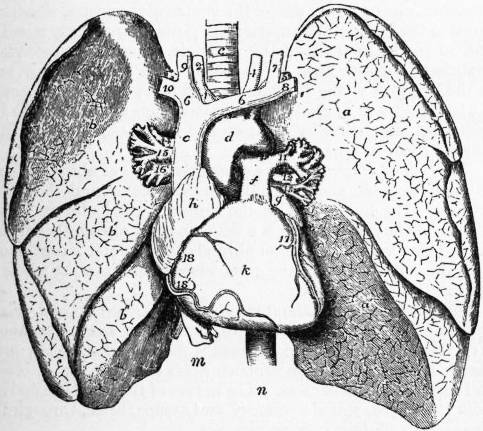
Fig. 1. The Thoracic Viscera.—In this diagram the lungs are turned to the side, and the pericardium removed to display the heart, a, upper, a′, lower lobe of left lung; b, upper, b′, middle, b″, lower lobe of right lung; c, trachea; d, arch of aorta; e, superior vena cava; f, pulmonary artery; g, left, and h, right auricle; k, right, and l, left ventricle; m, inferior vena cava; n, descending aorta; 1, innominate artery; 2, right, and 4, left common carotid artery; 3, right, and 5, left subclavian artery; 6, 6, right and left innominate vein; 7 and 9, left and right internal jugular veins; 8 and 10, left and right subclavian veins; 11, 12, 13, left pulmonary artery, bronchus and vein; 14, 15, 16, right pulmonary bronchus, artery and vein; 17 and 18, left and right coronary arteries.
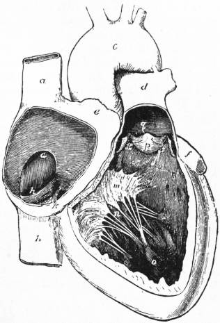
Fig. 2. Cavities of the Right Side of the Heart.—a, superior, and b, inferior vena cava; c, arch of aorta; d, pulmonary artery; e, right, and f, left auricular appendage; g, fossa ovalis; h, Eustachian valve; k, mouth of coronary vein; l, m, n, cusps of the tricuspid valve; o, o, papillary muscles; p, semilunar valve; q, corpus Arantii; r, lunula.
The right auricle is situated at the base of the heart, and its outline is seen on looking at the organ from in front. Into the posterior part of it open the two venae cavae (see fig. 2), the superior (a) above and the inferior (b) below. In front and to the left of the superior vena cava is the right auricular appendage (e) which overlaps the front of the root of the aorta, while running obliquely from the front of one vena cava to the other is a shallow groove called the sulcus terminalis, which indicates the original separation between the true auricle in front and the sinus venosus behind. When the auricle is opened by turning the front wall to the right as a flap the following structures are exposed:
1. A muscular ridge, called the crista terminalis, corresponding to the sulcus terminalis on the exterior.
2. A series of ridges on the anterior wall and in the appendage, running downward from the last and at right angles to it, like the teeth of a comb; these are known as Musculi pectinati.
3. The orifice of the superior vena cava (fig. 2, a) at the upper and back part of the chamber.
4. The orifice of the inferior vena cava (fig. 2, b) at the lower and back part.
5. Attached to the right and lower margins of this opening are the remains of the Eustachian valve (fig. 2, h), which in the foetus directs the blood from the inferior vena cava, through the foramen ovale, into the left auricle.
6. Below and to the left of this is the opening of the coronary sinus (fig. 2, k), which collects most of the veins returning blood from the substance of the heart.
7. Guarding this opening is the coronary valve or valve of Thebesius.
8. On the posterior or septal wall, between the two auricles, is an oval depression, called the fossa ovalis (fig. 2, g), the remains of the original communication between the two auricles. In about a quarter of all normal hearts there is a small valvular communication between the two auricles in the left margin of this depression (see “7th Report of the Committee of Collective Investigation,” J. Anat. and Phys. vol. xxxii. p. 164).
9. The annulus ovalis is the raised margin surrounding this depression.
10. On the left side, opening into the right ventricle, is the right auriculo-ventricular opening.
11. On the right wall, between the two caval openings, may occasionally be seen a slight eminence, the tubercle of Lower, which is supposed to separate the two streams of blood in the embryo.
12. Scattered all over the auricular wall are minute depressions, the foramina Thebesii, some of which receive small veins from the substance of the heart.
The right ventricle is a triangular cavity (see fig. 2) the base of which is largely formed by the auriculo-ventricular orifice. To the left of this it is continued up into the root of the pulmonary artery, and this part is known as the infundibulum. Its anterior wall forms part of the anterior surface of the heart, while its posterior wall is chiefly formed by the septum ventriculorum, between it and the left ventricle. Its lower border is the margo acutus already mentioned. In transverse section it is crescentic, since the septal wall bulges into its cavity. In its interior the following structures are seen:
1. The tricuspid valve (fig. 2, l, m, n) guarding against reflux of blood into the right auricle. This consists of a short cylindrical curtain of fibrous tissue, which projects into the ventricle from the margin of the auriculo-ventricular aperture, while from its free edge three triangular flaps hang down, the bases of which touch one another. These cusps are spoken of as septal, marginal and infundibular, from their position.
2. The chordae tendineae are fine fibrous cords which fasten the cusps to the musculi papillares and ventricular wall, and prevent the valve being turned inside out when the ventricle contracts.
3. The columnae carneae are fleshy columns, and are of three kinds. The first are attached to the wall of the ventricle in their whole length and are merely sculptured in relief, as it were; the second are attached by both ends and are free in the middle; while the third are known as the musculi papillares and are attached by one end to the ventricular wall, the other end giving attachment to the chordae tendineae. These musculi papillares are grouped into three bundles (fig. 2, o).
4. The moderator band is really one of the second kind of columnae carneae which stretches from the septal to the anterior wall of the ventricle.
5. The pulmonary valve (fig. 2, p) at the opening of the pulmonary artery has three crescentic, pocket-like cusps, which, when the ventricle is filling, completely close the aperture, but during the contraction of the ventricle fit into three small niches known as the sinuses of Valsalva, and so are quite out of the way of the escaping blood. In the middle of the free margin of each is a small knob called the corpus Arantii (fig. 2, q), and on each side of this a thin crescent-shaped flap, the lunula (fig. 2, r), which is only made of two layers of endocardium, whereas in the rest of the cusp there is a fibrous backing between these two layers.
The left auricle is situated at the back of the base of the heart, behind and to the left of the right auricle. Running down behind it are the oesophagus and the thoracic aorta. When it is opened it is seen to have a much lighter colour than the other cavities, owing to the greater thickness of its endocardium obscuring the red muscle beneath. There are no musculi pectinati except in the auricular appendage. The openings of the four pulmonary veins are placed two on each side of the posterior wall, but sometimes there may be three on the right side, and only one on the left. On the septal wall is a small depression like the mark of a finger-nail, which corresponds to the anterior part of the fossa ovalis and often forms a valvular communication with the right auricle. The auriculo-ventricular orifice is large and oval, and is directed downward and to the left. Foramina Thebesii and venae minimae cordis are found in this auricle, as in the right, although the chamber is one for arterial or oxidized blood.
At the lower part of the posterior surface of the unopened auricle, lying in the left auriculo-ventricular furrow, is the coronary sinus, which receives most of the veins returning the blood from the heart substance; these are the right and left coronary veins at each extremity and the posterior and left cardiac veins from below. One small vein, called the oblique vein of Marshall, runs down into it across the posterior surface of the auricle, from below the left lower pulmonary vein, and is of morphological interest.
The left ventricle is conical, the base being above, behind and to the right, while the apex corresponds to the apex of the heart and lies opposite the fifth intercostal space, 3½ in. from the mid line. The following structures are seen inside it:—
1. The mitral valve guarding the auriculo-ventricular opening has the same arrangement as the tricuspid, already described, save that there are only two cusps, named marginal and aortic, the latter of which is the larger.
2. The chordae tendineae and columnae carneae resemble those of the right ventricle, though there are only two bundles of musculi papillares instead of three. These are very large. A moderator band has been found as an abnormality (see J. Anat. and Phys. vol. xxx. p. 568).
3. The aortic valve has the same structure as the pulmonary, though the cusps are more massive. From the anterior and left posterior sinuses of Valsalva the coronary arteries arise. That part of the ventricle just below the aortic valve, corresponding to the infundibulum on the right, is known as the aortic vestibule.
The walls of the left ventricle are three times as thick as those of the right, except at the apex, where they are thinner. The septum ventriculorum is concave towards the left ventricle, so that a transverse section of that cavity is nearly circular. The greater part of it has nearly the same thickness as the rest of the left ventricular wall and is muscular, but a small portion of the upper part is membranous and thin, and is called the pars membranacea septi; it lies between the aortic and pulmonary orifices.
Structure of the Heart.—The arrangement of the muscular fibres of the heart is very complicated and only imperfectly known. For details one of the larger manuals, such as Cunningham’s Anatomy (London, 1910), or Gray’s Anatomy (London, 1909), should be consulted. The general scheme is that there are superficial fibres common to the two auricles and two ventricles and deeper fibres for each cavity. Until recently no fibres had been traced from the auricles to the ventricles, though Gaskell predicted that these would be found, and the credit for first demonstrating them is due to Stanley Kent, their details having subsequently been worked out by W. His, Junr., and S. Tawara. The fibres of this auriculo-ventricular bundle begin, in the right auricle, below the opening of the coronary sinus, and run forward on the right side of the auricular septum, below the fossa ovalis, and close to the auriculo-ventricular septum. Above the septal flap of the tricuspid valve they thicken and divide into two main branches, one on either side of the ventricular septum, which run down to the bases of the anterior and posterior papillary muscles, and so reach the walls of the ventricle, where their secondary branches form the fibres of Purkinje. The bundle is best seen in the hearts of young Ruminants, and it is presumably through it that the wave of contraction passes from the auricles to the ventricles (see article by A. Keith and M. Flack, Lancet, 11th of August 1906, p. 359).
The central fibrous body is a triangular mass of fibro-cartilage, situated between the two auriculo-ventricular and the aortic orifices. The upper part of the septum ventriculorum blends with it. The endocardium is a delicate layer of endothelial cells backed by a very thin layer of fibro-elastic tissue; it is continuous with the endothelium of the great vessels and lines the whole of the cavities of the heart.
The heart is roughly about the size of the closed fist and weighs from 8 to 12 oz.; it continues to increase in size up to about fifty years of age, but the increase is more marked in the male than in the female. Each ventricle holds about 4 f. oz. of blood, and each auricle rather less. The nerves of the heart are derived from the vagus, spinal accessory and sympathetic, through the superficial and deep cardiac plexuses.
Embryology.
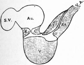
Fig. 3.—Formation of Septa. Diagram of the formation of some of the septa of the heart (viewed from the right side).
S.V. Sinus venosus. Au. Auricle. E.C. Endocardial cushions forming septum intermedium. V. Septum ventriculorum. T. Ar. Septum aorticum intruncus arteriosus. V.A. Ventral aorta.
In the article on the arteries (q.v.) the formation and coalescence of the two primitive ventral aortae to form the heart are noticed, so that we may here start with a straight median tube lying ventral to the pharynx and being prolonged cephalad into the ventral aortae and caudad into the vitelline veins. This soon shows four dilatations, which, from the tail towards the head end, are called the sinus venosus, the auricle, the ventricle and the truncus2 arteriosus. As the tubular heart grows more rapidly than the pericardium which contains it, it becomes bent into the form of an S laid on its side (∾), the ventral convexity being the ventricle and the dorsal the auricle. The passage from the auricle to the ventricle is known as the auricular canal, and in the dorsal and ventral parts of this appear two thickenings known as endocardial cushions, which approach one another and leave a transverse slit between them (fig. 3, E.C.). Eventually these two cushions fuse in the middle line, obliterating the central part of the slit, while the lateral parts remain as the two auriculo-ventricular orifices; this fusion is known as the septum intermedium. From the bottom (ventral convexity) of the ventricle an antero-posterior median septum grows up, which is the septum inferius or septum ventriculorum (fig. 3, V). Posteriorly (caudally) this septum fuses with the septum intermedium, but anteriorly it is free at the lower part of the truncus arteriosus. On referring to the development of the arteries (see Arteries) it will be seen that another septum starts between the last two pairs of aortic arches and grows downward (caudad) until it reaches and joins with the septum inferius just mentioned. This septum aorticum (formed by two ingrowths from the wall of the vessel which fuse later) becomes twisted in such a way that the right ventricle is continuous with the last pair of aortic arches (pulmonary artery), while the left ventricle communicates with the other arches (the permanent ventral aorta and its branches); it joins the septum ventriculorum in the upper part of the ventricular cavity and so forms the pars membranacea septi (fig. 3, T. Ar).
The fate of the sinus venosus and auricle must now be followed. Into the former, at first, only the two vitelline veins open, but later, as they develop, the ducts of Cuvier and the umbilical veins join in (see Veins). As the ducts of Cuvier come from each side the sinus spreads out to meet them and becomes transversely elongated. The slight constriction, which at first is the only separation between the sinus and the auricle, becomes more marked, and later the opening is into the right part of the auricle, and is guarded by two valvular folds of endocardium (the venous valves) which project into that cavity, and are continuous above with a temporary downgrowth from the roof, known as the septum spurium. Later the right side of the sinus enlarges, and so does the right part of the aperture, until the back part of the right side of the auricle and the right part of the sinus venosus are thrown into one, and the only remnants of the partition are the crista terminalis and the Eustachian and Thebesian Valves. The left part of the sinus venosus, which does not enlarge at the same rate as the right part, remains as the coronary sinus. It will now be seen why, in the adult heart, all the veins which open into the right auricle open into its posterior part, behind the crista terminalis. The septum spurium has been referred to as a temporary structure; the real division between the two auricles occurs at a later date than that between the ventricles and to the left of the septum spurium. It is formed by two partitions, the first of which, called the septum primum, grows down from the auricular roof. At first it does not quite reach the endocardial cushions in the auricular canal, already mentioned, but leaves a gap, called the ostium primum, between. This has nothing to do with the foramen ovale, which occurs as an independent perforation higher up, and at first is known as the ostium secundum. When it is established the septum primum grows down and meets the endocardial cushions, and so the ostium primum is obliterated. The septum secundum grows down on the right of the septum primum and is never complete; it grows round and largely overlaps the foramen ovale and its edges form the annulus ovalis, so that, in the later months of foetal life, the foramen ovale is a valvular opening, the floor of which is formed by the septum primum and the margins by the septum secundum. The closure of the foramen is brought about by adhesion of the two septa.
The pulmonary veins of the two sides at first join one another, dorsal to the left auricle, and open into that cavity by a single median trunk, but, as the auricle grows, this trunk and part of the right and left veins are absorbed into its cavity.
The mitral and tricuspid valves are formed by the shortening of the auricular canal which becomes telescoped into the ventricle, and the cusps are the remnants of this telescoping process.
The columnae carneae and chordae tendineae are the remains of a spongy network which originally filled the cavity of the primary ventricle.
The aortic and pulmonary valves are laid down in the ventral aorta, before it is divided into aorta and pulmonary artery, as four endocardial cushions; anterior, posterior and two lateral. The septum aorticum cuts the latter two into two, so that each artery has the rudiments of three cusps.
Abnormalities of the heart are very numerous, and can usually be explained by a knowledge of its development. They often cause grave clinical symptoms. A clear and well-illustrated review of the most important of them will be found in the chapter on congenital disease of the heart in Clinical Applied Anatomy, by C. R. Box and W. McAdam Eccles, London, 1906.
For further details of the embryology of the heart see Oscar Hertwig’s Entwicklungslehre der Wirbeltiere (Jena, 1902); G. Born, “Entwicklung des Säugetierherzens,” Archiv f. mik. Anat. Bd. 33 (1889); W. His, Anatomie menschlicher Embryonen (Leipzig, 1881-1885); Quain’s Anatomy, vol. i. (1908); C. S. Minot, Human Embryology (New York, 1892); and A. Keith, Human Embryology and Morphology (London, 1905).
Comparative Anatomy.
In the Acrania (e.g. lancelet) there is no heart, though the vessels are specially contractile in the ventral part of the pharynx.
In the Cyclostomata (lamprey and hag), and Fishes, the heart has the same arrangement which has been noticed in the human embryo. There is a smooth, thin-walled sinus venosus, a thin reticulate-walled auricle, produced laterally into two appendages, a thick-walled ventricle, and a conus arteriosus containing valves. In addition to these the beginning of the ventral aorta is often thickened and expanded to form a bulbus arteriosus, which is non-contractile, and, strictly speaking, should rather be described with the arteries than with the heart. In relation to human embryology the smooth sinus venosus and reticulated auricle are interesting. Between the auricle and ventricle is the auriculo-ventricular valve, which primarily consists of two cusps, comparable to the two endocardial cushions of the human embryo, though in some forms they may be subdivided. In the interior of the ventricle is a network of muscular trabeculae. The conus arteriosus in the Elasmobranchs (sharks and rays) and Ganoids (sturgeon) is large and provided with several rows of semilunar valves, but in the Cyclostomes (lamprey) and Teleosts (bony fishes) the conus is reduced and only the anterior (cephalic) row of valves retained. With the reduction of the conus the bulbus arteriosus is enlarged. So far the heart is a single tubular organ expanded into various cavities and having the characteristic ∾-shaped form seen in the human embryo; it contains only venous blood which is forced through the gills to be oxidized on its way to the tissues. In the Dipnoi (mud fish), in which rudimentary lungs, as well as gills, are developed, the auricle is divided into two, and the sinus venosus opens into the right auricle. The conus arteriosus too begins to be divided into two chambers, and in Protopterus this division is complete. This division of the heart is one instance in which mammalian ontogeny does not repeat the processes of phylogeny, because, in the human embryo, it has been shown that the ventricular septum appears before the auricular. This want of harmony is sometimes spoken of as the “falsification of the embryological record.”
In the Amphibia there are also two auricles and one ventricle, though in the Urodela (tailed amphibians) the auricular septum is often fenestrated. The sinus venosus is still a separate chamber, and the conus arteriosus, which may contain many or few valves, is usually divided into two by a spiral fold. Structurally the amphibian heart closely resembles the dipnoan, though the increased size of the left auricle is an advance. In the Anura (frogs and toads) the whole ventricle is filled with a spongy network which prevents the arterial and venous blood from the two auricles mixing to any great extent. (For the anatomy and physiology of the frog’s heart, see The Frog, by Milnes Marshall.)
In the Reptiles the ventricular septum begins to appear; this in the lizards is quite incomplete, but in the crocodiles, which are usually regarded as the highest order of living reptiles, the partition has nearly reached the top of the ventricle, and the condition resembles that of the human embryo before the pars membranacea septi is formed. The conus arteriosus becomes included in the ventricular cavity, but the sinus venosus still remains distinct, and its opening into the right ventricle is guarded by two valves which closely resemble the two venous valves in the auricle of the human embryo already referred to.
In the Birds the auricular and ventricular septa are complete; the right ventricle is thin-walled and crescentic in section, as in Man, and the musculi papillares are developed. The left auriculo-ventricular valve has three membranous cusps with chordae tendineae attached to them, but the right auriculo-ventricular valve has a large fleshy cusp without chordae tendineae. The sinus venosus is largely included in the right auricle, but remains of the two venous valves are seen on each side of the orifice of the inferior vena cava.
In the Mammals the structure of the heart corresponds closely with the description of that of Man already given. In the Ornithorynchus, among the Monotremes, the right auriculo-ventricular valve has two fleshy and two membranous cusps, thus showing a resemblance to that of the bird. In the Echidna, the other member of the order, however, both auriculo-ventricular valves are membranous. In the Edentates the remains of the venous valves at the opening of the inferior vena cava are better marked than in other orders. In the Ungulates the moderator band in the right ventricle is especially well developed, and the central fibrous body at the base of the heart is often ossified, forming the os cordis so well known in the heart of the ox.
The position of the heart in the lower mammals is not so oblique as it is in Man.
For further details, see C. Rose, Beitr. z. vergl. Anal. des Herzens der Wirbelthiere Morph. Jahrb., Bd. xvi. (1890); R. Wiedersheim, Vergleichende Anatomie der Wirbelthiere (Jena, 1902) (for literature); also Parker and Haswell’s Zoology (London, 1897).
(F. G. P.)
Heart Disease.—In the early ages of medicine, the absence of correct anatomical, physiological and pathological knowledge prevented diseases of the heart from being recognized with any certainty during life, and almost entirely precluded them from becoming the object of medical treatment. But no sooner did Harvey (1628) publish his discovery of the circulation of the blood, and its dependence on the heart as its central organ, than derangements of the circulation began to be recognized as signs of disease of that central organ. (See also under Vascular System.)
Among the earliest to profit by this discovery and to make important contributions to the literature of diseases of the heart and circulation were, R. Lower (1631-1691), R. Vieussens (1641-1716). H. Boerhave (1668-1738) and the great pathologists at the beginning of the 18th century, G. M. Lancisi (1654-1720), G. B. Morgagni (1682-1771) and J. B. Senac (1693-1770). The works of these writers form very interesting reading, and it is remarkable how careful were the observations made, and how sound the conclusions drawn, by these pioneers of scientific medicine. J. N. Corvisart (1755-1821) was one of the earliest to make practical use of R. T. Auenbrugger’s (1722-1809) invention of percussion to determine the size of the heart. R. T. H. Laennec (1781-1826) was the first to make a scientific application of mediate auscultation to the diagnosis of disease of the chest, by the invention of the stethoscope. J. Bouillaud (1796-1881) extended its use to the diagnosis of disease of the heart. To James Hope (1801-1841) we owe much of the precision we have now attained in diagnosis of valvular disease from abnormalities in the sounds produced during cardiac movements. This short list by no means exhausts the earlier literature on the subject, but each of these names marks an era in the progress of the diagnosis of cardiac disease. In later years the literature on this subject has become very copious.
The heart and great vessels occupy a position immediately to the left of the centre of the thoracic cavity. The anterior surface of the heart is projected against the chest wall and is surrounded on either side by the lungs, which are resonant organs, so that any increase in the size of the heart, “dilatation,” can be detected by percussion. By placing the hand on the chest, palpation, the impulse of the left ventricle, or apex beat, can normally be felt just below and internal to the nipple. Deviations from the normal in the position or force of the apex beat will afford important information as to the nature of the pathological changes in the heart. Thus, displacement downwards and outwards of the apex beat, with a forcible thrusting impulse, will indicate hypertrophy, or increase of the muscular wall and increased driving power of the left ventricle, whereas a similar displacement with a feeble diffuse impulse will indicate dilatation, or over-distension of its cavity from stretching of the walls.
By auscultation, or listening with a suitable instrument named a stethoscope over appropriate areas, we can detect any abnormality in the sounds of the heart, and the presence of murmurs indicative of disease of one or other of the valves of the heart.
The pericardium is a fibro-serous sac which loosely envelops the heart and the origin of the great vessels. Inflammation of this sac, or pericarditis, is apt to occur as a result of rheumatism, more especially in children. It may also occur as a complication of pneumonia. It is a serious affection associated with pain over the heart, fever, shortness of breath, rapid pulse and dilatation of the heart. As a result of the inflammation, fluid may accumulate in the pericardial sac, or the walls of the sac may become adherent to the heart and tend to embarrass its action. In favourable cases, however, recovery may take place without any untoward sequelae.
Diseases of the heart may be classified in two main groups, (1) Disease of the valves, and (2) Disease of the walls of the heart.
1. Valvular Disease.—Inflammation of the valves of the heart, or endocarditis, is one of the most common complications of rheumatism in children and young adults. More severe types, which are apt to prove fatal from a form of blood poisoning, may result when the valves of the heart are attacked by certain micro-organisms, such as the pneumococcus, which is responsible for pneumonia, the streptococcus and the staphylococcus pyogenes, the gonococcus and the influenza bacillus.
As a result of endocarditis, one or more of the valves may be seriously damaged, so that it leaks or becomes incompetent. The valves of the left side of the heart, the aortic and mitral valves, are affected far more commonly than those of the right side. It is indeed comparatively rarely that the latter are attacked. In the process of healing of a damaged valve, scar tissue is formed which has a tendency to contract, so that in some cases the orifice of the valve becomes narrowed, and the resulting stenosis or narrowing gives rise to obstruction of the blood stream. We may thus have incompetence or stenosis of a valve or both combined.
Valvular lesions are detected on auscultation over appropriate areas by the blowing sounds or murmurs to which they give rise, which modify or replace the normal heart sounds. Thus, lesions of the mitral valve give rise to murmurs which are heard at the apex beat of the heart, and lesions of the aortic valves to murmurs which are heard over the aortic area, in the second right intercostal space. Accurate timing of the murmurs in relation to the heart sounds enables us to judge whether the murmur is due to stenosis or incompetence of the valve affected.
If the valvular lesion is severe, it is essential for the proper maintenance of the circulation that certain changes should take place in the heart to compensate for or neutralize the effects of the regurgitation or obstruction, as the case may be. In affections of the aortic valve, the extra work falls on the left ventricle, which enlarges proportionately and undergoes hypertrophy. In affections of the mitral valve the effect is felt primarily by the left auricle, which is a thin walled structure incapable of undergoing the requisite increase in power to resist the backward flow through the mitral orifice in case of leakage, or to overcome the effects of obstruction in case of stenosis. The back pressure is therefore transmitted to the pulmonary circulation, and as the right ventricle is responsible for maintaining the flow of blood through the lungs, the strain and extra work fall on the right ventricle, which in turn enlarges and undergoes hypertrophy. The degree of hypertrophy of the left or right ventricle is thus, up to a certain point, a measure of the extent of the lesion of the aortic or mitral valve respectively. When the effects of the valvular lesion are so neutralized by these structural changes in the heart that the circulation is equably maintained, “compensation” is said to be efficient.