VOLUME II SLICE III Apollodorus to Aral (continued 11)
Reading edition
Fig. 29.—Diagram of the arterial system of A, Scorpio, and B, Limulus. The Roman numerals indicate the body somites and the two figures are adjusted for comparison. ce, Cerebral arteries; sp, supra-spinal or medullary artery; c, caudal artery; l, lateral anastomotic artery of Limulus. The figure B also shows the peculiar neural investiture formed by the cerebral arteries in Limulus and the derivation from this of the arteries to the limbs, III, IV, VI, whereas in Scorpio the latter have a separate origin from the anterior aorta.
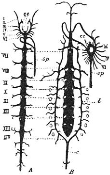
From Lankester, “Limulus an Arachnid.”
It must be pointed out that the presence or absence of such renal excretory tubes opening into the intestine appears to be a question of adaptation to the changed physiological conditions of respiration, and not of morphological significance, since a pair of renal excretory tubes of this nature is found in certain Amphipod Crustacea (Talorchestia, &c.) which have abandoned a purely aquatic life. This view has been accepted and supported by Professors Korschelt and Heider (16). An important fact in its favour was discovered by Laurie (17), who investigated the embryology of two species of Scorpio under Lankester’s direction. It appears that the Malpighian tubes of Scorpio are developed from the mesenteron, viz. that portion of the gut which is formed by the hypoblast, whereas in Hexapod insects the similar caecal tubes are developed from the proctodaeum or in-pushed portion of the gut which is formed from epiblast. In fact it is not possible to maintain that the renal excretory tubes of the gut are of one common origin in the Arthropoda. They have appeared independently in connexion with a change in the excretion of nitrogenous waste in Arachnids, Crustacea, and the other classes of Arthropoda when aerial, as opposed to aquatic, respiration has been established—and they have been formed in some cases from the mesenteron, in other cases from the proctodaeum. Their appearance in the air-breathing Arachnids does not separate those forms from the water-breathing Arachnids which are devoid of them, any more than does their appearance in certain Amphipoda separate those Crustaceans from the other members of the class.
Further, it is pointed out by Korschelt and Heider that the hinder portion of the gut frequently acts in Arthropoda as an organ of nitrogenous excretion in the absence of any special excretory tubules, and that the production of such caeca from its surface in separate lines of descent does not involve any elaborate or unlikely process of growth. In other words, the Malpighian tubes of the terrestrial Arachnida are homoplastic with those of Hexapoda and Myriapoda, and not homogenetic with them. We are compelled to take a similar view of the agreement between the tracheal air-tubes of Arachnida and other tracheate Arthropods. They are homoplasts (see 18) one of another, and do not owe their existence in the various classes compared to a common inheritance of an ancestral tracheal system.
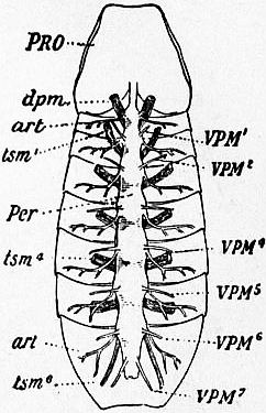
Fig. 30.—View from below of a scorpion (Buthus occitanus) opened and dissected so as to show the pericardium with its muscles, the lateral arteries, and the tergo-sternal muscles.
PRO, Prosoma. dpm, Dorso-plastral muscle. art, Lateral artery. tsm1, Tergo-sternal muscle (labelled dv in fig. 31) of the second (pectiniferous) mesosomatic somite; this is the most anterior pair of the series of six, none are present in the genital somite. tsm4, Tergo-sternal muscle of the fifth mesosomatic somite. tsm6, Tergo-sternal muscle of the enlarged first metasomatic somite. Per, Pericardium. VPM1 to VPM7, The series of seven pairs of veno-pericardiac muscles (labelled pv in fig. 31).
There is some reason to admit the existence of another more anterior pair of these muscles in Scorpio; this would make the number exactly correspond with the number in Limulus.
(After Lankester, Trans. Zool. Soc. vol. xi, 1883.)
Conclusions arising from the Close Affinity of Limulus and Scorpio.—When we consider the relationships of the various classes of Arthropoda, having accepted and established the fact of the close genetic affinity of Limulus and Scorpio, we are led to important conclusions. In such a consideration we have to make use not only of the fact just mentioned, but of three important generalizations which serve as it were as implements for the proper estimation of the relationships of any series of organic forms. First of all there is the generalization that the relationships of the various forms of animals (or of plants) to one another is that of the ultimate twigs of a much-branching genealogical tree. Secondly, identity of structure in two organisms does not necessarily indicate that the identical structure has been inherited from an ancestor common to the two organisms compared (homogeny), but may be due to independent development of a like structure in two different lines of descent (homoplasy). Thirdly, those members of a group which, whilst exhibiting undoubted structural characters indicative of their proper assignment to that group, yet are simpler than and inferior in elaboration of their organization to other members of the group, are not necessarily representatives of the earlier and primitive phases in the development of the group—but are very often examples of retrogressive change or degeneration. The second and third implements of analysis above cited are of the nature of cautions or checks. Agreements are not necessarily due to common inheritance; simplicity is not necessarily primitive and ancestral.
On the other hand, we must not rashly set down agreements as due to “homoplasy” or “convergence of development” if we find two or three or more concurrent agreements. The probability is against agreement being due to homoplasy when the agreement involves a number of really separate (not correlated) coincidences. Whilst the chances are in favour of some one homoplastic coincidence or structural agreement occurring between some member or other of a large group a and some member or other of a large group b, the matter is very different when by such an initial coincidence the two members have been particularized. The chances against these two selected members exhibiting another really independent homoplastic agreement are enormous: let us say 10,000 to 1. The chances against yet another coincidence are a hundred million to one, and against yet one more “coincidence” they are the square of a hundred million to one. Homoplasy can only be assumed when the coincidence is of a simple nature, and is such as may be reasonably supposed to have arisen by the action of like selective conditions upon like material in two separate lines of descent.4
So, too, degeneration is not to be lightly assumed as the explanation of a simplicity of structure. There is a very definite criterion of the simplicity due to degeneration, which can in most cases be applied. Degenerative simplicity is never uniformly distributed over all the structures of the organism. It affects many or nearly all the structures of the body, but leaves some, it may be only one, at a high level of elaboration and complexity. Ancestral simplicity is more uniform, and does not co-exist with specialization and elaboration of a single organ. Further: degeneration cannot be inferred safely by the examination of an isolated case; usually we obtain a series of forms indicating the steps of a change in structure—and what we have to decide is whether the movement has been from the simple to the more complex, or from the more complex to the simple. The feathers of a peacock afford a convenient example of primitive and degenerative simplicity. The highest point of elaboration in colour, pattern and form is shown by the great eye-painted tail feathers. From these we can pass by gradual transitions in two directions, viz. either to the simple lateral tail feathers with a few rami only, developed only on one side of the shaft and of uniform metallic coloration—or to the simple contour feathers of small size, with the usual symmetrical series of numerous rami right and left of the shaft and no remarkable colouring. The one-sided specialization and the peculiar metallic colouring of the lateral tail feathers mark them as the extreme terms of a degenerative series, whilst the symmetry, likeness of constituent parts inter se, and absence of specialized pigment, as well as the fact that they differ little from any average feather of birds in general, mark the contour feather as primitively simple, and as the starting-point from which the highly elaborated eye-painted tail feather has gradually evolved.
Applying these principles to the consideration of the Arachnida, we arrive at the conclusion that the smaller and simpler Arachnids are not the more primitive, but that the Acari or mites are, in fact, a degenerate group. This was maintained by Lankester in 1878 (19), again in 1881 (20); it was subsequently announced as a novelty by Claus in 1885 (21). Though the aquatic members of a class of animals are in some instances derived from terrestrial forms, the usual transition is from an aquatic ancestry to more recent land-living forms. There is no doubt, from a consideration of the facts of structure, that the aquatic water-breathing Arachnids, represented in the past by the Eurypterines and to-day by the sole survivor Limulus, have preceded the terrestrial air-breathing forms of that group. Hence we see at once that the better-known Arachnida form a series, leading from Limulus-like aquatic creatures through scorpions, spiders and harvest-men, to the degenerate Acari or mites. The spiders are specialized and reduced in apparent complexity, as compared with the scorpions, but they cannot be regarded as degenerate since the concentration of structure which occurs in them results in greater efficiency and power than are exhibited by the scorpion. The determination of the relative degree of perfection of organization attained by two animals compared is difficult when we introduce, as seems inevitable, the question of efficiency and power, and do not confine the question to the perfection of morphological development. We have no measure of the degree of power manifested by various animals—though it would be possible to arrive at some conclusions as to how that “power” should be estimated. It is not possible here to discuss that matter further. We must be content to point out that it seems that the spiders, the pedipalps, and other large Arachnids have not been derived from the scorpions directly, but have independently developed from aquatic ancestors, and from one of these independent groups—probably through the harvest-men from the spiders—the Acari have finally resulted.
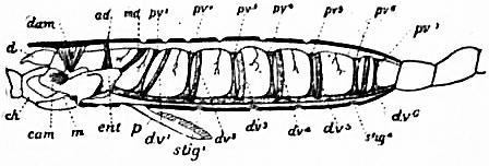
After Beck, Trans. Zool. Soc. Vol. xi., 1883.
Fig. 31.—Diagram of a lateral view of a longitudinal section of a scorpion.
d, Chelicera. ch, Chela. cam, Camerostome. m, Mouth. ent, Entosternum. p, Pecten. stig1, First pulmonary aperture. stig4, Fourth pulmonary aperture. dam, Muscle from carapace to a praeoral entosclerite. ad, Muscle from carapace to entosternum. md, Muscle from tergite of genital somite to entosternum (same as dpm in fig. 30). dv1 to dv6, Dorso-ventral muscles (same as the series labelled tsm in fig. 30). pv1 to pv7, The seven veno-pericardiac muscles of the right side (labelled VPM in fig. 30).
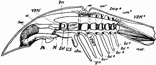
After Benham, Trans Zool. Soc. vol. xi, 1883.
Fig. 32.—Diagram of a lateral view of a longitudinal section of Limulus.
Suc, Suctorial pharynx. al, Alimentary canal. Ph, Pharynx. M, Mouth. Est, Entosternum. VS, Ventral venous sinus. chi, Chilaria. go, Genital operculum. br1 to br5, Branchial appendages, met, Unsegmented metasoma. Entap4, Fourth dorsal entapophysis of left side. tsm, Tergo-sternal muscles, six pairs as in Scorpio (labelled dv in fig. 31). VPM1 to VPM8, The eight pairs of veno-pericardiac muscles (labelled pv in fig. 31). VPM1 is probably represented in Scorpio, though not marked in figs. 30 and 31.
From Lankester, “Limulua an Arachnid.”
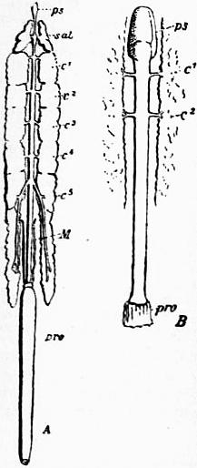
Fig. 33.—The alimentary canal and gastric glands of a scorpion (A) and of Limulus (B).
ps, Muscular suctorial enlargement of the pharynx. sal, Prosomatic pair of gastric caeca in Scorpio, called salivary glands by some writers. c1, and c2, The anterior two pairs of gastric caeca and ducts of the mesosomatic region. c3, c4 and c5. Caeca and ducts of Scorpio not represented in Limulus. M, The Malpighian or renal caecal diverticula of Scorpio. pro, The proctodaeum or portion of gut leading to anus and formed embryologically by an inversion of the epiblast at that orifice.
Leaving that question for consideration in connexion with the systematic statement of the characters of the various groups of Arachnida which follows on p. 299, it is well now to consider the following question, viz., seeing that Limulus and Scorpio are such highly developed and specialized forms, and that they seem to constitute as it were the first and second steps in the series of recognized Arachnida—what do we know, or what are we led to suppose with regard to the more primitive Arachnida from which the Eurypterines and Limulus and Scorpio have sprung? Do we know in the recent or fossil condition any such primitive Arachnids? Such a question is not only legitimate, but prompted by the analogy of at least one other great class of Arthropods. The great Arthropod class, the Crustacea, presents to the zoologist at the present day an immense range of forms, comprising the primitive phyllopods, the minute copepods, the parasitic cirrhipedes and the powerful crabs and lobsters, and the highly elaborated sand-hoppers and slaters. It has been insisted, by those who accepted Lankester’s original doctrine of the direct or genetic affinity of the Chaetopoda and Arthropoda, that Apus and Branchipus really come very near to the ancestral forms which connected those two great branches of Appendiculate (Parapodiate) animals. On the other hand, the land crabs are at an immense distance from these simple forms. The record of the Crustacean family-tree is, in fact, a fairly complete one—the lower primitive members of the group are still represented by living forms in great abundance. In the case of the Arachnida, if we have to start their genealogical history with Limulus and Scorpio, we are much in the same position as we should be in dealing with the Crustacea, were the whole of the Entomostraca and the whole of the Arthrostraca wiped out of existence and record. There is no possibility of doubt that the series of forms corresponding in the Arachnidan line of descent, to the forms distinguished in the Crustacean line of descent as the lower grade—the Entomostraca—have ceased to exist, and not only so, but have left little evidence in the form of fossils as to their former existence and nature. It must, however, be admitted as probable that we should find some evidence, in ancient rocks or in the deep sea, of the early more primitive Arachnids. And it must be remembered that such forms must be expected to exhibit, when found, differences from Limulus and Scorpio as great as those which separate Apus and Cancer. The existing Arachnida, like the higher Crustacea, are “nomomeristic,” that is to say, have a fixed typical number of somites to the body. Further, they are like the higher Crustacea, “somatotagmic,” that is to say, they have this limited set of somites grouped in three (or more) “tagmata” or regions of a fixed number of similarly modified somites—each tagma differing in the modification of its fixed number of somites from that characterizing a neighbouring “tagma.” The most primitive among the lower Crustacea, on the other hand, for example, the Phyllopoda, have not a fixed number of somites, some genera—even allied species—have more, some less, within wide limits; they are “anomomeristic.” They also, as is generally the case with anomomeristic animals, do not exhibit any conformity to a fixed plan of “tagmatism” or division of the somites of the body into regions sharply marked off from one another; the head or prosomatic tagma is followed by a trunk consisting of somites which either graduate in character as we pass along the series or exhibit a large variety in different genera, families and orders, of grouping of the somites. They are anomotagmic, as well as anomomeristic.
When it is admitted—as seems to be reasonable—that the primitive Arachnida would, like the primitive Crustacea, be anomomeristic and anomotagmic, we shall not demand of claimants for the rank of primitive Arachnids agreement with Limulus and Scorpio in respect of the exact number of their somites and the exact grouping of those somites; and when we see how diverse are the modifications of the branches of the appendages both in Arachnida and in other classes of Arthropoda (q.v.), we shall not over-estimate a difference in the form of this or that appendage exhibited by the claimant as compared with the higher Arachnids. With those considerations in mind, the claim of the extinct group of the trilobites to be considered as representatives of the lower and more primitive steps in the Arachnidan genealogy must, it seems, receive a favourable judgment. They differ from the Crustacea in that they have only a single pair of prae-oral appendages, the second pair being definitely developed as mandibles. This fact renders their association with the Crustacea impossible, if classification is to be the expression of genetic affinity inferred from structural coincidence. On the contrary, this particular point is one in which they agree with the higher Arachnida. But little is known of the structure of these extinct animals; we are therefore compelled to deal with such special points of resemblance and difference as their remains still exhibit. They had lateral eyes5 which resemble no known eyes so closely as the lateral eyes of Limulus. The general form and structure of their prosomatic carapace are in many striking features identical with that of Limulus. The trilobation of the head and body—due to the expansion and flattening of the sides or “pleura” of the tegumentary skeleton—is so closely repeated in the young of Limulus that the latter has been called “the trilobite stage” of Limulus (fig. 42 compared with fig. 41). No Crustacean exhibits this trilobite form. But most important of the evidences presented by the trilobites of affinity with Limulus, and therefore with the Arachnida, is the tendency less marked in some, strongly carried out in others, to form a pygidial or telsonic shield—a fusion of the posterior somites of the body, which is precisely identical in character with the metasomatic carapace of Limulus. When to this is added the fact that a post-anal spine is developed to a large size in some trilobites (fig. 38), like that of Limulus and Scorpio, and that lateral spines on the pleura of the somites are frequent as in Limulus, and that neither metasomatic fusion of somites nor post-anal spine, nor lateral pleural spines are found in any Crustacean, nor all three together in any Arthropod besides the trilobites and Limulus—the claim of the trilobites to be considered as representing one order of a lower grade of Arachnida, comparable to the grade Entomostraca of the Crustacea, seems to be established.
The fact that the single pair of prae-oral appendages of trilobites, known only as yet in one genus, is in that particular case a pair of uni-ramose antennae—does not render the association of trilobites and Arachnids improbable. Although the prae-oral pair of appendages in the higher Arachnida is usually chelate, it is not always so; in spiders it is not so; nor in many Acari. The bi-ramose structure of the post-oral limbs, demonstrated by Beecher in the trilobite Triarthrus, is no more inconsistent with its claim to be a primitive Arachnid than is the foliaceous modification of the limbs in Phyllopods inconsistent with their relationship to the Arthrostracous Crustaceans such as Gammarus and Oniscus.
Thus, then, it seems that we have in the trilobites the representatives of the lower phases of the Arachnidan pedigree. The simple anomomeristic trilobite, with its equi-formal somites and equi-formal appendages, is one term of the series which ends in the even more simple but degenerate Acari. Between the two and at the highest point of the arc, so far as morphological differentiation is concerned, stands the scorpion; near to it in the trilobite’s direction (that is, on the ascending side) are Limulus and the Eurypterines—with a long gap, due to obliteration of the record, separating them from the trilobite. On the other side—tending downwards from the scorpion towards the Acari—are the Pedipalpi, the spiders, the book-scorpions, the harvest-men and the water-mites.
The strange nobody-crabs or Pycnogonids occupy a place on the ascending half of the arc below the Eurypterines and Limulus. They are strangely modified and degenerate, but seem to be (as explained in the systematic review) the remnant of an Arachnidan group holding the same relation to the scorpions which the Laemodipoda hold to the Podophthalmate Crustacea.
We have now to offer a classification of the Arachnida and to pass in review the larger groups, with a brief statement of their structural characteristics.
In the bibliography at the close of this article (referred to by leaded arabic numerals in brackets throughout these pages), the titles of works are given which contain detailed information as to the genera and species of each order or sub-order, their geographical distribution and their habits and economy so far as they have been ascertained. The limits of space do not permit of a fuller treatment of those matters here.
Tabular Classification6 of the Arachnida.
Class. ARACHNIDA.
Grade A. ANOMOMERISTICA. Sub-Class. TRILOBITAE. Orders. Not satisfactorily determined. Grade B. NOMOMERISTICA. Sub-Class I. PANTOPODA. Order 1. Nymphonomorpha. Order 2. Ascorhynchomorpha. Order 3. Pycnogonomorpha. Sub-Class II. EU-ARACHNIDA. Grade a. delobranchia, Lankester (vel hydropneustea, Pocock). Order 1. Xiphosura. Order 2. Gigantostraca. Grade b. embolobranchia, Lankester (vel aeropneustea, Pocock). Section α. Pectinifera. Order 1. Scorpionidea. Sub-order a. Apoxypoda. Sub-order b. Dionychopoda. Section β. Epectinata. Order 2. Pedipalpi. Sub-order a. Uropygi. Tribe 1. Urotricha. Tribe 2. Tartarides. Sub-order b. Amblypygi. Order 3. Araneae. Sub-order a. Mesothelae. Sub-order b. Opisthothelae. Tribe 1. Mygalomorphae. Tribe 2. Arachnomorphae. Order 4. Palpigradi (= Microthelyphonidae). Order 5. Solifugae (= Mycetophorae). Order 6. Pseudoscorpiones (= Chelonethi). Sub-order a. Panctenodactyli. Sub-order b. Hemirtenodactyli. Order 7. Podogona (= Ricinulel). Order 8. Opiliones. Sub-order a. Laniatores. Sub-order b. Palpatores. Sub-order c. Anepignathi. Order 9. Rhynchostomi (= Acari). Sub-order a. Notostigmata. Sub-order b. Cryptostigmata. Sub-order c. Metastigmata. Sub-order d. Prostigmata. Sub-order e. Astigmata. Sub-order f. Vermiformia. Sub-order g. Tetrapoda.
Class. ARACHNIDA.—Euarthropoda having two prosthomeres (somites which have passed from a post-oral to a prae-oral position), the appendages of the first represented by eyes, of the second by solitary rami which are rarely antenniform, more usually chelate. A tendency is exhibited to the formation of a metasomatic as well as a prosomatic carapace by fusion of the tergal surfaces of the somites. Intermediate somites forming a mesosoma occur, but tend to fuse superficially with the metasomatic carapace or to become co-ordinated with the somites of the metasoma, whether fused or distinct to form one region, the opisthosoma (abdomen of authors). In the most highly developed forms the two anterior divisions (tagmata) of the body, prosoma and mesosoma, each exhibit six pairs of limbs, pediform and plate-like respectively, whilst the metasoma consists of six limbless somites and a post-anal spine. The genital apertures are placed in the first somite following the prosoma, excepting where a praegenital somite, usually suppressed, is retained. Little is known of the form of the appendages in the lowest archaic Arachnida, but the tendency of those of the prosomatic somites has been (as in the Crustacea) to pass from a generalized bi-ramose or multi-ramose form to that of uni-ramose antennae, chelae and walking legs.
The Arachnida are divisible into two grades of structure—according to the fixity or non-fixity of the number of somites building up the body:—
Grade A (of the Arachnida). ANOMOMERISTICA.—Extinct archaic Arachnida, in which (as in the Entomostracous Crustacea) the number of well-developed somites may be more or less than eighteen and may be grouped only as head (prosoma) and trunk or may be further differentiated. A telsonic tergal shield of greater or less size is always present, which may be imperfectly divided into well-marked but immovable tergites indicating incompletely differentiated somites. The single pair of palpiform appendages in front of the mouth has been found in one instance to be antenniform, whilst the numerous post-oral appendages in the same genus were bi-ramose. The position of the genital apertures is not known. Compound lateral eyes present; median eyes wanting. The body and head have the two pleural regions of each somite flattened and expanded on either side of the true gut-holding body-axis. Hence the name of the sub-class signifying tri-lobed, a condition realized also in the Xiphosurous Arachnids. The members of this group, whilst resembling the lower Crustacea (as all lower groups of a branching genealogical tree must do), differ from them essentially in that the head exhibits only one prosthomere (in addition to the eye-bearing prosthomere) with palpiform appendages (as in all Arachnida) instead of two. The Anomomeristic Arachnida form a single sub-class, of which only imperfect fossil remains are known.
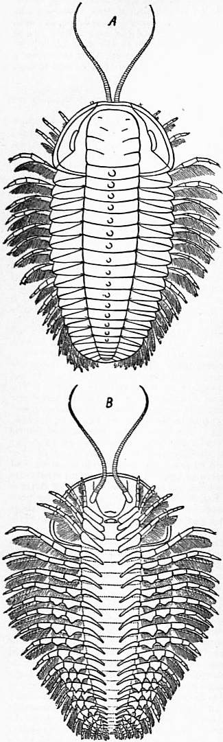
Fig. 34.—Restoration of Triarthrus Becki, Green, as determined by Beecher from specimens obtained from the Utica Slates (Ordovician), New York. A, dorsal; B, ventral surface. In the latter the single pair of antennae springing up from each side of the camerostome or hypostome or upper lip-lobe are seen. Four pairs of appendages besides these are seen to belong to the cephalic tergum. All the appendages are pediform and bi-ramose; all have a prominent gnathobase, and in all the exopodite carries a comb-like series of secondary processes.
Sub-class (of the Anomomeristica). TRILOBITAE.—The single sub-class Trilobitae constitutes the grade Anomomeristica. It has been variously divided into orders by a number of writers. The greater or less evolution and specialization of the metasomatic carapace appears to be the most important basis for classification—but this has not been made use of in the latest attempts at drawing up a system of the Trilobites. The form of the middle and lateral regions of the prosomatic shield has been used, and an excessive importance attached to the demarcation of certain areas in that structure. Sutures are stated to mark off some of these pieces, but in the proper sense of that term as applied to the skeletal structures of the Vertebrata, no sutures exist in the chitinous cuticle of Arthropoda. That any partial fusion of originally distinct chitinous plates takes place in the cephalic shield of Trilobites, comparable to the partial fusion of bony pieces by suture in Vertebrata, is a suggestion contrary to fact.
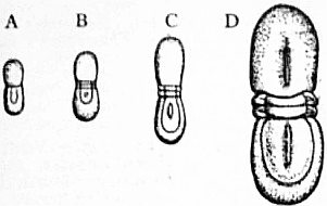
The Trilobites are known only as fossils, mostly Silurian and prae-Silurian; a few are found in Carboniferous and Permian strata. As many as two thousand species are known. Genera with small metasomatic carapace, consisting of three to six fused segments distinctly marked though not separated by soft membrane, are Harpes, Paradoxides and Triarthrus (fig. 34). In Calymene, Homalonotus and Phacops (fig. 38) from six to sixteen segments are clearly marked by ridges and grooves in the metasomatic tagma, whilst in Illaenus the shield so formed is large but no somites are marked out on its surface. In this genus ten free somites (mesosoma) occur between the prosomatic and metasomatic carapaces. Asaphus and Megalaspis (fig. 39) are similarly constituted. In Agnostus (fig. 40) the anterior and posterior carapaces constitute almost the entire body, the two carapaces being connected by a mid-region of only two free somites. It has been held that the forms with a small number of somites marked in the posterior carapace and numerous free somites between the anterior and posterior carapace, must be considered as anterior to those in which a great number of posterior somites are traceable in the metasomatic carapace, and that those in which the traces of distinct somites in the posterior or metasomatic carapace are most completely absent must be regarded as derived from those in which somites are well marked in the posterior carapace and similar in appearance to the free somites. The genus Agnostus, which belongs to the last category, occurs abundantly in Cambrian strata and is one of the earliest forms known. This would lead to the supposition that the great development of metasomatic carapace is a primitive and not a late character, were it not for the fact that Paradoxides and Atops, with an inconspicuous telsonic carapace and numerous free somites, are also Cambrian in age, the latter indeed anterior in horizon to Agnostus.
On the other hand, it may well be doubted whether the pygidial or posterior carapace is primarily due to a fusion of the tergites of somites which were previously movable and well developed. The posterior carapace of the Trilobites and of Limulus is probably enough in origin a telsonic carapace—that is to say, is the tergum of the last segment of the body which carries the anus. From the front of this region new segments are produced in the first instance, and are added during growth to the existing series. This telson may enlarge, it may possibly even become internally and sternally developed as partially separate somites, and the tergum may remain without trace of somite formation, or, as appears to be the case in Limulus, the telson gives rise to a few well-marked somites (mesosoma and two others) and then enlarges without further trace of segmentation, whilst the chitinous integument which develops in increasing thickness on the terga as growth advances welds together the unsegmented telson and the somites in front of it, which were previously marked by separate tergal thickenings. It must always be remembered that we are liable (especially in the case of fossilized integuments) to attach an unwarranted interpretation to the mere discontinuity or continuity of the thickened plates of chitinous cuticle on the back of an Arthropod. These plates may fuse, and yet the somites to which they belong may remain distinct, and each have its pair of appendages well developed. On the other hand, an unusually large tergal plate, whether terminal or in the series, is not always due to fusion of the dorsal plates of once-separate somites, but is often a case of growth and enlargement of a single somite without formation of any trace of a new somite. For the literature of Trilobites see (22*).
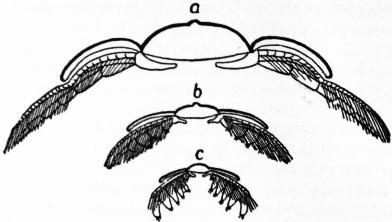
Fig. 35.—Triarthrus Becki, Green. a, Restored thoracic limbs in transverse section of the animal; b, section across a posterior somite; c, section across one of the sub-terminal somites.
(After Beecher.)
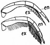
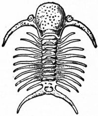
Fig. 36.—Triarthrus Becki, Green. Dorsal view of second thoracic leg with and without setae. en, Inner ramus; ex, Outer ramus. Fig. 37.—Deiphon Forbesii, Barr. One of the Cheiruridae. Silurian Bohemia.
(After Beecher.) (From Zittel’s Palaeontology.)
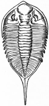
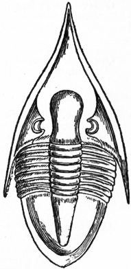
Fig. 38.—Dalmanites Kmulurus, Green. One of the Phacopidae, from the Silurian, New York. Fig. 39.—Megalaspis extenuatus. One of the Asaphidae allied to Illaenus, from the Ordovician of East Gothland, Sweden.
(From Zittel.) (From Zittel.)
Grade B (of the Aracknida) NOMOMERISTICA.—Arachnida in which, excluding from consideration the eye-bearing prosthomere, the somites are primarily (that is to say, in the common ancestor of the grade) grouped in three regions of six—(a) the “prosoma” with palpiform appendages, (b) the “mesosoma” with plate-like appendages, and (c) the “metasoma” with suppressed appendages. A somite placed between the prosoma and mesosoma —the prae-genital somite—appears to have belonged originally to the prosomatic series (which with its ocular prosthomere and palpiform limbs [Pantopoda], would thus consist of eight somites), but to have been gradually reduced. In living Arachnids, excepting the Pantopoda, it is either fused (with loss of its appendages) with the prosoma (Limulus,7 Scorpio), after embryonic appearance, or is retained as a rudimentary, separate, detached somite in front of the mesosoma, or disappears altogether (excalation). The atrophy and total disappearance of ancestrally well-marked somites frequently take place (as in all Arthropoda) at the posterior extremity of the body, whilst excalation of somites may occur at the constricted areas which often separate adjacent “regions,” though there are very few instances in which it has been recognized. Concentration of the organ-systems by fusion of neighbouring regions (prosoma, mesosoma, metasoma), previously distinct, has frequently occurred, together with obliteration of the muscular and chitinous structures indicative of distinct somites. This concentration and obliteration of somites, often accompanied by dislocation of important segmental structures (such as appendages and nerve-ganglia), may lead to highly developed specialization (individuation, H. Spencer), as in the Araneae and Opiliones, and, on the other hand, may terminate in simplification and degeneration, as in the Acari.
Fig. 40.—Four stages in the development of the trilobite Agnostus nudus. A, Youngest stage with no mesosomatic somites; B and C, stages with two mesosomatic somites between the prosomatic and telsonic carapaces; D, adult condition, still with only two free mesosomatic somites.
(From Korschelt and Heider.)
From Korschelt and Heider, after Barrande.
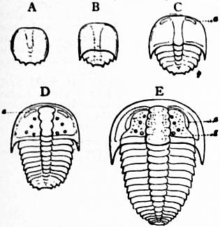
Fig. 41.—Five Stages in the development of the trilobite Sao hirsuta.
A, Youngest stage. B, Older stage with distinct pygidial carapace. C, Stage with two free mesosomatic somites between the prosomatic and telsonic carapaces. D, Stace with seven free intermediate somites. E, Stave with twelve free somites; the telsonic carapace has not increased in size. a, Lateral eye. g, So-called facial “suture” (not really a suture). p, Telsonic carapace.
Fig. 42.—So-called “trilobite stage” of Limulus polyphemus. A, Dorsal; B, ventral view.
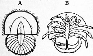
(from Korschelt and Heider, after Leuckart.)
The most important general change which has affected the structure of the nomomeristic Arachnida in the course of their historic development is the transition from an aquatic to a terrestrial life. This has been accompanied by the conversion of the lamelliform gill-plates into lamelliform lung-plates, and later the development from the lung-chambers, and at independent sites, of tracheae or air-tubes (by adaptation of the vasifactive tissue of the blood-vessels) similar to those independently developed in Peripatus, Diplopoda, Hexapoda and Chilopoda. Probably tracheae have developed independently by the same process in several groups of tracheate Arachnids. The nomomeristic Arachnids comprise two sub-classes—one a very small degenerate offshoot from early ancestors; the other, the great bulk of the class.
Sub-Class I. (of the Nomomeristica). PANTOPODA.—Nomomeristic Arachnids, in which the somites corresponding to mesosoma and metasoma have entirely aborted. The seventh, and sometimes the eighth, leg-bearing somite is present and has its leg-like appendages fully developed. Monomeniscous eyes with a double (really triple) cell-layer formed by invagination, as in the Eu-arachnida, are present The Pantopoda stand in the same relation to Limulus and Scorpio that Cyamus holds to the thoracostracous Crustacea. The reduction of the organism to seven leg-bearing somites, of which the first pair, as in so many Eu-arachnida, are chelate, is a form of degeneration connected with a peculiar quasi-parasitic habit resembling that of the crustacean Laemodipoda. The genital pores are situate at the base of the 7th pair of limbs, and may be repeated on the 4th, 5th, and 6th. In all known Pantopoda the size of the body is quite minute as compared with that of the limbs: the alimentary canal sends a long caecum into each leg (cf. the Araneae) and the genital products are developed in gonocoels also placed in the legs.
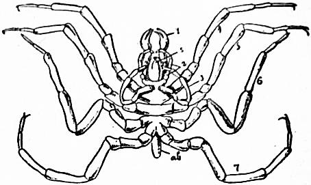
From Parker and Harwell’s Text-book of Zoology, after Hoek.
Fig. 43.—One of the Nymphonomorphous Pantopoda, Nymphon hispidum, showing the seven pairs of appendages 1 to 7; ab, the rudimentary opisthosoma; s, the mouth-bearing proboscis.
The Pantopoda are divided into three orders, the characters of which are dependent on variation in the presence of the full number of legs.
Order 1. (of the Pantopoda). Nymphonomorpha, Pocock (nov.) (fig. 43).—In primitive forms belonging to the family Nymphonidae the full complement of appendages is retained—the 1st (mandibular), the 2nd (palpiform), and the 3rd (ovigerous) pairs being well developed in both sexes. In certain derivative forms constituting the family Pallenidae, however, the appendages of the 2nd pair are either rudimentary or atrophied altogether.
Two families: 1. Nymphonidae (genus Nymphon), and 2. Pallenidae (genus Pallene).
Order 2. Ascorhynchomorpha, Pocock (nov.).—Appendages of the 2nd and 3rd pairs retained and developed, as in the more primitive types of Nymphonomorpha; but those of the 1st pair are either rudimentary, as in the Ascorhynchidae, or atrophied, as in the Colossendeidae. In the latter a further specialization is shown in the fusion of the body segments.
Two families. 1. Ascorhynchidae (genera Ascorhynchus and Ammothea); 2. Colossendeidae (genera Colossendeis and Discoarachne).
Order 3. Pycnogonomorpha, Pocock (nov.).—Derivative forms in which the reduction in number of the anterior appendages is carried farther than in the other orders, reaching its extreme in the Pycnogonidae, where the 1st and 2nd pairs are absent in both sexes, and the 3rd pair also are absent in the female. In the Hannoniidae, however, which resemble the Pycnogonidae in the absence of the 3rd pair in the female and of the 2nd pair in both sexes, the 1st pair are retained in both sexes.
Two families: 1. Hannoniidae (genus Hannonia); 2. Pycnogonidae (genera Pycnogonum and Phoxichilus).
Remarks.—The Pantopoda are not known in the fossil condition. They are entirely marine, and are not uncommon in the coralline zone of the sea-coast. The species are few, not more than fifty (23). Some large species of peculiar genera are taken at great depths. Their movements are extremely sluggish. They are especially remarkable for the small size of the body and the extension of viscera into the legs. Their structure is eminently that of degenerate forms. Many frequent growths of coralline Algae and hydroid polyps, upon the juices of which they feed, and in some cases a species of gall is produced in hydroids by the penetration of the larval Pantopod into the tissues of the polyp.
Sub-Class II. (of the Nomomeristic Arachnida). EU-ARACHNIDA.—These start from highly developed and specialized aquatic branchiferous forms, exhibiting a prosoma with six pediform pairs of appendages, an intermediate prae-genital somite, a mesosoma of six somites bearing lamelliform pairs of appendages, and a metasoma of six somites devoid of appendages, and the last provided with a post-anal spine. Median eyes are present, which are monomeniscous, with distinct retinal and corneagenous cell-layers, and placed centrally on the prosoma. Lateral eyes also may be present, arranged in lateral groups, and having a single or double cell-layer beneath the lens. The first pair of limbs is often chelate or prehensile, rarely antenniform; whilst the second, third and fourth may also be chelate, or may be simple palps or walking legs.
An internal skeletal plate, the so-called “entosternite” of fibro-cartilaginous tissue, to which many muscles are attached, is placed between the nerve-cords and the alimentary tract in the prosoma of the larger forms (Limulus, Scorpio, Mygale). In the same and other leading forms a pair of much-coiled glandular tubes, the coxal glands (coelomocoels in origin), is found with a duct opening on the coxa of the fifth pair of appendages of the prosoma. The vascular system is highly developed (in the non-degenerate forms); large arterial branches closely accompany or envelop the chief nerves; capillaries are well developed. The blood-corpuscles are large amoebiform cells, and the blood-plasma is coloured blue by haemocyanin.
The alimentary canal is uncoiled and cylindrical, and gives rise laterally to large gastric glands, which are more than a single pair in number (two to six pairs), and may assume the form of simple caeca. The mouth is minute and the pharynx is always suctorial, never gizzard-like. The gonadial tubes (gonocoels or gonadial coelom) are originally reticular and paired, though they may be reduced to a simpler condition. They open on the first somite of the mesosoma. In the numerous degenerate forms simplification occurs by obliteration of the demarcations of somites and the fusion of body-regions, together with a gradual suppression of the lamelliferous respiratory organs and the substitution for them of tracheae, which, in their turn, in the smaller and most reduced members of the group, may also disappear.
The Eu-arachnida are divided into two grades with reference to the condition of the respiratory organs as adapted to aquatic or terrestrial life.
Grade a (of the Eu-arachnida). delobranchia (Hydropheustea).
Mesosomatic segments furnished with large plate-like appendages, the 1st pair acting as the genital operculum, the remaining pairs being provided with branchial lamellae fitted for breathing oxygen dissolved in water. The prae-genital somite partially or wholly obliterated in the adult. The mouth lying far back, so that the basal segments of all the prosomatic appendages, excepting those of the 1st pair, are capable of acting as masticatory organs. Lateral eyes consisting of a densely packed group of eye-units (“compound” eyes).
ORDER 1. XIPHOSURA.—The prae-genital somite fuses in the embryo with the prosoma and disappears (see fig. 19). Not free-swimming, none of the prosomatic appendages modified to act as paddles; segments of the mesosoma and metasoma (= opisthosoma) not more than ten in number, distinct or coalesced.
Family—Limulidae (Limulus).
” *Belinuridae (Belinurus, Aglaspis, Prestwichia).
” *Hemiaspidae (Hemiaspis, Bunodes).
Fig. 44.—Dorsal view of Limulus polyphemus, Latr.
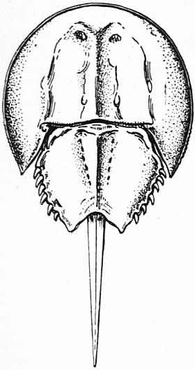
(From Parker and Haswell, Text book of Zoology after Leuckart.)
Remarks.-The Xiphosura are marine in habit, frequenting the shore. They are represented at the present day by the single genus Limulus (figs. 44 and 45; also figs. 7, 9, 11, to 15 and 20), often termed the king-crab, which occurs on the American coast of the Atlantic Ocean, but not on its eastern coasts, and on the Asiatic coast of the Pacific. The Atlantic species (L. polyphemus) is common on the coasts of the United States, and is known as the king-crab or horse-shoe crab. A single specimen was found in the harbour of Copenhagen in the 18th century, having presumably been carried over by a ship to which it clung.
A species of Limulus is found in the Buntersandstein of the Vosges; L. Walchi is abundant in the Oolitic lithographic slates of Bavaria.
Fig. 45.—Ventral view of Limulus polyphemus.
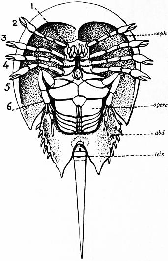
1 to 6, The six prosomatic pairs of appendages. abd, the solid opisthosomatic carapace. tels, the post-anal spine (not the telson as the lettering would seem to imply, but only its post-anal portion). operc, the fused first pair of mesosomatic appendages forming the genital operculum.
(From Parker and Haswell, Text book of Zoology, after Leuckart.)
The genera Belinurus, Aglaspis, Prestwichia, Hemiaspis and Bunodes consist of small forms which occur in Palaeozoic rocks. In none of them are the appendages known, but in the form of the two carapaces and the presence of free somites they are distinctly intermediate between Limulus and the Trilobitae. The young form of Limulus itself (fig. 40) is also similar to a Trilobite so far as its segmentation and trilobation are concerned. The lateral eyes of Limulus appear to be identical in structure and position with those of certain Trilobitae.
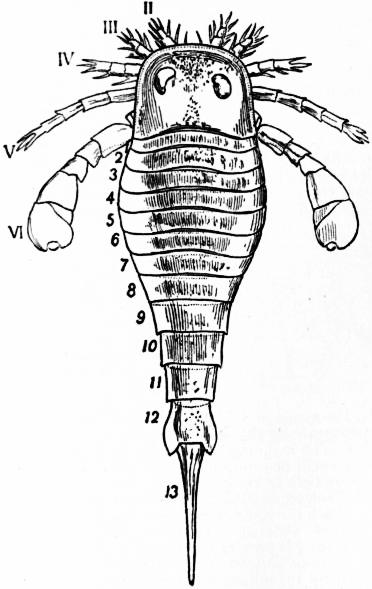
Fig. 46.—Eurypterus Fischeri, Eichwald. Silurian of Rootzikil. Restoration after Schmidt. The dorsal aspect is presented showing the prosomatic shield with paired compound eyes and the prosomatic appendages II. to VI. The small first pair of appendages is concealed from view by the carapace, 1 to 12 are the somites of the opisthosoma; 13, the post-anal spine.
(From Zittel’s Text-book of Palaeontology, The Macmillan Co, New York, 1896.)
Order 2. Gigantostraca (figs. 46, 47).—Free-swimming forms, with the appendages of the 6th or 5th and 6th pairs flattened or lengthened to act as oars; segments of mesosoma and metasoma (= opisthosoma), twelve in number.
Appendages of anterior pair very large and chelate.
Sub-order Pterygotomorpha, Pterygotidae (Pterygotus).
Appendages of anterior pair minute and chelate.
Sub-order Eurypteromorpha Stylonuridae (Stylonurus). Eurypteridae (Eurypterus, Slimonia).
From Zittel’s Palaeontology.
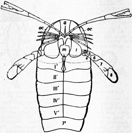
Fig. 47.—Pterygotus osiliensis, Schmidt. Silurian of Rootzikil. Restoration of the ventral surface, about a third natural size, after Schmidt.
a, Camerostome or epistoma. m, Chilarium or metasternite of the prosoma (so-called metastoma). oc, The compound eyes. 1 to 8, Segments of the sixth prosomatic appendage. I′ to V′, First five opisthosomatic somites. 7′, Sixth opisthosomatic somite.
[Observe the powerful gnathobases of the sixth pair of prosomatic limbs and the median plates behind m. The dotted line on somite I indicates the position of the genital operculum which was probably provided with branchial lamellae.]
Remarks.—The Gigantostraca are frequently spoken of as “the Eurypterines.” Not more than thirty species are known. They became extinct in Palaeozoic times, and are chiefly found in the Upper Silurian, though extending upwards as far as the Carboniferous. They may be regarded as “macrourous” Xiphosura; that is to say, Xiphosura in which the nomomeristic number of eighteen well-developed somites is present and the posterior ones form a long tail-like region of the body. There still appears to be some doubt whether in the sub-order Eurypteromorpha the first pair of prosomatic appendages (fig. 46) is atrophied, or whether, if present, it has the form of a pair of tactile palps or of minute chelae. Though there are indications of lamelliform respiratory appendages on mesosomatic somites following that bearing the genital operculum, we cannot be said to have any proper knowledge as to such appendages, and further evidence with regard to them is much to be desired. (For literature see Zittel, 22*.)
Grade b (of the Eu-arachnida). EMBOLOBRANCHIA (Aeropneustea).
In primitive forms the respiratory lamellae of the appendages of the 3rd, 4th, 5th and eth, or of the 1st and 2nd mesosomatic somites are sunk beneath the surface of the body, and become adapted to breathe atmospheric oxygen, forming the leaves of the so-called lung-books. In specialized forms these pulmonary sacs are wholly or partly replaced by tracheal tubes. The appendages of the mesosoma generally suppressed; in the more primitive forms one or two pairs may be retained as organs subservient to reproduction or silk-spinning. Mouth situated more forwards than in Delobranchia, no share in mastication being taken by the basal segments of the 5th and 6th pairs of prosomatic appendages. Lateral eyes, when present, represented by separate ocelli.
The prae-genital somite, after appearing in the embryo, either is obliterated (Scorpio, Galeodes, Opilioand others) or is retained as a reduced narrow region of the body, the “waist,” between prosoma and mesosoma. It is represented by a full-sized tergal plate in the Pseudo-scorpiones.
Restored after Thorell’s indications by R.I. Pocock.
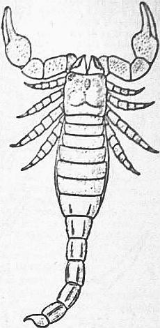
Fig. 48.—Dorsal view of a restoration of Palaeophonus nuncius, Thorell. The Silurian scorpion from Gothland.
Section α. Pectinifera.—The primitive distinction between the mesosoma and the metasoma retained, the latter consisting of six somites and the former of six somites in the adult, each of which is furnished during growth with a pair of appendages. Including the prae-genital somite (fig. 16), which is suppressed in the adult, there are thirteen somites behind the prosoma. The appendages of the 1st and 2nd mesosomatic somites persisting as the genital operculum and pectones respectively, those of the 3rd, 4th, 5th and 6th somites (? in Palaeophonus) sinking below the surface during growth in connexion with the formation of the four pairs of pulmonary sacs (see fig. 17). Lateral eyes monostichous.
Order 1. Scorpiones.—Prosoma covered by a single dorsal shield, bearing typically median and lateral eyes; its sternal elements reduced to a single plate lodged between or behind the basal segments of the 5th and 6th pairs of appendages. Appendages of 1st pair tri-segmented, chelate; of 2nd pair chelate, with their basal segments subserving mastication; of 3rd, 4th, 5th and 6th pairs similar in form and function, except that in recent and Carboniferous forms the basal segments of the 3rd and 4th are provided with sterno-coxal (maxillary) lobes, those of the 4th pair meeting in the middle line and underlying the mouth. The five posterior somites of the metasoma constricted to form a “tail,” the post-anal sclerite persisting as a weapon of offence and provided with a pair of poison glands (see figs. 8, 10, 12, 13, 14, 15, 21 and 22).
Sub-order Apoxypoda.—The 3rd, 4th, 5th and 6th pairs of appendages short, stout, tapering, the segments about as wide as long, except the apical, which is distally slender, pointed, slightly curved, and without distinct movable claws.
Family—Palaeophonidae, Palaeophonus (figs. 48 and 49).
Sub-order Dionychopoda.—The 3rd, 4th, 5th and 6th pairs of appendages slender, not evenly tapering, the segments longer than wide; the apical segment short, distally truncate, and provided with a pair of movable claws. Basal segments of the 5th and 6th pairs of appendages abutting against the sternum of the prosoma (see fig. 10 and figs. 51, 52 and 53).
Family—Pandinidae (Pandinus, Opisthophthalmus, Urodacus).
” Vejovidae (Vaejovis, Jurus, Euscorpius, Broteas).
” Bothriuridae (Bothriurus, Cercophonius).
” Buthidae (Buthus, Centrums).
” *Cyclophthalmidae (Cydophthalmus) Carboniferous.
” *Eoscorpiidae (Eoscorpius, Centromachus) Carboniferous.
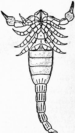
Fig. 49.—Ventral view of a restoration of Palaeophonus Hunteri, Pocock, the Silurian scorpion from Lesmahagow, Scotland. Restored by R.I. Pocock. The meeting of the coxae of all the prosomatic limbs in front of the pentagonal sternum; the space for a genital operculum; the pair of pectens, and the absence of any evidence of pulmonary stigmata are noticeable in this specimen.
(See Pocock, Quart Jour. Micr. Sci., 1901.)
Remarks on the Order Scorpiones.—The Scorpion is one of the great animals of ancient lore and tradition. It and the crab are the only two invertebrates which had impressed the minds of early men sufficiently to be raised to the dignity of astronomical representation. It is all the more remarkable that the scorpion proves to be the oldest animal form of high elaboration which has persisted to the present day. In the Upper Silurian two specimens of a scorpion have been found (figs. 48, 49), one in Gothland and one in Scotland, which would be recognized at once as true scorpions by a child or a savage. The Silurian scorpion Palaeophonus, differs, so far as obvious points are concerned, from a modern scorpion only in the thickness of its legs and in their terminating in strong spike-like joints, instead of being slight and provided with a pair of terminal claws. The legs of the modern scorpion (fig. 10; fig. 51) are those of a terrestrial Arthropod, such as a beetle; whilst those of the Silurian scorpion are the legs of an aquatic Arthropod, such as a crab or lobster. It is probable that the Silurian scorpion was an aquatic animal, and that its respiratory lamellae were still projecting from the surface of the body to serve as branchiae. No trace of “stigmata,” the orifices of the lung-chambers of modern scorpions, can be found in the Scottish specimen of Palaeophonus, which presents the ventral surface of the animal to view. On the other hand, no trace of respiratory appendages excepting the pectens can be detected in the specimen (see fig. 49).
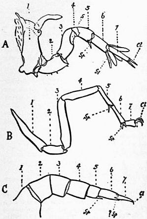
Fig. 50.—Comparison of the sixth prosomatic limb of a recent scorpion (B), of Palaeophonus (C), and of Limulus (A), showing their agreement in the number of segments; in the existence of a movable spine, Sp, at the distal border of the fifth segment; in the correspondence of the two claws at the free end of the limb of Scorpio with two spines similarly placed in Limulus; and, lastly, in the correspondence of the three talon-like spines carried on the distal margin of segment six of recent scorpions with the four larger but similarly situated spines on the leg of Limulus; s, groove dividing the ankylosed segments 4 and 5 of the Limulus leg into two.
(After Pocock, Q. J. Mic. Sci., 1901.)
From Lankester, Journ. Linn. Soc. Zool. vol. xvi., 1881.
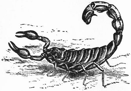
Fig. 51.—Drawing from life of the desert scorpion, Buthusaustralis, Lin., from Biskra, N. Africa.
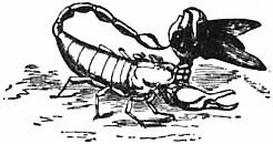
Fossil scorpions of the modern type are found in the Coal Measures. At the present day scorpions of various genera are found in all the warm regions of the world. In Europe they occur as far north as Bavaria and the south of France. The largest species measure 9 in. from the front of the head to the end of the sting, and occur in tropical India and Africa. Between 200 and 300 species are known. The scorpions use their large chelae for seizing prey and for fighting with one another. They never use the sting when (as frequently happens) they attack another scorpion, because, as was ascertained by A.G. Bourne (24), the poison exuded by the sting has no injurious effect on another scorpion nor on the scorpion itself. The stories of a scorpion stinging itself to death when placed in a circle of burning coals are due to erroneous observation. When placed in such a position the scorpion faints and becomes inert. It is found (Bourne, 24) that some species of scorpion faint at a temperature of 40° Cent. They recover on being removed to cooler conditions. A scorpion having seized its prey (usually a large insect, or small reptile or mammal) with the large chelae brings its tail over its head, and deliberately punctures the struggling victim twice with its sting (fig. 52). The poison of the sting is similar to snake-poison (Calmette), and rapidly paralyses animals which are not immune to it. It is probably only sickly adults or young children of the human race who can be actually killed by a scorpion’s sting. When the scorpion has paralysed its prey in this way, the two short chelicerae are brought into play (fig. 53). By the crushing action of their pincers, and an alternate backward and forward movement, they bring the soft blood-holding tissues of the victim close to the minute pin-hole aperture which is the scorpion’s mouth. The muscles acting on the bulb-like pharynx now set up a pumping action (see Huxley, 26); and the juices—but no solid matter, excepting such as is reduced to powder—are sucked into the scorpion’s alimentary canal. A scorpion appears to prefer for its food another scorpion, and will suck out the juices of an individual as large as itself. When this has taken place, the gorged scorpion becomes distended and tense in the mesosomatic region. It is certain that the absorbed juices do not occupy the alimentary canal alone, but pass also into its caecal off-sets which are the ducts of the gastric glands (see fig. 33).
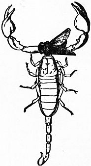
From Lankester, Journ. Linn. Soc. From Lankester, Journ. Linn. Soc.
Fig. 52.—Drawing from life of the Italian scorpion Euscorpius italicus, Herbst, holding a blue-bottle fly with its left chela, and carefully piercing it between head and thorax with its sting. Two insertions of the sting are effected and the fly is instantly paralysed by the poison so introduced into its body. Fig. 53.—The same scorpion carrying the now paralysed fly held in its chelicerae, the chelae liberated for attack and defence. Drawn from life.
All Arachnida, including Limulus, feed by suctorial action in essentially the same way as Scorpio.
Scorpions of various species have been observed to make a hissing noise when disturbed, or even when not disturbed. The sound is produced by stridulating organs developed on the basal joints of the limbs, which differ in position and character in different genera (see Pocock, 27). Scorpions copulate with the ventral surfaces in contact. The eggs are fertilized, practically in the ovary, and develop in situ. The young are born fully formed and are carried by the mother on her back. As many as thirty have been counted in a brood. For information as to the embryology of scorpions, the reader is referred to the works named in the bibliography below. Scorpions do not possess spinning organs nor form either snares or nests, so far as is known. But some species inhabiting sandy deserts form extensive burrows. The fifth pair of prosomatic appendages is used by these scorpions when burrowing, to kick back the sand as the burrow is excavated by the great chelae.
References to works dealing with the taxonomy and geographical distribution of scorpions are given at the end of this article (28).
Section β. Epectinata.—The primitive distinction between the mesosoma and the metasoma wholly or almost wholly obliterated, the two regions uniting to form an opisthosoma, which never consists of more than twelve somites and never bears appendages or breathing-organs behind the 4th somite. The breathing-organs of the opisthosoma, when present, represented by two pairs of stigmata, opening either upon the 1st and 2nd (Pedipalpi) or the 2nd and 3rd somites (Solifugae, Pseudo-scorpiones), or by a single pair upon the 3rd (? 2nd) somite (Opiliones) of the opisthosoma, there being rarely an additional stigma on the 4th (some Solifugae). The appendages of the 2nd somite of the opisthosoma absent, rarely minute and bud-like (some Amblypygi), never pectiniform. A prae-genital somite is often present either in a reduced condition forming a waist (Pedipalpi, Araneae, Palpigradi) or as a full-sized tergal plate (Pseudo-scorpiones); in some it is entirely atrophied (Solifugae, Holosomata, and Rhynchostomi). Lateral eyes when present diplostichous.
Remarks.—The Epectinate Arachnids do not stand so close to the aquatic ancestors of the Embolobranchia as do the Pectiniferous scorpions. At the same time we are not justified in supposing that the scorpions stand in any way as an intermediate grade between any of the existing Epectinata and the Delobranchia. It is probable that the Pedipalpi, Araneae, and Podogona have been separately evolved as distinct lines of descent from the ancient aquatic Arachnida. The Holosomata and Rhynchostomi are probably offshoots from the stem of the Araneae, and it is not unlikely (in view of the structure of the prosomatic somites of the Tartarides) that the Solifugae are connected in origin with the Pedipalpi. The appearance of tracheae in place of lung-sacs cannot be regarded as a starting-point for a new line of descent comprising all the tracheate forms; tracheae seem to have developed independently in different lines of descent. On the whole, the Epectinata are highly specialized and degenerate forms, though there are few, if any, animals which surpass the spiders in rapidity of movement, deadliness of attack and constructive instincts.
From Lankester, Q. J. Mic. Sci. N.S. vol. xxi., 1881.
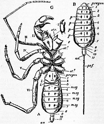
Fig. 54.—Thelyphonus, one of the Pedipalpi.
A, Ventral view. I, Chelicera (detached). II, Chelae. III, Palpiform limb. IV to VI, The walking legs. stc, Sterno-coxal process (gnathobase) of the chelae. st1, Anterior sternal plate of the prosoma. st2, Posterior sternal plate of the prosoma. pregen, Position of the prae-genital somite (not seen). l, l, Position of the two pulmonary sacs of the right side. 1 to 11, Somites of the opisthosoma (mesosoma plus metasoma). msg, Stigmata of the tergo-sternal muscles. an, Anus. B, Dorsal view of the opisthosoma of the same. pregen, The prae-genital somite. p, The tergal stigmata of the tergo-sternal muscles. paf, Post-anal segmented filament corresponding to the post-anal spine of Limulus.
Order 2. Pedipalpi (figs. 54 to 59).—Appendages of 1st pair bisegmented, without poison gland; of 2nd pair prehensile, their basal segments underlying the proboscis, and furnished with sterno-coxal (maxillary) process, the apical segment tipped with a single movable or immovable claw; appendages of 3rd pair different from the remainder, tactile in function, with at least the apical segment many-jointed and clawless. The ventral surface of the prosoma bears prosternal, metasternal and usually mesosternal chitine-plates (fig. 55). A narrow prae-genital somite is present between opisthosoma and prosoma (figs. 55, 57). Opisthosoma consisting of eleven somites, almost wholly without visible appendages. Intromittent organ of male beneath the genital operculum (= sternum of the 1st somite of opisthosoma).
Fig. 55.—Thelyphonus sp. Ventral view of the anterior portion of the body to show the three prosomatic sternal plates a, b, c, and the rudimentary sternal element of the prae-genital somite; opisth 1, first somite of the opisthosoma.
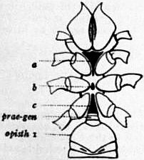
From a drawing made by Pickard—Cambridge, under the direction of R.I. Pocock.
Note.—The possibility of another interpretation of the anterior somites of the mesosoma and the prae-genital somite must be borne in mind. Possibly, though not probably, the somites carrying the two lung-sacs correspond to the first two lung-bearing somites of Scorpio, and it is the genital opening which has shifted. The same caution applies in the case of the Araneae. Excalation of one or of two anterior mesosomatic somites, besides the prae-genital somite, would then have to be supposed to have occurred also.
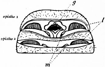
Fig. 56—Thelyphonus assamensis ♂. Ventral surface of the anterior region of the opisthosoma, the first somite being pushed upwards and forwards so as to expose the subjacent structures. opistho 1, First somite of the opisthosoma; opistho 2, second do.; g, genital aperture; l, edges of the lamellae of the lung-books; m, stigmata of tergo-sternal muscles.
(Original drawing by Pocock.)
Sub-order a. Uropygi.—Prosoma longer than wide, its sternal area very narrow, furnished with a large prosternal and metasternal plate, and often with a small mesosternal sclerite. Appendages of 2nd pair with their basal segments united in the middle line and incapable of lateral movement; appendages of 3rd pair with only the apical segment many-jointed. Opisthosoma without trace of appendages; its posterior somites narrowed to form a movable tail for the support of the post-anal sclerite, which has no poison glands.
Tribe 1. Urotricha.—Dorsal area of prosoma covered with a single shield (? two in Geralinura), bearing median and lateral eyes. Post-anal sclerite modified as a long, many-jointed feeler. Appendages of 2nd pair folding in a horizontal plane, completely chelate, the claw immovably united to the sixth segment. Respiratory organs present in the form of pulmonary sacs.
Family—Thelyphonidae (Thelyphonus (fig. 54), Hypoctonus, *Geralinura).
Tribe 2. Tartarides.—Small degenerate forms with the dorsal area of the prosoma furnished with two shields, a larger in front covering the anterior four somites, and a smaller behind covering the 5th and 6th somites; the latter generally subdivided into a right and left portion. There is also a pair of narrow tergal sclerites interposed between the anterior and posterior shields. Eyes evanescent or absent. Appendages of 2nd pair folding in a vertical plane, not chelate, the claw long and movable. Post-anal sclerite short and undivided. No distinct respiratory stigmata behind the sterna of the 1st and 2nd somites of the opisthosoma.
Family-Hubbardiidae (Schizomus, Hubbardia) (figs. 57-59).
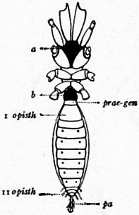
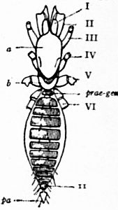
Fig. 57.—Schizomus crassicaudatus, one of the Tartarid Pedipalpi. Ventral view of a female with the appendages cut short near the base. Fig. 58.—Schizomus crassicaudatus, a Tartarid Pedipalp. Dorsal view of a male with the appendages cut short.
a, Prosternum of prosoma. b, Metasternum of prosoma. prae-gen, The prae-genital somite. I opisth, First somite of the opisthosoma. II opisth, Eleventh somite of the opisthosoma. pa, Post-anal lobe of the female (compare the jointed filament in Thelyphonus, fig. 54). I to VI. The prosomatic appendages. a, Anterior plate. b, Posterior plate of the prosomatic carapace. prae-gen, Tergum of the prae-genital somite. 11, The eleventh somite of the opisthosoma. pa, Post-anal lobe of the male—a conical body with narrow basal stalk.
(Original drawing by Pickard-Cambridge, directed by Pocock.) (Original as preceding.)
Sub-order b. Amblypygi.—Prosoma wider than long, covered above by a single shield bearing median and lateral eyes, which have diplostichous ommatea. Sternal area broad, with prosternal, two mesosternal, and metasternal plates, the prosternum projecting forwards beneath the coxae of the 2nd pair of appendages. Appendages of 2nd pair folding in a horizontal plane; their basal segments freely movable; claw free or fused; basal segments of 4th and 5th pairs widely separated by the sternal area; appendages of 3rd pair with all the segments except the proximal three, forming a many-jointed flagellum. Opisthosoma without post-anal sclerite and posterior caudal elongation: with frequently a pair of small lobate appendages on the sternum of the 3rd somite. Respiratory organs, as in Urotricha.
Family—Phrynichidae (Phrynichus, Damon).
” Admetidae (Admetus, Heterophrynus).
” Charontidae (Charon, Sarax).
(Family ?)—*Graeophonus.
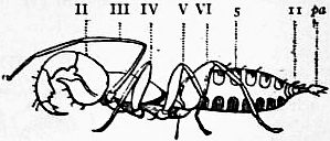
Fig. 59.—Schizomus crassicaudatus, one of the Pedipalpi. Lateral view of a male. II to VI, the prosomatic appendages, the first being concealed (see fig. 58); 5, the fifth, and 11, the eleventh tergites of the opisthosoma; pa, the conical post-anal lobe.
(Original as preceding.)
Remarks.—The Pedipalpi are confined to the tropics and warmer temperate regions of both hemispheres. Fossil forms occur in the Carboniferous. The small forms known as Schizomus and Hubbardia are of special interest from a morphological point of view. The Pedipalpi have no poison glands. (Reference to literature (29).)
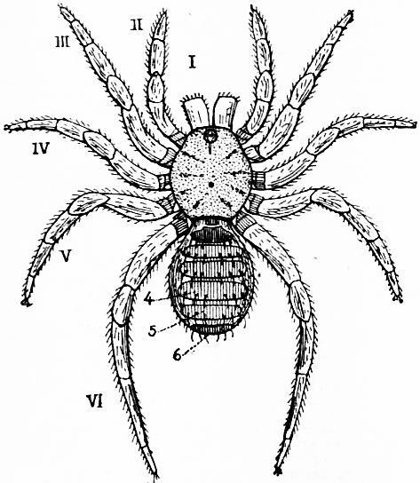
Fig. 60.—Liphistius desultor, Schiödte, one of the Araneae Mesothelae. Dorsal view. I to VI, the prosomatic appendages; 4, 5, 6, the fourth, fifth and sixth tergites of the opisthosoma. Between the bases of the sixth pair of limbs and behind the prosomatic carapace is seen the tergite of the small prae-genital somite.
(Original by Pickard-Cambridge and Pocock.)
Order 3. Araneae (figs. 60 to 64.).—Prosoma covered with a single shield and typically furnished with median and lateral eyes of diplostichous structure, as in the Amblypygi. The sternal surface wide, continuously chitinized, but with prosternal and metasternal elements generally distinguishable at the anterior and posterior ends respectively of the large mesosternurm. Prosternum underlying the proboscis. Appendages of 1st pair have two segments, as in Pedipalpi, but are furnished with poison gland, and are retroverts. Appendages of 2nd pair not underlying the mouth, but freely movable and, except in primitive forms, furnished with a maxillary lobe; the rest of the limb like the legs, tipped with a single claw and quite unmodified (except in ♂). Remaining pairs of appendages similar in form and function, each tipped with two or three claws. Opisthosoma when segmented showing the same number of somites as in the Pedipalpi; usually unsegmented, the prae-genital somite constricted to form the waist; the appendages of its 3rd and 4th somites retained as spinning mammillae. Respiratory organs (see fig. 63, stg), as in the Amblypygi, or with the posterior pair, rarely the anterior pair as well, replaced by tracheal tubes. Intromittent organ of male in the apical segment of the 2nd prosomatic appendage.
Fig. 61.—Liphistius desultor. Ventral view with the prosomatic appendages cut short excepting the chelicerae (1) whose sharp retroverts are seen. Between the bases of the prosomatic limbs an anterior and a posterior sternal plate (black) are seen. 1, The sternum of the first opisthosomatic or genital somite covering the genital aperture and the first pair of lung-sacs. In front of it the narrow waist is formed by the soft sternal area of the praegenital somite; 2, the sternite of the second opisthosomatic somite covering the posterior pair of lung-sacs; 3 and 4, the spinning appendages (limbs) of the opisthosoma; a, inner, b, outer ramus of the appendage; 11, sternite of the eleventh somite of the opisthosoma: in front of it other rudimentary sternites; an, anus.
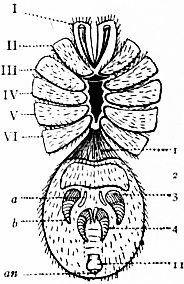
(Original as above.)
Sub-order a. Mesothelae (see figs. 60 to 62).—Opisthosoma distinctly segmented, furnished with 11 tergal plates, as in the Amblypygi; the ventral surface of the 1st and 2nd somites with large sternal plates, covering the genital aperture and the two pairs of pulmonary sacs, the sternal plates from the 6th to the 11th somites represented by integumental ridges, weakly chitinized in the middle. The two pairs of spinning appendages retain their primitive position in the middle of the lower surface of the opisthosoma far in advance of the anus on the 3rd and 4th somites, each appendage consisting of a stout, many-jointed outer branch and a slender, unsegmented inner branch. Prosoma as in the Mygalomorphae, except that the mesosternal area is long and narrow.
Family—Liphistiidae (Liphistius, *Arthrolycosa).
Sub-order b. Opisthothelae (see fig. 63).—Opisthosoma without trace of separate terga and sterna, the segmentation merely represented posteriorly by slight integumental folds and the sterna of the 1st and 2nd somites by the opercular plates of the pulmonary sacs. The spinning appendages migrate to the posterior end of the opisthosoma and take up a position close to the anus; the inner branches of the anterior pair either atrophy or are represented homogenetically by a plate, the cribellum, or by an undivided membranous lobe, the colulus.
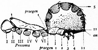
Fig. 62.—Liphistius desultor. Lateral view.
I to VI, Appendages of the prosoma cut off at the base. o, Ocular tubercle. prae-gen, The prae-genital somite. 1 and 2, Sternites of the first and second opisthosomatic somites. 3 and 4, Appendages of the third and fourth opisthosomatic somites, which are the spinning organs, and in this genus occupy their primitive position instead of migrating to the anal region as in other spiders. 5, Tergite of the fifth opisthosomatic somite. 11, Eleventh opisthosomatic somite; an, Anus.
(Original.)
Tribe 1. Mygalomorphae.—The plane of the articulation of the appendages of the 1st pair to the prosoma (the retrovert) vertical, the basal segment projecting straight forwards at its proximal end, the distal segment or fang closing backwards in a direction subparallel to the long axis of the body. Two pairs of pulmonary sacs.
Families—Theraphosidae (Avicularia, Poecilotheria). Barychelidae (Barychelus, Plagiobothrus). Dipluridae (Diplura, Macrothele). Ctenizidae (Cteniza, Nemesia). Atypidae (Atypus, Calommata).
Tribe 2. Arachnomorpnae.—The plane of the articulation of the appendages of the 1st pair to the prosoma horizontal, the basal segment projecting vertically downwards, at least at its proximal end, the distal segment or fang closing inwards nearly or quite at right angles to the long axis of the body. The posterior pulmonary sacs (except in Hypochilus) replaced by tracheal tubes; the anterior and posterior pairs replaced by tracheal tubes in the Caponiidae.
Principal families—Hypochilidae (Hypochilus). Dysderidae (Dysdera, Segestria). Caponiidae (Caponia, Nops). Filistatidae (Filistata). Uloboridae (Uloborus, Dinopis). Argiapidae (Nephila, Gasteracantha). Pholcidae (Pholcus, Artema). Agelenidae (Tegenuria). Lycosidae (Lycosa). Clubionidae (Clubiona, Olios, Sparassus) Gnaphosidae (Gnaphosa, Hemiclaea). Thomisidae (Thomisus). Attidae (Salticus). Urocteidae (Uroctea). Eresidae (Eresus).
Remarks on the Araneae.—The Spiders are the most numerous and diversified group of the Arachnida; about 2000 species are known. No noteworthy fossil spiders are known; the best-preserved are in amber of Oligocene age. Protolycosa and Arthrolycosa occur in the Carboniferous. Morphologically, the spiders are remarkable for the concentration and specialization of their structure, which is accompanied with high physiological efficiency. The larger species of Bird’s Nest Spiders (Avicularia), the opisthosoma of which is as large as a bantam’s egg, undoubtedly attack young birds, and M’Cook gives an account of the capture in its web by an ordinary house spider of a small mouse. The “retrovert” or bent-back first pair of appendages is provided with a poison gland opening on the fang or terminal segment. Spiders form at least two kinds of constructions—snares for the capture of prey and nests for the preservation of the young. The latter are only formed by the female, which is a larger and more powerful animal than the male. Like the scorpions the spiders have a special tendency to cannibalism, and accordingly the male, in approaching the female for the purpose of fertilizing her, is liable to be fallen upon and sucked dry by the object of his attentions. The sperm is removed by the male from the genital aperture into a special receptacle on the terminal segment of the 2nd prosomatic appendage. Thus held out at some distance from the body, it is cautiously advanced by the male spider to the genital aperture of the female.
Fig. 63.—Ventral view of a male mygalomorphous spider.
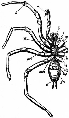
I to VI, The six pairs of prosomatic appendages. a, Copulatory apparatus of the second appendage. b, Process of the fifth joint of the third appendage. M, Mouth. pro, Prosternite of the prosoma. mes, Mesosternite of the prosoma: observe the contact of the coxae of the sixth pair of limbs behind it; compare Liphistius (fig. 61) where this does not occur. stg, Lung aperture. gn, Genital aperture. a, Anus with a pair of backwardly migrated spinning appendages on each side of it; compare the position of these appendages in Liphistius (fig. 61).
(From Lankester, “Limulus an Arachnid.”)
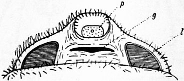
Fig. 64.—Liphistius desultor. Under side of the uplifted genital or first opisthosomatic somite of the female; g, genital aperture; p, pitted plate, probably a gland for the secretion of adhesive material for the eggs; l, the edges of the lamellae of the lung-books of the first pair.
(Original drawing by Pocock.)
For an account of the courtship and dancing of spiders, of their webs and floating lines, the reader is referred to the works of M‛Cook (30) and the Peckhams (31), whilst an excellent account of the nests of trap-door spiders is given by Moggridge (32). References to systematic works will also be found at the end of this article (33).
Order 4. Palpigradi = Microthelyphonidae (see fig. 65).—Prosoma covered above by three plates, a larger representing the dorsal elements of the first four somites, and two smaller representing the dorsal elements of the 5th and 6th.
Its ventral surface provided with one prosternal, two mesosternal and one metasternal plate. Appendages of 1st pair consisting of three segments, completely chelate, without poison gland; of 2nd pair slender, leg-like, tipped with three claws, the basal segment without sterno-coxal process taking no share in mastication, and widely separated from its fellow of the opposite side; 3rd, 4th, 5th and 6th appendages similar in form to the 2nd and to each other.
Proboscis free, not supported from below by either the prosternum or the basal segments of the appendages of the 2nd pair.
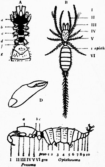
Fig. 65.—Koenenia mirabilis, Grassi, one of the Palpigradi.
A, Ventral view of prosoma and anterior region of opisthosoma with the appendages cut off near the base; a and b, prosternites; c, mesosternite; and d, metasternite of the prosoma; f, ventral surface of the prae-genital somite; g, sternite of the genital somite (first opisthosomatic somite). B, Dorsal view. I to VI, prosomatic appendages; 1 opisth, genital somite (first opisthosomatic somite). C, Lateral view, I to VI, prosomatic appendages; a, b, c, the three tergal plates of the prosoma; prae-gen, the prae-genital somite; 1 to 10, the ten somites of the opisthosoma. D, Chelicera.
(Original drawing by Pocock and Pickard-Cambridge, after Hansen and Sörensen.)
Opisthosoma consisting of only ten somites, which have no tergal and sternal elements, the prae-genital somite contracted to form a “waist,” as in the Pedipalpi; the last three narrowed to form a caudal support for the many-jointed flagelliform telson, as in the Urotricha. Respiratory organs atrophied.
Family—Koeneniidae (Koenenia).
Remarks.—An extremely remarkable minute form originally described by Grassi (34) from Sicily, and since further described by Hansen (35). Recently the genus has been found in Texas, U.S.A. Only one genus of the order is known.
Order 5. Solifugae = Mycetophorae (see figs. 66 to 69).—Dorsal area of prosoma covered with three distinct plates, two smaller representing the terga of the 5th and 6th somites, and a larger representing those of the anterior four somites, although the reduced terga of the 3rd and 4th are traceable behind the larger plate. The latter bears a pair of median eyes and obsolete lateral eyes on each side. Sternal elements of prosoma almost entirely absent, traces of a prosternum and metasternum alone remaining. Rostrum free, not supported by either the prosternum or the basal segments of the appendages. Appendages of 1st pair large, chelate, bisegmented, articulated to the sides of the head-shield; appendages of 2nd pair simple, pediform, with protrusible (? suctorial) organ, and no claws at the tip; their basal segments united in the middle line and furnished with sterno-coxal process. Remaining pairs of appendages with their basal segments immovably fixed to the sternal surface, similar in form, the posterior three pairs furnished with two claws supported on long stalks; the basal segments of the 6th pair bearing five pairs of tactile sensory organs or malleoli. The prae-genital somite is suppressed. Opisthosoma composed of ten somites. Respiratory organs tracheal, opening upon the ventral surface of the 2nd and 3rd, and sometimes also of the 4th somite of the opisthosoma. A supplementary pair of tracheae opening behind the basal segment of the 4th appendage of the prosoma.
(? Intromittent organ of male lodged on the dorsal side of the 1st pair of prosomatic appendages.)
Families—Hexisopodidae (Hexisopus). Solpugidae (Solpuga, Rhagodes). Galeodidae (Galeodes).
Remarks.—These most strange-looking Arachnids occur in warmer temperate, and tropical regions of Asia, Africa and America. Their anatomy has not been studied, as yet, by means of freshly-killed material, and is imperfectly known, though the presence of the coxal glands was determined by Macleod in 1884. The proportionately enormous chelae (chelicerae) of the first pair of appendages are not provided with poison glands; their bite is not venomous.
Fig. 66.—Galeodes sp., one of the Solifugae. Ventral view to show legs and somites.
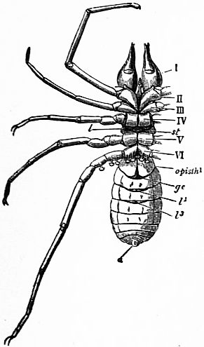
I to VI, The six leg-bearing somites of the prosoma. opisth 1, First or genital somite of the opisthosoma. ge, Site of the genital aperture. st, Thoracic tracheal aperture. l2, Anterior tracheal aperture of the opisthosoma in somite 2 of the opisthosoma. l3, Tracheal aperture in somite 3 of the opisthosoma. a, Anus.
(From Lankester, “Limulus an Arachnid.”)
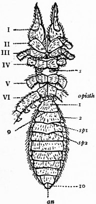
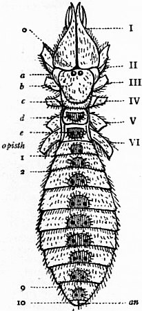
Fig. 67.—Galeodes sp., one of the Solifugae. Ventral view with the appendages cut off at the base. Fig. 68.—Galeodes sp., one of the Solifugae. Dorsal view.
I to VI, Prosomatic appendages. s, Prosomatic stigma or aperture of the tracheal system. 1, First opisthosomatic sternite covering the genital aperture g. 2, Second opisthosomatic sternite covering the second pair of tracheal apertures sp1. sp2, The third pair of tracheal apertures. 10, The tenth opisthosomatic somite. an, The anal aperture. I to VI, Bases of the prosomatic appendages. o, Eyes. a, Lateral region of the cephalic plate to which the first pair of appendages are articulated. b, Cephalic plate with median eye. c, Dorsal element of somites bearing third and fourth pairs of appendages. d, Second plate of the prosoma with fifth pair of appendages. e, Third or hindermost plate of the prosoma beneath which the sixth pair of legs is articulated. 1, 2, 9, 10, First, second, ninth and tenth somites of the opisthosoma. an, Anus.
(Original by Pickard-Cambridge and Pocock.) (Original.)
Galeodes has been made the means of a comparison between the structure of the Arachnida and Hexapod insects by Haeckel and other writers, and it was at one time suggested that there was a genetic affinity between the two groups—through Galeodes, or extinct forms similar to it. The segmentation of the prosoma and the form of the appendages bear a homoplastic similarity to the head, pro-, meso-, and meta-thorax of a Hexapod with mandibles, maxillary palps and three pairs of walking legs; while the opisthosoma agrees in form and number of somites with the abdomen of a Hexapod, and the tracheal stigmata present certain agreements in the two cases. Reference to literature (36).
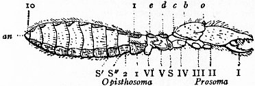
Fig. 69.—Galeodes sp., one of the Solifugae.
I to VI, The six prosomatic limbs cut short. o, The eyes. b, c, Demarcated areae of the cephalic or first prosomatic plate corresponding respectively to appendages I, II, III, and to appendage IV (see fig. 68). d, Second plate of the prosoma-carrying appendage V. e, Third plate of the prosoma-carrying appendage VI. The prae-genital somite is absent. 1, First somite of the opisthosoma. 2, Second do. S, Prosomatic tracheal aperture between legs IV and V. S′ and S″, Opisthosomatic tracheal apertures. 10, Tenth opisthosomatic somite. an, Anus.
(Original.)
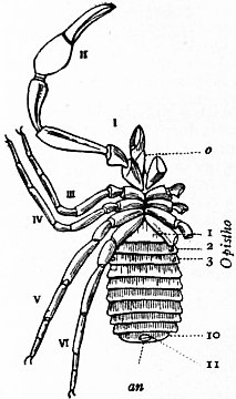
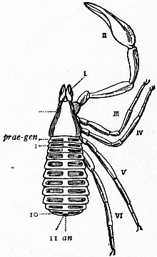
Fig. 70.—Garypus litoralis, one of the Pseudoscorpiones. Ventral view. Fig. 71.—Garypus litoralis, one of the Pseudoscorpiones. Dorsal view.
I to VI, Prosomatic appendages. o, Sterno-coxal process of the basal segment of the second appendage. 1, Sternite of the genital or first opisthosomatic somite; the prae-genital somite, though represented by a tergum, has no separate ternal plate. 2 and 3, Sternites of the second and third somites of the opisthosoma, each showing a tracheal stigma. 10 and 11, Sternites of the tenth and eleventh somites of the opisthosoma. an, Anus. I to VI, The prosomatic appendages. o, Eyes. prae-gen, Prae-genital somite. 1, Tergite of the genital or first opisthosomatic somite. 10, Tergite of the tenthsomite of the opisthosoma. 11, The evanescent eleventh somite of the opisthosoma. an, Anus.
(Original by Pocock and Pickard-Cambridge.) (Original.)
Order 6. Pseudoscorpiones = Chelonethi, also called Chernetidia (see figs. 70, 71, 72).—Prosoma covered by a single dorsal shield, at most furnished with one or two diplostichous lateral eyes; sternal elements obliterated or almost obliterated. Appendages of the 1st pair bisegmented completely chelate, furnished with peculiar organs, the serrula and the lamina. Appendages of 2nd pair very large and completely chelate, their basal segments meeting in the middle line, as in the Uropygi, and provided in front with membranous lip-like processes underlying the proboscis. Appendages of the 3rd, 4th, 5th and 6th pairs similar in form and function, tipped with two claws, their basal segments in contact in the median ventral line. The prae-genital somite wide, not constricted, with large tergal plate, but with its sternal plate small or inconspicuous. Opisthosoma composed, at least in many cases, of eleven somites, the 11th somite very small, often hidden within the both. Respiratory organs in the form of tracheal tubes opening by a pair of stigmata in the 2nd and 3rd somites of the opisthosoma. Intromittent organ of male beneath sternum of the 1st somite of the opisthosoma.
Sub-order a. Panctenodactyli.—Dorsal plate of prosoma (carapace) narrowed in front; the appendages of the 1st pair small, much narrower, taken together, than the posterior border of the carapace. Serrula on movable digit of appendages of 1st pair fixed throughout its length, and broader at its proximal than at its distal end; the immovable digit with an external process.
Family—Cheliferidae (Chelifer (figs. 70, 71, 72), Chiridium).
” Garypidae (Garypus).
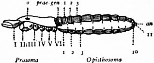
Fig. 72.—Garypus litoralis, one of the Pseudoscorpiones. Lateral view.
I to VI, of the six prosomatic appendages. o, Eyes. prae-gen, Tergite of the prae-genital somite. 1, Genital or first opisthosomatic somite. 2, 3, 10, The second, third and tenth somites of the opisthosoma. 11, The minute eleventh somite; an, the anus.
(Original.)
Sub-order b. Hemictenodactyli.—Dorsal plate of prosoma scarcely narrowed in front; the appendages of the 1st pair large, not much narrower, taken together, than the posterior border of the carapace. The serrula or the movable digit free at its distal end, narrowed at the base; no external lamina on the immovable digit.
Family—Obisiidae (Obisium, Pseudobisium).
” Chthoniidae (Chthonius, Tridenchthonius).
Remarks.—The book-scorpions—so called because they were, in old times, found not unfrequently in libraries—are found in rotten wood and under stones. The similarity of the form of their appendages to those of the scorpions suggests that they are a degenerate group derived from the latter, but the large size of the prae-genital somite in them would indicate a connexion with forms preceding the scorpions. Reference to literature (37).
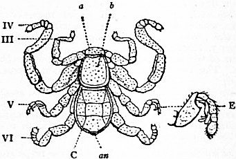
Fig. 73.—Cryptostemma Karschii, one of the Podogona. Dorsal view of male.
III to VI, The third, fourth, fifth and sixth appendages of the prosoma. a, Movable (hinged) sclerite (so-called hood) overhanging the first pair of appendages. b, Fused terga of the prosoma followed by the opisthosoma of four visible somites. an, Orifice within which the caudal segments are withdrawn. E, Extremity of the fifth appendage of the male modified to subserve copulation.
(Original drawing by Pocock and Pickard-Cambridge.)
Order 7. Podogona = Ricinulei (see figs. 73 to 76).—Dorsal area of prosoma furnished with two shields, a larger behind representing, probably, the tergal elements of the somites, and a smaller in front, which is freely articulated to the former and folds over the appendages of the 1st pair. Ventral area without distinct sternal plates. Appendages of 1st pair, bisegmented, completely chelate. Appendages of 2nd pair, with their basal segments uniting in the middle line below the mouth, weakly chelate at apex. Appendages of 3rd, 4th, 5th and 6th pairs similar in form; their basal segments in contact in the middle line and immovably welded, except those of the 3rd pair, which have been pushed aside so that the bases of the 2nd and 4th pairs are in contact with each other. A movable membranous joint between the prosoma and the opisthosoma, the generative aperture opening upon the ventral side of the membrane. Prae-genital somite suppressed; the opisthosma consisting of nine segments, whereof the first and second are almost suppressed and concealed within the joint between the prosoma and the opisthosoma; the following four large and manifest, and the remaining three minute and forming a slender generally-retracted tail like that of Thelyphonus. Respiratory organs tracheal, opening by a pair of spiracles in the prosoma above the base of the fifth appendage on each side. Intromittent organ of male placed at the distal end of the appendage of the 5th pair.
Family—Cryptostemmidae (Cryptostemma, Poliochera), Carboniferous.
Fig. 74.—Cryptostemma Karschii, anterior aspect of the prosoma with the “hood” removed. I to IV, first to fourth appendages of the prosoma; a, basal segment of the second pair of appendages meeting its fellow in the middle line (see fig. 75).
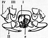
(Original drawing by Pocock and Pickard-Cambridge.)
Remarks on the Podogona.—The name given to this small but remarkable group has reference to the position of the male intromittent organ (fig. 73, E). They are small degenerate animals with a relatively firm integument. Not more than four species and twice that number of specimens are known. They have been found in West Africa and South America. A fact of special interest in regard to them is that the genus Poliochera, from the Coal Measures, appears to be a member of the same group. The name Cryptostemma, given to the first-known genus of the order, described by Guérin-Méneville, refers to the supposed concealment of the eyes by the movable cephalic sclerite. Reference to literature (38).
Fig. 75.—Cryptostemma Karschii, one of the Podogona. Ventral view.
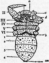
I to VI, The six pairs of appendages of the prosoma, the last three cut short. 1, 2, 3, 4, The four somites of the opisthosoma. a. Visible hood overhanging the first pair of appendages. b, Position of the genital orifice. c, Part of 3rd appendage. d, Fourth segment of 2nd appendage. Observe that the basal segment of appendage III does not meet its fellow in the middle line.
(Original drawing by Pocock and Pickard-Cambridge.)
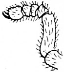
Fig. 76.—Cryptostemma Karschii. Extremity of the fifth pair of appendages of the female for comparison with that of the male E in fig. 73.
Order 8. Opilione (see fig. 77).—Dorsal area of prosoma covered by a single shield usually bearing a pair of eyes. Sternal elements much reduced. Appendages of 1st pair large, three segmented and completely chelate; of 2nd pair either simple and pediform, or prehensile and subchelate; of remaining four pairs, similar in form, ambulatory in function; the basal segment of the 2nd, 3rd and sometimes of the 4th pairs of appendages furnished with sterno-coxal (maxillary) lobe. Opisthosoma confluent throughout its breadth with the prosoma, with the dorsal plate of which its anterior tergal plates are more or less fused; at most ten opisthosomatic somites traceable; the generative aperture thrust far forwards between the basal segments of the 6th appendages. Prae-genital somite suppressed. Respiratory organs tracheal, opening by a pair of stigmata situated immediately behind the basal segments of the 6th pair of appendages on what is probably the sternum of the 2nd opisthosomatic somite and also in some cases upon the 5th segment of the legs.
Intromittent organ of male lying within the genital orifice.
Sub-order a. Laniatores.—Orifice of foetid glands opening above the coxa of the 4th appendage, not raised upon a tubercle. Orifice of coxal gland situated just behind that of the foetid gland. Sternal plate of prosoma long and narrow, with a distinct prosternal element underlying the mouth. Coxae of 4th, 5th and 6th appendages immovable. Appendages of 2nd pair, strong, usually prehensile and spiny. Genital orifice covered by an operculum.
Families—Gonoleptidae (Gonoleptes, Goniasoma).
Biantidae (Biantes).
Oncopodidae (Oncopus, Pelitnus).
Trioenonychidae (Trioenonyx, Acumontia).
Sub-order b. Palpatores.—Orifice of foetid glands opening above the coxa of the 3rd appendage, not raised upon a tubercle. Orifice of coxal gland situated between the coxae of the 5th and 6th appendages. Sternal plate of prosoma usually short and wide, rarely longer than broad; with a larger or smaller prosternal element underlying the mouth. Coxae of 3rd, 4th, 5th and 6th appendages movable or immovable. Appendages of 2nd pair weak, pediform not prehensile. Genital orifice covered by an operculum.
Families—Phalangiidae (Phalangium, Gagrella).
Ischyropsalidae (Ischyropsalis, Taracus).
Nemastomidae (Nemastoma).
Trogulidae (Trogulus, Anelasmocephalus).
Sub-order c. Cyphophthalmi (Anepignathi).—Orifice of foetid glands opening on a tubercle situated near the lateral border of the carapace above the base of the 5th appendage. Orifice of coxal gland probably situated at base of coxa of 5th appendage; sternal plate of prosema minute or absent; no prosternal element underlying the mouth. Coxae of 5th and 6th, and usually also of 4th appendages immovable. Appendages of 2nd pair weak, pediform, not prehensile. Genital orifice not covered by an operculum.
Families—Sironidae (Siro, Pettalus).
Stylocellidae (Stylocellus).
Remarks on the Opiliones.—These include the harvest-men, sometimes called also daddy-long-legs, with round undivided bodies and very long, easily-detached legs. The intromittent organs of the male are remarkable for their complexity and elaboration. The confluence of the regions of the body and the dislocation of apertures from their typical position are results of degeneration. The Opiliones seem to lead on from the Spiders to the Mites. Reference to literature (39).
Fig. 77.—Stylocellus sumatranus, one of the Opiliones; after Thorell. Enlarged.
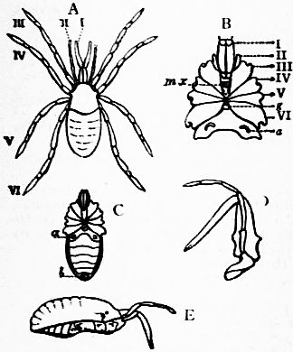
A, Dorsal view; I to VI, the six prosomatic appendages. B, Ventral view of the prosoma and of the first somite of the opisthosoma, with the appendages I to VI cut off at the base; a, tracheal stigma; mx, maxillary processes of the coxae of the 3rd pair of appendages; g, genital aperture. C, Ventral surface of the prosoma and opisthosoma; a, tracheal stigma; b, last somite. D, Lateral view of the 1st and 2nd pair of appendages. E, Lateral view of the whole body and two 1st appendages, showing the fusion of the dorsal elements of the prosoma into a single plate, and of those of the opisthosoma into an imperfectly segmented plate continuous with that of the prosoma.
Apparently related to the Opiliones are two extinct groups, the Anthracomarti and Phalangiotarbi, which are not known to have survived the Carboniferous period. In the Anthracomarti the opisthosoma was movably articulated to the prosoma, and consisted of from eight to ten segments furnished with movable lateral plates, the anal segment being overlapped dorsally by a laminate expansion of the preceding segment. The carapace of the prosoma was unsegmented and often bore a pair of eyes. The appendages of the 2nd pair were slender and pediform; those of the 3rd, 4th, 5th and 6th pairs were similar in form and ambulatory in function with their basal segments arranged round a sternal area as in the order Araneae. The best-known genera were Anthracomartus and Eophognus.
In the Phalangiotarbi the appendages resembled those of the Anthracomarti, except that the basal segments of the last four pairs were usually approximated in the middle line leaving a long and narrow sternal area between; and the carapace of the prosoma was unsegmented. The prosoma and opisthosoma were broadly confluent and probably immovably welded together. The opisthosoma consisted of eight or nine segments, whereof the anterior five or six were very short in the dorsal region, and the posterior three exceptionally large with the anal orifice terminal.
Several genera have been established, the best-characterized being Geraphognus and Architarbus.
Order 9. Rhynchostomi = Acari (see fig. 78).—Degenerate Arachnids resembling the Opiliones in many structural points, but chiefly distinguishable from them by the following features:—The basal segments of the appendages of the 2nd pair are united in the middle line behind the mouth, those of the 3rd, 4th, 5th and 6th pairs are widely separated and not provided with sterno-coxal (maxillary) lobes, and take no share in mastication; the respiratory stigmata, when present, belong to the prosoma, and the primitive segmentation of the opisthosoma has entirely or almost entirely disappeared.
Sub-order a. Notostigmata.—Opisthosoma consisting of ten segments defined by integumental grooves, each of the anterior four of these furnished with a single pair of dorsally-placed spiracles or tracheal stigmata.
Family—Opilioacaridae (Opilioacarus).
Sub-order b. Cryptostigmata.—Integument hard, strengthened by a continuously chitinized dorsal and ventral sclerite. Tracheae typically opening by stigmata situated in the articular sockets (acetabula) of the 3rd, 4th, 5th and 6th pairs of appendages.
Family—Oribatidae (Oribata, Nothrus, Hoplophora).
Sub-order c. Metastigmata.—Integument mostly like that of the Cryptostigmata. Tracheae opening by a pair of stigmata situated above and behind the base of the 4th or 5th or 6th pair of appendages.
Families—Gamasidae (Gamasus, Pteroptus).
Argasidae (Argas, Ornithodoros).
Ixodidae (Ixodes, Rhipicephalus).
Sub-order d. Prostigmata.—Integument soft, strengthened by special sclerites, those on the ventral surface of the prosoma apparently representing the basal segments of the legs embedded in the skin. Tracheae, except in the aquatic species in which they are atrophied, opening by a pair of stigmata situated close to or above the base of the appendages of the 1st pair (mandibles).
Families—Trombidiidae (Trombidium, Tetranychus).
Hydrachnidae (Hydrachna, Atax).
Halacaridae (Halacarus, Leptognathus).
Bdellidae (Bdella, Eupodes).
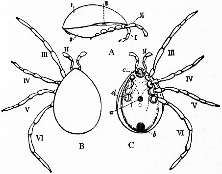
Fig. 78.—Holothyrus nitidissimus, one of the Acari; after Thorell.
A, Lateral view with appendages III to VI removed; 1, plate covering the whole dorsal area, representing the fused tergal sclerites of the prosoma and opisthosoma; 2, similarly-formed ventral plate; 3, tracheal stigma. B, Dorsal view of the same animal; II to VI, 2nd to 6th pairs of appendages. The 1st pair of appendages both in this and in C are retracted. C, Ventral view of the same; II to VI as in B; a, genital orifice; b, anus; c, united basal segments of the second pair of appendages; d, basal segment of the 6th prosomatic appendage of the right side. The rest of the appendage, as also of app. Ill, IV and V, has been cut away.
Sub-order e. Astigmata.—Degenerate, mostly parasitic forms approaching the Prostigmata in the development of integumental sclerites and the softness of the skin, but with the respiratory system absent.
Families—Tyroglyphidae (Tyroglyphus, Rhizoglyphus).
Sarcoptidae (Sarcoptes, Analges).
Sub-order f. Vermiformia.—Degenerate atracheate parasitic forms with the body produced posteriorly into an annulated caudal prolongation, and the 3rd, 4th, 5th and 6th pairs of appendages short and only three-jointed.
Family—Demodicidae (Demodex).
Sub-order g. Tetrapoda.—Degenerate atracheate gall-mites in which the body is produced posteriorly and annulated, as in Demodex, but in which the appendages of the 3rd and 4th pairs are long and normally segmented and those of the 5th and 6th pairs entirely absent.
Family—Eriophyidae (Eriophyes, Phyllocoptes).
Remarks on the Rhynchostomi.—The Acari include a number of forms which are of importance and special interest on account of their parasitic habits. The ticks (Ixodes) are not only injurious as blood-suckers, but are now credited with carrying the germs of Texas cattle-fever, just as mosquitoes carry those of malaria. The itch-insect (Sarcoptes scabiei) is a well-known human parasite, so minute that it was not discovered until the end of the 18th century, and “the itch” was treated medicinally as a rash. The female burrows in the epidermis much as the female trap-door spider burrows in turf in order to make a nest in which to rear her young. The male does not burrow, but wanders freely on the surface of the skin. Demodex folliculorum is also a common parasite of the sebaceous glands of the skin of the face in man, and is frequent in the skin of the dog. Many Acari are parasitic on marine and freshwater molluscs, and others are found on the feathers of birds and the hair of mammals. Others have a special faculty of consuming dry, powdery vegetable and animal refuse, and are liable to multiply in manufactured products of this nature, such as mouldy cheese. A species of Acarus is recorded as infesting a store of powdered strychnine and feeding on that drug, so poisonous to larger organisms. Reference to literature (40).
Authorities cited by numbers in the text.—1. Strauss-Dürckheim (as reported by MM. Riester and Sanson in an appendix to the sixth volume of the French translation of Meckel’s Anatomy, 1829); 2. Lankester, “Limulus an Arachnid,” Quart. Journ. Micr. Sci. vol. xxi. N.S., 1881; 3. Idem, “On the Skeletotrophic Tissues of Limulus, Scorpio and Mygale,” Quart. Journ. Micr. Sci. vol. xxiv. N.S., 1884; 4. Idem. Trans. Zool. Soc. vol. xi., 1883; 5. Lankester and A.G. Bourne, “Eyes of Limulus and Scorpio,” Quart. Journ. Micr. Sci. vol. xxiii. N.S., Jan. 1883; 6. Milne-Edwards, A., “Recherches sur l’anatomie des Limules,” Ann. Sci. Nat. 5th Series, Zoologie, vol. xvii., 1873; 7. Owen, Richard, “Anatomy of the King-Crab,” Trans. Linn. Soc. Lond., vol. xxviii., 1872; 8. Kishinouye, “Development of Limulus longispina,” Journal of the Science College of Japan, vol. v., 1892; 9. Brauer, “Development of Scorpion,” Zeitschrift für wiss. Zoologie, vol. lix., 1895; 10. Hansen, H.J., “Organs and Characters in Different Orders of Arachnida,” Entomol. Meddel. vol. iv. pp. 137-149; 11. Watase, “On the Morphology of the Compound Eyes of Arthropods,” Studies from the Biolog. Lab. Johns Hopkins University, vol. iv. pp 287-334; 12. Newport, George, “Nervous and Circulatory Systems in Myriapoda and Macrourous Arachnids,” Phil. Trans. Roy. Soc., 1843; 13. Lankester, “Coxal Glands of Limulus, Scorpio and Mygale,” Quart. Journ. Micr. Sci. vol. xxiv. N.S., 1884; 13A. W. Patten and A.P. Hazen, “Development of the Coxal Glands of Limulus,” Journ. of Morphology, vol. xvi., 1900; 13B. Bernard, “Coxal Glands of Scorpio,” Ann. and Mag. Nat. Hist. vol. xii., 1893, p. 55; 14. Benham, “Testis of Limulus,” Trans. Linn. Soc., 1882; 15. Lankester, “Mobility of the Spermatozoa of Limulus,” Quart. Journ. Micr. Sci. vol. xviii. N.S., 1878; 16. Korschelt and Heider, Entwickelungsgeschichte (Jena, 1892), ibique citata; 17. Laurie, M., “The Embryology of a Scorpion,” Quart. Journ. Micr. Sci. vol. xxxi. N.S., 1890, and “On Development of Scorpio fulvipes,” ibid. vol. xxxii., 1891; 18. Lankester (Homoplasy and Homogeny), “On the Use of the term Homology in Modern Zoology,” Ann. and Mag. Nat. Hist., 1870; 19. Idem, “Degeneration, a Chapter in Darwinism,” 1878, reprinted in the Advancement of Science (Macmillan, 1890); 20. Idem, “Limulus an Arachnid,” Q. J. Micr. Sci. vol. xxi. N.S.; 21. Claus, “Degeneration of the Acari and Classification of Arthropoda,” Anzeiger d. k. k. Akad. Wissen. Wien, 1885; see also Ann. and Mag. Nat. Hist. (5) vol. xvii., 1886, p. 364, and vol. xix. p. 225; 22. Lindstrom, G., “Researches on the Visual Organs of the Trilobites,” K. Svenska Vet. Akad. Handl. xxxiv. No. 8, pp. 1-86, Pls. i.-vi., 1901; 22*. Zittel, American edition of his Palaeontology (the Macmillan Co., New York), where ample references to the literature of Trilobitae and Eurypteridae will be found; also references to literature of fossil Scorpions and Spiders; 23. Hoek, “Report on the Pycnogonida,” Challenger Expedition Reports, 1881; Meinert, “Pycnogonida of the Danish Ingolf Expedition,” vol. iii., 1899; Morgan, “Embryology and Phylogeny of the Pycnogonids,” Biol. Lab. Baltimore, vol. v., 1891; 24. Bourne, A.G., “The Reputed Suicide of the Scorpion,” Proc. Roy. Soc. vol. xlii. pp. 17-22; 25. Lankester, “Notes on some Habits of Scorpions,” Journ. Linn. Soc. Zool. vol. xvi. p. 455, 1882; 26. Huxley, “Pharynx of Scorpion,” Quart. Journ. Micr. Sci. vol. viii. (old series), 1860, p. 250; 27. Pocock, “How and Why Scorpions hiss,” Natural Science, vol. ix., 1896; cf. idem, “Stridulating Organs of Spiders,” Ann. and Mag. Nat. Hist. (6), xvi. pp. 230-233; 28. Kraepelin, Das Thierreich (Scorpiones et Pedipalpi) (Berlin, 1899); Peters, “Eine neue Eintheilung der Skorpione,” Man. Akad. Wiss. Berlin, 1861; Pocock, “Classification of Scorpions,” Ann. and Mag. Nat. Hist. (6) xii., 1893; Thorell and Lindstrom, “On a Silurian Scorpion,” Kongl. Svens. Vet. Akad. Handl. xxi. No. 9, 1885; 29. Cambridge, O.P., “A New Family (Tartarides) and Genus of Thelyphonidea,” Ann. and Mag. Nat. Hist. (4) x., 1872, p. 413; Cook, “Hubbardia, a New Genus of Pedipalpi,” Proc. Entom. Soc. Washington, vol. iv., 1899; Thorell, “Tartarides, &c.” Ann. Mus. Genova, vol. xxvii., 1889; 30. M Cook, American Spiders and their Spinning Work (3 vols.; Philadelphia, 1889-1893); 31. Peckham, “On Sexual Selection in Spiders,” Occasional Papers Nat. Hist. Soc. Wisconsin, vol. i. pp. 1-113, 1889; 32. Moggridge, Harvesting Ants and Trap-Door Spiders (1873); 33. Bertkau, Ph., Arch. f. Naturgesch. vol. xlviii. pp. 316-362; Idem, same journal, 1875, p. 235, and 1878, p. 351; Cambridge, O.P., “Araneidea” in Biologia Centr. Americana, vols. i. and ii. (London, 1899); Keyserling, Spinnen Amerikas (Nuremberg, 1880-1892); Pocock, “Liphistius and the Classification of Spiders,” Ann. and Mag. Nat. Hist. (6) x., 1892; Simon, Hist. nat. des Araignées, vols. i. and ii., 1892, 1897; Wagner, “L’Industrie des Araneína,” Mem. Acad. St-Pétersbourg; Idem, “La Mue des Araignées,” Ann. Sci. Nat. vol. vi.; 34. Grassi, G.B. “Intorno ad un nuovo Aracnide artrogastro (Koenenia mirabilis) &c.” Boll. Soc. Ent. Ital. vol. xviii., 1886; 35. H.J. Hansen and Sörensen, “The Order Palpigradi, Thorell (Koenenia), and its Relationships with other Arachnida,” Ent. Tidskr. vol. xviii. pp. 233-240, 1898; Kraepelin, Das Thierreich (Berlin, 1901); 36. Bernard. “Compar. Morphol. of the Galeodidae,” Trans. Linn. Soc. Zool. vol. vi., 1896, ibique citata; Dufour, “Galeodes,” Mém. prés. Acad. Sci. Paris, vol. xvii., 1862; Kraepelin, Das Thierreich (Berlin, 1901); Pocock, “Taxonomy of Solifugae,” Ann. and Mag. Nat. Hist. vol. xx.; 37. Balzan, “Voyage au Vénézuela (Pseudoscorpiones),” Ann. Soc. Entom. France, 1891, pp. 497-522; 38. Guérin-Méneville, Rev. Zool., 1838, p. II; Karsch, “Ueber Cryptostemma Guer.” Berliner entom. Zeitschrift, xxxviii. pp. 25-32, 1892; Thorell, “On an apparently new Arachnid belonging to the family Cryptostemmidae,” Westv. Bihang Svenska Vet. Akad. Handligar, vol. xvii. No. 9, 1892; 39. Hansen and Sorensen, On Two Orders of Arachnida (Cambridge, 1904); Sörensen, “Opiliones laniatores,” Nat. Tidskr. (3) vol. xiv., 1884; Thorell, “Opilioni,” Ann. Mus. Genova, vol. viii., 1876; 40. Berlese, “Acari, &c., in Italia reperta” (Padova, 1892); Canestrini, Acarofauna Italiana (Padova, 1885); Canestrini and Kramer, “Demodicidae and Sarcoptidae” in Das Thierreich (Berlin, 1899); Michael, “British Oribatidae,” Ray Soc.; Idem, “Oribatidae” in Das Thierreich (Berlin, 1898); Idem, “Progress and Present State of Knowledge of Acari,” Journ. Roy. Micr. Soc., 1894; Nalepa, “Phytoptidae,” Das Thierreich (Berlin, 1898); Trouessart, “Classification des Acariens,” Rev. Sci. Nat. de l’ouest. p. 289, 1892; Wagner, Embryonal Entwick, von Ixodes (St Petersburg, 1803); 41. Bertkau, Ph., “Coxaldrusen der Arachniden,” Sitzb. Niederl. Gesellsch., 1885; 42. Patten, W., “Brain and Sense Organs of Limulus,” Quart. Journ. Mic. Sci. vol. xxxv., 1894; see also his “Origin of Vertebrates from Arachnids,” ibid. vol. xxxi.