Part 3 — SECTION VIII. THE SKIN AND THE SENSE-ORGANS.
George Haslam translation
THE SKIN AND THE SENSE-ORGANS.
LITERATURE.
I. THE SKIN.
Ascherson, Ueber die Hautdrüsen der Frösche. Arch. f. Anat. u. Physiol. 1810, p. 15.
Axmann, Beiträge zur mikroskopischen Anatomie und Physiologie des Ganglien-nervensystems. 1853.
de Betta, Erpétologia delle provincie Venete e del Tirolo meridionale. 1857.
Biesiadecki, Ueber Basenbildung und Epithelregeneration an der Schwimmhaut des Frosches. Untersuchungen aus d. Krakauer path.-anatom. Institut. 1872, pp. 60–84. Abstract in Centralbl. f. d. med. Wiss. 1873, No. 7, p. 106.
Bimmermann, E. H., Ueber den Einfluss der Nerven auf die Pigmentzellen des Frosches. Dissert. Strasburg, 1878.
Bimmermann, E. H., Ueber den Einfluss der Nerven auf die Pigmentzellen des Frosches. Centralbl. f. d. med. Wiss. 1879, p. 545.
de Blainville, M. H. M. Ducrotay, De l’Organisation des animaux. Paris, 1822, pp. 142, 225.
Bolaw, Beitrag zur Kenntniss der Amphibienhaut. Göttingen, 1866.
Bruch, Beiträge zur Naturgeschichte und Classification der nackten Amphibien. Würzburger naturwiss. Zeitsch. 1864. Vol. III.
Brücke, Untersuchungen über den Farbenwechsel des afrikanischen Chamaeleon. Sitzungsb. der Wiener Acad. 1852. Vol. IV, p. 196.
Bugnion, Recherches sur les organes sensitifs qui se trouvent dans l’épiderme du Protée et de l’Axolotl. Dissert. Zürich.
Canini, A., Die Endigungen der Nerven in der Haut des Froschlarvenschwanzes. Arch. f. Anat. u. Physiol. 1880, pp. 143–153.
Ciaccio, J. V., Intorno alla minuta fabbrica della pella della Rana esculenta. Palermo, 1866. Giornale di Scienze naturali ed economiche. Vol. II, pp. 103–158.
Ciaccio, J. V., On the distribution of the nerves to the skin of the frog, with physiological remarks on the ganglia connected with the cerebro-spinal nerves. Trans. Micros. Soc. London, 1864. Vol. XII, pp. 15–31.
Czermak, J. N., Ueber die Hautnerven des Frosches. Arch. f. Anat. u. Physiol. 1849, p. 252.
Da Collin, Danemarks Froer og Tudser. Naturhist. Tidsskrift. 1870.
Dewitz, Ueber das verschiedene Aussehen der gereizten und ruhenden Drüsen im Sehenballe des Laubfrosches. Biol. Centralbl. 1883, p. 558.
Dogiel, Neue Untersuchungen über den pupillenerweiterden Muskel, etc. Arch. f. mik. Anat. 1886. Vol. XXVII, p. 403.
Duméril et Bibron, Erpétologie générale. 1841. Vol. VIII.
Eberth, C. J., Untersuchungen zur normalen und path. Anatomie der Froschhaut. Leipzig, 1869.
Eberth, C. J., Zur Entwickelung der Gewebe im Schwanze der Froschlarven. Arch. f. mik. Anat. 1866. Vol. II, pp. 490–503.
Eckhard, Ueber den Bau der Hautdrüsen der Kröten und die Abhängigkeit der Entleerung ihres Secretes vom centralen Nervensystem. Arch. f. Anat. u. Physiol. 1849.
Ehrmann, S., Ueber Nervenendigungen in den Pigmentzellen der Froschhaut. Sitzungsb. der Wiener Acad. 1882. Vol. LXXXIV, Pt. III, pp. 165–170.
Engelmann, T. W., Pflüger’s Arch. f. d. ges. Physiol. 1871, Vol. IV, p. 321, and 1872, Vol. V, pp. 500–513. (Contractile glands in skin of frog.)
Harless, E., Ueber die Chromatophoren des Frosches. Zeitsch. f. wiss. Zool. 1854, Vol. V, p. 372; also in Münchener Gelehrten Anzeiger, 1853, No. 35.
Harley, G., On the organs of cutaneous respiration; principally on those of Rana temporaria. Trans. Micros. Soc. London, 1857. Vol. V, p. 148.
Hensche, A., Ueber die Drüsen und glatten Muskeln in der äusseren Haut von Rana temporaria. Zeitsch. f. wiss. Zool. 1856. Vol. VII, p. 273.
Henslow, Way in which toads shed their skin. Annals of Nat. History, 1850, p. 69.
Hoffmann, C. K., Bronn’s Klassen und Ordnungen des Thierreichs. Leipzig und Heidelberg, 1873–1878. Vol. VI.
Huber, O., Ueber Brustwarzen bei Rana temporaria, L. Zeitsch. f. wiss. Zool. 1887. Vol. XLV, p. 664.
Huizinga, D., Untersuchungen über die Innervation der Gefäsze in der Schwimmhaut des Frosches. Pflüger’s Arch. 1875. Vol. XI, p. 207.
Huxley, T. H., Tegumentary organs. Todd and Bowman’s Cyclopædia of Anat. and Physiol. p. 500.
Klein, E., On the lymphatic system of the skin and mucous membranes. Quart. Journ. Micros. Sci. 1881. Vol. XXI, pp. 379–406.
Kölliker, A., Stiftchenzellen in der Epidermis von Froschlarven. Zoolog. Anzeiger. 1885.
Krause, Handbuch der Gewebelehre. 1862.
Krukenberg, C. F. W., Die Hautfarbstoffe der Amphibien, in Vergleich. Physiol. Studien, by same author, 1882, pp. 43–49.
Kutschenko, N., Ueber die Krappfärbung der Froschgewebe. Arch. f. mik. Anat. 1882. Vol. XXVIII, pp. 360–364.
Langer, Ueber das Lymphgefässsystem des Frosches. Wiener Sitzungsberichte. 1867. Vol. LV, pp. 593–636.
Langerhans, Anat.-hist. Untersuchungen über Fische und Reptilien. Berlin, 1853. Journal de Medicine. Vol. XI.
Leboucq, H., Recherches sur le développement et la terminaison des nerfs chez les larves des Batraciens. Bull. de l’acad. roy. de Belgique. 1876. Vol. XLI, p. 4.
Leydig, F., Histologisch-anatomische Untersuchungen über Fische und Reptilien. 1853.
Leydig, F., Tastkörperchen und Muskelstruktur. Arch. f. Anat. u. Physiol. 1856, pp. 150–159.
Leydig, F., Lehrbuch der Histologie. 1857.
Leydig, F., Ueber die Nervenendigung in den sogenannten Schleimkanälen der Fische und über entsprechende Organe der durch Kiemen athmenden Amphibien. Arch. f. Anat. u. Physiol. 1861.
Leydig, F., Ueber die Organe eines sechsten Sinnes. Nov. act. acad. Leop. Carol. 1868. Vol. XXXIV, pp. 1–102.
Leydig, F., Ueber die allgemeinen Bedeckungen der Amphibien. Arch. f. mik. Anat. 1876. Vol. XII, p. 119.
Leydig, F., Ueber die Schleichenlurche. Zeitsch. f. wiss. Zool. Vol. XVIII.
Leydig, F., Ueber die Schwanzflosse, Tastkörperchen und Endorgane der Nerven bei Batrachiern. Arch. f. mik. Anat. 1876. Vol. XII, pp. 513–527.
Leydig, F., Die anuren Batrachier der deutschen Fauna. Bonn, 1877, p. 123.
Lister, J., On the cutaneous pigmentary system of the frog. Phil. Trans. 1857. Vol. CXLVIII, p. 627.
Macallum, A. B., The nerve terminations in the cutaneous epithelium of the tadpole. Quart. Journ. Micros. Sci. 1886. Vol. XXVI, p. 53.
Mayer, A., Ueber das Epithelium bei den Amphibien. Froriep’s Notizen, 1839. Vol. IX, coll. 49–51.
Menke, Rana rubita, Linn. Zeitschr. Isis von Oken. 1827. Vol. XX, p. 172.
Merkel, Ueber die Endigungen der sensiblen Nerven in der Haut der Wirbelthiere. Rostock, 1880.
Meyer, Ueber die Abhängigkeit der Gefässe und Pigmentzellen beim Frosch von dem Nerveneinfluss. Virchow’s Arch. 1854. Vol. VI, p. 581.
Openchowski, T., Histologisches zur Innervation der Drüsen. Pflüger’s Arch. 1882. Vol. XXVII, pp. 223–232.
Pfitzner, W., Die Epidermis der Amphibien. Morph. Jahrb. 1880. Vol. VI, p. 469.
Rainey, On the structure of the cutaneous follicles of the toad. Quart. Journ. Micros. Sci. Vol. III, p. 257.
Roesel, Historia natur. rana. nostrat. Nürnberg, 1758, p. 3, etc.
Rudneff, Ueber die epidermiodale Schicht der Froschhaut. Arch. f. mik. Anat. 1865. Vol. I, p. 295.
Sattler, E. E., Die Verwendung des Lapisstiftes zur Untersuchung der Epithelien. Arch. f. mik. Anat. 1882. Vol. XXI, p. 672.
Schneider, Historia amphibiorum. 1739.
Schultze, F. E., Epithel und Drüsenzellen. Arch. f. mik. Anat. 1867. Vol. III, p. 145.
Schultze, F. E., Ueber cuticulare Bildungen und Verhornung von Epithelzellen bei den Wirbelthieren. Arch. f. mik. Anat. 1869. Vol. V, p. 295.
Schultze, M., Ueber die Nervenendigung in der Netzhaut des Auges bei Menschen und Thieren. Centralbl. f. med. Wiss. 1869, p. 885.
Stieda, L., Ueber den Bau der Haut des Frosches. Arch. f. Anat. u. Physiol. 1865, pp. 52–66.
Stricker, S., and Spina, A., Untersuchungen über die mechan. Leistungen d. acinösen Drüsen. Wiener med. Jahrb. 1880, p. 355; und Wiener Sitzungsb. 1880. (Skin and Membrana nictitans.)
Swammerdam, J., Biblia naturae. Vol. II, p. 808.
Szcesny, Beiträge zur Kenntniss der Textur der Froschhaut. Dissert. inaug. Dorpat, 1867.
Turner, Way in which toads shed their skins. Annals of Nat. History, 1850, p. 430.
Wagner, R., Handwörterbuch der Physiologie. Vol. III, p. 389.
Walter, Ueber die Drüsen des Daumenballens des Froschmännchens. Verhandl. d. naturf. Vereins der pr. Rheinlande und Westphalen. 1851, p. 351.
v. Wittich, Die grüne Farbe der Haut unserer Frösche, etc. Arch. f. Anat. u. Physiol. 1854, p. 41.
v. Wittich, Entgegnung auf Herm. Harless’s über die Chromatophoren des Frosches. Arch. f. Anat. u. Physiol. 1854, p. 264.
II. SPECIAL ORGANS OF TACTILE SENSATION.
a. The lateral sense-organs.
Beard, J., On the segmental sense-organs of the lateral line and on the morphology of the vertebrate auditory organs. Zool. Anz. 1884. Vol. VII, p. 162.
Bugnion, E., Recherches sur les organes sensitifs qui se trouvent dans l’épiderme du Protée et de l’Axolotl. Dissert. inaug. Zürich. Tire du Boll. No. 7, de la société vaucloise des Sc. nat. Vol. XII. Lausanne, 1873. Verhandl. d. schweiz. naturforsch. Gesells. Zürich, 1873. Vol. LVI, p. 49.
Froriep, A., Ueber Anlagen von Sinnesorganen am Facialis, Glossopharyngeus und Vagus. Arch. f. Anat. u. Physiol. 1885.
Hoffmann, C. K., Bronn’s Klassen und Ordnungen des Thierreichs. Leipzig and Heidelberg, 1873–1878.
v. Kölliker, A., Ueber neue Sinnesorgane von Froschlarven. Sitzungsb. Würzburg. 1885, p. 79.
Krause, W., Die Nervenendigung innerhalb der terminal Körperchen. Arch. f. mik. Anat. 1880. Vol. XIX, pp. 53–137.
Leydig, F., Ueber die Organe eines sechsten Sinnes. Nova acta Acad. Leopold. Carol. Dresden, 1868. Vol. XXXIV, pp. 1–102.
Leydig, F., Ueber die allgemeinen Bedeckungen der Amphibien. Arch. f. mik. Anat. 1875. Vol. XII, pp. 513–527.
Leydig, F., Ueber Tastkörperchen und Muskelstructur. Arch. f. Anat. u. Physiol. 1856, p. 150.
Malbranc, M., Bemerkung betreffend die Sinnesorgane der Seitenlinie der Amphibien. Centralbl. f. med. Wiss. 1875, p. 5.
Malbranc, M., Von der Seitenlinie und ihren Sinnesorganen bei Amphibien. Zeitsch. f. wiss. Zool. 1876. Vol. XXVI, pp. 24–86.
Schultze, F. E., Ueber die Nervenendigung in den sogenannten Schleimkanälen der Fische und über entsprechende Organe der durch Kiemen athmenden Amphibien. Arch. f. Anat. u. Physiol. 1861, p. 759.
Schultze, F. E., Ueber die Sinnesorgane der Seitenlinie bei Fischen und Amphibien. Arch. f. mik. Anat. 1870. Vol. VI, p. 62.
b. The touch-corpuscles of Golgi.
Golgi, C., Della terminazione dei nervi nei tendini e di un nuovo apparato nervoso terminale musculo-tendineo. Atti della Società Italiana di scienze naturali. Milan, 1879. Vol. XXI; and abstracted in Centralbl. f. med. Wiss. 1879, p. 725.
Golgi, C., Intorno alla distribuzione e terminazione dei nervi nei tendini dell’ uomo e di altri vertebrali. Estratto dai Rendiconti del R. Istituto Lombardo. Serie II. Vol. XI. Fasc. IX. Milan, 1878. Abstract in Centralbl. f. med. Wiss. 1879. Vol. 41, p. 725.
Löwe, L., Ueber eine eigenthümliche Art von Gelenknervenkörperchen beim Frosch. Arch. f. mik. Anat. 1880. Vol. XVI, p. 613.
III. THE ORGANS OF TASTE.
Beale, L. S., New observations upon the minute anatomy of the frog’s tongue. Phil. Trans. 1865, p. 443.
Biedermann, W., Ueber morphologische Veränderungen der Zungendrüsen des Frosches, etc. Wiener acad. Sitzungsb. 1882. Vol. LXXXVI, Pt. III, p. 67.
Billroth, T., Ueber die Epithelzellen der Froschzunge, etc. Arch. f. Anat. u. Physiol. 1858, p. 159.
Engelmann, T. W., Ueber die Endigungsweise der Geschmacksnerven des Frosches. Centralbl. f. med. Wiss. 1867, p. 785.
Engelmann, T. W., Ueber die Endigungen der Geschmacksnerven in der Zunge des Frosches. Zeitsch. f. wiss. Zool. 1868. Vol. XVIII, p. 142.
Engelmann, T. W., Die Geschmacksorgane. Stricker’s Handbuch der Gewebelehre. 1872.
Fixsen, C., De linguae raninae structura. Dorpat, 1857.
Gottschau, M., Ueber Geschmacksorgane der Wirbelthiere. Biol. Centralbl. 1882, pp. 240–248.
Hartmann, R., Ueber die Endigungsweise der Nerven in den Papillae fungiformes der Froschzunge. Arch. f. Anat. u. Physiol. 1863, p. 634.
Hoyer, Mikroskopische Untersuchungen über die Zunge des Frosches. Arch. f. Anat. u. Physiol. 1859, p. 481.
Hoyer, Ueber die Epithelzellen der Froschzunge, etc. Arch. f. Anat. u. Physiol. 1858, p. 163; also in Deutscher Klinik, 1857. No. 21.
Hyrtl, Ueber abwickelbare Gefässknäuel in der Zunge der Batrachier. Wiener Acad. Sitzungsb. 1863. Vol. XLVIII, Pt. I, p. 437.
Key, E. A., Ueber die Endigungsweise des Geschmacksnerven in der Zunge des Frosches. Arch. f. Anat. u. Physiol. 1861, p. 329.
Lépine (On the nerve supply of the glands of the tongue). Arbeiten aus d. physiol. Anstalt zu Leipzig, 1870, p. 113.
Molin, R., Ueber die Theilung der Nervenprimitivröhren in den Papillae fungiformes der Froschzunge. Wiener acad. Sitzungsb. 1849. Vol. III, p. 183.
Schultze, F. E., Die Geschmacksorgane der Froschlarven. Arch. f. mik. Anat. 1870. Vol. VI, p. 407.
Waller, A., Minute structure of the organ of taste in vertebrate animals. Proc. Roy. Soc. 1848. Vol. V, p. 751.
Waller, A., Minute structure of the papillae and nerves of the tongue of the frog and toad. Phil. Trans. 1848. Pt. I, p. 139.
Waller, A., Microscopic examination of some of the principal tissues of the tongue, etc. Phil. Mag. 1847. Vol. XXX, p. 277.
IV. THE NOSE.
Babuchin, Das Geruchsorgan, in Stricker’s Handbuch der Gewebelehre. 1872, p. 964.
de Blainville, M. H. M. Ducrotay, L’Organisation des animaux. Paris, 1822, p. 328.
Blaue, J., Untersuchungen über den Bau der Nasenschleimhaut bei Fischen und Amphibien, namentlich über Endknorpel als Endapparate des Nervus olfactorius. Arch. f. mik. Anat. 1884. Vol. XXX, pp. 231–309. Zool. Anzeiger. Vol. V, pp. 657–680.
Born, G., Ueber die Nasenhöhlen und den Thränennasengang der Amphibien. Morph. Jahrb. 1875. Vol. II, p. 4.
Born, G., Ueber die Nasenhöhlen und den Thränennasengang der Amphibien. Breslau. Habilit.-Schrift. 1877.
Born, G., Die Nasenhöhlen und der Thränennasengang der amnioten Wirbelthiere. Morph. Jahrb. 1879. Vol. V, p. 62. 1882. Vol. VIII, pp. 188–232.
v. Brunn, Die Membrana limitans olfactoria. Centralbl. f. med. Wiss. 1874, p. 709.
v. Brunn, Untersuchungen über das Riechepithel. Arch. f. mik. Anat. 1875. Vol. XI, pp. 468–478.
v. Brunn, Weitere Untersuchungen über das Riechepithel u. sein Verhalten zum N. olfactorius. Arch. f. mik. Anat. 1880. Vol. XVII, p. 141.
Cisoff, Zur Kenntniss der Regio olfactoria. Centralbl. f. med. Wiss. 1874, pp. 689–691.
Cisoff, Zur Frage über die Endigung der Riechnerven beim Frosche. Arbeiten der naturf. Gesells. an der Universität Kassan. 1879. Vol. VIII (in Russian).
Clarke, J. Lockhart, Ueber den Bau des Bulbus olfactorius und der Geruchsschleimhaut. Zeitsch. f. wiss. Zool. 1862. Vol. XI, p. 31.
Colossanti, Untersuchungen über die Durchschneidung des N. olfactorius bei Fröschen. Arch. f. Anat. u. Physiol. 1875.
Dirkinck-Holmfield-Christmas, J., Experimentalle Undersögelen Bijgningen. Nordiskt medicinskt Arkiv., 1883. Vol. XXVI, pp. 50–60.
Dogiel, A., Ueber die Drüsen der Nasenschleimhaut, besonders die Bowmanschen Drüsen. Arch. f. mik. Anat.
Dogiel, A., Ueber den Bau des Geruchsorgans bei Garroirren, Knochenfischen, und Amphibien. Arch. f. mik. Anat. 1887. Vol. XXIX, p. 74.
Dogiel, A., Ueber den Bau des Geruchsorgans bei Fischen und Amphibien. Biol. Centralbl. Vol. I, pp. 428–431.
Dogiel, A., Ueber die Drüsen d. Regio olfactoria. Arch. f. mik. Anat. 1885. Vol. XXVI, p. 50.
Ecker, E., Bericht über die Verhandl. der Gesellschaft für Beförd. der Naturw. zu Freiburg, 1855, p. 199; and in Zeitsch. f. wiss. Zool. 1856, Vol. VIII, p. 303.
Eckhard, C., Beiträge zur Anatomie und Physiologie. Pt. I, 1855, p. 77.
Eckhard, C., Ueber die Endigungsweise der Geruchsnerven. Beiträge zur Anat. u. Physiol. Giessen, 1882. Vol. I, p. 77.
Erichsen, J., De textura nervi olfactorii ejusque ramorum. Dissert. inaug. 1857.
Exner, S., Untersuchungen über die Riechschleimhaut des Frosches. Sitzungsb. der Wiener Acad. 1871. Vol. LXIII, Pt. II, p. 44.
Exner, S., Weitere Studien über die Structur der Riechschleimhaut bei Wirbelthieren. Sitzungsb. der Wiener Acad. 1872. Vol. LXV, Pt. III, p. 7.
Exner, S., Fortgesetzte Studien über die Endigungsweise des Geruchsnerven. Sitzungsb. der Wiener Acad. 1878. Vol. LXXVI, Pt. III, p. 171.
Gastaldi, Nuove Ricerche sovra la terminazione del nervo olfatt. Mem. della reale Acad. della scienze di Torino. 1858. Vol. XVII, Series II, p. 369.
Hoffmann, Lehrbuch der Anatomie der Sinnesorgane.
Hoffmann, C. K., Ondersockingen over den anat. bouw van de membrana olfactoria en het periph. uitiende van den Nervus olfactorius. Dissert. inaug. 1866.
Hoffmann, C. K., Amphibia in Bronn’s Klassen und Ordnungen des Thierreichs. Leipzig und Heidelberg, 1873–1878. Vol. VI, pp. 335–347.
Horn, H., Ueber die Endschlingen des Geruchsnerven (nervus olfactorius) der Rana temporaria. Arch. f. Anat. u. Physiol. 1850.
Hoyer, H., De tunica mucosa narium textura. Dissert. inaug. Berol., 1857.
Hoyer, H., Ueber die mikroskopischen Verhältnisse der Nasenschleimhaut, etc. Arch. f. Anat. u. Physiol. 1860, p. 50.
Langer, C., Ueber das Lymphgefässsystem des Frosches. Wiener Acad. Sitzungsb. 1867. Vol. LV, Pt. I, p. 615.
Legal, E., Die Nasenhöhlen und der Thränennasengang der amnioten Wirbelthiere. Morph. Jahrb. 1882. Vol. VIII, p. 353.
Martin, Studies from the physiological laboratory in the University of Cambridge. Pt. I. 1873.
Paschutin, V., Ueber den Bau der Schleimhaut der Regio olfactorio beim Frosch. Medicinisky Wjestnik. 1872. Nos. 38–40.
Paschutin, V., Ueber den Bau der Schleimhaut der Regio olfactoria des Frosches. Arbeiten aus d. phys. Laborat. Leipzig, 1873.
Paulsen, E., Ueber die Drüsen der Nasenschleimhaut besonders die Bowman’schen Drüsen. Arch. f. mik. Anat. 1886. Vol. XXVI, pp. 307–321.
Piersol, G. A., Beiträge zur Histologie der Harderschen Drüsen der Amphibien. Arch. f. mik. Anat. 1887. Vol. XXIX, p. 594.
Reichert, B., Arch. f. Anat. u. Physiol. 1857, p. 39. Bericht.
Schultze, M., Ueber die Endigungsweise des Geruchsnerven und die Epithelial-gebilde der Nasenschleimhaut. Monatsb. Acad. z. Berlin. 1856, p. 504.
Schultze, M., Untersuchungen über den Bau der Nasenschleimhaut, namentlich die Structur und Endigungsweise der Geruchsnerven bei dem Menschen und den Wirbelthieren. Halle, 1862. Vol. VII, pp. 32–41; also published in Berlin, 1875. Abstract in Centralbl. f. med. Wiss. 1863, p. 97.
Seeberg, Disq. microsc. de textura membr. pitnit. nasi. Dorpat, 1855. Dissert. inaug.
Stirling, W., and Macdonald, The minute structure of the palatine nerves of the frog and the termination of nerves in blood-vessels and glands. Journ. of Anat. and Physiol. 1884. Vol. XVII, p. 293.
Wiedersheim, R., Ueber die Kopfdrüsen der Amphibien. Sitzungsb. d. phys.-med. Gesell. in Würzburg, 1876. Febr. 5.
Wiedersheim, R., Die Kopfdrüsen der geschwänzten Amphibien und die Glandula intermaxillaris der Anuren. Zeitsch. f. wiss. Zool. 1876. Vol. XXVII, pp. 1–50.
Wiedersheim, R., Lehrbuch der vergl. Anatomie der Wirbelthiere. Pt. I. Jena, 1882.
V. THE EAR.
Chatin, J., Recherches pour servir a l’histoire du noyau dans l’épithelin auditif des batrachiens. Annales des sciences nat. Zool. 1883. Vol. XVI, Series VI, Art. No. 2.
Comparetti, A., in gymnasio patavino observationes anatomicae de aure interna comparatae. Patavii, 1789.
Crombie, J. M., On the membrana tympani. Journ. of Anat. and Physiol. 1883, pp. 523–536.
Cuvier, G., Leçons d’anatomie comparée. Paris, 1800. Vol. II.
Deiters, O., Ueber das innere Gehörorgan der Amphibien. Arch. f. Anat. u. Physiol. 1862, p. 277.
Eberth, C. J., Zur Kenntniss der Verbreitung der glatten Muskelfasern (Tympanic membrane). Zeitsch. f. wiss. Zool. 1863. Vol. XII, p. 364.
Ferré, Étude sur la crête auditive chez les vertébrés. Ann. Sci. Nat. Bordeaux. Vol. I, No. III.
Hasse, C., Die Histologie des Bogenapparates und des Steinsacks der Frösche. Zeitsch. f. wiss. Zool. 1868. Vol. XVIII, p. 71.
Hasse, C., Das Gehörorgan der Frösche. Zeitsch. f. wiss. Zool. 1868. Vol. XVIII, p. 359.
Hasse, C., Das knöcherne Labyrinth der Frösche. Anatomische Studien, 2nd Pt. 1872, p. 377.
Hasse, C., Die vergleichende Morphologie und Histologie des häutigen Gehörorgans der Wirbelthiere. Supplement to the Anatomische Studien. Vol. I, 1873.
Hasse, C., Die Lymphbahnen des inneren Ohres der Wirbelthiere. In the Anatomische Studien of Hasse, 1873. No. XIX.
Hasse, C., Das Gehörorgan der Wirbelthiere von Gustav Retzius. Arch. f. Ohrenheilk. 1884. Vol. XXI, pp. 314–324.
Hoffmann, Lehrbuch der Anatomie der Sinnesorgane.
Hoffmann, Bronn’s Klassen und Ordnungen des Thierreichs. Leipzig and Heidelberg, 1873–1878. Vol. VI, pp. 304–335.
Ibsen, J., Anatomiske Undersögelser over orets Labyrinth. Udgivet vet P. L. Panum. Kjöbenhawn, 1846 (abstract in Virchow and Hirsch’s Jahresb. 1881, p. 88).
Kuhn, Ueber das häutige Labyrinth der Amphibien. Arch. f. mik. Anat. 1880. Vol. XVII, pp. 479–550.
Kuhn, Zur Anatomie des inneren Ohres der Wirbelthiere. Compt. rend. du III Congrès internat. d’Otologie à Bàle, 1884, pp. 228–238.
Leydig, F., Lehrbuch der Histologie des Menschen und der Thiere. Frankfurt a. M. 1857.
Leydig, F., Ueber neuere den Aquaeductus vestibuli des Menschen und der Thiere betreffende Forschungen. Sitzungsb. d. Niederrheinischen naturf. Gesellsch. in Bonn, 1874. Vol. XXXIV, p. 124.
Moldenhauer, Vergleichende Histologie des Trommelfells. Arch. f. Ohrenheilk. 1878, p. 1.
Pappenheim, S., Ueber Verbreitung der Flimmerbewegung. Arch. f. Anat. u. Physiol. 1840, p. 533.
Retzius, G., On hörselnervens ändningssätt i maculae och cristae acusticae. Nordiskt medicinskt arkiv. 1871. Vol. III.
Retzius, G., Zur Kenntniss der Morphologie des inneren Ohres bei Wirbelthieren. Nord. Med. Ark. 1880. Vol. XII, No. 12.
Retzius, G., Zur Kenntniss des Gehörorgans der Wirbelthiere. Arch. f. Anat. u. Physiol. 1880, pp. 235–244.
Retzius, G., Das Gehörorgan der Wirbelthiere. Pts. I and II, 1881. Stockholm.
Retzius, G., Das Gehörorgan der Wirbelthiere. Congr. internat. de Copenhagen. Anatomy, p. 29.
Retzius, G., Das Gehörorgan der Fische und Amphibien. Pt. I, 1881. Stockholm.
Retzius, G., Zur Kenntniss der Morphologie des inneren Ohres bei Wirbelthieren. Nord. Med. Ark. 1880. Vol. XII, p. 6.
Retzius, G., Ueber die peripherische Endigungsweise der Gehörnerven. Biol. Untersuch. Stockholm and Leipzig, pp. 51–61.
Schultze, F. E., Zur Kenntniss der Endigungsweise der Hörnerven bei Fischen und Amphibien. Arch. f. Anat. u. Physiol. 1862, p. 381.
Stannius, H., Handbuch der Zootomie von v. Siebold und Stannius. Berlin, 1856. 2nd Edit., Pt. II.
Tafani, A., L’organo dell’ udito. Firenze, 1885.
Waldeyer, W., Hörnerv und Schnecke. Stricker’s Handbuch der Gewebelehre, pp. 915–963.
Weber, E., Amtlicher Bericht über die neunzehnte Versammlung deutscher Naturforscher und Aerzte in Braunschweig im Sept. 1841. Printed Braunschweig, 1842.
Windischmann, De penitiori auris in amphibiis structura Lipsiae, 1831.
VI. THE EYE.
a. The cornea and sclerotic.
Angelucci, A., Ueber den Bau u. die Entwickelung des vorderen Uvealtractus der Vertebraten. Centralbl. f. d. med. Wiss. 1879. No. 24, p. 417.
Altmann, R., Ueber die Verwerthbarkeit der Corrosion in der mikroskopischen Anatomie. Arch. f. mik. Anat. 1879. Vol. XVI, p. 471 (Corneal corpuscles).
Arnold, J., Die Vorgänge bei der Regeneration epithelialer Gebilde. Virchow’s Arch. 1869. Vol. XLVI, p. 168.
Arnold, J., Experimentale Untersuchungen über die Entwicklung der Blutcapillaren. Virchow’s Arch. 1871. Vol. LIII, p. 70. 1872. Vol. LIV, p. 1.
Boddaert, Zur Histologie der Cornea. Centralbl. f. med. Wiss. 1871, p. 337.
Ciaccio, J. V., On the nerves of the cornea and of their distribution in the corneal tissue of man and animals. Trans. Micros. Soc. London, 1863. Vol. XI, p. 77.
Clasow, E., Om corneal epithel. Upsala läkareförenings förhandlingar. 1869. Vol. IV, p. 411.
Cohnheim, J., Ueber die Endigung der sensiblen Nerven in der Hornhaut. Virchow’s Arch. 1867. Vol. XXXVIII, p. 343.
Durante, F., Sulla terminazione dei nervi della cornea. Richerche fatte nel laborat. di anat. normal. Roma, pubbl. dal dott. F. Tocharo. 1873, pp. 81–87.
Engelmann, T. V., Ueber die Hornhaut des Auges. Leipzig, 1867.
v. Ewetsky, Ueber das Endothel der Membrana Descemeti. Untersuch. aus d. pathol. Institut. Zürich, 1875. Vol. III.
Frey, H., Handbuch der Histologie und Histochemie.
Generisch, Zur Lehre von den Saftkanälchen in der Cornea. Med. Jahrb. der Gesellsch. d. Aerzte in Wien. 1871, p. 1.
Guye, A. G., Over bekercellen en in het byzonder over de bekercellen in de membrana nictitans van den Kïkvonch. Nederl. Tydschrift v. Gewesh. 1868. Vol. II, p. 135.
Hansen, A., Untersuchungen über die entzündlichen Veränderungen der Hornhautkörper. Wiener Med. Jahrb. 1871, p. 210.
Harpeck, C., Ueber die Bedeutung der nach Silberimprägnation auftretenden weissen lücken- und spaltähnlichen Figuren in der Cornea. Arch. f. Anat. u. Physiol. 1864, p. 222.
Hartmann, R., Ueber die durch den Gebrauch der Höllensteinlösung künstlich dargestellten Lymphgefässanhänge, Saftcanälchen und epithelähnlichen Bildungen. Arch. f. Anat. u. Physiol. 1864, p. 235.
Heiberg, H., Ueber die Neubildung des Hornhaut-Epithels. Wiener Med. Jahrb. 1871.
Heisrath, F., Ueber d. Zusammenhang d. vordern Augenkammer mit d. vord. Ciliarvenen. Arch. f. mik. Anat. 1878. Vol. XV, p. 209.
Helfreich, Ueber die Nerven der Conjunctiva und Sclera. Würzburg, 1870.
Henle, J., Handbuch der systematischen Anatomie, 1866. Vol. II.
His, W., Untersuchungen über der Bau der Hornhaut, in Verhandlungen der phys.-med. Gesellschaft in Würzburg. 1856. Vol. IV, p. 90.
His, W., Ueber die Einwirkung des salpetersauren Silberoxydes auf die Hornhaut. Schweizerische Zeitsch. f. Heilkunde. 1864. Vol. II, p. 1.
Hoffmann, F. A., Ueber Contractilitätsvorgänge im vorderen Epithel der Froschhornhaut. Diss. Berlin, 1868.
Hoffmann, F. A., Epithelneubildung auf der Cornea. Virchow’s Arch. 1870. Vol. LI, P. 373.
Hosch, F., Ueber die angebliche Contractilität der Knorpelzellen und Hornhaut-körperchen. Pflüger’s Arch. f. d. ges. Physiol. 1873. Vol. VII, p. 515.
Hoyer, H., Ueber den Austritt von Nervenfasern in das Epithel der Hornhaut. Arch. f. Anat. u. Physiol. 1866, pp. 180–195.
Hoyer, H., Ueber die Nerven der Hornhaut. Arch. f. mik. Anat. 1873. Vol. IX, p. 220.
Klebs, E., Das Epithel der hintern Hornhautfläche. Centralbl. f. d. med. Wiss. 1864, p. 513.
Klein, E., Some remarks on the finer nerves of the cornea. Monthly Journ. Micros. Sci. 1872. Vol. VII, pp. 156–164.
Klein, E., Beiträge zur Kenntniss der peripherischen Verzweigung markloser Nervenfasern. Centralbl. f. d. med. Wiss. 1871, p. 594.
Klemensiewicz, R., Karyokinese in den fixen Hornhautzellen bei Entzündung. Centralbl. f. d. med. Wiss. 1884, p. 163.
v. Kölliker, A., Ueber die Nerven der Hornhaut. Mittheilungen der naturforschenden Gesellschaft in Zürich. 1849.
v. Kölliker, A., Ueber die Nervenendigungen in der Hornhaut. Würzb. naturw. Zeitschr. 1866. Vol. VI, p. 121.
Königstein, L., Histologische Notizen. Arch. f. Ophthalmologie. 1881. Vol. XXVII, p. 56.
Königstein, L., Beobachtungen über die Nerven der Cornea und ihre Gefässe. Wiener Sitzungsber. 1877. Vol. LXXVI, Pt. III, p. 17.
Königstein, L., Beobachtungen über die Nerven der Cornea und ihre Gefässe. Wiener Sitzungsber. 1877. Vol. LXXVI, Pt. III, p. 37.
Krause, W., Ueber das vordere Epithel der Cornea. Göttinger Nachrichten, 1870. Arch. f. Anat. u. Physiol. 1870.
Krause, W., Anatomischer Jahresbericht für 1871 in Prager Vierteljahrsschrift.
Kühne, W., Untersuchungen über Protoplasma und die Contractilität. 1864.
Lavdowsky, M., Das Saugadersystem und die Nerven der Cornea. Arch. f. mik. Anat. 1872. Vol. VIII, p. 538.
Leber, T., Zur Kenntniss der Imprägnationsmethoden der Hornhaut und ähnlicher Gewebe. Arch. f. Ophthalmologie. 1867. Vol. XIV, p. 300.
Leydig, F., Lehrbuch der Histologie. 1857.
Lightbody, W. H., Observations on the comparative anatomy of the cornea of vertebrates. Journ. of Anat. and Phys. 1867. Vol. I, p. 15.
Lipmann, H., Ueber die Endigung der Nerven im eigentlichen Gewebe und im hinteren Epithel der Hornhaut des Frosches. Virchow’s Arch. 1869. Vol. XLVIII, p. 218.
Lott, G., Ueber den feineren Bau und die physiologische Regeneration der Epithelien, insbesondere des Cornea-Epithels. Centralbl. f. d. med. Wiss. 1871.
Moseley, H. N., Some remarks on the nerves of the cornea of the rabbit and frog. Quart. Journ. Micros. Sci. 1871. Vol. XI, p. 261.
Müller, C. F., Histologische Untersuchungen über die Cornea. Virchow’s Arch. 1867. Vol. XLI, p. 110.
v. Recklinghausen, F., Notiz über Silberimprägnation. Virchow’s Arch. 1860. Vol. XIX, p. 451.
v. Recklinghausen, F., Die Lymphgefässe und ihre Bedeutung zum Bindegewebe. Berlin, 1862.
Robinski, Die Kittsubstanz auf Reaction des Argentum nitricum. Arch. f. Anat. u. Physiol. 1871, p. 184.
Rollett, A., Ueber das Gefüge der Substantia propria corneae. Sitzungsb. Wiener Akad. 1859. Vol. XXXIII, p. 516.
Rollett, A., Ueber die Contractilität der Hornhautkörper und die Hornhauthöhlen. Centralbl. f. d. med. Wiss. 1871, p. 193.
Rollett, A., Ueber die Hornhaut. Stricker’s Gewebelehre. 1872, p. 1091.
Saemisch, T., Beiträge zur normalen und pathologischen Anatomie des Auges (Nerves of Cornea). Leipzig, 1862.
Sattler, E. E., Die Verwendung des Lapisstiftes sur Untersuchung der Epithelien. Arch. f. mik. Anat. 1882. Vol. XXI, p. 672.
Schalygen, C., Ueber Hornhautepithel und besonders über Vermehrung der Zellen desselben. Arch. f. Ophthalmol. 1866. Vol. XII, p. 83.
Schneider, A., Ueber die Vermehrung der Epithelzellen der Hornhaut. Würzb. naturw. Zeitsch. 1862. Vol. III, p. 105.
Schwalbe, G., Ueber ein mit Endothel bekleidetes Höhlensystem zwischen Choroidea und Sclerotica. Central. f. d. med. Wiss. Berlin, 1868. No. 54.
Schweigger, Ueber den Bau der Hornhaut. Allg. med. Centralzeitung. Berlin, 1862. No. 4.
Schweigger-Seidel, F., Ueber die Grundsubstanz und die Zellen der Hornhaut des Auges. Berichte d. math.-physik. Classe der Königl. Sächs. Gesellsch. d. Wissenschaft. 1869. Vol. XXI, pp. 305–359.
Stricker, S., Conjunctiva und Sclerotica im Handb. der Lehre von den Geweben des Menschen und der Thiere. 1872, p. 1142.
Swaen, M. A., Des éléments cellulaires et des canaux plastiques dans la cornée de la grenouille. Bulletin de l’Acad. roy. de Belgique. 1876. 2nd Series, Vol. XLII, pp. 144–183.
Tamamscheff, T., Ueber die Membrana Demoursiana. Centralbl. f. d. med. Wiss. Berlin, 1869.
Waldeyer, W., Microscopische Anatomie der Cornea, Sclera, Lider und Conjunctiva, in Handbuch der gesammten Augenheilkunde. 1874.
Wolff, W., Die Nerven der Cornea. Arch. f. mik. Anat. 1882. Vol. XX, p. 373.
Zielonko, J., Ueber die Entwicklung und Proliferation von Epithelien und Endothelien. Arch. f. mik. Anat. 1874. Vol. X, p. 351.
b. The lens, choroid, iris, appendages, anterior and posterior chambers.
Altmann, R., Ueber Corrosion in der Histologie. Centralbl. f. d. med. Wiss. 1878, p. 245.
Altmann, R., Ueber die Verwerthbarkeit der Corrosion in der mikroskopischen Anatomie. Arch. f. mik. Anat. 1879. Vol. XVI, p. 471 (Blood-vessels of the choroid).
Angelucci, A., Ueber den Bau und die Entwickelung des vorderen Uveatractus der Vertebraten. Centralbl. f. d. med. Wiss. 1879, p. 417.
Arnold, Die Linse. Handbuch der gesammten Augenheilkunde. 1874.
Barkau, A., Beiträge zur Entwicklungsgeschichte des Auges der Batrachier. Wiener Sitzungsber. 1866. Vol. LIV, Pt. I, pp. 70–75.
Becker, Untersuchungen über den Bau der Linse bei dem Menschen und den Wirbelthieren. Arch. f. Ophthalmologie. 1863. Vol. IX, Pt. II, pp. 1–42.
Biedermann, W., Zur Histologie und Physiologie der Schleimsekretion (Membrana nictitans). Wiener Sitzungsber. 1886. Vol. XCIV.
Born, G., Ueber die Nasenhöhlen und den Thränennasengang der Amphibien. Morph. Jahrb. 1875. Vol. II, p. 4.
Bubuchin, Die Linse. In Stricker’s Handb. der Gewebelehre. 1872.
Burow, De vasis sanguif. ran. Diss. Regiomonti, 1838.
Ciaccio, C. V., Beobachtungen über den inneren Bau des Glaskörpers im Auge des Menschen und der Wirbelthiere im Allgemeinen. Moleschott’s Untersuchungen. 1870. Vol. X, p. 385.
Faber, C., Der Bau der Iris des Menschen und der Wirbelthiere. Leipzig, 1876.
Frey, Handbuch der Histologie und Histochemie des Menschen. 1874.
Fubini, S., Beiträge zum Studium der Krystalllinse. Moleschott’s Untersuchungen. 1876. Vol. XI, p. 291.
Golubew, Beiträge zur Kenntniss des Baues und der Entwicklungsgeschichte der Capillargefässe des Frosches. Arch. f. mik. Anat. 1869. Vol. V, p. 49.
Grünhagen, Ueber das Vorkommen eines Dilatator pupillae in der Iris. Zeitsch. f. rat. Med. Vol. XXVIII, pp. 178, 186.
Heisrath, F., Ueber den Zusammenhang der vorderen Augenkammer mit den vorderen Ciliarvenen. Arch. f. mik. Anat. 1878. Vol. XV, p. 209.
Henle, J., Zur Anatomie der Crystalllinse. Abhandl. Gesells. d. Wiss. zu Göttingen. 1878. Vol. XXIII.
Henle, J., Zur Entwicklungsgeschichte der Krystalllinse und zur Theilung des Zellkerns. Arch. f. mik. Anat. 1882. Vol. XX, p. 413.
Hirschberg, Zur Dioptric und Ophthalmologie der Amphibienaugen. Arch. f. Anat. u. Physiol. 1887, p. 493.
Iwanoff, Beiträge zur normalen und pathologischen Anatomie des Froschglaskorpers. Centralbl. f. d. med. Wiss. 1868, p. 129.
Klein, E., On the peripheral distribution of non-medullated nerve-fibres (Nerves of the nictitating membrane). Quart. Journ. Micros. Sci. 1872. Vol. XII, pp. 21–32.
Koganeï, J., Untersuchungen über den Bau der Iris des Menschen und der Wirbelthiere. Arch. f. mik. Anat. 1885. Vol. XXV, p. 1.
Koganeï, J., Untersuchungen über den Bau der Iris. Sitzungsber. d. Acad. d. Wiss. Berlin, 1885, p. 105.
v. Kölliker, A., Handbuch der Gewebelehre. 1867.
Langer, C., Ueber das Lymphgefässsystem des Frosches. Wiener Acad. Sitzungsb. 1867. Vol. LV, Pt. I, p. 603.
Leydig, F., Anatomisch-histologische Untersuchungen über Fische und Reptilien, 1853.
Leydig, F., Lehrbuch der Histologie des Menschen und der Thiere. 1857.
Manz, W., Ueber den Mechanismus der Nickhautbewegung beim Frosche. Berichte über d. Verhandl. d. naturforsch. Gesell. Freiburg, 1862. Vol. II, p. 391.
Moriggia, A., Ueber die beste Darstellungsweise und die Entwicklung der Röhrchen der Krystalllinse. Moleschott’s Untersuchungen. 1870. Vol. X, p. 658.
Openchowski, T., Histologisches zur Innervation der Drüsen. Pflüger’s Arch. f. d. ges. Physiol. 1882. Vol. XXVII, pp. 223–232.
Petit, Sur l’œil de la grenouille. Mém. de l’académie d. Sciences, 1737; see also Lehrbuch d. vergleich. Anatomie, Stannius, Berlin, 1846, p. 198.
Robinski, S., Zur Anatomie, Physiologie und Pathologie der Augenlinse des Menschen und der Wirbelthiere. Arch. f. Anat. u. Physiol. 1872, p. 178.
Robinsky, S., Die Augenlinsensterne des Menschen und der Wirbelthiere. Centralbl. f. d. med. Wiss. 1877, p. 51.
Sattler, E. E., Die Verwendung des Lapisstiftes zu Untersuchungen der Epithelien. Arch. f. mik. Anat. 1882. Vol. XXI, p. 672.
Stricker, S., Untersuchungen über die capillaren Blutgefässe in der Nickhaut des Frosches. Wiener Sitzungsb. 1865, Vol. LI, Pt. II, pp. 16–27; and Moleschott’s Untersuchungen, 1866, Vol. X, pp. 168–180.
Stricker, S., Untersuchungen über die mechan. Leistungen d. acirrösen Drüsen. Wiener med. Jahrb. 1880, p. 355; also Wiener Sitzungsb. 1880 (Membrana nictitans and skin); also in Moleschott’s Untersuchungen. 1870. Vol. X, p. 237.
Stricker, S., Studien über den Bau und das Leben der capillaren Blutgefässe. Wiener Sitzungsb. 1866. Vol. LII, Pt. II, p. 379.
Stricker, S., Untersuchungen über die capillaren Blutgefässe in der Nickhaut des Frosches. Moleschott’s Untersuchungen. 1870. Vol. X, p. 168.
Thin, G., Contribution to the anatomy of the lens. Journ. of Anat. and Physiol. 1876. Vol. X, p. 223.
Valentin, Handbuch der Physiologie v. Wagner. 1842.
Virchow, H., Ueber die Gefässe im Auge und in der Umgebung des Auges beim Frosche. Zeit. f. wiss. Zool. 1881. Vol. XXXV, pp. 247–281.
Virchow, H., Ueber die Gefässe des Kopfes und des Auges des Frosches. Verhandl. Würzburg. 1881. Vol. XV, p. xxxiv.
Virchow, H., Mittheilungen zur vergleichenden Anatomie des Wirbelthierauges. Versamml. deutsch. Naturf. und Aerzte. Strassburg. 1885, pp. 409, 410.
Virchow, H., Ueber den ciliaren Muskel des Frosches. Verhandl. d. physiol. Gesell. zu Berlin. 1885, p. 571.
Virchow, H., Ueber die verschiedenen Formen des Ligamentum pectinatum iridis. Versamml. d. Naturf. und Aerzte. 1885, p. 409.
Zimmermann, W., Ueber circumvasale Safträume der Glaskörpergefässe von Rana esculenta. Arch. f. mik. Anat. 1886. Vol. XXVII, p. 410.
c. The retina.
Babuchin, Vergleichende histologische Studien. Würzburger naturwissensch. Zeitsch. 1864. Vol. V, p. 127.
Beauregard, M. H., Contribution à l’étude du rouge rétinien. Journ. de l’anat. et de la physiol. 1879. Vol. XV, p. 161.
Boll, F., Zur Anatomie und Physiologie der Retina. Berlin. Acad. Monatsber. 1876, p. 783.
Dennissenko, G., Vorläufige Bemerkungen zur Lehre über den Bau der Netzhaut. Mitt. aus dem embryolog. Institut. d. Wiener Universität, 1880, Vol. II, p. 1; Abstract in Centralbl. f. d. med. Wiss. 1880, P. 739.
Dennissenko, G., Ueber den Bau der äusseren Körnerschicht der Netzhaut bei den Wirbelthieren. Arch. f. mik. Anat. 1881. Vol. XIX, p. 395.
Dietl, M. J., Beitrag zur Kenntniss des feineren Baues der Stäbchenaussenglieder in der Netzhaut des Frosches. Arch. f. Augen- u. Ohrenheilkunde. 1878. Vol. VII, pp. 17–24.
Dobrowolsky, Die Doppelzapfen. Arch. f. Anat. u. Physiol. 1871.
Dobrowolsky, Zur Anatomie der Retina. Arch. f. Anat. u. Physiol. 1871.
Ewart, J. C., Notes on the minute structure of the retina and vitreous humour. Journ. of Anat. and Physiol. 1875. Vol. IX.
Ewart, J. C., and Thin, G., On the structure of the retina. Journ. of Anat. and Physiol. 1876. Vol. IX, p. 166.
Frisch, Gestalten des Choroidalpigments. Wiener Sitzungsber. 1868. Vol. LVIII, pp. 316–320.
Heinemann, C., Beiträge zur Anatomie der Retina. Arch. f. mik. Anat. 1877. Vol. XIV, p. 409.
Helfreich, Ophthalmoscopische Mittheilungen über den Purpur der Retina. Centralbl. f. d. med. Wiss. 1877, p. 113.
Henle and Merkel, Ueber die sogenannte Bindesubstanz der Centralorgane des Nervensystems. Zeitsch. f. rat. Med. 3rd Series, 1869. Vol. XXXIV.
Hensen, Ueber das Sehen in der Fovea centralis. Virchow’s Arch. 1867. Vol. XXXIX, p. 75.
Hoffmann, Lehrbuch der Anatomie der Sinnesorgane.
Hoffmann, Bronn’s Thierbuch. Leipzig and Heidelberg, 1873–1878. Vol. VI, pp. 274–297.
Hulke, J. W., A contribution to the anatomy of the amphibian and reptilian retina. Quart. Journ. Micros. Sci. 1864, Vol. IV, p. 236; and London Ophthalmic Hospital Reports, 1864.
Hulke, J. W., On the retina of amphibia and reptiles. Journ. of Anat. and Physiol. 1867. Vol. I, p. 94.
Kühne, W., Ueber den Sehpurpur. Centralbl. f. d. med. Wiss. 1877, p. 193.
Kühne, W., Fortgesetzte Untersuchungen über die Retina und die Pigmente des Auges. Heidelberger physiol. Untersuch. 1878, Vol. II, p. 89; Abstract in Centralbl. f. d. med. Wiss. 1879, p. 276.
v. Kölliker, A., Zur Anatomie und Physiologie der Retina. Verhandl. d. phys.-med. Gesellsch. Würzburg, 1852. Vol. III, p. 316.
v. Kölliker, A., Microscopische Anatomie. 1854. Vol. II.
v. Kölliker, A., Handbuch der Gewebelehre. 1867.
Krause, W., Die Nervenendigung in der Retina. Arch. f. mik. Anat. 1876. Vol. XII, pp. 742–790.
Krause, W., Die Nervenendigung innerhalb der terminalen Körperchen. Arch. f. mik. Anat. 1881. Vol. XIX, p. 53.
Landolt, Beitrag zur Anatomie der Retina vom Frosch, Salamander und Triton. Arch. f. mik. Anat. 1871. Vol. VII, p. 81.
Leydig, F., Anatomisch-histologische Untersuchungen über Fische und Reptilien. 1853.
Leydig, F., Die Farbe der Retina und das Leuchten der Augen. Arch. f. Naturgesch. 1877. Vol. XXXIII, p. 8.
Manz, W., Ueber den Bau der Retina des Frosches. Zeitsch. f. rat. Med. 3rd Series, 1861. Vol. X, pp. 301–322.
Manz, W., Die Ganglienzellen der Froschnetzhaut. Zeitsch. f. rat. Med. 3rd Series, 1866. Vol. XXVIII.
Merkel, Zur Kenntniss der Stäbchenschicht der Retina. Arch. f. Anat. u. Physiol. 1870, p. 642.
Morano, F., Die Pigmentschicht der Retina. Arch. f. mik. Anat. 1872. Vol. VIII, p. 81.
Morano, F., Stomata in der Pigmentschicht der Retina. Centralbl. f. d. med. Wiss. 1875, p. 67.
Müller, H., Histologie der Netzhaut. Zeitsch. f. wiss. Zool. 1851–52. Vol. III, p. 234.
Müller, H., Ueber sternförmige Zellen der Retina. Würzb. Verhandl. 1852. Vol. II, pp. 216–218.
Müller, H., Ueber einige Verhältnisse der Netzhaut bei Menschen und Thieren. Würzb. Verhandl. 1853. Vol. IV, p. 96.
Müller, H., and Kölliker, A., Retina-Tafel. Plate XIX in Ecker’s Icones Physiol. Leipzig, 1854.
Müller, H., Observations sur la structure de la rétine de certains animaux. Compt. rend. 1856, Vol. XLIII, p. 743; and Annales hist. nat. 1856, Vol. XVIII, p. 492.
Müller, H., Anatomisch-physiologische Untersuchungen über die Retina des Menschen und der Wirbelthiere. Zeitsch. f. wiss. Zool. 1857. Vol. VIII, p. 27.
Müller, W., Ueber die Stammentwicklung des Sehorgans der Wirbelthiere. Leipzig, 1874, Festgabe an Carl Ludwig; Abstract in Centralbl. f. d. med. Wiss. 1877, pp. 372 and 388.
Ogneff, J., Histogenese der Retina. Centralbl. f. d. med. Wiss. 1881, p. 641.
Ogneff, J., Ueber die moleculäre Schicht und die sogenannte reticuläre Substanz der Retina. Centralbl. f. d. med. Wiss. 1883, p. 801.
Oppenheimer, L. S., Die Stäbchen in der Netzhaut der Froschembryonen. Schenk’s Embryol. Mitt. Wien. 1878, p. 163.
Ranvier, L., Traité technique d’histologie, sixième fasicule. 1882.
Ritter, Ueber den Bau der Stäbchen und äusseren Endigungen der Radialfasern an der Netzhaut des Frosches. Arch. f. Ophthalmol. 1859.
Ritter, Zur Histologie des Auges. Arch. f. Ophthalmol. 1868. Vol. XI.
Schifferdecker, Studien zur vergleichenden Histologie der Retina. Arch. f. mik. Anat. 1886. Vol. XXVIII, p. 305.
Schultze, M., Zur Anatomie und Physiologie der Retina. Arch. f. mik. Anat. 1866. Vol. II, pp. 175–286.
Schultze, M., Ueber Stäbchen und Zapfen der Retina. Arch. f. mik. Anat. 1867. Vol. III, p. 215.
Schultze, M., Bemerkungen zu dem Aufsatze des Dr. W. Steinlin. Arch. f. mik. Anat. 1868. Vol. IV, pp. 10–21.
Schultze, M., Die Retina. Stricker’s Handbuch der Gewebelehre. 1871.
Schultze, M., Ueber die Nervenendigung in der Netzhaut des Auges bei Menschen und bei Thieren. Arch. f. mik. Anat. 1869. Vol. V, p. 380.
Schwalbe, Lehrbuch der Anatomie der Sinnesorgane. Erlangen, 1885, p. 392, etc.
Steinlin, W., Zur Anatomie und Physiologie der Retina. Arch. f. mik. Anat. 1868. Vol. IV, p. 10.
THE SKIN AND THE SENSE-ORGANS.
I. THE SKIN AND ITS APPENDAGES.
The general characters and the colouring of the skin have already been described (pp. 4 to 7). The skin consists of the cutis vera or corium, and the cuticle or epidermis, and is possessed of numerous glands.
Fig. 228.
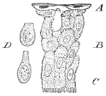
Vertical section through the epidermis from the head of Rana esculenta; after F. E. Schultze. Magnified 600 times.
A Horny layer.
B Middle layer.
C Deep layer.
D Isolated mucous-cells.
a. The epidermis (Figs. 228, 230) consists of several layers of epithelial cells, those of the deepest layer being more or less columnar in form, those of the middle layer shorter and polygonal, while those of the superficial layer are flattened, very transparent, and horny.
All the surfaces of these cells are serrated, the serrations being, however, with difficulty seen on the free surface of the superficial cells (Leydig). Each cell has a distinct nucleus; in the deeper cells the nucleus is oval, broad, and rounded, in the superficial cells flattened and thin.
Fig. 229.
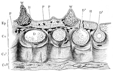
Vertical section through the skin of the back; after Wiedersheim.
Co Superficial layer of cutis. Co1 Middle layer of cutis. Co2 Deep layer of cutis. D Cutaneous glands. D1 Ducts of cutaneous glands. Ep Epidermis. H Horny layer of epidermis. P Pigment-cells. W Papillae.
The surface of the epidermis (Fig. 230) forms a very beautiful mosaic of flat cells, chiefly hexagonal in form, with pale, central, oval nuclei. Here and there two adjacent cells appear to enclose a semilunar space (Fig. 228 D); these spaces are occupied by peculiar cells (goblet-cells, E. Schultze; mucous cells, Leydig), which do not belong to the superficial cells but the layer of cells immediately beneath the horny layer (Schultze, Pfitzner). The cells are rounded or flask-shaped, and closely resemble the epithelial goblet-cells or chalice-cells. According to Rudneff they open on the free surface by stomata; this is, however, denied by Schultze and others.
According to Pfitzner these cells secrete a substance, which has an important function in connection with the process of casting the skin, which consists in the separation of the upper layer from that below brings about a complete shedding of the skin (Wiedersheim).
Fig. 230. Surface view of epidermis of Rana temporaria; after Eberth. A Epidermal cell. B Stoma-cell. C Attached cuticle from duct of gland. D Protoplasm and nucleus of stoma-cell.
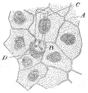
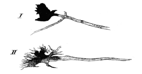
Fig. 231. Nerve terminations in the branched pigment-cells of the cutis; after Ehrmann. Hartnack, Obj. 8, Oc. 3. I. Pigment ceasing abruptly at junction of nerve. II. Pigment gradually ceasing along the nerve.
[The horny layer is, for the most part, very thin, as it consists of one or two layers of flattened cells only (Schultze), but in some situations, as on the back and especially on the under surface of the toes, it is much thickened, and is then rough.
Deeply pigmented, branched cells, capable of contractile movements, are also found, somewhat sparsely distributed, in the epidermis (Leydig, H. Müller, Schultze).]
b. The cutis (Fig. 229, Co, Co1, Co2). The epidermis is generally attached to the corium by means of a continuous layer of branched cells, which is deeply stained when the animals are fed with madder (Katschenko). Many of these cells are pigmented. This layer is seldom flat, but is raised into papillae and folds, which are repeated by the superimposed epidermis. In addition to this layer the corium has, except in the webs and supplemental toes, three distinct layers of connective-tissue, together with much unstriped muscle-fibre (Eberth).
The superficial layer (Fig. 229 Co) is a loosely-meshed, much pigmented, vascular layer; it forms a loose support for the numerous glands, and is traversed by numerous nerves.
The middle layer (Co1) forms the groundwork of the cutis; it is much firmer and more compact than the superficial layer; in section it appears as a broad band, bounded superficially by a sharp line. It is chiefly composed of closely packed connective-tissue fibres, which have, for the main part, a horizontal or wavy course; at certain points, however, vertical fibres are seen passing towards the surface and hiding the sharp contour (between Co and Co1). At such points the two sets of fibres form a sort of basket-work arrangement.
The deepest layer (Fig. 229 Co2) is composed of very delicate, white and yellow elastic fibres, and vessels and nerves; it attaches the whole integument to the underlying organs. By the looseness of its structure it forms an important lymph-space.
[The muscle-fibre of the cutis is very unevenly distributed. It is found somewhat freely in the region of the back, the dorsal surface of the head, and the neck; less freely on the dorsal surfaces of the extremities, very sparsely on the abdomen, breast, and ventral surfaces of the extremities; in the feet it appears to be absent (Eberth).
The pigment of the skin. As already mentioned pigmented cells occur in the epidermis, but by far the greater quantity of the cutaneous pigment is found in the cutis, and more especially in the loose layer of branched cells, which form the boundary between epidermis and true cutis. These cells, known as chromatophore-cells, have been carefully investigated by Harless. They play an extremely important part in bringing about the well-known changes in the colouring of the skin. Bimmerman has proved that these cells are influenced by stimulation of the nerves supplying the region in which they occur. Ehrmann has been able to trace a direct connection between the nerve-fibres and the pigment-cells (Fig. 231 I, II). In such cases the pigment was sometimes sharply marked off, at others was gradually lost along the nerve.]
c. The papillae and ridges of the skin are classified by Leydig as follows: (1) Small ridges, (2) larger ridges, (3) papillae with touch-bodies, (4) papillae without touch-bodies, (5) papillae with the ducts of glands, (6) capillaries in the form of papillae, (7) small elevations, including the lateral organs.
[All these structures are derived from the cutis, the epithelium above simply repeating the elevation, and being itself either not increased in thickness or only to a slight degree. (For further description of some of these papillae, see Organs of Tactile Sensation, p. 380.)
Peculiar wart-like papillae are found on the articulations of the first phalanges with the metatarsus, on the same articulations and on the articulations between the first and second phalanges of the third and fifth toes, and on the articulations between the first, second, and third phalanges of the fourth toe. These papillae are connected with the long flexor tendons by fine tendinous bands (Klein).
Temporary papillae are developed in the females of Rana temporaria during the breeding season; according to O. Huber (l. c.), who has described them, their distribution and structure is as follows:—
During the breeding season the skin of the sides of the trunk of female specimens of Rana temporaria is rough; the small elevations, to which this roughness is due, are each about the size of the head of a pin, and have the appearance of small, rounded, white or pale rose-coloured, warty growths. The papillae reach their greatest development and are most numerous on the sides of the trunk (Fig. 232 I); they may be traced on to the tympanic membrane, where they are arranged in a ring, and in some cases under the eyes to the sides of the snout.
Anteriorly the papillae are absent from the median portion of the dorsal surface of the trunk, but posteriorly, behind a line joining the anterior extremities of the two iliac bones, the papillae cover the whole surface.
The papillae are large and very numerous around the arms, and pass by gradual, transitionary stages into the ordinary papillae of the region. Further they may be traced backwards along the anterior or extensor surface of the thigh, the upper surface of the leg, foot, and fifth toe. Occasionally a few papillae were found on flexor surfaces of the third and fourth metatarsus, and very rarely were any discovered on the fore-limbs. They occur in no other batrachian of Germany (Huber).
Fig. 232.
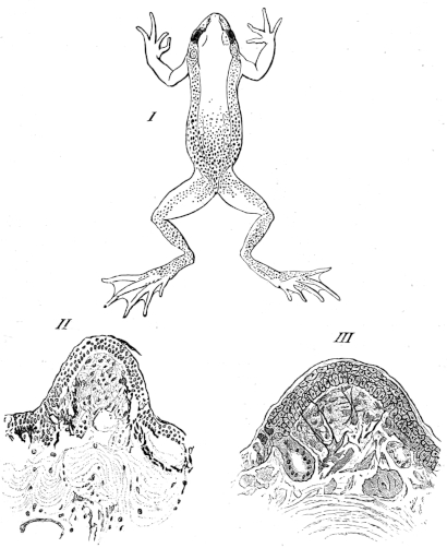
I. Diagram to show the distribution of the temporary papillae in Rana temporaria; after Huber.
II, III. Vertical sections through a temporary papilla; after Huber.
Each papilla (Fig. 232 II and III) consists of a hemispherical elevation of the cutis, and is covered either by a layer of normal epidermis or in some very rare cases the epidermis was slightly thickened. The papilla consists of a firm connective-tissue stroma, and is from 0.2–0.4 mm. in height, 0.4–0.5 mm. broad (after hardening in alcohol). Such glands as exist in the papilla usually open laterally (Fig. 232 III), and the blood-vessels are numerous.
The epidermis covering the papilla is well supplied with nerve-fibres, derived from large ganglion-cells in the papilla (Fig. 232 III). The branched pigment-cells, so numerous in the rest of the skin, are absent from the upper parts of the papillae (Fig. 232 II); the pigment-cells may cease either abruptly or gradually.
The rose-colour of the papillae is due to the presence of a golden red pigment. After the breeding season the papillae are gradually lost, and the pigment-cells wander into the unoccupied parts.
Huber compares the arrangement of the nerves to that of the ‘touch-spots’ of Merkel.
d. The glands of the skin are of two kinds, serous and mucous; they have been fully described by Engelmann and Openchowski.
(1) The serous glands (Körnerdrüsen, Engelmann) are large, averaging in diameter 0.2–0.4 mm., but others vary in size from 0.13–0.8 mm. in diameter. They are found chiefly on the dorsum of the trunk and arranged in groups, which vary in dimensions from 3–20 mm. in length and 2–4 mm. in breadth. They may be found in any part between the ear-region and the anus, on the dorsal surface of the thigh, especially towards the outer and inner borders. They are found very sparsely distributed on the ventral surface of the trunk and on other parts of the extremities, though as a rule a few are found on the dorsal surface of the feet and near the phalanges. They are wanting in the nictitating membrane.
The glands have a rounded form, with the ducts placed at their superficial poles. The glands possess three coats; externally is a coat of connective-tissue, the fibres of which are arranged closely together, and cross one another at acute angles: these fibres are continuous with the connective-tissue of the surrounding cutis. Inside this coat is a thick layer of muscle-fibre (0.005–0.015 mm.); the muscle-cells are arranged meridionally, with the one pole towards the superficial surface of the gland, the other pole at the deep surface (Hensche). Many of the fibres, however, extend only one-fourth or one-third of this distance.
The innermost coat is a simple layer of epithelial cells, which rests directly on the muscle-layer; no basement-membrane or space of any kind having been discovered between the two layers. When examined in the recent state these cells are conical or cylindrical in shape, and have the appearance of goblet-cells: they possess a delicate but distinct cell-wall, but are open towards the lumen of the gland. One or sometimes two rounded nuclei are found towards the base of the gland, and surrounded by a small amount of granular protoplasm; the rest of the cell is occupied by a mass of small, rounded, highly refractive granules of about 0.002–0.01 mm. in diameter. These granules are also to be found in the protoplasm of the cell; they contain a substance capable of swelling freely, and present many points of resemblance to the granules found in the cells of the oviducts.
These glands represent the poison-glands or lateral glands of toads, and the ear-glands or parotids of Caecilia (Leydig). According to Leroux81, Gratiolet and Cloëz82, the reaction of these glands in toads varies; according to du Bois-Reymond83 the reaction is, in frogs, acid; Engelmann84 found the reaction of these glands, in frogs, to be neutral, frequently, however, with a tendency to either acidity or alkalinity.
These glands correspond with the ‘large glands’ of Hensche, the ‘contractile glands’ of Stieda, the ‘large, dark glands’ of Eberth, and the ‘large contractile glands’ of Leydig’s earlier publications.
(2) The mucous glands are, as a rule, smaller, more numerous, and more evenly distributed than the serous glands. Usually they lie in such close proximity as almost to touch one another; according to Engelmann an average number of sixty is found in one square millimeter; on the abdomen 62–68; flexor surfaces of the posterior extremities in some places 76, but in others only 30–40; on the webs only 2–6 to the square millimeter. On the nictitating membrane they are arranged in two or three parallel rows, are large, and closely applied to each other. They are wanting on the deeper surface of the nictitating membrane.
The glands are generally rounded in form and have a short neck (Fig. 229). Glands in the recent condition, and not fully contracted, have a diameter of 0.06–0.21 mm., the majority measure 0.12–0.16 mm. in diameter. The upper pole of the gland is usually placed directly under the boundary between epidermis and cutis, and, as a rule, about 0.06–0.1 mm. beneath the superficial surface of the skin. The duct usually passes directly to the surface, in a straight line.
The structure of the glands corresponds, in general, with that of the serous glands. Externally is a coat of connective-tissue, which is, however, more firmly attached to the muscular coat than in the case of the serous glands, and may easily be mistaken for a structureless basement-membrane85 (Eberth). The muscular coat is very thin, and consists of 16–20 flat, spindle-shaped fibres; extending from the upper to the lower pole as in the serous glands. At a short distance (0.015–0.022 mm.) from the upper or superficial pole each fibre has an ellipsoid thickening, composed of granular protoplasm, and containing a clear oval nucleus.
The inner coat is composed of a single layer of epithelium, placed directly on the muscular coat. The epithelium has a thickness of about 0.01–0.02 mm.; it is, however, usually thicker towards the base than towards the duct. The boundaries between the cells are very indistinct, and so give the epithelium the appearance of a continuous layer of protoplasm. The glandular epithelium is not directly continuous with the epithelium of the duct.
The shape of the cells depends upon their physiological condition. In a state of semi-contraction of the gland, the cells are cubical, and their free borders pushed into the lumen in a slight convexity; when the gland is fully distended the cells are two or three times as broad as deep; in a fully contracted gland the cells are conical or cylindrical. In the first-mentioned condition their diameter varies from 0.006–0.018 mm. The larger glands are estimated to possess about 150 cells, the smaller only thirty to forty (Engelmann).
The cells appear to have no cell-wall, certainly they possess none on their free surfaces. Their protoplasm distends very freely on the addition of water. As a rule some cells are much more granular than others of the same gland; each cell has a rounded oval, clear nucleus, and vacuoles are frequently met with.
The lumen of the gland contains a colourless, transparent, watery fluid, which contains mucous material.
The ducts of the glands are thick-walled cylinders, circular in section. Their diameter varies from 0.02–0.05 mm., their length is equal to the depth of the superimposed epidermis. The wall of the duct is lined with two or three concentric layers of highly refractive, nucleated cells. The cells are arranged parallel to the length of the duct: in transverse section they are sickle-shaped or semilunar. In the upper third or fourth of the duct the inner wall is lined with a cuticle, very resistant to the action of acids or alkalies: it is thrown off with the skin in the process of casting. In the horny layer of the epidermis this cuticle is partially closed by a stoma-cell (Eberth), (Fig. 230), which possesses a tri-radiate opening.
The glands correspond with the ‘non-contractile glands’ of Hensche, the ‘dark and bright glands’ of Stieda, the ‘glandule mezzane, piccole e piccolissime (ad epitelio pavimentoso)’ of Ciaccio, and the ‘small, dark, and medium-sized, bright glands’ of Eberth.
Fig. 233.
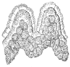
Vertical section through the epidermis of the supplemental toe of Rana esculenta; after E. Schultze. Magnified 400 times.
e. The peculiarities of the skin of the supplemental toe and webs. The supplemental toe presents, in the male, a rounded, oval swelling (Fig. 234 I, D), with the end of the digit projecting from the distal end.
Normally this swelling is usually of a grey colour; during the breeding season, however, the swelling enlarges, and becomes of a black or deep brown colour (Leydig).
The epidermis covering this swelling (Fig. 233) is much thickened and produced into projecting papillae. The cutis also shows a marked thickening and an increase in vascularity; the glands are of the kind described as mucous glands (Engelmann). They present, however, an immense increase in size, and at the same time are so closely applied to one another as to form the main part of the enlargement (see also Organs of Tactile Sensation).
In the webs the various layers of the corium are more or less fused together, and cannot be distinguished from each other; the glands are, as a rule, smaller than on the general surface of the skin.
Fig. 234.
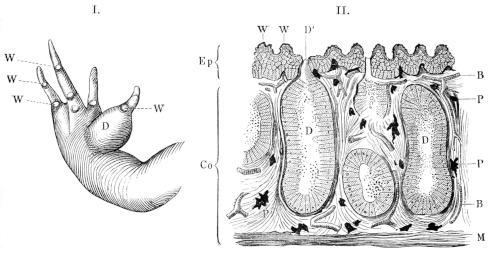
Fore-foot of a male frog. Vertical section through the swelling on the supplemental toe of a male frog.
D Glandular swelling on the supplemental toe. W Warty papillae on the inter-phalangeal joints. B Blood-vessels. Co Cutis. D Glands. Ep Epidermis. M Muscles. P Pigment-cells.
f. The blood-vessels and lymphatics of the skin.
(1) The blood-vessels of the skin are arranged in two networks; the one is situated immediately beneath the epidermis, and consists of fine capillaries, forming a fine-meshed network, which enclose the ducts of the glands. The meshes vary considerably in size and shape (Hyrtl and Langer). The second network of vessels is situated under the cutis; the vessels forming it are small, and the meshes formed are large. From this network vertical branches pass to form the superficial network; these branches usually course along the strands of vertical fibres already described, and break up to form capillaries only when they reach the deeper surface of the epidermis (Langer), (Fig. 235 I).
Fig. 235.
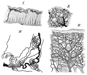
The blood-vessels and lymphatics of the skin; after Langer. Arteries striped, lymphatics shaded.
I. Vertical section through skin of thigh.
II. Horizontal view of arteries and lymphatics of the skin.
III. Peculiar tortuous arteries of the lamina inguinalis.
IV. Arteries and lymphatics of the web.
In some situations, however, the vertical branches divide earlier; this is especially the case in parts where the glands are very closely packed, as in the nictitating membrane, the upper lip, and the swelling of the supplemental toe (in the male). In these parts the division takes place at the deeper surface of the glands (Langer).
A peculiar arrangement of the cutaneous blood-vessels is found in the lamina inguinalis, where a chain of reddish points can be made out with the naked eye; examined more carefully (Fig. 235 III) each reddish point is found to be a complex loop of capillaries. They extend from the skin to the hinder lymph-hearts, where they form a close network which partially invests the lymph-hearts (Langer).
(2) The lymphatics of the skin (see also p. 252), like the arteries, reach the deeper surface of the epidermis by coursing along the bands of vertical fibres; they then form a network of capillaries with rounded meshes (Fig. 235 I, II, IV), and lying immediately under the epidermis, and a network which surrounds the various glands. The lymphatic-capillaries are as a rule larger than the blood-capillaries. The lymphatic network lies beneath the blood network, and the vessels of the two systems branch independently (Langer).
In those parts where the glands are placed closely together this arrangement is modified, and corresponds with the modifications found in the blood-capillaries in these regions. The vertical branches divide so as to form a network below the glands, and from this vertical branches pass in the same direction between the glands to form a secondary network on the superficial surface of the glands (Langer).
g. The nerves of the skin. The cutis is very richly supplied with both medullated and non-medullated nerves. In the subcutaneous tissue the nerves destined for the skin branch freely to form numerous fine twigs, which, without actually inosculating, form a fine meshwork. From the larger trunks numerous vertical branches pass vertically to form a secondary network, from which both coarse and very fine twigs pass to encircle the glands. The fine fibres which supply the glands are non-medullated and possess oval nuclei; their diameter varies from 0·001–0·002 mm. The number of nerve-fibres is not much smaller than that of the muscle-fibres (Engelmann). (See also Organs of Tactile Sensation.)]
II. THE ORGANS OF TACTILE SENSATION.
The organs of tactile sensation are the nerve-plexus of the epidermis, the touch-spots of Merkel, the lateral sense-organs, and the touch-corpuscles of the tendons.
a. [The nerve-plexus of the epithelium is very incomplete as compared with the plexus found in the epithelium of the cornea; here and there a few non-medullated fibres have been traced a short distance between the epithelial cells. No such mode of nerve-terminations in the epithelium as is described by Eberth and Macullum in the tadpole can be found in the adult.]
b. The touch-spots. What knowledge we possess of these organs is due to the labours of Leydig, Ciaccio, Eberth, and more particularly those of Merkel.
At the base of each papilla, which contains such a touch-organ, is found a number of colourless, flattened cells, arranged either in a single layer, or heaped up and connected with very fine nerve-fibres. The flat surface of the cells is parallel to the surface of the body, and they do not form a separate or circumscribed body, they are therefore better spoken of as touch-spots than as true touch-bodies (Merkel). The larger organs are found in the papillae, but smaller ones may be found on any part of the skin.
Fig. 236.
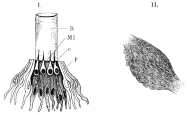
I. Lateral sense-organ of tadpole of frog. Half-schematic.
c Central zone (nerve-epithelium). Ml Membrana limitans. p Peripheral zone (sustentacular cells). R Hyaline tube.
II. Touch-corpuscle from the sheath of a digital tendon of the frog; after Löwe. Schieck, Oc. O., Obj. 9.
These organs are best seen in the prominence or swelling upon the supplemental toe during the breeding season; they are numerous on the dorsal surface of the trunk, but occur most frequently on the under surface of the hinder feet (Planta pedis). After the breeding season the organs of the thumb undergo a retrogressive change, which results in a network of spindle-shaped and branched cells with fibres (Wiedersheim).
c. The lateral sense-organs attain their highest development in the tadpole; in the adult the organs have undergone a retrogressive change, the result of which is that the organs are diminished in size; the whole organ sinks by the formation of a tube, which is then closed by a mucous secretion, consequently the organ is functionless: with this loss in function and change in position of the organs a corresponding diminution of the ramus lateralis nervi vagi occurs (Merkel).
Each lateral organ (Fig. 236 I) consists of a slightly elevated papilla, with the centre depressed, and in each may be distinguished a central and a peripheral zone (c, p). The central zone consists of a group of pear-shaped cells, with the narrower ends directed towards the free surface; each is connected below with a nerve-fibril, while above it bears a short, stiff cilium. The cilia are enclosed in a delicate hyaline tube (R), which is open superficially, closed by the papilla below; consequently the cilia are in direct contact with the surrounding medium. The peripheral zone is a layer of pale, flattened, cylindrical cells (p); they possess, at their upper borders, a perforated membrana limitans (Ml), through which the cilia pass. These cells serve as a support to the central cells, and are themselves surrounded by ordinary epithelial cells.
The distribution and arrangement of the lateral organs is alike in fish and larval amphibia. They are most numerous on the head, where they surround the eye, and are continued forwards to the snout and on to the lower jaw. All these organs on the head are in connection with the trigeminal nerve. A line of these organs passes from the hinder part of the circumference of the eye along the gill-cover to the neck, where the lines of opposite sides are usually, though not constantly, joined by a transverse line of the same organs; thence the lines are continued along the trunk to the tip of the tail. In fish there exists, as a rule, only one pair of such longitudinal lines (Linea lateralis); in anura-larvae, proteus, and in all salamanders there are three pairs: of these one pair lies near the vertebral column, the second at the junction of the flank with the abdomen, and the third corresponds to the lateral line in fish (Malbranc).
At an early developmental period each metamere possesses one pair of such lateral organs; in later life, however, this simple arrangement is lost, and each segment usually possesses a group of organs.
The organs of the trunk and tail are supplied by branches of the Ramus lateralis nervi vagi. Both in fish and amphibia a reparative process, by means of fission, occurs; as a consequence the organs are found in various stages of development.
d. [The touch-corpuscles of Golgi and Löwe (Fig. 236 II) are found on the joints of the digits. They are surrounded by connective-tissue and are spindle-shaped; the two ends of the spindle are glassy in appearance, the middle portion is fibrous. In the thickest part of the corpuscle is a zone of nuclear bodies.
They are regarded as touch-bodies (Golgi and Löwe). Golgi (l. c.) describes two kinds of touch-organs in connection with muscle and tendon; one class correspond with those just described, he names them ‘nervous muscle-tendon organs.’ The second class he compares with the touch-bodies found in the conjunctiva.]
III. THE ORGANS OF TASTE.
The organs of taste are not confined to the surface of the tongue, but are also found on the mucous membrane of the roof of the mouth, especially in the neighbourhood of the vomerine teeth; on both roof and floor of the mouth these organs can be traced to the commencement of the oesophagus.
[The mucous membrane of the tongue possesses two kinds of papillae and numerous glands.
a. The filiform papillae are the more numerous; they are conical or thread-like in form, and consist of connective-tissue, with a few striated muscle-fibres; they include blood-capillaries, but no nerves have been traced into them (Leydig). They are covered with ciliated epithelium and goblet-cells.
b. The fungiform papillae are much larger, and are paler than the filiform papillae. The free ends of these papillae are broader than the bases, and when the epithelium is removed the free ends present a concavity. From the base to the border of the free surface each papilla is covered with the usual oval, ciliated epithelium, but at this border an abrupt transition takes place. The epithelium covering the end of the papillae is of three kinds: goblet-cells, cylindrical cells, and forked cells; the three forms of cells are quite characteristic, and no intermediate transitionary forms are met with.
(1) The goblet-cells (Fig. 237 I) are arranged vertically to the free surface of the papilla; they are from 0.020–0.024 mm. in length, and from 0.01–0.02 mm. in diameter. In the lower third of the cell is found a nucleus of about 0.008 mm. in diameter, and this encloses a nucleolus of about 0.001 mm. in diameter. Close underneath the nucleus the cell is contracted to form an irregular process or foot. The contents of the cells are a very finely granular, transparent protoplasm. These cells form the outermost part of the epithelium, covering the end of the papilla; all the cells belonging to the same papilla are of the same size. In consequence of the mutual pressure which they exert on each other they present, in transverse section, six-sided outlines (Fig. 237, 2). The nuclei of adjacent cells are placed at almost exactly the same level. By the action of reagents on these cells very remarkable forms are obtained, in consequence of the protoplasm swelling and being forced upwards (Fig. 237, 5).
Fig. 237.
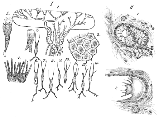
I. Various parts from the fungiform papillae; after Engelmann. 1. Nerves of a fungiform papilla. Magnified 450 times. 2. Surface view of the epithelium, after five minutes’ action of iodized serum. Magnified 600 times. 3. Goblet-cell with adjacent forked cell. Magnified 450 times. 4. Portion of the papilla, after removal of the goblet- and forked cells; only cylindrical cells remaining. Magnified 400 times. 5. Goblet-cell, with swollen contents. Magnified 450 times. 6‑13. Various forms of isolated forked cells. Magnified 450 times. II. Sections of two glands of the tongue; after Biederman. 1. Resting-gland. 2. Gland after stimulation.
(2) The cylindrical cells (Fig. 237 I) have rounded free extremities, which reach to the general surface of the epithelium; the remainder of the cell is cylindrical except towards its lower extremity, where it is slightly dilated and encloses an oval nucleus: the protoplasm of the cells is very finely granular. These cells rest on the connective-tissue of the papilla, and are in close juxtaposition, so that several hundreds of them are found on one papilla.
The spaces between the upper parts of the cylindrical cells are occupied by the goblet-cells and by the forked cells.
(3) The third form of cell found on the fungiform papillae are named forked cells (Fig. 237 I) by Engelmann. The body of the cells has an ellipsoidal form, is from 0.006–0.008 mm. in its longer diameter, and 0.003–0.004 mm. in the shorter diameter. Processes arise from both poles. The peripheral processes arise by a short common stem, which then divides into two or rarely three branches; the whole process is from 0.004–0.008 mm. in length, and is always just sufficiently long to reach the general surface of the epithelium: when the peripheral process is long the central is usually shorter, and vice versâ.
The central processes arise by a stem from 0.001–0.002 mm. in thickness, which usually divides dichotomously. The length of this process may be as much as 0.025 mm. or almost nil.
The forked cells are about twice as numerous as the goblet-cells, they occupy the spaces between the cylindrical cells and the goblet-cells. The branched central processes form a network on the connective-tissue of the papilla, which is, at this point, perforated by a rich plexus of fine non-medullated nerve-fibrils. It is not decided whether the processes of various cells inosculate. Engelmann regards these forked cells as the taste-cells and as the sensory nerve-endings.
The bodies of the fungiform papillae contain blood-vessels and nerves, the latter enter as medullated nerves, but lose the medulla, somewhat abruptly towards the upper end of the papilla, where they form a sort of nerve-cushion (Nervenkissen, Engelmann).]
Taste-organs are also found on the roof and other parts of the mouth, and present many points of resemblance to the lateral sense-organs. Like them they present a peripheral zone and a central zone; the cells of the latter, however, are not longer than the peripheral cells, and possess no cilia (J. van der Hoeven, Merkel).
IV. THE NOSE.
The anterior nares or nostrils are two small openings, placed directly in front of the anterior angle of the eye; the distance between the eye and the corresponding nostril being, in the case of adult animals, about five or six millimeters. The anterior nares are surrounded by rims, which are contracted below, and so form very short, tentacular-like prominences.
The posterior nares (choanae) are about four millimeters from the anterior nares, consequently the long axis of the nose is, approximately, of this length. (For the external muscles of the nose, see p. 59.)
The boundaries of the nasal cavities are as follows:—the roof of each cavity is formed by the dorsal plate of the sphenethmoid, the nasal bone, and the premaxillary bone; the floor is formed by the vomer and the palatine bone, the inner wall by the vertical septum of the sphenethmoid, the outer wall by the premaxillary and maxillary bones, the anterior wall by the premaxillary bone, and the posterior wall by the sphenethmoid. The cartilaginous portion of the nasal skeleton (see also p. 27) projects into the general cavity and subdivides it into various sinuses, which have been the subject of careful investigation by Born and Wiedersheim.
a. The nasal cavities are best examined by means of serial transverse sections; in a section through the anterior nares (Figs. 238, 239) there will be seen three sinuses on either side:—
(1) The superior sinus (on) is large, rounded, and placed against the cartilaginous septum; the sinus is lined with olfactory epithelium. Posteriorly the sinus extends beyond the posterior nares, and is bounded by the anterior surface of the sphenethmoid. In front it ends in a rounded concavity, likewise lined with olfactory mucous membrane. Opposite the anterior nares the cavity is partially subdivided, by a longitudinal process on the floor, into two cavities; the anterior nares open into the external chambers, and therefore not directly into the main cavity of the superior sinus. The superior sinus communicates with the inferior sinus by a narrow, almost vertical, slit.
(2) The inferior sinus (un) is narrow and flattened from above downwards; it is placed against the maxillary bone, and represents the maxillary sinus of the frog. Externally it possesses a descending arm.
The inferior sinus is continued backwards and opens on its inner side into the posterior nares, or rather into the mouth. The descending arm extends only a short distance forwards, to terminate near the point where the superior and inferior sinuses communicate by only a narrow opening. This sinus is on the whole broader in front than behind, and has a general direction from without, inwards and forwards underneath the superior sinus.
Fig. 238.
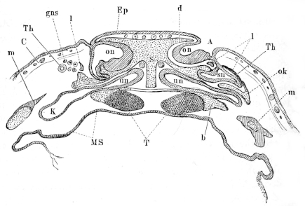
Fig. 239. Frontal sections through the nose of two tadpoles; after G. Born. A Anterior naris. b Cartilaginous floor. C Cutis. d Cartilaginous roof. Ep Epidermis. gi Intermaxillary gland. gni Lower nasal gland. gns Upper nasal gland. K Maxillary sinus. l Concha narium (os lachrymale). m Maxillary bone. MS Oral mucous membrane. ok Cartilage. on Upper blind sac. R Pharyngeal gland. S Nasal septum. se Septum between the upper and lower nasal cavity. sn External blind sac. T Trabeculae. Th Lachrymal duct. un Lower blind sac.
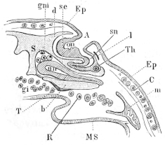
(3) The lateral sinus (sn) is situated in the partition between the superior and inferior sinuses, or rather between the external chamber of the upper sinus and the inferior sinus. The lateral sinus is triangular in form, being narrow in front and broad behind; at its inner posterior angle it opens on the free border of the horizontal partition between the superior and inferior sinuses; this opening is, however, continued along its roof, so that the lateral sinus opens also into the superior sinus.
b. The nasal cartilages. The two nasal cavities are completely separated by the cartilaginous septum (Figs. 238 and 239 S), and are for the most part lined by cartilage. The anterior end is formed of concave cartilages, while the posterior, being situated in the sphenethmoid, is usually more or less ossified in the adult. The posterior wall has two openings: a larger near the septum for the olfactory nerve, and a smaller, more externally, for the nasal branch of the trigeminal nerve.
The anterior wall is more complex than the posterior, being thicker in the middle than at the sides, and possessing three blind sacs for the three sinuses. From the anterior wall two processes project backwards between the sacs and enclose them more or less completely.
Of the three sacs or cavities only the lower is completely surrounded by cartilage. The upper sac is in part bounded by a shell-shaped, concave cartilage (Concha narium, of various authors; Os lachrymale, Born), which covers it anteriorly and externally, and is attached by a small base to the roof of the lower sac (Fig. 239, l), its upper border bounding the anterior naris (Fig. 15). The upper and external parts of the upper sac have no cartilage. The cartilages of the outer wall of the lower, blind sac extend backwards to the point where the maxillary sinus commences to descend: the roof, however, is prolonged further backwards by two small cartilaginous processes; the inner is short, the outer forms the roof of the descending arm of the sinus, and joins a cartilaginous process, which commences at the anterior portion of the roof of the nasal cavity, passes backwards and downwards to a broad plate, and forms an incomplete outer wall to the nasal cavity. There are also three cartilaginous processes—α. One arising from the outer side of the floor at the level of its junction with the anterior wall; it is a flattened process and passes outwards, and bifurcates at its end to meet the premaxillary and maxillary bones (this is the Oberkieferfortsatz of Ecker), (p. 28, Fig. 14 n″). β. A delicate process on either side, described by Wiedersheim (see p. 280). γ. The third pair of processes are fully described for the first time by Born; each arises at the lower border of the corresponding Concha narium, passes forwards and downwards under the ascending process of the premaxillary to the point where process β is attached to this bone. The processes are flat and approach one another obliquely; in older animals they are united at the point of contact.
At the junction of the anterior wall, the septum, and the floor is a large aperture in the cartilaginous capsule, through which passes the chief nasal branch of the trigeminus, which supplies numerous branches to the intermaxillary gland.
c. The glands of the nasal region are Bowman’s glands, the intermaxillary glands (Wiedersheim), the lower nasal gland (Born), the upper nasal gland (Born), the pharyngeal gland (Born), and the lachrymal duct.
(1) The intermaxillary gland (see p. 280).
(2) The lower nasal gland is placed along the septum and extends as far back as the posterior nares, opening by its ducts (Figs. 238 and 239 gni) into the inferior blind sac of the nasal cavity. In histological structure it corresponds with the intermaxillary glands, except that the glandular tubes are somewhat smaller, and the glandular epithelium stains somewhat less easily with carmine (Born).
(3) The upper nasal gland occupies the space between the Concha narium (Os lachrymale, Born) and the neighbouring cartilages, it also surrounds the anterior naris and the opening of the lachrymal canal (Figs. 238 and 239 gns). The numerous ducts open on the mucous membrane covering the oblique cartilage (above described, as passing from in front, downwards and backwards, and forming an incomplete outer wall of the cavity), and its hinder prolongation.
(4) The pharyngeal gland is placed transversely behind the posterior nares, and surrounds the vomerine teeth. A portion of the ducts open into the posterior nares, the remainder on the mucous membrane of the pharyngo-oral cavity at two symmetrically placed points (Fig. 238 R).
(5) The lachrymal duct (see p. 428) opens into the nasal cavity at the point where the lateral sinus opens into the external chamber of the superior nasal sinus.
Fig. 240.
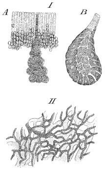
I. A. Bowman’s glands in situ from Rana temporaria; after C. K. Hoffmann. Magnified 150 times.
B. Section of Bowman’s gland; after C. K. Hoffmann. Magnified 300 times.
II. Vessels of nasal mucous membrane of Rana esculenta; after Langer. Blood-vessels striped, lymphatics shaded.
(6) [The glands of Bowman (Fig. 240 I) are freely distributed in the nasal mucous membrane. Each gland is usually rounded or flask-shaped, and consists of a single layer of large epithelial cells possessing distinct nuclei and nucleoli. The epithelium of the glands possesses no basement membrane (M. Schultze and Hoffmann86), but is bounded externally by a layer of nerve-fibres and connective-tissue.
The glands situated more superficially have straight ducts opening on the surface; those placed more deeply have usually curved ducts (Paschutin).]
d. The mucous membrane of the nasal cavities. That part of the superior sinus immediately around the anterior naris is lined with stratified epithelium; the rest of the nasal cavity is lined with columnar ciliated epithelium The epithelial layer rests on a subepithelial network, and this again on a submucous connective-tissue layer possessing numerous vessels and nerves.
Fig. 241.
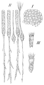
Separations from the olfactory mucous membrane of Rana temporaria; after C. K. Hoffmann.
I. Surface view of the olfactory mucous membrane. Magnified 350 times.
II. Epithelial and olfactory cells. Magnified 600 times.
III. Epithelial cells. Magnified 600 times.
(1) The epithelial layer (Fig. 241) consists of columnar cells, which in the olfactory region are of two kinds; in other regions the epithelium consists of closely-applied ciliated, thick cells, possessing large oval nuclei, and having irregular, branched bases or ‘feet.’ The cells are 0.032–0.048 mm. long; the nuclei 0.016–0.018 mm long, and 0.006–0.008 mm. broad. The free borders of the cells bear a number of fine cilia.
In the olfactory region a second set of cells, olfactory cells, are met with; these cells (Fig. 241 II) possess each an oval body, enclosing a large nucleus, and a peripheral and central process. The peripheral processes reach to the general surface of the surrounding epithelium and there terminate in a number (5–8) of stiff cilia; these cilia are sometimes 0.09 mm. long, they are thicker at their bases than the ordinary cilia (Schultze); according to Hoffmann, a second system of stiffer and longer cilia is met with; of which each olfactory cell possesses as a rule only one. The bodies of the olfactory cells are 0.009–0.010 mm. long, and 0.007–0.008 mm. broad; the peripheral processes vary considerably in length, according to the position of the body (0.03–0.05 mm.). The central processes vary from 0.02–0.03 mm. in length. The peripheral processes are considerably thicker than the central processes. No membrana limitans olfactoria has been discovered in the frog.
(2) The second layer consists of a network of processes belonging to the bases of the central processes of the superimposed epithelial and olfactory cells. Numerous highly refractive nuclei possessing nucleoli are situated in the meshes of this network; they correspond in all particulars with the nuclei of the olfactory cells (M. Schultze). According to Exner the branched processes of the epithelial cells and of the olfactory cells unite to form a complex plexus. Other observers, Paschutin, Cissoff, v. Brunn, Schultze, and Hoffmann oppose this view, and hold that the central processes of the olfactory cells do not unite with the processes of the epithelial cells, but that they are in direct continuity with the fibrils of the olfactory nerve. This view is probably correct.
The submucous layer has a loose connective-tissue matrix, which encloses the glands, nerves, and vessels; according to Paschutin two pigment-layers may be distinguished: the one, immediately under the middle layer of this mucous membrane, is continuous and deeply pigmented; the second is deeper and does not form a continuous layer. This layer is extremely rich in blood-vessels and lymphatics (Langer and Paschutin), (Fig. 240 II). The lymphatics are relatively very large and are very numerous (Langer).
In this layer the central processes of the olfactory cells form bundles of fibres, lying parallel with the surface of the mucous membrane (Paschutin, Cissoff, and Schultze).]
V. THE EAR.
(Re-written by the translator from Das Gehörorgan der Wirbelthiere, by G. Retzius, 1881.)
The organ of hearing is divisible into two parts, the tympanum or middle ear, and the labyrinth or internal ear; an external ear is absent, unless a very slight depression of the tympanic membrane be regarded as such.
A. The tympanum (Cavum tympani) is a cavity, bounded externally by the tympanic membrane and internally by the capsule of the internal ear; it communicates by means of the Eustachian tube (tuba Eustachii) with the pharyngo-oral cavity (Fig. 178).
Fig. 242.
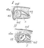
I. The tympanic membrane of Rana esculenta; natural size. After Retzius.
II. The tympanum as seen after removing the tympanic membrane; natural size. After Retzius.
at Annulus membranae tympani.
col Columella.
ct Fissure-like cavity.
dm M. depressor maxillae.
mt Tympanic membrane.
sq Squamosal.
t M. temporalis.
a. The tympanic membrane lies immediately underneath the skin, but can be separated from that structure; externally it possesses a very slight depression, the only trace of an external ear found in the frog.
The tympanic membrane is of a rounded oval form, being a little wider in the transverse than in the longitudinal diameter; the membrane is directed outwards.
After detaching the skin (Fig. 242 I) the membrane is seen to be attached by its circumference to a ring of cartilage (Annulus membranae tympani; see also p. 26) (at); the ring is attached anteriorly and above to the squamosal bone (sq), and in the rest of its circumference to the surrounding soft parts, i.e. the M. depressor maxillae (dm), the M. temporalis (t), and their fasciae. In the middle of the membrane is a small, rounded, white part (col) which can be traced backwards and upwards towards the circumference.
The membrane consists of fibres of connective-tissue which radiate peripherally from the central point of attachment of the columella; in the peripheral portion unstriated muscular fibres are also found (Leydig).
Internally the tympanic membrane is covered by columnar epithelium, a continuation of the mucous membrane lining the tympanic cavity.
b. The tympanic cavity (Fig. 242 II) is seen, after removing the tympanic membrane, as a flattened, funnel-shaped cavity. The walls of the cavity are lined with a pigmented mucous membrane, under which the cartilaginous ring (annulus membranae tympani) extends internally to form the greater part of the wall of the cavity. The cavity is an elongated oval slit (Fig. 242 II, ct), with its longer axis directed from above and in front, downwards and backwards, and leading inwards; it is bounded above, below, and in front by the squamosal, behind by the soft parts; above in the roof is the cartilaginous part of the columella. The inner or deeper portion of the tympanic cavity is bounded in front by the squamosal, and by the squamous process of the prootici (proc. squamosus prootici) in front and above; internally by the cartilage (primordial-cranium) between the prootic and the exoccipital; behind by the M. depressor maxillae. This part of the cavity is rounded and covered with mucous membrane, and has in its roof the bony part of the columella, which covers the foramen ovale (fenestra ovalis) by its oval, widened end-piece. This deeper portion of the cavity communicates by a short, wide Eustachian tube with the pharyngo-oral cavity; the tube is wide and is of a rounded oval form in section: anteriorly, externally, and internally it is bounded by the pterygoids, posteriorly by soft parts, in which is embedded the styloid process.
The tympanic cavity can be examined from without after removing the tympanic membrane, or from below by means of the Eustachian tube.
Fig. 243.
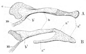
The columella; after Retzius. Magnified eight times.
A. Seen from above.
B. Seen from behind.
a′ Extrastapedial (Parker). Outer cartilaginous portion attached to middle of tympanic membrane (Retzius).
a″ Attachment of extrastapedial to mediostapedial.
a‴ Suprastapedial (Parker). Portion embedded in mucous membrane (Retzius).
b′ Mediostapedial (Parker). Bony part (Retzius).
b″ Process of mediostapedial.
c Interstapedial (Parker). Inner cartilaginous piece (Retzius).
m Insertion of small muscle.
c. The columella auris (Figs. 12, 243) is described by Retzius as consisting of three portions, of which the middle is bony, the external and internal cartilaginous. Parker divides it into four parts (see pp. 25, 26).
The extrastapedial (Fig. 243 a′, a″) is attached to the middle of the tympanic membrane by the oval surface opposite a′, and is attached by the process a‴ to the annulus tympanicus; this process of Retzius is the suprastapedial of Parker.
The mediostapedial (Parker), or middle bony piece of Retzius (Figs. 12 a′, 243 b, b′), is narrow externally but widens internally to articulate with the interstapedial. Just before reaching the latter it gives off a process (b′), to which are attached a few fibres of striated muscle (m).
The interstapedial (Figs. 12 a, 243 c) is cartilaginous; it is thick with a sharply cut-off, slightly concave end, which is placed against the fenestra ovalis. The inner surface of the interstapedial is, however, distinctly larger than the opening of the fenestra ovalis, consequently it does not fit in accurately, but is attached to the border of the opening by means of connective-tissue; the margins of the fenestra ovalis are hollowed (Fig. 245 II) so as to form a fossa fenestrae ovalis, and it is really to the margin of this fossa that the connective-tissue capsule of the interstapedial is attached.
B. The labyrinth or internal ear is contained in a capsule formed of bone and cartilage.
a. The capsule of the labyrinth (Fig. 244) is formed of two bones, the prootic and the exoccipital (according to Hasse this includes the opisthotic), which are united by cartilage belonging to the primordial-cranium. The fronto-parietal, squamosal, and parasphenoid take only an indirect part in its formation.
Fig. 244.
Antero-posterior section through the capsule of the right labyrinth of Rana esculenta; after Retzius. Enlarged five times.
ac. Aquaeductus cochleae.
av. Aquaeductus vestibuli.
cp. Posterior semicircular canal.
cra. Canalis rami anterioris acustici.
crp. Canalis rami posterioris acustici.
fr. Fenestra rotunda.
fsc. Fovea sacculi et cochleae.
ft. Trigeminal foramen.
kn. Cartilaginous suture.
ol. Exoccipital.
pr. Prootic.
ps. Parasphenoid.
On the whole the anterior half of the capsule is formed by the prootic, and the hinder half by the exoccipital; the cavity has its long axis directed from within and above, downwards and outward; the cavity is comparatively large and rounded, and contains the membranous ear.
Four surfaces can be distinguished: a supero-external, an interno-inferior, an anterior, and a posterior (Retzius).
(1) The supero-external surface is divided into two parts by a strong, transverse, bony ridge, the processus squamosus prootici. The upper half is concave, is directed upwards and outwards, and is formed by the prootic; it is separated from the anterior surface by a bony ridge, which marks the position of the anterior semicircular canal. Posteriorly and medianly it is separated from the posterior surface by a cartilaginous ridge, running from above, downwards and outwards, which marks the position of the posterior semicircular canal. The cartilaginous hinder root of the processus squamosus prootici arises in the cartilaginous ridge just mentioned, and runs outwards and forwards, covering the external semicircular canal.
The lower half of the supero-external surface (under the processus squamosus) is irregularly concave; immediately under the proc. squamosus is a shallow groove in the prootic; it is continued on the exoccipital to the jugular foramen. The remaining part of this surface consists of a rounded cartilage, and is part of the primordial-cranium cartilage between the prootic and the exoccipital; posteriorly it has a small oval aperture, the foramen ovale (fenestra vestibulare), which with the groove is covered by the columella. Above the aperture the cartilage narrows and is continued to the processus squamosus.
(2) The posterior surface is directly continuous with the supero-external surface, and is formed by the exoccipital. This surface lies behind the ridge formed by the posterior semicircular canal, it is concave, and has two small apertures, separated by a narrow, bony process, and situated near the jugular foramen; these are the foramen rotundum (fr.) and the aquaeductus cochleae (ac.).
(3) The anterior surface is altogether bony and formed by the prootic (pr.); the anterior surface of this part of the wall is continuous with the surface of the cranium, and abuts on to the large trigeminal foramen (ft.); below it is continuous with the under surface of the cranium, below and externally it articulates with the pterygoid.
(4) The interno-inferior surface is as a whole hollowed internally, the upper part being pushed in towards the cranial cavity. It is formed by the prootic anteriorly, by the exoccipital posteriorly, and is completed by the cartilage lying between these bones. In the middle of the upper part of the cartilage is a small oval opening (av.), the ap. aquaeductus vestibuli: about midway between this aperture and the jugular foramen is a second opening in the cartilage (cra.), the opening of the canalis rami anterioris (vestibularis) acustici: a little behind and above this is the opening of the canalis rami posterioris (cochlearis) acustici, situated in the exoccipital. The cartilage between the prootic and exoccipital in the lower half of the interno-inferior surface rests on the parasphenoid.
b. The position of the soft parts in the capsule. The large outer, rounded part of the cavity contains the saccule and cochlea (fovea sacculi et cochleae) (fsc.). Above, the cavity is more irregular, and contains the utricle and the sinus utriculi superior. In front and externally, imbedded in a deep groove, are the anterior and external ampullae (fovea ampullae anterioris et amp. externae); to these are attached the corresponding semicircular canals. The anterior semicircular canal opens above and externally into the canal of the sinus utriculi superior; the posterior passes into a groove for the posterior ampulla (fovea ampullae posterioris), and opens above and internally into the posterior semicircular canal. The external semicircular canal courses in the processus squamosus prootici, the posterior in the cartilage (Fig. 244 cp) between the prootic and exoccipital, and the anterior in the prootic. The upper and posterior part of the anterior semicircular canal is imbedded in cartilage, on which rests the parasphenoid.
c. The perilymphatic space (Fig. 245 I and II). The membranous labyrinth does not occupy the whole space enclosed by the hard parts but is surrounded by the perilymphatic space, which contains the perilymph. The periosteum and perichondrium form the outer lining of the space, and are for the most part pigmented:
The perilymphatic space is widest in the lower part of the organ, in the region of the fovea sacculi et cochleae, especially externally and behind (per); in front it is much narrower (per1). A little higher, in the region of the tegmentum vasculosum, the space is wanting, as the membranous labyrinth is here attached to the periosteum.
Around the utricle and saccule the space is comparatively wide; it is continued into the bony canals and ampullae. In the semicircular canals the space is wider on the concave side than on the convex side, the membranous canals being placed eccentrically, as are also the ampullae, though to a less extent. The periosteum and perichondrium lining this cavity form a very delicate, pigmented membrane, formed of numerous fine elastic fibres which cross each other irregularly; its inner surface is lined with an incomplete layer of branched protoplasmic cells with large oval nuclei. From this membrane numerous irregular bundles of fibres, in the form of trabeculae, pass into the space and form a rich network, which is attached internally to the outer surface of the membranous labyrinth and holds it in position. Free nucleated leucocytes are occasionally met with in the meshes of this network. The perilymphatic space is prolonged into two subsidiary cavities:—
Fig. 245.
The membranous labyrinth of Rana esculenta, within its natural covering of periosteum; after Retzius.
I. Seen from the side; magnified ten times.
II. The hinder half; magnified ten times. Seen obliquely from above, and in front, and from the side.
aa Anterior ampulla. adf Apertura fenestrae ovalis. al. Apertura lagenae. ap. Posterior ampulla. apb. Opening into the pars basilaris. ca Anterior semicircular canal. ce External semicircular canal. cp Posterior semicircular canal. dfo Ductus fenestrae ovalis. dp Ductus perilymphaticus. dp′ Saccus perilymphaticus. l. Lagena cochlea. ms. Macula sacculia. pb Pars basilaris cochleae. per Wider part of perilymphatic space. per1 Narrower part of perilymphatic space. ra. Ramus anterior. rn. Ramulus neglectus. rp. Ramus posterior. rs. Ramulus sacculi. s. Saccule. sfo Saccus fenestrae ovalis. spl Saccus perilymphaticus. tv Tegmentum vasculosum. u Utricle.
(1) The ductus fenestra ovalis (Retzius), (Fig. 245 dfo) commences as an opening (adf) immediately opposite the foramen ovale of the hard capsule, which leads into a moderately large canal, the structure under consideration. It passes forwards and outwards through the fenestra ovalis between the interstapedial and the outer wall of the capsule into the oval depression (fossa fenestrae ovalis), and rapidly widens to form a short, flattened, blind sac (saccus fenestrae ovalis), which is lodged in the fossa (sfo). Its walls are thin and pigmented.
Fig. 246.
Part of the outer wall of the perilymphatic space: after Retzius. Vérick’s Syst., Obj. III, Oc. 3.
per. Periosteum.
pg. Perilymphatic network.
zn. Leucocytes.
(2) The ductus perilymphaticus (Hasse), (Fig. 245 dp) passes above and behind the ductus fenestrae ovalis, behind the auditory-nerve, and near the cochlea, then behind and above the lagena to the aquaeductus cochleae (Fig. 244 ac), where it forms a short, wide tube, which passes backwards and inwards through this canal to the canalis jugularis; here it lies close to the nerves and forms a short oval sac (saccus perilymphaticus) (dp′), which communicates with the sub-arachnoid space of the cranial cavity by means of a tube from the neck of the sac. The walls of this structure are thin and formed of connective-tissue with very few pigment-cells. The other extremity of the tube passes to the pars basilaris and under the sinus post. utri., between it and the pars neglecta; it then courses to the outer side of the hinder end of the external semicircular canal: the tube then bends downwards to the outer side of the utricle and saccule to open into the general perilymphatic space.