SECTION III. THE NERVOUS SYSTEM.
George Haslam translation
THE NERVOUS SYSTEM.
LITERATURE.
Ahlborn, F., Ueber die Bedeutung der Zirbeldrüse. Zeit. f. wiss. Zool. 1884. Vol. XL, pp. 331–337.
Allen, H., The Spinal Cord in Batrachia and Reptilia. Proc. Acad. Nat. Sci. Philadelphia. 1883, pp. 56–57.
Arndt, A. W., Untersuchungen über die Ganglienkörper des Nervus sympathicus. Arch. f. mikrosk. Anat. 1874. Vol. X, pp. 208–241.
Arndt, R., Untersuchungen über die Endigung der Nerven in den quergestreiften Muskelfasern. Arch. f. mikrosk. Anat. 1873. Vol. IX, p. 481.
Arndt, R., Untersuchungen über die Ganglienkörper der Spinalganglien. Arch. f. mikrosk. Anat. 1875. Vol. XI, p. 140.
Arnold, J., Zur Histologie der Lungen des Frosches. Virchow’s Arch. 1863. Vol. XXVIII, p. 433.
Arnold, J., Histologische Verhältnisse des Frosch-Sympathicus. Centralbl. f. d. med. Wiss. 1864, p. 657.
Arnold, J., Ueber die feineren histologischen Verhältnisse der Ganglienzellen in dem Sympathicus des Frosches. Virchow’s Arch. 1865. Vol. XXXII, p. 1.
Arnold, J., Die Spinalfasern im Sympathicus des Frosches. Arch. f. Anat. und Physiol. 1866, p. 398.
Arnold, J., Das Gewebe der organischen Muskeln. Leipzig, 1869; and Chap. IV in Stricker’s Handbook.
Arnstein, C., and Gonjaew, K., Ueber die Nerven des Verdauungskanals. Bericht. f. Physiol. u. Histologie. Mittheilung. aus. d. 4 Vers. wissensch. Naturforsch. zu Kasan. Pflüger’s Arch. d. ges. Physiol. 1874. Vol. VIII, pp. 614–615.
Aubert, H., Die Innervation der Kreislaufsorgane, Hermann’s Handbuch der Physiologie. Leipzig, 1880. Vol. IV, Pt. I, p. 377.
Axmann, De Gangliorum systematicis structura persitiori eiusque functionibus. Berolini, 1847.
Axmann, Beiträge zur mikroskopischen Anatomie und Physiologie des Ganglien-nervensystems. 1853, p. 20.
Baculo, B., Nuove ricerche intorno l’apparechio ganglionare intrinseco dei cuori linfatici. Naples, 1885.
Balfour, F. M., Treatise on Comparative Embryology. London, 1880.
Balfour, F. M., Handbuch der vergleichenden Embryologie. Aus dem Englischen von B. Vetter. Jena, 1880.
Beale, L. S., On the Structure and Formation of the so-called Apolar, Unipolar, and Bipolar Nerve-cells of the Frog. Phil. Trans. 1863, p. 543. (Hyla arborea.)
Beale, L. S., Further observations in favour of the view that nerve-fibres never end in voluntary muscle. Proc. Roy. Soc. 1863. Abstract in Quart. Journ. Micros. Sci. 1863. Vol. XII, p. 668.
Beale, L. S., New observations upon the structure, etc. of certain nervous centres. Proc. Roy. Soc. 1860. Vol. III.
Beale, L. S., On the Distribution of Nerves to the elementary fibres of Striped Muscle. Phil. Trans. 1864, p. 611.
Beale, L. S., Of very fine nerve-fibres ramifying in certain fibrous tissues and trunks, and plexuses consisting entirely of very fine nerve-fibres in the bladder of the frog. Beale’s Archives of Medicine, 1864. Vol. IV, pp. 19–251.
Beard, J., The Ciliary or Motor-oculi ganglion and the Ganglion of the ophthalmicus profundus in Sharks. Anatom. Anzeiger. 1887. Vol. II, p. 565.
Beck, K., Zur Kenntniss der Herznerven. Arch. f. mik. Anat. 1885. Vol. XXIV, pp. 11–19.
Bellonci, G., Intorno alla struttura e alle connessioni dei lobi olfattorii negli arthropodi superiori e nei vertebrati. Atti Accad. Lincei Ann. 279 Mem. Accad. Bologna, Vol. XIII, pp. 555–564: and in Arch. Ital. Biol. 1883; Vol. III, pp. 191–196.
Bellonci, G., Sulla terminazione centrale del nervo ottico nei mammiferi. Mem. Accad. Bologna, Vol. VI, pp. 199–205: and in Arch. Ital. Biol. 1884, Vol. VI, pp. 405–411.
Bidder, F. H., Die Endigungsweise der Herzzweige des N. vagus beim Frosch. Arch. f. Anat. u. Physiol. 1839, p. 1.
Bidder, F. H., Zur Lehre von dem Verhältniss der Ganglienkörper zu den Nervenfasern. Nebst einem Anhange von Dr. A. W. Volkmann. Leipzig, 1847.
Bidder, F. H., Zur Lehre vom Verhalten der Ganglienkörper, u. s. w. Leipzig, 1847.
Bidder, F. H., Ueber functionelle verschiedene und räumlich getrennte Nerven-centra im Froschherzen. Arch. f. Anat. u. Physiol. 1852, p. 163.
Bidder, F. H., Endigungsweise der Herzzweige des N. vagus beim Frosch. Arch. f. Anat. und Physiol. 1868, p. 1.
Bidder, F. H., Zur näheren Kenntniss des Froschherzens und seiner Nerven. Arch. f. Anat. u. Physiol. 1866, p. 1.
Bidder, F. H., and Kupffer, Untersuchungen über die Textur des Rückenmarks. Leipzig, 1857.
Bidder, F. H., and Volkmann, A. W., Die Selbständigkeit des sympathischen Nervensystems, durch anatomische Untersuchungen nachgewiesen. Leipzig, 1842.
Bidder and Gregory, Beiträge zur Physiologie der Herzbewegung beim Frosche. Dorpat, 1865.
Bikfalvi, K., Ueber die Hornscheide der markhaltigen Nervenfasern. Orvoster-meszettudomanyi Ertesito, 1884, p. 133. Abstract in Centralbl. f. d. med. Wiss. 1886, p. 34.
Biroege, E. A., Die Zahl der Nervenfasern u. der motor. Ganglienzellen im Rückenmark des Frosches. Arch. f. Anat. u. Physiol. 1882. Physiol. Abth., p. 435.
Bischoff, Nervi accessorii Willisii anatomia et physiologia. Heidelberg, 1832.
Blasius, G., Gerardi Blasii Anatomia Animalium. Amstelodami, 1681.
Blattmann, A., Mikrosk. anatom. Darstellung der Centralorgane des Nervensystems bei den Batrachiern. Dissert. Zürich, 1850.
Boll, F., Studj sulle immagini microscopiche della fibra nervosa midollare. Atti della R. accademia dei Lincei, 1876–1877. Vol. II.
Boll, F., Ueber Zersetzungsbilder der markhaltigen Nervenfasern. Arch. f. Anat. u. Physiol. 1877, p. 288.
Bojanus, Testudinis anatomia. 1819.
Bosse, De ganglior. spinal. vi in nutr. radic. poster. nervor. spinal. Dissert. 1859.
Boveri, T., Beiträge zur Kenntniss der Nervenfasern. Abhandl. d. bayr. Akad. d. Wiss. 1885. Vol. XV, p. 421.
Bremer, D. L., Die Nerven der Capillaren, der kleinen Arterien und Venen. Arch. f. mik. Anat. Vol. XXI. 1882, p. 663.
Bremer, L., Ueber die Muskelspindeln sowie über Structur, Neubildung u. Innervation der quergestreiften Muskelfaser. Waldeyer’s Arch. 1883. Vol. XXII, p. 2.
Bremer, L., Ueber die Endigungen der markhaltigen und marklosen Nerven im quergestreiften Muskel. Arch. f. mikrosk. Anat. 1882. Vol. XXI, p. 165.
Brenner, A., Ueber das Verhältniss des nervus laryngeus inferior vagi zu einigen Aortenvarietäten des Menschen und zu dem Aortensystem der durch Lungen athmenden Wirbelthiere überhaupt. His u. Braune’s Arch. 1883, pp. 373–397.
Budge, J., Ueber den Verlauf der Nervenfasern im Rückenmark des Frosches. Arch. f. Anat. u. Physiol. 1844, p. 160.
Budge, J., Ueber die Bewegung der Iris. (Concerning ganglia of motor-oculi, p. 36.)
Budge, J., Wagner’s Handwörterb. d. Physiologie. 1846. Vol. III, p. 451.
Burdon-Sanderson, J., Circulation of the Blood. Handbook for the Physiological Laboratory. London, 1873.
Calberla, E., Ueber die Endigungsweise der Nerven in den quergestreiften Muskeln bei Amphibien. Zeit. f. wiss. Zool. 1874. Vol. XXIV, p. 164.
Calberla, E., Studien über die Entwicklung der quergestreiften Muskeln und Nerven der Amphibien und Reptilien. Arch. f. mik. Anat. 1875. Vol. XI, p. 442.
Carus, Versuch einer Darstellung des Nervensystems und Gehirns. Leipzig, 1814.
Champness, F., The septum atrium of the Frog and Rabbit. Journ. of Anat. and Physiol. 1874.
Christmas-Dirckinck-Holmfeld, J., Experimentaluntersuchungen über den Bau der Regio olfactoria. Nord. med. ark. 1883. Vol. XV, No. 3.
Ciaccio, G. V., On the nerves of the Cornea, and their distribution in the corneal tissue of man and animals. Proc. Roy. Soc. 1863. Abstract in Trans. Micros. Soc. London, 1863. Vol. XI, pp. 77–93.
Ciaccio, G. V., On the distribution of nerves to the skin of the frog, with physiological remarks on the ganglia connected with the cerebro-spinal nerves. Quart. Journ. Micros. Sci. 1864. Vol. IV, pp. 15–31.
Ciaccio, G. V., Intorno alla minuta fabbrica delle pelle Rana esculenta. Giornale di Scienze naturali ed economiche. Palermo, 1866. Vol. II, p. 103.
Clarke, J. Lockhart, Ueber den Bau des Bulbus olfactorius und der Geruchsschleimhaut. Zeit. f. wiss. Zool. 1862. Vol. XI, p. 31.
Cornil and Ranvier, A Manual of Pathological Histology. London, 1880, p. 359, Fig. 199 (Ganglia of heart).
Courvoisier, G., Beobacht. über den sympathischen Gränzstrang. Arch. f. mik. Anat. 1866. Vol. II, p. 13.
Courvoisier, L. G., Ueber die spinalen und sympathischen Zellen des Frosches. Centralbl. f. d. med. Wiss. 1867, p. 897.
Courvoisier, G., Ueber die Zellen der Spinalganglien, sowie des Sympathicus beim Frosch. Arch. f. mikrosk. Anat. 1868. Vol. IV, p. 125.
Cuvier, Leçons d’anatomie comparée. 2nd Edit. Vol. II.
Cyon, Ueber die Nerven des Peritoneum. Berichte über die Verhandlungen d. königl. sächs. Gesellsch. d. Wissensch. zu Leipzig. Math.-physik. Classe. 1868. Vol. XX, p. 119.
Czermak, J. N., Ueber die Hautnerven des Frosches. Arch. f. Anat. u. Physiol. 1849, p. 252. Leipzig, 1869.
Czermak, J. N., Ueber die Herznerven des Frosches, Rana temporaria. Arch. f. Anat. u. Physiol. 1851, p. 160.
v. Darkschewitsch, L., Zur Anatomie der Glandula pinealis. Neurol. Cent. Bl. 1886. Vol. V, p. 29.
v. Deen, De differentia et nexu inter nervos vitae animalis et vitae organicae. Diss. inaugur. Lugduni-Bat., 1834.
v. Deen, Over de zijdelingsche Takken dar swervende zenuw van den Proteus anguineus, aus Bijdragen tot de Naturkundige. Wettenschappen, 1834.
v. Deen, Ueber den Ramus lateralis n. vagi bei den Batrachiern. Arch. f. Anat. u. Physiol. 1834, p. 477 (abstract from Diss. inaugur. De differentia et nexu inter nervos vitae animalis et vitae organicae). Lugduni-Bat.
Deiters, O., Untersuchungen über Gehirn und Rückenmark. Braunschweig, 1865.
Denissenko, G., Zur Frage über den Bau der Kleinhirnrinde bei verschiedenen Klassen von Wirbelthieren. Arch. f. mikrosk. Anat. 1877. Vol. XIV, p. 203.
Dietl, M. J., Beobachtungen über Theilungsvorgänge an Nervenzellen. Wiener Sitzungsber. 1874. Vol. LXIX, Pt. III, p. 71.
Dietl, M. J., Casuistische Beiträge zur Morphologie der Nervenzellen. Wiener Sitzungsber. 1874. Vol. LXIX, Pt. III, p. 80.
Dogiel, J., Die Ganglienzellen und Nerven des Herzventrikels beim Frosche. Arch. f. mikrosk. Anat. 1877. Vol. XIV, p. 470.
Dogiel, J., Die Ganglienzellen des Herzens bei verschiedenen Thieren und beim Menschen. Arch. f. mikrosk. Anat. 1877. Vol. XVII, p. 471.
Dogiel, J., Die Nervenzellen und Nerven des Herzventrikels beim Frosche. Arch. f. mikrosk. Anat. 1882. Vol. XXI, p. 21.
Duméril, A. M. C., and Bibron, G., Erpétologie générale ou histoire naturelle complète des reptiles. Paris, 1841.
Eberth, Untersuchungen zur normalen und pathologischen Anatomie der Froschhaut. Leipzig, 1869.
Ecker, A., Icones physiologicae. Leipzig, 1851 to 1859.
Eckhard, C., Beiträge zur Anatomie und Physiologie. Giessen, 1860. Vol. II.
Eckhard, C., Experimentale Physiologie des Nervensystems. Giessen, 1867, p. 208 (Ganglion cells of heart).
Edinger, L., Nachtrag zu dem Bericht über Leistungen auf dem Gebiete der Anatomie des Centralnervensystems. Schmidt’s Jahresb. 1887.
Egorow, W., Ueber die Nervenzellen der Lungen. Centralbl. f. med. Wiss. 1879. Vol. XVII, p. 305.
Eichholtz, De piscium atque amphibiorum nudorum lobis opticis atque olfactoriis. Berolini, 1841.
Engelmann, T. W., Zur Physiologie des Ureter. Pflüger’s Arch. f. d. ges. Physiol. 1869. Vol. II, p. 243. (Treats also of histological details of frog’s bladder.)
Engelmann, T. W., Ueber die Discontinuität des Axencylinders und den fibrillären Bau der Nervenfasern. Pflüger’s Arch. f. d. ges. Physiol. 1880. Vol. XXII, p. 1.
Engelmann, T. W., Der Bulbus aortae des Froschherzens. Pflüger’s Arch. f. d. ges. Physiol. 1882. Vol. XXIX, p. 425.
Fischer, E., Ueber die Endigung der Nerven im quergestreiften Muskel der Wirbelthiere. Arch. f. mik. Anat. 1876. Vol. XIII, pp. 365–390.
Fischer, J. G., Amphibiorum nudorum neurologiae specimen primum. Berlin, 1843; and Arch. f. Anat. u. Physiol. 1844, p. 57.
Fowelin, C., De causa mortis post nervos vagos dissectos instantis. Inaug. Dissert. Dorpati, 1851.
Frenkel, S., Nerv und Epithel am Froschlarvenschwanze. Arch. f. Anat. u. Physiol. 1886. Physiol. Abth., p. 405.
Frey, Handb. d. Histologie u. Histochemie d. Menschen. 3rd Edit., p. 341. (Ganglia of heart.)
Friedländer, Ueber die nervösen Centralorgane des Froschherzens. Untersuch. aus d. physiol. Laborat. zu Würzburg. 1867.
Frommann, Ueber Structur der Nervenzellen. Virchow’s Arch. 1864. Vol. XXXI, p. 129.
Fubini, S., Gewicht des centralen Nervensystems im Vergleich zu dem Körpergewicht der Thiere, bei R. esculenta u. R. temporaria, Moleschott’s Untersuchungen zur Naturlehre d. Menschen. 1881. Vol. XII, pp. 455–461.
Gad, J., Ueber Centrum und Leitungsbahnen im Rückenmark des Frosches. Verhandl. der Physiol. Gesellsch. zu Berlin, No. 10, 1884. Arch. f. Anat. u. Physiol. 1884, p. 304. Verhandl. d. physick. med. Gesellsch. zu Würzburg. 1884. Vol. XVIII.
Gaskell, W. H., On the augmentor (accelerator) nerves of the heart of cold-blooded animals. Journ. of Physiol. 1884. Vol. V, p. 46.
Gaskell, W. H., and Gadow, On the anatomy of the cardiac nerves in certain cold-blooded animals. Journ. of Physiol. 1884. Vol. V, p. 362.
Gaskell, W. H., The Structure, Distribution, and Function of the Nerves which innervate the Visceral and Vascular System. Journ. of Physiol. 1886. Vol. VII, p. 1.
Gegenbaur, C., Grundzüge der vergleichenden Anatomie. 1870. 2nd Edition, p. 728.
Gerlach, J., Ueber das Verhalten der Nerven in den quergestreiften Muskelfäden der Wirbelthiere. Sitzungsber. d. phys.-med. Societät zu Erlangen. 1873. Vol. V, p. 97.
Gerlach, J., Das Verhältniss der Nerven zu den willkürlichen Muskeln der Wirbelthiere. Leipzig, 1874.
Gerlach, J., Ueber die Nervenendigungen in der Muskulatur des Froschherzens. Virchow’s Arch. 1876. Vol. LXVI, pp. 187–223.
Gerlach, J., Das Verhältniss der nervösen und contractilen Substanz d. querge-streiften Muskels. Arch. f. mikrosk. Anat. 1877. Vol. XIII, p. 399.
Giuliani, Sulla struttura del midollo spinale della Lacerta viridis. Memorie della R. academia dei Lincei. Roma, 1878.
Goette, A., Kurze Mittheilungen aus der Entwicklungsgeschichte der Unke. Arch. f. mik. Anat. 1873. Vol. IX, p. 396.
Goette, A., Entwickelungsgeschichte der Unke. Leipzig, 1875.
Golgi, C., Sulla fina anatomia degli organi centrali del sistema nervoso. Milano, 1866.
Golgi, C., Sui Nervi dei Tendini dell’ Uomo e di altri Vertebrati e di un nuovo organo nervoso terminale muscolo-tendineo. Estr. dalle Memoire della Reale Acc. di Torino. 1880. Vol. XXXII, p. 29.
Gonjaew, K., Die Nerven des Nahrungsschlauches. Arch. f. mikrosk. Anat. 1875. Vol. XI, pp. 479–496.
de Graaf, H. W., Zur Anatomie u. Entwicklung d. Epiphyse bei Amphibien u. Reptilien. Zool. Anz. 1886. Vol. IX, No. 219.
de Graaf, H. W., Over den bouw der epiphyse bij de amphibien. Tijdschr. der Nederl. dierkundige Vereeniging. 1886. Vol. II, p. 1.
de Graaf, H. W., Bijdrage tot de kennis van der bouw ende entwickkeling der epiphyse bij amphibiën en reptiliën. Inaug.-Diss. Leiden, 1886. Abstract in Centralbl. f. d. med. Wiss. 1886, p. 706.
Grant, R., Comparative Anatomy. London, 1839.
Grant, R., Umrisse der vergleichenden Anatomie von R. E. Grant. Aus dem Englischen von C. C. Schmidt, M.D. Leipzig, 1842.
Gruenhagen, A., Ueber ein Endothelialelement der Nervenprimitivscheide. Arch. f. mik. Anat. 1884. Vol. XXIII, p. 380.
Gscheidlen, R., Ueber Nervenendigung in den glatten Muskelfasern. Arch. f. mik. Anat. 1877. Vol. XIV, pp. 320–332.
Guillot, Exposition anatomique de l’organisation des centres nerveux dans les quatres classes d’animaux vertébrés. Paris, 1844.
Hannover, A., Die Chromsäure, ein vorzügliches Mittel bei mikroskopischen Unter-suchungen. Arch. f. Anat. u. Physiol. 1840, pp. 549–556. (Describes the connection between fine fibres and small ganglia in the central nervous system of fish, frogs, and birds.)
Hannover, A., Recherches microscopiques sur le système nerveux. Recherches sur le cerveau de la grenouille, pp. 20–22. Kopenhagen, Paris, and Leipzig, 1844.
Harless, Ern., Ueber die Functionen verschiedener Parthien des Rückenmarks der Amphibien. Arch. f. Anat. u. Physiol. 1846, p. 74.
Hartmann, D. R., Ueber die Endigungsweise der Nerven in den Papillae fungi-formes der Froschzunge. Arch. f. Anat. u. Physiol. 1863, p. 634.
Heidenhain, Disquisitiones de nervis organisque centralibus cordis. Dissert. inaug. Berol. 1854; and in Arch. f. Anat. u. Physiol. 1858, p. 479.
Hénoque, Du mode de distribution et de la terminaison des nerfs dans les muscles lisses. Arch. de l’Anat. et de la Physiol. 1870.
Hensen, V., Ueber die Entwicklung des Gewebes und der Nerven im Schwanze der Froschlarve. Virchow’s Arch. 1864. Vol. XXXI, p. 51.
Hensen, V., Ueber die Nerven im Schwanz der Froschlarven. Arch. f. mik. Anat. 1868. Vol. IV, p. 111.
Hesse, F., Zur Kenntniss der peripherischen markhaltigen Nervenfaser. His u. Braune’s Arch. f. Anat. u. Physiol. 1879. Vol. VI, p. 89. Abstract in Centralbl. f. d. med. Wiss. 1880, p. 324.
His, W., Ueber die Endigung der Gefässnerven. Virchow’s Arch. 1863. Vol. XXVIII, p. 427.
Hjelt, De nervis cerebralibus parteque cephalica nervi sympathici Bufonis cinerei adnotata quaedam. Helsingforsiae, 1852.
Hoffmann, C. K., in Bronn’s Klassen und Ordnungen des Thierreichs. Leipzig and Heidelberg, 1873–1878. Vol. VI. Amphibia.
Holl, M., Ueber den Bau der Spinalganglien. Wiener Sitzungsber. 1876. Vol. LXXII, Pt. III, p. 31.
Horn, H., Ueber die Endschlingen des Geruchsnerven (Nervus olfactorius) in Rana temporaria. Arch. f. Anat. und Physiol. 1850.
Huizinga, D., Untersuchungen über die Innervation der Gefässe in der Schwimm-haut des Frosches. Pflüger’s Arch. f. d. ges. Physiol. 1875. Vol. XI, pp. 207–221.
Huxley, T. H., Article Amphibia. Encyclopaedia Britannica, 9th Edition.
Isquierdo, V., Ueber die Endigungsweise der sensiblen Nerven. Arch. f. mikrosk. Anat. 1880. Vol. XVII, p. 367.
Jacubowitsch, M. N., Terminaisons des nerfs à la périphérie et dans les différents organes ou terminaisons périphériques du système nerveux en général. Comptes rendus. 1860. Vol. L, p. 859.
Jantschitz, J., Materialien zur Anatomie der Nerven des Pericardiums. Rudneff’s Journ. f. normale u. patholog. Histologie u. klinische Med. St. Petersburg, 1874, pp. 417–442. (In Russian, Abstract in Centralbl. f. d. med. Wiss. 1874.)
Johnson, A., and Sheldon, L., Note on the Development of the Newt (Triton cristatus). Quart. Journ. Micros. Sci. 1886. Vol. XXVI, p. 573.
Kandarazki, M., Ueber die Nerven der Respirationswege. His u. Braune’s Arch. f. Anat. u. Entwickelungsgesch. 1881, pp. 1–11.
Kanhel, G., Studien über Innervation der Lymphherzen. Medic. Jahrb. 1886, pp. 392–420.
Karabanowitsch, D., Ueber den Bau des Rückenmarkes vom Frosche. Arbeiten der St. Petersburger Gesellsch. der Naturforscher. St. Petersburg, 1872, pp. 402–421.
Key, A., and Retzius, G., Studien in der Anatomie des Nervensystems. Arch. f. mikrosk. Anat. 1873. Vol. IX, p. 308.
Klebs, E., Die Nerven der organischen Muskeln. Centralbl. f. wiss. Med. 1863, p. 561.
Klebs, E., Die Nerven der organischen Muskelfasern. Virchow’s Arch. 1865. Vol. XXXII, p. 168.
Klein, E., Beiträge zur Anatomie der ungeschwänzten Batrachier. Württemberger Jahreshefte. 1850, pp. 1–84.
Klein, E., Auerbach’s Plexus in the Intestine of Frog and Toad. Quart. Journ. Microsc. Sci. 1873. Vol. XIII, pp. 377–380.
Klein, E., Some remarks on the finer nerves of the Cornea. Monthly Micros. Journ. 1872. Vol. VII, pp. 156–164.
Klein, E., On the peripheral distribution of non-medullated nerve-fibres. Quart. Journ. Microsc. Sci. 1871. Vol. XI. New Series, p. 405.
Klug, F., Ueber die Herznerven des Frosches. Arch. f. Anat. u. Entwicklungsgesch. 1881, pp. 330–346.
Klug, F., Ueber die Beschleunigungsnerven des Froschherzens. Centralbl. f. med. Wissensch. Berlin, 1881, pp. 945–948.
v. Kölliker, A., Neurologische Bemerkungen. Zeit. f. wiss. Zool. 1849. Vol. I, p. 135.
v. Kölliker, A., Handbuch der Gewebelehre. 5th Edit. 1861, p. 163. (On nerve-fibres of frog’s heart.)
v. Kölliker, A., Vorläufige Mittheil. über den Bau des Rückenmarks bei niederen Wirbelthieren. Zeit. f. wiss. Zool. 1858. Vol. IX, p. 1.
v. Kölliker, A., Ueber die letzten Endigungen der Nerven in Muskeln des Frosches. Würzburger naturwissensch. Zeitschrift. Vol. III. Sitzungen, 8th and 22nd March, 1862.
v. Kölliker, A., Untersuchungen über die letzten Endigungen der Nerven. Zeit. f. wiss. Zool. 1863. Vol. XII, p. 149.
v. Kölliker, A., Histologische Studien an Batrachierlarven. Zeit. f. wiss. Zool. 1886. Vol. XII, pp. 1–4.
v. Kölliker, A., Ueber die Vitalität der Nervenrohren der Frösche. Würzburg. Verhandl. 1857. Vol. VII, p. 145.
Kollmann, J., Ueber den Verlauf des Lungenmagennerven in der Bauchhöhle. Zeit. f. wiss. Zool. 1859–1860. Vol. X, p. 413.
Kollmann, J., and Arnstein, Die sympathischen Ganglienzellen des Frosches. Zeitschr. f. Biologie. 1866. Vol. II, p. 271.
Königstein, L., Beobachtungen über die Nerven der Cornea und ihre Gefasse. Wiener Sitzungsber. 1878. Vol. LXXVI, Pt. III, p. 37.
Köppen, M., Zur Anatomie des Froschgehirns. Neurologisches Centralblatt. 1888.
Köppen, M., Zur Anatomie des Froschgehirns. Arch. f. Anat. u. Physiologie, 1888.
Korybutt-Daszkiewicz, W., Ueber die Entwicklung der Nerven aus Plasmazellen beim Frosche. Arch. f. mikrosk. Anat. 1878. Vol. XV, p. 1.
Krause, W., Die Nervenendigungen in den Froschmuskeln. Internat. Monats. f. Anat. u. Histologie. 1884. Vol. I, p. 194.
Krause, W., Ueber die Drüsennerven. Zeitschr. f. rat. Medicin. Dritte Reihe. Vol. XXIII, p. 60.
Krohn, A., Ueber den Ramus lateralis n. vagi bei niedrigen Amphibien. Fror. Not. 1836. Vol. XLVIII, p. 1043.
Krohn, A., Ergänzungen der Nachricht über den n. lateralis der Froschlarven. Fror. Not. 1838. Vol. VII, n. 137.
Kühne, W., Die Endigungsweise der Nerven in den Muskeln und das doppelsinnige Leitungsvermögen der motorischen Nervenfaser. Monatsbericht der Königl. Akademie der Wissenschaften zu Berlin, 19 May, 1859.
Kühne, W., Die peripher. Endorgane der motorischen Nerven. Leipzig, 1862.
Kühne, W., Zur Lehre von den Endplatten der Nervenhügel. Virchow’s Arch. 1865. Vol. XXXIV, p. 412.
Kühne, W., Zur Histologie der motorischen Nerven. W. Kühne’s Heidelberger Untersuchungen, 1879. Vol. II, p. 187. Abstract in Centralbl. f. d. med. Wiss. 1879, p. 405.
Kühne, W., Ueber Nervenendigungen in den Muskeln nach Beobachtungen von M. B. von Syckel. Verhandl. des naturf. Vereins z. Heidelberg, 1884. Vol. III, p. 238.
Kühne, W., Widerlegen der Bemerkung E. du Bois-Reymonds über mehrfache Nervenendigungen in einer Muskelfaser. Zeitschr. f. Biol. 1884. Vol. XX, pp. 531–539.
Kühne, W., Ueber das doppelsinnige Leitungsvermögen der Nerven. Zeitschr. f. Biol. 1886. Vol. XXII, p. 305.
Kuhnt, J. H., Die peripherischen markhaltigen Nervenfasern. Arch. f. mik. Anat. 1877. Vol. XIII, p. 427.
Kupffer, De medullae spinalis textura in Ranis. 1854. Dissert. inaug.
Kutschin, Zur Structur des Nervengewebes. Centralbl. f. d. med. Wiss. 1865, p. 561.
Küttner, C., De origine n. sympathici ranarum, ex nervorum dissectorum mutationibus dijudicatur. Dorpat, 1854. Dissert. inaug.
Lahousse, P., Die Structur des Nervenplexus in der Vorhofscheidewand des Frosch-herzens. Arch. f. Anat. u. Physiol. 1886, p. 19.
Langerhans, P., Zur Histologie des Herzens. Virchow’s Arch. 1873. Vol. LVIII, p. 65.
Lantermann, A. J., Bemerkungen über den feineren Bau der markhaltigen Nervenfasern. Centralbl. f. d. med. Wiss. 1874, p. 706.
Lantermann, A. J., Ueber den feineren Bau der markhaltigen Nervenfasern. Arch. f. mikrosk. Anat. 1877. Vol. XIII, p. 1.
Lavdowsky, N., Die feinere Structur und die Nervenendigungen der Froschharn-blase. Arch. f. Anat. und Physiol. 1872, p. 55.
Lavdowsky, N., Das Saugadersystem und die Nerven der Cornea. Arch. f. mikrosk. Anat. 1872. Vol. VIII, p. 538.
Lavdowsky, N., Zum Nachweis der Axencylinderstructurbestandtheile von mark-haltigen Nervenfasern. Centralbl. f. d. med. Wiss. 1879, pp. 865, 881.
Lavdowsky, N., Ueber die Fortsätze der Nervenzellen in den Herzganglien. Arch. f. mik. Anat. 1887. Vol. XXIX, p. 609.
Lehmann, J. C., Ueber die Nervenendigungen und das Vorkommen von mikroskopischen Ganglien in den Gefasswandungen. Zeit. f. wiss. Zool. 1864. Vol. XIV, pp. 346–352.
Lenhossek, M., Untersuchungen über die Spinalganglien des Frosches. Arch. f. mikrosk. Anat. 1886. Vol. XXVI, pp. 370–453.
Leuret, Anatomie comparée du système nerveux de l’homme et des animaux vertébrés. Paris. Vols. I, II.
Lipmann, Die Nerven der organischen Muskeln. Dissert. Berlin, 1869.
Lominsky, T., Zur Frage über die Teilung der Nervenzellen. Centralbl. f. d. med. Wiss. 1882, p. 434.
Longet, Anatomie et Physiologie du système nerveux de l’homme et des animaux vertébrés. Paris, 1842.
Löwit, M., Die Nerven der glatten Muskulatur. Wiener Sitzungsb. 1875. Vol. LXXI, Pt. III, p. 355.
Löwit, M., Beiträge zur Kenntniss der Innervation des Froschherzens. Pflüger’s Arch. f. d. ges. Physiol. Vol. XXIII, p. 313; Vol. XXV, p. 399; Vol. XXVIII, p. 312; Vol. XXIX, p. 469.
Löwit, M., Ueber die Gegenwart von Ganglienzellen im Bulbus aortae des Frosch-herzens. Pflüger’s Arch. f. d. ges. Physiol. 1883. Vol. XXXI, pp. 88–94.
Löwit, M., Beiträge zur Kenntniss der Innervation des Herzens. Pflüger’s Arch. f. d. ges. Physiol. 1881. Vol. XXV, pp. 399–496.
Luchsinger, B., Zur Innervation d. Lymphherzen. Pflüger’s Arch. f. d. ges. Physiol. 1880. Vol. XXIII, p. 304.
Ludwig, C., Ueber die Herznerven des Frosches. Arch. f. Anat. u. Physiol. 1848, p. 139.
Macallum, A. B., The Nerve Terminations in the Cutaneous Epithelium of the Tadpole. Quart. Journ. Micros. Sci. 1885. Vol. XXVI, p. 53.
Maddox, On the apparent relation of Nerve to Connective-tissue Corpuscles. Monthly Microsc. Journ. Vol. IX, p. 109.
Maier, R., Die Ganglien in den harnabführenden Wegen des Menschen und einiger Thiere. Arch. f. pathol. Anat. u. Physiol. 1881. Vol. LXXXV, p. 49.
Marchi, P., Beobachtungen über Wimper-Epithel. Arch. f. mik. Anat. 1866. Vol. II, p. 467.
Marshall, A. M., The segmental value of the Cranial Nerves. Journ. of Anat. and Physiol. Vol. XVI, pp. 305–354.
Marshall, A. M., The Frog: an introduction to Anatomy and Physiology. Manchester and London, 1885.
Masius and Vaulair, De la situation et de l’étendue des centres réflexes de la moelle épinière chez la grenouille. Bruxelles, 1870.
Mason, J. J., A new group of Nerve-cells in the Spinal Cord of the Frog. New York Med. Journ. 1879.
Mason, J. J., Microscopic Studies on the Central Nervous System of Reptiles and Batrachia. Journ. of Nerv. and Mental Diseases. New York. 1880, Vol. VII, p. 8; 1881, Vol. VIII, p. 7.
Mason, J. J., Minute Structure of the Central Nervous System of certain Reptiles and Amphibia of America. Newport, 1884.
Mayer, A. F. J. C., Ueber das Gehirn der Amphibien und ein zoologisches Gesetz. Fror. Notizen, 1833. Vol. XXXVII, pp. 49–53.
Mayer, S., Zur Lehre der Struktur der Spinalganglien und der peripherischen Nerven in Rana esculenta. Arch. f. Anat. u. Physiol. 1858, p. 274.
Mays, K., Histo-physiol. Untersuchungen über die Verbreitung der Nerven in den Muskeln. Zeitschr. f. Biol. Vol. XX, p. 449.
Mays, K., Ueber die Nervatur des Musculus rectus abdominis des Frosches. Heidelberg, 1886.
Mays, K., Nervenfasertheilungen in den Nervenstämmen der Froschmuskeln. Zeitschr. f. Biologie. 1886. Vol. XXII, p. 354.
Meckel, A., System der vergleichenden Anatomie. Halle, 1833.
Meckel, J. F., Beiträge zur vergleichenden Anatomie. Leipzig, 1811.
Michel, Ueber den Bau des chiasma nervorum opticum. v. Gräfe’s Arch. 1864. Vol. XIX, p. 59.
Müller, J., Beiträge zur Anatomie und Naturgeschichte der Amphibien. Tiedemann’s Zeitschr. 1831. Vol. IV, pp. 190–275.
Namias, M., Sui ganglii miocardici della Rana. Osservazioni ed esperienze in Lo Spallanzani. 1881. Vol. X, pp. 402–419.
Niccolucci, G., Sul sistema nervoso e circolatorio della Salamandra aquaivola. Napoli, 1842.
Niura, M., Untersuchungen über die motorischen Nervenendigungen der querge-streiften Muskelfasern. Virchow’s Arch. 1886. Vol. CV, p. 129.
Obersteiner, Ueber einige Lymphräume im Gehirne. Wiener Sitzungsber. 1870. Vol. LXI, Pt. 1, p. 57.
Odenias, M. W., Undersökungar öfver de sensibla muskelnervena. Nord. Medic. Arch. Vol. IV, No. 18.
Onodi, A. D., Ueber die Entwicklung des sympathischen Nervensystems. Arch. f. mik. Anat. 1886. Vol. XXVI, p. 61.
Onodi, A. D., Ueber die Gangliengruppen der hinteren u. vorderen Nervenwurzeln. Centralbl. f. wiss. Med. 1885. Nos. 16 and 17.
v. Openchowski, Th., Beitrag zur Kenntniss d. Nervenendigungen im Herzen. Arch. f. mik. Anat. 1883. Vol. XXII, pp. 408–419.
v. Openchowski, Th., Histologisches zur Innervation der Drüsen. Pflüger’s Arch. f. d. Ges. Physiol. 1882. Vol. XXVII, p. 223.
Osborne, H. F., Observations upon the Urodele Amphibian Brain. Zool. Anz. 1884. Vol. VII, p. 679.
Osborne, H. F., Observations on the presence of the corpus callosum in the brains of Amphibians and Reptiles. Zool. Anz. 1886. Vol. IX, p. 200.
Osborne, H. F., Note upon the cerebral commissures in the lower vertebrata and a probable fornix rudiment in the brain of Tropidonotus. Zool. Anz. 1886. Vol. IX, p. 577.
Partsch, C., Beiträge zur Kenntniss des Vorderdarmes einiger Amphibien und Reptilien. Arch. f. mik. Anat. 1877. Vol. XIV, p. 179.
Pertik, O., Untersuchungen über Nervenfasern. Arch. f. mik. Anat. 1881. Vol. XIX, p. 183.
Pfitzner, Nervenendigungen im Epithel. Morph. Jahrb. 1882. Vol. VII, pp. 726–745.
Popoff, N., Die Nerven der Gallenblase. Rudneff’s Journ. f. normale und pathol. Histologie u. klin. Med. St. Petersburg, 1872. (In Russian. Abstract in Virchow and Hirschfeld’s Arch.)
Priestley, J., An account of the anatomy and physiology of the batrachian lymph-hearts. Journ. of Physiol. 1878. Vol. I, p. 1.
Ranvier, L., Recherches sur l’histologie et la physiologie des nerfs. Arch. de physiol. normale et pathologique. 1872. Vol. IV, pp. 427–446.
Ranvier, L., Appareil nerveux terminaux des muscles de la vie organique. Leçons d’anat. générale. Paris, 1880. p. 350.
Ranvier, L., Des tubes nerveux en T et de leurs relations avec les cellules ganglionaires. Compt. rend. 1875. Vol. LXXXI, p. 1274.
Ranvier, L., Leçons sur l’histologie du système nerveux. 1878. Vol. I, pp. 98–101.
Rawitz, B., Ueber die Structur der Zellen in den Spinalganglien, etc. Arch. f. mik. Anat. 1880. Vol. XVIII, p. 283.
Rawitz, B., Ueber den Bau der Spinalganglien. Arch. f. mik. Anat. 1880. Vol. XVIII, p. 283.
Rawitz, B., Ueber den Bau der Spinalganglien. Arch. f. mik. Anat. 1882. Vol. XXI, p. 244.
Reichert, K. E., Ueber das Verhalten der Nervfasern bei dem Verlauf der Vertheilung und Endigung in einem Hautmuskel des Frosches, Rana temporaria. Arch. f. Anat. u. Physiol. 1851, p. 29.
Reissner, E., Der Bau des centralen Nervensystems der ungeschwänzten Batrachier. Dorpat, 1864.
Remak, R., Anatomische Beobachtungen über das Gehirn, das Rückenmark und die Nervenwurzel. Arch. f. Anat. u. Physiol. 1841, pp. 506–522.
Remak, R., Multipolare Ganglienzellen. Berliner Monatsberichte. 1854, p. 26.
Retzius, G., Undersökung öson cerebrospinalganglionus nervseller and särskild häusyn till dessus ut löpare. Nord. Med. Arkiv. 1879.
Retzius, G., Untersuchungen über die Nervenzellen der cerebrospinalen Ganglien, etc. Arch. f. Anat. u. Physiol. 1880, p. 360.
Reynier, Les nerfs du cœur. Paris, 1880 (pp. 32–37, ganglia of heart).
Rosenberger, De centris motuum cordis disquisitiones anatomico-pathologicae. Inaug. Diss. Dorpat, 1850.
Rouget, Note sur la terminaison des nerfs moteurs dans les muscles chez les reptiles, les oiseaux et les mammifères. Compt. rend. 1862. Vol. LV, pp. 548–555.
Rumph, T., Zur Histologie der Nervenfaser und des Axencylinders. W. Kühne’s Heidelberger Untersuchungen. 1879. Vol. II, p. 355.
Sachs, C., Die Nerven der Sehnen. Reichert u. du Bois-Reymond’s Arch. 1875, p. 402.
Sander, J., Die Spinalfasern im Sympathicus des Frosches. Arch. f. Anat. u. Physiol. 1866, p. 398.
Sanders, A., Contributions to the Anatomy of the Central Nervous System in Vertebrate Animals. Phil. Trans. 1882. Pt. III, p. 927.
Scheel, Ueber die Kreuzung der Sehnerven im Chiasma bei den Wirbelthieren u. beim Menschen. Klin. Monatsbl. f. Augenheilk. Vol. XII.
Scherhej, M. L., Zur Lehre der Innervation der Lymphherzen. Arch. f. Anat. u. Physiol. 1879. Physiol. Abth., p. 227.
Scherkey, M. L., Ueber die Feststellung und Bedeutung der Centren der Lymphherzen im Rückenmark. Dissert. Berlin, 1878. Abstract in Centralbl. f. d. med. Wiss. 1879, p. 117.
Schiefferdecker, P., Beiträge zur Kenntniss des Baus der Nervenfasern. Arch. f. mik. Anat. 1887. Vol. XXX, p. 435.
Schiess, Versuch einer speciellen Neurologie der Rana esculenta. St. Gallen und Bern, 1857.
Schmidt, M., Beiträge zur Kenntniss des Rückenmarkes der Amphibien. Zeit. f. Naturwiss. Vol. IV, pp. 1–45.
Schmidt, M., Beitätge zur Kenntniss des Rückenmarkes der Amphibien. Zeit. f. Naturwiss. Vol. LVIII, pp. 1–44.
Schöbl, J., Ueber die Blutgefässe des cerebrospinalen Nervensystems der Urodelen. Arch. f. mikrosk. Anat. 1882. Vol. XX, p. 87.
Schöbl, J., Ueber Wundernetze und divertikelbildende Capillaren bei nackten Amphibien und in pathologischen Neoplasmen. Arch. f. mik. Anat. 1885. Vol. XXV, p. 89.
Schönn, Ueber das angebliche Epithel des Rückenmarks und Centralcanals. Stettin, 1865.
Schönn, Ueber die Entwicklung des Rückenmarks. Stettin, 1865.
Schramm, Neue Untersuchungen über den Bau der Spinalganglien. Diss. Würzburg, 1864.
Schulgin, M. A., Lobi optici der Vögel; vergl. anat. Studien. Zool. Anz. 1885. Nos. 84 and 85.
Schultze, H., Axencylinder und Ganglienzelle. Reichert u. du Bois-Reymond’s Arch. 1878, p. 259.
Schwalbe, G., Bemerkungen über die Kerne der Ganglienzellen. Jenaische Zeitschr. f. Naturwissensch. 1876. Vol. X, pp. 25–40.
Schwalbe, G., Ueber den Bau der Spinalganglien nebst Bemerkungen über die sympathischen Ganglienzellen. Arch. f. mik. Anat. 1868. Vol. IV, p. 45.
Schwalbe, G., Lehrbuch der Anatomie der Sinnesorgane. Erlangen, 1885.
Schwalbe, G., Das Ganglion oculomotorii. Jenaische Zeitsch. f. Naturwissensch. 1879. Vol. XIII, p. 173.
Schwalbe, G., Handbuch der Neurologie. Erlangen, 1881.
Schweiger-Seidel, Das Herz. Stricker’s Gewebelehre, p. 177. (Ganglion cells of heart.)
Serres, C., Anatomie comparée du cerveau des quatres classes des animaux vertébrés. Paris, 1824.
Sokoloff, A. A., Ueber die Nervenendigungen in den Muskeln ausgehungerter Frösche. Med. Bote. St. Petersburg, 1876. Nos. 11, 15, and 16.
Sokolow, Sur les transformations des terminaisons des nerfs dans les muscles de la grenouille après la section des nerfs. Arch. de physiologie. 1864, p. 308.
Solly, S., The Brain. London, 1837.
Sommé, C. L., Recherches sur l’anatomie comparée du cerveau. Anvers, chez Ancelle, 1824.
Spencer, W. B., On the presence and structure of the parietal eye in Lacertilia. Quart. Journ. Micros. Sci. 1886. Vol. XXVII, p. 165.
Spina, A., Untersuchungen des lebenden Bindegewebes. Oesterr. med. Jahrb. 1885.
Stannius, Lehrbuch der vergleichenden Anatomie der Wirbelthiere. Berlin, 1856.
Stannius, Handbuch der Zootomie, 2nd Edit., 1856. Berlin. And Müller’s Arch. 1852, p. 85.
Stannius, Handb. der Anatomie der Wirbelthiere. 2nd Edit., 1854.
Stieda, L., Studien über das centrale Nervensystem der Wirbelthiere. Zeitschr. f. wiss. Zool. 1870. Vol. XX, p. 273.
Stieda, L., Zur vergleichenden Anatomie und Histologie des Cerebellum. Reichert u. du Bois-Reymond’s Arch. 1865, pp. 407–433.
Stirling, W., On the Nerves of the Lungs of the Newt. Journ. of Anat. and Physiol. 1882, p. 96.
Stirling, W., and Macdonald, F., The minute structure of the palatine nerves of the frog and the termination of nerves in blood vessels and glands. Journ. of Anat. and Physiol. 1884. Vol. XVII, p. 293.
Swan, Illustrations of the Comparative Anatomy of the Nervous System. London.
v. Thanhoffer, L., Neuer Nervenapparat im Dünndarm. Centralb. f. d. med. Wissen. 1883, pp. 33–35.
v. Thanhoffer, L., A csigolyaközti dúczsejtek szerkezetéhez. Értekezések á termé-szettudományok köréböl. Kiadjá a. m. t. Akadémia. 1877.
Tiedemann, Anatomie und Bildungsgeschichte des Hirns. 1816.
Toel, G. H., Die Ranvierschen Schnürringe markhaltiger Nervenfasern und ihr Verhältniss zu den Neurilemmkernen. Dissert. Zürich, 1875.
Toloschinoff, Ueber das Verhalten der Nerven zu den glatten Muskelfasern der Froschharnblase. Arch. f. mik. Anat. 1869. Vol. V, p. 509; and in Stricker’s Gewebelehre, p. 142.
Török, A., Beiträge zur Kenntniss der ersten Anlagen der Sinnesorgane und der primären Schädelformation bei den Batrachiern. Wiener Sitzungsber. 1886. Vol. LII, Pt. I, p. 646.
Traugott, Ein Beitrag zur feineren Anatomie des Rückenmarks von Rana temporaria. Dissert. Dorpat, 1861.
Treviranus, G. R., Untersuchungen über den Bau und die Functionen des Gehirns. 1820.
Treviranus, G. R., Ueber die hinteren Hemisphaeren des Gehirns der Vögel, Amphibien und Fische. Zeit. f. Physiol. von Tiedemann und Treviranus. Heidelberg and Leipzig, 1831. Vol. IV, p. 39.
Treviranus, G. R., Bemerkungen über das Nervensystem des Fisches und über einige bisher unbeachtete Theile dieses Thieres in G. R. and L. Chr. Treviranus. Verm. Schriften Anat. u. Physiol. Vol. I, 1816, p. 94.
Tschiriew, S., Sur les terminaisons nerveuses dans les muscles striés. Arch. de physiol. norm. et pathol. 1879. Vol. VI, 2 sér., p. 89.
Valentin, Die letzten Enden der Nerven. Nova acta academ. caesar. Leop. 1836. Vol. XVIII, p. 51.
Vignal, Recherches sur l’appareil ganglionaire du cœur des vertébrés. Laboratoire d’histologie du Collège de France. Travaux de l’année 1881, p. 186. Also in Arch. de physiol. norm. et pathol. 1881, pp. 694–738, and pp. 910–934.
Vogt, C., Beiträge zur Neurologie der Reptilien. Neuchâtel, 1840.
Vogt, C., Zoologische Briefe. Frankfurt a. M., 1851, p. 197, etc.
Volkmann, A. W., Von dem Baue und den Verrichtungen der Kopfnerven des Frosches. Arch. f. Anat. u. Physiol. 1838, p. 70.
Volkmann, A. W., Ueber die Faserung des Rückenmarks und des sympathischen Nervensystems. Arch. f. Anat. u. Physiol. 1838, p. 274.
Volkmann, A. W., Ueber das Verhältniss des Nervus sympathicus zu dem übrigen Nervensystem beim Frosch. Fror. Not. 1842. Vol. XXI, p. 305.
Volkmann, A. W., Nachweise der Nervencentra, von welchen die Bewegung der Lymph- und Blutgefässherzen ausgeht. Arch. f. Anat. u. Physiol. 1844, p. 419.
Wagner, R., Icones Zootomicae. Leipzig, 1841.
Wagner, R., Sympathische Ganglien des Herzens. Handwörterbuch d. Physiol. Vol. III, p. 461.
xWagner, R., Lehrbuch der vergleichenden Anatomie. Leipzig, 1834–35, pp. 400–403.
Wagner, R., Icones Physiologicae. Leipzig, 1840.
Waldeyer, W., Zur Anatomie und Physiologie der Lymphherzen von Rana und Emys europaea. Studien des physiologischen Instituts zu Breslau. 1865. Vol. III.
Waller, A., On the minute structure of the Papillae and Nerves of the Tongue of the Frog and Toad. Phil. Trans. 1849, p. 139.
Waters, W. H., On the local Vaso-motor functions of the spinal nerves. Journ. of Physiol. 1885. Vol. VI, p. 460.
de Watteville, A Description of the Cerebral and Spinal Nerves of Rana esculenta. Journ. of Anat. and Physiol. 1875. Vol. IX, p. 145.
Weber, Beiträge zur Anatomie u. Physiologie. Bonn, 1832.
Weismann, Ueber das Wachsen der quergestreiften Muskeln nach Beobachtungen am Frosch. Henle und Pfeufer’s Zeitschr. 1860. Vol. X, p. 264.
Wiedersheim, R., Zur Anatomie des Froschgehirns. Zool. Anz. 1881. No. 66, p. 497.
Wiedersheim, R., Ueber das Parietal-Auge der Saurier. Anat. Anz. 1886, p. 148.
Wiedersheim, R., Elements of Comparative Anatomy of Vertebrates, translated by W. Newton Parker. London, 1886.
Wiedersheim, R., Lehrbuch der vergleichenden Anatomie der Wirbelthiere auf Grundlage d. Entwicklungsgeschichte. Jena, 1886. 2nd Edit.
Wolff, W., Ueber Nervenendigungen im quergestreiften Muskel. Arch. f. mik. Anat. 1881. Vol. XIX, p. 331.
Wolff, W., Die Innervation der glatten Muskulatur. Arch. f. mik. Anat. 1882. Vol. XX, p. 361.
Wolff, W., Ueber freie, sensible Nervenendigungen. Arch. f. mik. Anat. 1882. Vol. XX, p. 377.
Wyman, J., Anatomy of the nervous system of Rana pipiens. Smithsonian Contributions to Knowledge. 1853. Vol. V.
Wyman, J., Results of microscopical examination of the structure of the brain and spinal cord in Frogs. Boston, Nat. Hist. Soc. 1851–1854. Vol. V, p. 107.
THE NERVOUS SYSTEM.
I. THE CENTRAL NERVOUS SYSTEM.
(Re-written by the translator.)
A. The Spinal Cord (Fig. 97).
1. External form. The spinal cord is, in comparison with the brain, somewhat small; the two organs are directly continuous, and present no distinct line of demarcation: the point of origin of the first spinal nerve is, therefore, arbitrarily accepted as the anterior limit of the spinal cord; posteriorly it terminates in the filum terminale.
The spinal cord is flattened dorso-ventrally, and is constricted at a point (pars media, Reissner) somewhat anterior to its middle: in consequence of this constriction the cord has two enlargements; an anterior smaller, and a posterior larger (intumescentiae anterior v. posterior), from which arise the nerves of the brachial and lumbar plexuses respectively. At about the sixth or seventh vertebra, the hinder enlargement diminishes rather abruptly to form the so-called conus medullaris; this is continued into the filum terminate, which enters the cavity of the urostyle. The hinder portion of the lumbar nerves forms a cauda equina, the constituent nerves of which surround the filum terminale.
A dorsal longitudinal fissure (sulcus longitudinalis superior) is well marked in the middle line of the dorsal surface of the posterior enlargement; anteriorly and posteriorly it rapidly fades away, its position being merely indicated by a small amount of connective-tissue and a blood-vessel.
The ventral longitudinal fissure (sulcus longitudinalis inferior) is well marked throughout the length of the cord. Neither fissure can be traced in the hinder part of the conus medullaris or in the filum terminale.
Ten pairs of nerves arise from the spinal cord, each nerve arising by two roots, a ventral and a dorsal (anterior and posterior), from points near the ventral and dorsal longitudinal sinuses respectively: the two roots unite at their point of exit from the vertebral canal through the intervertebral foramen; each dorsal root possesses a ganglion.
Fig. 97. The nervous system of Rana esculenta, from the ventral surface. (From Icones physiologicae by A. Ecker. Pl. XXIV, Fig. 1.) F Facial nerve. G Ganglion of pneumogastric nerve. He Cerebral hemisphere. Lc Optic tract. Lop Optic lobe. M Boundary between medulla oblongata and spinal cord. M1‑10 Spinal nerves. MS Connection between fourth spinal nerve and sympathetic chain. N Nasal sac. Ni Sciatic nerve. No Crural nerve. o Eyeball. S Trunk of sympathetic. S1‑10 Sympathetic ganglia. Sm Rami communicantes of sympathetic. Sp Continuation of sympathetic into head. I Olfactory nerve. II Optic nerve. III Motor oculi nerve. IV Trochlear nerve. V Trigeminal and facial nerves. Va Ramus ophthalmicus of trigeminal. Vc Ramus maxillo-mandibularis of trigeminal. Vd Mandibular branch of trigeminal. Ve Hyomandibular branch of facial. Vg Gasserian ganglion. Vs Upper end of sympathetic trunk in connection with Gasserian ganglion. VI Abducens nerve. VII Facial nerve. VIII Auditory nerve. X Glossopharyngeal and pneumogastric nerves. X1 Ramus anterior of glossopharyngeal. X2 Ramus Posterior of glossopharyngeal. X3–4 Branches of pneumogastric.
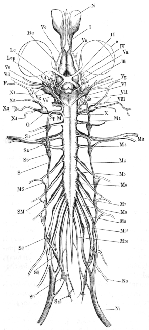
2. Internal structure. As in other vertebrates, the spinal cord consists of white and grey matter, the latter being surrounded by the former; the relative amount of the one to the other varies in different parts of the cord.
A transverse section of the cord presents the same general characteristics as a similar section from a bird or mammal. The grey matter lies in the middle, surrounding the central canal, and is prolonged into each half of the section by ventral and dorsal horns or cornua. The grey matter is surrounded by the white, but is not so sharply marked off from this in amphibia as in birds and mammals.
A section through the anterior enlargement is almost quadrangular in outline, and somewhat broader below than above; the ventral longitudinal fissure is well marked and deep, while the dorsal is indistinct. The dorsal horns are narrow and short, the ventral longer and much broader. The space between the dorsal horns is narrow and deep, that between the ventral shallow; the outer boundary of the grey matter is almost straight, and the central canal is placed above the centre of the section.
In the constricted portion of the cord (pars media) the relative proportion of the white matter is increased at the expense of the grey; the horns are less distinct, the dorsal being directed outwards and the ventral somewhat flattened. The central canal is in the centre of the section.
The posterior enlargement shows the ventral horns projecting outwards, very much enlarged, and approaching the periphery; the space between them is semilunar. The dorsal horns are well developed, especially at their upper parts; the space between them is narrow and deep. The outer margin of the grey matter is again nearly a straight line. The central canal is below the centre of the section.
The arrangement of the parts in the conus medullaris approaches that in the pars media: the horns are much diminished in size, and posteriorly they entirely disappear, the ventral horns persisting longer than the dorsal. The central canal approaches the lower surface.
No cornua can be recognised in the filum terminale behind the origin of the last pair of spinal nerves. The grey matter has here a circular outline, with the exception of a very slight indentation below; the white matter is almost absent: the central canal is on the lower border of the section in the anterior part, while posteriorly it occupies almost the entire space below the pia mater.
The Central Canal lies in the median line, and always presents a distinct lumen; in the two enlargements this lumen forms a vertical ellipse51, while in the pars media it is circular. The canal is lined by a single layer of ciliated columnar epithelium, the cells of which are usually conical, with their bases directed towards the lumen; but other forms may be noticed. These cells average about 0·040 mm. in length and about 0·002 mm. in width at their broadest part. The peripheral extremities of the cells have processes, which, in the case of the cells above and below the cord, may often be traced as far as the pia mater; the processes of the lateral cells are less distinct and seem to be shorter. Anteriorly the central canal opens into the floor of the fourth ventricle. The termination of the central canal, posteriorly, has been described by Masius and Vanlair. According to these observers, the canal towards the hinder end of the filum terminale increases very gradually in width, until its epithelial cells are immediately underneath the pia mater, its walls then converge somewhat more sharply to close the cavity. From the point where the canal commences to increase in width, a change takes place in its epithelial wall, which, instead of having a single layer of cells, now possesses two to three layers; towards the posterior end it is again thinned out to a single layer.
The grey matter varies in different parts of the cord and in different parts of the same section. In a section through the anterior enlargement a portion of the grey matter, placed immediately above the central canal, is easily distinguished by its vertically-placed, elliptical outline, and by its transparency: it is known as the substantia reticularis. It consists of a mesh-work of branched cells, through which course distinctly contoured fine commisural fibres which are derived chiefly from the dorsal cornua, the vertical from the epithelial cells of the roof of the central canal. In a series of sections the substantia reticulosa may be seen to originate indistinctly in the most anterior part of the cord; in the anterior enlargement it already possesses the elliptical form mentioned, and in the pars media it commences to send off lateral processes to either side. In the posterior enlargement it reaches its greatest absolute development, as it here surrounds the central canal; while further backwards it encroaches until, together with the remaining grey matter, it constitutes almost the whole of the filum terminale.
Immediately underneath the central canal is a narrow band, which immediately connects the grey matter of the two sides; above it is bounded by the epithelium of the central canal, below by medullated fibres. This septum medium (Reissner) may be traced backwards from the anterior extremity of the cord to the posterior enlargement. It consists chiefly of transverse fibres, together with vertical fibres derived from the epithelial cells on the floor of the canal, and like the substantia reticulosa, is distinguished by its greater transparency from the rest of the grey matter.
The grey matter consists of connective-tissue and nervous elements, which are very intimately united; each of these elements including its special cells and fibres. The nerve-cells vary much in size and appearance. A prominent group of large cells in the ventral cornua, the lateral group (Stieda), is very conspicuous. The individual cells are seldom rounded, but are usually spindle-shaped or angular, and each possesses one to five processes, which may often be traced through considerable distances. These cells have an average length of 0·040 mm., and are about 0·016 mm. broad; they are somewhat larger in the anterior enlargement than in the posterior, and are also more numerous in a section from the anterior enlargement than in a section from the posterior. In the terminal filament they gradually disappear. Some of their processes are continued into the lateral columns (Köppen).
Smaller, spindle-shaped or triangular cells are scattered irregularly throughout the grey matter, without forming distinct groups, though for descriptive purposes those of the centre have been named the central group. In a stained, transverse section of the cord are seen numerous nuclei; these undoubtedly belong both to small nerve-cells and to connective-tissue cells, but except under the most favourable conditions the two are indistinguishable. The larger nerve-cells present the usual characters of nerve-cells: more or less granular contents, nucleus, well-marked nucleolus, more or less marked fibrillation; their processes are usually more homogeneous. The cells are frequently pigmented.
The processes of the cells belonging to the lateral group radiate in all possible directions, but certain well-marked processes directed towards the middle line can always be made out. From the lowest part of this group a few processes may be traced into the ventral roots. The small cells seem to be chiefly arranged vertically, although their processes radiate in all directions (Stieda).
The Fibres of the grey substance are nearly all non-medullated. They may be traced in all directions, but the best-marked groups are either vertical or transverse. The vertical fibres appear to arise from the central grey matter, and to ascend in larger or smaller bundles towards the periphery. The transverse fibres are arranged chiefly in the two commissures: the dorsal commissure (commissura superior) is the smaller; the fibres are parallel, and show no trace of decussation; externally they radiate in various directions to the dorsal horns. The ventral commissure is composed of two layers, the upper grey (commissura inferior grisea), the lower white (commissura inferior alba): both are interrupted by the septum medium. A well-marked decussation of the fibres is seen in the middle line; the exact mode of termination of these fibres has not been made out, but many appear to communicate with the large cells of the ventral horns.
The white matter consists chiefly of longitudinal, medullated fibres, in which various columns may be distinguished. The dorsal columns are separated from the lateral by a process of neuroglia, continued from the general investment of neuroglia lying under the pia mater. The line of separation between the lateral and ventral columns is not well marked; it is about the line which would be formed by prolonging the ventral horns to the surface.
The fibres vary in size, and fibres of all sizes may be found in any particular part of a transverse section; still the fibres of the ventral columns have an average greater diameter than those of the lateral columns, and the fibres of the dorsal columns are finer than those of the lateral. The largest fibres of the ventral column are placed near the ventral fissure and on its lower border; they attain their greatest development in the posterior enlargement; these fibres frequently contain two or three axis-cylinders each. Köppen suggests that they may represent the formatio reticularis of higher animals.
In the lateral columns the larger fibres are placed close to the grey matter, these columns also receive fibres from the cells of the lateral group.
In the dorsal columns the radiating root-fibres never reach the median plane, but leave an area of purely longitudinal fibres on each side of the dorsal fissure; these represent Goll’s columns, and have a club-shaped outline in transverse section.
The white matter is pierced in all directions by fine connective-tissue fibres and bundles of fibres which radiate from the grey matter; some branch and join with others to form a network, others pass almost uninterruptedly to the periphery. From the connective-tissue cells of the pia mater, processes pass into the white matter and assist in completing the connective-tissue matrix for the nervous elements.
Dorsal roots of the spinal nerves. Each root consists of a single bundle of nerve fibres, which suddenly bends at the periphery of the cord in order to descend vertically through the white matter towards the dorsal horn; the fibres divide into three sets, one to the dorsal commissure, a second to the upper horn itself, and the third helps to form the dorsal columns.
Ventral roots of the spinal nerves. Each ventral root consists of three or four delicate threads, which may be traced to the ventral cornua, which they reach after a vertical or very slightly oblique course through the white matter. Other fibres arise from the ventral columns, but these oblique fibres never extend to the median plane.
Pigment is found distributed irregularly through a section from any part of the spinal cord; it is increased in amount in those parts in which there is an increased amount of grey matter. The pigmentation is always found more marked in the lower parts of the ventral horns than in other parts.
B. The Brain (Figs. 98, 102, 103, and 105).
1. General description. From behind forwards, the dorsal surface of the brain presents the following parts for examination: the medulla oblongata, the cerebellum, the optic lobes, the thalamencephalon, the cerebral hemispheres, and the olfactory lobes.
The medulla oblongata is a direct continuation of the spinal cord; it is wider anteriorly than posteriorly, and is separated in front from the optic lobes by a vertical plate of nervous matter, the cerebellum. The optic lobes are two symmetrical ovoid bodies touching each other in the median plane, and together forming the widest part of the brain. In front of the optic lobes is the thalamencephalon, with a thick vascular membrane, the choroid plexus, lying on its upper surface, and connected with the pineal gland; the thalamencephalon extends forwards between the posterior ends of the cerebral hemispheres. The cerebral hemispheres are two symmetrical ovoid bodies, narrow in front, wider and slightly diverging behind: each hemisphere is prolonged forwards to form an olfactory lobe.
The ventral surface has in front the olfactory lobes, then the cerebral hemispheres, behind these the lamina terminalis, the tuber cinereum, the optic chiasma, the pituitary body, the crura cerebri, and lastly the medulla oblongata, in the order here given.
The various cranial nerves (Figs. 102 and 103) may be seen to arise as follows:—The olfactory nerve (I, I′, L.ol′) arises directly from the anterior end and outer side of the corresponding olfactory lobe, and from the cerebral hemisphere. The optic nerve (To and II) arises, as the optic tract, from the side of the brain below the optic lobe, whence it passes to the chiasma on the under surface of the brain. The oculo-motor (III) takes its origin from the ventral surface close to the median line and between the crura cerebri. The pathetic or trochlear nerve (IV) is attached to the dorsal surface between the optic lobes and the cerebellum. The trigeminal nerve (V) arises from the side and anterior part of the medulla oblongata. The abducens (VI) arises behind the pituitary body close to the median line from the ventral surface of the medulla halfway between the sulcus and the origin of the vagus. The facial and auditory (VII and VIII) nerves arise from the medulla oblongata behind the trigeminal nerve, the facial being in front of the auditory nerve. The glossopharyngeal (IX) nerve arises, in common with the pneumogastric nerve (X), behind the auditory nerve.
2. The several parts of the brain.
a. The medulla oblongata (Figs. 98, 102, and 103 Mo) α. External form. The medulla is limited behind by the origin of the first pair of spinal nerves, at which point a very faint constriction is sometimes found: it extends forwards as far as the cerebellum. It gradually widens as it passes forwards until just before it reaches its anterior limit, where it presents a shallow but sharp constriction. The dorsal surface is characterised by the presence of a deep, triangular fossa, the fourth ventricle (Fig. 98 S.r), (ventriculus quartus, Stieda; sinus rhomboideus s. sinus triangularis, Reissner; fossa rhomboidalis); the sides of the triangle are, however, not quite straight, but are slightly bent outwards just before they converge towards the posteriorly-directed apex; the base of the triangle is formed by the cerebellum. By careful examination, the ventricle is seen to be continued for a short distance under the cerebellum, where it opens into the Sylvian aqueduct. In the floor of the fourth ventricle is a well-marked median longitudinal fissure (sulcus centralis), (Fig. 98 S). Into the posterior part of the ventricle opens the central canal of the spinal cord. As the fourth ventricle is formed by the white matter passing to either side, and the simultaneous flattening of the grey matter, the floor of the fourth ventricle is composed of grey matter.
Fig. 98.
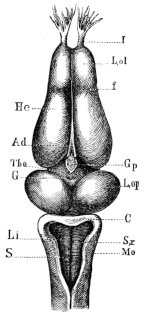
Dorsal view of brain of Rana esculenta.
Ad Choroid plexus.
C Cerebellum.
f Groove between cerebral hemispheres and olfactory lobes.
G Opening in the roof of the third ventricle.
Gp Pineal body.
Hc Cerebral hemispheres.
I Olfactory nerve.
Li Wall of fourth ventricle.
L.ol Olfactory lobe.
L.op Optic lobe.
Mo Medulla oblongata.
S Longitudinal fissure of the fourth ventricle.
S.r Fourth ventricle.
Tho Thalamencephalon.
The fourth ventricle is closed in by a highly vascular membrane, the choroid plexus of the fourth ventricle (plexus choroideus ventriculi quarti, Reissner; velum medullare posterius). The blood-vessels of the plexus will be described together with the other vessels of the brain (p. 162). They are supported by a connective-tissue matrix, and the whole covered with flattened epithelium, which in the fourth ventricle is ciliated and often pigmented.
The ventral surface of the medulla oblongata (Fig. 102 Mo) has a median ventral longitudinal fissure, a direct continuation of that of the cord; in the anterior part of the medulla oblongata there is also to either side of this a lateral fissure, continued on to the crura cerebri; these fissures correspond to the positions of the two rami posteriores of the internal carotid arteries; they are always well seen in microscopical sections. The medulla oblongata is so intimately connected with the pars commissuralis (pp. 149, 150) that the minute anatomy of the two is best described at the same time.
β. Internal structure. Examined by means of serial sections, the medulla oblongata is seen to have, in comparison with the cord, an increased amount of grey matter; this is especially the case in its anterior part. The floor and inner parts of the walls of the ventricle are formed of grey matter, in which the largest-sized cells have disappeared, to be replaced by medium-sized cells. Traced from behind, the ventral horns of the cord are seen to increase in size and to be more widely separated until they form two isolated masses, while the dorsal horns gradually diminish; at the same time they are forced outwards and upwards, until they lie under the floor of the ventricle, and so extend to the pars peduncularis.
Grey matter. The substantia reticularis is not present, but the septum medium extends forwards as far as the pars peduncularis. The central canal extends upwards at the expense of the tissue above it, and is here pear-shaped; at the same time the dorsal longitudinal fissure deepens until the two meet in the fourth ventricle; beyond this point one cannot speak of dorsal and ventral horns.
Small nerve cells are irregularly distributed throughout the whole of the grey matter and cannot be grouped; the larger cells, on the other hand, are arranged in distinct groups which have special relations with the nerves arising from the part. Occasionally these groups are not so isolated as usual; in this case processes of one group can be traced into another group (Reissner). Of these groups the chief are:
Fig. 99.
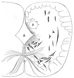
Transverse section through hinder end of Medulla oblongata (magnified 30–80)52, from Stieda.
b Inferior commissure.
f Dorsal horns.
g Ventral horns.
h Fourth ventricle.
i Nucleus centralis.
k Isolated mass of grey matter in which longitudinal fibres of the pneumogastric nerve course.
1. The nucleus centralis (upper inner group, Reissner), nucleus medullae oblongatae, Stieda (Fig. 99 i), is a group of cells found towards the hinder end of the medulla oblongata, on either side of and below the central canal; the group can be traced under the floor of the fourth ventricle to about its middle. The cells are rounded or spindle-shaped, the processes directed upwards, downwards, or outwards; their average size is 0·040–0·048 mm. long by 0·020 mm. broad.
2. The auditory nucleus (nucleus acusticus, Reissner, Stieda), (Fig. 100 n) is a large group of cells found in the wall of the fourth ventricle opposite the point of origin of the auditory nerve. The cells are rounded, pear-shaped, or of spindle form, and interspersed between the nerve fibres; these cells have an average length of 0·040 mm., and are about half as broad. The fibres of the auditory nerve radiate from their superficial origin in all directions through the grey matter towards these cells, and evidently communicate with them (Fig. 101 p). One small group (Fig. 101 r) passes to a lower level than the rest, and is regarded by Stieda as the true auditory centre. Köppen considers that the auditory nerve has a threefold origin: (1) from small cells on the median surface of the auditory area; (2) from the large cells between the above; (3) from a group of free nuclei on the dorsal surface of the auditory area.
Fig. 100.
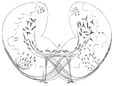
Transverse section through the Medulla oblongata at the point of origin of the abducens nerve, from Stieda. (Magnified 30 x 80.)
h Fourth ventricle.
m Abducens nerve.
n Auditory nucleus.
o Abducens nucleus.
3. The trigeminal nucleus (nucleus trigeminus), (Fig. 101 q) lies in part beneath the auditory nucleus but extends further forwards. It forms a rounded group of cells placed under the outer angle of the grey matter. The cells are somewhat crowded together, and are chiefly of an elongated spindle-form, with their processes directed obliquely downwards and outwards. The fibres of the trigeminal nerve separate into two groups; the upper group is best traced in a horizontal section, the fibres curving round to join the longitudinal fibres continued from the dorsal columns of the cord. The fibres of the lower, smaller group pass transversely inwards to the trigeminal nucleus. According to Reissner the latter fibres are motor, the former sensory. Probably other nerves are connected with the hinder part of this group.
4. The abducens nucleus (Fig. 100 o). From its superficial origin, the fibres of the abducens nerve may be traced vertically upwards to a small, rounded, grey mass; at this point the mass is somewhat isolated, but further forwards it may be traced as belonging to the central grey matter; it contains small spindle cells.
Fig. 101.
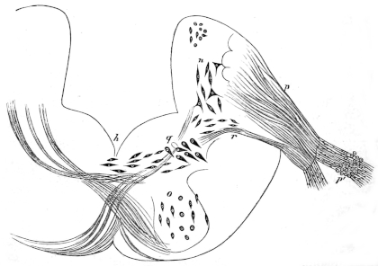
Transverse section of the Medulla oblongata, at the point of origin of the auditory nerve, from Stieda. (Magnified 30 x 80.)
h Fourth ventricle.
n Auditory nucleus.
o Abducens nucleus.
p Auditory nerve.
p′ Ganglion of auditory nerve.
q Hinder portion of trigeminal nerve.
r Bundle of fibres arising from trigeminal nucleus and joining the auditory nerve.
5. The pneumogastric nucleus. The pneumogastric, with its numerous irregular roots, arises from the side of the medulla oblongata. The hindermost fibres can be traced as a small bundle, passing almost transversely through the white matter to the outer margin of the grey matter. The larger portion of the fibres is placed in front of these; part of this seems to be directly continuous with the longitudinal fibres of the white matter; a second part, however, can be traced from the surface transversely through the white matter to the grey matter. These latter fibres, together with those of the group first described, do not arise from the grey matter in this part of the medulla oblongata, but curve round and run backwards longitudinally through the grey matter, thus forming a rounded bundle of fibres (Fig. 99 k). Between these fibres are interspersed small nerve-cells and nuclei which disappear as the fibres approach the white matter. The vagus undoubtedly receives fibres from the grey matter throughout a long course, and again receives a large bundle just before leaving the grey matter. The more exact origin of the various fibres has not been traced.
6. The nucleus magnus (Reissner and Stieda) is a very peculiar group of cells placed on either side, in the most anterior portion of the pars commissuralis, that is, immediately underneath the valvula cerebelli. The large cells are arranged in a transverse section in a single row so as to enclose a pear-shaped space on either side, which has its long axis directed from above, downwards and outwards, the narrower end being above. In longitudinal section the line of cells is seen to be open in front. The space enclosed by these cells is occupied by a granular ground-substance which contains only few nuclei. Bellonci is of opinion that these nuclei represent the corpora quadrigemina posteriora of higher animals.
White matter. In the hinder part of the medulla oblongata the arrangement of the white matter resembles that of the white matter of the spinal cord; further forwards the white matter of the dorsal surface commences to pass to either side, and ultimately it forms the outer part of the walls of the fourth ventricle. The fibres of the white matter of the ventral surface are unchanged in direction as they proceed forwards. The fibres of the anterior part of the medulla are thinner than those of the posterior portion (Stieda), according to Reissner they gradually thin as they pass forwards. The fibres are nearly all longitudinal, such transverse and oblique fibres as are present being chiefly in connection with the various nerve-roots and the commissures.
The commissura superior is naturally lost in consequence of the opening of the central canal into the fourth ventricle; the commissura inferior is increased in the anterior half and decreased in the posterior half of the medulla oblongata; in the latter the fibres become more and more oblique, and decussate very freely; ultimately they seem to be either continued as longitudinal fibres or to join the ganglia.
Near the pars commissuralis is a transverse arched band of fibres, passing from the under surface of one half of the cord over the ventral longitudinal fissure through the septum medium to the under surface of the opposite half; part of the fibres are continued upwards along the periphery to the cerebellum, part to the nucleus magnus. Vertical, straight, or slightly arched fibres are found in the walls of the fourth ventricle.
A section from the medulla oblongata has a larger amount of pigment than a section from the spinal cord, and the anterior portion of the medulla oblongata contains more than the posterior portion. The pigment is chiefly found in a curved line, placed in the lower and outer parts of the grey matter; the amount present varies in different specimens.
b. The Cerebellum and Valvula cerebelli.
α. External form. The cerebellum is a thin, semilunar plate, which projects between the optic lobes and the fourth ventricle, its base covering the most anterior part of the ventricle; the posterior surface possesses a very faint median fissure (Reissner). The valvula cerebelli (Velum medullare anterius, Reissner) is the thin lamella which connects the anterior surface of the cerebellum with the optic lobes.
β. Minute structure. By means of longitudinal, vertical sections, the posterior surface of the cerebellum is seen to be covered with epithelium; in the lower part of the surface this is columnar or conical, above it is flattened: immediately beneath, that is in front of this is a finely granular layer, with very closely packed and granular nuclei. In front of these is a stratum of nerve-fibres forming the second layer of the cerebellum.
Fig. 102.
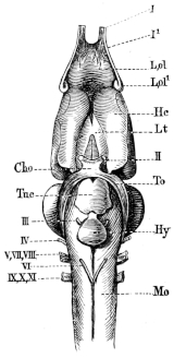
Ventral view of brain of Rana esculenta.
Cho Optic chiasma.
Hc Cerebral hemispheres.
Hy Pituitary body.
L.ol Olfactory lobe.
L.ol1 Origin of olfactory nerve from the cerebral hemisphere.
Lt Lamina terminalis.
Mo Medulla oblongata.
To Optic tract.
Tu.c Tuber cinereum.
I 1st root of the olfactory nerve.

I1 2nd
II Optic nerve.
III Oculo-motor nerve.
IV Trochlear nerve.
VI Abducens nerve.
V, VII, VIII Trigeminal, facial, and auditory nerves.
IX, X, XI Glossopharyngeal, pneumogastric, and accessory nerves.
Still more anteriorly is the third layer of the cerebellum, an irregular double layer of large cells (Purkinje’s cells, Denissenko); the cells have an average length and breadth of 0.040 mm. and 0.015 mm respectively; they are pear-shaped or of spindle-form, and possess usually two well-marked processes, one passing into the layer behind, the other forwards into the anterior layer to be immediately described, while other less distinct processes radiate irregularly in all directions. The fourth and most anterior layer of the cerebellum is a thick stratum of nerve fibres with numerous nuclei (0.006 to 0.008 mm. diameter). The fibres are for the most part arranged transversely, but some course in various directions. These fibres underlie the flattened epithelium which covers the anterior surface of the cerebellum.
The fibres of the second layer course, for the most part, in an almost vertical plane; they connect the cerebellum with the optic lobes (processus cerebelli ad corpora bigemina) and with other parts of the brain.
The fibres of the fourth layer receive numerous long processes from the large cells of Purkinje; they form a large commissural system, which can be followed ventrally on each side into the pars commissuralis. A part of the fibres ends here in the grey matter, a second portion enters the auditory area and forms a descending auditory root, a third part joins the lateral columns (in the medulla oblongata), and more anteriorly some join the ventral columns. The descending fibres from the cerebellum, together with the fibrae arcuatae found in the ventral columns, indicate the presence of a pons Varolii. The fibres of this ventral commissure decussate only on its dorsal surface (Köppen).
The Valvula cerebelli contains a few medullated fibres and the roots of the trochlear nerves; these pass from the medulla oblongata into the valvula cerebelli, cross in the median line, and then proceed forwards as the trochlear nerves.
The pigment in the pars commissuralis is arranged in a curved line similar to that found in the medulla oblongata, but the line is shortened at either extremity, and consequently does not extend into the cerebellum.
c. The optic lobes and Crura cerebri. (Corpora geminata and Pars peduncularis, Reissner; Lobus opticus, Stieda; Vierhügel, Tiedemann; Vierhügel (Zweihügel) and Pedunculi cerebri, Schiess; Mesencephalon, Huxley.)
α. External form (Figs. 98, 102, 103 L.op). The optic lobes are two prominent ovoid bodies placed immediately in front of the cerebellum, and connected with it by the valvula cerebelli; posteriorly they touch each other in the median plane, while anteriorly they diverge and thus constitute the widest part of the brain; in the angle thus formed is the thalamencephalon. The optic lobes are always more darkly pigmented than any other part of the central nervous system.
Fig. 103.
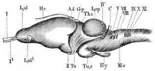
Lateral view of brain of Rana esculenta.
Ad Choroid plexus.
C Cerebellum.
Gp Pineal body.
Hc Cerebral hemisphere.
Hy Pituitary body.
L.ol Olfactory lobe.
L.ol1 Disc at origin of second root.
L.op Optic lobe.
Mo Medulla oblongata.
Tho Thalamencephalon.
To Optic tract.
Tu.c Tuber cinereum.
I 1st root of the olfactory nerve.
I1 2nd
II Optic nerve.
IV Trochlear nerve.
V Trigeminal nerve.
VII Facial nerve.
VIII Auditory nerve.
IX, X, XI Glossopharyngeal, pneumogastric, and accessory nerves.
The Crura cerebri are two columns of white matter, placed beneath the optic lobes, and partly hidden by the pituitary gland. At their junction with the medulla oblongata, or rather with the pars commissuralis, is a very slight transverse fissure; at the same point the ventral longitudinal fissure is interrupted by an extremely small grey tubercle (Stieda).
β. Internal structure. From the anterior extremity of the fourth ventricle a canal, the Sylvian aqueduct (Aqueductus Sylvii, iter a tertio ad quartum ventriculum), may be traced forwards under the cerebellum, in the median line of this section of the brain. At about opposite the middle of the length of the optic lobes the canal is dilated and communicates with the cavities or ventricles (Ventriculi lobi optici, Stieda) enclosed by these; a general cavity is formed, which in transverse section has something of the form of the letter T. The roof of the cavity is thinner than the floor; this is especially the case in the median plane opposite the superior longitudinal fissure between the optic lobes; the floor is thinned in the middle line by the descending portion of the cavity. The cavity of each optic lobe extends both forwards and backwards beyond its point of communication with the dilated Sylvian aqueduct, hence in a transverse section taken in front of this point (Fig. 104 h′) the cavity of either side appears to be isolated; in a horizontal and longitudinal section (Fig. 105 Aq) the general arrangement of the parts may be well seen.
Fig. 104.
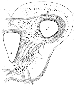
Transverse section through the anterior portion of the optic lobes opposite the origin of the motor-oculi nerve; from Stieda (magnified 30–80).
h Lower portion of cavity.
h′ Lateral portion of cavity.
u Ganglia of oculi-motor nerve.
v Oculo-motor nerve.
w Large cells of the optic lobe.
x Roof of optic lobe.
y Posterior commissure.
z Anterior diverticulum of the cavity.
The grey matter is chiefly arranged in a layer so as to surround the cavity (Fig. 104), this layer being deeper on either side of the descending portion of the cavity than elsewhere. For the rest the grey matter is much interspersed among the white matter, except at the circumference of the section, which is entirely formed of white matter. It contains a large number of small cells, of which the nuclei are alone visible; in the parts mentioned where the layer is most marked these cells are arranged in oblique rows, between which pass fine bundles of medullated fibres (Fig. 104, between u and h′). A group of large cells (Fig. 104 u) is found on either side of the middle line and under the floor of the cavity; the cells are about 0.032 mm. long, and 0.016 mm. broad; the oculo-motor nerve may be traced to this group, which is the oculo-motor nucleus. A small commissure of decussating fibres connects the nuclei of opposite sides (Köppen).
The substance of the roof of the optic lobes (Fig. 104 x) is arranged in very distinct layers: above is a layer free from cells; the fibres of this layer are extremely fine and delicate, and have not been accurately traced; under this is a nuclear layer; a second layer of fine fibres follows, which is succeeded by a second layer of nuclei in a granular matrix, and lastly epithelium (Stieda). (Reissner describes three nuclear layers in Bufo variabilis, and this is also the case in R. temporaria, G. H.; according to Köppen, the number is variable.) In the anterior portion of the roof a distinct bundle of fibres may be made out; externally they bend downwards, and can be traced as far as the crura cerebri.
Fibres corresponding with the commissural and arched fibres of the medulla oblongata are continued into the hinder portion of the crura cerebri, the change from pars commissuralis to crura cerebri being very gradual. The longitudinal white fibres are much increased in number in the crura cerebri, and a portion of them can be traced to the nucleus magnus.
A peculiar irregular group of large cells (Fig. 104 w) is found where the roof meets the crura cerebri; these vary in diameter from 0.024 to O.040 mm., and their processes are very indistinct.
The fibres of the optic tracts arise, according to Köppen, from two different origins: the one lies on the hindermost part of the optic lobe; from this point the fibres curve downwards and forwards to form longitudinal fibres; this root Köppen names the ventral ascending root, it can be traced through the entire length of the organ. The second root arises in the tectum opticum near the longitudinal fissure; it is smaller than the foregoing, and has been named the dorsal ascending optic root. The fibres of these two roots unite anteriorly near the posterior commissure, at which point they receive additional fibres (Köppen). Bellonci traces a large proportion of the fibres of the optic tract to the nucleus magnus, which pair of nuclei, as already stated, he regards as the posterior pair of corpora quadrigemina of higher animals.
The pars peduncularis is the continuation of the pars commissuralis underneath the optic lobes; a gelatinous mass lying in the median plane and containing numerous isolated nuclei (Ganglion interpedunculare) divides it into two lateral halves. The longitudinal fibres are ungrouped posteriorly, but arranged in rounded strands in the middle, especially dorsally; anteriorly the grouped arrangement is lost and the number of fibres diminished.
The pigment of this region has, in a transverse section, an outline which has something the form of a lyre; commencing on either side of the median line, and underneath the deepest portion of the cavity, the pigment line passes, first, directly outwards; then suddenly turns upwards and slightly outwards parallel with the wall of the deeper part of the cavity; it then curves outwards to pass below the cavity of the optic lobe, where it divides, one portion passing outwards, the other between the Sylvian aqueduct and the ventricle of the optic lobe.
d. The Thalamencephalon (Huxley), (Lobus ventriculi tertii, Stieda; Thalami optici, Reissner; Thalamus opticus s. Lobus ventriculi tertii, Stannius; Ganglien der Haemisphaeren, Carus).
α. External form. From above (Fig. 98 Tho) the thalamencephalon is seen as a lozenge-shaped mass lying in front of the optic lobes, and behind and between the diverging posterior ends of the cerebral hemispheres; it is covered by a thick vascular membrane, the choroid plexus, through which passes the pedicle of the pineal body (Glandula pinealis). On removing the choroid plexus a small aperture is seen in the roof of the thalamencephalon, connecting the hollow pedicle of the pineal gland with the third ventricle. The ventricle appears as a narrow slit in the median line, its walls being formed by the optic thalami. By pressing aside the cerebral hemispheres the posterior commissure (Commissura posterior) may be seen lying quite in front and deep in the cleft of the ventricle. Immediately behind the pedicle of the pineal body is a slight but well-marked depression (Fig. 98 G), the origin of which has not been investigated (Wiedersheim).
The choroid plexus is continued forwards between the cerebral hemispheres (Figs. 98, 103 Ad) for some distance, and terminates in a fine thread of connective-tissue.
The under surface of the thalamencephalon (Fig. 102) is divided into two parts by the optic chiasma (Cho): the anterior portion (Lt) is the lamina terminalis (Substantia cinerea anterior); the posterior (Tu.c) the tuber cinereum. The lamina terminalis is bounded on either side by the cerebral hemispheres. The tuber cinereum (Figs. 102, 103 Tu.c) is a small median swelling immediately behind the optic chiasma, and caused by the depression of the floor of the third ventricle to form the infundibulum (Diverticulum infundibuli, Reissner).
Fig. 105.
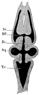
Horizontal section through the brain to show the ventricles.
Aq Ventricles of optic lobes and the aqueduct of Sylvius.
Dv Third ventricle.
MF Foramen of Monroe.
Sv Lateral ventricle.
Vv Fourth ventricle.
The pituitary body (Hypophysis cerebri) is a flattened sac, placed behind the tuber cinereum and continuous with it by means of the infundibulum.
β. Internal structure. The aqueduct of Sylvius, after communicating with the ventricles of the optic lobes, again contracts (Fig. 105), but still remains somewhat larger than before. In the thalamencephalon the Sylvian aqueduct opens into the third ventricle, which gradually assumes the form of a vertical slit with the walls bulging slightly outwards in their upper parts. The thin roof of this ventricle, where complete, contains a band of transverse fibres. The floor is depressed both before and behind the part immediately above the optic chiasma, the posterior depression lying above the tuber cinereum, which here descends towards the infundibulum: a transverse section through this portion of the third ventricle has the form of a square standing on one angle, the superior angle being produced into the vertical slit of the general cavity. The anterior depression is formed by the general cavity being prolonged downwards and forwards to the lamina terminalis in the form of a narrow and shallow slit.
The white and grey matter of the thalamencephalon are only so far distinct in that the portion immediately surrounding the cavity is darker than the rest of the section. In the immediate neighbourhood of the cavity are many small cells and nuclei, which become scarcer further from the ventricle; they are arranged in rows, separated by a fibrillated matrix. On either side is a distinct bundle of longitudinal fibres, the ‘round bundle’ of Köppen, which come from the optic lobes but appear to arise from either the pars commissuralis or the medulla oblongata, and to receive additional fibres from the optic thalami; they pass forwards to the base of the cerebral hemispheres (Stieda). A second set of longitudinal fibres arises in the substance of the tuber cinereum and passes forwards to the hemispheres (strand of the Tuber-cinereum and Thalamus, Köppen); this band, together with the ‘round bundle,’ form a crus cerebri (Köppen).
The commissural fibres of the thalamencephalon are: (1) a commissura transversa Halleri in the posterior portion of the organ; (2) an optic commissure, consisting of fibres arising from the thalamencephalon (thalamencephalic root) and crossing the median line to join the optic tract of the opposite side; (3) a probable commissure between the optic nerves just in front of the chiasma opticorum; the existence of the latter is not yet proved beyond doubt; (4) the large commissure of transverse fibres found in the roof of the third ventricle; whether the fibres decussate or not is uncertain (Köppen).
The fibres of the large commissure of the roof are, in part, continued into the strand of the Tuber-cinereum above mentioned, and thus conducted to the posterior parts of the hemispheres (Köppen). A group of fibres (Meynert’s band, Köppen) is found in each lateral wall of the third ventricle; they pass from the region of the nucleus parvus downwards in a curved course almost parallel with the external border of the thalamencephalon.
A distinct group of cells may be noted in this section of the brain, an arciform or circular group (Nucleus parvus, Reissner; ganglion of the habenula, Köppen) of large spindle cells (average diameter 0.016 mm.), placed under the upper border close to the third ventricle; the group extends alongside the whole length of the ventricle. A second group lying in the middle and posterior parts beneath the ventricle, Köppen names the ‘ventral nucleus’ of the thalamencephalon.
The pigment in the posterior part of the thalamencephalon is arranged in a manner similar to that in the optic lobes and crura cerebri; anteriorly where the third ventricle is prolonged forwards and downwards the arrangement is different; the pigment lies in a curved line above the process of the ventricle, with its concavity directed downwards, each end bifurcating, in order that one branch may pass upwards, the other downwards.
The pineal body53 is a small vesicle placed underneath the skin above the fronto-parietal bones; in the embryo it is connected with the third ventricle by means of the pedicle54 already mentioned; the skin covering the body is always paler than the surrounding skin, and the usual cutaneous glands are absent in this part; the paler spot on the head may always be found, but is more distinct in Rana temporaria than in Rana esculenta. The structure on the roof of the third ventricle, which is usually known as the pineal body, is nothing more than a thickened portion of the choroid plexus, and consists of a group of convoluted vessels surrounded by pia mater, which is described by Wyman as being covered with ciliated epithelium (R. pipiens). The true pineal body is a small body with an outer connective-tissue capsule, derived from the pia mater; this encloses an irregular mass of epithelial cells; according to de Graaf a twig of the ramus supramaxillaris reaches it subcutaneously, and a blood-vessel accompanies the pedicle through the foramen parietale. According to Darkschewitsch, the pedicle contains medullated nerve-fibres derived directly from the brain.
The pituitary body (Figs. 102, 103 Hy) when examined with a lens is seen to consist of two portions: an anterior, superior, and smaller white portion, and a larger, inferior, posterior, and reddish portion. The anterior portion has the form of a very small, flat disk, and is enclosed in a connective-tissue capsule which sends in larger and smaller processes. In either transverse or longitudinal section it is seen to be formed of two horizontal layers separated by a line of blood-vessels and connective-tissue. The upper layer consists of a granular and reticular matrix, containing many nuclei (averaging 0.006 to 0.010 mm. diameter), and divided into irregular rounded or polyhedral spaces by bands of tissue derived from the capsule. This layer is more vascular than the lower. The lower layer consists of a mass of clear, nucleated rounded or polyhedral cells (0.016 to 0.024 mm. in diameter; nuclei from 0.008 to O.012 mm. in diameter, Reissner), pierced by very fine connective-tissue septa derived from the capsule. The septa are, for the most part, vertical and longitudinal (Reissner), the blood-vessels are very few.
Fig. 106.
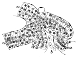
Section through the lower division of the pituitary body (magnified 360 times); from Stieda.
a Tubes lined with epithelium.
b Blood-vessels.
The posterior larger portion of the pituitary body (Fig. 106) is also compressed from above downwards, and in transverse section as an oval outline. It possesses an external thin connective-tissue capsule, which sends in fine processes to support a mass of convoluted tubes, between which course a few blood-vessels; these tubes possess an outer nucleated basement-membrane, and are lined with a single layer of more or less cylindrical epithelium, which entirely fills the tube; hence the tubes possess no lumen. The tubes are from 0.04 to 0.08 mm. in diameter; the cells are clear or granular, and possess distinct, rounded nuclei.
e. The Cerebral Hemispheres and Olfactory Lobes. The cerebral hemispheres (Lobi hemisphaerici, Stieda; Lobi cerebrales, Reissner; Centralmasse des Geruchssinns, Carus; Hemisphaeren des grossen Hirns, Tiedemann; Grosse Hemisphaeren, Schiess; Prosencephalon, Huxley). The olfactory lobes (Tubercula olfactoria, Stieda; Lobi olfactorii, Reissner; Riechkolben, Schiess; Rhinencephalon, Huxley).
α. External form (Figs. 98, 102, 103 Hc and L.ol). The two cerebral hemispheres form together the largest section of the brain; from above they are seen to be separated by a dorsal longitudinal fissure, which is here well marked: each hemisphere is an ovoid body with the smaller end directed forwards and continuous with the corresponding olfactory lobe; the posterior end forms one half of the anterior boundary of the thalamencephalon. The olfactory lobes are two elongated, rounded bodies directly continuous with the corresponding cerebral hemispheres, and likewise partially separated in the median line by a dorsal longitudinal fissure: at the point of union of the cerebral hemispheres and olfactory lobes is a faintly marked transverse depression.
On the ventral surface the parts are again marked off from one another by a corresponding ventral longitudinal fissure and a transverse groove; the two longitudinal fissures being continuous, anteriorly, between the olfactory lobes. The cerebral hemispheres appear to be more widely separated behind (Fig. 102) than is the case on the dorsal surface, and in the space so formed is the lamina terminalis (Lt). Seen from the side, the slight depression of the upper surface, between the cerebral hemispheres and the olfactory lobes, is seen to be continued downwards and slightly backwards to join the corresponding groove on the inferior surface.
The longitudinal fissures are shallow and do not meet, except at one point, at about the middle of the cerebral hemispheres (Fig. 105). The olfactory bulbs arise superficially (Figs. 102, 103 I′ and L.ol) from the whole length of the olfactory lobe, between the anterior extremity (I′) and the posterior (L.ol), where they are also attached to the cerebral hemispheres.
β. Internal structure. The cerebral hemispheres and olfactory lobes are hollow, the common cavity of each side being known as the lateral ventricle (Ventriculus lateralis); these ventricles communicate with each other, and with the third ventricle (Fig. 105). The narrow aperture by which the lateral ventricles communicate is known as the Foramen of Monro (MF); it communicates with the third ventricle posteriorly, and with the space between the cerebral hemispheres anteriorly, and thus forms a common cavity (Ventriculus communis loborum hemisphaericorum, Stieda).
Fig. 107.
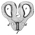
Transverse section through the hinder portion of the cerebral hemispheres; from Stieda.
c Lateral ventricle.
c′ Common ventricle of Stieda.
d Longitudinal fibres.
f Anterior prolongation of the third ventricle.
Fig. 108.
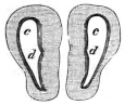
Transverse section near the middle of the cerebral hemispheres.
c Lateral ventricle.
d Groove on the inner wall.
In general terms, each ventricle may be said to be a semilunar cavity, prolonged backwards and forwards (Fig. 107 c); the outer wall is always more or less concave, while the inner varies according to the part of the hemisphere examined. At the anterior and posterior extremities the inner wall is convex and bulges into the cavity (Fig. 107); in the middle portion of the cavity the inner wall presents a longitudinal groove (Ventriculi lateralis cornu internum, Reissner), (Fig. 108 d), and consequently the inner wall has here two rounded masses, an upper and a lower (Corpus striatum, Wiedersheim), projecting into it. By tracing them backwards and forwards, the lower swelling is seen to increase at the expense of the upper, while at the same time the lateral grooves disappear; the outline of the cavity shown in Fig. 107 is then obtained. The roof of the ventricle is arched and broader than the floor, which, in the middle part, exists only as a vertical slit (Fig. 108), (Ventriculi lateralis cornu inferius, Reissner): towards the anterior and posterior extremities it widens and becomes shallower (Fig. 107).
The cerebral hemispheres and olfactory lobes are composed of a fine granular matrix, enclosing spindle-shaped, rounded, or pear-shaped nerve cells and nuclei, and containing very fine fibres. The cells (Fig. 109 b) are more numerous towards the ventricle, and somewhat sparse towards the superficial surface. The cells are of two chief sizes, the smaller and more numerous average 0.004 mm. to 0.008 mm. in diameter; they are found chiefly in the deeper portions of the section, but also form a very thin irregular layer beneath the pia mater: the larger cells have an average diameter of 0.010 mm. to 0.012 mm., and are placed towards the periphery, especially in the dorsal part of the inner wall.
Fig. 109.
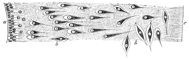
From a transverse section through one of the cerebral hemispheres; from Stieda. (Magnified 360 times.)
a Epithelium of lateral ventricle.
b Nerve cells.
c Connective-tissue processes from the Pia mater.
In this irregular collection of cells the following centres have been described: (1) The nucleus, through which the corpus callosum passes (Köppen); (2) the lower internal or median cell-area (Osborne), situated above the foregoing nucleus in the posterior and middle portions of the hemispheres; (3) the upper internal cell-area (Osborne) is the area of large cells in the dorsal part of the inner wall; (4) the Corpus striatum (Osborne) is a mass of cells between the corpus callosum and the commissura anterior; Köppen doubts the correctness of Osborne’s opinion, and suggests that a group of cells found in the wall of the third ventricle in front of ‘Meynert’s band’ may perhaps be a corpus striatum.
A bundle of longitudinal medullated nerve-fibres, the ‘round bundle,’ is found on either side of the median line (Fig. 107 d), and near the lower border in the posterior portion of this region; these fibres can be traced from the posterior section of the thalamencephalon; they diminish in number as they course through the base of the cerebral hemispheres, and are ultimately lost in the lower anterior part of the outer walls. A second strand of longitudinal fibres is the continuation of that of the Tuber-cinereum above described, which can be followed to the outer wall of the posterior part of the ventricles, and to the anterior commissure.
The commissures are: (1) The corpus callosum, a large bundle of transverse fibres, seen best in a transverse section, at the point of junction between the lamina terminalis and the cerebral hemispheres, forming an arch over the roof of the anterior prolongation of the third ventricle. The fibres course to the inner and anterior parts of the hemispheres, and are situated chiefly behind the foramen Monroi. To this commissure must be added those fibres which unite the two olfactory lobes, and possibly the fibres (Commissura posterior) found in the roof of the third ventricle (Köppen). (2) The Commissura anterior (Stieda), a smaller set, found immediately under the floor of the common ventricle, forming in their course outwards a curve, with the concavity directed downwards. This commissure connects the ‘round bundles’ of opposite sides, and those fibres coursing with the ‘round bundles’ to the olfactory lobes constitute the pars olfactoria of the commissura anterior. To this commissure must also be added some fibres found on the ventral surface of the commissura anterior and connecting the two strands of the Tuber-cinereum; an unusually coarse strand of these fibres can be traced to the inner wall of the ventricle, and is termed the pars olfactoria interna by Osborne.
The general structure of the olfactory lobes resembles that of the hemispheres; the olfactory nerves arise each by two roots, an outer and inner. The outer root arises from the outer wall near the groove between the corresponding hemisphere and olfactory lobe; the inner or anterior root arises from the anterior surface of the olfactory lobe. Both roots have a peculiar method of origin from the extremely fine fibrillar network of the matrix (Nerve-fibre-conglomerate, Köppen), in which are rounded dark bodies known as ‘glomeruli;’ in the ‘glomeruli’ dark points and nuclei are seen, between larger and smaller bands of nerve-fibres. Köppen holds that all the sensory nerves of the brain arise in a similar manner.
A decussation takes place between the two inner roots of the olfactory nerves; possibly the external roots are connected by means of the commissura anterior.
Very little pigment exists in the cerebral hemispheres or olfactory lobes, the greater portion is found in the upper part of the inner walls of the cerebral hemispheres.
The epithelium of the ventricles of the brain, like that of the central canal of the spinal cord, consists of conical cells with their bases directed towards the cavity, and their apices directed peripherally and prolonged into distinct processes (Figs. 104, 109 a). In such situations, as the choroid plexuses, where nervous tissue is absent and the cavity is completed by pia mater alone, the epithelial cells are flattened. Everywhere else it is ciliated55 and possesses distinct round nuclei which are as broad as the cells themselves. The epithelium is somewhat irregularly and sparsely pigmented; the ventral parts of the central canal of the spinal cord, of the fourth ventricle, and of the Sylvian aqueduct are always more pigmented than the dorsal parts.
PLAN OF THE ARRANGEMENT OF THE NERVE-FIBRES OF THE CENTRAL NERVOUS SYSTEM, ACCORDING TO KÖPPEN.
Nerve fibre strands in the white matter. Nerve-fibre strands in the grey matter. Commissures. Nerve-fibres which connect the strands of white fibres.
The spinal cord. 1. The ventral columns, including the strand of coarse fibres. 1. Longitudinal fibres in the dorsal portion of the grey matter. 1. The ventral commissure.

2. The lateral columns. 2. The dorsal commissure (rudimentary).
3. The dorsal columns, including Goll’s column.
The medulla oblongata. 1. The ventral columns, including the strand of coarse fibres and dorsal longitudinal strand. 1. Longitudinal fibres of the Substantia gelatinosa Rolandi, and which give off ascending vagal and trigeminal roots. 1. The ventral commissure: a. Dorsal portion. b. Ventral portion. 1. The fibrae arcuatae.

2. The lateral columns, which give off an ascending (recurrent) vagus-root. 2. (The dorsal commissure is wanting.)
3. The dorsal columns. a. Ascending trigeminal root. b. Auditory root. c. Descending auditory root.
The cerebellum and the pars commissuralis. 1. The ventral columns, including the dorsal longitudinal strand (the strand of coarse fibres has disappeared). 1. Longitudinal fibres of the grey matter. Small ascending trigeminal root. 1. The ventral commissure. 1. The fibrae arcuatae.
2. The lateral columns (each receives a strand from the cerebellum). 2. The dorsal commissure, cerebellar commissure and trochlear decussation.
3. The dorsal columns. a. Large ascending trigeminal root. b. Descending auditory root.
The optic lobes. 1. The ventral columns, including the dorsal longitudinal strand. (Wanting.) 1. The ventral commissure. 1. The fibrae arcuatae.

2. (The lateral columns cannot now be distinguished from the ventral columns.) 2. The dorsal commissure. a. Commissura posterior. b. Marginal commissure.
3. The dorsal columns: two ascending opticus-roots.
The thalamencephalon and the cerebral hemispheres. 1. The ventral and dorsal columns. a. Fibres from the optic lobes. b. The ‘round bundle.’ c. The ‘Thalamus-Tuber-cinereum strand.’ (Wanting.) 1. The ventral commissure: a. Commissura transversa Halleri. b. Commissura anterior. 1. The fibrae arcuatae.

2. The dorsal commissure, the corpus callosum.
C. The Coverings of the Brain and Spinal Cord.
1. The Pia mater closely invests the whole of the brain and spinal cord; it may, in fact, be regarded as the flattened, outermost layer of the connective-tissue material which supports the nervous elements; fine processes (Fig. 109 c) pass radially from its inner surface to join the general connective-tissue matrix or neuroglia. It is usually pigmented, those portions covering the optic lobes and spinal cord being especially deeply pigmented: on the cerebral hemispheres it contains very little pigment; at times, indeed, in R. temporaria, pigment is absent from this part.
The pia mater is continued on to the choroid plexuses and pituitary body, and on to the pineal body by means of the pedicle; the membrane is very vascular, and forms, especially for the cerebral hemispheres and the olfactory lobes, a very important source for the direct supply of blood-vessels.
2. The Dura mater, or lining membrane of the cranial cavity and vertebral canal, is a connective-tissue membrane containing many very much branched, pigmented cells. This membrane is not so deeply pigmented as the pia mater, except in that portion covering the cerebral hemispheres and the olfactory lobes, which is much darker than the corresponding portion of the pia mater.
3. The arachnoid membrane is the layer of endothelial cells covering the inner surface of the dura mater: by means of the blood-vessels and nerves, etc., it is continued on to the pia mater of the spinal cord and brain, which it in like manner closely invests. Masses of calcareous crystals are found between the epineurium and the arachnoid (Wiedersheim) on each spinal nerve at its exit from the intervertebral foramen, also on the trigeminal nerves in the cranium. Additional smaller and more irregular masses are found on the dorsal part of the dura mater of the spinal cord.