SECTION II. THE MUSCLES. (continued 2)
George Haslam translation
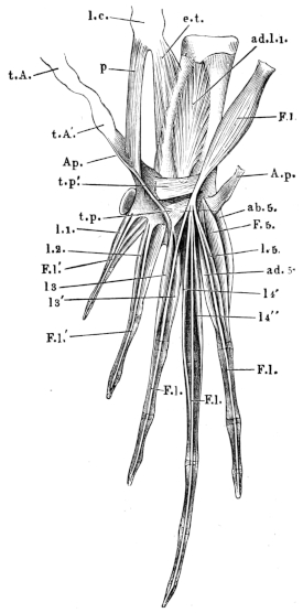

133. Aponeurosis plantaris (Fig. 88 A.p.).
As already mentioned, the tendon of the m. gastrocnemius is, at the heel, continued into a strong aponeurosis, and possesses at this place a thickening48. The aponeurosis is of triangular form, the base directed towards the toes and attached to these. The lateral borders of this triangle are continued into weaker fasciae, which are attached to the two long tarsal bones. The inner border in particular gives a strong bundle of fibres to the astragalus, and is thus connected with the fascia of the dorsum of the foot; externally this aponeurosis is attached to a cartilage (Dugès, os sesamoïde, l. c., n. 66), situated on the plantar surface of the joint between the calcaneus and the os metatarsi IV.
134. Ligamentum calcanei (Ecker), (Figs. 88, 89 l.c.).
If the tendo Achillis be divided and the aponeurosis plantaris reflected towards the toes, there is seen, arising from the posterior head of the tibio-fibula, in its whole breadth, a ligament (between the tib. posticus and peroneus). This ligament forms a firm surface on which the thickened portion of the tendo Achillis may move, and which evidently represents the tuberosity of the heel bone. I have therefore named the entire structure the ligamentum calcanei.
Several muscles arise from this ligament, viz.:—
135. M. extensor tarsi (Klein), (Figs. 88, 89 e.t.).
Dugès, tibio-sous-astragalien (regarded by him as the homologue of the tibialis posticus).
Covered by the two succeeding muscles, this muscle arises from the ligamentum calcanei, runs backwards, and is inserted into the whole length of the plantar surface of the astragalus. It extends the foot.
136. M. plantaris (Figs. 88, 89 p).
This takes its origin from the ligamentum calcanei to the inner side and below the last muscle. It passes backwards and is inserted into the dorsal surface of the aponeurosis plantaris.
137. M. flexor digitorum III, IV, V longus (Ecker), (Figs. 88, 89 F.l.).
Dugès, péronéo-sous-phalangettien (flexor dig. longus).
This muscle also arises from the ligamentum calcanei to the outer side of the m. plantaris. It runs backwards over the aponeurosis plantaris, and at the free margin of this passes somewhat suddenly into a strong tendon, which traverses an aponeurotic canal, formed by fibres of the aponeurosis passing above and below the tendon, from the cartilaginous enlargement in the tendon to the median border of the aponeurosis. The tendon forms three slips for the fifth, fourth, and third toes: these are slightly attached to the aponeurosis, but reach beyond its free border to be inserted into the terminal phalanges of the toes mentioned, and are held down by fibrous bands. (The small m. lumbricales arising from these tendons are described below.)
138. M. flexor digitorum I, II longus (Ecker), (Figs. 88, 89 F.l′.).
The tendons, by means of which the terminal phalanges of the first and second toes are flexed, arise directly from the posterior border of the aponeurosis.
139, 140. MM. transversi plantae (Ecker), (Fig. 89 t.p., t.p′.).
The movements of the aponeurosis and through it the movements of the tendons attached to it are influenced not only by the m. gastrocnemius and plantaris, but also by two other muscles, which together have been described as the flexor dig. longus internus (Klein), or as the tarso-sous-phalangettien (Dugès, 221).
a. M. transversus plantae posterior (t.p.).
This takes its origin from the plantar cartilage, widens as it courses backwards and inwards to be inserted into the dorsal surface of the aponeurosis plantaris; from this the tendons for the first and second toes (flexor dig. longus I and II) arise, thus there is no direct connection between the muscular fibres of the two muscles.
b. M. transversus plantae anterior (t.p′.).
This muscle is covered by the last; broader anteriorly, it arises from the calcaneum, and is inserted to the inner side of the last muscle into the dorsal surface of the aponeurosis. This muscle, like that just described, is not continued directly into the tendon of the flexor of the toes, they cannot therefore be regarded as flexors of the toes.
141 to 149. Musculi lumbricales (Fig. 89 l.1–5).
(A.) From the posterior border of the aponeurosis plantaris arise:—
(1) M. lumbricalis digiti I (l.1).
Dugès, tendini-sous-phalangien de pouce, n. 185.
This is inserted into the anterior extremity of the proximal phalanx.
(2) M. lumbricalis digiti II (l.2).
Dugès, tendini-sous-phal. du I doigt, n. 186 (and 187), double according to Dugès.
Similarly inserted.
(3 and 4) MM. lumbricales digiti III (l.3).
Dugès, tendini-sous-phal. du III doigt, n. 188, single according to Dugès.
One of these (l3) is inserted into the anterior extremity of the basal phalanx, the other (l3′) into a corresponding position on the middle phalanx.
(5) M. lumbricalis digiti IV.
Dugès, tendini-sous-phal. du IV doigt.
Inserted into the anterior extremity of the basal phalanx.
(B.) Arising from the tendons of the flexor longus III, IV, V:
(6 and 7) MM. lumbricales digiti IV (l.4′. and l.4″.).
Dugès, tendini-sous-phalanginien du IV doigt, n. 201, 202.
The first (l.4′) is inserted into the posterior end of the basal phalanx, the second (l.4″) in a similar position into the second phalanx.
(8) M. lumbricalis digiti V (l.5.).
Dugès, tendini-sous-phalangien du V doigt, n. 203.
This arises from the tendon for the fifth toe, runs backwards as a thin tendon alongside that of the flexor, and is inserted into the middle phalanx.
Special Muscles of the Separate Toes.
(I.) Muscles of the rudimentary great toe.
150. M. abductor hallucis (Figs. 88 a.h., 90).
Dugès, tibio-sous-tarsien, n. 163.
This muscle arises from the inner border of the aponeurosis plantaris, at the spot where the m. plantaris is inserted, it may therefore almost be regarded as a continuation of this muscle; it is inserted into the anterior border of the rudimentary toe.
(II.) Muscles of the great and first toes.
151. M. adductor longus digiti I (Figs. 89, 90 ad.l1).
Dugès, calcanéo-scaphoidien, n. 164.
This powerful muscle arises from the plantar surface of the calcaneum and astragalus; its fibres converge towards a tendon lying in its middle, which becomes free towards its posterior end; the tendon passing inwards under the ligamentum tarsi transversum through a groove on the hinder extremity of the astragalus in an arched direction, to be inserted into the os naviculare, which bears the rudimentary great toe and the os metatarsi I.
(III.) Muscles of the first toe.
152. M. abductor longus digiti I (Figs. 88 al.1, 90 a.b.1).
It takes its origin from the inner border of the aponeurosis plantaris, and lies on the plantar surface of the abductor hallucis. Its tendon runs in the hollow which the great toe forms, and is inserted into the inner side of the os metatarsi I.
153. M. flexor brevis digiti I (Fig. 90 F.b.1).
A small, thin muscle, which arises from the anterior extremity of the os metatarsi II, and is inserted into the basal phalanx of the first toe. In this small muscle, and also in the m. lumbrical. dig. V (Fig. 89 l.5.), the nerve distribution may be very well seen.
154. M. opponens dig. I (Fig. 90 op).
Dugès, sous-metatarso-phalangien du pouce, n. 192.
This is situated to the inner side of the preceding muscle, arises from the os metatarsi II somewhat narrow, widens in a fan-like manner towards the os metatarsi I, into the anterior half of which it is inserted.
(IV.) Muscles of the second toe.
155. M. flexor metatarsi dig. II (Fig. 90 F.m.2).
Dugès, sous-tarso-metatarsien du II doigt.—Klein, extensor metatarsi.
By a narrow origin from the hinder end of the calcaneum, posteriorly this muscle becomes broader and is inserted, somewhat thinned out, into the plantar surface of the second os metatarsi.
156. M. flexor dig. II proprius (Fig. 90 F.p.2).
Dugès, sous-metatarso-phalangien, n. 194.
It arises from the plantar surface of the os metatarsi II, and is inserted by a thin tendon into the plantar surface of the first phalanx.
Dugès describes this and the analogous muscles of the third, fourth, and fifth toes (F.p.3, 4, 5) as the interossei plantares, a nomenclature which, to me, does not appear correct, as these muscles lie entirely on the plantar surface, and do not adduct or abduct the toes, but flex them.
(V.) Muscles of the third toe.
Fig. 90.
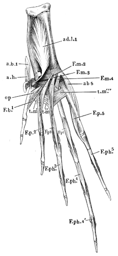
Muscles of plantar surface of foot of Rana esculenta.
a.b.1 M. abductor longus digiti I.
ab5 M. abductor digiti V.
ad.l.1 M. adductor longus digiti I.
a.h. M. abductor hallucis.
F.b.1 M. flexor brevis digiti I.
F.m.2, 3, 4 M. flexores metatarsi II, III, IV.
F.p.2, 3, 4, 5 MM. flexor. proprii digit. 2, 3, 4, 5.
F.ph.3, 4, 4′, 5 MM. flexor. phalang. digit. 3, 4, 5.
op M. opponens digiti I.
t.m′ MM. transversi metatarsi 1, 2, 3.

t.m″
t.m‴
157. M. flex. metatarsi dig. III (Ecker), (Fig. 90 F.m.3).
Dugès, sous-calcaneo-métatarsien du III doigt, n. 174.—Klein, extensor metatarsi.
Like the corresponding muscle of the second toe (n. 155), this arises by a small tendon from the hinder extremity of the calcaneum, runs backwards and is inserted into the plantar surface of the os metatarsi III. The greatest portion of the muscle lies beneath the m. transversus metatarsi (tm), the most anterior portion, however, lies above this. Dugès has described this latter portion as a special muscle (n. 176, metatarso-métatarsien du III), the same description holds good for the second toe (n. 177, Dugès).
158. M. flexor dig. III proprius (Ecker), (Fig. 91 Fp3).
Dugès, sous-métatarso-phalangien du III doigt, n. 195.
Like the corresponding muscle of the second toe, this arises from the plantar surface of the os metatarsi III, and is inserted by a thin tendon into the plantar surface of the anterior extremity of the first phalanx.
159. M. flexor phalangum proprius digiti III (Ecker), (Fig. 91 F.ph.3).
Dugès, phalango-phalangien, n. 204.
This arises by tendon from the plantar surface of the basal phalanx, and is inserted into the anterior extremity of the terminal phalanx.
(VI.) Muscles of the fourth toe.
160. M. flexor metatarsi dig. IV (Fig. 91 F.m.4).
Dugès, métatarso-métatarsien, n. 175.
Similarly to the corresponding muscle of the third toe, this one arises from the hinder extremity of the calcaneum, and is inserted into the anterior portion of the plantar surface of the os metatarsi IV, but not into the greater portion of this surface, as in the second and third os metatarsi.
161. M. flexor dig. IV proprius (Fig. 91 Fp4).
Dugès, sous-métatarso-phalangien du IV doigt (interosseus plantaris), n. 196.
The origin and insertion are similar to those of the muscles of the second and third toes.
Fig. 91.
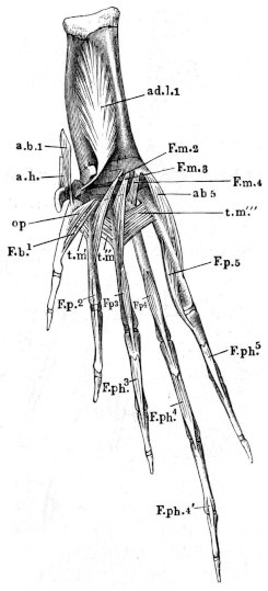
Muscles of plantar surface of foot of Rana esculenta.
a.b.1 M. abductor longus digiti I.
ab5 M. abductor digiti V.
ad.l.1 M. adductor longus digiti I.
a.h. M. abductor hallucis.
F.b.1 M. flexor brevis digiti I.
F.m.2, 3, 4 M. flexores metatarsi II, III, IV.
F.p.2, 3, 4, 5 MM. flexor. proprii digit. 2, 3, 4, 5.
F.ph.3, 4, 4′, 5 MM. flexor. phalang. digit. 3, 4, 5.
op M. opponens digiti I.
t.m′ MM. transversi metatarsi 1, 2, 3.
t.m″
t.m‴
162. M. flexor brevis dig. IV.
A small muscle, which arises with the m. flexor brevis of the fifth toe, from enlargement of aponeurosis plantaris, and is inserted into the basal phalanx of the fourth toe.
163. M. flexor phalangum proprius dig. IV anterior (Fig. 91 F.ph.4).
Dugès, phalango-phalanginien, n. 205.
It resembles the corresponding muscle of the third toe.
164. M. flexor phalangum proprius posterior (Fig. 91 F.ph.4′).
Dugès, phalangino-phalangettien, n. 209.
This extends from the plantar surface of the second phalanx to the anterior extremity of the third.
(VII.) Muscles of the small toe.
165. M. abductor dig. V (Fig. 91 a.b.5).
Dugès, calcanéo-ex-métatarsien du digitule, n. 169.
This muscle arises from the hinder extremity of the calcaneum, and is inserted into the outer surface of the fifth metatarsal.
166. M. adductor dig. V (Fig. 89 a.d.5).
Dugès, sous-tarso-in-phalangien, n. 191.
This arises from the cartilage of the plantar aponeurosis, and is inserted into the hinder end of the fifth metatarsal.
167. M. flexor brevis dig. V (Fig. 89 F.5).
Dugès, sous-tarso-ex-phalangien, n. 190.
Arising from the same place as the preceding, it extends to the basal phalanx.
168. M. flexor dig. V. proprius (Fig. 91 F.p.5).
(Analogues of Muscles, n. 156, 158, 161; see note at n. 156).—Dugès, sous-métatarso-phalangien, n. 197 (=inteross. plant.).
This muscle arises from the plantar and inner surfaces of the fifth metatarsal, and is inserted into the basal phalanx.
169. M. flexor phalangum proprius (Fig. 91 F.ph.5).
Dugès, phalango-phalanginien, n. 206.
Its origin and insertion are as in the corresponding muscles of the second, third, and fourth toes, n. 159, 163, 164, 169.
170 to 172. MM. interossei (Fig. 91 t.m.).
Dugès, 1, 2, and 3, intermétatarsien, n. 170, 171, 172.
These are three muscles with their fibres arranged transversely, which occupy the anterior portion of the spaces between the metatarsals, and approximate these. The first arises (tm′) from the edge of the plantar surface of the first metatarsal, and is inserted into the corresponding part of the second: the second (tm″) passes from this point to the third, and the third (tm‴) to the fifth.
B. Muscles of the Dorsal Surface.
173. M. flexor tarsi posterior (Ecker), (Fig. 92 F.t.).
Dugès, péronéo-sus-astragal, n. 161.
This is a strong muscle, which has a narrow origin from the outer side of the tibio-fibula, directly over the distal extremity, and is inserted into the dorsal surface of the astragalus from the middle to the distal extremity.
Fig. 92.
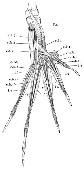
Dorsal view of muscles of foot of Rana esculenta.
a.b.1 M. abductor brevis dig. I.
e.b.1 M. extensor brevis dig. I.
e.b.2 M. extensor brevis dig. II.
e.b.3 M. extensor brevis dig. III.
e.b.4 M. extensor brevis 1 dig. IV.
e.b.4′ M. extensor brevis 2 dig. IV.
e.b.5 M. extensor brevis dig. V.
e.l.1 M. extensor longus dig. I.
e.l.2 M. extensor longus dig. II.
e.l.3 M. extensor longus dig. III.
e.l.4 M. extensor longus dig. IV.
e.l.5 M. extensor longus dig. V.
F.t M. flexor tarsi posterior.
i.2 to i.10 MM. interossei
Muscles of the first toe.
174. M. extensor longus dig. I (Fig. 92 e.l.1).
Dugès, calcanéo-sus-métatarsien du pouce, n. 166.
This lies to the outer side of and near the foregoing muscle; it arises by a long, strong head from the middle of the calcaneum, runs obliquely backwards and inwards to unite with a small head which arises from the common epiphysis of the astragalus and calcaneum, in common with the m. extensor dig. II. The muscle so formed passes into a flat tendon to be inserted into the basal phalanx of the first toe.
175. M. extensor brevis dig. I (Fig. 92 e.b.1).
A small, short muscle which arises from the os naviculare and is inserted into the dorsal surface of the first metatarsal.
176. M. abductor brevis dig. I (Fig. 92 a.b.1).
Dugès, ex-tarso-métatarsien du pouce, n. 168.
This small muscle is situated near the preceding; it arises from the first bone of the supplemental toe, and is inserted into the inner side of the first metatarsal.
Muscles of the second toe.
177. M. extensor dig. II longus (Fig. 92 e.l.2).
Dugès, calcanéo-sus-phalangien du II doigt, n. 182.
This arises from the calcaneum, in common with the m. extensor longus of the third toe, and is inserted into the basal phalanx of the second toe by a tendon.
178. M. extensor dig. II brevis (Fig. 92 e.b.2).
Dugès, astragalo-sus-phalangien du II, n. 183.
This is partly covered by the preceding muscle: it arises from the astragalus, and unites with the tendon of the preceding muscle.
Muscles of the third toe.
179. M. extensor dig. III longus (Fig. 92 e.l.3).
Dugès, sus-astragalo-phalangien du médius, n. 199.
This arises with the extensor longus of the second toe from the calcaneum: its tendon unites with that of the following muscle.
180. M. extensor dig. III brevis (Fig. 92 e.b.3).
Dugès, astragalo-sus-phalangien du médius, n. 181.
This muscle arises from the astragalus, and is inserted with the tendon of the preceding muscle into the dorsal surface of the basal phalanx.
Muscles of the fourth toe.
Fig. 93.
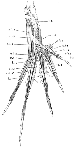
Dorsal view of muscles of foot of Rana esculenta.
a.b.1 M. abductor brevis dig. I.
e.b.1 M. extensor brevis dig. I.
e.b.2 M. extensor brevis dig. II.
e.b.3 M. extensor brevis dig. III.
e.b.4 M. extensor brevis 1 dig. IV.
e.b.4′ M. extensor brevis 2 dig. IV.
e.b.5 M. extensor brevis dig. V.
e.l.1 M. extensor longus dig. I.
e.l.2 M. extensor longus dig. II.
e.l.3 M. extensor longus dig. III.
e.l.4 M. extensor longus dig. IV.
e.l.5 M. extensor longus dig. V.
F.t M. flexor tarsi posterior.
i.2 to i.10 MM. interossei.
181. M. extensor longus dig. IV (Fig. 93 e.l.4).
Dugès, péroneo-sus-phalangien du IV, n. 178.
A narrow muscle arising in common with the flexor tarsi inf., but external to it. It is inserted into the tendinous expansion on the dorsum of the toe.
182. MM. extensores dig. IV breves (Fig. 93 e.b.4 and e.b.4′).
Dugès, calcanéo-sus-phalangien and sus-calcanéo-phalanginien, n. 179 and 198.
These two small muscles arise from the calcaneum: one is inserted into the basal phalanx, the other, by means of a long thin tendon, passes along with the tendon of the corresponding m. interosseus to the second phalanx, to which it is attached.
Muscles of the fifth toe.
183. M. extensor dig. V longus (Fig. 93 e.l.5).
Dugès, calcanéo-sus-métatarsien, n. 165.
A strong muscle which covers almost the whole length of the calcaneum; it arises from the anterior extremity of this bone, runs backwards to the outer side of the dorsum of the foot, and is attached to the outer surface of the fifth metatarsal.
184. M. extensor dig. V brevis (Fig. 93 e.b.5).
Dugès, calcanéo-sus-phalangien du V, n. 180.
This is a long thin muscle, which, together with the muscle last described, arises from the calcaneum and is inserted into the basal phalanx of the fifth toe.
185. M. abductor dig. V brevis (Fig. 93 i.10).
This, the last m. interosseus, arises from the outer side of the fifth metatarsal, and is inserted into the hinder extremity of the second phalanx of the same toe.
186 to 195. MM. interossei dorsales (Fig. 93 i.1 to i.10).
First toe.
(1) The analogue of the first m. interosseus is the m. abductor brevis dig. I (a.b.1).
(2) The second m. interosseus (i.2) arises from the outer side of the first metatarsal, and is inserted into the outer surface of the second phalanx of the same toe, dorsally to the m. opponens and flexor brevis dig. I.
Second toe.
Fig. 94.
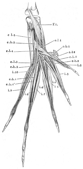
Dorsal view of muscles of foot of Rana esculenta.
a.b.1 M. abductor brevis dig. I.
e.b.1 M. extensor brevis dig. I.
e.b.2 M. extensor brevis dig. II.
e.b.3 M. extensor brevis dig. III.
e.b.4 M. extensor brevis 1 dig. IV.
e.b.4′ M. extensor brevis 2 dig. IV.
e.b.5 M. extensor brevis dig. V.
e.l.1 M. extensor longus dig. I.
e.l.2 M. extensor longus dig. II.
e.l.3 M. extensor longus dig. III.
e.l.4 M. extensor longus dig. IV.
e.l.5 M. extensor longus dig. V.
F.t M. flexor tarsi posterior.
i.2 to i.10 MM. interossei.
(3) The third m. interosseus (i.3) takes origin from the anterior part of the outer surface of the first metatarsal by a thin tendon; it is inserted into the inner surface of the second phalanx by a similar tendon.
(4) The fourth m. interosseus (i.4) arises from the outer side of the second metatarsal, and is inserted into the outer surface of the second phalanx.
Third toe.
(5) The fifth m. interosseus arises from the outer surface of the second metatarsal, and is inserted by a thin tendon into the inner surface of the second phalanx of the same toe.
(6) The sixth arises from the outer surface of the third metatarsal, and is inserted laterally by a thin tendon into the hinder extremity of the second phalanx of the same toe.
Fourth toe.
(7) The seventh m. interosseus (i.7) arises by a thin tendon from the hinder extremity of the astragalus, and by fleshy fibres from the hinder extremity of the fourth metatarsal; it is inserted by a thin tendon into the inner surface of the second phalanx of the same toe.
(8) The eighth m. interosseus (i.8) takes its origin from the outer surface of the fourth metatarsal and from the inner surface of the fifth: it is inserted into the hinder extremity of the second phalanx.
Fifth toe.
(9) The ninth m. interosseus (i.9) arises from the inner surface of the fifth metatarsal, and is inserted into the hinder extremity of the second phalanx.
(10) The tenth m. interosseus has already been described as the abductor digiti V brevis (Fig. 94 i.10).
If we take the fourth toe as the axis of movement, the first, third, fifth, seventh, eighth, and tenth mm. interossei will abduct from an imaginary line which runs through this toe. The second, fourth, sixth, and ninth will adduct towards this line. If we compare them with those of man (in whose case the second toe affords the central line), those of the first group must be regarded as mm. interossei dorsales, of the latter as plantares. Dugès holds all these interossei (with the exception of the first, abductor brevis dig. I) to be mm. interossei dorsales (métatarso-sus-phalangettiens, n. 210, 211, 212, 214, 215, 216, 218, 219). Those which I have described as flexores proprii digitorum, he regards as interossei volares.
V. MUSCLES OF THE SKIN.
Fig. 95.
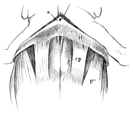
Pectoral region of Rana esculenta.
cp M. cutaneus pectoris.
p″ Port. abdominalis of the m. pectoralis.
196. M. cutaneus pectoris (Fig. 95 cp).
Dugès, abdomino-guttural, n. 53.—Klein, abdomino-cutaneus.—Zenker, subcutaneus pectoris.
This is an elongated quadrangular muscle, attached by its hinder margin to the side of the xiphisternum, and to the superficial surface of the aponeurosis of the m. obliquus externus. The fibres course forwards and slightly outwards to be inserted, at a very acute angle, into the skin: lying between the two muscles and attached to them on either side is a thin fascia, which, together with the muscles and the septum thoracicum, assists in closing a triangular space between these structures and the deeper muscles.
The thinness and transparency of this muscle render it especially suitable for the investigation of nerve-terminations49.
197. M. cutaneus dorsi (Fig. 96 cd).
Dugès, pubio-dorso-cutané, n. 56.—Zenker, cutaneus iliacus.
Fig. 96.
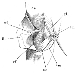
Hind portion of back and thigh of Rana esculenta.
cd M. cutaneus dorsi.
cx. Coccyx.
gl. M. glutaeus.
H Skin reflected to left side.
oc M. obliquus externus.
rf M. rectus femoris anterior.
sm M. semimembranosus.
v.e M. vastus externus.
This muscle has a narrow origin in the space between the muscles of the belly and of the thigh, and is here inserted into the fasciae, which are attached to the pubic symphysis; it passes inwards and upwards to the outer side of the anterior insertion of the m. rectus femoris anticus, then widens and is inserted by diverging fibres into the inner surface of the skin of the hinder portion of the back.
198. M. coccygeo-cutaneus.
Dugès, coccy-dorso-cutanés, n. 57.
The muscular fibres which Dugès has described under this name, lie covered by the m. pyriformis, behind the m. coccygeo-iliacus, etc., and above the muscles of the rectum. They arise from the hinder extremity of the urostyle: they appear to be connected with the rectal muscles, and pass outwards to be attached to the skin.
Among the cutaneous muscles may perhaps be included the m. submaxillaris (p. 62).
The fibrils passing from the flexor tendons to the warty bodies on the plantar surface of the toes are described50 with the skin (p. 373).