SECTION III. THE NERVOUS SYSTEM. (continued 2)
George Haslam translation
D. The Vessels of the Brain and Spinal Cord. (Fig. 110.)
The arteries of these organs are derived from the internal carotids and the arteriae vertebrales. As soon as the internal carotid arteries reach the cranial cavity each divides into two branches, an anterior (Ramus anterior, Schöbl56) and a posterior (Ramus posterior, Schöbl); the anterior branches course forwards on the lower part of the surface of the brain as far as the anterior portion of the thalamencephalon, where they again divide; one division, arteria lobi hemisphaerici inferior externa (Schöbl), continues forwards along the outer surface of the cerebral hemisphere and of the olfactory lobe as far as the olfactory bulb, where it may still be distinctly seen; it gives off branches to the adjacent parts in its course forwards. The vessels of the two sides communicate with each other by means of delicate transverse vessels (Arteriae communicantes anteriores). The second division, arteria lobi hemisphaerici superior interna (Schöbl), of the ramus anterior courses on the thalamencephalon to the dorsal surface, gives a large branch to the choroid plexus of the fourth ventricle, and runs forwards in the dorsal longitudinal fissure; it supplies vessels to all the neighbouring parts.
The rami posteriores converge as they course backwards, and ultimately unite to form an arteria basilaris, which is continued in the median line of the under surface of the spinal cord as the arteria spinalis anterior. The ramus posterior gives off, in its course, many small vessels to the neighbouring parts, and two larger vessels on either side, one of which, the arteria lobi optici (Schöbl), is distributed to the optic lobes, while the other passes to the pituitary body.
The branches of these vessels form a network in the pia mater, from which the nervous tissue is supplied; they also send numerous branches directly into the brain and cord, and these have a similar arrangement for all parts of the brain with the exception of the cerebral hemispheres and olfactory lobes; more or less vertical branches arise from the posterior parts of the upper borders of the rami anteriores, from the whole of the rami posteriores, and from the arteria basilaris; these course upwards from their place of origin into the corresponding part of the brain, give off a few branches in the white matter, and then branch freely, and at somewhat sharp angles, in the grey matter. In the pars commissuralis a large branch may be traced from the ramus posterior on either side upwards into the cerebellum almost to its upper border. The vessels of the cerebral hemispheres and olfactory lobes seem to possess no other definite arrangement than that described above. The grey matter seems, on the whole, to be more vascular than the white; the vessels of the latter are chiefly arranged radially to the surface, and run in courses which are more or less straight; the vessels of the grey matter are more irregular and sinuous.
The Arteria spinalis anterior courses along the whole length of the spinal cord, giving off lateral branches and communicating with rami spinales (branches of the Arteria vertebralis). These branches form a plexus in the pia mater, from which vessels pass at irregular points into the cord; one set of small vessels, described by Reissner, pass in a straight course from the superior longitudinal sinus towards the substantia reticularis, where they divide. Other branches pass directly from the arteria spinalis anterior, through the ventral longitudinal fissure, and there divide; the twigs as a rule avoiding the septum medium and passing in greater part towards the ventral horns. The vessels in the white matter are for the most part radial and straight, while in the more vascular grey matter they are irregular and more sinuous in their course.
Fig. 110.
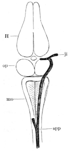
Diagram to show the Vena spinalis posterior, the Venae spinales superiores, and the origin of the Vena jugularis interna.
H Hemispheres.
ji Internal jugular.
op Optic lobes.
mo Medulla oblongata.
spp Vena spinalis posterior.
The choroid plexus of the third ventricle (Plexus choroideus ventriculi tertii, Reissner) lies, as already described, on the roof of the third ventricle; it is somewhat triangular in form (Plexus venosus triangularis, Schöbl), and is evidently a prolongation of the pia mater, with an increased supply of vessels. It receives, at its anterior angle, veins from the cerebral hemispheres and the adjacent parts. The under surface is covered with a layer of ciliated pavement epithelium. At the posterior angles of the plexus the veins communicate on either side (Fig. 110) with the anterior lateral prolongation of the vena spinalis superior, and with the internal jugular vein. The small body usually named the pineal body, and placed on the roof of the ventricle, is only a small thickened portion of the plexus, and consists of a group of convoluted blood-vessels. This plexus sends a considerable bunch of vessels into the upper part of the third ventricle. The choroid plexus of the fourth ventricle (Plexus choroideus ventriculi quarti, Reissner) is a triangular membrane, slightly attached to the borders of the fourth ventricle. Its upper surface is flat and bounded at either side by a large vein, the vena spinalis superior; anteriorly it is bounded by the cerebellum. The under surface is not flat; in the middle line is a slight furrow corresponding to the position of a median vessel, which may, when injected, be seen from the dorsal surface; from it a number of short vessels pass outwards and slightly backwards to the outer border. The connective-tissue between these vessels is pushed down into the cavity; in this manner one obtains a double row of flattened villous-looking bodies, which frequently have their tips pigmented. The under surface of the plexus is clothed with flattened, ciliated epithelium.
On either side of the pituitary body is another small venous plexus (Plexus lateralis, Schöbl); it communicates above with the posterior angle of the choroid plexus of the third ventricle; externally, with the internal jugular vein; and internally with its fellow of the opposite side by more or less irregular transverse vessels, which, together with a median vein from the fissure between the cerebral hemispheres, form an irregular circulus venosus around the pituitary body. These plexuses receive blood from the lower part of the cerebral hemispheres, the thalamencephalon, the optic lobes, and from the pituitary body. The veins of the spinal cord open into the dorsal, vena spinalis posterior (Fig. 110 spp), which bifurcates at the posterior angle of the fourth ventricle, one division passing to either side and forming, as already described, the outer border of the choroid plexus; anteriorly it opens into the internal jugular vein (Fig. 110 ji).
The blood-vessels, both arteries and veins, are irregularly pigmented, both on the surface and in the interior of the central nervous system; those of the cerebral hemispheres and olfactory lobes have very little or no pigment.
DESCRIPTION OF THE FIGURES ON PLATE I.
Fig. 111. Dorsal view of the orbit, etc.; deep dissection.
c Course of the palatine nerve. II Optic nerve.
db Fibrous plate. III Motor oculi nerve.
dm M. depressor maxillae. IV Trochlear nerve.
ef Terminal branches of the ophthalmic nerve. Vb R. palatinus of the trigeminal nerve.

F Facial with R. anterior of the glossopharyngeal. Vc R. maxillo-mandibularis
g,g,g Terminal twigs of the ophthalmic nerve. Vc′ R. maxillaris
h Muscular twigs of the Ram. maxillaris. Vd R. mandibularis
ics M. intertransversar. capitis superior. Ve(VII) Facial nerve.
ii Twigs of upper eyelid. Vg Gasserian ganglion.
K Terminal twigs of the Ram. maxillaris. VN Trigeminus.
la M. levator anguli scapulae. VS Sympathetic nerve.
pt M. pterygoideus. VI Abducens nerve.
sc M. sterno-cleido-mastoideus. VII′ Facial nerve.
Sp Sympathetic nerve. VIII Auditory nerve.
t M. temporalis. X1 Ram. anterior of the glossopharyngeal.
tr Nasal branch. X2 Glossopharyngeal nerve.
* Union of facial nerve with Ram. anterior of the glossopharyngeal nerve. X3 Pneumogastric nerve.
I Olfactory nerve. XG Ganglion nervi vagi.
XI Accessory nerve.
Fig. 112. Dorsal view of the orbit, etc.; superficial dissection.
e External branch of the ophthalmic nerve. Va Ophthalmic nerve.
f Internal branch of the ophthalmic nerve. Vc R. maxillo-mandibularis.
g,g,g Terminal twigs of the ophthalmic nerve. Vc′ R. maxillaris of the trigeminal nerve.
II Optic nerve. Vd R. mandibularis of the trigeminal nerve.
III Motor oculi nerve. VI Abducens nerve.
IV Trochlear nerve. VI′ Ciliary nerves.
Fig. 113. View of roof of mouth; mucous membrane, etc., removed.
a Branch of the Ram. palatinus to the Harderian gland. ri M. rectus internus.
b Branch of the Ram. palatinus to the intermaxillary gland. ri′ M. rectus inferior.
c Inosculating-branch of the Ram. palatinus. sc M. sterno-cleido-mastoideus.
Ch Internal naris. uk Mandible.
d Cutaneous branch of the Ram. palatinus. † Terminal branch of Ram. palatinus.
db Fibrous plate. * Union of facial nerve with Ram. anterior of the glossopharyngeal.
F Ramus mandibularis of the facial nerve. III Motor oculi nerve.
Gl Intermaxillary gland. Vb Ramus palatinus of trigeminal nerve.
HD Harderian gland. Vc Ramus maxillo-mandibularis of the trigeminal nerve.
l Eyeball. Vc′ Ramus maxillaris of the trigeminal nerve.
m M. masseter. Vd R. mandibularis.
Ms.ob.i. M. obliquus inferior. Ve(VII) Facial nerve.
OK Upper jaw. X Pneumogastric nerve.
r M. retractor bulbi. X1 Glossopharyngeal nerve.
Fig. 11457. Lateral dissection of head, etc., to show the cranial nerves, etc.
c Cardiac nerve. pp Pharyngeal branches of the pneumogastric nerve.
F Union of facial and Ram. anterior of the glossopharyngeal nerve. tt′ M. temporalis.
h Ram. hyoideus of the glossopharyngeal nerve. us Muscular twigs of the Ram. mandibularis of the trigeminal nerve.
la M. levator angi scapulae. Z Tongue.
lgd M. longissimus dorsi. Va Terminal twigs of the ophthalmic nerve.
m M. masseter. Vc Ram. maxillaris of the trigeminal nerve.
mt Ram. mandibularis of the glossopharyngeal nerve. Vd Ram. mandibularis of the trigeminal nerve.
M1 Hypoglossal nerve. Ve(VII) Facial nerve.
M2 Brachial nerve. X1 Ram. anterior of the glossopharyngeal nerve.
Oh Anterior cornu of the hyoid bone. X2 Glossopharyngeal nerve.
oi M. obliquus internus. X3 Pneumogastric nerve.
ph1 M. petrohyoideus I. X3g Ram. gastrici of the pneumogastric nerve.
ph2 M. petrohyoideus II. X3l Ram. laryngeus.
ph3 M. petrohyoideus III. X3p Ram. pulmonales of the pneumogastric nerve.
ph4 M. petrohyoideus IV. XI Accessory nerve of the pneumogastric nerve.
Fig. 115. Dissection of the floor of the mouth.
dm M. depressor maxillae. ph3 M. petrohyoideus III.
F United facial nerve and Ram. anterior of the glossopharyngeal nerve. ph4 M. petrohyoideus IV.
gh M. geniohyoideus. sc M. sterno-cleido-mastoideus.

gh′ sh M. sterno-hyoideus.
h Ram. hyoideus of the glossopharyngeal nerve. sm M. mylohyoideus (submaxillaris).
H Heart. smt M. submentalis.
Lg Lung. TT Thyroid.
m′ Ram. mandibularis of the glossopharyngeal nerve. uk Mandible.
M1 Hypoglossal nerve. us Muscular twigs of the Ram. mandibularis.
M2 Brachial nerve. X2 Glossopharyngeal nerve.
Oh Anterior cornu of the hyoid bone. X3 Pneumogastric nerve.
oh Momohyoideus. X3c Rami cardiaci of pneumogastric nerve.
ph1 M. petrohyoideus I. X3l Ram. laryngeus of the pneumogastric nerve.
ph2 M. petrohyoideus II. X3p Ram. pulmonalis of the pneumogastric nerve.
II. THE PERIPHERAL NERVOUS SYSTEM.
E. The Cranial Nerves.
(To facilitate reference the original arrangement of this part has been altered.)
[There are ten pairs of cranial nerves in the frog, which are numbered in order from before backwards. The mode of origin of these nerves, and their deeper relations with the respective parts of the brain to which they belong, have already been described.]
1. The olfactory nerve (N. olfactorius), [(Figs. 97, 98, 102, 103, 111 I) runs a very short course only a few lines in length, and escapes from the cranium by an opening in the cartilage of the sphenethmoid into the nasal cavity, where it divides into two branches, each of which breaks up into a brush of filaments, to be distributed in the olfactory mucous membrane. The nerve contains no white fibres]. (See organ of smell, p. 385.)
2. The optic nerve (N. opticus), (Figs. 97, 102, 103, 111 II) [arises, as already described, by the optic tract, and joins with its fellow at the optic chiasma or commissure, where part of the fibres pass over to the opposite side (according to Michel all the fibres cross). Each optic nerve then courses outwards, piercing the cartilage of the cranium and so reaching the eyeball]. (See organ of sight, p. 408.)
3. The motor oculi (N. oculomotorius, Oculo-motor, Motor communis), (Figs. 102, 111, 112, 113 III). From its origin it courses outwards and forwards, perforating with a slight obliquity the cartilaginous wall of the cranium, just in front of the Gasserian ganglion (Fig. 116 III); on reaching the orbit it divides into two branches, between the Levator bulbi and the Rectus internus and inferior. One branch, Ramus superior, runs over and parallel to the Ramos ophthalmicus Trig. and enters the under surface of the Rectus superior; the second, lower branch, Ramus inferior, supplies filaments to the Rectus internus and inferior and to the Obliquus inferior. Apparently it exchanges fibres with the ophthalmic division of the trigeminal nerve.
[That portion of the motor-oculi between its branch to the Rectus superior and to that to the Rectus internus contains a number of nerve cells; according to Schwalbe (l. c., p. 235 and Pt. XII, Fig. 4) these cells are arranged in four groups or ganglia; the second group forms a very slight prominence, and is covered by a very fine layer of nerve fibres, it also gives off some extremely fine nerves to the eyeball; this is perhaps a ciliary ganglion (Ganglion ciliare, Schwalbe). Beard, however, names it ‘mesocephalic ganglion.’ The other three groups are simply clusters of cells between the fibres of the nerve.]
4. The pathetic or trochlear nerve (N. trochlearis, patheticus), (Figs. 102, 103, and 112 IV). From its origin it courses forwards with and then crosses the motor-oculi; it perforates very obliquely the cartilaginous lateral wall of the cranium, in front of the motor oculi but above and very slightly behind the optic foramen (Fig. 116); it runs parallel to and in company with the Ramus opthalmicus trigemini, and appears to exchange a few fibres with it, which, however, according to de Watteville, Stannius, Cuvier, and Wyman, is not really the case. In this course it lies first to the inner, then to the outer side of the ophthalmic, over which it passes to supply the Rectus superior. [The pathetic and ophthalmic nerves are sometimes enclosed in a common sheath (de Watteville).]
5. The trigeminal nerve (N. trigeminus), (Figs. 97, 102, 103, 111, 112, 113, and 114 V) is the largest of the cranial nerves in the frog; from its origin it runs outwards and forwards to the skull wall, and just before reaching this enters the large Gasserian ganglion. It then passes through the cranial wall immediately in front of the auditory capsule, and divides at once into two main branches (Fig. 111 Vg, etc.), the Ramus ophthalmicus and the Ramus maxillo-mandibularis.
The Gasserian ganglion is a large, yellow, oval ganglion placed in a depression in the outer wall of the cranium; it is covered by a ‘periganglionic gland,’ which is similar in structure to that of a spinal ‘periganglionic’ gland (see p. 180), the fibrous capsule being stronger. This ganglion receives three other nerves besides the trigeminal, viz. the sixth and seventh nerves, and branches of the sympathetic.
According to de Watteville, these nerves are arranged on the lower surface of the ganglion, so that the sympathetic is below, the sixth above it, then the seventh, while the fifth is uppermost; the sympathetic splits into several bundles which join the various branches of the ganglion; the sixth divides into two bundles, one of which usually joins the ophthalmic, while the other makes its exit independently; the seventh splits into two bundles, one of which leaves the ganglion as the hyomandibular branch, the other gives some fibres to the palatine branch of the trigeminal nerve, and is then continued as the palatine branch of the facial.]
a. The Ramus ophthalmicus (Ramus nasalis, Fischer; Ophthalmic or Orbital Branch, Wyman; Orbito-nasal or Ophthalmic Nerve, de Watteville), (Figs. 111 and 112 Va). After leaving the Gasserian ganglion the nerve is directed forwards parallel to the side of the cranium, between this and the eyeball. It lies beneath the Rectus superior, but above all the other muscles of the eyeball and the optic nerve. At the anterior end of the orbit it divides into two branches (e and f) which pass through apertures (Foramen pro ramo nasali) in the cartilage of the sphenethmoid, to reach the nasal cavity, where they lie between the cartilage and mucous membrane. In this position the nerves supply branches to the mucous membrane and then pierce the skull to appear on the surface (Figs. 111, 112 g,g,g, 114 Va), the skin of which they supply. The branches inosculate with each other and communicate with the anterior twigs of the Ramus maxillaris (Fig. 114 Vc) and with those of the Ramus palatinus (Fig. 113 Vb).
In its course through the orbit the Ramus ophthalmicus gives off:—
(1) Near the Gasserian ganglion one, two, or three small branches, which communicate with the trochlear nerve, and may then be traced to the sclerotic of the eyeball; some of the twigs enter near the optic nerve, others nearer the cornea. They are regarded as Ciliary nerves (Nervi ciliares).
(2) A large branch, the palatine nerve (Ramus palatinus), (Figs. 111 and 113 Vb, b). This nerve, after receiving the palatine branch of the facial, descends on the inner border of the Levator bulbi to the mucous membrane of the mouth, to which it gives numerous filaments; it then runs parallel and near to the median line, lying on the base of the skull; and near the front of the orbit it divides into three branches. The smallest (a) supplies the Harderian gland (HD); the second (b) continues in the course of the original nerve, pierces the vomer, and divides, giving filaments to the mucous membrane in its whole course, while the terminal twigs supply the intermaxillary gland (Gl) and the surrounding structures. One of these (†) ascends on the anterior border of the sphenethmoid to the intermaxillary (internasal, Born) space to supply the numerous glands and to inosculate by one or two twigs with the nasal branch (tr) of the trigeminal. The third branch of the palatine nerve (c) curves directly outwards, just behind the palatine bone, to the inner surface of the maxillary bone; it then courses backwards, pierces the fibrous plate (db) between the eyeball and the pterygoid bone, and ultimately unites with superior maxillary division of the trigeminal nerve (Vc′). Just before piercing the fibrous plate it gives off a tolerably large nerve (d) to the mucous membrane, and in its whole course it gives off twigs to the mucous membrane and surrounding structures; many of these twigs inosculate with other terminal twigs of the trigeminal.
[This nerve has been minutely described by Stirling and Macdonald; these observers find that the branches form a very fine and close plexus in the mucous membrane, etc.; fibres were traced to blood-vessels, glands, etc., and ganglion cells were found scattered along the nerves. The fibres are both medullated and non-medullated, and many of the cells are described as ‘spiral cells’ (see page 201) such as those described by Arnold in the sympathetic system of the frog.]
b. The Ramus maxillo-mandibularis (Ramus maxillaris, Ecker and Hoffmann; Supra-maxillary, Humphry; Upper Maxillary Branch, Wyman), (Figs. 111, 112, and 113 Vc). This nerve is the largest division of the trigeminal; it runs directly outwards behind the eyeball, in front of the auditory capsule, and between the temporal and pterygoid muscles. After a very short course it divides into the maxillary and mandibular nerves. The nerve gives off before its division a few fine filaments (i,i) to the hinder half of the upper eyelid and to the surrounding skin, also a branch (h) [not correctly drawn in Fig. 111] which divides to supply twigs to the temporal and pterygoid muscles.
(1) The Ramus maxillaris (Ramus supramaxillaris, Ecker; Ramus maxillaris superior, Hoffmann; Upper Maxillary Branch, Wyman; Supramaxillary, Humphry and de Watteville), (Figs. 111, 112, 113, and 114 Vc) runs outwards and then forwards, between the eyeball and the outer wall of the orbit, to the margin of the upper jaw (K); a large portion terminates in a number of small branches for the supply of the skin of the lower eyelid, the upper lip, and of the parts between the tympanic membrane and the external nares. Some of these twigs inosculate with twigs of the palatine and ophthalmic nerves. The second portion of the nerve unites with the palatine nerve, as already described.
(2) The Ramus mandibularis (Ramus maxillaris inferior, Hoffmann; Mandibular or Lower Jaw Branch, Wyman; Inframaxillary, Humphry and de Watteville), (Figs. 111, 112, 113, 114, and 115 Vd, us). As far as the outer margin of the eyeball, this nerve runs parallel to and behind the Ramus maxillaris, in which course it supplies branches to the temporal and pterygoid muscles; it then curves backwards, outwards, and downwards to the under surface of the squamosal bone, where it supplies a twig to the Depressor maxillae, and then perforates the Masseter: in this manner it reaches the outer surface of the mandible, just behind the insertion of the temporal muscle; it then courses forwards, under the skin, to the Symphysis menti. In this course the nerve gives off numerous branches to the skin and surrounding parts, it also supplies the mylo-hyoid and submental muscles; one branch (Figs. 114 and 115 us) is larger than the rest, and supplies the under surface of the floor of the mouth and lower lip.
6. The abducens nerve (Nervus abducens, Ecker and Hoffmann; included in the trigeminal, Wyman), (Figs. 111 and 112 VI). This very slender nerve courses along the inner wall of the cranium from its origin to the Gasserian ganglion (q.v.) which it joins, and leaves this in contact with the ophthalmic division of the trigeminal nerve: the nerve then bifurcates in the orbit; the outer branch supplies the Rectus externus, the inner inosculates with small twigs of the ophthalmic division of the trigeminal, and then gives off a number of small ciliary nerves, already described, and one special branch to the Retractor bulbi.
7. The facial nerve (N. facialis, Ecker and Hoffmann; Facialis (Portio dura), Wyman; Facial or Jugular Nerve, Stannius), (Figs. 111, 113, and 114 Ve VII) arises immediately behind the trigeminal, and runs forwards, first in company with the auditory nerve, then alone, to the Gasserian ganglion (q.v.), with which it unites; it again appears at the posterior angle of the ganglion, and escapes from the cranium in company with, and immediately behind, the Ramus mandibularis of the trigeminal, and divides at once into two branches.
a. The Ramus palatinus at once unites with the palatine branch of the ophthalmic division of the trigeminal nerve. Wiedersheim doubts this arrangement, and holds that the two portions arise in common from the Gasserian ganglion; neither has Wyman (Rana pipiens) described this branch of the facial. [De Watteville states that the Ramus palatinus and the palatine branch of the ophthalmic nerve are separated by the carotid artery. In urodeles the two nerves are distinct.]
b. The Ramus hyomandibularis (Ramus jugularis, Hoffmann; Facial Nerve, Wyman), (Figs. 113 and 114, Ve VII). The nerve is directed outwards and backwards so as to pass around the bony wall of the auditory capsule, it then crosses over the inner end of the columella, with which it is in close contact, and is then joined, under cover of the sterno-cleido-mastoideus (sc), by a branch (Ramus communicans) of the glossopharyngeal (see Fig. 111 *). The single trunk (F) so formed is directed downwards in the posterior wall of the Eustachian tube to just above the angle of the mandible, where it divides into three branches, or sometimes into two, in which case a third nerve is supplied by one of the others, usually by the Ramus hyoideus.
(1) The Ramus mandibularis (Ramus mentalis, Hoffmann; Third Trunk of the Facial, Wyman), (Figs. 114 mt and 115 m′) passes inside the angle of the jaw and courses forwards, between the skin and mylo-hyoid muscle, parallel to the Ramus mandibularis of the trigeminal nerve, as far as the symphysis. It supplies small twigs to the neighbouring parts of the mucous membrane of the mouth.
(2) The Ramus hyoideus (Ramus jugularis, Hoffmann; Second Trunk of the Facial, Wyman), (Figs. 114, 115 h) is the largest division of the facial; it courses forwards subcutaneously over the deltoideus and the hinder fibres of the mylo-hyoideus to the anterior cornu of the hyoid bone, supplying its muscles and the skin of the throat and sternal region.
(3) The Ramus auricularis (First Trunk of the Facial, Wyman); is a small branch, directed outwards; it supplies a branch to the walls of the tympanic cavity, and is finally distributed in the skin under the tympanic membrane and behind the angle of the mouth.
Fig. 116.
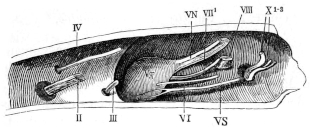
Right half of skull of Rana esculenta, seen from within.
II Optic nerve.
III Motor oculi nerve.
IV Trochlear nerve.
VN Root of the trigeminal nerve.
Vg Gasserian ganglion.
VS Sympathetic nerve.
VI Abducens nerve.
VII1 Facial nerve.
VIII Auditory nerve.
X1–3 Pneumogastric and glossopharyngeal nerves.
8. The auditory nerve (N. acusticus, Ecker, Hoffmann; Auditory Nerve, Wyman). This nerve has a very short course in the cranium; it reaches the auditory organ by a foramen (Fig. 116 VIII) in the auditory capsule, and is then distributed in the ear, with which it will be described.
9. The glossopharyngeal nerve (N. glossopharyngeus, Ecker; Ramus glosso-pharyngeus, Hoffmann; Glossopharyngeal, Wyman; Glossopharyngeal of the Vagus, Müller), (Figs. 102 and 103 X, 111, 113, 114, and 115 X2) arises in common with the pneumogastric nerve, and quits the skull with it through an opening (Canalis nervi vagi) immediately behind the auditory capsule, and at once divides into two branches; both of which lie under the MM. in tertransversarii capitis and are covered by the Depressor mandibulae.
a. The Ramus anterior (Verbindungsast des Glossopharyngeus, Wiedersheim; Ramus communicans, Hoffmann; Uniting Branch, Wyman; Laryngeal Branch of the Vagus, Volkmann; Communicans ad facialem, de Watteville), (Figs. 111, 113, and 114 X1). This nerve curves downwards and forwards around the auditory capsule and beneath the Depressor mandibuli to join the facial nerve, as already described.
b. The Ramus posterior (N. glossopharyngeus, Wiedersheim; Ramus lingualis, Hoffmann), (Figs. 111, 113, and 114 X2). This nerve runs downwards and forwards to the ventral surface of the pharynx, dips underneath the Petrohyoideus IV, to appear again, after a short interval, by piercing the Petrohyoideus II or III, courses parallel to and behind the anterior cornu of the hyoid bone, and thus reaches the floor of the mouth. The nerve then runs forwards in a very sinuous course, close to the median line, and between the Geniohyoideus and Hyoglossus; in its course on the floor of the mouth it crosses the hypoglossal nerve. It supplies the petrohyoid muscles, and gives numerous small branches to the mucous membrane of the pharynx (Ramus pharyngeus, Hoffmann).
10. The pneumogastric or vagus nerve (Vagus; Ramus intestinalis nervi vagi, Fischer, Hoffmann; Vagal Trunk, Wyman; Vaso-sympathetic, Gaskell), (Figs. 111, 113, 114, and 115 X3). This nerve arises in common with the glossopharyngeal; the two nerves leave the skull together by an opening in the exoccipital bone (Canalis nervi vagi); immediately outside the skull they acquire a ganglionic enlargement (Ganglion condyloideum, Ganglion nervi vagi), (Fig. 111 XG); in this course the glossopharyngeal lies in front of the pneumogastric, which it then leaves. The pneumogastric lies first upon the M. levator anguli scapulae, then running backwards and downwards along the hinder border of the Petrohyoideus IV it comes to the side of the pharynx; it is covered by the trapezius, and passes between the hypoglossal nerve and the Aorta ascendens; arriving at the Arteria pulmonalis, it gives off its terminal branches. [Gaskell has proved that this nerve contains both sympathetic and vagal elements, and that it is therefore really a vago-sympathetic.]
The branches and communications of the pneumogastric nerve are:—
a. Communications between the ganglion of the pneumogastric and the sympathetic system (Fig. 111 Sp), which again connect it with the Gasserian ganglion of the trigeminal nerve (Fig. 111 VS, Vg); (see sympathetic system).
b. [The Ramus cutaneus dorsalis (Fischer) is considered by Stannius and Fürbringer to be the homologue of the Ramus auricularis; it passes outwards between the digastricus and temporalis to be distributed in the skin of the suprascapular region. This nerve is the persistent portion of the N. lateralis nervi vagi of the tadpole.]
c. During its course over the petrohyoidei the vagus gives off a few twigs (Fig. 114 pp), which form a fine plexus to supply these muscles and the pharynx [also a twig to the trachea, according to Hoffmann].
d. The Ramus accessorius (Fürbringer, Hoffmann, Ecker), (Fig. 114 XI) is usually a single small nerve which runs between the intertransversarii muscles and the trapezius: it supplies the under surface of the latter muscle.
e. The Ramus scapularis (Hoffmann) is a very slender nerve arising near the Ramus accessorius; it passes along the under surface of the trapezius to the inner surface of the interscapularis, which it supplies.
f. The Ramus laryngeus (Recurrens vagi), (Figs. 114 and 115 X3l) runs for some distance parallel to the pneumogastric, separated from it by the petrohyoideus IV; arriving at the hinder cornu of the hyoid bone the nerve loops round the Arteria pulmonalis, and divides into two branches for the supply of the larynx.
g. The Rami gastrici (Fig. 114 X3g) are usually two in number: they pierce the partial diaphragm formed by the anterior fibres of the M. obliquus internus and terminate in the walls of the stomach.
h. The Rami pulmonales (Figs. 114 and 115 X3p) also perforate the partial diaphragm, and then course along the pulmonary arteries to the lungs.
i. The Ramus cardiacus (Figs. 114 and 115 X3c). This nerve is usually smaller on the right side than on the left; it passes along the dorsal surface of the pulmonary artery and Vena cava superior to the Sinus venosus; in this course it gives off two or three twigs to the roots of the lungs: the two nerves communicate just before reaching the heart and pass on to the auricular septum, whence they are distributed to the heart.
j. A slender Laryngeal nerve arises from the vagus alongside the Ramus cardiacus; it courses along the hinder end of the greater cornu of the hyoid to the outer side of the pharynx, which it pierces to pass to the larynx.
F. The Spinal Nerves.
I. General description. Ten pairs of nerves arise, as already described (p. 135), from the spinal cord; each nerve has two roots, a ventral or anterior, and a dorsal or posterior, which unite at their points of exit from the intervertebral foramen: just before, and for a short distance beyond this union, each dorsal root bears a ganglionic enlargement.
a. The length and direction of the various Nerve-roots vary greatly; the roots of the anterior spinal nerves run a very short course, almost transversely outwards, from their points of origin to the intervertebral foramina: the roots of the middle and posterior nerves, in consequence of the vertebral column being considerably longer than that part of the cord belonging to it, pass obliquely backwards to the foramina, the hinder nerves of the Cauda equina running for a considerable distance in the vertebral canal (Fig. 116). The relations of these roots are as follows:—
(1) The roots of the first nerve (N. hypoglossus) arise from the spinal cord at a point between the first and second vertebrae; they run transversely outwards to escape between the first and second vertebrae. The dorsal root is extremely slender.
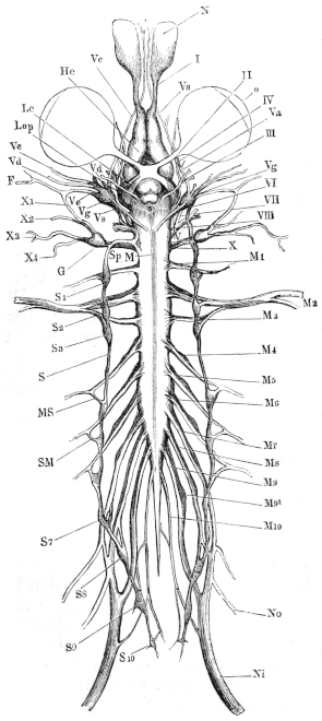
Fig. 117. The nervous system of Rana esculenta, from the ventral surface. (From Icones physiologicae by A. Ecker. Pl. XXIV, Fig. 1.) F Facial nerve. G Ganglion of pneumogastric nerve. He Cerebral hemisphere. Lc Optic tract. Lop Optic lobe. M Boundary between medulla oblongata and spinal cord. M1‑10 Spinal nerves. MS Connection between fourth spinal nerve and sympathetic chain. N Nasal sac. Ni Sciatic nerve. No Crural nerve. o Eyeball. S Trunk of sympathetic. S1‑10 Sympathetic ganglia. SM Rami communicantes of the same. Sp Continuation of sympathetic into head. I Olfactory nerve. II Optic nerve. III Motor oculi nerve. IV Trochlear nerve. V Trigeminal and facial nerves. Va Ramus ophthalmicus. Vc Ramus maxillaris. Vd Mandibular branch of trigeminal. Ve Hyomandibular branch of facial. Vg Gasserian ganglion. Vs Upper end of sympathetic trunk in connection with Gasserian ganglion. VI Abducens nerve. VII Facial nerve. VIII Auditory nerve. X Glossopharyngeal and pneumogastric nerves. X1 Ramus anterior of glossopharyngeal. X2 Ramus posterior of glossopharyngeal. X3, 4 Branches of pneumogastric.
(2) The roots of the second nerve (N. brachialis) arise at the level of the second vertebra and leave the vertebral canal between the second and third vertebrae.
(3) The roots of the third nerve arise from the cord between the second and third vertebrae, and pass out between the third and fourth vertebrae.
(4) The fourth nerve arises by its two roots at the level of the fourth vertebra, and quits the vertebral canal between the fourth and fifth vertebrae.
(5) The fifth nerve arises opposite the fourth vertebra, and passes outwards and slightly backwards to the foramen between the fifth and sixth vertebrae.
(6) The roots of the sixth nerve are attached to the cord opposite the fourth vertebra, and leave the vertebral canal between the sixth and seventh vertebrae.
(7) The seventh nerve arises from the cord at a point between the fourth and fifth vertebrae, and leaves the canal between the seventh and eighth vertebrae.
(8) The eighth nerve quits the cord at a point opposite the articulation between the fifth and sixth vertebrae, then runs backwards to the eighth vertebra, and escapes between this and the ninth vertebra.
(9) The ninth nerve arises at the level of the sixth vertebra, and passes out between the sacrum (ninth vertebra) and the urostyle.
(10) The tenth nerve (N. coccygeus) arises from the cord immediately behind the ninth nerve and opposite the sixth vertebra, and courses alongside the terminal filament to pass out through a foramen in the urostyle (canalis coccygeus).
b. [The Spinal ganglia (Ganglia intervertebralia).
At their exit from the intervertebral foramen the two roots of each nerve unite and bear a ganglionic enlargement, which is in intimate connection with the dorsal root, but is merely in contact with the ventral root. The ganglia lie58 in the large intervertebral depressions, upon the under surface of the Proc. obliqui, above and behind, being in relation with the bodies of the vertebrae internally, and the Musculi intertransversarii externally (Fig. 119); ventrally the ganglia are, more or less, covered by the ‘periganglionic glands.’ The ganglia vary much in size; that of the first spinal nerve is the smallest. The ganglion is not attached to the dorsal root alone but is prolonged a short distance beyond the point of union of the two roots; this is best seen in the nerves of the lumbar plexus. Each ganglion is a yellowish-white rounded or oval body, with an outer thick (sometimes 0.15 mm. thick) sheath of connective-tissue, connected by the epineurium of the nerve roots with the Dura mater. This sheath is composed of bundles of parallel, wavy, white, connective-tissue fibres, which enclose a large number of connective-tissue corpuscles, and here and there contains brown pigment.
Fig. 118.
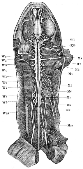
Ventral view of the brain and spinal cord, to show the points of exit of the spinal nerves.
M1‑10 Spinal nerves.
VG Trigeminal ganglion.
W1‑10 Vertebrae.
XG Ganglion of vagus.
By means of longitudinal and transverse sections it may be seen that the ganglion surrounds the ventral root, but that it forms a thinner layer on that side, where the dorsal root joins the ventral; although at this point the two roots and the ganglion are firmly united together, the thick fibrous sheath of the ganglion prevents any communication between the nervous elements of the ventral root and those of the ganglion.
The sheath contains nerve-cells and fibres, and sends in a few fine processes to support these structures. The fibres of the posterior root enter and pass out of the ganglion without suffering any changes, except that the nerve fibres are slightly separated by the presence of a few nerve-cells, and that a distinct increase in the number of nerve fibres takes place. The nerve-cells, of which the ganglion is chiefly composed, and upon the number of which its size directly depends, are arranged chiefly around the fibres of the dorsal root; such few as lie between the fibres are smaller than the rest and vary more in their relative numbers; at times one or two isolated cells are found in the capsule or even in the adjoining ‘periganglionic gland.’ The cells near the capsule are somewhat smaller than the deeper cells.59 Each of these cells, which are usually pear-shaped, possesses only one process; the cell membrane is thick, resistant, and possessed of an external nucleated, endothelial covering, the space around being probably a pericellular lymph-space; it often contains one to three small fat-globules; the protoplasm of these has, according to v. Lenhossék, a concentric fibrillation; the nucleus is round, clear, and distinct, and relatively larger the smaller the nerve-cell; it is usually placed in the centre of the cell.
Fig. 119.
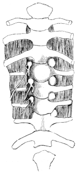
Ventral view of the spinal ganglia; on the right side they are still hidden by the ‘periganglionic glands.’
Fig. 120.
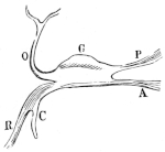
Schema of spinal ganglion.
A Ventral root.
C Ramus communicans.
G Ganglion
O Dorsal division.
P Dorsal root.
R Ventral division.
In that portion of the cell towards the process is a portion brighter and less easily stained than the rest of the cell (Polarkernen, Courvoisier); this apparently possesses one or two nuclei, and is regarded as a cell by Lenhossék (Polarzellen); the process of each cell soon acquires a medullated sheath, and after a short course, 0.09 mm. to 0.35 mm., divides usually about the third constriction (Lenhossék). These processes all pass peripherally, even those which appear at first to pass in the opposite direction curve round again; it is chiefly due to these fibres that the spinal nerve is larger than its two roots.
In addition to the above, smaller, irregular cells from 5 µ to 7 µ in length are found; Rawitz regards them as young cells, Lenhossék as cells capable of (entwicklungsfähige) developing into nerve-cells. The ganglia contain only few blood-vessels.]
The above description is founded upon that of Lenhossék, and has been confirmed by the translator in every particular, except that only very indistinct and unsatisfactory indications of the concentric fibrillation so clearly delineated by Lenhossék were obtained; the structure of the Gasserian ganglion was found to correspond exactly with the description here given of the structure of the spinal ganglia. The nucleus was oftener at the side than at the centre of the cell.
c. The ‘Periganglionic Glands’ (Periganglionäre Kalkdrüsen, v. Lenhossék; Kalksäckchen, Ecker, and others; Crystal capsules, Wyman; Calcareous Sacs or Masses, of other writers), (Fig. 119). These bodies are found on the ganglia of all the spinal nerves and on the Gasserian ganglion of the trigeminal nerve; they have recently been carefully investigated by v. Lenhossék. According to his description there are usually two to each ganglion, lying on its sides and ventral surface; each consists of a connective-tissue capsule which sends in a few fine trabeculae to support a system of glandular tubes: these usually run parallel to the long axis of the gland, and are about 14 µ to 15 µ in diameter; each tube is lined with a single layer of somewhat columnar epithelium, the cells of which have sharp, distinct outlines and oval nuclei.
A membrana propria was not made out with certainty; the lumen of the tubes is wide and contains a milky fluid, which gives the whole structure its characteristic appearance; it has long been known (Blasius, 1681, mentions the fact, l.c., p. 291) that this fluid contains calcareous matter, which effervesces and dissolves in the presence of hydrochloric acid; the crystals vary in form but are chiefly oval (Wyman).
These glands bear no definite proportion to the size of the nerves to which they are attached (Fig. 118); they already contain calcareous matter during the tadpole stage before the limbs are developed (Stannius, Wyman). No ducts have been traced to these glands. This description, founded upon that of Lenhossék, can be confirmed by the translator in every particular; his sections, however, would lead him to believe that the glands are far more vascular than the description and the drawings of Lenhossék imply.]
d. The Branches of the complete spinal nerves formed by the union of the dorsal and ventral roots with the ganglion (Fig. 120) are usually described as two, a dorsal and a ventral branch, which latter at once gives off a Ramus communicans to the sympathetic system; these two branches are given off almost immediately beyond the ganglion.
II. The Individual Nerves.
A. The Dorsal Branches (Fig. 121).
Fig. 121.
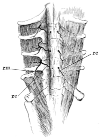
Dorsal branches of the spinal nerves. The Fascia dorsalis is dissected, removed from the left side.
rc Rami cutanei.
rm Rami musculares.
The dorsal branches are smaller than the ventral; they pass upwards between the inner borders of the Musc. intertransversarii and the articular processes of the vertebrae to reach the under surface of the M. longissimus dorsi; the general course of these nerves is very similar for all; each nerve gives off—
(a) A twig to the Musc. intertransversarius and then divides into two branches.
(b) A Ramus muscularis (Fig. 121 rm), which passes outwards to supply the M. longissimus dorsi.
(c) A Ramus cutaneus (Fig. 121 rc); this nerve continues under the extensor muscle of the back, running on the arch of the vertebra towards the middle line; it then pierces the muscle and Fascia dorsalis to reach the large dorsal lymph-sac (Sacc. cranio-dorsalis), through which it courses to end in the skin. In its course through the sac the nerve is connected with a small artery and vein by a small amount of connective-tissue, the whole being enclosed in a sheath of endothelium continuous with that of the general cavity.
The three anterior cutaneous nerves pierce the Fascia dorsalis near the spinous processes, the fourth a little to the side, the fifth perforates the M. coccygeo-iliacus at about the junction of its anterior and middle thirds; the sixth and seventh perforate the same muscle, but more posteriorly. There are seven of these Rami cutanei, the first coming from the second spinal nerve, the rest from the succeeding six spinal nerves.
B. The Ventral Branches.
The ventral branches of the spinal nerves are larger than their corresponding dorsal branches: each nerve gives off immediately a Ramus communicans to the sympathetic system (Figs. 117 SM, 120 C); these Rami communicantes will be described with the sympathetic system; the remaining portions of the ventral divisions of the original spinal nerves are usually known as the spinal nerves; they will be described as such in detail.
1. The hypoglossal nerve60 (N. hypoglossus, N. spinalis I), (Figs. 114 115, and 117 M1), or first spinal nerve, is of small size; it leaves the vertebral canal between the first and second vertebrae to lie between the Musculi intertransversarii and the pharynx, where it has in front the Arteria vertebralis, behind the Arteria and Nervus brachialis; passing under the Levator anguli scapulae, it touches the Aorta ascendens and crosses the Vagus and the Carotid-gland, and thus reaches the space between the Musculi sterno-hyoidei and the Petro-hyoideus; then curving forwards sharply it turns under the Mylo-hyoideus to course between the fibres of the Genio-hyoideus to the root of the tongue, where it ends. It first lies to the inner and then to the outer side of the glossopharyngeal nerve. In its course the nerve gives off the following branches:—
(a) One or two communicating twigs (Figs. 114 and 122) to the second spinal nerve.
(b) Muscular twigs to the Longissimus dorsi, Intertransversarii capitis, Levator anguli scapulae, and the Retrahens scapulae.
(c) When near the glossopharyngeal it supplies twigs to the Geniohyoideus, Sternohyoideus, and Omohyoideus, where it bifurcates.
(d) One of the branches passes inwards to supply the Hyoglossus.
(e) The other terminal branch passes forwards with the Ramus lingualis and the glossopharyngeal, between the fibres of the Geniohyoideus, which it partially supplies, and sends twigs to the neighbouring parts.
(f) [Hoffmann describes a communicating branch to the pneumogastric nerve, which the translator has not been able to discover, and which no other observer has mentioned.]
Fig. 122.
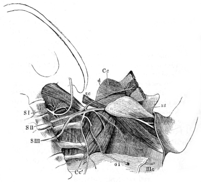
The brachial plexus.
Cc N. coraco-clavicularis.
Cc′ Nerve to musc. obliq. abdom internus.
d Deltoid muscle.
la Musc. lev. ang. scap.
ld Musc. lat. dorsi.
oi Musc. obliq. abdom. intern.
sc Musc. sterno-mastoid.
ss Musc. subscapularis.
SI First spinal nerve.
SII Second spinal nerve.
SIII Third spinal nerve.
IIc Ramus cutaneus axillaris.
IIIc Ramus cutaneus abdominalis.
2. The second spinal or brachial nerve (N. brachialis, N. spinalis II), (Fig. 122) is a large nerve, leaving the vertebral canal between the second and third vertebrae; it then accompanies the Art. axillaris along the anterior border of the transverse process of the third vertebra, over the hinder end of the Musc. levator scapulae and the anterior end of the m. transverso-scapularis major, to the inner border of the Musc. subscapularis, under which it passes into the arm. In this course it gives off or receives the following branches:—
(a) It first receives a branch from the second spinal nerve.
(b) It gives off a large branch, the N. coraco-clavicularis (Fig. 122 Cc), which first accompanies a branch of the Art. axillaris on the Musc. levat. scapulae, then courses forwards and outwards over the Musc. subscapularis to pass from above into the Foramen ovale between the clavicle and coracoid bones. It gives off two branches:
(1) One immediately beyond its origin, which runs backwards over the Musc. subscapularis to supply the Musc. obliq. abdominis internus (Fig. 122 Cc′).
(2) While in the Foramen ovale, the N. coraco-clavicularis bifurcates; the anterior twig runs forwards and outwards to the deltoideus and also supplies a recurrent filament to the Musc. sterno-radialis; the posterior twig enters the upper surface of the Musc. stero-radialis.
(c) At the outer extremity of the Musc. transverso-scapularis the brachial nerve gives off a posterior branch, which at once bifurcates:
(1) The first branch passes into the under surface of the Musc. latissimus dorsi (ld), giving a twig to the Infraspinatus.
(2) The second is the Ramus cutaneus axillaris (IIc); it passes on the under surface and posterior border of the Musc. latissimus dorsi to the skin of axilla and dorsal surface of the upper arm.
(d) Several small branches are given off from the upper and anterior surfaces of the brachial nerve near the Foramen ovale; they accompany an artery to pass forwards and upwards between the Musc. subscapularis and the M. transverso-scapularis on the one side, and the long head of the M. triceps brachialis on the other, and are lost in the M. deltoideus and the M. infraspinatus.
The N. brachialis continues its course by passing between the long and inner heads of the M. triceps, reaches the arm and splits into two nerves, the N. ulnaris and the N. radialis.
Fig. 123.
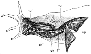
Nerves of the central surface of the arm. The hand pronated.
rc Upper cutaneous branch of the N. radialis.
rc′ Lower cutaneous branch 4-,f the N radialis
U N. ulnaris.
uc R. cutan. sup. of the N. ulnaris.
uc′ R. cutan. inf. of the N ulnaris.
up R. pectoralis of the N. ulnaris.
[I.] The N. ulnaris (Fig. 123 U) escapes from cover of the long head of the M. triceps and runs obliquely over its inner head towards the outer side of the Plica cubiti; in this course it supplies four branches:
α. The Ramus subscapularis passes dorsally to the M. subscapularis.
β. A Ramus pectoralis (Fig. 123 up), passes forwards to the M. abdomino-pectoralis and to the skin of the pectoral region (R. cutaneus pectoralis).
γ. The N. cutaneus antibrachii superior s. medialis (uc) passes to the skin of the inner side of the forearm; this and the two following nerves are given off near the Plica cubiti.
δ. The N. cutaneus antibrachii inferior s. dorsalis s. musculo-cutaneus (uc′) at once supplies branches to the M. flexor carpi radialis61, and then runs downwards upon the muscle and bifurcates:—
[1] The R. lateralis is distributed, by two twigs, in the M. flexor carpi ulnaris and in the skin of the second finger, to which it supplies the Rami digitales volaris and dorsalis.
[2] The R. dorsalis, after giving a cutaneous twig to the second finger, is distributed by numerous twigs to the skin of the dorsal surface of the hand.
The N. ulnaris then passes between the M. flexor carpi radialis and the tendon of the M. sterno-radialis, sinks deeply into the Plica cubiti, where it lies between the M. flexor carpi radialis and the Flexor carpi ulnaris, then between the latter and the Flexor anti-brachii medialis on the one side and the Flexor digitorum communis on the other: it supplies branches to all these muscles, and while still in the forearm divides into two terminal branches:—
α. The R. ulnaris medialis is the smaller; it runs inwards, passes under the tendon of the M. extensor carpi ulnaris into the palm of the hand and ends as the N. volaris digiti V medialis.
Fig. 124.
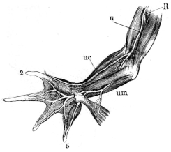
Nerves of the ventral surface of the arm. The hand supinated, and the superficial layer (Flex. carpi rad. and uln., Flex. dig. commun.) has been removed.
R N. radialis.
u N. ulnaris.
uc R. cutan. inf. of the N. ulnaris.
um Bifurcation of the N. ulnaris.
β. The R. ulnaris lateralis passes near the thumb-rudiment, covered by the M. abductor pollicis, deeply into the palm of the hand, and supplies by bifurcating branches the adjacent sides of the second, third, fourth, and fifth fingers (R. volares); it also gives twigs to the muscles of the palm.
[II.] The N. radialis (Figs. 123, 124, and 125 R) passes immediately beyond its origin from the N. brachialis, between the humerus and the M. triceps, runs along the bone to its outer side; in this course it gives off:—
α. Rami musculares to the separate parts of the M. triceps.
β. The Ramus cutaneus superior (Fig. 125 rc) to the skin of the outer surface of the upper arm and forearm.
The nerve then sinks deeply under the origins of the M. extensor carpi ulnaris and the M. extensor digitorum communis, where it bifurcates after giving off some small twigs:—
γ. Small muscular twigs to the extensor muscles of the hand and fingers.
Fig. 125.
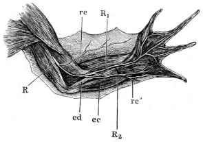
The N. radialis.
ec M. extensor carpi ulnaris.
ed M. extensor digit. comm.
R N. radialis.
R1 R. lateralis.
R2 R. medialis.
rc R. cutaneus superior.
rc′ R. cutaneus inferior.
δ. The R. radialis medialis (R2), the smaller terminal branch, supplies a branch to the M. extensor digit. comm. longus and the skin over the carpus (Ramus cutaneus inferior, rc′), and ends on the muscles of the little finger as the R. dorsalis digiti V medialis.
ε. The R. radialis lateralis (R), the larger of the two branches, supplies the extensor muscles of the fingers and gives bifurcating branches, Rami digitales dorsales, to the adjacent sides of the second, third, fourth, and fifth fingers (the Ramus dorsalis lateralis of the second finger is supplied by the N. ulnaris).
3. The third spinal nerve (N. spinalis III) (Fig. 122 SIII) leaves the vertebral canal by the foramen between the third and fourth vertebrae, and runs outwards and forwards upon the large transverse process of the third vertebra to the brachial nerve (N. spinalis II). Its behaviour at this point is subject to considerable variation; at times it joins the brachial nerve completely, at other times the two nerves are simply in contact; or lastly, it may send a small twig to the brachial nerve. Externally to this point the two nerves supply a number of small branches, which may belong almost entirely to either one nerve or the other; these nerves again inosculate in a variable manner, to form an axillary plexus. The more constant branches are:—
(1) One or more twigs to the Musc. transverso-scapularis major.
(2) Several branches, usually two larger and one smaller; they course in the M. obliq. abdom. internus (Fig. 122 oi) to the M. rectus, where they give off branches, then pierce the muscle to reach the skin (R. cutaneus abdominalis), (Fig. 122 IIIc).
Fig. 126.
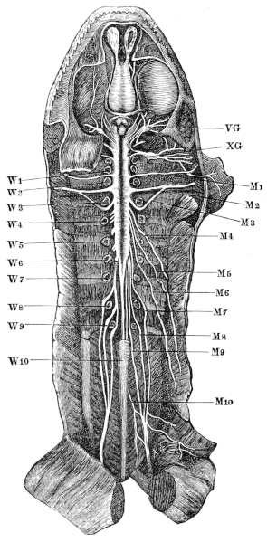
Ventral view of the brain and spinal cord, to show the points of exit of the spinal nerves.
M1‑10 Spinal nerves.
VG Trigeminal ganglion.
W1‑10 Vertebrae.
XG Ganglion of the vagus.
4. The fourth spinal nerve (N. spinalis IV) (Fig. 126 M4) appears between the fourth and fifth vertebrae; it runs on the ventral surface of the transverse process of the fifth vertebra and upon the Musc. intertransversarius obliquely outwards and downwards, and reaches the deeper surface of the Musc. obliquus internus, into which it descends, about opposite the articulation between the eighth and ninth vertebrae; it then bifurcates into:—
(1) A Ramus cutaneus abdominalis, which pierces the muscle and supplies the skin.
(2) A Ramus muscularis, which supplies the broad abdominal muscle and the M. rectus abdominis.
5. The fifth spinal nerve (N. spinalis V) (Fig. 126 M5) emerges through the foramen between the fifth and sixth vertebrae, runs obliquely downwards and outwards over the transverse processes of the sixth and seventh vertebrae and the corresponding M. intertransversarii, and at a point nearly opposite the middle of the urostyle pierces the M. obliquus internus.
The rest of its course corresponds exactly with that of the fourth spinal nerve.
6. The sixth spinal nerve (N. spinalis VI) (Fig. 126 M6), after escaping from the vertebral canal between the sixth and seventh vertebrae, runs obliquely downwards and outwards on the under surfaces of the transverse processes of the seventh, eighth, and ninth vertebrae, then under the iliac bone to a point opposite the hinder half of the urostyle, where it descends under cover of the M. obliquus abdom. internus. In the rest of its course it repeats the corresponding courses of the fourth and fifth spinal nerves.
7, 8, 9, and 10. The seventh, eighth, ninth, and tenth spinal nerves (Nervi spinales VII, VIII, IX, and N. spinalis X s. N. coccygeus) (Figs. 126 and 127 M7, M8, M9, and M10) are best described together, as they are intimately associated to form the sciatic plexus (Plexus ischiadicus, Plexus cruralis).
Within the vertebral canal the roots of these nerves form the Cauda equina; the seventh nerve leaves the canal between the seventh and eighth vertebrae, the eighth nerve between the eighth and ninth vertebrae, the ninth between the sacrum or ninth vertebra and the urostyle, and the tenth by the Canalis coccygeus in the urostyle; from these points the four nerves run obliquely on the ventral surface of the M. ilio-coccygeus to the pelvis, where they form the sciatic plexus. Although subject to some variation, the usual arrangement is that the seventh and eighth nerves unite to form a trunk, which then receives the ninth nerve; the large nerve so formed is the sciatic nerve: a branch of the tenth usually then joins either the sciatic plexus or the sciatic nerve; the plexus gives off or receives the following branches:—
(a) Like other spinal nerves, these nerves give off Rami communicantes (see Sympathetic System).
(b) The M. ilio-hypogastricus (Fig. 127 M7a). This arises from the seventh spinal nerve, before its union with the eighth spinal nerve; it runs obliquely outwards and downwards on the Musc. ilio-coccygeus and under the border of the M. obliquus abdom. internus, where it bifurcates:—
(1) One branch, the Ramus cutaneus abdominalis, pierces the M. obliq. abdom. internus in the same fashion as the cutaneous branches of the fourth, fifth, and sixth spinal nerves; it supplies the skin of the abdomen.
(2) The second or Ramus muscularis supplies the flat abdominal muscles and the M. rectus abdominis.
(c) The M. cruralis (Fig. 127 M7b) also arises from the seventh spinal nerve at or just beyond its point of union with the eighth spinal nerve. It accompanies the A. cruralis on the pelvic wall to the thigh, where it lies upon the M. ilio-psoas in a triangle between the M. rectus fem. anterior and the M. adductor longus, and divides into two main branches:—
(1) Muscular twigs to the M. ilio-psoas, M. rectus femoris anticus, MM. adductores longus and brevis.
(2) The Ramus cutaneus femoris, which runs downwards in the hinder wall of the Lymph-sac (Saccus iliacus), and supplies the skin of the under and outer surfaces of the thigh.
Fig. 127. The sciatic plexus, from a drawing by Wiedersheim. a Communicating nerve from the ninth spinal nerve. a′ Nerve to musc. lev. ani. b Nerve to oviduct. c Urostyle. cc Communicating twig from the ninth spinal nerve. ci M. coccygeoiliacus. c.I M. coccygeosacralis. d Nerve to bladder. d′ Twig to the m. iliacus. e Twig from sciatic plexus to coccygeal plexus. i M. intertransversarius. il M. ilio-coccygeus. ip M. ilio-psoas. lev M. lev. ani. oi M. obliq. abdom. internus. t′ Transverse process of sacrum. Ao Aorta. Com Point of union of nerves a and cc. Is Ischium. M5 Fifth spinal nerve. M6 Sixth spinal nerve. M7 Seventh spinal nerve. M8 Eighth spinal nerve. M9 Ninth spinal nerve. M10 Tenth spinal nerve. M7a M. iliohypogastricus. M7b M. cruralis. Ni Sciatic nerve. Ovd Oviduct. Rect′ Rectum. S7, S8, S9, S10 Rami communicantes of seventh, eighth, ninth, and tenth spinal nerves. Ves Bladder. 8, 9 Eighth and ninth vertebrae.
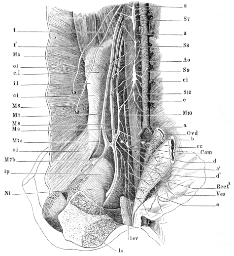
(d) The Ramus dorsalis is a very small nerve; according to Waldeyer it possesses only twenty nerve-fibres. It arises from the coccygeal nerve immediately beyond the Canalis coccygeus, perforates the M. ilio-coccygeus, and runs on its dorsal surface obliquely over the lymph-heart, without supplying it; the nerve then pierces the fascia to supply the skin of the back and thigh.
(e) The Ramus abdominalis also arises from the coccygeal nerve; it is larger than the R. dorsalis, and arises at the same point; the nerve runs on the ventral surface of the M. ilio-coccygeus towards the lymph-heart, and inosculates with the sympathetic. It is from this nerve that the branch to the sciatic plexus is usually given off; it further supplies a varying number of branches, which with branches derived directly or indirectly from the sciatic nerve, form a plexus (Plexus coccygeus); the two plexuses may together be regarded as a Plexus ischio-coccygeus or a Plexus sacro-coccygeus.
(f) In addition to the Rami communicantes given off by the spinal nerves near the vertebral column, the sciatic plexus supplies a few twigs, usually two (Fig. 127).
(g) Other small twigs pass from the sympathetic system forwards and outwards to join the sciatic plexus or the sciatic nerve. According to Waldeyer two of these are very constant.
(h) Branches to the oviduct (Fig. 127 b).
(i) Branches to the rectum (Fig. 127 cc).
(k) Branches to the bladder (Fig. 127 d).
(l) Branches to the M. levator ani (Fig. 127 a′).
(m) A branch to the lymph-heart, which runs along the anterior border of the M. lev. ani, on to its dorsal surface, and then direct to the lymph-heart.
Variations in the sciatic plexus. The arrangement of the nerves in the sciatic plexus is subject to many variations; according to Wiedersheim most of these variations belong to two chief classes: either the N. coccygeus inosculates directly with the ninth spinal nerve by one or several branches, or it joins the ninth nerve indirectly by uniting with its branches. A case of the latter arrangement is seen in Fig. 127, and is thus described by Wiedersheim:—
‘After cutting through the pelvic symphysis and drawing to one side the contained viscera, namely, the hinder end of the oviduct, the rectum, and the bladder, one sees a row of small twigs (a, b, cc, d, and e) arising from the inner, hinder, and anterior surfaces of the ninth spinal nerve: the first (a) runs backwards parallel with the N. coccygeus over the M. ilio-coccygeus to join this nerve at the point marked Com. The twig cc behaves in like manner after receiving a twig from b. The twig b arises from the inner surface of the ninth nerve between the two foregoing and close to its union with the eighth spinal nerve; from this origin twig b passes almost transversely outwards to the hinder extremity of the oviduct (“uterus”), and partly to the rectum, crossing in its course twig a, the sciatic nerve, and the urostyle. Its branches form a net-like plexus with the terminal branches of the last sympathetic ganglion and with a branch cc from the point of union Com.
‘A second branch (a′), arising from the point of junction Com, is a continuation of the N. coccygeus (M10); it passes vertically downwards towards the hinder end of the cloaca and at the upper border of the Levator ani, divides into two branches, which are distributed to the inner and outer surfaces of this muscle and to the cloaca. Other branches pass dorsally to the lymph-hearts, while a third set pass to the hindermost part of the bladder.
‘These three sets of nerves, to the M. coccygeus, M. levator ani, and to the lymph-hearts, are not supplied entirely by the N. coccygeus, as this is reinforced by one or more branches (e) from the sciatic plexus: this branch (e) supplies twigs to the M. ilio-coccygeus, which is also supplied anteriorly from the trunk of the N. coccygeus.
‘The bladder receives a special branch (d), which arises from the sciatic plexus at the junction of the eighth and ninth spinal nerves; this nerve gives a twig (d′) to the M. iliacus.’
Fig. 128.
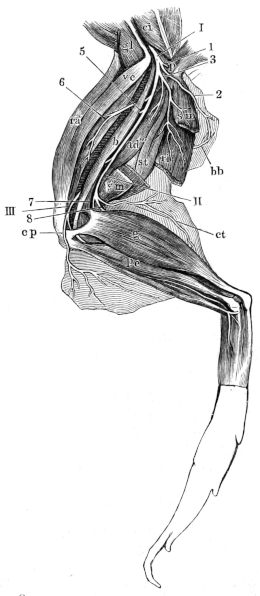
Distribution of the sciatic nerve.
I The sciatic nerve.
II N. tibialis.
III N. peroneus.
1 Branches to the M. pyramidalis.
2 N. cut. fem. posterior.
3 Large collateral branch.
5 Branch to the M. biceps.
6 Branch accompanying the art. circumfl. genu lateralis sup.
7 Branch of tibial nerve to the M. gastrocnemius.
8 N. suralis.
ad‴ M. adductor magnus.
b M. biceps.
bb Twig accompanying the art. cutanea.
ci M. coccygeo-iliacus.
cp N. cut. cruris lateralis.
ct R. cut. cruris posterior.
g M. gastrocnemius.
gl M. glutaeus.
p M. pyramidalis.
pe M. peroneus.
ra M. rectus anterior.
ri′ M. rectus internus minor.
sm M. semimembranosus.
st M. semitendinosus.
ve M. vastus externus.
I. The sciatic nerve (N. ischiadicus) (Fig. 128 I) is the largest nerve of the body; it passes under the M. coccygeus, between the origins of the M. vastus externus and of the M. pyramidalis: lies then between the M. biceps and the M. pyramidalis, and later between the M. biceps and the M. semimembranosus; lastly, it bifurcates under the M. biceps to form the N. tibialis (II) and the N. peroneus (III). In this course it gives off:—
(a) A twig to the M. coccygeo-iliacus, while still in the pelvis.
(b) The N. cutaneus femoris posterior (Fig. 128, 2), which passes between the M. pyramidalis and the M. vastus externus, to appear behind and beneath the former; it accompanies an artery of like name to supply the skin of the hinder and inner surfaces of the thigh.
(c) A little below the foregoing it gives off a collateral branch (Fig. 128, 3), which passes under the M. pyramidalis and divides to form—
(1) A branch to the upper third of the M. semimembranosus.
(2) A branch to the Rectus internus minor. A twig of this branch (bb) pierces the muscle transversely in company with the Arteria cutanea, and passes to the skin of the middle of the inner surface of the thigh.
(3) Branches to both heads of the semitendinosus.
(4) Branches to the M. adductor magnus.
(d) Branches covered by the M. pyramidalis to the M. quadrat. femoris and M. obturator.
(e) A branch (5) forwards to the M. biceps.
(f) A branch (6) which accompanies the Art. circumflexa genu lateralis sup. forwards to the M. extensores cruris, the M. vastus externus, and the M. rectus anterior.
II. The Nervus tibialis (Figs. 128 II, 129 T) passes backwards and inwards to supply the extensors of the foot and the flexors of the toes. Its branches are:—
(a) The R. cutaneus cruris posterior (Figs. 128 and 129 ct), accompanied by an artery and vein, passes to the skin of the calf.
(b) A twig to the upper part of the gastrocnemius (Fig. 129 g′). The main nerve, after supplying this twig, bifurcates to form the next two nerves.
(c) The Nervus suralis (Figs. 128 8; 129 s) runs downwards on the inner border of the M. gastrocnemius and gives off:—
(1) A twig (g″) to the upper third of the muscle.
Fig. 129.
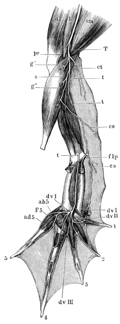
Nerves of the leg and sole of the foot.
ab5 Branch to the M. abduct. digit. V.
ad5 Branch to the M. adduct. digit. V.
ar M. rectus anterior.
b M. biceps.
cs Ramus cutaneus medius.
ct Ramus cutaneus cruris posterior.
dvI Ramus digitalis volaris I.
dvII Branch supplying R. digit. volaris I and II.
dvIII Second branch of the N. tibialis.
F5 Branch to the M. flex. brev. digit. V.
flp Branches to the M. plantaris and the M. flexor digitorum.
g′ Branch to the M. gastrocnemius.
g″ Branch to the M. gastrocnemius.
pc N. peroneus.
s N. suralis.
sm M. semimembranosus.
T, t Nervus tibialis.
(2) The Ramus cutaneus medius (cs) is given off below the middle of the M. gastrocnemius; it passes to the skin of the lower third of the leg. After giving off the latter branch, the N. suralis runs downwards along the inner aspect of the Tendo achillis to the Aponeurosis plantaris of the sole of the foot as far as the calcar, and gives off:—
(3) Branches (flp) to the Musc. plantaris and the M. flexor digitorum.
(4) A branch to the M. abductor hallucis.
(5) The R. digitalis volaris I (dvI) supplies the inner side of the first toe by its terminal twig.
(d) The Nervus tibialis (Fig. 129 t) passes downwards on the hinder surface of the tibio-fibula, sinks into the M. tibialis posticus to appear again at its hinder border; it then runs over the ankle-joint into the sole of the foot, where it lies midway in the space between the two Ossa tarsi and between the M. extensor tarsi and the M. abductor digiti I longus; the nerve then runs downwards in the groove of the small Os tarsi and divides to form three branches:—
(1) The first branch (dvII) runs to the space between the second and third fingers, where it bifurcates:—
(α) The Rami digital. volaris I run transversely over the muscles of the second toe, supply the muscles of the first toe, and bifurcate to form the R. digital. volaris I and II.
(β) The Ram. digit. volar. II divides in the space between the second and third fingers to supply the Flex. phalang. and the adjacent sides of these toes.
(2) The second branch (dvIII) runs over the M. flex. metatarsi of the third toe to the space between the third and fourth toes, and bifurcates to supply the adjacent sides of these toes and the web between them.
(3) The third branch at once divides to supply:—
(α) The M. transv. metatarsi.
(β) The MM. lumbricales of the fourth toe.
(γ) The M. abductor digit. V (ab5), the M. flexor brevis dig. V (F5), and the M. adductor dig. V (ad5); it then ends as—
(δ) A bifurcating branch forming the R. digit. volar. of the fourth and fifth toes.
III. The Nervus peroneus (N. peroneus communis superior, Ecker), (Fig. 130 pc) is the second division of the sciatic nerve; it passes between the outer head of origin of the M. gastrocnemius and the tendon of insertion of the M. biceps, it then lies on the tibio-fibula between the M. gastrocnemius and the M. peroneus, where it is accompanied by the Vena tibialis postica, then runs downwards upon the M. extensor cruris and the Flexor tarsi ant., and passes under the M. tibial. anticus and bifurcates; its branches are:—
Fig. 130.
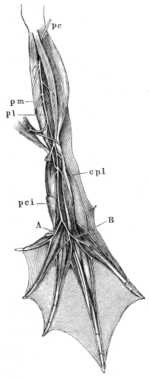
Distribution of the N. peroneus.
A Inner terminal twig of N. peron. comm. inf.
B Outer terminal twig of N. peron. comm. inf.
cpl N. cutaneus dorsi pedis lateralis.
pc N. peroneus.
pci N. peroneus communis inferior.
pl N. peroneus lateralis.
pm N. peroneus medialis.
a. The N. cutaneus cruris lateralis (Fig. 128 cp); which, like other cutaneous nerves, runs in a common sheath together with an artery and vein to the skin. It arises close to the sciatic nerve.
b. Muscular branches to the MM. peroneus, tibialis anticus, extensor cruris brevis, and the flexor tarsi anterior.
c. The N. peroneus medialis (Fig. 130 pm) is the smaller of the two terminal branches of the N. peroneus; it courses with the Art. tibialis antica under the MM. flexores tarsi anterior and posterior and supplies the latter.
d. The N. peroneus lateralis (Fig. 130 pl) is the larger terminal branch of the N. peroneus; it passes downwards between the heads of the M. tibialis anticus to the tendon of origin of the Flexor tarsi posterior, where it gives off two branches (Nos. 1 and 2); the nerve then unites with the N. peroneus lateralis to form a common stem, the N. peroneus communis inferior (Ecker). The N. peroneus lateralis gives off:—
(1) The N. cutaneus dorsi pedis lateralis (cpl), which passes to the skin of the outer side of the dorsum of the foot.
(2) A second branch to the M. extensor of the fourth and fifth toes.
e. The N. peroneus communis inferior (Fig. 130 pci) runs on the dorsum of the foot in company with the Art. dorsalis pedis, underneath the MM. extensores digiti I and II; it supplies several branches and then bifurcates. It gives off:—
(1) Branches to the MM. extensor longus and brevis digiti I.
(2) Rami digitales dorsales to the adjacent sides of the first and second toes.
(3) Branches to the MM. extensores digiti II.
(4) The inner, terminal branch (Fig. 130 A) at once divides:—
(α) The outer branch runs between the third and fourth toes as far as the commencement of the web, where it bifurcates to form two Rami cutanei, which course along the adjacent sides of these toes as far as their apices.
(β) The inner branch passes to the extensor muscle, and, in part, to the adjacent sides of the third and fourth toes.
(5) The outer, terminal branch (Fig. 130 B) of the N. peroneus communis inferior passes to the muscles of the fourth and fifth toes, and supplies Rami cutanei dorsales to the outer side of the fourth and inner side of the fifth toes.
Cutaneous branches of the N. tibialis supply the outer side of the fifth and inner side of the first toe.
G. The Sympathetic System (Sympathicus).
(Re-written by the translator.)
The sympathetic cord or chain is a row of nervous ganglia (vertebral or lateral ganglia), connected by nerve-fibres, and lying on either side of the vertebral column (Figs. 117 and 131); with the exception of the last spinal nerve there is usually one sympathetic ganglion associated with each spinal nerve; in the case of the tenth spinal nerve there may be only one ganglion or as many as twelve.
The first ganglion (Figs. 111, opposite ics; 117 S1) is placed on the hypoglossal nerve just as it emerges from the first intervertebral foramen; it is large, but smaller than the second; its Ramus communicans is represented by several fine and very short fibres, which connect the ganglion with the nerve. This ganglion is connected with the second by two or three nervous threads, between which passes the subclavian artery, a true Annulus Vieussenii being thus formed (de Watteville). The other ganglia are connected by single bands of fibres. The first ganglion supplies also branches to the axillary artery and to the cardiac plexus.
The second ganglion (Figs. 117 and 131) is the largest, and is closely applied to the brachial nerve; as in the case of the first ganglion and hypoglossal nerve, it is attached to the second spinal nerve without a distinctly marked Ramus communicans.
The third ganglion (Ganglion cardiacum basale, Gaskell and Gadow) is sometimes fused with the second, but is usually close to the third spinal nerve: it has a short but distinct Ramus communicans.
Fig. 131.
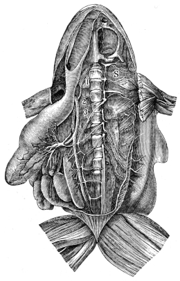
Sympathetic cord. From Ecker (Icones physiologicae, Pl. XXIV, Fig. 3).
The heart, lungs, and liver have been removed; the stomach, intestine, kidneys, and testes drawn to the right side; the left sympathetic cord is thus pulled to the right side to expose the Rami communicantes.
S Sympathetic cord attached to the ganglion of the vagus.
The numbers refer to the ganglia, which are enumerated from before backwards.
Behind the third ganglion the sympathetic cord is continued backwards along the corresponding aortic arch, then parallel with and close to the abdominal aorta (Figs. 117, 127, and 131), receiving Rami communicantes, which are long and well marked, from each of the spinal nerves; the fourth, fifth, and sixth nerves usually supply each one Ramus communicans, the seventh two, and the eighth and ninth each two or three: from the tenth nerve it receives three or more, as many as twelve having been noted. The ganglia are usually more or less spindle-shaped or flattened and triangular; the hinder part of the cord usually receives in addition two or three branches from the sciatic plexus and twigs from the R. abdominalis of the N. coccygeus.
The branches and communications of the sympathetic cords are as follows:—
a. Communicating branches between the two cords; these are extremely numerous and irregular, forming a net-like plexus, which surrounds the abdominal aorta and other adjacent structures, and gives off numerous small twigs to the neighbouring vessels and organs.
b. Communications with the cranial nerves; these are two branches (Wiedersheim), (Figs. 111 and 116) which pass from the first ganglion to the Ganglion nervi vagi, where one terminates, the other leaves the ganglion to pass on to the Gasserian ganglion (Figs. 111 and 116 VS): according to Gaskell a single nerve passes from the first ganglion to the Ganglion nervi vagi, whence one portion of its fibres is continued to the Gasserian ganglion, the remainder accompanying the pneumogastric nerve without any connection with the ganglion; he therefore names this nerve the vago-sympathetic. (See Gasserian ganglion, p. 168.)
c. Communications with the spinal nerves; these are:—
(1) The Rami communicantes.
(2) Communications between the sympathetic ganglia or their branches and the spinal nerves or their branches (Fig. 117); by means of these connections, fibres of the sympathetic system are conducted by the spinal nerves and their branches to all parts of the body.
d. Branches to the heart, which form the following ganglia:—
(1) A relatively large plexus lies on the auricles in the median plane immediately beneath the division of the M. hyoglossus. It supplies a network of fibres to the auricles and the adjacent large vessels. It is said to communicate at various points with the pneumogastric nerve.
(2) A smaller ganglion of oval form, supplies twigs to the neighbouring vessels and a communicating branch to the hypoglossal nerve (Wiedersheim).
e. Branches to the abdominal viscera; these form intricate plexuses by which the organs are supplied: the one best known is the solar plexus (Fig. 131); it is formed chiefly from branches derived from the third, fourth, and fifth ganglia, and supplies the stomach, etc.; other plexuses for the various viscera are known by corresponding names, such are the Plexus hepaticus, renalis, genitalis, haemorrhoidalis, and vesicalis.
The sympathetic system is characterised by the fact that the branches form intricate plexuses, which include numerous ganglia and which are very irregular; it is also characteristic that most of its fibres are non-medullated. According to the investigations of Gaskell, the fibres of the sympathetic system arise in mammalia as very fine medullated fibres from the posterior vesicular (Clarke’s) columns (Mason has recently described cells in the frog’s spinal cord, which he holds to be homologous with the cells of these columns); they leave the cord by both the ventral and dorsal roots of the spinal nerves, and are thus connected with two sets of ganglia, (1) with the ganglia of the dorsal roots, and (2) through the Rami communicantes with the sympathetic ganglia (vertebral or lateral ganglia); these two sets of ganglia Gaskell terms proximal. By means of the branches from the sympathetic ganglia (Rami efferentes) part of the fibres pass to another set of ganglia, the solar plexus, etc., which he terms prevertebral or collateral; from these the fibres pass to be distributed to the various viscera and blood-vessels, where a fourth set of very small ganglia (terminal ganglia) is found. The prevertebral and terminal ganglia are together classed as distal ganglia. Gaskell holds that the fine medullated fibres from the cord lose their medullary sheath in one or other of these ganglia according to the function they fulfil.
The inhibitory fibres of the heart and vaso-dilator fibres of the blood-vessels continue as white fibres along the vago-sympathetic and spinal nerves to the distal ganglia (Bidder’s ganglion, etc.), where the medullary sheath disappears: whereas the ‘augmentor’ fibres of the heart and vaso-constrictor fibres of the blood-vessels lose their medullary sheath in the proximal ganglia and pass on as non-medullated fibres. In the same way the nerve-fibres that bring about contraction of the circular muscle fibres of the hollow viscera lose their medullary sheaths in the proximal ganglia, while those fibres, the influence of which negatives the former, become pale fibres in the distal ganglia.
Waters has demonstrated that in the frog the various spinal nerves have each a localised physiological action upon the blood-vessels and muscular walls of various parts of the alimentary canal: he shows that
The third spinal nerve supplies the oesophagus.
The fourth spinal nerve supplies the stomach.
The fifth spinal nerve supplies the upper third of the small intestine.
The sixth spinal nerve supplies the lower two thirds of the small intestine.
The seventh spinal nerve supplies the large intestine.
The eighth spinal nerve supplies the bladder, this supply being, however, not so definite as the others given above.
It has long been known that the branches of the spinal ganglia (ganglia of the posterior roots) are together larger (one-third, Lenhossék), and contain more fibres than the ventral and dorsal roots together; this is supposed to be in part due to an acquisition of new fibres derived from the ganglia. The majority of recent observers hold that each ganglionic cell has only one process, which, however, soon bifurcates; whether any of the fibres so formed pass as far as the cord or beyond its blood-vessels is doubted by most observers, and denied by Gaskell. These remarks and the description of the cells of the spinal ganglia (p. 176) hold good for the lateral or vertebral ganglia and the prevertebral ganglia (solar ganglion, etc.) of the sympathetic system; the terminal ganglia will be described with the organs in which they are found.
H. Histological Notes on the Nervous System.
(In order to render the foregoing description of the nervous system more complete, the following notes have been added by the translator.)
[The histological elements of the nervous system are nerve-cells and nerve-fibres; of these the nerve-cells have been described with the parts in which they occur; it may simply be remarked that later observers have been unable to discover the ‘spiral cells’ described by Beale, Arnold, and others. The nerve-fibres, as in most other animals, are of two kinds, medullated and non-medullated.
1. Medullated nerve-fibres or white fibres are found in all cranial and spinal nerves, with the exception of the olfactory nerves, and in many of the sympathetic nerves (see Sympathetic System); also in the white matter of the brain and spinal cord; examined microscopically the fibres are seen to consist of an external sheath or neurilemma, a medullary sheath, and an axis-cylinder:—
a. The neurilemma (Sheath of Schwann, Outer or Primitive Sheath) is a nucleated endothelial layer covering the nerve-fibre; it is continuous with the corresponding coat of the nerve-cells, and is uninterrupted throughout the length of the nerve; at the nodes, however, it dips down towards the axis-cylinder, the circular groove so formed being filled with cement substance.
b. The medullary sheath (White substance of Schwann); the presence of this sheath is the chief cause of the whiteness of these nerves; the thickness of the sheath varies considerably, and towards the ultimate distribution of the nerve it is entirely lost. At more or less regular intervals along the course of the nerve-fibre the continuity of the medullary sheath is broken, and gives the fibres the appearance of being constricted at these places; such constrictions are known as nodes of Ranvier; the portion between two such nodes being termed an internode. Each internode possesses an oval, flattened, granular nucleus at about its middle and placed between the neurilemma and the medullary sheath; the nucleus has a nucleolus. In the fresh state the medullary sheath seems to be fluid; it is of a fatty nature.
Medullary segments are caused by breaks in the continuity of the medullary sheath, which are seen only in nerves which are no longer in their normal condition, and are especially well marked after treatment with osmic acid. The breaks are oblique; hence the conical end of one segment fits into the funnel-shaped end of the next. How far they correspond to pre-existent structures is uncertain. The segments in the frog vary in length from 0.010 to 0.040 mm.
c. The axis-cylinder is the essential part of every nerve-fibre; it shows a longitudinal striation corresponding to the fine fibrils (primitive fibrillae) of which it consists; these fibrils often exhibit minute varicosities: at times it has the appearance of being invested with a very delicate structureless sheath. It is continuous through the nodes of Ranvier.
2. Non-medullated nerve-fibres (Grey or Varicose Fibres; Fibres of Remak); these occur chiefly in branches and plexuses of the sympathetic system; they consist of a neurilemma and an axis-cylinder, which agree exactly with the corresponding elements found in the medullated fibres.
The nerve-fibres, whether medullated or non-medullated, are bound together by connective-tissue to form nerves. A number of fibres bound together by connective-tissue to form a slender cord is known as a funiculus; a small nerve may consist of one such funiculus; the sheath surrounding it is known as the perineurium, and sends in supporting processes between the fibres (endoneurium): when several funiculi are bound together to form a large nerve the common sheath is known as the epineurium. These sheaths support nerves (nervi nervorum) and vessels (vasa vasorum) supplying the nervous elements, and their intercellular spaces form lymph-canals. The whole nerve is surrounded by an endothelial coat, which helps to form a lymph-space, which more or less completely surrounds the nerve.
The ultimate distribution of the nerve-fibres will be included in the description of the various organs in which they end.]